NAPQI inhibition of glutathione synthetase 1 The acetaminophen metabolite N-acetyl-p-benzoquinone imine (NAPQI) inhibits glutathione synthetase in vitro; a clue to the mechanism of 5-oxoprolinuric acidosis? Valerie Walker 1 , Graham A Mills 2 , Mary E Anderson 3 , Brandall L Ingle 4 , John M Jackson 5 , Charlotte L Moss 6 , Hayley Sharrod-Cole 1 and Paul J Skipp 6 1 Department of Clinical Biochemistry, University Hospital Southampton NHS Foundation Trust, Southampton SO16 6YD UK; [email protected]; [email protected]2 School of Pharmacy and Biomedical Sciences, University of Portsmouth, Portsmouth PO1 2DT UK; [email protected]3 Department of Chemistry and Biochemistry, Texas Woman’s University, Denton 76204 USA; [email protected]4 Center for Advanced Scientific Computing and Modeling, Department of Chemistry, University of North Texas, Denton 76203 USA; [email protected]5 NIHR Southampton Biomedical Research Centre, Southampton General Hospital, Southampton SO16 6YD UK; [email protected]6 Centre for Proteomic Research, Centre for Biological Sciences, University of Southampton, Southampton SO17 1BJ UK; [email protected]; [email protected]Running title: NAPQI inhibition of glutathione synthetase Corresponding author: Dr V Walker, Department of Clinical Biochemistry, C Level, MP6, South Block, Southampton General Hospital, Tremona Road, Southampton, SO16 6YD, UK [email protected]TEL +44 2381 206436 FAX +44 2381 206011 Key words: protein arylation, acetaminophen toxicity, glutathione deficiency, γ-glutamyl cycle Abbreviations: NAPQI, N-acetyl-p-benzoquinone imine; GS, glutathione synthetase; γ-GCS, γ- glutamylcysteine synthetase; γ-GCT, γ-glutamyl cyclotransferase; γ-GACT γ-glutamylamine cyclotransferase; GAB, glutamyl-L-α-aminobutyric acid; APAP-Cys, acetaminophen-cysteine
Transcript
NAPQI inhibition of glutathione synthetase
1
The acetaminophen metabolite N-acetyl-p-benzoquinone imine (NAPQI) inhibits glutathione synthetase in vitro; a clue to the mechanism of 5-oxoprolinuric acidosis? Valerie Walker1, Graham A Mills2, Mary E Anderson3, Brandall L Ingle4, John M Jackson5, Charlotte L Moss6, Hayley Sharrod-Cole1 and Paul J Skipp6 1Department of Clinical Biochemistry, University Hospital Southampton NHS Foundation Trust, Southampton SO16 6YD UK; [email protected]; [email protected] 2School of Pharmacy and Biomedical Sciences, University of Portsmouth, Portsmouth PO1 2DT UK; [email protected] 3Department of Chemistry and Biochemistry, Texas Woman’s University, Denton 76204 USA; [email protected] 4Center for Advanced Scientific Computing and Modeling, Department of Chemistry, University of North Texas, Denton 76203 USA; [email protected] 5NIHR Southampton Biomedical Research Centre, Southampton General Hospital, Southampton SO16 6YD UK; [email protected]
6Centre for Proteomic Research, Centre for Biological Sciences, University of Southampton, Southampton SO17 1BJ UK; [email protected]; [email protected] Running title: NAPQI inhibition of glutathione synthetase Corresponding author: Dr V Walker, Department of Clinical Biochemistry, C Level, MP6, South Block, Southampton General Hospital, Tremona Road, Southampton, SO16 6YD, UK [email protected] TEL +44 2381 206436 FAX +44 2381 206011 Key words: protein arylation, acetaminophen toxicity, glutathione deficiency,
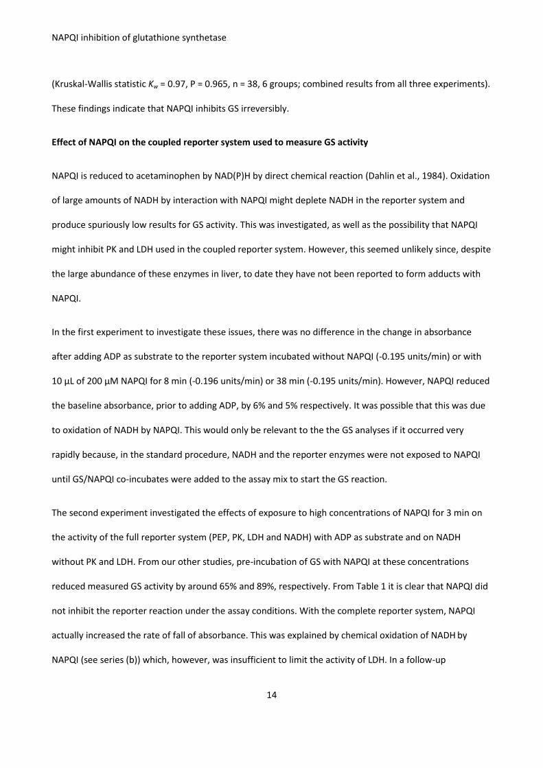
For supportive evidence that NAPQI did not inhibit PK or LDH, the effect of increasing the amounts of PK
and LDH in the standard GS assay procedure was investigated. Doubling the amounts of reporter enzymes
had no effect on the measured activity of GS pre-incubated with NAPQI. The mean (range) of GS activities
(μmol/mg/min) of four replicates for each experimental condition were: normal versus double PK/LDH:
without NAPQI: 6.7 (6.7-7.1) versus 6.7 (6.7-6.7); with 200 μM NAPQI: 2.4 (2.0-2.8) versus 2.4 (2.0-2.8).
Collectively, the findings show that under the assay conditions used, NAPQI added in concentrations up to
400 μM did not inhibit PK or LDH. At the higher concentrations it probably oxidised NADH to a small extent
but this did not limit the reporter reaction. It might have caused a small spurious increase in measured GS
activity, but not a decrease. Thus the assay was valid.
Computer modeling
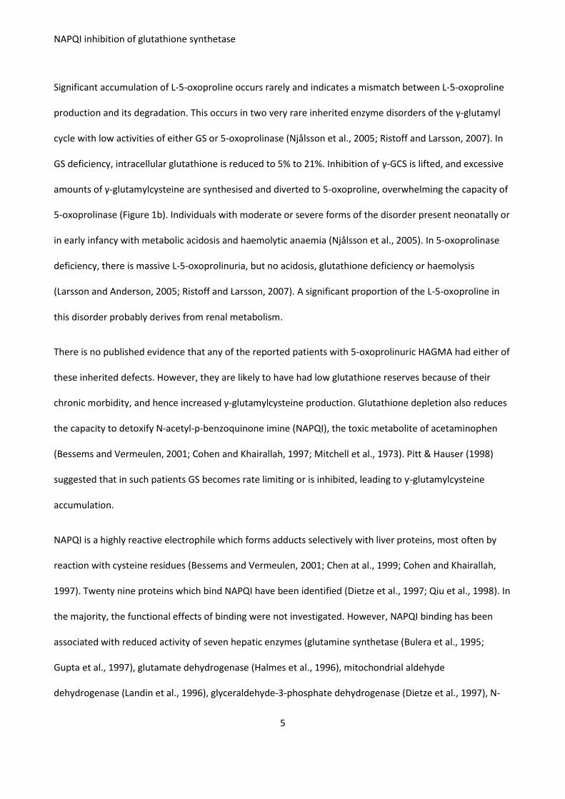
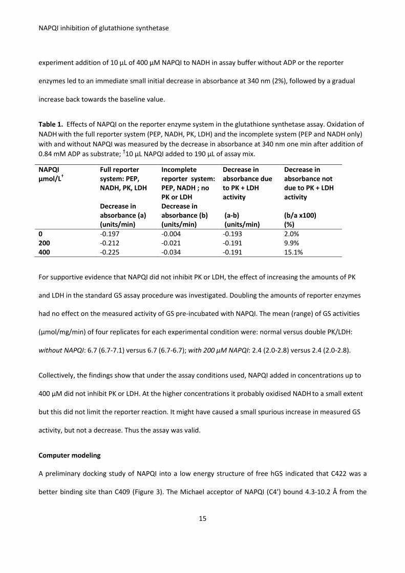
A preliminary docking study of NAPQI into a low energy structure of free hGS indicated that C422 was a
better binding site than C409 (Figure 3). The Michael acceptor of NAPQI (C4’) bound 4.3-10.2 Å from the
NAPQI inhibition of glutathione synthetase
16
C409 sulphur. The poses near C409 exhibited a high variation in orientation with several poses on the
exterior of the protein, indicating that C409 was not an ideal binding site for NAPQI. In contrast, all poses
near C422 nestled deeply into the binding pocket with the Michael acceptor on NAPQI 3.0-3.8 Å from the
C422 sulphur.
Figure 3. Docked poses near C409 and C422 show the specificity of the C422 binding site, relative to the
non-specific binding near C409.
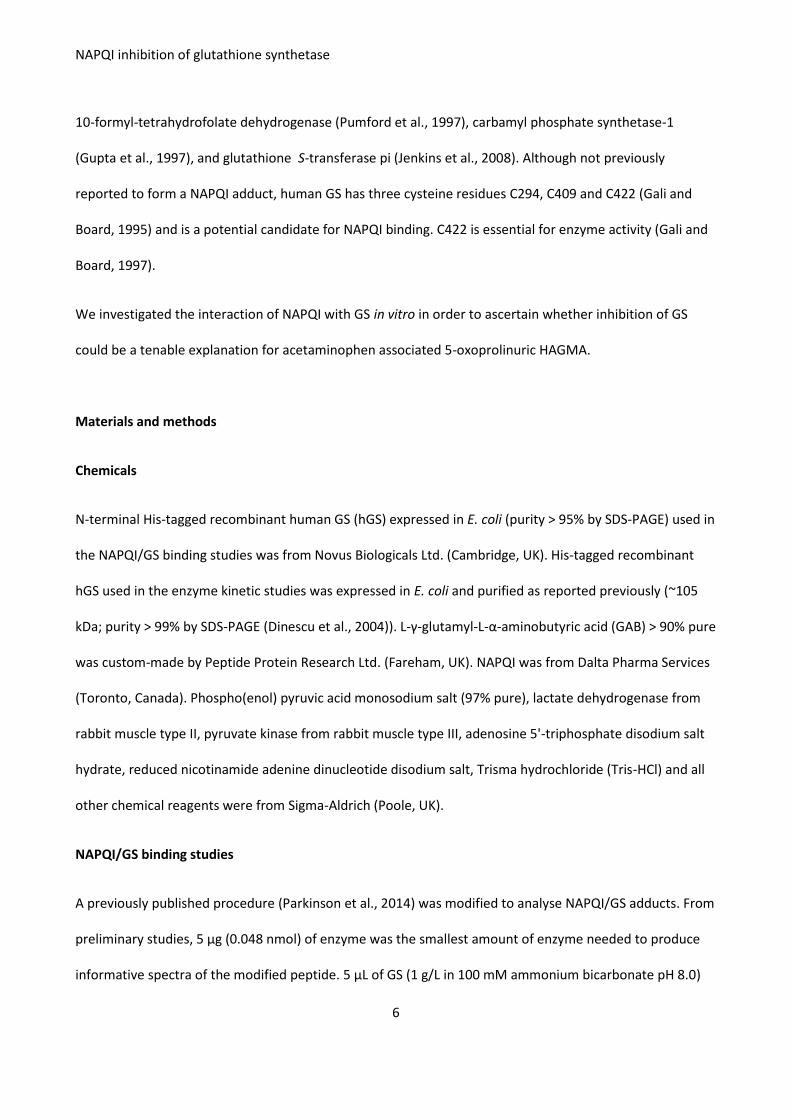
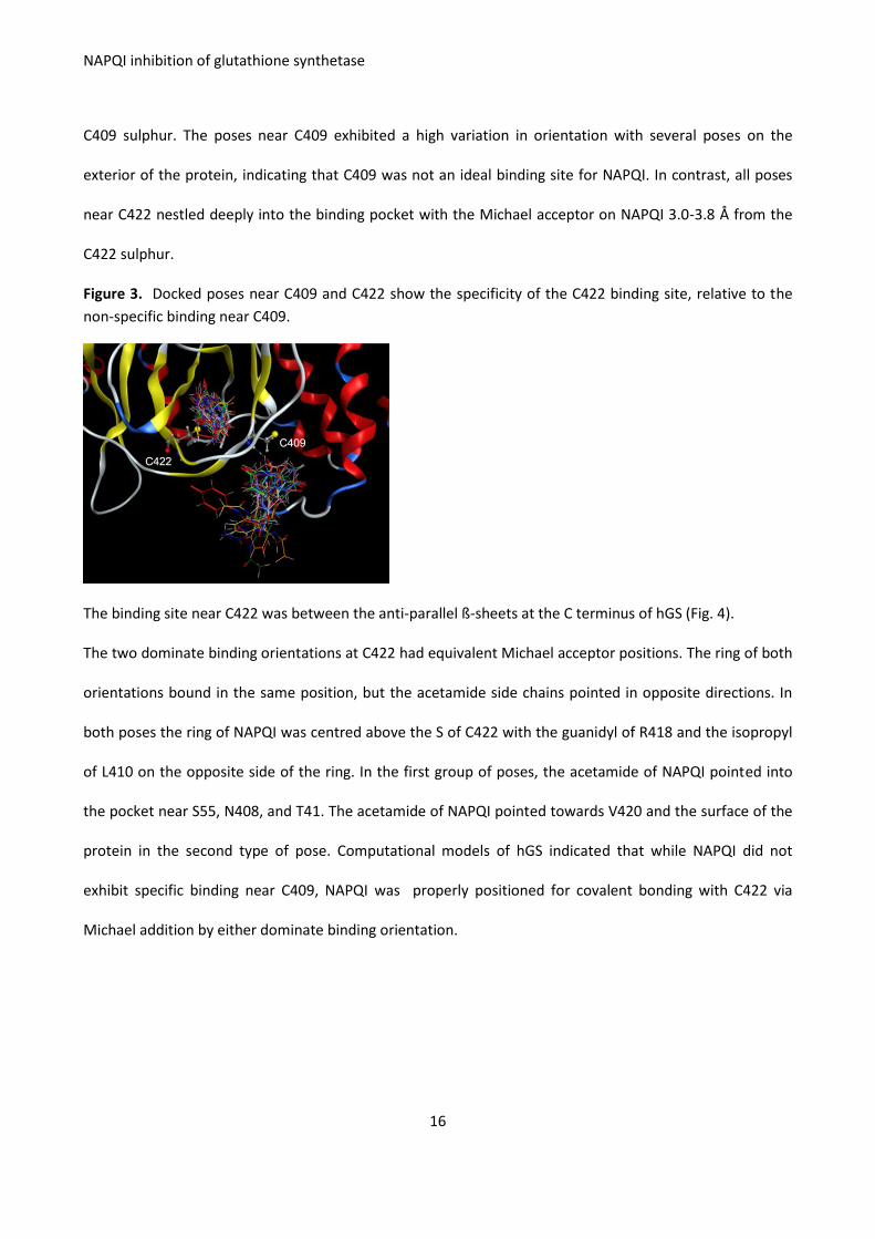
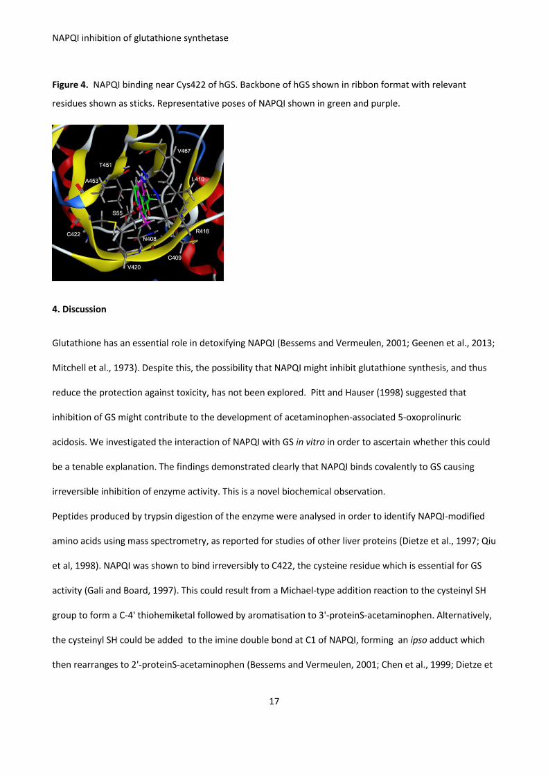
The binding site near C422 was between the anti-parallel ß-sheets at the C terminus of hGS (Fig. 4).
The two dominate binding orientations at C422 had equivalent Michael acceptor positions. The ring of both
orientations bound in the same position, but the acetamide side chains pointed in opposite directions. In
both poses the ring of NAPQI was centred above the S of C422 with the guanidyl of R418 and the isopropyl
of L410 on the opposite side of the ring. In the first group of poses, the acetamide of NAPQI pointed into
the pocket near S55, N408, and T41. The acetamide of NAPQI pointed towards V420 and the surface of the
protein in the second type of pose. Computational models of hGS indicated that while NAPQI did not
exhibit specific binding near C409, NAPQI was properly positioned for covalent bonding with C422 via
Michael addition by either dominate binding orientation.
NAPQI inhibition of glutathione synthetase
17
Figure 4. NAPQI binding near Cys422 of hGS. Backbone of hGS shown in ribbon format with relevant
residues shown as sticks. Representative poses of NAPQI shown in green and purple.
4. Discussion
Glutathione has an essential role in detoxifying NAPQI (Bessems and Vermeulen, 2001; Geenen et al., 2013;
Mitchell et al., 1973). Despite this, the possibility that NAPQI might inhibit glutathione synthesis, and thus
reduce the protection against toxicity, has not been explored. Pitt and Hauser (1998) suggested that
inhibition of GS might contribute to the development of acetaminophen-associated 5-oxoprolinuric
acidosis. We investigated the interaction of NAPQI with GS in vitro in order to ascertain whether this could
be a tenable explanation. The findings demonstrated clearly that NAPQI binds covalently to GS causing
irreversible inhibition of enzyme activity. This is a novel biochemical observation.
Peptides produced by trypsin digestion of the enzyme were analysed in order to identify NAPQI-modified
amino acids using mass spectrometry, as reported for studies of other liver proteins (Dietze et al., 1997; Qiu
et al, 1998). NAPQI was shown to bind irreversibly to C422, the cysteine residue which is essential for GS
activity (Gali and Board, 1997). This could result from a Michael-type addition reaction to the cysteinyl SH
group to form a C-4' thiohemiketal followed by aromatisation to 3'-proteinS-acetaminophen. Alternatively,
the cysteinyl SH could be added to the imine double bond at C1 of NAPQI, forming an ipso adduct which
then rearranges to 2'-proteinS-acetaminophen (Bessems and Vermeulen, 2001; Chen et al., 1999; Dietze et
NAPQI inhibition of glutathione synthetase
18
al., 1997). Human GS has two identical subunits, each with a central active site surrounded by mobile loops
that control activity and bind the substrates (Dinescu et al., 2007; Gali and Board, 1995; Polekhina et al.,
1999). While C422 is not directly involved with the active site, it is located within a mini-barrel which
contributes one wall to the ATP-binding site and it may have a structural role (Polekhina et al., 1999). The
modeling studies showed that NAPQI can nestle deeply in the molecular pocket containing C422. Covalent
binding of NAPQI at this site might interfere with catalytic loop motions and block the active site, thus
reducing GS activity.
NAPQI was shown to inhibit GS in vitro through an irreversible interaction. From the combined results of
three experiments, NAPQI in concentrations ranging from 25 μM to 400 μM inhibited the activity of 81
mg/L (0.77 μM) of GS by 16 % to 89%. Inhibition was significantly dose-related. Other proteins and
glutathione were not included in the assays, and we do not know whether these concentrations are
biologically relevant. The amount of free NAPQI in hepatocytes cannot be quantified directly because of its
reactivity. Exposure of mouse hepatocytes in vitro to 250 μM and 500 μM of NAPQI, but not to 100 μM,
caused toxic changes (cell surface blebs) (Moore et al., 1985). Intracellular concentrations are probably
much lower in vivo. Protein derived acetaminophen (APAP)-cysteine is measured in plasma as an indicator
of the amount of NAPQI-protein adduct formed in liver. Plasma concentrations range from <1 to 27 μM
after overdose (Heard et al., 2011; Muldrew et al., 2002; McGill et al., 2013). However, these probably
under-estimate the liver concentrations. In experimental animals given acetaminophen, damage to the liver
by ischaemia/reperfusion increased blood levels of APAP-cysteine significantly (McGill et al., 2013).
Despite speculation, there is no published evidence that any of the 50 patients reported to 2014 with
acetaminophen-associated 5-oxoprolinuric HAGMA had a genetic defect of the γ-glutamyl cycle. GS was
measured in fibroblasts or red cells in only four and was normal. One also had normal 5-oxoprolinase
activity (Brohan et al., 2014; Liss et al., 2013; Pitt and Hauser 1998). In addition, we measured red cell GS in
one of our own patients and found a normal value (not reported). Heterozygotes for inherited GS
NAPQI inhibition of glutathione synthetase
19
deficiency have GS activity of 55 +/- 13% (mean [SD]) of normal and are asymptomatic (Njålsson et al.,
2005). Inhibition of the activity of a normal GS enzyme by NAPQI could cause 5-oxoprolinuria, but why
would this only occur in such a small sub-group of the world population who consume acetaminophen?
We suggest that reduced detoxification of NAPQI resulting from glutathione depletion, and its constant
production from repeated chronic ingestion of acetaminophen, could increase intracellular NAPQI to levels
which inhibit GS. This could contribute to 5-oxoprolinuria.
The patients reported were at high risk of glutathione depletion because of a poor intake of cysteine,
chronic liver disease, or on-going glutathione losses through degradation during sepsis-induced oxidative
and nitrosative stress, often in combination (Emmett, 2014; Malmezat et al., 2000; Wu et al., 2004).
Glutathione depletion increases the risk for acetaminophen toxicity (Mitchell et al., 1973). This was shown
when glutathione synthesis was reduced in γGCS-deficient rats (Akai et al., 2007) and mice (McConnachie
et al., 2007; Slitt et al. 2005), and in lymphocytes of a human patient with inherited GS deficiency (Spielberg
and Gordon, 1981). In early studies in mice, covalent binding of NAPQI to proteins did not start until 30-45
min after exposure to large toxic doses of acetaminophen (≥ 350 mg/kg) which reduced liver glutathione by
75%. However, this still left a significant amount of glutathione (1 mM) (Mitchell et al., 1973). In more
recent studies, protein adducts formed within 15 min (Muldrew et al., 2002), and were detectable even
after non-toxic acetaminophen doses as low as 15 mg/kg, with only a minimal decrease in glutathione
(McGill et al., 2013). These later studies suggest that NAPQI binding to proteins and glutathione occurs
simultaneously (and independently) from the onset of NAPQI production, and that severe glutathione
depletion is not a pre-requisite for protein binding. In fact, protein adducts were detectable in serum of
healthy adults taking therapeutic doses of acetaminophen (Heard et al., 2011). However, glutathione
depletion does increase the amount of adduct formed because less glutathione is available to scavenge
NAPQI (McGill et al., 2013; Mitchell et al., 1973).
NAPQI inhibition of glutathione synthetase
20
By releasing γ-GCS from feedback inhibition, glutathione depletion increases γ-glutamylcysteine synthesis.
Expression and activity of γ-GCS are increased further by oxidative and nitrosative stress, inflammation and
inflammatory cytokines (Malmezat et al., 2000; Wu et al., 2004). Compared with γ-GCT, GS has a much
higher activity and affinity for γ-glutamylcysteine. Normally therefore, the flood of increased γ-
glutamylcysteine is directed towards glutathione synthesis to replenish stores (Wu et al., 2004). It could be
diverted to 5-oxoproline if GS activity was inhibited or over-whelmed.
In humans who survive an acute acetaminophen overdose, exposure of hepatocytes to NAPQI is curtailed
rapidly. Data for 5-oxoproline are lacking since organic acid analyses are not part of the routine work-up of
such patients. However, two (possibly three) of the 50 cases reported with acetaminophen-induced 5-
oxoprolinuria described above had acute acetaminophen toxicity. Hence it is possible that 5-oxoprolinuria
may occur after acute overdose but resolves and passes undetected. The situation is different in patients
with low glutathione because of an underlying chronic morbidity. Their glutathione reserves are drained
constantly by repeated challenge from acetaminophen ingestion. In this chronic situation, NAPQI could
accumulate when the capacity to replace the glutathione losses becomes inadequate. Continuation of
acetaminophen, even in therapeutic doses, could then cause toxicity and increase adduct formation.
In such patients with complex morbidities, other factors, for example accelerated protein catabolism, could
increase 5-oxoproline production (Figure 1a). Cysteine deficiency arising from an inadequate intake of
cysteine and its precursor methionine, and urinary losses of sulphated acetaminophen metabolites, could
also contribute. Cysteine is the rate-limiting substrate for glutathione synthesis. In vitro, incubation of γ-
GCS with L-glutamate without cysteine resulted in production of phosphorylated glutamate which readily
converted to 5-oxoproline (Orlowski and Meister 1971). Emmett (2014) proposed that through this process
cysteine depletion, together with increased activity of γ-GCS due to low glutathione levels, would lead to
accumulation of 5-oxoproline. Demonstration of low hepatocyte concentrations of γ-glutamylcysteine
would support this proposal, but there are no published data.
NAPQI inhibition of glutathione synthetase
21
Although not investigated, an alternative explanation could be that NAPQI inhibits 5-oxoprolinase. This
enzyme has essential cysteine residues and is rapidly inactivated by the sulfhydryl group inhibitor, N-
ethylmaleimide (Williamson and Meister, 1982). Acidosis is not a feature of inherited 5-oxoprolinase
deficiency (Ristoff and Larsson, 2007), but might be provoked by simultaneous GS inhibition or renal failure.
Other possible explanations for 5-oxoprolinuric acidosis are highly unlikely in the majority of cases. Glycine
deficiency may contribute to mild 5-oxoprolinuria in children with severe protein-energy malnutrition, but
deficiency of this non-essential amino is otherwise exceptionally rare (Emmett, 2014). Foods rich in
glutamine (notably tomato juice) which cyclises to 5-oxoproline are unlikely sources. Flucloxacillin was
proposed in one case report (Croal et al., 1998), and listed as a cause since, but this is still unproven. The D-
isomer probably accounts for 5-oxoprolinuria reported during treatment with the γ-aminobutyric acid
(GABA) antagonist vigabatrin (Larsson et al., 2005).
Conclusions
NAPQI binds covalently to GS causing irreversible inhibition of enzyme activity in vitro. These novel
preliminary findings are the first indication that NAPQI may inhibit glutathione production. This has not
been explored so far, despite the pivotal role of glutathione in protection against acetaminophen toxicity.
The biological relevance of GS inhibition should be investigated in cell cultures. The possibility that NAPQI
may also inhibit other enzymes in the glutathione and supportive methionine transsulphuration pathways
should be explored. The hypothesis that inhibition of GS by NAPQI could contribute to the development of
acetaminophen associated 5-oxoprolinuric HAGMA merits further investigation.
Acknowledgements
The study was supported by the Southampton Hospital Charity, Southampton General Hospital, UK; Charity
registration number 1051543; Fund no. 0182. The work was also funded by the National Institute of Health
(NIH) Grant R15GM086833 (MEA) and a Texas Woman’s University Research Enhancement Grant (MEA).
NAPQI inhibition of glutathione synthetase
22
We thank Theresa Brown for assistance. The funders had no role in study design, data collection and
analysis, decision to publish, or preparation of the article.
Declaration of interest
The authors report no declarations of interest
Appendices
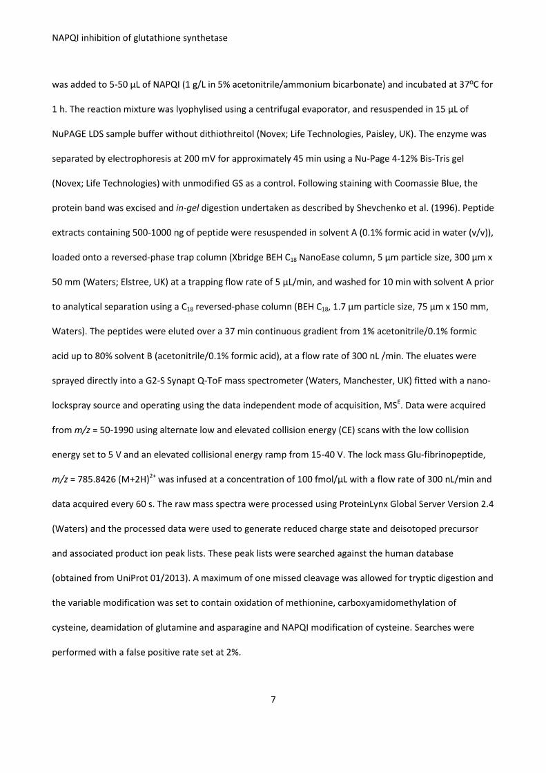
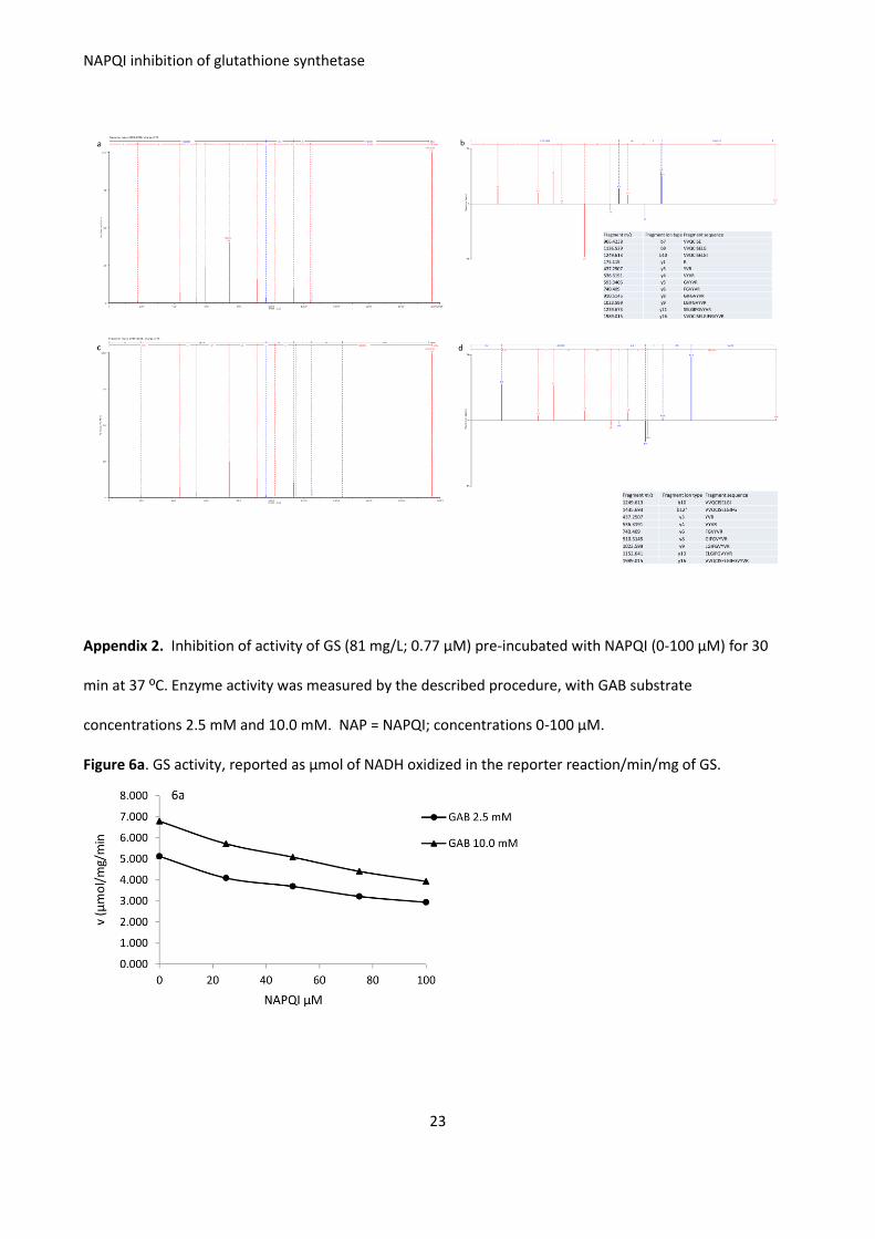
Appendix 1. Mass spectra and error plots of fragment ions of the GS peptide modified by NAPQI after co-
incubation of 5 μg of hGS with 15 μg of NAPQI for 60 min at 37 °C. Results for two independent
experiments.
Figure 5
Figure 5a. Mass spectrum of the modified peptide VVQCISELGIFGVYVR (MW = 1989.026) from experiment
1. NAPQI (∆mass = 149.0477 Da), carboamidomethylation (∆mass = 57.021 Da) and deamidation are isolate
to fragment ions from y16 to y11 corresponding to the amino acids VVQCI.
Figure 5b. Error plot of fragment ions from the modified peptide VVQCISELGIFGVYVR (MW = 1989.026) from experiment 1. NAPQI, carboamidomethylation and deamidation are isolate to fragment ions from y16 to y11 corresponding to the amino acids VVQCI. Figure 5c. Mass spectrum of the modified peptide VVQCISELGIFGVYVR (MW = 1989.026) from experiment 2. NAPQI, carboamidomethylation and deamidation are isolate to fragment ions from y16 to y10 corresponding to the amino acids VVQCIS. Figure 5d. Error plot of fragment ions from the modified peptide VVQCISELGIFGVYVR (MW = 1989.026) from experiment 2. NAPQI, carboamidomethylation and deamidation are isolate to fragment ions from y16 to y10 corresponding to the amino acids VVQCI.
NAPQI inhibition of glutathione synthetase
23
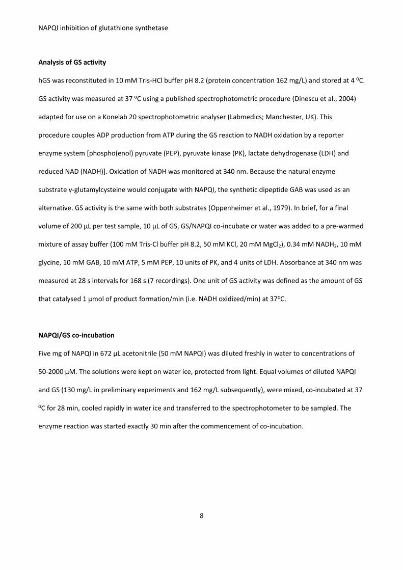
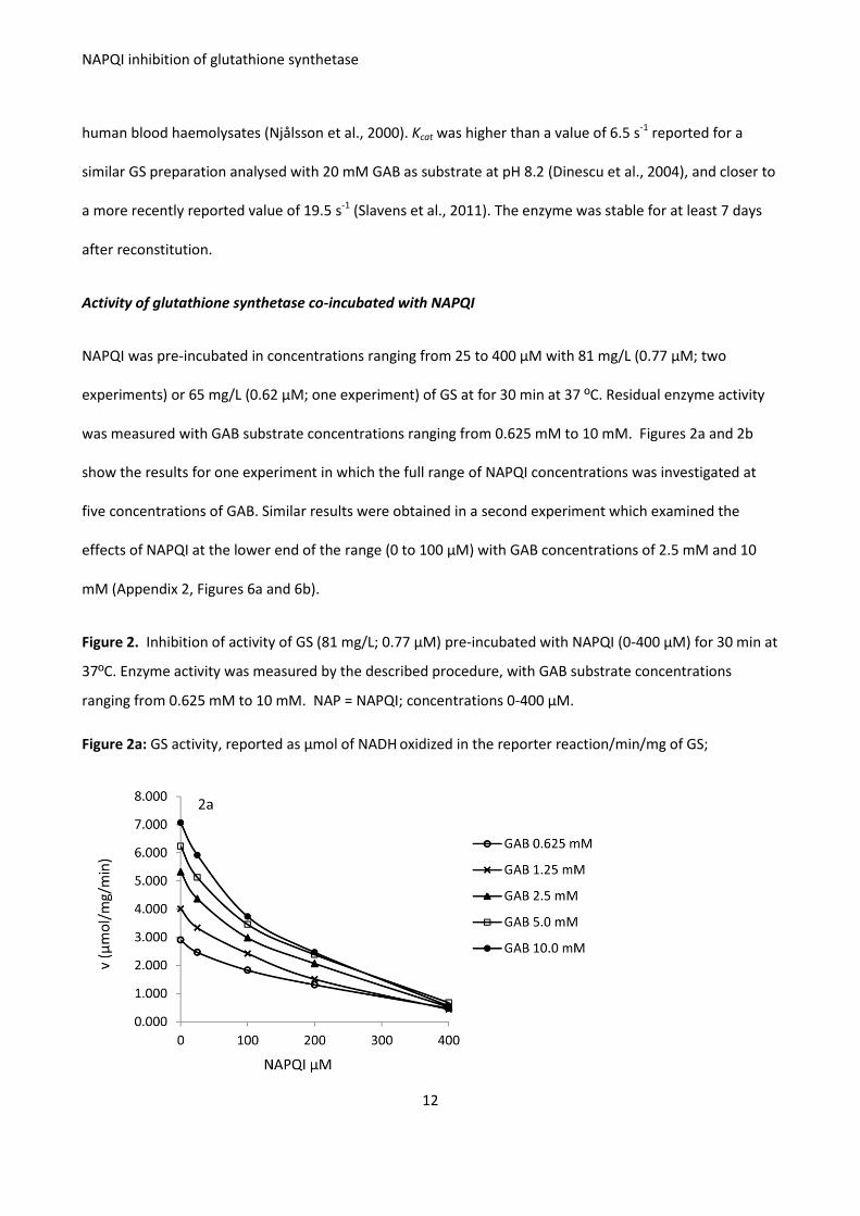
Appendix 2. Inhibition of activity of GS (81 mg/L; 0.77 μM) pre-incubated with NAPQI (0-100 μM) for 30
min at 37 ºC. Enzyme activity was measured by the described procedure, with GAB substrate
concentrations 2.5 mM and 10.0 mM. NAP = NAPQI; concentrations 0-100 μM.
Figure 6a. GS activity, reported as μmol of NADH oxidized in the reporter reaction/min/mg of GS.
NAPQI inhibition of glutathione synthetase
24
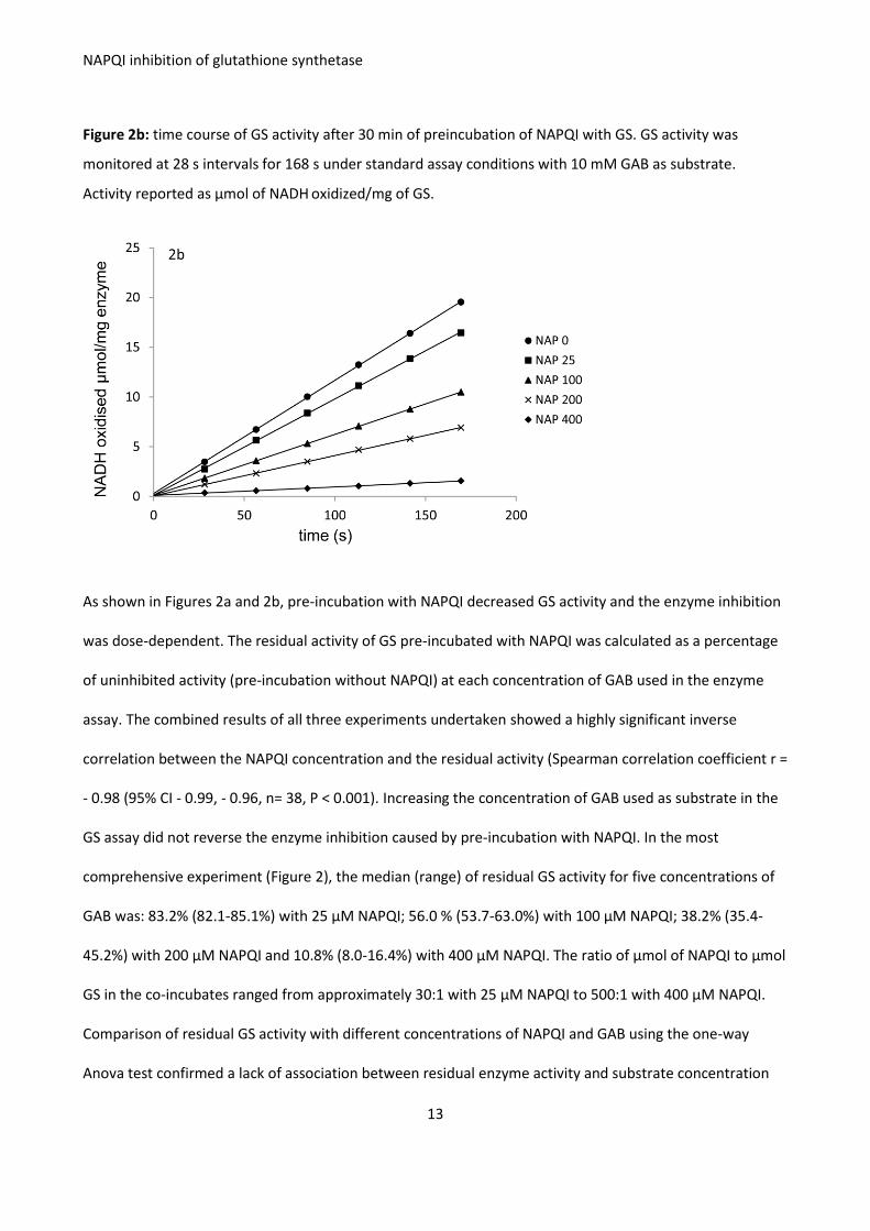
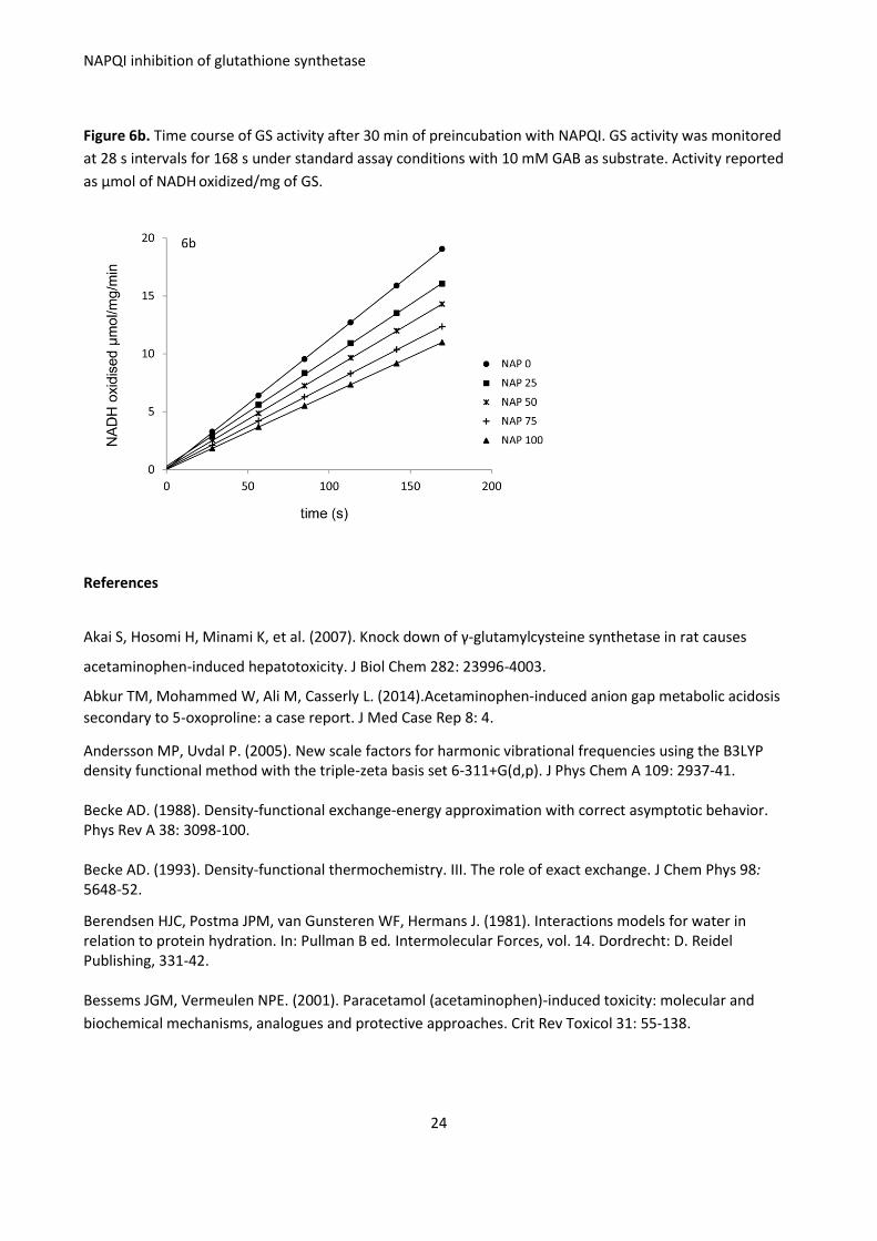
Figure 6b. Time course of GS activity after 30 min of preincubation with NAPQI. GS activity was monitored
at 28 s intervals for 168 s under standard assay conditions with 10 mM GAB as substrate. Activity reported
as μmol of NADH oxidized/mg of GS.
References
Akai S, Hosomi H, Minami K, et al. (2007). Knock down of γ-glutamylcysteine synthetase in rat causes
Abkur TM, Mohammed W, Ali M, Casserly L. (2014).Acetaminophen-induced anion gap metabolic acidosis
secondary to 5-oxoproline: a case report. J Med Case Rep 8: 4.
Andersson MP, Uvdal P. (2005). New scale factors for harmonic vibrational frequencies using the B3LYP density functional method with the triple-zeta basis set 6-311+G(d,p). J Phys Chem A 109: 2937-41. Becke AD. (1988). Density-functional exchange-energy approximation with correct asymptotic behavior. Phys Rev A 38: 3098-100. Becke AD. (1993). Density-functional thermochemistry. III. The role of exact exchange. J Chem Phys 98: 5648-52.
Berendsen HJC, Postma JPM, van Gunsteren WF, Hermans J. (1981). Interactions models for water in relation to protein hydration. In: Pullman B ed. Intermolecular Forces, vol. 14. Dordrecht: D. Reidel Publishing, 331-42. Bessems JGM, Vermeulen NPE. (2001). Paracetamol (acetaminophen)-induced toxicity: molecular and
Brohan J, Donnelly M, Fitzpatrick GJ. (2014). Metabolic acidosis with a raised anion gap associated with high
5-oxoproline levels; an under-recognised cause for metabolic acidosis in intensive care. J Clin Toxicol 4: 6.
http://dx.doi.org/10.4172/2161-0495.1000220
Bulera SJ, Birge RB, Cohen SD, Khairallah EA. (1995). Identification of the mouse liver 44-kDa
acetaminophen-binding protein as a subunit of glutamine synthetase. Toxicol Appl Pharmacol 134: 313-20.
Chen W, Shockcor JP, Tonge R, et al. (1999). Protein and nonprotein cysteinyl thiol modification by N-Acetyl-p-benzoquinone imine via a novel ipso adduct. Biochem 38: 8159-66. Cohen, SD, Khairallah, EA. (1997). Selective protein arylation and acetaminophen-induced hepatotoxicity.
Drug Metab Rev 29: 59-77.
Corbeil CR, Williams CI, Labute P. (2012). Variability in docking success rates due to dataset preparation. J Comput-Aided Mol Des 26: 775-86. Cornell WD, Cieplak P, Bayly CL, et al. (1995). A second generation force fields for the simulation of proteins, nucleic acids, and organic molecules. J Am Chem Soc 117: 5179-97. Croal BL, Glen ACA, Kelly CJG, Logan RW. (1998). Transient 5-oxoprolinuria (pyroglutamic aciduria) with
systemic acidosis in an adult receiving antibiotic therapy. Clin Chem 44: 336-40.
Dahlin DC, Miwa GT, Lu AY, Nelson SD. (1984). N-acetyl-p-benzoquinine imine: A cytochrome P-450-mediated oxidation product of acetaminophen. Proc Natl Acad Sci USA 81: 1327-31. De Jesus MC, Ingle BL, Barakat KA, et al. (2014). The role of strong electrostatic interactions at the dimer interface of human glutathione synthetase. Protein J 33: 403-9.
Dietze EC, Schäfer A, Omichinski JG, Nelson SD. (1997). Inactivation of glyceraldehyde-3-phosphate
dehydrogenase by a reactive metabolite of acetaminophen and mass spectral characterization of an
arylated active site peptide. Chem Res Toxicol 10: 1097-103.
Dinescu A, Anderson ME, Cundari TR. (2007). Catalytic loop motion in human glutathione synthetase: a
molecular modeling approach. Biochem Biophys Res Commun 353: 450-6.
Dinescu A, Cundari TR, Bhansali VS, et al. (2004). Function of conserved residues of human glutathione synthetase. J Biol Chem 279: 22412-21. Emmett M. (2014). Acetaminophen toxicity and 5-oxoproline (pyroglutamic acid): a tale of two cycles, one
an ATP-depleting futile cycle and the other a useful cycle. Clin J Am Soc Nephrol 9: 191-200.
Gali RR, Board PG. (1995). Sequencing and expression of a cDNA for human glutathione synthetase.
Biochem J 310: 353-8.
Gali RR, Board PG. (1997). Identification of an essential cysteine residue in human glutathione synthase.
Geenen S, Guallar-Hoyas C, Michopoulos F et al. (2011). HPLC-MS/MS methods for the quantitative analysis
of 5-oxoproline (pyroglutamate) in rat plasma and hepatic cell line culture medium. J Pharm Biomed Anal
56: 655-63.
Geenen S, du Preez FB, Snoep JL, et al. (2013). Glutathione metabolism modelling: a mechanism for liver
drug-robustness and a new biomarker strategy. Biochim Biophys Acta 1830: 4943-59.
Ghauri, FYK, McLean, AEM, Beales D, et al. (1993). Induction of 5-oxoprolinuria in the rat following chronic
feeding with N-acetyl 4-aminophenol (paracetamol). Biochem. Pharmacol. 46: 953-7.
Gupta S, Rogers LK, Taylor SK, Smith CV. (1997).Inhibition of carbamyl phosphate synthetase-1 and
glutamine synthetase by hepatotoxic doses of acetaminophen in mice. Toxicol Appl Pharmacol 146: 317-27.
Halmes NC, Hinson JA, Martin BM, Pumford NR. (1996). Glutamate dehydrogenase covalently binds to a
reactive metabolite of acetaminophen. Chem Res Toxicol 9: 541-6.
Heard KJ, Green JL, James LP, et al. (2011). Acetaminophen-cysteine adducts during therapeutic dosing and
following overdose. BMC Gastroenterol. 11:20.
Hess B, Kutzner C, van der Spoel D, Lindahl E. (2008). GROMACS 4: algorithms for highly efficient load-balanced, and scalable molecular simulations. J Chem Theory Comput 4: 435-47. Hornak V, Abel R, Okur A, et al. (2006). Comparison of multiple amber force fields and development of improved protein backbone parameters. Proteins 65: 712-25. Jenkins RE, Kitteringham NR, Goldring CEP, et al. (2008). Glutathione-S-transferase pi as a model protein for
the characterisation of chemically reactive metabolites. Proteomics 8: 301-5.
Labute P. (2008). The generalized Born/volume integral implicit solvent model: estimation of the free energy of hydration using London dispersion instead of atomic surface area. J Comput Chem 29: 1693-8. Landin JS, Cohen SD, KhairallahEA. (1996). Identification of a 54-kDa mitochondrial acetaminophen-binding
protein as aldehyde dehydrogenase. Toxicol Appl Pharmacol 141: 299-307.
Larsson A, Ristoff E, Anderson ME. (2005). Glutathione synthetase deficiency and other disorders of the -glutamyl cycle. In: Valle D, Beaudet AL, Vogelstein, et al., eds. The Online Metabolic Bases of Inherited Disease. McGraw-Hill Medical, NY. http://ommbid.mhmedical.com/content.aspx?sectionid=62677375&bookid=971&jumpsectionID=62677377&Resultclick=2&q=glutathione
Liss DB, Paden MS, Schwarz ES, Mullins ME. (2013). What is the clinical significance of 5-oxoproline (pyroglutamic acid) in high anion gap metabolic acidosis following paracetamol (acetaminophen) exposure? Clin Toxicol 51: 817-27.
Luo J-L, Huang C-S, Babaoglu K, Anderson ME. (2000). Novel kinetics of mammalian glutathione synthetase: characterization of γ-glutamyl substrate cooperative binding. Biochem Biophys Res Commun 275: 577-81. McConnachie LA, Mohar I, Hudson FN, et al. (2007). Glutamate cysteine ligase modifier subunit deficiency and gender as determinants of acetaminophen-induced hepatotoxicity in mice. Toxicol Sci 99: 628-36.
Malmezat T, Breuillé D, Capitan P, et al. (2000). Glutathione turnover is increased during the acute phase of sepsis in rats. J Nutr 130: 1239-46. McGill MR, Lebofsky M, Norris H-RK, et al. (2013) Plasma and liver acetaminophen-protein adduct levels in
mice after acetaminophen treatment: dose-response, mechanisms, and clinical implications. Toxicol Appl
Pharmacol 269: 240-9.
Mehta AN, Emmett JB, Emmett M. (2008). GOLD MARK: an anion gap mnemonic for the 21st century.
Lancet 372: 892.
Mitchell JR, Jollow DJ, Potter WZ, et al. (1973). Acetaminophen-induced hepatic necrosis. IV. Protective role
of glutathione. J Pharmacol Exp Ther 187: 211-7.
Muldrew KL, James LP, Coop L, et al. (2002). Determination of acetaminophen-protein adducts in mouse
liver and serum and human serum after hepatotoxic doses of acetaminophen using high-performance liquid
chromatography with electrochemical detection. Drug Metab Dispos; 30: 446-51
Moore, Thor H, Moore G, et al. (1985). The toxicity of acetaminophen and N-acetyl-p-benzoquinone imine in isolated hepatocytes is associated with thiol depletion and increased cytosolic Ca2+. J Biol Chem 260: 13035-40. Njålsson R, Carlsson K, Olin B, et al. (2000). Kinetic properties of missense mutations in patients with glutathione synthetase deficiency. Biochem J 349: 275-9. Njalsson R, Norgren S, Larsson A et al. (2001). Cooperative binding of γ-glutamyl substrate to human glutathione synthetase. Biochem Biophys Res Commun 289: 80-4. Njålsson R, Ristoff E, Carlsson K, et al. (2005). Genotype, enzyme activity, glutathione level, and clinical
phenotype in patients with glutathione synthetase deficiency. Hum Genet 116: 384-9.
Oakley AJ, Coggan M, Board PG. (2010). The identification and characterization of γ-glutamylamine cyclotransferase: an enzyme responsible for γ-glutamyl-ϵ-lysine catabolism. J Biol Chem 285: 9642-8. Oppenheimer L, Wellner VP, Griffith OW, Meister A. (1979). Glutathione Synthetase. Purification from rat kidney and mapping of the substrate binding sites. J Biol Chem 254: 5184-90. Orlowski M and Meister A. (1971). Partial reactions catalyzed by glutamylcysteine synthetase and evidence for an activated glutamate intermediate. J Biol Chem 246:7095-105. Parkinson E, Boyd P, Aleksic M, et al. (2014). Stable isotope labelling method for the investigation of protein
haptenation by electrophilic skin sensitisers. Toxicol Sci 142: 239-49.
Pitt JJ, Hauser S. (1998). Transient 5-oxoprolinuria and high anion gap metabolic acidosis: clinical and
biochemical findings in eleven subjects. Clin Chem 44: 1497-503.
Polekhina G, Board PG, Gali RR, et al. (1999). Molecular basis of glutathione synthetase deficiency and a
rare gene permutation event. EMBO J 18: 3204-13.
Pumford NR, Halmes NC, Martin BM, et al. (1997). Covalent binding of acetaminophen to N-10-formyl-
tetrahydrofolate dehydrogenase in mice. J Pharmacol Exp Ther 280: 501-5
NAPQI inhibition of glutathione synthetase
28
Qiu Y, Benet LZ, Burlingame AL. (1998). Identification of the hepatic protein targets of reactive metabolites
of acetaminophen in vivo in mice using two-dimensional gel electrophoresis and mass spectrometry. J Biol
Chem 28: 17940-53.
Ristoff E, Larsson A. (2007). Inborn errors in the metabolism of glutathione. Orphanet J Rare Dis 2:16. doi:
10.1186/1750-1172-2-16.
Shevchenko A, Jensen ON, Podtelejnikov AV, et al. (1996). Linking genome and proteome by mass
spectrometry: large-scale identification of yeast proteins from two dimensional gels. Proc Natl Acad Sci USA
93: 14440-5.
Slavens KD, Brown TR, Barakat KA, et al. (2011).Valine 44 and valine 45 of human glutathione synthetase are key for subunit stability and negative cooperativity. Biochem Biophys Res Commun 410: 597-601. Slitt AML, Dominick PK, Roberts JC, Cohen SD. (2005). Effect of ribose cysteine pretreatment on hepatic and renal acetaminophen metabolite formation and glutathione depletion. Basic Clin Pharmacol Toxicol 96: 487-94. Spielberg SP, Gordon GB. (1981). Glutathione synthetase-deficient lymphocytes and acetaminophen
toxicity. Clin Pharmacol Ther 29: 51-5.
Stephens PJ, Devlin FJ, Chabalowski CF, Frisch MJ. (1994). Ab initio calculation of vibrational absorption and circular dichroism spectra using density functional force fields. J Phys Chem 98: 11623-7. Wang J, Cieplak P, Kollman PA. (2000). How well does a restrained electrostatic potential (RESP) model perform in calculating conformational energies of organic and biological molecules? J Comput Chem 21: 1049-74. Williamson JM, Meister A. (1982). Effect of sulfhydryl group modification on the activities of 5-oxo-L-prolinase. J Biol Chem 257: 9161-72. Wu G, Fang Y-Z, Yang S, et al. (2004). Glutathione metabolism and its implications for health. J Nutr 134: