Zurich Open Repository and Archive University of Zurich Main Library Strickhofstrasse 39 CH-8057 Zurich www.zora.uzh.ch Year: 2006 The allocation of a ruminant feeding type to the okapi (Okapia johnstoni) on the basis of morphological parameters Clauss, Marcus ; Hummel, J ; Völlm, J ; Lorenz, A ; Hofmann, R R Abstract: As the anatomy of the digestive tract of the okapi (Okapia johnstoni) has not been described in relation to the ruminant feeding type classifcation to date, we here report anatomical measurements on a complete digestive tract of an okapi euthanised in captivity and measurements of its ingesta particle size and forestomach protozoa species, supplemented with other information on two okapi forestomachs and anatomical literature data. The digestive tract of the okapi is characterised by a comparatively small reticulorumen (wet contents 9.8 % of body weight) with weak rumen pillars (thickness 7-10 mm), a shallow reticular honeycomb structure (reticular crest height 1-2 mm) and a small omasum (curvature 28- 33 cm); particularly long papillae unguiculiformes have been reported and were found in one forestomach investigated, but are not a consistent fnding. The ratio of the length of the small vs. large intestine was low (1.3-1.8). The liver investigated was comparatively large (1.56 % of body weight). Faecal particle size investigated was large compared to other ruminant data. Forestomach protozoa were almost exclusively Entodinium species. All these parameters are in accord with a classifcation as a typical browser according to Hofmann (1989). However, the parotid glands investigated represented only 0.071 % of body weight, which is within the range typically reported for grazers. Potential causes and consequences of this fnding are briefy discussed. Posted at the Zurich Open Repository and Archive, University of Zurich ZORA URL: https://doi.org/10.5167/uzh-3514 Book Section Originally published at: Clauss, Marcus; Hummel, J; Völlm, J; Lorenz, A; Hofmann, R R (2006). The allocation of a ruminant feeding type to the okapi (Okapia johnstoni) on the basis of morphological parameters. In: Fidgett, Andrea; Clauss, Marcus; Eulenberger, Klaus; Hatt, Jean-Michel; Hume, Ian; Janssens, Geert Paul Jules; Nijboer, Joeke. Zoo animal nutrition Vol. III. Fürth: Filander, 253-270.
Transcript
Zurich Open Repository andArchiveUniversity of ZurichMain LibraryStrickhofstrasse 39CH-8057 Zurichwww.zora.uzh.ch
Year: 2006
The allocation of a ruminant feeding type to the okapi (Okapia johnstoni) onthe basis of morphological parameters
Clauss, Marcus ; Hummel, J ; Völlm, J ; Lorenz, A ; Hofmann, R R
Abstract: As the anatomy of the digestive tract of the okapi (Okapia johnstoni) has not been describedin relation to the ruminant feeding type classification to date, we here report anatomical measurementson a complete digestive tract of an okapi euthanised in captivity and measurements of its ingesta particlesize and forestomach protozoa species, supplemented with other information on two okapi forestomachsand anatomical literature data. The digestive tract of the okapi is characterised by a comparativelysmall reticulorumen (wet contents 9.8 % of body weight) with weak rumen pillars (thickness 7-10 mm), ashallow reticular honeycomb structure (reticular crest height 1-2 mm) and a small omasum (curvature 28-33 cm); particularly long papillae unguiculiformes have been reported and were found in one forestomachinvestigated, but are not a consistent finding. The ratio of the length of the small vs. large intestine waslow (1.3-1.8). The liver investigated was comparatively large (1.56 % of body weight). Faecal particle sizeinvestigated was large compared to other ruminant data. Forestomach protozoa were almost exclusivelyEntodinium species. All these parameters are in accord with a classification as a typical browser accordingto Hofmann (1989). However, the parotid glands investigated represented only 0.071 % of body weight,which is within the range typically reported for grazers. Potential causes and consequences of this findingare briefly discussed.
Posted at the Zurich Open Repository and Archive, University of ZurichZORA URL: https://doi.org/10.5167/uzh-3514Book Section
Originally published at:Clauss, Marcus; Hummel, J; Völlm, J; Lorenz, A; Hofmann, R R (2006). The allocation of a ruminantfeeding type to the okapi (Okapia johnstoni) on the basis of morphological parameters. In: Fidgett,Andrea; Clauss, Marcus; Eulenberger, Klaus; Hatt, Jean-Michel; Hume, Ian; Janssens, Geert Paul Jules;Nijboer, Joeke. Zoo animal nutrition Vol. III. Fürth: Filander, 253-270.
M. Clauss, J. Hummel, J. Völlm, A. Lorenz, R. R. HofmannThe allocation of a ruminant feeding type to the okapi (Okapia johnstoni) on the basis of morphological parameters
All rights reserved. No part of this book may be reproduced or transmi�ed in any form, by any means, electronic or mechanical, including photocopying, or by any information storage and retrieval system, without wri�en permission from the publisher, except for the inclusion of brief quotations in a review.
Cover photo: Rob Doolaard (Ro�erdam Zoo)
ISBN-10 3-930831-57-0ISBN-13 978-3-930831-57-9
www.filander.com
Printed in the Federal Republic of Germany
253
M. Clauss1, J. Hummel2, J. Völlm3, A. Lorenz4, R. R. Hofmann4
The allocation of a ruminant feeding type to the okapi (Okapia johnstoni) on the basis of
morphological parameters
Presented at the Joint Nutrition Symposium at Antwerp, Belgium, 2002
Abstract
As the anatomy of the digestive tract of the okapi (Okapia johnstoni) has not been described in relation to the ruminant feeding type classification to date, we here report anatomical measurements on a complete digestive tract of an okapi eutha-nised in captivity and measurements of its ingesta particle size and forestomach protozoa species, supplemented with other information on two okapi forestomachs and anatomical literature data. The digestive tract of the okapi is characterised by a comparatively small reticulorumen (wet contents 9.8 % of body weight) with weak rumen pillars (thickness 7–10 mm), a shallow reticular honeycomb structure (retic-ular crest height 1–2 mm) and a small omasum (curvature 28–33 cm); particularly long papillae unguiculiformes have been reported and were found in one forestom-ach investigated, but are not a consistent finding. The ratio of the length of the small vs. large intestine was low (1.3–1.8). The liver investigated was comparatively large (1.56 % of body weight). Faecal particle size investigated was large compared to other ruminant data. Forestomach protozoa were almost exclusively Entodinium species. All these parameters are in accord with a classification as a typical browser according to Hofmann (1989). However, the parotid glands investigated represented only 0.071 % of body weight, which is within the range typically reported for graz-ers. Potential causes and consequences of this finding are briefly discussed.
1 Division of Zoo Animals, Exotic Pets and Wildlife, Vetsuisse Faculty, University of Zurich, Switzerland, [email protected]
2 Zoological Garden of Cologne, and Institute of Animal Science, University of Bonn, Germany
3 Zoological Garden of Basle, Switzerland4 Institute of Zoo and Wildlife Research (IZW) Berlin, Germany
254
n M. C����� �� ��. n
Introduction
Although the ruminant digestive tract has been described in its morphologi-cal variety for a very long time (e. g. Flower 1872, Short et al. 1965), it was the anatomical work of Hofmann (1969, 1973, 1985, 1988, 1989, 1991, 1999) that not only provided a comparative framework within the ruminant guild by a�ributing certain morphological peculiarities to one of three feeding types (concentrate selectors/browsers, intermediate feeders, grazers), but also provided a link between the functional anatomy and the ecology of ruminants. Although the major body of morphological observations stems from Hofmann himself, other researchers have contributed to the knowl-edge of wild ruminant anatomy (e. g. Church and Hines 1978, Kay 1987, Kamler 2001, Clauss et al. 2005). This type of research has been particularly fruitful in the Australasian region (Agungpriyono et al. 1992, Fraser 1996, Yamamoto et al. 1998, Forsyth and Fraser 1999, Jiang et al. 2002).
In spite of this scientific output, a large number of species – in particular from the South American and Asian continent – remain to be described. From within the African ruminants, the digestive tract of one flagship spe-cies for many zoological gardens in terms of conservation efforts, but also in terms of visual a�ractivity, the okapi, has not been studied in relation to Hofmann’s classification. Nevertheless, the okapi has been used repeat-edly as a typical example for a browsing species in morphological, and in particular in palaeontological databases (Janis and Ehrhardt 1988, Gordon and Illius 1988, Janis1990, Solounias and Moelleken 1993, Solounias et al. 1995, 2000). This unequivocal classification can be based on solid litera-ture evidence that the okapi is an exclusive browser: Hart and Hart (1988, 1989) and Hart (1992) report that okapis do not include monocots in their diet. Already Burne (1917) reported on remnants of natural food in the oesophagus of an okapi from the wild: a bolus of small fragments of leaves of trees which were identified as fragments of dicotyledonous plants, with grasses being apparently absent. This author cites different other authors who all observed the absence of grass and the complete focus on leaves in the natural diet of the okapi (Wilmet 1913, Christy 1915).
In order to investigate the morphological adaptations of the okapi diges-tive tract to its strict dicotyledonous diet, and to facilitate a comparison with other ruminant species, we dissected one captive okapi that was eutha-nised in a zoological collection and inspected the conserved forestomach of another captive individual; additionally, we collated the available data measurements on the okapi digestive tract from the literature.
Methods
The anatomical dissection of an okapi (animal I) that was euthanized because of dental wear and disease was performed according to methods described by Hofmann (1969, 1973). As the digestive tract of okapi has been documented visually in excellent form by Burne (1917, 1939) and Neuville
255
n O���� ��������� ������� n
and Derscheid (1929), visualisation of anatomical parts was kept to a mini-mum. Samples of the contents of the reticulorumen and the abomasum were frozen and stored until sieve analysis according to Clauss et al. (2002). A sample of fresh rumen contents was fixed in formalin and analysed for protozoa species by microscope (10×) a�er staining with methylgreen using Ogimoto and Imai (1981) as a reference.
Additionally, the formalin-fixed forestomach of another okapi (animal II) that died of unknown causes was inspected. Further additional data came from Hofmann (unpublished) from the dissection of a captive animal (animal III) in 1988. Other available data on okapi digestive anatomy were collated from the literature. For an overview of the investigated animals, see Table 1.
Burne (1917) investigated a young specimen that came directly from the wild, and Neuville and Derscheid (1929) studied a two year-old female from Antwerp zoo. Burne (1939) gives no indication where the second individual he dissected came from.
Feeding regime of the animals was as follows (all amounts as fresh ma�er): Animal (I) was offered alfalfa hay ad libitum, smaller amounts of browse, 2100 g of a pelleted feed , 450 g carrots, 450 g apples, 450 g banana, 600 g vegetables (each of the la�er five distributed over 3 meals). Animal (II) was offered alfalfa hay ad libitum, smaller amounts of browse depend-ing on the season, 600 g ca�le pellet, 150 g oats, 150 g dried forage meal (alfalfa), 50 g of a mineral pellet, 600 g apple, 600 g banana, 600 g carrots and about 350 g vegetables (the la�er eight distributed over 2 meals per day). For animal (III), diet could not be reconstructed in detail.
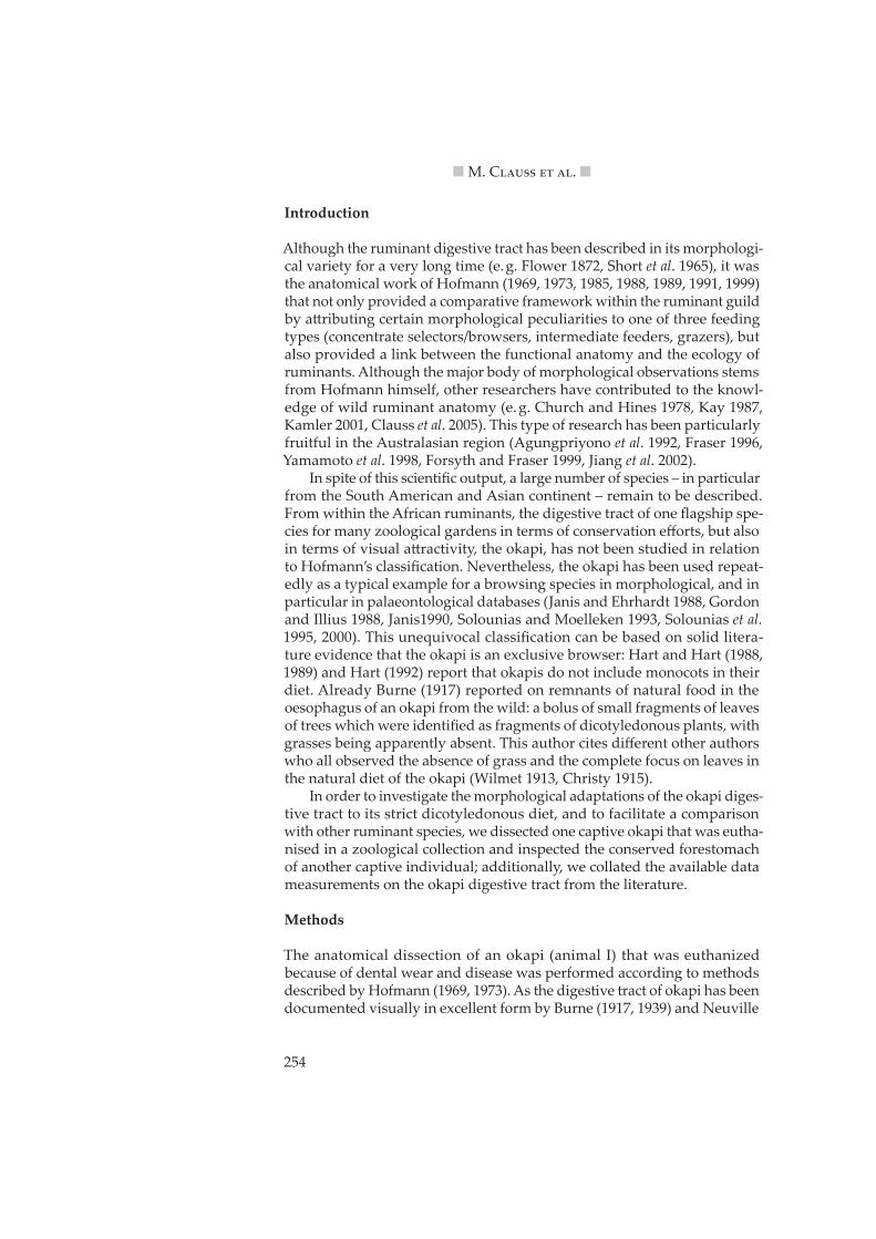
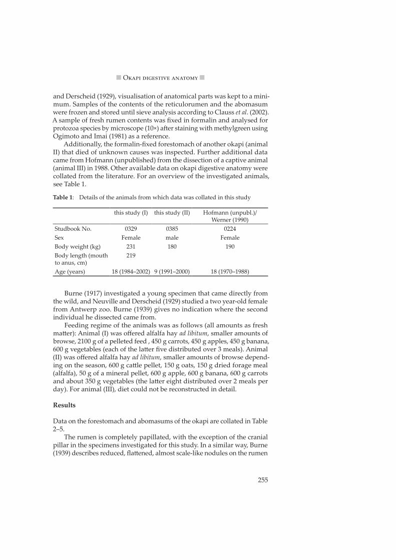
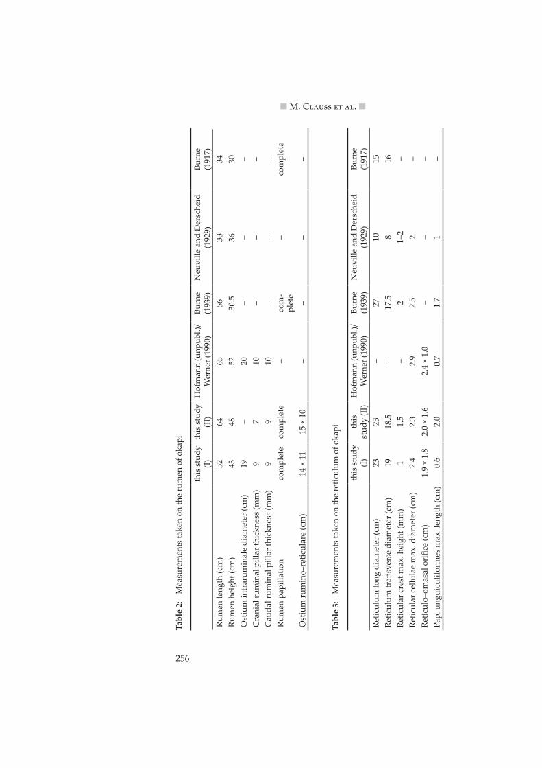
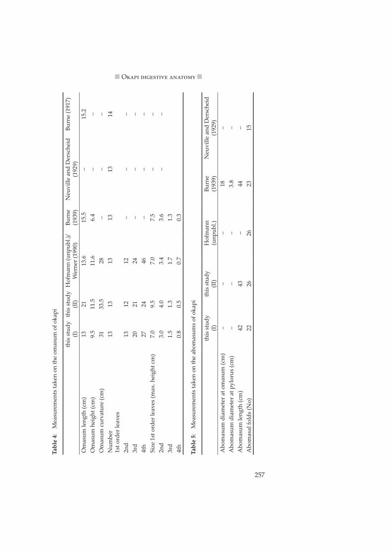
Results
Data on the forestomach and abomasums of the okapi are collated in Table 2–5.
The rumen is completely papillated, with the exception of the cranial pillar in the specimens investigated for this study. In a similar way, Burne (1939) describes reduced, fla�ened, almost scale-like nodules on the rumen
Table 1: Details of the animals from which data was collated in this study
this study (I) this study (II) Hofmann (unpubl.)/Werner (1990)
Studbook No. 0329 0385 0224Sex Female male FemaleBody weight (kg) 231 180 190Body length (mouth to anus, cm)
219
Age (years) 18 (1984–2002) 9 (1991–2000) 18 (1970–1988)
256
n M. C����� �� ��. n
Tabl
e 2:
M
easu
rem
ents
take
n on
the
rum
en o
f oka
pi
this
stu
dy
(I)th
is s
tudy
(II
)H
ofm
ann
(unp
ubl.)
/W
erne
r (19
90)
Burn
e(1
939)
Neu
ville
and
Der
sche
id(1
929)
Burn
e(1
917)
Rum
en le
ngth
(cm
)52
6465
5633
34Ru
men
hei
ght (
cm)
4348
5230
.536
30O
stiu
m in
trar
umin
ale
diam
eter
(cm
)19
–20
––
–C
rani
al ru
min
al p
illar
thic
knes
s (m
m)
97
10–
––
Cau
dal r
umin
al p
illar
thic
knes
s (m
m)
99
10–
––
Rum
en p
apill
atio
nco
mpl
ete
com
plet
e–
com
-pl
ete
–co
mpl
ete
Ost
ium
rum
ino–
retic
ular
e (c
m)
14 ×
11
15 ×
10
––
––
Tabl
e 3:
M
easu
rem
ents
take
n on
the
retic
ulum
of o
kapi
this
stu
dy
(I)th
is
stud
y (II
)H
ofm
ann
(unp
ubl.)
/W
erne
r (19
90)
Burn
e(1
939)
Neu
ville
and
Der
sche
id
(192
9)Bu
rne
(191
7)Re
ticul
um lo
ng d
iam
eter
(cm
)23
23–
2710
15Re
ticul
um tr
ansv
erse
dia
met
er (c
m)
1918
.5–
17.5
816
Retic
ular
cre
st m
ax. h
eigh
t (m
m)
11.
5–
21–
2–
Retic
ular
cel
lula
e m
ax. d
iam
eter
(cm
)2.
42.
32.
92.
52
–Re
ticul
o–om
asal
ori
fice
(cm
)1.
9 ×
1.8
2.0
× 1.
62.
4 ×
1.0
––
–Pa
p. u
ngui
culif
orm
es m
ax. l
engt
h (c
m)
0.6
2.0
0.7
1.7
1–
257
n O���� ��������� ������� n
Tabl
e 4:
M
easu
rem
ents
take
n on
the
omas
um o
f oka
pi
this
stu
dy
(I)th
is s
tudy
(II
)H
ofm
ann
(unp
ubl.)
/W
erne
r (19
90)
Burn
e (1
939)
Neu
ville
and
Der
sche
id
(192
9)Bu
rne
(191
7)
Om
asum
leng
th (c
m)
1321
13.6
15.5
–15
.2O
mas
um h
eigh
t (cm
)9.
511
.511
.66.
4–
–O
mas
um c
urva
ture
(cm
)31
33.5
28–
––
Num
ber
1st o
rder
leav
es13
1313
1313
14
2nd
1312
12–
––
3rd
2021
24–
––
4th
2724
46–
––
Size
1st
ord
er le
aves
(max
. hei
ght c
m)
7.0
9.5
7.0
7.5
––
2nd
3.0
4.0
3.4
3.6
––
3rd
1.5
1.3
1.7
1.3
4th
0.8
0.5
0.7
0.3
Tabl
e 5:
M
easu
rem
ents
take
n on
the
abom
asum
s of
oka
pi
this
stu
dy
(I)th
is s
tudy
(II
)H
ofm
ann
(u
npub
l.)Bu
rne
(1
939)
Neu
ville
and
Der
sche
id
(192
9)A
bom
asum
dia
met
er a
t om
asum
(cm
)–
––
18–
Abo
mas
um d
iam
eter
at p
ylor
us (c
m)
––
–3.
8–
Abo
mas
um le
ngth
(cm
)42
43–
44–
Abo
mas
al fo
lds
(No)
2226
2623
15
258
n M. C����� �� ��. n
pillars. The shallowness of the reticular honeycomb structure already caught the a�ention of Burne (1917, 1939), Neuville and Derscheid (1929) and Scheidegger (1950), who all present pictures of this finding in their publi-cations; Langer (1973) reported the maximal reticular crest height in okapi to be 3.9 mm, with a maximal diameter of the reticular cellulae of 2.8 cm. Particularly long papillae unguiculiformes had been noted by Burne (1939) and Neuville and Derscheid (1929) who also presented pictures of this finding. One of the two forestomachs examined for this work displayed approximately 30 very long papillae unguiculiformes with long, thin, kerati-neous tips that pointed into the reticulo-omasal orifice. The fact that the omasum appears rather small for a ruminant of this size had been com-mented upon by Scheidegger (1950), and Clauss et al. (2006) measured an average total absorptive surface for the okapi omasal leaves of 2029 cm2. Langer (1973) counted a total of 18 omasal leaves (all orders combined) in his okapis, a number that appears low compared to the other results on this parameter.
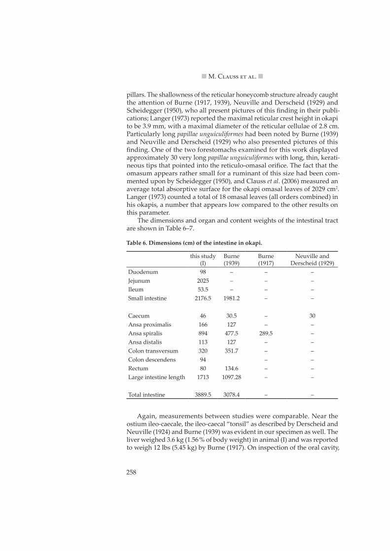
The dimensions and organ and content weights of the intestinal tract are shown in Table 6–7.
Again, measurements between studies were comparable. Near the ostium ileo-caecale, the ileo-caecal “tonsil” as described by Derscheid and Neuville (1924) and Burne (1939) was evident in our specimen as well. The liver weighed 3.6 kg (1.56 % of body weight) in animal (I) and was reported to weigh 12 lbs (5.45 kg) by Burne (1917). On inspection of the oral cavity,
Table 6. Dimensions (cm) of the intestine in okapi.
animal (I) showed severe and irregular tooth wear. The incisor arcade, which was reported to measure 43.7 mm by Gordon and Illius (1988), was of similar size with 40.5 mm in this animal. The total weight of both parotid glands was 164 g and of both mandibular glands 144 g (0.071 and 0.062 % of body weight, respectively). Burne (1917) stated that the tongue, which he measured at 35.6 cm, had a free part that was more developed than in any other ruminants except for giraffe. In animal (I), the tongue measured 40 cm with a torus of 15.5 cm and a free part of 17 cm (ratio corpus:torus 1.58; free part 42.5 % of total length).
The rumen contents of animal (I) had a pH of 6.22 and contained only protozoa belonging to the genera of Entodinium (E. dubardi, E. longinucleatum, E. simulans, E. rectangulatum, E. simplex, E. caudatum, C. exiguum, E. parvum, E. rostratum) and Dasytricha (D. ruminatum). The particle size distribution of ingesta in the reticulorumen and distal to it is displayed in Table 8.
Discussion
The data collection on the digestive anatomy of okapi shows that in general, data are in good accord across studies. The animals studied by Burne (1917)
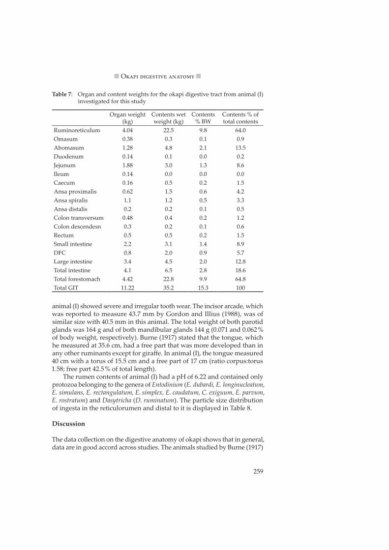
Table 7: Organ and content weights for the okapi digestive tract from animal (I) investigated for this study
and Neuville and Derscheid (1929) were evidently younger specimens that had not yet fully matured.
In order to interpret such findings on a quantitative basis, comparative data sets have to be used against which the okapi values can be compared. This method has been used repeatedly, e. g. by Jiang et al. (2002) for Mon-golian gazelles.
Forestomach parameters
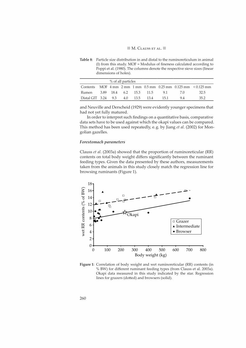
Clauss et al. (2003a) showed that the proportion of ruminoreticular (RR) contents on total body weight differs significantly between the ruminant feeding types. Given the data presented by these authors, measurements taken from the animals in this study closely match the regression line for browsing ruminants (Figure 1).
Table 8: Particle size distribution in and distal to the ruminoreticulum in animal (I) from this study. MOF = Modulus of fineness calculated according to Poppi et al. (1980). The columns denote the respective sieve sizes (linear dimensions of holes).
% of all particlesContents MOF 4 mm 2 mm 1 mm 0.5 mm 0.25 mm 0.125 mm < 0.125 mmRumen 3.89 18.4 6.2 15.3 11.5 9.1 7.0 32.5Distal GIT 3.24 9.3 4.0 13.5 13.4 15.1 9.4 35.2
02
468
101214
1618
0 100 200 300 400 500 600 700 800
wet
RR
cont
ents
(% o
f BW
)
Body weight (kg)
GrazerIntermediate
Okapi
Browser
Figure 1: Correlation of body weight and wet ruminoreticular (RR) contents (in % BW) for different ruminant feeding types (from Clauss et al. 2003a). Okapi data measured in this study indicated by the star. Regression lines for grazers (do�ed) and browsers (solid).
261
n O���� ��������� ������� n
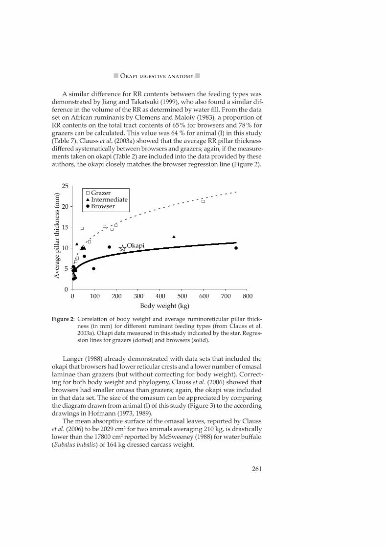
A similar difference for RR contents between the feeding types was demonstrated by Jiang and Takatsuki (1999), who also found a similar dif-ference in the volume of the RR as determined by water fill. From the data set on African ruminants by Clemens and Maloiy (1983), a proportion of RR contents on the total tract contents of 65 % for browsers and 78 % for grazers can be calculated. This value was 64 % for animal (I) in this study (Table 7). Clauss et al. (2003a) showed that the average RR pillar thickness differed systematically between browsers and grazers; again, if the measure-ments taken on okapi (Table 2) are included into the data provided by these authors, the okapi closely matches the browser regression line (Figure 2).
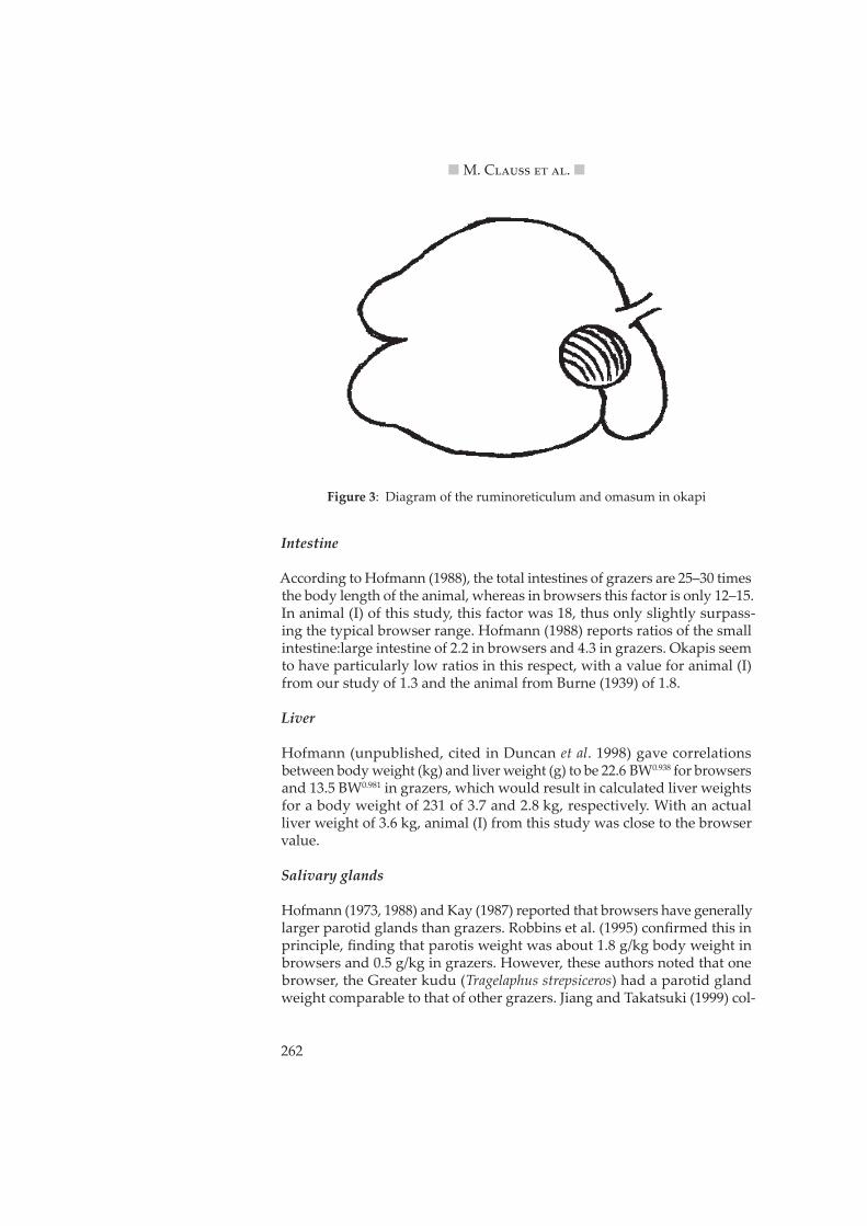
Langer (1988) already demonstrated with data sets that included the okapi that browsers had lower reticular crests and a lower number of omasal laminae than grazers (but without correcting for body weight). Correct-ing for both body weight and phylogeny, Clauss et al. (2006) showed that browsers had smaller omasa than grazers; again, the okapi was included in that data set. The size of the omasum can be appreciated by comparing the diagram drawn from animal (I) of this study (Figure 3) to the according drawings in Hofmann (1973, 1989).
The mean absorptive surface of the omasal leaves, reported by Clauss et al. (2006) to be 2029 cm2 for two animals averaging 210 kg, is drastically lower than the 17800 cm2 reported by McSweeney (1988) for water buffalo (Bubalus bubalis) of 164 kg dressed carcass weight.
Figure 2: Correlation of body weight and average ruminoreticular pillar thick-ness (in mm) for different ruminant feeding types (from Clauss et al. 2003a). Okapi data measured in this study indicated by the star. Regres-sion lines for grazers (do�ed) and browsers (solid).
0
5
10
15
20
25
0 100 200 300 400 500 600 700 800Body weight (kg)
Ave
rage
pill
ar th
ickn
ess
(mm
) GrazerIntermediateBrowser
Okapi
262
n M. C����� �� ��. n
Intestine
According to Hofmann (1988), the total intestines of grazers are 25–30 times the body length of the animal, whereas in browsers this factor is only 12–15. In animal (I) of this study, this factor was 18, thus only slightly surpass-ing the typical browser range. Hofmann (1988) reports ratios of the small intestine:large intestine of 2.2 in browsers and 4.3 in grazers. Okapis seem to have particularly low ratios in this respect, with a value for animal (I) from our study of 1.3 and the animal from Burne (1939) of 1.8.
Liver
Hofmann (unpublished, cited in Duncan et al. 1998) gave correlations between body weight (kg) and liver weight (g) to be 22.6 BW0.938 for browsers and 13.5 BW0.981 in grazers, which would result in calculated liver weights for a body weight of 231 of 3.7 and 2.8 kg, respectively. With an actual liver weight of 3.6 kg, animal (I) from this study was close to the browser value.
Salivary glands
Hofmann (1973, 1988) and Kay (1987) reported that browsers have generally larger parotid glands than grazers. Robbins et al. (1995) confirmed this in principle, finding that parotis weight was about 1.8 g/kg body weight in browsers and 0.5 g/kg in grazers. However, these authors noted that one browser, the Greater kudu (Tragelaphus strepsiceros) had a parotid gland weight comparable to that of other grazers. Jiang and Takatsuki (1999) col-
Figure 3: Diagram of the ruminoreticulum and omasum in okapi
263
n O���� ��������� ������� n
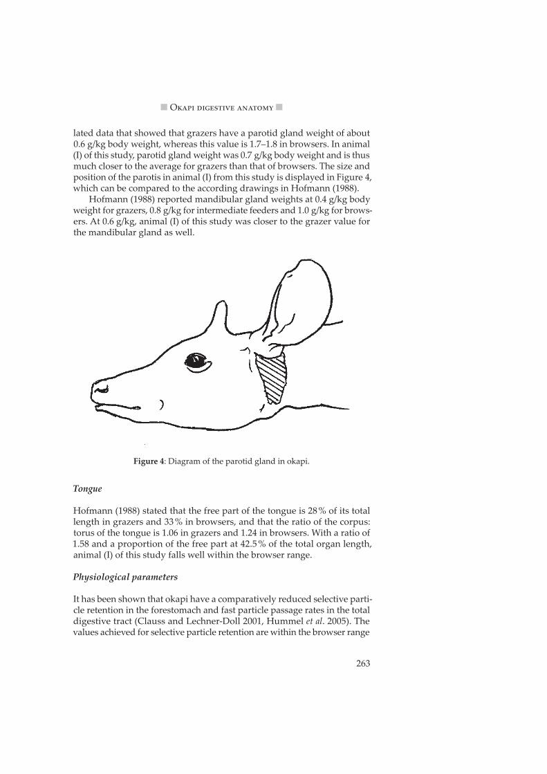
lated data that showed that grazers have a parotid gland weight of about 0.6 g/kg body weight, whereas this value is 1.7–1.8 in browsers. In animal (I) of this study, parotid gland weight was 0.7 g/kg body weight and is thus much closer to the average for grazers than that of browsers. The size and position of the parotis in animal (I) from this study is displayed in Figure 4, which can be compared to the according drawings in Hofmann (1988).
Hofmann (1988) reported mandibular gland weights at 0.4 g/kg body weight for grazers, 0.8 g/kg for intermediate feeders and 1.0 g/kg for brows-ers. At 0.6 g/kg, animal (I) of this study was closer to the grazer value for the mandibular gland as well.
Tongue
Hofmann (1988) stated that the free part of the tongue is 28 % of its total length in grazers and 33 % in browsers, and that the ratio of the corpus:torus of the tongue is 1.06 in grazers and 1.24 in browsers. With a ratio of 1.58 and a proportion of the free part at 42.5 % of the total organ length, animal (I) of this study falls well within the browser range.
Physiological parameters
It has been shown that okapi have a comparatively reduced selective parti-cle retention in the forestomach and fast particle passage rates in the total digestive tract (Clauss and Lechner-Doll 2001, Hummel et al. 2005). The values achieved for selective particle retention are within the browser range
Figure 4: Diagram of the parotid gland in okapi.
264
n M. C����� �� ��. n
for this parameter. The protozoal fauna of the forestomach of browsing ruminants has been repeatedly described as being dominated by Entodinium species only (for a compilation of literature see Clauss and Lechner-Doll 2001, Behrend et al. 2004), which were also by far the dominating protozoa in animal (I) of this study. Although large faecal particles and a high modulus of fineness in the faeces of okapi have been reported (Clauss et al. 2002), the values obtained from animal (I) even exceed the reported ones. While this can be a�ributed to the poor condition of the masticatory apparatus of this animal – a condition well-known in aged captive okapis (Raphael 1999) – the fact that such a high proportion of large particles does leave the ruminoreticulum is nevertheless remarkable.
Conclusions
The number of anatomical parameters measured in this study is smaller than that listed by Hofmann (1988) for the comparative investigation of ruminant digestive morphology. Among these are the masseter muscles, the sublingual salivary glands, and most notably histological parameters, all of which were not measured on animal (I) in this study. Nevertheless, the measured and reported parameters indicate that major anatomical and physiological parameters in okapi are within the typical browser range, and probably reflect adaptations to the peculiarities of its natural diet, which have been described in detail by Hofmann (1973, 1988, 1989, 1999), Clauss and Lechner-Doll (2001) and Clauss et al. (2002, 2003a). Admi�edly, the method of allocating a feeding type on the basis of anatomical parameters appears especially reliable if the respective opposite (grazer or browser) shall be ruled out. A differentiation between any extreme feeding type and the intermediate feeders would be more speculative in this respect.
The notable exception in this unanimous classification is the size of the salivary glands measured in this study. One of the functions of the salivary glands in browsing ruminants is the production of tannin-binding proteins (Austin et al. 1989, Hagerman and Robbins 1993, Juntheikki 1996, Fickel et al. 1998). One might speculate that the lack of natural forage with its secondary plant compounds in captivity could mean that the salivary glands are not developed according to their potential in captive animals; however, experi-mental work has shown that in contrast to rodents, the secretion of salivary tannin-binding proteins is not induced by tannin ingestion in ruminants (Austin et al. 1989, Clauss et al. 2003b). Therefore, the salivary glands of captive okapi might well represent the state natural for this species. When dissecting two captive giraffes that showed morphological adaptations to long periods of grass hay feeding (Hofmann and Matern 1988), Hofmann and Matern (unpubl.) observed relatively small salivary glands in these specimens. While it cannot be ruled out that captive individuals have less developed salivary glands than free-ranging individuals, these findings could also indicate relatively smaller salivary glands in giraffids as com-pared to other browsers. Although the relatively larger salivary glands of
265
n O���� ��������� ������� n
browsing ruminants are a robust finding in comparative anatomy (Kay 1987, Hofmann 1988) that has been confirmed by statistical evaluations of available data (Robbins et al. 1995, Jiang and Takatsuki 1999), it has been observed that the Greater kudu (Tragelaphus strepsiceros) is an exception to this rule, and that other members of the Tragelaphinae have salivary glands that are not as large as would be expected because of their dietary habits (Robbins et al. 1995). The Greater kudu is the only browsing ruminant spe-cies which has been reported to suffer losses in ecological situations where overbrowsing leads to increased dietary tannin levels in their natural forage (Van Hoven 1991). In this context, one might speculate that the large variety of plants included in the natural diet of okapi (Hart and Hart 1989) is an adaptation to reduce the intake of any one particular secondary compound in large amounts (c. f. Westoby 1974), and that a low ability to cope with such secondary compounds confines the okapi to its narrow natural habitat. Evidently, data on the tannin-binding capacity of okapi (and kudu) saliva is warranted, as well as a larger survey of ruminant salivary gland anatomy and analyses of tanninc levels in wild ruminant forages.
Knowing the morphophysiological adaptations of ruminants can help to understand the limitations to the survival (and breeding success) of a species both in captivity and in the wild. For captive management, the typi-cal “browser” digestive anatomy should caution against a common health problem in this feeding type, rumen acidosis (Marholdt and Hofmann 1991), due to the fact that these animals seem to be particularly reluctant to ingest traditional roughage sources used in zoos – hays – in sufficient proportions (Clauss et al. 2003c). The data collection of this study demon-strates that the criteria defined by Hofmann (1969, 1973, 1988, 1989) allow a classification of okapi that is in good accord with reports on its natural diet. Further comparative studies on ruminant digestive anatomy should be encouraged.
Acknowledgements
We thank Nadia Robert and Christian Wenker for their cooperation, and B. Peters and C. Wi�e for support in literature acquisition.
Yamashita, T. (1992): Morphological study on the stomach of the Lesser mouse deer (Tragulus javanicus) with special reference to the internal surface. J. Vet. Med. Sci. 54: 1063–1069
Austin, P. J.; Suchar, L. A.; Robbins, C. T.; Hagerman, A. E. (1989): Tannin-binding proteins in saliva of deer and their absence in saliva of sheep and ca�le. J. Chem. Ecol. 15: 1335–1347
Behrend, A.; Lechner-Doll, M.; Streich, W. J.; Clauss, M. (2004): Seasonal faecal excretion, gut fill, and solute and particle marker retention
266
n M. C����� �� ��. n
in mouflon (Ovis ammon musimon), and a comparison with roe deer (Capreolus capreolus). Acta Theriol. 49: 259–267
Burne, R. H. (1917): Notes on some of the viscera of an okapi (Okapia john-stoni). Proc. Zool. Soc. Lond., pp. 187–208
Burne, R. H. (1939): Description of the stomach, intestine, liver, and pan-creas of the okapi (Okapia johnstoni). Proc. Zool. Soc. Lond. 109: 451–479
Christy, C. (1915): The okapi. Nature 95: 506–507Church, D. C.; Hines, W. H. (1978): Ruminoreticular characteristics of elk.
J. Wildl. Manage. 42: 654–659Clauss, M.; Lechner-Doll, M. (2001): Differences in selective particle reten-
tion as a key factor in the diversification of ruminants. Oecologia 129: 321–327
Clauss, M.; Lechner-Doll, M.; Streich, W. J. (2002): Faecal particle size dis-tribution in captive wild ruminants: an approach to the browser/graz-er-dichotomy from the other end. Oecologia 131: 343–349
Clauss, M.; Lechner-Doll, M.; Streich, W. J. (2003a): Ruminant diversifica-tion as an adaptation to the physicomechanical characteristics of for-age. A reevaluation of an old debate and a new hypothesis. Oikos 102: 253–262
Clauss, M.; Lason, K.; Gehrke, J.; Lechner-Doll, M.; Fickel, J.; Grune, T.; Streich, W. J. (2003b): Captive roe deer (Capreolus capreolus) select for low amounts of tannic acid but not quebracho: fluctuation of prefer-ences and potential benefits. Comp. Biochem. Physiol. B 136: 369–382
Clauss, M.; Kienzle, E.; Ha�, J. M. (2003c): Feeding practice in captive wild ruminants: peculiarities in the nutrition of browsers/concentrate selectors and intermediate feeders. A review. In: Zoo animal nutrition II (A. L. Fidge�, M. Clauss, U. Gansloßer, J.-M. Ha�, J. Nijboer, eds.). Fürth: Filander Verlag, pp. 27–52
Clauss, M.; Hummel, J.; Vercammen, F.; Streich, W. J. (2005): Observations on the macroscopic digestive anatomy of the Himalayan tahr (Hemitragus jemlahicus). Anat. Hist. Embryol. 34: 276–278
Clauss, M.; Hofmann, R. R.; Hummel, J.; Adamczewski, J.; Nygren, K.; Pitra, C.; Streich, W. J.; Reese, S. (2006): The macroscopic anatomy of the omasum of free-ranging moose (Alces alces) and muskoxen (Ovibos moschatus) and a comparison of the omasal laminal surface area in 34 ruminant species. J. Zool. Lond. (in press)
Clemens, E. T.; Maloiy, G. M. O. (1983): Digestive physiology of East African wild ruminants. Comp. Biochem. Physiol. A 76: 319–333
Derscheid, J. M.; Neuville, H. (1924): Recherches anatomiques sur l’okapi. I. Le caecum et la glande ileo-caecale. Rev. Zool. Bot. Afr. 12: 498–507
267
n O���� ��������� ������� n
Duncan, P.; Tixier, H.; Hofmann, R. R.; Lechner-Doll, M. (1998): Feeding strategies and the physiology of digestion in roe deer. In: The European roe deer: the biology of success (R. Andersen, P. Duncan, J. D. C. Linnell, eds.). Stockholm: Scandinavian University Press, pp. 91–116
Fickel, J.; Göritz, F.; Joest, B. A.; Hildebrandt, T.; Hofmann, R. R.; Breves, G. (1998): Analysis of parotid and mixed saliva in roe deer. J. Comp. Physiol. B 168: 257–265
Flower, W. H. (1872): Lectures on the comparative anatomy of the organs of digestion of the mammalia. Lecture IX. Medical Times and Gaze�e 2: 319–322
Forsyth, D. M.; Fraser, K. W. (1999): Seasonal changes in the rumen mor-phology of Himalayan tahr (Hemitragus jemlahicus) in the Two Thumb Range, South Island, New Zealand. J. Zool. Lond. 249: 241–248
Fraser, K. W. (1996): Comparative rumen morphology of sympatric sika deer (Cervus nippon) and red deer (C. elaphus scoticus) in the Ahimanawa and Kaweka Ranges, central North Island, New Zealand. Oecologia 105: 160–166
Gordon, I. J.; Illius, A. W. (1988): Incisor arcade structure and diet selection in ruminants. Functional Ecol. 2: 15–22
Hagerman, A. E.; Robbins, C. T. (1993): Specificity of tannin-binding sali-vary proteins relative to diet selection by mammals. Can. J. Zool. 71: 628–633
Hart, J. A. (1992): Forage selection, forage availability and use of space by okapi (Okapia johnstoni) a rainforest giraffe in Zaire. In: Proceedings International Symposium „Ongulés/Ungulates ‚91“ (F. Spitz, G. Janeau, G. Gonzalez, S. Aulagnier, eds.). Toulouse, p. 18
Hart, J. A.; Hart, T. B. (1988): A summary report on the behaviour, ecology and conservation of the okapi (Okapia johnstoni) in Zaire. Acta Zool. Pathol. Antv. 80: 19–28
Hart, J. A.; Hart, T. B. (1989): Ranging and feeding behaviour of okapi (Okapia johnstoni) in the Ituri forest of Zaire: food limitation in a rain-forest herbivore? Symp. Zool. Soc. Lond. 61: 31–50
Hofmann, R. R. (1969): Zur Topographie und Morphologie des Wieder-käuermagens im Hinblick auf seine Funktion. Nach vergleichenden Untersuchungen an Material ostafrikanischer Wildarten. Zbl. Vet. Med. Suppl. 10: 1–180
Hofmann, R. R. (1973): The ruminant stomach. East African Monographs in Biology Volume II. Nairobi: East African Literature Bureau
Hofmann, R. R. (1985): Digestive physiology of the deer – their morpho-physiological specialisation and adaptation. R. Soc. N. Zeal. Bull. 22: 393–407
Hofmann, R. R. (1988): Morphophysiological evolutionary adaptations of the ruminant digestive system. In: Aspects of digestive physiology in ru-
268
n M. C����� �� ��. n
minants (A. Dobson, M. J. Dobson, eds.). Ithaca NY: Cornell University Press, pp. 1–19
Hofmann, R. R. (1989): Evolutionary steps of ecophysiological adaptation and diversification of ruminants: a comparative view of their diges-tive system. Oecologia 78: 443–457
Hofmann, R. R. (1991): Endangered tropical herbivores – their nutritional requirements and habitat demands. In: Recent advances on the nutrition of herbivores (Y. W. Ho, H. K. Wong, N. Abdullah, Z. A. Tajuddin, eds.). UPM Serdang: Malaysia Society of Animal Production, pp. 27–34
Hofmann, R. R. (1999): Functional and comparative digestive system anat-omy of Arctic ungulates. Rangifer 20: 71–81
Hofmann, R. R.; Matern, B. (1988): Changes in gastrointestinal morphol-ogy related to nutrition in giraffes: a comparison of wild and zoo spec-imens. Int. Zoo Yb. 27: 168–176
Hummel, J.; Clauss, M.; Zimmermann, W.; Johanson, K.; Norgaard, C.; Pfeffer, E. (2005): Fluid and particle retention in captive okapi (Okapia johnstoni). Comp. Biochem. Physiol. A 140: 436–444
Janis, C. M. (1990): Correlation of cranial and dental variables with dietary preferences in mammals: a comparison of macropodoids and ungu-lates. Mem. Queensland Mus. 28: 349–366
Janis, C. M.; Ehrhardt, D. (1988): Correlation of relative muzzle width and relative incisor width with dietary preference in ungulates. Zool. J. Linn. Soc. 92: 267–284
Jiang, Z.; Takatsuki, S. (1999): Constraints on feeding type in ruminants: a case of morphology over phylogeny. Mammal Study 24: 79–89
Jiang, Z.; Takatsuki, S.; Li, J.; Wang, W.; Ma, J.; Gao, Z. (2002): Feeding type and seasonal digestive strategy of Mongolian gazelles in China. J. Mammal. 83: 91–98
Juntheikki, M.R. (1996): Comparison of tannin-binding proteins in saliva of Scandinavian and North American moose. Biochem. Syst. Ecol. 24: 595–601
Kamler, J. (2001): Morphological variability of forestomach mucosal mem-brane in red deer, fallow deer, roe deer and mouflon. Small Rum. Res. 41: 101–107
Kay, R. N. B. (1987): Weights of salivary glands in ruminant animals. J. Zool. Lond. 211: 431–436
Langer, P. (1973): Vergleichend-anatomische Untersuchungen am Magen der Artiodactyla. II. Untersuchungen am Magen der Tylopoda und Ruminantia. Gegenbaurs Morphol. Jb. 119: 633–695
Langer, P. (1988): The mammalian herbivore stomach. Stu�gart: Gustav Fischer
269
n O���� ��������� ������� n
Marholdt, F.; Hofmann, R. R. (1991): Makro- und mikroskopische Veränderungen der Pansenschleimhaut von Zoo- und Wildwieder-käuern – ein Befundbericht mit Hinweisen zur artgerechten Fü�erung. Arbeitstagung der Zootierärzte im deutschsprachigen Raum 11: 19–34
McSweeney, C. S. (1988): A comparative study of the anatomy of the omas-um in domesticated ruminants. Aust. Vet. J. 65: 205–207
Neuville, H.; Derscheid, J. M. (1929): Recherches anatomiques sur l‘okapi Okapia johnstoni. IV. L’estomac. Rev. Zool. Bot. Afr. 16: 373–419
Ogimoto, K.; Imai, S. (1981): Atlas of rumen microbiology. Tokyo: Japan Scientific Society Press
Poppi, D. P.; Norton, B. W.; Minson, D. J.; Hendricksen, R. E. (1980): The va-lidity of the critical size theory for particles leaving the rumen. J. Agric. Sci. Camb. 94: 275–280
Raphael, B. L. (1999): Okapi medicine and surgery. In: Zoo and wild animal medicine: current therapy 4 (M. E. Fowler, R. E. Miller, eds.). Philadelphia: WB Saunders, pp. 646–649
Robbins, C. T.; Spalinger, D. E.; Van Hoven, W. (1995): Adaptation of rumi-nants to browse and grass diets: are anatomical-based browser-grazer interpretations valid? Oecologia 103: 208–213
Scheidegger, S. (1950): Pathologisch-anatomische Untersuchung des Okapi „Bambe“. Acta Trop. 7: 133–150
Short, H. L.; Medin, D. E.; Anderson, A. E. (1965): Ruminoreticular charac-teristics of mule deer. J. Mammal. 46: 196–199
Solounias, N.; Moelleken, S. M. C. (1993): Tooth microwear and premaxil-lary shape of an archaic antelope. Lethaia 26: 261–268
Solounias, N.; Moelleken, S. M. C.; Plavcan, J. M. (1995): Predicting the diet of extinct bovids using masseteric morphology. J. Vertebr. Paleontol. 15: 795–805
Solounias, N.; McGraw, W. S.; Hayek, L. A.; Werdelin, L. (2000): The pale-odiet of the Giraffidae. In: Antelopes, deer, and relatives. Fossil record, be-havioral ecology, systematics, and conservation (E. S. Vrba, G. B. Schaller, eds.). New Haven: Yale University Press, pp. 83–95
Werner, A. (1990): Vergleichende morphometrische Untersuchungen am Blä�ermagen von 17 Wiederkäuern unterschiedlicher Ernährungs-typen. [dissertation]. Giessen: University of Giessen.
Wilmet (1913): L’okapi. C. R. Acad. Sci. Paris 156: 2006–2008Van Hoven, W. (1991): Mortalities in kudu populations related to chemical
defence in trees. J. Afr. Zool. 105: 141–145Westoby, M. (1974): An analysis of diet selection by large generalist herbi-
vores. Am. Nat. 108: 290–304
270
n M. C����� �� ��. n
Yamamoto, Y.; Atoji, Y.; Agungpriyono, S.; Suzuki, Y. (1998): Morphologi-cal study of the forestomach of the Japanese serow (Capricornis cris-pus). Anat. Hist. Embryol. 27: 73–81


![COURSE AL60D: ADVANCED RUMINANT PRODUCTIONostasp.brinkster.net/downloads/al60d2012.pdfAGLS6004 [AL60D] Advanced Ruminant Production Advanced Ruminant Production Gary Wayne Garcia 02/09/2012](https://static.documents.pub/doc/80x56/5e52eefa225a0e0647002013/course-al60d-advanced-ruminant-agls6004-al60d-advanced-ruminant-production-advanced.jpg)


![Ruminant Digestion[1]](https://static.documents.pub/doc/80x56/5532bfab4a795968588b46f1/ruminant-digestion1.jpg)


