PICTORIAL REVIEW The anatomical compartments and their connections as demonstrated by ectopic air Ana Frias Vilaça & Alcinda M. Reis & Isabel M. Vidal Received: 4 April 2013 /Revised: 10 July 2013 /Accepted: 17 July 2013 /Published online: 25 September 2013 # The Author(s) 2013. This article is published with open access at Springerlink.com Abstract Air/gas outside the aero-digestive tract is abnor- mal; depending on its location, it is usually called emphy- sema, referring to trapped air/gas in tissues, or ectopic air/ gas. It can be associated to a wide range of disorders, and although it usually is an innocuous condition, it should prompt a search for the underlying aetiology, since some of its causes impose an urgent treatment. In rare instances, it may itself represent a life-threatening condition, depending on the site involved and how quickly it evolves. Abnormal air/gas beyond viscera and serosal spaces, reaches its location following some anatomic boundaries, such as fascia, which may help search the source; however if the air pressure exceeds the strength of the tissues, or the time between the aggression and the imaging is too long, the air/gas is almost everywhere, which may hinder its cause. Good knowledge of the anatomic spaces and how they connect between them facilitates the quick detection of the cause. Teaching points • Ectopic air can be depicted on conventional radiographs; but CT is more sensitive and accurate • Visceral and retropharyngeal spaces directly communicate with mediastinum • Renal fascia is a single multilaminated structure, which contains potential space Keywords Subcutaneous emphysema . Pneumomediastinum Pneumoretroperitoneum . Fascia . Anatomy Introduction Air/gas is normally seen in some body structures, such as in paranasal sinuses, and in the respiratory and gastroin- testinal tract. However, when present in subcutaneous tis- sues, cervical, mediastinal, retroperitoneal, extraperitoneal abdomen and pelvis spaces, involving muscular fibres or interstitial tissues, it is abnormal, indicating a “pathological process”, and represents a challenge to search for the underlying aetiology. Based on the embryologic development, an anatomi- cal continuum exists between the spaces allowing air/ gas to spread along them. Since air/gas may be found distant from its point of origin, a detailed anatomical knowledge is crucial in the assessment of patients with emphysema/ectopic air/gas, contributing to a clear and attempted diagnosis. In this article, we will briefly list local sources of ectopic air/gas in each space, followed by a revision of the local anatomy, highlighting the main connections among spaces. Pathogenesis and imaging Even though the word emphysema does not differentiate its composition, gas is different from atmospheric air, in both source and composition. Gas consists of carbon dioxide and nitrogen, produced by fermentation of glucose by some bacteria [1]. Several clinical conditions may present with emphysema (listed on the respective anatomic space). Often the cause is A. Frias Vilaça (*) Department of Imagiology, Hospital-Escola da Universidade Fernando Pessoa, Gondomar, Porto, Portugal e-mail: [email protected]A. Frias Vilaça : A. M. Reis : I. M. Vidal Department of Radiology, Centro Hospitalar de Entre Douro e Vouga, Rua Dr Cândido Pinho, 4520-211 Santa Maria da Feira, Portugal Insights Imaging (2013) 4:759–772 DOI 10.1007/s13244-013-0278-0
Transcript
PICTORIAL REVIEW
The anatomical compartments and their connectionsas demonstrated by ectopic air
Ana Frias Vilaça & Alcinda M. Reis & Isabel M. Vidal
Received: 4 April 2013 /Revised: 10 July 2013 /Accepted: 17 July 2013 /Published online: 25 September 2013# The Author(s) 2013. This article is published with open access at Springerlink.com
Abstract Air/gas outside the aero-digestive tract is abnor-mal; depending on its location, it is usually called emphy-sema, referring to trapped air/gas in tissues, or ectopic air/gas. It can be associated to a wide range of disorders, andalthough it usually is an innocuous condition, it shouldprompt a search for the underlying aetiology, since someof its causes impose an urgent treatment. In rare instances,it may itself represent a life-threatening condition,depending on the site involved and how quickly itevolves. Abnormal air/gas beyond viscera and serosalspaces, reaches its location following some anatomicboundaries, such as fascia, which may help search thesource; however if the air pressure exceeds the strengthof the tissues, or the time between the aggression and theimaging is too long, the air/gas is almost everywhere,which may hinder its cause. Good knowledge of theanatomic spaces and how they connect between themfacilitates the quick detection of the cause.Teaching points• Ectopic air can be depicted on conventional radiographs; but
CT is more sensitive and accurate• Visceral and retropharyngeal spaces directly communicate
with mediastinum• Renal fascia is a single multilaminated structure, which
contains potential space
Keywords Subcutaneous emphysema . PneumomediastinumPneumoretroperitoneum . Fascia . Anatomy
Introduction
Air/gas is normally seen in some body structures, such asin paranasal sinuses, and in the respiratory and gastroin-testinal tract. However, when present in subcutaneous tis-sues, cervical, mediastinal, retroperitoneal, extraperitonealabdomen and pelvis spaces, involving muscular fibres orinterstitial tissues, it is abnormal, indicating a “pathologicalprocess”, and represents a challenge to search for theunderlying aetiology.
Based on the embryologic development, an anatomi-cal continuum exists between the spaces allowing air/gas to spread along them. Since air/gas may be founddistant from its point of origin, a detailed anatomicalknowledge is crucial in the assessment of patients withemphysema/ectopic air/gas, contributing to a clear andattempted diagnosis.
In this article, we will briefly list local sources of ectopicair/gas in each space, followed by a revision of the localanatomy, highlighting the main connections among spaces.
Pathogenesis and imaging
Even though the word emphysema does not differentiate itscomposition, gas is different from atmospheric air, in both sourceand composition. Gas consists of carbon dioxide and nitrogen,produced by fermentation of glucose by some bacteria [1].
Several clinical conditions may present with emphysema(listed on the respective anatomic space). Often the cause is
A. Frias Vilaça (*)Department of Imagiology, Hospital-Escola da UniversidadeFernando Pessoa, Gondomar, Porto, Portugale-mail: [email protected]
A. Frias Vilaça :A. M. Reis : I. M. VidalDepartment of Radiology, Centro Hospitalar de Entre Douro eVouga, Rua Dr Cândido Pinho, 4520-211 Santa Maria da Feira,Portugal
iatrogenic, a well-known risk of certain procedures; infectionand “high pressure” movements also play an important role.
Pathologically, one of the following mechanisms might beimplicated: (1) disruption of cutaneous or mucosal barriers1
(e.g. bone fractures, injectable administration, drainage cath-eters, complications of intubation, endoscopic and surgicalprocedures, gastrointestinal tract tear); (2) infectious processwith gas forming microorganisms; or (3) spontaneous alveolarrupture, when the pressure gradient between air-filled alveoliand their surrounding interstitial space is sufficient to causealveolar rupture [2].
Solid parenchymatous organs and serous membranes have arelatively great resistance to the diffusion of air/gas. Instead, airdiffusion is easy through the least resistant loose areolar andfascial structures and through the communicating neurovascularstructures where the fascia is anatomically discontinued [3–5].
Ectopic air/emphysema can be depicted on conventionalradiographs. However, computed tomography (CT) is highlysensitive in the detection of abnormal air/gas, especially in“lung window” settings (window width, 1,500 HU; windowcentre, −800 HU). Soft-tissue windows such as mediastinal(window width, 400 HU; window centre, 40 HU) and abdom-inal (window width, 350 HU; window centre, 50 HU), withtheir better spacial resolution contributes to specific depictionof the anatomical location and extent [6] (Fig. 1).
Clinical impact and treatment
When leakage of air/gas is greater than reabsorption, progres-sive accumulation in various tissue planes occurs. Commonly,subcutaneous tissue offers the least resistance to expansion.
It is usually a benign and self-limited condition, causingpainless swelling of the tissues and, once its cause is resolved,in the majority of patients it spontaneously resolves in a fewdays (Fig. 2); high-flow oxygen therapy facilitates reabsorptionof nitrogen from the distended tissues.
However, if rapidly onset, it can be associated with extremediscomfort, disfigurement, anxiety and, rarely, compartmentalsyndrome, especially in the neck, mediastinum and chest wall,compromising the upper airway and jugular venous drainage(raising intracranial pressure), reducing the cardiac output bycompressing the heart and great vessels, and restricting theventilation, or even causing pacemaker malfunction [5, 7, 8].In children, whose tracheal rings are less resistant, massivepneumomediastinum may lead to compression and obstruc-tion of the trachea-bronchial tree [9]. These emergencies
impose prompt diagnosis (which is usually clinic) and treat-ment, including tracheal intubation and/or emergencytracheostomy.
Imaging studies are helpful to confirm the diagnosis ondoubtful cases, exclude local associated complications, deter-mine the extension, and monitor the evolution.
Retroperitoneal causes of ectopic air are hardly ever life-threatening in themselves, as no vital structures are underconcern. Thus, treatment will mainly depend on the type oflesion/injury and the patient’s clinical presentation. As a gen-eral rule, small perforations that are expected to spontaneouslyclose may be conservatively managed—by “nothing bymouth”, intravenous hydration and broad-spectrum intrave-nous antibiotics, as well as serial imaging studies. Largeperforations are indicated for surgical treatment [10].
Surgical procedures are a frequent cause of sparse air bub-bles, usually in the proximity of the intervention site. Thecomplete absorption of ectopic gas often occurs within 48–72 h, thus conservative-expectant management is the option,if the patient is otherwise asymptomatic. Serial imaging studiesare performed when patients are suspected to have any earlypostoperative complication; evidence of increased ectopic gas/air is worrisome and may be related to an anastomotic leak orinfectious complication. In the era of laparoscopic surgery, oneshould be aware that insufflated gas (usually carbon dioxide) issometimes temporarily retained within the abdomen, and maycause transient upper abdominal and shoulder pain, which maypersist for about 3 days [6, 11].
Gas-forming infections are a special matter of concern;clinical outcome will largely depend on a prompt diagnosisand treatment. Thus, imaging plays an important role toachieve the correct diagnosis and extension of the disease.Generally, successful treatment requires aggressive manage-ment of the infection with systemic antimicrobial therapy andcontrol of septic shock. Most of the time, these patients arecandidates for surgical interventions such as debridement orpercutaneous drainage in emphysematous pancreatitis andsurgical/percutaneous drainage or even nephrectomy in re-fractory emphysematous pyelonephritis. Patients withnecrotising fasciitis are early submitted to surgical debride-ments of the affected areas in order to remove all non-viabletissue and help relieve the cause of systemic toxicity [1].
Cervical space
Local causes of cervicofacial emphysema
1. Loss of oral cavity or pharyngeal mucosal integrity.Sources might be related to endoscopic and surgical pro-cedures (such as teeth extraction, oral, maxillary andfacial procedures, and more rarely tonsillectomy) andforeign body traumatism.
1 The mechanism for the appearance of air/gas in serosal cavities (pneu-mothorax, pneumopericardium and pneumoperitoneum) and in solidorgans (including aerobilia and portal venous gas) are beyond the scopeof this article; these will be referred only as a cause of abnormal air/gas inthe anatomical areas discussed herein.
760 Insights Imaging (2013) 4:759–772
2. Increased pressure on air pathways, such as excessivecoughing, sneezing, vomiting and nose blowing.
3. Trauma to any of the air-containing structures such asparanasal sinuses or tracheal rupture.
4. Soft tissue infections that may arise in the scalp, paranasalsinus, face, oral cavity (e.g. dental abscess) or neck.Necrotising fasciitis is an aggressive infection complicatedby gangrene that quickly diffuses through fascial planes; itis usually related to odontogenic infections in immuno-compromised patients, though it has also been describedafter trauma and tonsillectomy.
Cervical anatomy
Two major “fascial” layers enclose infrahyoid neck structures:the former superficial fascia, according to TerminologiaAnatomica (1998) now known as subcutaneous layer (telasubcutanea ) and the (deep) cervical fascia. The telasubcutanea is a fat-filled layer of connective tissue thatsurrounds the neck and contains the platysma, superficiallymph nodes, nerves and vessels. It allows the skin to
easily glide over deeper structures and extends all overthe body [12, 13].
Although some controversy exists in the description/nomenclature of the (deep) cervical fascia, for an easy under-standing, it is commonly accepted that it consists of threelayers: superficial, middle and deep layers, which divide thedeep neck into multiple spaces (Fig. 3).
The superficial (or investing) layer lies between the super-ficial cervical fascia and the muscles of the neck; it splits toencircle the sternocleidomastoid and trapezius muscles, toblend posteriorly with the ligamentum nuchae, and splitsagain between the angle of mandible and the mastoid processto enclose the parotid gland. The middle fascia extends fromthe skull base superiorly to the mediastinum inferiorly andanteriorly from the hyoid bone to thoracic inlet; it is dividedinto muscular and visceral divisions (also called pretrachealand buccopharyngeal, in the suprahyoid neck), surroundingthe strap muscles and the visceral structures of the neck,respectively. The deep layer of cervical fascia surrounds thedeep muscles of the neck and the cervical vertebrae; it extendsfrom the skull base into the mediastinum and has two
Fig. 2 A 44-year-old man with silicosis and CPOD (chronic pulmonaryobstructive disease), with an upper right lobe nodule, who developedsecondary pneumothorax after biopsy; the lung did not expand withdrainage, due to the development of broncho-pleural fistula; the drain
was accidentally removed and the patient developed rapidly progressiveemphysema. a Axial CT scan in lung windows depicts extensive subcu-taneous emphysema and pneumomediastinum. b Axial CT scan 5 dayslater, shows that all ectopic air has been reabsorbed
Fig. 1 Imaging subcutaneous emphysema. Duodenal perforation compli-cating ERCP in a 75-year old woman. a Chest conventional PA radiogra-phy shows linear lucent areas in subcutaneous and chest wall tissues. b, cContrast-enhanced CTscan at the level of the kidneys. The “lung window”(a) easily depicts the presence of air dissecting the retroperitoneum as well
as extending to the abdominal wall; the “soft tissue window” (b) accuratelylocalizes ectopic air/gas in the different planes of retroperitoneum, and inthe abdominal muscular and subcutaneous tissue. Air is also seen in thecontrast filled gallbladder (red star) with an air-fluid level (ERCP-relatedfindings) and transverse colon (blue triangle)
Insights Imaging (2013) 4:759–772 761
divisions: the alar and the prevertebral layers; the alar layerforms the posterior and lateral wall of the retropharyngealspace, and bridges the transverse processes of the vertebrae;the prevertebral layer encloses the paraspinal muscles. Thealar layer is attached to the prevertebral layer by loose con-nective tissue only, and thus an easily distended space is foundbetween them. This space is limited above by the base of theskull, while below it extends behind the oesophagus into theposterior mediastinum to the diaphragm, creating a conduit forfree movement of air/gas between mediastinum and neck.This space has been named “dangerous space”, as it allowsthe spread of the infection between the neck and mediastinum.The prevertebral layer laterally forms a sheath for the brachialnerves and subclavian vessels in the posterior triangle of theneck, and it is continued under the clavicle as the axillarysheath [6, 12, 14, 15] (Fig. 3c and d).
So, the layers of the cervical fascia define important spacesthat limit air spread along cervical tissues:
1. Pretracheal space . The space anterior to the trachea andposterior to the strap muscles and pretracheal fascia. Its
upper limit is bound by the thyroid cartilage and below inthe mediastinum by the pericardium and parietal pleura atthe level of the carina.
2. Visceral space . As suggested by the name, this infra-hyoid space encloses the thyroid and parathyroid glands,larynx and trachea, and pharynx and oesophagus. Thisspace continues into the chest, creating a conduit of freemovement of air/gas between the neck and the mediasti-num [2].
3. Carotid space . This space is surrounded by the carotidsheath. It is formed by the fusion of the major layers ofcervical fascia and contains the carotid artery, internaljugular vein and vagus nerve, and descends into the chestwith these structures.
4. Retropharyngeal space . Fat-filled space between the mid-dle layer of cervical fascia anteriorly and the alar layer ofthe deep layer posteriorly and laterally; extends from theskull base to the level of the T4 vertebral body, andinferiorly it connects with the danger space.
5. Perivertebral space. Enclosed by the prevertebral fascia,this space is continuous from the skull to the coccyx.
Fig. 3 Cervical emphysema in a 63-year-old man with tracheal ruptureafter endotracheal intubation (respiratory failure in CPOD patient com-plicated by lung infection). Contrast-enhanced CTscan at supra–hyoid (aand b) and infra-hyoid (c and d) cervical levels, illustrate the cervicalfascia and its layers. The superficial or investing layer (green line) issuperiorly attached to the superior nuchal line of the occipital, the mastoidprocess of the temporal bone and the inferior border of the mandible, andinferiorly to the manubrium, clavicles and scapula; laterally it mergeswith subcutaneous tissues of the neck. The middle fascia (pink line) runs,anteriorly, from the hyoid downwards in front of the trachea and largevessels, ultimately blending with the fibrous pericardium; and posteriorly
from the skull base, attaches to the prevertebral fascia, and merges withthe investing fascia at the lateral borders of the infrahyoid muscles. Thedeep or prevertebral layer (blue line) encircles the paraspinous andperivertebral muscles; it runs from the skull base downwards to thecoccyx; its more anterior layer contribute to the posterior and lateral wallof the retropharyngeal space and insert on the diaphragm; it gives off athin lamina, the alar layer, attached to the prevertebral layer by looseconnective tissue, creating a potential space between mediastinum andneck, the dangerous space (orange shaded area)—for free movement ofair/gas between mediastinum and neck
762 Insights Imaging (2013) 4:759–772
Air/gas spread to the upper limbs
The superficial cervical fascia laterally merges with subcuta-neous tissues of the neck.
The cervicoaxillary canal with its fibrous neurovascularsheath, enclosing the first part of the axillary artery, the axillaryvein and the brachial plexus, forms a passageway between theneck and the upper limb, also allowing air/gas to go through.
The muscular fascia also enables air/gas spread betweenthe trunk and the upper limbs, as happens with the upper limbmuscular insertions of scapular girdle, and the extensions offascia in between (e.g. the floor of the axilla is formed by thefascia extending among the pectoralis major and the latissimusdorsi) [16].
Where is the connection between neck and chest?
There are no barriers on the subcutaneous tissues all over thebody. So, free movement of air/gas is possible just under theskin. An equivalent free air/gas flow is possible in theperivertebral space. The visceral and retropharyngeal spacesdirectly communicate with the mediastinum, creating a con-duit for free movement of air/gas between mediastinum andneck [2] (Fig. 4).
Mediastinal space
A pathological accumulation of air/gas in mediastinum iscalled pneumomediastinum.
Local causes of pneumomediastinum
1. Large airways: mainly related to traumatic events.
2. Small airways: whenever the pressure gradient betweenthe air-filled alveoli and the surrounding interstitium in-creases, it might be sufficient to cause alveolar rupture.Air then spreads along the perivascular and peribronchialfascial sheaths and reaches the mediastinum—theMacklin effect (Fig. 5a and b). This may occur in thesetting of: (a) air trapping due to airway narrowing ormucous plugging (e.g. asthma), (b) straining against aclosed glottis (e.g. forced coughing, parturition, andweight-lifting), (c) blunt or barotrauma, including vigor-ous cardiopulmonary resuscitation [2, 17].
If under high pressure (because of an abrupt increase inpressure or insufficient decompression into cervical andsubcutaneous tissues and/or to retroperitoneum),pneumomediastinum may lead to rupture of the medias-tinal pleura, with air decompressing to the pleural cavity.Nevertheless, the opposite is not true—pneumothoraxdoes not lead to pneumomediastinum in normal circum-stances [2]. Although it is difficult to prove this sequenceby imaging, it is important to be aware that one of thepotential complications following alveolar rupture ispneumothorax. Once pneumothorax is established, if thepatient is on positive—pressure ventilation it should bewithdrawal, if feasible, and a tension pneumothoraxshould be considered—a life-threatening condition thatrequires immediate drainage [2].
3. Oesophagus: oesophageal perforations are full-thicknesswall tears that may have several aetiologies: (a) iatrogenic(e.g. endoscopic procedures, nasogastric tube placement,endotracheal tube misplacement, thoracic surgery; (b)forceful emesis (Boerhave syndrome); (c) oesophagealinflammatory and neoplastic diseases; (d) postoperativestatus (oesophageal anastomosis); (e) caustic injury; (f)foreign bodies.
Fig. 4 The same patient as inFig. 3: tracheal rupture afterendotracheal intubation. Sagittal(a) and coronal (b) reformattedCTscan illustrate cervico-thoraciccontinuum throughretropharyngeal (orange) andvisceral (in between pink lines)spaces
Insights Imaging (2013) 4:759–772 763
Once an oesophageal perforation occurs, along withair, contaminated saliva and gastric contents, especiallybile and strong acids enter the mediastinum, resulting inmediastinitis. On CT, densification of the mediastinal fatalong with air/gas is highly suspicious of mediastinitis, alife-threatening septic condition, which requires urgentintervention and intensive care unit admission (Fig. 5c).
Mediastinal anatomy
According to Terminologia Anatomica the fasciae of the trunkare listed as parietal, extraserosal and visceral. The fasciaparietalis is defined as a sheet or band of fibrous tissue whichlies outside the parietal layer of a serosa and lines the wall of abody cavity; the parietal fascia of the thorax is the endothoracicfascia and that of the abdomen is the endoabdominal fascia.The endothoracic fascia is firmly attached to the costal pleura,and posteriorly blends with the prevertebral fascia [18].
The mediastinum lies between the right and left pleura; itextends anteriorly from the sternum (undersurfaced by theendothoracic fascia), to the vertebral column posteriorly(prevertebral fascia), and contains all the thoracic visceraexcept for the lungs. Despite being academically divided intoupper, anterior, middle and posterior compartments, it repre-sents a single space and thus air/gas freely moves inside(Fig. 6).
Where is the link between chest and abdominal cavity?
During the early embryonic life, the coelomic cavity is formed,lined by a serous membrane, and deep to it lies the subserosalspace; the integrity of this space is maintained during thedivision of the coelomic cavity, forming a thoracoabdominalcontinuum, which crosses the diaphragmatic hiatusesconnecting the subperitoneal and subpleural portions of thesubserous space [19, 21] (Fig. 7).
The parietal pleura is attached to the thoracic wall by theendothoracic fascia. This fascia lines the diaphragm superiorly,while the transversalis fascia, which is the innermost layer of theendoabdominal fascia, lines its inferior surface [18]. The costalinsertions of the diaphragm on the inner surfaces of the lowersix ribs and their cartilages provide a separation between thesessleeves. However, three anterior defects in the diaphragmaticattachments provide blending of the two fasciae: [1] one in themidline, between the two slips of sterna origin of the diaphragmthat arise from the back of xiphoid process and [2] twoparasagittal gaps between the sternal and costal origins of thediaphragm—the sternocostal triangles or foramen of Morgagni,through which run internal mammary vessels that continue inthe abdomen as the superior epigastric vessels (Fig. 7a and b).Therefore, air/disease processes that follow these diaphragmaticdefects may enter a space between the transversalis fascia andthe parietal peritoneum, the properitoneal (or preperitoneal)space, and spread through it laterally to the flank, arriving intothe retroperitoneum[18, 20, 21].
Fig. 5 Local sources of pneumomediastinum. a , b Spontaneouspneumomediastinum due to prolonged and forceful Valsalva manoeuvresin 21-year-old man. Axial CT scan with “lung window” demonstrates theMacklin effect: air is seen along the perivascular and peribronchial fascialsheaths (a), continuous to the mediastinum (red star) (b). c Oesophagealperforation by fishbone complicated by mediastinitis in a 61-year-old
woman. Axial CT scan after oral iodinated contrast ingestion, distendingthe oesophagus (red square); extraluminal air bubbles (red arrows) areseen in mediastinum together with increased mediastinal fat attenuation,as well as multiple reactive lymph nodes. Normal permeable airways areseen (blue triangle) at level of carina
Fig. 6 Unilateral secondary spontaneous pneumothorax in a 69-year-oldman with silicosis, and extensive emphysematous changes in lungs, withbullae. Axial CT scan depicts air spread in the mediastinum as a contin-uum space, and subcutaneous planes after percutaneous drainage ofpneumothorax (chest tube not shown)
764 Insights Imaging (2013) 4:759–772
Posteriorly, endothoracic and transversalis fasciae are con-tinuous behind the diaphragm, at lumbocostal arches and theaortic and oesophageal hiatus, providing communication pointbetween mediastinum and retroperitoneum (Fig. 7b) [21].
Besides these fully accepted congenital communications,there are also areas of structural weakness of embryologicorigin, as tiny focal defects on the tendinous part of thediaphragm or the rare septum transversum lacunar aplasia,which allows transphrenic migration of air/gas [19].
The vena caval foramen is not considered part of thethoracoabdominal continuum because the wall of this vesselsticks to the margins of the foramen and thus interrupts con-tinuity of the subserous space [20].
Thus, subserous space provides not only avenues throughwhich vital structures come from thorax to abdomen and viceversa but also pathways for bidirectional spread of diseaseprocesses [20].
Retroperitoneal space
As in mediastinum, a pathological accumulation of air/gas inretroperitoneum is called pneumoretroperitoneum.
Local causes of pneumoretroperitoneum
1. Retroperitoneal hollow viscera rupture, non-traumatic(e.g. perforated duodenal ulcer, diverticulitis, perforatedcarcinoma, foreign body perforation) or traumatic (e.g.endoscopic evaluation of the gastrointestinal tract, endo-scopic retrograde cholangiopancreatography [ERCP] with/out biopsy, surgery, blunt or penetrating abdominal trauma).
2. Emphysematous infections (e.g. emphysematous pancre-atitis, which represents a superimposed infection withgas-forming bacteria in previously acute pancreatitis,and may also be a sign of end-organ infarction; emphy-sematous pyelonephritis, usually associated with poorlycontrolled diabetes mellitus, both life-threatening condi-tions, with poor prognosis) (Fig. 8) [1].
3. Residual air from retroperitoneal surgery (e.g. urological/adrenal, anterolateral approach of spinal surgery).
Retroperitoneal anatomy
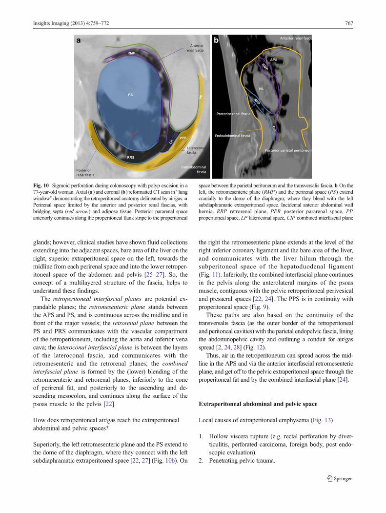
The retroperitoneum is the space between the posterior parie-tal peritoneum and the transversalis fascia, and extends fromthe diaphragm superiorly to the pelvis inferiorly. Althoughtraditionally divided in three distinct compartments, the anteriorpararenal, the perirenal and the posterior renal spaces, by theanterior (Gerota or Toldt) and the posterior (Zuckerlandl) renalfasciae, more recent work has demonstrated that the renal fasciais a single multilaminated structure, which contains potentialspace, named the interfascial plane; these potential spaces arerepresented by the retromesenteric, retrorenal, lateroconal andcombined interfascial planes [22–24] (Fig. 9).
The anterior pararenal space (APS) is bounded anteriorly bythe posterior parietal peritoneum, laterally by the lateroconal fasciaand posteriorly by the anterior renal fascia; it contains the pancre-atic gland, retroperitoneal segments of the duodenum, ascending
Fig. 7 The same patient as in Fig. 2. Axial CT in the “lung window”depicts the thoracoabdominal continuum. a The anterior blending of theendothoracic and endoabdominal fascias outlined by air: one in themidline, between the two slips of sternal origin of the diaphragm on theback of xiphoid process, and two parasagittal between the sternal and
costal origins of the diaphragm—the sternocostal triangles or foramen ofMorgagni. b The crura have separated, forming the oesophageal hiatus.The subpleural space of the thorax and subperitoneal space of the abdo-men are in continuity through the subserous space within the oesophagealand aortic hiatus
Fig. 8 Emphysematous necrotising pancreatitis in a 64-year-old woman.Contrast-enhanced axial CT scan demonstrates a mottled collection of gasbubbles mainly involving the body and tail of the pancreas; inflammatorychanges in the surrounding fat are seen. Gas spread is depicted along theretromesenteric plane and along properitoneal space (red star). RMPretromesenteric plane, APS anterior pararenal space, PP properitoneal space
Insights Imaging (2013) 4:759–772 765
and descending colon and the mesenteric root, as well as adiposetissue. This space is continuous across the midline [25].
The posterior pararenal space (PPS) is bound anteriorlyby the posterior renal fascia and posteriorly by the transversalisfascia, and it only contains adipose tissue, lymphatics andblood vessels. Medially, it is limited by the margin of the psoasmuscle. The posterolateral portion of PPS has anatomic conti-nuity with the properitoneal flank “stripe” (a layer of areolartissue and variable amount of fat in between the transversalisfascia and parietal peritoneum) Therefore, air arising within orentering this space may spread to the anterior abdominal wall.PPS also continues superiorly as a thin subdiaphragmatic layerof extraperitoneal fat [18, 19, 25, 26].
The perirenal space (PS) lies between the anterior andposterior renal fascia; it is a paired inverted cone shaped oftissue containing the kidney, the proximal portion of thecollecting system, renal and perirenal vasculature, perirenallymphatics, adrenal glands and variable amount of fat. Thespace is divided into multiple compartments by thin fibrouslamellae , which form bridging septa crossing the perinephricfat and connect the renal capsule and the anterior and posteriorrenal fascia, providing a bidirectional channel between spaces[24] (Fig. 10a).
There is a considerable disagreement with the exact ar-rangement of the renal fascia. In Meyers’ concept the renalfasciae are closed above and below the kidney and adrenal
Fig. 9 Duodenal perforation complicating ERCP in a 60-year-old man.Contrast-enhanced CTscan at the level of right renal hilum (a , b) and iliaccrest (c) shows the retroperitoneal and interfascial planes. a Anteriorpararenal space (APS) is limited by posterior parietal peritoneum andanterior renal fascia, with midline continuity; posterior pararenal space(PPS) between posterior renal fascia and transversalis layer ofendoabdominal fascia; perirenal space (PS) between anterior and posteriorrenal fascias. Renal and lateroconal fasciae are laminated, defining poten-tial spaces: the retromesenteric (RMP), the retrorenal (RRP) andlateroconal planes (LP) that all communicate at fascial trifurcation. b Axial
image focused on fascial trifurcation. Ectopic gas is seen extendingthrough the retromesenteric plane (RMP), retrorenal plane (RRP) andlateroconal plane (LP), and meeting at the fascial trifurcation (red star).Posterior pararenal space (PPS) anteriorly continues as a fat stripe in theproperitoneal space (PP). c Inferior extension of the interfascial plane,delineated by ectopic gas/air. The retromesenteric and the retrorenal planesapproximate one another as the fat cone of perirenal fat diminishesinferiorly, resulting in the combined interfascial plane (CIP); it continuesin the pelvis along the anterolateral margins of the psoas muscle contigu-ous with the pelvic retroperitoneal perivesical and presacral spaces
766 Insights Imaging (2013) 4:759–772
glands; however, clinical studies have shown fluid collectionsextending into the adjacent spaces, bare area of the liver on theright, superior extraperitoneal space on the left, towards themidline from each perirenal space and into the lower retroper-itoneal space of the abdomen and pelvis [25–27]. So, theconcept of a multilayered structure of the fascia, helps tounderstand these findings.
The retroperitoneal interfascial planes are potential ex-pandable planes; the retromesenteric plane stands betweenthe APS and PS, and is continuous across the midline and infront of the major vessels; the retrorenal plane between thePS and PRS communicates with the vascular compartmentof the retroperitoneum, including the aorta and inferior venacava; the lateroconal interfascial plane is between the layersof the lateroconal fascia, and communicates with theretromesenteric and the retrorenal planes; the combinedinterfascial plane is formed by the (lower) blending of theretromesenteric and retrorenal planes, inferiorly to the coneof perirenal fat, and posteriorly to the ascending and de-scending mesocolon, and continues along the surface of thepsoas muscle to the pelvis [22].
How does retroperitoneal air/gas reach the extraperitonealabdominal and pelvic spaces?
Superiorly, the left retromesenteric plane and the PS extend tothe dome of the diaphragm, where they connect with the leftsubdiaphramatic extraperitoneal space [22, 27] (Fig. 10b). On
the right the retromesenteric plane extends at the level of theright inferior coronary ligament and the bare area of the liver,and communicates with the liver hilum through thesubperitoneal space of the hepatoduodenal ligament(Fig. 11). Inferiorly, the combined interfascial plane continuesin the pelvis along the anterolateral margins of the psoasmuscle, contiguous with the pelvic retroperitoneal perivesicaland presacral spaces [22, 24]. The PPS is in continuity withproperitoneal space (Fig. 9).
These paths are also based on the continuity of thetransversalis fascia (as the outer border of the retroperitonealand peritoneal cavities) with the parietal endopelvic fascia, liningthe abdominopelvic cavity and outlining a conduit for air/gasspread [2, 24, 28] (Fig. 12).
Thus, air in the retroperitoneum can spread across the mid-line in the APS and via the anterior interfascial retromesentericplane, and get off to the pelvic extraperitoneal space through theproperitoneal fat and by the combined interfascial plane [24].
Extraperitoneal abdominal and pelvic space
Local causes of extraperitoneal emphysema (Fig. 13)
1. Hollow viscera rupture (e.g. rectal perforation by diver-ticulitis, perforated carcinoma, foreign body, post endo-scopic evaluation).
2. Penetrating pelvic trauma.
Fig. 10 Sigmoid perforation during colonoscopy with polyp excision in a77-year-old woman. Axial (a) and coronal (b) reformatted CTscan in “lungwindow” demonstrating the retroperitoneal anatomy delineated by air/gas. aPerirenal space limited by the anterior and posterior renal fascias, withbridging septa (red arrow) and adipose tissue. Posterior pararenal spaceanteriorly continues along the properitoneal flank stripe to the properitoneal
space between the parietal peritoneum and the transversalis fascia. b On theleft, the retromesenteric plane (RMP) and the perirenal space (PS) extendcranially to the dome of the diaphragm, where they blend with the leftsubdiaphramatic extraperitoneal space. Incidental anterior abdominal wallhernia. RRP retrorenal plane, PPR posterior pararenal space, PPproperitoneal space, LP lateroconal space, CIP combined interfascial plane
Insights Imaging (2013) 4:759–772 767
3. Residual air from previous surgery (e.g. abdominoperinealamputation, hysterectomy, caesarean).
4. Emphysematous infection: e.g. necrotising fasciitis ofperineal, genital or perianal regions (Fournier gangrene)is a polymicrobial infection complicated by thrombosis ofsmall subcutaneous vessels and rapid gangrenousinvolvement of the surrounding skin and deep fascia;although the diagnosis is based on clinical examination,CT plays an important role not only in confirming thediagnosis, but essentially to assess the extent of disease toplan the surgical treatment, sometimes identifying a po-tential underlying cause [1].
Extraperitoneal abdominal and pelvic anatomy
As already stated, the abdominopelvic cavity is entirely linedby the endoabdominal (parietal abdominal) fascia, whoseinnermost layer is the transversalis fascia; this name is derivedfrom its location between the inner surface of the transverseabdominal muscle and the parietal peritoneum in the anteriorand lateral abdominal wall; nevertheless, it also undersurfacesthe diaphragm, is the anterior surface of the anterior longitu-dinal ligament, paravertebral, iliopsoas and internal obturatormuscles; moreover, it delineates the lateral walls and dia-phragm of the pelvis [4, 18].
Fig. 11 Duodenal perforation complicating ERCP in a 75-year-old wom-an. Sagittal reformatted (a) and axial (b) contrast-enhanced CT scandemonstrates the right superior extension of the interfascial plane. The
multilaminated retroperitoneum is open towards the upper abdominalextraperitoneal space: the bare area of the liver, and communicates withliver hilum through the subperitoneal space of the hepatoduodenal ligament
Fig. 12 Anastomotic colorectalleak during a CTcolonoscopy in a81-year-old woman with a historyof excision of recto-sigmoidadenocarcinoma 8 yearspreviously. Coronal (a) andsagittal (b) reformatted CT scan.Continuity of the transversalisfascia with the parietal endopelvicfascia is depicted
768 Insights Imaging (2013) 4:759–772
Anatomical and embryological differences dictate thatthe fascia transversalis should be considered differentlyfrom the supra-abdominal and infra-abdominal regions.The arcuate line, which is an horizontal line that de-marcates the lower limit of the posterior layer of therectus sheath, separates the cranial from the caudal partof the abdominal wall; below this line, the aponevrosisof internal oblique, external oblique and abdominaltransversal muscles pass anterior to the rectus muscle,and the fascia transversalis becomes stronger and func-tions as an aponevrosis, attached to the abdominal rectusmuscle [18].
In the infraumbilical properitoneal space are the urachus andthe obliterated umbilical arteries, which are developmentalremnants, forming the medial and the median umbilical liga-ments, respectively [18].
The endopelvic fascia is the internal investing fasciaof the pelvis and consists of two layers, the parietallayer (how the transversalis fascia is named at thislevel) and visceral layer ; it courses along the lateral bordersof the pelvic organs, and encircles the perirectal fat, at thispoint named mesorectal fascia. Both sleeves are continu-ous and attached to the diaphragmatic part of the pelvicfascia along the tendinous arch (a thickened band on theupper layer of the diaphragmatic part of the pelvic fasciaat the level of a line extending from the lower part of thepubic symphysis to the spine of the ischium) [14, 29](Fig. 12).
The umbilicovesical fascia runs inferiorly from the umbi-licus, posterior to tranversalis fascia and anterior to parietalperitoneum, with a triangular configuration with its apex at theumbilicus. It surrounds the urachus and obliterated umbilicalarteries. Courses on the pelvis below the peritoneal reflection
and extends to the pelvis floor surrounding the urinary blad-der, then blending with the endopelvic visceral fascia alongthe lateral aspects of the lower uterus or seminal vesicles andthe rectum.
Pelvic cul-de-sacs between the rectum and bladder (therectovesical space in males), and rectum and uterus (theDouglas cul-de-sac in females) are shaped by parietal perito-neum reflections. These reflections extend inferiorly, and theiranterior and posterior sleeves are fused, originating septa thatseparate the urological or genital tract from the rectum:rectovesical and rectovaginal septum .
Thus, a compartmental pelvis may be described as follows[4, 30, 31]:
1. Prevesical spaceAnterior and lateral to the umbilicovesical fascia,
and posterior to the trasversalis fascia. Prevesical spacelaterally communicates with the properitoneal spaceof the abdominal wall and flanks; posteroinferiorly itsurrounds the lateral walls of the urinary bladder, andanteroinferiorly it forms the retropubic space (the spaceof Retzius) [32] (Fig. 11).
2. Paravesical spaceMedially is limited by the umbilicovesical fascia,
which fuses inferiorly with the visceral sleeve of theendopelvic fascia. The lateral boundary is the parietalendopelvic fascia, and the superior one the peritoneum(Fig. 11).
3. Presacral spaceRepresents the posterior communication between
both paravesical spaces. It is located between therectal and parietal sleeves of the endopelvic fascia(Fig. 11).
Fig. 13 Local causes of extraperitoneal pelvic emphysema. (a) Sigmoidperforation (necrotising acute colitis probably due to fecaloma) in a 75-year-old female. Unenhanced-CTscans of pelvis shows gas pockets in thesigmoid mesocolon, extending superiorly to retroperitoneum through thecombined interfascial plane. (b ) Fournier gangrene in a 67-year-old
diabetic man. Contrast-enhanced CT scan shows fluid and air pocketstracking in the corpora cavernosa, extending anteriorly to an enlargedscrotal sac containing gas and cranially to the ischiorectal fossa andsubcutaneous tissue
Insights Imaging (2013) 4:759–772 769
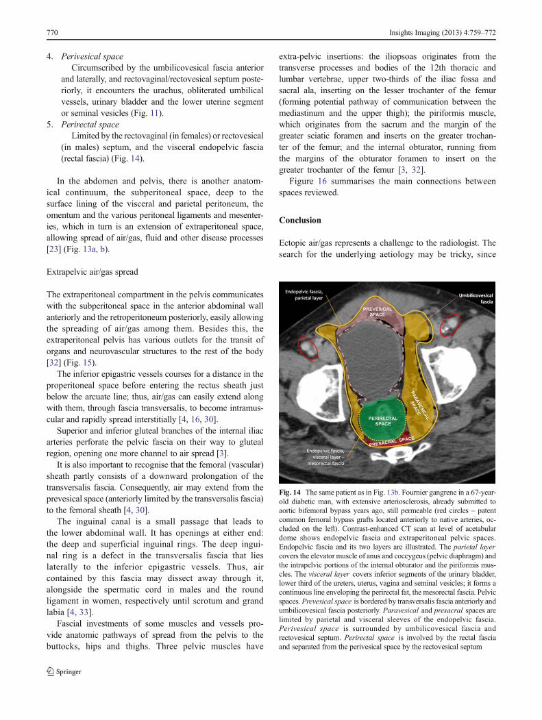
4. Perivesical spaceCircumscribed by the umbilicovesical fascia anterior
and laterally, and rectovaginal/rectovesical septum poste-riorly, it encounters the urachus, obliterated umbilicalvessels, urinary bladder and the lower uterine segmentor seminal vesicles (Fig. 11).
5. Perirectal spaceLimited by the rectovaginal (in females) or rectovesical
(in males) septum, and the visceral endopelvic fascia(rectal fascia) (Fig. 14).
In the abdomen and pelvis, there is another anatom-ical continuum, the subperitoneal space, deep to thesurface lining of the visceral and parietal peritoneum, theomentum and the various peritoneal ligaments and mesenter-ies, which in turn is an extension of extraperitoneal space,allowing spread of air/gas, fluid and other disease processes[23] (Fig. 13a, b).
Extrapelvic air/gas spread
The extraperitoneal compartment in the pelvis communicateswith the subperitoneal space in the anterior abdominal wallanteriorly and the retroperitoneum posteriorly, easily allowingthe spreading of air/gas among them. Besides this, theextraperitoneal pelvis has various outlets for the transit oforgans and neurovascular structures to the rest of the body[32] (Fig. 15).
The inferior epigastric vessels courses for a distance in theproperitoneal space before entering the rectus sheath justbelow the arcuate line; thus, air/gas can easily extend alongwith them, through fascia transversalis, to become intramus-cular and rapidly spread interstitially [4, 16, 30].
Superior and inferior gluteal branches of the internal iliacarteries perforate the pelvic fascia on their way to glutealregion, opening one more channel to air spread [3].
It is also important to recognise that the femoral (vascular)sheath partly consists of a downward prolongation of thetransversalis fascia. Consequently, air may extend from theprevesical space (anteriorly limited by the transversalis fascia)to the femoral sheath [4, 30].
The inguinal canal is a small passage that leads tothe lower abdominal wall. It has openings at either end:the deep and superficial inguinal rings. The deep ingui-nal ring is a defect in the transversalis fascia that lieslaterally to the inferior epigastric vessels. Thus, aircontained by this fascia may dissect away through it,alongside the spermatic cord in males and the roundligament in women, respectively until scrotum and grandlabia [4, 33].
Fascial investments of some muscles and vessels pro-vide anatomic pathways of spread from the pelvis to thebuttocks, hips and thighs. Three pelvic muscles have
extra-pelvic insertions: the iliopsoas originates from thetransverse processes and bodies of the 12th thoracic andlumbar vertebrae, upper two-thirds of the iliac fossa andsacral ala, inserting on the lesser trochanter of the femur(forming potential pathway of communication between themediastinum and the upper thigh); the piriformis muscle,which originates from the sacrum and the margin of thegreater sciatic foramen and inserts on the greater trochan-ter of the femur; and the internal obturator, running fromthe margins of the obturator foramen to insert on thegreater trochanter of the femur [3, 32].
Figure 16 summarises the main connections betweenspaces reviewed.
Conclusion
Ectopic air/gas represents a challenge to the radiologist. Thesearch for the underlying aetiology may be tricky, since
Fig. 14 The same patient as in Fig. 13b. Fournier gangrene in a 67-year-old diabetic man, with extensive arteriosclerosis, already submitted toaortic bifemoral bypass years ago, still permeable (red circles – patentcommon femoral bypass grafts located anteriorly to native arteries, oc-cluded on the left). Contrast-enhanced CT scan at level of acetabulardome shows endopelvic fascia and extraperitoneal pelvic spaces.Endopelvic fascia and its two layers are illustrated. The parietal layercovers the elevator muscle of anus and coccygeus (pelvic diaphragm) andthe intrapelvic portions of the internal obturator and the piriformis mus-cles. The visceral layer covers inferior segments of the urinary bladder,lower third of the ureters, uterus, vagina and seminal vesicles; it forms acontinuous line enveloping the perirectal fat, the mesorectal fascia. Pelvicspaces. Prevesical space is bordered by transversalis fascia anteriorly andumbilicovesical fascia posteriorly. Paravesical and presacral spaces arelimited by parietal and visceral sleeves of the endopelvic fascia.Perivesical space is surrounded by umbilicovesical fascia andrectovesical septum. Perirectal space is involved by the rectal fasciaand separated from the perivesical space by the rectovesical septum
770 Insights Imaging (2013) 4:759–772
air can be detected almost anywhere (away from itssource). Continuity is described between neck, medias-tinum, retroperitoneum and the pelvis on the basis of
anatomy and fascial-defined spaces; vascular perforationsof the fasciae and muscle insertions also constitute potentialconduits between compartments.
Fig. 16 Ectopic air/gas flow chart through different anatomical spaces
Fig. 15 Air spread from pelvis to inferior limbs. a, b The same patient as inFig. 2: a 44-year-old man with broncho-pleural fistula secondary to pneumo-thorax drainage and subsequent rapidly progressive emphysema from neck togroin. a Axial CT in “lung window” shows air spread through the thinnedtransversalis fascia and abdominal rectus below the level of arcuate line,involving inferior epigastric vessels. Air is seen anteriorly to iliac muscles(brown arrow). b Axial CT caudal to a demonstrates air spreading out the
pelvis along with iliac vessels through the femoral canal. Air dissecting alongmuscular fibres and sheaths, as can be seen from the pelvis along iliac muscleuntil its insertion in the lesser femoral trochanter (brown line). The leastresistance of subcutaneous tissues allow easy spread of subcutaneous emphy-sema. c, d A 60-year-old patient with colonic perforation at optical colonos-copy. Sagittal (c) and coronal (d) reformatted CT images illustrate air spreadalong the right inguinal (red star) canal and into the scrotal sac
Insights Imaging (2013) 4:759–772 771
Acknowledgments Ana Frias Vilaça and Alcinda Reis contributedequally to this work.
Authors thank Liliana Leite for language revision of the article. Thiswork was previously presented as an education exhibit at the 2009 RSNAannual meeting (Space: LL-ER2494).
Disclosures The authors declare no conflicts of interest. No fundingwas received for this work.
Open Access This article is distributed under the terms of the CreativeCommons Attribution License which permits any use, distribution, andreproduction in any medium, provided the original author(s) and thesource are credited.
References
1. Grayson D, Abbott R, Levy A, Sherman P (2002) Emphysematousinfections of the abdomen and pelvis: a pictorial review. Radiographics22:543–561
2. Maunder RJ, Pierson DJ, Hudson LD (1984) Subcutaneous andmediastinal emphysema. Pathophysiology, diagnosis, and manage-ment. Arch Intern Med 144:1447–1453
3. Meyers M, Goodman K (1975) Pathways of extrapelvic spread ofdisease: anatomic-radiologic correlation. AJR Am J Roentgenol 125:900–909
4. Mastromatteo JF et al (1997) Communications of the pelvicextraperitoneal spaces and their relation to the abdominal extraperitonealspaces: helical CT cadaver study with pelvic extraperitoneal injections.Radiology 202:523–530
5. Srinivas R, Singh N, Agarwal R, Aggarwal AN (2007) Managementof extensive subcutaneous emphysema and pneumomediastinum bymicro-drainage: time for a re-think? Singapore Med J 48:e323
6. Prokop M, Galanski M (2003) Spiral and multislice computed to-mography of the body. Thieme, Stuttgart New York
7. Williams DJ, Jaggar SI, Morgan CJ (2005) Upper airway obstructionas a result of massive subcutaneous emphysema following accidentalremoval of an intercostal drain. Br J Anaesth 94(3):390–392
8. Sucena M, Coelho F, Almeida T, Gouveia A, Hespanhol V (2010)Massive subcutaneous emphysema—management using subcutane-ous drains. Rev Port Pneumol 16:321–329
9. Fortes F, Sennes LU, Fortes FSG, Imamura R, Tsuji DH (2007)Cervical emphysema as an early complication of tonsillectomy. ArqInt Otorrinolaringol 11:65–69
10. Fujii L, Lau D, Fleischer DE, Harrison ME (2010) Successfulnonsurgical treatment of pneumomediastinum, pneumothorax,pneumoperitoneum, pneumoretroperitoneum and subcutaneousemphysema following ERCP. Gastroenterol Res Pract 2010:289135
11. Tsai HW, Chen YJ, Ho CM et al (2011) Maneuvers to decreaselaparoscopy-induced shoulder and upper abdominal pain a random-ized controlled study. Arch Surg 146:1360–1366
12. Smoker WR, Harnsberger HR (1991) Differential diagnosis of headand neck lesions based on their space of origin. 2. The infrahyoidportion of the neck. AJR Am J Roentgenol 157:155–159
13. Kumka M, Bonar J (2012) Fascia: a morphological description andclassification system based on a literature review. J Can ChiroprAssoc 56:179–191
14. Standring S (ed) (2004) Gray’s anatomy: the anatomical basis ofclinical practice. Churchill Livingstone, Oxford
15. Warshafsky D, Goldenberg D, Kanekar SG (2012) Imaging anatomyof deep neck spaces. Otolaryngol Clin N Am 45:1203–1221
16. Macéa JR, Fregnani JHTG (2006) Anatomy of the thoracic wall,axilla and breast. Int J Morphol 24:691–704
17. Zylak CM, Standen JR, Barnes GR, Zylak CJ (2000)Pneumomediastinum revisited. Radiographics 20:1043–1057
18. Skandalakis PN, Zoras O et al (2006) Transversalis, endoabdominal,endothoracic fascia: who’s who? Am Surg 72:16–18
19. Lidid L, Valenzula J, Villarroel C, Alegria J (2013) Crossing the barrier:when the diaphragm is not a limit. AJR Am J Roentgenol 200:W62–W70
20. Oliphant M, Berne AS, Meyers MA (1999) The subserousthoracoabdominal continuum: embryologic basis and diagnostic im-aging of disease spread. Abdom Imaging 24:211–219
21. Kleinman P, Brill P, Whalen J (1978) Anterior pathway fortransdiaphragmatic extension of pneumomediastinum. AJR Am JRoentgenol 131:271–275
22. Lee SL, Ku YM, Rha SE (2010) Comprehensive reviews of theinterfascial plane of the retroperitoneum: normal anatomy and path-ologic entities. Emerg Radiol 17:3–11
23. Tirkes T, Sandrasegaran K, Patel AA et al (2012) Peritoneal andretroperitoneal anatomy and its relevance for cross-sectional imaging.Radiographics 32:437–451
24. Gore R, Balfe DM, Aizenstein RI, Silverman PM (2000) The greatescape: interfascial decompression planes of the retroperitoneum.AJR Am J Roentgenol 175:363–370
25. Love L, Meyers M, Churchill R, Reynes C, Moncada R, Gibson D(1981) Computed tomography of extraperitoneal spaces. AJR Am JRoentgenol 136:781–789
26. Meyers MA (1974) Radiological features of the spread and localiza-tion of extraperitoneal gas and their relationship to its source. Radi-ology 111:17–26
27. Lim JH, Kim B, Auh YH (1998) Anatomical communications of theperirenal space. Br J Radiol 71:450–456
28. Aikawa H, Tanoue S, Okino Y, Tomonari K, Miyake H (1998) Pelvicextension of retroperitoneal fluid: analysis in vivo. AJR Am JRoentgenol 171:671–677
29. Chen N, Min PO, Liu ZYet al (2010) Radiologic and anatomic studyof the extraperitoneal space associated with the rectum. AJR Am JRoentgenol 194:642–652
30. Auh YH, Rubenstein WA, Schneider M et al (1986) Extraperitonealparavesical spaces: CT delineation with US correlation. Radiology159:319–328
31. Meyers M (2000) Dynamic radiology of the abdomen: normal andpathologic anatomy, 5th edn. Springer, New York
32. Tan CH, Vikram R, Boonsirikamchai P et al (2011) Pathways ofextrapelvic spread of pelvic disease: imaging findings. Radiographics31:117–133
33. Bhosale PR, Patnana M, Viswanathan C, Szklaruk J (2008) Theinguinal canal: anatomy and imaging features of common and un-common masses. Radiographics 28:819–835