The Anionic (9-Methyladenine)-(1-Methylthymine) Base Pair Solvated by Formic Acid. A Computational and Photoelectron Spectroscopy Study Piotr Storoniak,* ,† Kamil Mazurkiewicz, † Maciej Haranczyk, ‡ Maciej Gutowski, § Janusz Rak, † Soren N. Eustis, | Yeon Jae Ko, | Haopeng Wang, | and Kit H. Bowen* ,| Department of Chemistry, UniVersity of Gdan ´sk, Sobieskiego 18, 80-952 Gdan ´sk, Poland, Computational Research DiVision, Lawrence Berkeley National Laboratory, 1 Cyclotron Road, Mail Stop 50F-1650, Berkeley, California 94720-8139, Chemistry-School of Engineering and Physical Sciences, Heriot-Watt UniVersity, Edinburgh EH14 4AS, U.K., and Department of Chemistry, Johns Hopkins UniVersity, Baltimore, Maryland 21218 ReceiVed: May 21, 2010; ReVised Manuscript ReceiVed: July 26, 2010 The photoelectron spectrum for (1-methylthymine)-(9-methyladenine) ··· (formic acid) (1MT-9MA ··· FA) anions with the maximum at ca. 1.87 eV was recorded with 2.54 eV photons and interpreted through the quantum-chemical modeling carried out at the B3LYP/6-31+G(d,p) level. The relative free energies of the anions and their calculated vertical detachment energies suggest that only seven anionic structures contribute to the observed PES signal. We demonstrate that electron binding to the (1MT-9MA ··· FA) complex can trigger intermolecular proton transfer from formic acid, leading to the strong stabilization of the resulting radical anion. The SOMO distribution indicates that an excess electron may localize not only on the pyrimidine but also on the purine moiety. The biological context of DNA-environment interactions concerning the formation of single-strand breaks induced by excess electrons has been briefly discussed. I. Introduction Low energy electrons (LEEs; 0-20 eV) are the main secondary product of water radiolysis. 1,2 Shortly after the discovery 3 of Sanche et al. that LEEs are capable of inducing single- (SSBs) and double-strand (DSBs) breaks in plasmid DNA, special interest in the interactions between LEEs and these biopolymers has emerged. Currently, however, the detailed mechanism of the LEE-induced DNA strand break formation is still under discussion. 4 The most commonly accepted pos- sibility assumes that an electron, initially captured by a nucleobase as a transient or stable anion, 5-8 is transferred to the phosphate group, which initiates SSB formation, i.e., rupture of the sugar-phosphate bond. Among DNA components, pyrimidine nucleobases appear to be most susceptible to an electron attack, as suggested by the relative values of their gas phase experimental 9,10 and computational 11-14 electron affinities (EAs). Therefore, most mechanistic proposals assume the involvement of pyrimidine anionic states in the LEE-induced DNA cleavage. In the gas phase, the isolated nucleobases form adiabatically stable dipole bound anions, 15-17 while their valence anions were found unbound or only weakly bound. 12 The following order of the adiabatic electron affinities (AEA) of the valence anions of canonical nucleobases was predicted theoretically by Sevilla et al.: U ≈ T > C > A > G. 13,18 Using a combination of the B3LYP method with several basis sets ranging from 6 to 31G(d) to 6311++G(2d,p), they obtained 13 positive adiabatic electron affinities for the valence bound (VB) anions of pyrimidines which fall in the range between 0.0 and 0.2 eV, whereas those for purines were predicted to be negative (-0.35 and -0.75 eV for A and G, respectively). Similarly, a study by Schaefer et al., 12 employing a range of density functionals and the DZP++ basis set, suggests that the covalent anions of uracil and thymine are bound by ca. 0.05-0.25 eV, the electron affinities of cytosine and guanine are close to zero, and the electron affinity of adenine is substantially negative (-0.28 eV). Finally, the most recent and accurate theoretical estimates obtained by Svozil et al. 19 and Mazurkiewicz et al. 20 for thymine and Bachorz et al. 21 for uracil also demonstrate that the AEAs of valence anions of those nucleobases are close to zero. While the stability of isolated, canonical, valence anions of nucleobases is uncertain, they may occur in the gas phase provided that additional inter- or intramolecular interactions are present. Indeed, the photoelectron spectroscopy (PES) experi- ments carried out by the Bowen group 22 demonstrated that the dipole bound anion of uracil is gradually converted to its VB anion when uracil forms a binary complex, as occurs with xenon and also with water. 22 The same experimental technique was used by the Weinkauf group 23 to investigate the anions of cytosine, thymine, and uracil in the presence of a specific number of water molecules. In both studies, it was found that even a single water molecule stabilizes the valence anions of the studied nucleobases. 23 Similarly, the evidence for stabiliza- tion of the valence anion of adenine upon solvation by water or methanol was obtained from the Rydberg electron transfer (RET) experiments of Schermann 24 and also in photoelectron experiments of Bowen. 25 Finally, employing the PCM model, Sevilla et al. 13 demonstrated that in bulk water all the nucleo- bases form stable valence anions. Proton transfer (PT) induced by electron attachment may be regarded as an extreme case of the stabilization of nucleobase valence anions via hydrogen bonding. As a matter of fact, in a series of studies employing a combination of anion photoelectron spectroscopy with computational methods, we demonstrated that * To whom correspondence should be addressed. E-mail: pondros@ chem.univ.gda.pl (P.S.); [email protected] (K.H.B.). † University of Gdan ´sk. ‡ Lawrence Berkeley National Laboratory. § Heriot-Watt University. | Johns Hopkins University. J. Phys. Chem. B XXXX, xxx, 000 A 10.1021/jp104668h XXXX American Chemical Society
Transcript
The Anionic (9-Methyladenine)-(1-Methylthymine) Base Pair Solvated by Formic Acid. AComputational and Photoelectron Spectroscopy Study
Piotr Storoniak,*,† Kamil Mazurkiewicz,† Maciej Haranczyk,‡ Maciej Gutowski,§ Janusz Rak,†
Soren N. Eustis,| Yeon Jae Ko,| Haopeng Wang,| and Kit H. Bowen*,|
Department of Chemistry, UniVersity of Gdansk, Sobieskiego 18, 80-952 Gdansk, Poland, ComputationalResearch DiVision, Lawrence Berkeley National Laboratory, 1 Cyclotron Road, Mail Stop 50F-1650,Berkeley, California 94720-8139, Chemistry-School of Engineering and Physical Sciences, Heriot-WattUniVersity, Edinburgh EH14 4AS, U.K., and Department of Chemistry, Johns Hopkins UniVersity,Baltimore, Maryland 21218
ReceiVed: May 21, 2010; ReVised Manuscript ReceiVed: July 26, 2010
The photoelectron spectrum for (1-methylthymine)-(9-methyladenine) · · · (formic acid) (1MT-9MA · · ·FA)anions with the maximum at ca. 1.87 eV was recorded with 2.54 eV photons and interpreted through thequantum-chemical modeling carried out at the B3LYP/6-31+G(d,p) level. The relative free energies of theanions and their calculated vertical detachment energies suggest that only seven anionic structures contributeto the observed PES signal. We demonstrate that electron binding to the (1MT-9MA · · ·FA) complex cantrigger intermolecular proton transfer from formic acid, leading to the strong stabilization of the resultingradical anion. The SOMO distribution indicates that an excess electron may localize not only on the pyrimidinebut also on the purine moiety. The biological context of DNA-environment interactions concerning theformation of single-strand breaks induced by excess electrons has been briefly discussed.
I. Introduction
Low energy electrons (LEEs; 0-20 eV) are the mainsecondary product of water radiolysis.1,2 Shortly after thediscovery3 of Sanche et al. that LEEs are capable of inducingsingle- (SSBs) and double-strand (DSBs) breaks in plasmidDNA, special interest in the interactions between LEEs and thesebiopolymers has emerged. Currently, however, the detailedmechanism of the LEE-induced DNA strand break formationis still under discussion.4 The most commonly accepted pos-sibility assumes that an electron, initially captured by anucleobase as a transient or stable anion,5-8 is transferred tothe phosphate group, which initiates SSB formation, i.e., ruptureof the sugar-phosphate bond. Among DNA components,pyrimidine nucleobases appear to be most susceptible to anelectron attack, as suggested by the relative values of their gasphase experimental9,10 and computational11-14 electron affinities(EAs). Therefore, most mechanistic proposals assume theinvolvement of pyrimidine anionic states in the LEE-inducedDNA cleavage.
In the gas phase, the isolated nucleobases form adiabaticallystable dipole bound anions,15-17 while their valence anions werefound unbound or only weakly bound.12 The following orderof the adiabatic electron affinities (AEA) of the valence anionsof canonical nucleobases was predicted theoretically by Sevillaet al.: U ≈ T > C > A > G.13,18 Using a combination of theB3LYP method with several basis sets ranging from 6 to 31G(d)to 6311++G(2d,p), they obtained13 positive adiabatic electronaffinities for the valence bound (VB) anions of pyrimidineswhich fall in the range between 0.0 and 0.2 eV, whereas those
for purines were predicted to be negative (-0.35 and -0.75eV for A and G, respectively). Similarly, a study by Schaeferet al.,12 employing a range of density functionals and theDZP++ basis set, suggests that the covalent anions of uraciland thymine are bound by ca. 0.05-0.25 eV, the electronaffinities of cytosine and guanine are close to zero, and theelectron affinity of adenine is substantially negative (-0.28 eV).Finally, the most recent and accurate theoretical estimatesobtained by Svozil et al.19 and Mazurkiewicz et al.20 for thymineand Bachorz et al.21 for uracil also demonstrate that the AEAsof valence anions of those nucleobases are close to zero.
While the stability of isolated, canonical, valence anions ofnucleobases is uncertain, they may occur in the gas phaseprovided that additional inter- or intramolecular interactions arepresent. Indeed, the photoelectron spectroscopy (PES) experi-ments carried out by the Bowen group22 demonstrated that thedipole bound anion of uracil is gradually converted to its VBanion when uracil forms a binary complex, as occurs with xenonand also with water.22 The same experimental technique wasused by the Weinkauf group23 to investigate the anions ofcytosine, thymine, and uracil in the presence of a specificnumber of water molecules. In both studies, it was found thateven a single water molecule stabilizes the valence anions ofthe studied nucleobases.23 Similarly, the evidence for stabiliza-tion of the valence anion of adenine upon solvation by wateror methanol was obtained from the Rydberg electron transfer(RET) experiments of Schermann24 and also in photoelectronexperiments of Bowen.25 Finally, employing the PCM model,Sevilla et al.13 demonstrated that in bulk water all the nucleo-bases form stable valence anions.
Proton transfer (PT) induced by electron attachment may beregarded as an extreme case of the stabilization of nucleobasevalence anions via hydrogen bonding. As a matter of fact, in aseries of studies employing a combination of anion photoelectronspectroscopy with computational methods, we demonstrated that
† University of Gdansk.‡ Lawrence Berkeley National Laboratory.§ Heriot-Watt University.| Johns Hopkins University.
J. Phys. Chem. B XXXX, xxx, 000 A
10.1021/jp104668h XXXX American Chemical Society
the VB anions of nucleobases are largely stabilized due to PTin their binary complexes with amino acids,26-28 inorganicacids,29,30 alcohols,31 formic acid,32-34 and other nucleobases,35,36
as well as within the anionic nucleotide of adenine.37 Asindicated by the latter example, not only intermolecular but alsointramolecular interactions may stabilize the VB anions ofnucleobases. Indeed, the AEAs for 2′-deoxyribonucleosides,calculated at the B3LYP/DZP++ level by Schaefer et al.,38 aresubstantially positive (recall that AEAs for the isolated nucleo-bases are negative or around zero), i.e., 0.44, 0.33, 0.09, and0.06 eV for dT, dC, dG, and dA, respectively. The two formervalues were also confirmed within the B3LYP/6-31+G(d)studies by Sevilla et al.39 It is worth mentioning that theexperimental estimates of AEA for 2′-deoxythymidine, 2′-deoxycytidine, and 2′-deoxyadenosine, measured by Bowen etal.40 with the use of anion photoelectron spectroscopy, correlatewell with those obtained theoretically. Employing the densityfunctional method, Schaefer et al.41 investigated the effect of asugar moiety on the electron affinities of the AT base pair. Theypredicted the AEA of the 2′-deoxyriboadenosine:2′-deoxyri-bothymidine pair (dAdT) to be substantially higher as comparedwith that of the corresponding AT dimer.
Complementary base pairs are especially interesting in thecontext of electron attachment, as they constitute the funda-mental fragments of DNA. There are several computationalreports on the adiabatic stability of the Watson-Crick anionicdimers of AT and GC.35-37,42-47 While the attachment of anelectron does not induce proton transfer in the 9-methylad-enine · · ·1-methylthymine base pair,43 the proton transferred inthe 9-methylguanine · · ·1-methylcytosine base pair is more stableby 3.1 kcal/mol than its intact WC anionic configuration.35
The present report is a continuation of our studies on thebehavior of nucleobase pairs complexed with an external speciesupon electron attachment.48 In the current experimental-compu-tational effort, the photoelectron spectrum for the [1-methylth-ymine · · ·9-methyladenine · · · formic acid]- anions was recordedin the gas phase, and therefore, the possible configurations ofthe calculated complexes were not limited to the biologicallysignificant Watson-Crick arrangement of the AT base pair. Ourcomputational studies resulted in the anionic trimers which arelikely to be responsible for the measured PES feature. In themost stable structure, FA interacts with the O8 atom of1-methylthymine and C8 site of 9-methyladenine and a protonis transferred from formic acid to O8.
The studied trimer models interactions that may be presentin a double-stranded DNA-protein complex. Indeed, formicacid could mimic the side chain of acidic amino acid of a proteinand the AT base pair is one of the base pairs present in double-stranded DNA. The current work demonstrates that in such asystem an excess electron may localize not only on thepyrimidine base but also on purine, and this is especially evidentfor the Watson-Crick configuration of AT. Since in double-stranded DNA the proton donor-acceptor sites of nucleobases(especially those of purines) are involved in additional interac-tions with side chains of amino acids of proteins,49 our findingsmay be relevant to DNA damage processes occurring inbiological systems. This issue is briefly discussed in theconcluding remarks.
II. Methods
Experimental Details. Anion photoelectron spectroscopy(PES) is conducted by crossing beams of mass-selected negativeions and fixed frequency photons and energy-analyzing theresultant photodetached electrons. This technique is governed
by the following energy conserving relationship: hν ) EBE +EKE, where hν is the photon energy, EBE is the electron bindingenergy, and EKE is the measured electron kinetic energy.
The apparatus has been described previously.4,50 Anions wereproduced in supersonic expansion, nozzle-ion source, where amixture of the nucleic acid bases and formic acid was heatedto approximately 180-200 °C and coexpanded with 1-2 atmof argon through a 25 µm nozzle. A negatively biased hotfilament, placed very close to the expansion, injected low energyelectrons into the jet, which, in the presence of an axial magneticfield, formed a microplasma. Anions were then extracted andmass-selected with a 90° magnetic sector mass spectrometer.The mass-selected ion beam was then crossed with an intracavityargon ion laser beam, and the photodetached electrons wereenergy-analyzed with a hemispherical electron energy analyzer.The typical resolution of the electron energy analyzer is 25 meV,and photodetachment of electrons was accomplished with ∼200circulating watts of 2.54 eV photons.
Computational Details. In order to interpret the photoelec-tron spectrum of the (1MT-9MA · · ·FA)- anion, we haveperformed quantum-chemical calculations considering possiblecombinations of the 1-methylthymine · · ·9-methyladenine basepair with formic acid. The purpose of the methylation ofnucleobases was to sequester the structures in which the N1-Hand N9-H protons of thymine and adenine, respectively, areinvolved in hydrogen bonding. As a consequence, this madethe studied complexes more realistic models, since the above-mentioned protons are not present in DNA, i.e., the N9/N1 sitesof the considered nucleobases take part in the glycosidic bondin their respective nucleotides. Our previous experimental-theoretical studies43 demonstrated that the gas phase AT pairwas not a suitable model for reproducing interactions inbiological systems, since the biologically irrelevant configurationof the neutral and anion complex, involving the N1(T) andN9(A) atoms in hydrogen bonding, was favored under the PESconditions. Moreover, methylating of nucleobases allows oneto limit significantly the number of possible arrangements underconsideration. Furthermore, methylation exerts only a minoreffect on the energy of distant hydrogen bonds.43
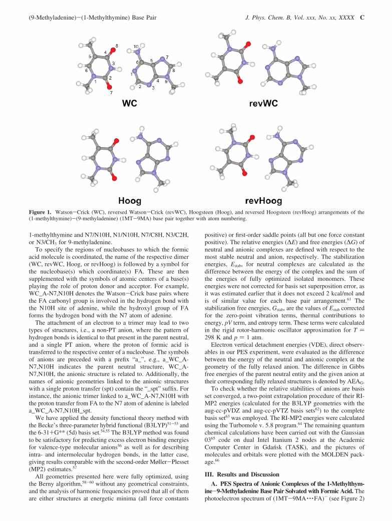
The 1MT and 9MA nucleobases can be hydrogen bonded toeach other in the four ways (see Figure 1) labeled asWatson-Crick (WC), reversed Watson-Crick (revWC), Hoogs-teen (Hoog), and reversed Hoogsteen (revHoog). In both theWC and revWC structures, the N1 and N10H (from the N1side; for atom numbering, see Figure 1) of 9MA participate intwo stabilizing hydrogen bonds, and the dimers differ in 1MTorientation. Namely, in the WC arrangements, a proton acceptorsite of 1-methylthymine is the O8 atom, while, in the revWCfamily, the O7 one is. In all the considered complexes, the protondonating site of 1MT is N3H. In the Hoogsteen and reversedHoogsteen scheme, the nucleobases are paired utilizing the N7and N10H (from the N7 side) atoms of 9MA. The 1MTorientation distinguishes the Hoog from revHoog structures, inan analogous fashion to the WC and revWC structures.
The third component of the considered complexes, formicacid, possesses both proton donor and acceptor properties andis coordinated to the available centers of the MAMT base pair.FA can be attached to the proton donor and proton acceptorcenters of the single base or can simultaneously interact withboth bases via the proton acceptor center of pyrimidine, O7 orO8, and proton donor center of purine, C2H, C8H, or N10H.Building the structures stabilized by two hydrogen bonds inwhich FA is coordinated to a single base, we considered thefollowing pairs of molecular centers: O8/CH3 or O7/CH3 for
B J. Phys. Chem. B, Vol. xxx, No. xx, XXXX Storoniak et al.
1-methylthymine and N7/N10H, N1/N10H, N7/C8H, N3/C2H,or N3/CH3 for 9-methyladenine.
To specify the regions of nucleobases to which the formicacid molecule is coordinated, the name of the respective dimer(WC, revWC, Hoog, or revHoog) is followed by a symbol forthe nucleobase(s) which coordinate(s) FA. These are thensupplemented with the symbols of atomic centers of a base(s)playing the role of proton donor and acceptor. For example,WC_A-N7,N10H denotes the Watson-Crick base pairs wherethe FA carbonyl group is involved in the hydrogen bond withthe N10H site of adenine, while the hydroxyl group of FAforms the hydrogen bond with the N7 atom of adenine.
The attachment of an electron to a trimer may lead to twotypes of structures, i.e., a non-PT anion, where the pattern ofhydrogen bonds is identical to that present in the parent neutral,and a single PT anion, where the proton of formic acid istransferred to the respective center of a nucleobase. The symbolsof anions are preceded with a prefix “a_”, e.g., a_WC_A-N7,N10H indicates the parent neutral structure, WC_A-N7,N10H, the anionic structure is related to. Additionally, thenames of anionic geometries linked to the anionic structureswith a single proton transfer (spt) contain the “_spt” suffix. Forinstance, the anionic trimer linked to a_WC_A-N7,N10H withthe proton transfer from FA to the N7 atom of adenine is labeleda_WC_A-N7,N10H_spt.
We have applied the density functional theory method withthe Becke’s three-parameter hybrid functional (B3LYP)51-53 andthe 6-31+G** (5d) basis set.54,55 The B3LYP method was foundto be satisfactory for predicting excess electron binding energiesfor valence-type molecular anions56 as well as for describingintra- and intermolecular hydrogen bonds, in the latter case,giving results comparable with the second-order Møller-Plesset(MP2) estimates.57
All geometries presented here were fully optimized, usingthe Berny algorithm,58-60 without any geometrical constraints,and the analysis of harmonic frequencies proved that all of themare either structures at energetic minima (all force constants
positive) or first-order saddle points (all but one force constantpositive). The relative energies (∆E) and free energies (∆G) ofneutral and anionic complexes are defined with respect to themost stable neutral and anion, respectively. The stabilizationenergies, Estab, for neutral complexes are calculated as thedifference between the energy of the complex and the sum ofthe energies of fully optimized isolated monomers. Theseenergies were not corrected for basis set superposition error, asit was estimated earlier that it does not exceed 2 kcal/mol andis of similar value for each base pair arrangement.61 Thestabilization free energies, Gstab, are the values of Estab correctedfor the zero-point vibration terms, thermal contributions toenergy, pV term, and entropy term. These terms were calculatedin the rigid rotor-harmonic oscillator approximation for T )298 K and p ) 1 atm.
Electron vertical detachment energies (VDE), direct observ-ables in our PES experiment, were evaluated as the differencebetween the energy of the neutral and anionic complex at thegeometry of the fully relaxed anion. The difference in Gibbsfree energies of the parent neutral entity and the given anion attheir corresponding fully relaxed structures is denoted by AEAG.
To check whether the relative stabilities of anions are basisset converged, a two-point extrapolation procedure of their RI-MP2 energies (calculated for the B3LYP geometries with theaug-cc-pVDZ and aug-cc-pVTZ basis sets62) to the completebasis set63 was employed. The RI-MP2 energies were calculatedusing the Turbomole v. 5.8 program.64 The remaining quantumchemical calculations have been carried out with the Gaussian0365 code on dual Intel Itanium 2 nodes at the AcademicComputer Center in Gdansk (TASK), and the pictures ofmolecules and orbitals were plotted with the MOLDEN pack-age.66
III. Results and Discussion
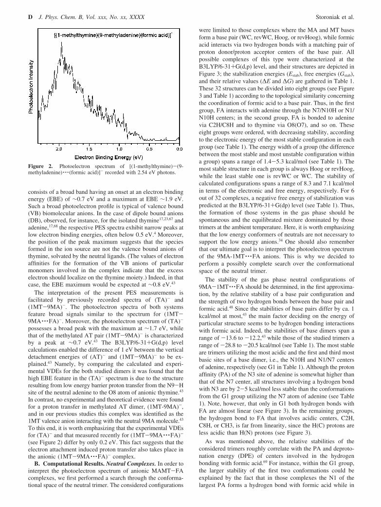
A. PES Spectra of Anionic Complexes of the 1-Methylthym-ine-9-Methyladenine Base Pair Solvated with Formic Acid. Thephotoelectron spectrum of (1MT-9MA · · ·FA)- (see Figure 2)
Figure 1. Watson-Crick (WC), reversed Watson-Crick (revWC), Hoogsteen (Hoog), and reversed Hoogsteen (revHoog) arrangements of the(1-methylthymine)-(9-methyladenine) (1MT-9MA) base pair together with atom numbering.
(9-Methyladenine)-(1-Methylthymine) Base Pair J. Phys. Chem. B, Vol. xxx, No. xx, XXXX C
consists of a broad band having an onset at an electron bindingenergy (EBE) of ∼0.7 eV and a maximum at EBE ∼1.9 eV.Such a broad photoelectron profile is typical of valence bound(VB) biomolecular anions. In the case of dipole bound anions(DB), observed, for instance, for the isolated thymine17,23,67 andadenine,17,68 the respective PES spectra exhibit narrow peaks atlow electron binding energies, often below 0.5 eV.4 Moreover,the position of the peak maximum suggests that the speciesformed in the ion source are not the valence bound anions ofthymine, solvated by the neutral ligands. (The values of electronaffinities for the formation of the VB anions of particularmonomers involved in the complex indicate that the excesselectron should localize on the thymine moiety.) Indeed, in thatcase, the EBE maximum would be expected at ∼0.8 eV.43
The interpretation of the present PES measurements isfacilitated by previously recorded spectra of (TA)- and(1MT-9MA)-. The photoelectron spectra of both systemsfeature broad signals similar to the spectrum for (1MT-9MA · · ·FA)-. Moreover, the photoelectron spectrum of (TA)-
possesses a broad peak with the maximum at ∼1.7 eV, whilethat of the methylated AT pair (1MT-9MA)- is characterizedby a peak at ∼0.7 eV.43 The B3LYP/6-31+G(d,p) levelcalculations enabled the difference of 1 eV between the verticaldetachment energies of (AT)- and (1MT-9MA)- to be ex-plained.43 Namely, by comparing the calculated and experi-mental VDEs for the both studied dimers it was found that thehigh EBE feature in the (TA)- spectrum is due to the structureresulting from low energy barrier proton transfer from the N9-Hsite of the neutral adenine to the O8 atom of anionic thymine.43
In contrast, no experimental and theoretical evidence were foundfor a proton transfer in methylated AT dimer, (1MT-9MA)-,and in our previous studies this complex was identified as the1MT valence anion interacting with the neutral 9MA molecule.43
To this end, it is worth emphasizing that the experimental VDEsfor (TA)- and that measured recently for (1MT-9MA · · ·FA)-
(see Figure 2) differ by only 0.2 eV. This fact suggests that theelectron attachment induced proton transfer also takes place inthe anionic (1MT-9MA · · ·FA)- complex.
B. Computational Results. Neutral Complexes. In order tointerpret the photoelectron spectrum of anionic MAMT-FAcomplexes, we first performed a search through the conforma-tional space of the neutral trimer. The considered configurations
were limited to those complexes where the MA and MT basesform a base pair (WC, revWC, Hoog, or revHoog), while formicacid interacts via two hydrogen bonds with a matching pair ofproton donor/proton acceptor centers of the base pair. Allpossible complexes of this type were characterized at theB3LYP/6-31+G(d,p) level, and their structures are depicted inFigure 3; the stabilization energies (Estab), free energies (Gstab),and their relative values (∆E and ∆G) are gathered in Table 1.These 32 structures can be divided into eight groups (see Figure3 and Table 1) according to the topological similarity concerningthe coordination of formic acid to a base pair. Thus, in the firstgroup, FA interacts with adenine through the N7/N10H or N1/N10H centers; in the second group, FA is bonded to adeninevia C2H/C8H and to thymine via O8(O7), and so on. Theseeight groups were ordered, with decreasing stability, accordingto the electronic energy of the most stable configuration in eachgroup (see Table 1). The energy width of a group (the differencebetween the most stable and most unstable configuration withina group) spans a range of 1.4-5.3 kcal/mol (see Table 1). Themost stable structure in each group is always Hoog or revHoog,while the least stable one is revWC or WC. The stability ofcalculated configurations spans a range of 8.3 and 7.1 kcal/molin terms of the electronic and free energy, respectively. For 6out of 32 complexes, a negative free energy of stabilization waspredicted at the B3LYP/6-31+G(dp) level (see Table 1). Thus,the formation of those systems in the gas phase should bespontaneous and the equilibrated mixture dominated by thosetrimers at the ambient temperature. Here, it is worth emphasizingthat the low energy conformers of neutrals are not necessary tosupport the low energy anions.34 One should also rememberthat our ultimate goal is to interpret the photoelectron spectrumof the 9MA-1MT · · ·FA anions. This is why we decided toperform a possibly complete search over the conformationalspace of the neutral trimer.
The stability of the gas phase neutral configurations of9MA-1MT · · ·FA should be determined, in the first approxima-tion, by the relative stability of a base pair configuration andthe strength of two hydrogen bonds between the base pair andformic acid.48 Since the stabilities of base pairs differ by ca. 1kcal/mol at most,43 the main factor deciding on the energy ofparticular structure seems to be hydrogen bonding interactionswith formic acid. Indeed, the stabilities of base dimers span arange of -13.6 to -12.2,43 while those of the studied trimers arange of -28.8 to -20.5 kcal/mol (see Table 1). The most stableare trimers utilizing the most acidic and the first and third mostbasic sites of a base dimer, i.e., the N10H and N1/N7 centersof adenine, respectively (see G1 in Table 1). Although the protonaffinity (PA) of the N3 site of adenine is somewhat higher thanthat of the N7 center, all structures involving a hydrogen bondwith N3 are by 2-5 kcal/mol less stable than the conformationsfrom the G1 group utilizing the N7 atom of adenine (see Table1). Note, however, that only in G1 both hydrogen bonds withFA are almost linear (see Figure 3). In the remaining groups,the hydrogen bond to FA that involves acidic centers, C2H,C8H, or CH3, is far from linearity, since the H(C) protons areless acidic than H(N) protons (see Figure 3).
As was mentioned above, the relative stabilities of theconsidered trimers roughly correlate with the PA and deproto-nation energy (DPE) of centers involved in the hydrogenbonding with formic acid.69 For instance, within the G1 group,the larger stability of the first two conformations could beexplained by the fact that in those complexes the N1 of thelargest PA forms a hydrogen bond with formic acid while in
Figure 2. Photoelectron spectrum of [(1-methylthymine)-(9-methyladenine) · · · (formic acid)]- recorded with 2.54 eV photons.
D J. Phys. Chem. B, Vol. xxx, No. xx, XXXX Storoniak et al.
the two remaining structures FA interacts with adenine throughthe N7 atom of lower basicity.
One should, however, realize that simple arguments basedon the basicity/acidity of centers involved in hydrogen bonding
(9-Methyladenine)-(1-Methylthymine) Base Pair J. Phys. Chem. B, Vol. xxx, No. xx, XXXX E
are not always able to explain the relative stabilities of thestudied complexes. The analyzed structures are complicatedsystems where many molecular centers interact simultaneously.For example, despite the fact that N7 is substantially less basicthan N3, WC_A-N7,C8H and revWC_A-N7,C8H are morestable than WC_A-N3,C2H and revWC_A-N3,C2H, respec-tively. Probably, stabilizing interactions between formic acidand N10H present in WC_A-N7,C8H and revWC_A-N7,C8H(see Figure 3), as well as destabilizing effects between thecarbonyl group of FA and the O7/O8 oxygen in WC_A-N3,C2Hand revWC_A-N3,C2H (see Figure 3), are responsible for theobserved “anomaly”. Therefore, it seems to be difficult to predictthe relative stability of 9MA-1MT · · ·FA complexes withoutdoing the actual calculations.
Anionic Complexes. All 32 neutral structures described inthe previous section support adiabatically stable valence anions(see Table 2 and Figure 4 and Table S1 and Figure S1 of theSupporting Information). Our previous reports on the base pair
anions as well as on the anionic complexes of nucleobases withmolecules having proton donor properties together with theposition of the current photoelectron spectrum suggest that anintermolecular proton transfer induced by electron attachmentis also involved in [MAMT · · ·FA]-. This prompted us to carryout quantum chemical calculations not only for the parent anionsbut also for systems where proton transfer from formic acid tothe proton acceptor center of a base takes place. Since our poolof neutral structures comprises 32 complexes, the B3LYPoptimizations should end up with 32 anions having the patternof hydrogen bonds identical to the respective neutrals as wellas with 32 proton transfer anions. The actual number of anionsis, however, smaller than the theoretical value of 64 (see TableS1, Supporting Information); i.e., it amounts to only 47configurations, since some of the anionic structures convergedto the same geometries. For instance, a_WC_T-O8,CH3 con-verged to a_WC_T-O8_A-N10H; we did not observe protontransfer to the thymine O7 atom; therefore, only non-PTgeometries are characterized for the complexes where formicacid interacts with this center. Similarly, the optimization ofa_revHoog_T-O8_A-C8H and a_Hoog_T-O8_A-N10H con-verged to proton transferred structures with proton bonded tothe O8 atom of thymine, a_revHoog_T-O8_A-C8_spt anda_Hoog_T-O8_A-N10H_spt (see Figure S1, Supporting Infor-mation), which means that a barrier-free proton transfer waspredicted for those types of anionic complexes.
The energetic characteristics of anionic geometries aregathered in Tables 2 and S1 (Supporting Information), whilethe distributions of their SOMO orbital are depicted in Figures4 and S2 (Supporting Information). In all non-PT structures,the excess electron localizes primarily on the thymine moiety.The relative stability of the studied anions comprises a rangeof 16.6 and 13.4 kcal/mol in the electronic and free energy scale,respectively (see Table S1, Supporting Information). Moreover,the adiabatic stability of those anions spans a range of 0.37-0.97eV (see Table S1, Supporting Information). Hence, all of thestudied structures possess AEAG larger than those calculatedfor the MAMT configurations (0.26-0.36 eV)43 at a similar levelof theory. Apparently, the presence of formic acid additionallystabilizes the trimeric anions.
The most stable anions were predicted for those complexeswhere the hydroxyl group of formic acid interacts with the O8atom of thymine. Due to the relative electron affinities ofcanonical forms of particular nucleobases,13,18 an extra electronhas the tendency to localize on thymine and then its O8 atombecomes one of the thymine atomic centers characterized by
TABLE 1: Values of Stabilization Energy (Estab) andStabilization Free Energy (Gstab) as Well as Their RelativeValues (∆E and ∆G) for the Neutral 1MT-9MA · · ·FAComplexes as Calculated at the B3LYP/6-31+G** (5d)Levela
TABLE 2: Relative Electronic Energies and Free Energies(∆E and ∆G) Calculated with Respect to the Most StableAnion Together with the Adiabatic Electron Affinities(AEAG) and Electron Vertical Detachment Energies (VDE)for the Seven Low Energy Anionic Complexes Predicted atthe B3LYP/6-31+G** (5d) Levela
a ∆E and ∆G are given in kcal/mol, while VDE and AEAG, ineV. b The relative MP2 energies extrapolated to the complete basisset (CBS) limit. c The relative MP2 CBS energies supplementedwith the B3LYP ZPEs, thermal energies, and entropy terms.
F J. Phys. Chem. B, Vol. xxx, No. xx, XXXX Storoniak et al.
the highest density of excess electron.20 This explains theobserved increased stability of the configurations involvingFA · · ·O8(MT) interactions.
Proton transfer between formic acid and the anionic basepair is an important factor deciding on the stability of[1MT-9MA · · ·FA]-. Indeed, the a_revHoog_T-O8_A-C8H_sptand a_Hoog_T-O8_A-N10H_spt anions of the largest electronadiabatic affinities are also the most stable in terms of electronicand free energy (see Tables 2 and S1, Supporting Information).Electron attachment to the mentioned above neutral complexestriggers barrier free proton transfer (BFPT); thus, only PTstructures are minima on the potential energy surface, whiletheir non-PT counterparts, a_revHoog_T-O8_A-C8H and
a_Hoog_T-O8_A-N10H, do not exist. For the other complexes,both the non-PT and PT geometries frequently support adiabati-cally stable anions. This, for instance, holds for the complexeswhere formic acid interacts with adenine (see Table S1 andFigure S2, Supporting Information). When its N7/N1 and N10Hcenters form hydrogen bonds with FA, the proton transferstructure is always more stable than the non-PT one (by 0.6-4.9kcal/mol in terms of the electronic energy, see Table S2,Supporting Information). For the remaining centers of adenine,the situation can be reversed, although then the difference instability between both types of anions never exceeds 1.4 kcal/mol.
The PT structures are more favored compared to the non-PTones in those cases where proton transfer brings about acomplete localization of an extra charge on adenine. Inspectionof the SOMO distribution depicted in Figure S2 (SupportingInformation) indicates that these are a_WC_A-N7,N10H_spt anda_revWC_A-N7,N10H_spt. Indeed, for these two anions, theproton transfer structures are by as much as 3.4 and 4.9 kcal/mol, respectively, more stable, in terms of electronic energy,than their non-PT counterparts, a_WC_A-N7,N10H and a_revW-C_A-N7,N10H (see Table S2, Supporting Information). Al-though proton transfer to adenine always increases the amountof excess charge localized on that base, only for the twomentioned above anions electron transfer is complete. Thus, thepartial charge localization on adenine is probably one of thereasons that makes some of the PT structures somewhat lessstable than their parent non-PT configurations.
In the anionic complexes in which adenine interacts with FA,proton transfer leads to a partial or almost complete electrontransfer form the initial thymine anion. Thus, one could wonderif this phenomenon allows for a second charge transfer fromN3H of thymine to the available center of adenine. This secondproton transfer could additionally stabilize an electron residingon the adenine moiety. In order to answer this question, we didfurther calculations for the complexes where MAMT possessesthe Watson-Crick configuration, an arrangement of the basepair which is most abundant in double-stranded DNA. Moreover,we chose a trimer configuration involving the most frequentpattern of hydrogen bonds for adenine that were identified viathe analysis of interactions in a protein-nucleic acid databasecomprising over 1000 complexes.49 In Figure 5, the process ofan electron attachment triggering two consecutive proton transferreactions is sketched for the WC_A-N7,N10H complex. At-tachment of an electron leads to the a_WC_A-N7,N10H anionadiabatically stable by 3.4 kcal/mol. The first proton transfer,from formic acid, is favored again by 3.4 kcal/mol and leads tothe anion in which the electron is practically completelylocalized on the adenine molecule (see Figure 5). Finally, thesecond proton transfer from the N3H of thymine to the N1 atomof adenine provides additional stabilization by 0.5 kcal/mol andresults in a complete localization of an extra electron on adenine.Thus, our computational results suggest that if adenine in DNAinteracts with a sufficiently acidic external proton donor thenintermolecular proton transfers may occur and an unpairedelectron becomes completely localized on adenine. A similarconclusion has been published recently.4,34,48 This findingquestions the commonly accepted paradigm that in DNAelectrons localize on pyrimidines rather than on purines whenthe cell is exposed to high energy radiation.
C. Interpretation of the PES Experiment. We make theassumption that the anions which contribute to the photoelectronspectrum are in some degree of thermodynamic equilibrium inthe ion source. Therefore, only the low energy anionic structures
Figure 4. Optimized structures of the seven low energy 1MT-9MA · · ·FA anionic complexes and their singly occupied molecularorbitals plotted with a contour value of 0.03 bohr-3/2.
(9-Methyladenine)-(1-Methylthymine) Base Pair J. Phys. Chem. B, Vol. xxx, No. xx, XXXX G
explain the result of the PES experiment. Using the relativestabilities, ∆G’s (see Tables 2 and S1, Supporting Information),we choose seven anions which might be responsible for theexperimental picture. These anions differ in ∆G by 4 kcal/molat most, and this seems to be a reasonable threshold. The eighthmost stable structure, a_Hoog_T-O7_A-C8H, differs from theleast stable one from the set of the chosen seven configurations by1.83 kcal/mol, and its absolute ∆G amounts to 5.98 kcal/mol. Thelatter value leads, for T ) 298 K, to the equilibrium contributionof a_Hoog_TO7_A-C8H equal to 4.1 × 10⁻³ percent of the amountrepresented by the most stable a_revHoog_T-O8_A-C8H_spt anion.
The VDEs for the chosen anions span a relatively large rangeof 1.3-2.06 eV (see Table 2) which agrees with an exceptionallybroad peak observed in the experimental PES spectrum (seeFigure 2).
The most stable anion a_revHoog_T-O8_A-C8H_spt ischaracterized by VDE of 2.06 eV. Similarly, a relatively highvertical stability, VDE ) 1.96 eV, possesses the second moststable structure, a_Hoog_T-O8_A-N10H_spt, which in termsof free energy is less stable than the a_revHoog_T-O8_A-C8H_spt anion by only 0.72 kcal/mol. Note that the VDEs ofthe above-mentioned anions reproduce very well the observedmaximum in the PES spectrum, 1.9 eV, especially when theyare shifted by -0.15 eV, a usual shift observed for protontransferred anions involving nucleobases.4 There are also non-PT structures in the pool of low energy anions, a_WC_T-O8_A-N10H and a_Hoog_T-O8,CH3. In terms of electronic energy,they are negligibly more stable than their PT counterparts (seeTable 2) and separated from them by a low kinetic barrier equalto 0.13 and 0.29 kcal/mol, respectively. However, on the freeenergy surface, these barriers become negative, -1.08 and-1.53 kcal/mol, respectively, indicating that the equilibriumbetween PT and non-PT species is attained immediately afterelectron attachment.
The vertical stability of the non-PT anions was calculated tobe 1.35 and 1.30 eV, respectively. These VDEs correspond fairlywell to the experimental spectrum, where a significant value ofthe ion’s signal is recorded in the range 1.0-1.5 eV (see Figure2). The instability of these structures with regard to the moststable anion amounts to 2.93 and 4.15 kcal/mol, respectively,and seems to be somewhat too large to ensure their significantcontributions. However, we did additional MP2 calculations atthe B3LYP geometries and we extrapolated our results to thecomplete basis set limit (MP2 CBS). As indicated by datagathered in the second and third columns of Table 2, the relativeinstabilities of non-PT structures decreased at the MP2 CBSlevel. Probably inclusion of higher order correlation terms (atthe coupled cluster level, for instance) would further diminishthe difference between non-PT and PT structures.
In summary, the low energy structures identified within thepresent study explain the position and shape of the experimentalspectrum. Several anions both of the PT and non-PT typecontribute to the measured PES signal.
D. Biological Relevance. The lowest energy geometryidentified within the current study was determined to be a singleproton transfer anion of the reversed Hoogsteen type where FAinteracts simultaneously with both bases. Populated in the gasphase are also the structures with the Hoogsteen and Watson-Crick patterns of hydrogen bonding. While the biologicalimportance of the WC configuration does not require any specialcomments, the Hoogsteen and reversed Hoogsteen arrangementsare much less common as far as the DNA molecule is concerned.They are, however, essential for building up a three-dimensionalstructure of large RNAs.70
In order to compare our computational results to the PESdata, the conformational space of the AT pair was not limitedto its WC arrangement, as in the gas phase the geometryconstraints of DNA are not present. Despite the lack of suchlimitations, the same configurations which are relevant to theDNA molecule turned out to also be populated in the gas phase.From the above statements, it follows that our computational-experimental study allowed us to characterize the propensityto bind an excess electron for the biologically relevant con-figurations of the MAMT base pair.
Formic acid, on the other hand, may be viewed as a modelof a medium-strength proton donor. To this end, it is worthemphasizing that in the cell DNA never appears in isolation.Instead, it interacts with a number of molecular systems, havingvarious proton donor properties, such as histones, replicationand repair enzymes, hydrated metal cations, H3O+ ions, etc.Although the trimer complexes studied in this work aresimplistic models of the cellular DNA, one should realize thatan excess electron is being attached to a single nucleobase alsoin the biopolymer and the environment (water, DNA itself,proteins, etc.) modifies this process somewhat. From thisperspective, the attachment of an electron is a local phenomenon.Due to the distance dependence of various types of interactions,the most important effects related to the presence of environmentare exerted by its components that are adjacent to the site whichbinds an electron such as complementary bases and proton donorspecies. Therefore, the characteristics obtained for the modeltrimers studied within the current work should be consideredas a first but reasonable approximation of an electron attachmentphenomenon to the whole complex system.
Perhaps the most important biological context of our studylies in the identification of the SPT anionic structures that aremore stable than the parent non-PT geometries. A prominentexample of such a case is the a_WC-A-N7,N10H anion forwhich the possible proton transfers are depicted in Figure 5.Although this anion is not populated in the gas phase, it shouldrepresent one of the most abundant configurations in double-stranded DNA. Two consecutive proton transfers in this anioniccomplex make the excess electron completely localized onadenine (see the discussion in the Anionic Complexes subsec-tion), and such an effect should have profound consequencesas far as the electron induced damage of DNA is concerned.4,34,48
Figure 5. Possible proton transfer reactions triggered by electron attachment to the WC_A-N7,N10H complex. The numbers above the arrowsindicate the difference in the electronic energy between product and substrate.
H J. Phys. Chem. B, Vol. xxx, No. xx, XXXX Storoniak et al.
Namely, both our35,36 and other44-46 studies demonstrate thatelectron attachment to cytosine in the GC base pair induces lowenergy barrier proton transfer from the N1 atom of guanine tothe N3 site of the anionic cytosine. This intermolecular PTprocess neutralizes a negative charge of the cytosine anion whichprevents further electron transfer to the phosphate in the DNAstrand; the latter process directly precedes the formation of SSBsin DNA. On the other hand, the analogous PT process is notallowed in the AT- anion owing to its energetic barriers. Thus,an electron captured by thymine involved in the AT base pairmay lead directly to DNA single-strand break, while theformation of cytosine anion induces PT within the GC- basepair which halts the subsequent formation of SSBs. If, however,the neutralizing proton would come from some other externalproton donor of sufficiently low deprotonation energy (fromformic acid, for instance, as in the complexes studied withinthe current work), the negative charge of thymine in AT- wouldbe neutralized by the PT process analogous to that describedabove for the GC anion. Accordingly, regardless of the type ofelectron-induced PT, the development of the SSB-type damagewould be stopped by the formation of the neutral monohydro-radical of thymine, adenine, or the cation radical of adenine.
From the foregoing discussion, it would seem that proteinspresent in living organisms might play a role of DNA protectoragainst high energy radiation, not only because they constitutea first layer of a DNA-protein complex (e.g., histones innucleus), being, thus, a main defender against water radiolysisproducts, but also since the proton donor groups of the sidechains of amino acids may qualitatively change the behaviorof the anionic sites formed due to DNA interactions withelectrons.
IV. Conclusions
The shape of the photoelectron spectrum obtained for the1MT-9MA · · ·FA anions suggests that several low energystructures are involved in the thermodynamic equilibrium underthe conditions of our PES experiment. The B3LYP/6-31+G(d,p)level calculations for complexes comprising four possiblearrangements of the 1MT-9MA base pair, i.e., Watson-Crick,reversed Watson-Crick, Hoogsteen, and reversed Hoogsteen,and the formic acid molecule enabled these low energygeometries to be identified. These anions differ substantiallywith VDEs that fall in the EBE region covered by the registeredPES spectrum. Moreover, the calculated differences in therelative stabilities of those structures justify their presence inthe equilibrated gas phase mixture in non-negligible amounts.
We demonstrated that electron binding to the (1MT-9MA) · · · (proton donor) complex can trigger intermolecularproton transfer that leads to the strong stabilization of theresulting radical anion. Indeed, five out of seven structurescontributing to the PES signal are formed from the neutralcomplexes due to electron attachment that is followed by thebarrier-free proton transfer from the formic acid molecule.
The SOMO distribution calculated for the considered geom-etries indicates that all the anions are of valence type. In someproton transferred geometries, the excess electron is significantlylocalized to the adenine moiety. This finding suggests that, whileDNA interacts with its environment, the excess electron maybe captured by purine rather than pyrimidine bases whichquestions the generally accepted paradigm that pyrimidines arethe main target of DNA-electron interactions.
Finally, the DNA interactions with a cellular environment(water, proteins, metal complexes, etc.), which may be apotential source for external protons, could block the electron-induced formation of SSBs in DNA.
Acknowledgment. This work was supported by (i) the PolishMinistry of Science and Higher Education (MNiSW) underGrant Nos. N N204 023135 (J.R.) and DS/8221-4-0140-10(P.S.); (ii) the U.S. Department of Energy under contract DE-AC02-05CH11231 and through a 2008 Seaborg Fellowship atLawrence Berkeley National Laboratory (M.H.); and (iii) theU.S. National Science Foundation under Grant No. CHE-0809258 (K.H.B.). The calculations were performed at theAcademic Computer Center in Gdansk (TASK).
Supporting Information Available: Relative electronicenergies and free energies calculated with respect to thea_revHoog_T-O8_A-C8H_spt anion together with the adiabaticelectron affinities and electron vertical detachment energies forall anionic complexes considered within the current work.Optimized structures of these anions as well as their SOMOorbital distribution. This material is available free of charge viathe Internet at http://pubs.acs.org.
References and Notes
(1) Miller, J. H.; Wilson, W. E.; Ritchie, R. H. Direct ionization ofDNA in solution. In Computational Approaches in Molecular RadiationBiology; Varma, M. N., Chatterjee, A., Eds.; Plenum Press: New York,1994; p 65.
(2) Michael, B. R.; O’Neill, P. Science 2000, 287, 1603–1604.(3) Boudaiffa, B.; Cloutier, P.; Hunting, D.; Huels, M. A.; Sanche, L.
zyk, M.; Dabkowska, I.; Bachorz, R. A.; Gutowski, M.; Radisic, D.; Stokes,S. T.; Eustis, S. N.; Wang, D.; Li, X.; Ko, Y. J.; Bowen, K. H. In RadiationInduced Molecular Phenomena in Nucleic Acids: A ComprehensiVeTheoretical and Experimental Analysis; Shukla, M. K., Leszczynski, J., Eds.;Springer: Amsterdam, The Netherlands, 2008; Vol. 5, pp 619-667.
(5) Simons, J. Acc. Chem. Res. 2006, 39, 772–779.(6) Dabkowska, I.; Rak, J.; Gutowski, M. Eur. Phys. J. D 2005, 35,
429–435.(7) Bao, X.; Wang, J.; Gu, J.; Leszczynski, J. Proc. Natl. Acad. Sci.
U.S.A. 2006, 103, 5658–5663.(8) Gu, J.; Wang, J.; Leszczynski, J. J. Am. Chem. Soc. 2006, 128,
9322–9323.(9) Seidel, C. A. M.; Schulz, A.; Sauer, M. H. M. J. Phys. Chem. 1996,
100, 5541–5553.(10) Aflatooni, K.; Gallup, G. A.; Burrow, P. D. J. Phys. Chem. A 1998,
102, 6205–6207.(11) Wetmore, S. D.; Boyd, R. J.; Eriksson, L. A. Chem. Phys. Lett.
2000, 322, 129.(12) Wesolowski, S. S.; Leininger, M. L.; Pentchev, P. N.; Schaefer,
H. F., III. J. Am. Chem. Soc. 2001, 123, 4023–4028.(13) Li, X.; Cai, Z.; Sevilla, M. D. J. Phys. Chem. A 2002, 106, 1596–
1603.(14) Haranczyk, M.; Gutowski, M. J. Am. Chem. Soc. 2005, 127, 699–
706.(15) Oyler, N. A.; Adamowicz, L. J. Phys. Chem. 1993, 97, 11122–
11123.(16) Hendricks, J. H.; Lyapustina, S. A.; de Clercq, H. L.; Snodgrass,
J. T.; Bowen, K. H. J. Chem. Phys. 1996, 104, 7788–7791.(17) Defrancois, C.; Abdoulcarime, H.; Schermann, J. J. Chem. Phys.
1996, 104, 7792–7794.(18) Sevilla, M. D.; Besler, B.; Colson, A.-O. J. Phys. Chem. 1995, 99,
Phys. 2005, 5, 840.(20) Mazurkiewicz, K.; Bachorz, R. A.; Gutowski, M.; Rak, J. J. Phys.
Chem. B 2006, 48, 24696–24707.(21) Bachorz, R. A.; Rak, J.; Gutowski, M. Phys. Chem. Chem. Phys.
2005, 7, 2116–2125. Bachorz, R. A.; Klopper, W.; Gutowski, M. J. Chem.Phys. 2007, 126, 085101(1-7).
(22) Hendricks, J. H.; Lyapustina, S. A.; de Clercq, H. L.; Bowen, K. H.J. Chem. Phys. 1998, 108, 8–11.
(23) Schiedt, J.; Weinkauf, R.; Neumark, D. M.; Schlag, E. W. Chem.Phys. 1998, 239, 511–524.
(24) Periquet, V.; Moreau, A.; Carles, S.; Schermann, J.; Desfrancois,C. J. Electron Spectrosc. Relat. Phenom. 2000, 106, 141–151.
(25) Eustis, S.; Wang, D.; Lyapustina, S.; Bowen, K. H. J. Chem. Phys.2007, 127, 224309/1–224309/6.
(26) Gutowski, M.; Dabkowska, I.; Rak, J.; Xu, S.; Nilles, J. M.; Radisic,D.; Bowen, K. H., Jr. Eur. Phys. J. D 2002, 20, 431–439.
(9-Methyladenine)-(1-Methylthymine) Base Pair J. Phys. Chem. B, Vol. xxx, No. xx, XXXX I
(27) Dabkowska, I.; Rak, J.; Gutowski, M.; Nilles, J. M.; Stokes, S. T.;Bowen, K. H., Jr. J. Chem. Phys. 2004, 120, 6064–6071.
(28) Dabkowska, I.; Rak, J.; Gutowski, M.; Nilles, J. M.; Stokes, S. T.;Radisic, D.; Bowen, K. H., Jr. Phys. Chem. Chem. Phys. 2004, 6, 4351–4357.
(29) Haranczyk, M.; Bachorz, R.; Rak, J.; Gutowski, M.; Radisic, D.;Stokes, S. T.; Nilles, J. M.; Bowen, K. H., Jr. J. Phys. Chem. B 2003, 107,7889–7895.
(30) Haranczyk, M.; Rak, J.; Gutowski, M.; Radisic, D.; Stokes, S. T.;Nilles, J. M.; Bowen, K. H., Jr. Isr. J. Chem. 2004, 44, 157–170.
(31) Haranczyk, M.; Rak, J.; Gutowski, M.; Radisic, D.; Stokes, S. T.;Bowen, K. H., Jr. J. Phys. Chem. B 2005, 109, 13383–13391.
(32) Haranczyk, M.; Dabkowska, I.; Rak, J.; Gutowski, M.; Nilles, J. M.;Stokes, S. T.; Radisic, D.; Bowen, K. H., Jr. J. Phys. Chem. B 2004, 108,6919–6921.
(33) Mazurkiewicz, K.; Haranczyk, M.; Storoniak, P.; Gutowski, M.;Rak, J.; Radisic, D.; Eustis, S. N.; Wang, D.; Bowen, K. H., Jr. Chem.Phys. 2007, 342, 215–222.
(34) Mazurkiewicz, K.; Haranczyk, M.; Gutowski, M.; Rak, J.; Radisic,D.; Eustis, S. N.; Wang, D.; Bowen, K. H., Jr. J. Am. Chem. Soc. 2007,129, 1216–1224.
(35) Szyperska, A.; Rak, J.; Leszczynski, J.; Li, X.; Ko, Y. J.; Wang,H.; Bowen, K. H. J. Am. Chem. Soc. 2009, 131, 2663–2669.
(36) Szyperska, A.; Rak, J.; Leszczynski, J.; Li, X.; Ko, Y. J.; Wang,H.; Bowen, K. H. ChemPhysChem 2010, 11, 880–888.
(37) Kobyleecka, M.; Gu, J.; Rak, J.; Leszczynski, J. J. Chem. Phys. 2008,128, 044315.
(38) Richardson, N. A.; Gu, J.; Wang, S.; Xie, Y.; Schaefer, H. F., III.J. Am. Chem. Soc. 2004, 126, 4404–4411.
(39) Li, X.; Sanche, L.; Sevilla, M. D. Radiat. Res. 2006, 165, 721–729.
(40) Stokes, S. T.; Li, X.; Grubisic, A.; Ko, Y. J.; Bowen, K. H., Jr.J. Chem. Phys. 2007, 127, 084321-6.
(41) Gu, J.; Xie, Y.; Schaefer, H. F., III. J. Phys. Chem. B 2005, 109,13067–13075.
(42) Richardson, N. A.; Wesolowski, S. S.; Schaefer, H. F. J. Phys.Chem. B 2003, 107, 848–853.
(43) Radisic, D.; Bowen, K. H.; Dabkowska, I.; Storoniak, P.; Rak, J.;Gutowski, M. J. Am. Chem. Soc. 2005, 127, 6443–6450.
(44) Colson, A.-O.; Besler, B.; Close, D. M.; Sevilla, M. D. J. Phys.Chem. 1992, 96, 661–668.
(45) Richardson, N. A.; Wesolowski, S. S.; Schaefer, H. F. J. Am. Chem.Soc. 2002, 124, 10163–10170.
(46) Li, X.; Cai, Z.; Sevilla, M. D. J. Phys. Chem. B 2001, 105, 10115–10123.
(47) Colson, A.-O.; Besler, B.; Sevilla, M. D. J. Phys. Chem. 1992, 96,9787–9794.
(48) Mazurkiewicz, K.; Haranczyk, M.; Gutowski, M.; Rak, J. Int. J.Quantum Chem. 2007, 107, 2224–2232.
(49) Hoffman, M. M.; Khrapov, M. A.; Cox, J. C.; Yao, J.; Tong, L.;Ellington, A. D. Nucleic Acid. Res. 2004, 32, D174–D181.
(50) Coe, J. V.; Snodgrass, J. T.; Friedhoff, C. B.; McHugh, K. M.;Bowen, K. H., Jr. Chem. Phys. 1986, 84, 618.
(51) Becke, A. D. Phys. ReV. A 1988, 38, 3098–3100.
(52) Becke, A. D. J. Chem. Phys. 1993, 98, 5648–5652.(53) Lee, C.; Yang, W.; Parr, R. G. Phys. ReV. B 1988, 37, 785–789.(54) Ditchfield, R.; Hehre, W. J.; Pople, J. A. J. Chem. Phys. 1971, 54,
724–728.(55) Hehre, W. J.; Ditchfield, R.; Pople, J. A. J. Chem. Phys. 1972, 56,
2257–2261.(56) Rienstra-Kiracofe, J. C.; Tschumper, G. S.; Schaefer, H. F.; Nandi,
S.; Ellison, G. B. Chem. ReV. 2002, 102, 231–282.(57) van Mourik, T.; Price, S. L.; Clary, D. C. J. Phys. Chem. A 1999,
103, 1611–1618.(58) Fogarasi, G.; Zhou, X.; Taylor, P.; Pulay, P. J. Am. Chem. Soc.
1992, 114, 8191–8201.(59) Baker, J. J. Comput. Chem. 1993, 14, 1085–1100.(60) Peng, C.; Ayala, P. Y.; Schlegel, H. B.; Frisch, M. J. J. Comput.
Chem. 1996, 17, 49–56.(61) Li, X.; Cai, Z.; Sevilla, M. D. J. Phys. Chem. A 2002, 106, 9345–
9351.(62) Kendall, R. A.; Dunning, T. H., Jr.; Harrison, R. J. J. Chem. Phys.
Olsen, J.; Wilson, A. K. Chem. Phys. Lett. 1998, 286, 243.(64) Ahlrichs, R.; Bar, M.; Haser, M.; Horn, H.; Kolmel, C. Chem. Phys.
Lett. 1989, 162, 165.(65) Frisch, M. J.; Trucks, G. W.; Schlegel, H. B.; Scuseria, G. E.; Robb,
M. A.; Cheeseman, J. R.; Montgomery, J. A., Jr.; Vreven, T.; Kudin, K. N.;Burant, J. C.; Millam, J. M.; Iyengar, S. S.; Tomasi, J.; Barone, V.;Mennucci, B.; Cossi, M.; Scalmani, G.; Rega, N.; Petersson, G. A.;Nakatsuji, H.; Hada, M.; Ehara, M.; Toyota, K.; Fukuda, R.; Hasegawa, J.;Ishida, M.; Nakajima, T.; Honda, Y.; Kitao, O.; Nakai, H.; Klene, M.; Li,X.; Knox, J. E.; Hratchian, H. P.; Cross, J. B.; Bakken, V.; Adamo, C.;Jaramillo, J.; Gomperts, R.; Stratmann, R. E.; Yazyev, O.; Austin, A. J.;Cammi, R.; Pomelli, C.; Ochterski, J. W.; Ayala, P. Y.; Morokuma, K.;Voth, G. A.; Salvador, P.; Dannenberg, J. J.; Zakrzewski, V. G.; Dapprich,S.; Daniels, A. D.; Strain, M. C.; Farkas, O.; Malick, D. K.; Rabuck, A. D.;Raghavachari, K.; Foresman, J. B.; Ortiz, J. V.; Cui, Q.; Baboul, A. G.;Clifford, S.; Cioslowski, J.; Stefanov, B. B.; Liu, G.; Liashenko, A.; Piskorz,P.; Komaromi, I.; Martin, R. L.; Fox, D. J.; Keith, T.; Al-Laham, M. A.;Peng, C. Y.; Nanayakkara, A.; Challacombe, M.; Gill, P. M. W.; Johnson,B.; Chen, W.; Wong, M. W.; Gonzalez, C.; Pople, J. A. Gaussian 03,revision C.02; Gaussian, Inc.: Wallingford, CT, 2004.
(66) Schaftenaar, G.; Noordik, J. H. J. Comput.-Aided Mol. Des. 2000,14, 123–134.
(67) Desfrancois, C.; Abdoul-Carime, H.; Carles, S.; Periquet, V.;Moreau, A.; Schermann, J. P.; Smith, D. M. A.; Adamowicz, L. J. Chem.Phys. 1999, 110, 11876–11883.
(68) Carles, S.; Lecomte, F.; Schermann, J. P.; Desfrancois, C. J. Phys.Chem. A 2000, 104, 10662–10668.
(69) Dabkowska, I.; Rak, J.; Gutowski, M. J. Phys. Chem. A 2002, 106,7423–7433.
(70) Sponer, J. E.; Spackova, N.; Kulhanek, P.; Leszczynski, J.; Sponer,J. J. Phys. Chem. A 2005, 109, 2292–2301.
JP104668H
J J. Phys. Chem. B, Vol. xxx, No. xx, XXXX Storoniak et al.


![Technical Specifications For W. Eustis Avenue …dnhiggins.com/docs/10-5593 spec - Specs[1].pdfInclusion of FDOT Standard Specifications ... 2010 W. EUSTIS AVENUE DRAINAGE IMPROVEMENTS](https://static.documents.pub/doc/80x56/5b22045a7f8b9a90188b45f5/technical-specifications-for-w-eustis-avenue-spec-specs1pdfinclusion-of-fdot.jpg)

![Untitled-9 [pages.jh.edu]pages.jh.edu/~ryugolab/pdfs/2009_ryugo_limb.pdf · is defined as any change in behavior as a result of experi- ence. Behavior is shaped by the interactions](https://static.documents.pub/doc/80x56/5b449e8c7f8b9ae0668bd4a6/untitled-9-pagesjhedupagesjheduryugolabpdfs2009ryugolimbpdf-is.jpg)


![Eustis Bank Custom OM 2.14.19 [Read-Only]€¦ · Eustis, FL 32726 Offering Memorandum. Non-Endorsements ... The City of Eustis is located within central Lake County, Florida and](https://static.documents.pub/doc/80x56/6052716457a9c4221d69d190/eustis-bank-custom-om-21419-read-only-eustis-fl-32726-offering-memorandum.jpg)
