The application of molecular markers in the study ofdiversity in acarology: a review
M. NAVAJAS1,∗ and B. FENTON21CBGP – INRA, Campus International de Baillarguet, CS 30 016, 34 988Montferrier–sur–Lez Cedex, France2Scottish Crop Research Institute, Invergowrie, Dundee, DD25DA, Scotland, UK
Abstract. The application of molecular markers to the study of ticks and mites has recentlyyielded new insights into their population structures and taxonomic relationships. Ticks havebeen studied at individual, population and species level. Mites are a more diverse group andthose that have been studied to the same degree as the ticks include the Tetranychidae (spidermites), Phytoseiidae (predatory mites) and the Eriophyidae. Population variation has also beenstudied in the important bee parasitic miteVarroa jacobsoniOudemans. The methods usedto study these organisms have much in common. At the individual level these range fromgeneral approaches, such as AFLP, RAPD or DALP, to highly specific microsatellite analysis.Although these markers also work at the population and species level, additional analysis ofspecific nuclear or mitochondrial genes has been conducted either by RFLP or sequencing.Molecular applications have had particular success in facilitating the identification of taxo-nomically difficult species, understanding population structures and elucidating phylogeneticrelationships.
Describing, quantifying and classifying diversity is a long-standing task forbiologists. Morphological description has been widely used for such purposesin acarology and has given rise to much successful research. The techniquesinvolved generally use direct observation of phenotypic differences betweenorganisms. Studies of tick and mite chromosomes have led to an interestinghypothesis that ticks and mites have distinct origins (Oliver, 1977), an ob-servation that is a suitable challenge for a molecular phylogenetic analysis.Other studies of the total genome of ticks include analysis of the base com-position ofAmblyomma americanum(L.) and the estimate of the genome size
as 1.04× 10⁹ nucleotide base pairs (Palmeret al., 1994). Of this 35.8% wasunique sequence and most of the rest (60%) was either moderately or highlyrepetitive.
Towards the end of the 20th Century the advent of molecular techniqueshas generated the potential to investigate DNA at the individual base-pair.This is clearly a much more direct way of measuring and quantifying thegenetic variation within and between species. This review is focused on therecent applications of molecular markers for describing genetic diversity atboth inter- and intra-specific levels in the Acari. The molecular biologicalapproach has been particularly undertaken in Acari of economic interest inthe agricultural, medical and veterinary sciences. Among the phytophagousmites, the families Tetranychidae and Eriophyidae have been central to thistype of DNA-based research. The molecular biology approach is, however, ofincreasing importance in two other economically important taxa, the Varroidaeand the Phytoseiidae. In medical and veterinary sciences knowledge of thegenetic diversity of the Ixodidae has been considerably enhanced by the ap-plication of molecular techniques. This review will mostly focus on thesediverse groups. It will describe different biochemical and molecular tech-niques used as well as the principal genome regions studied. Following this ageneral survey of the results obtained in this field is presented.
Biochemical and Molecular Techniques Used in Genetics of the Acari
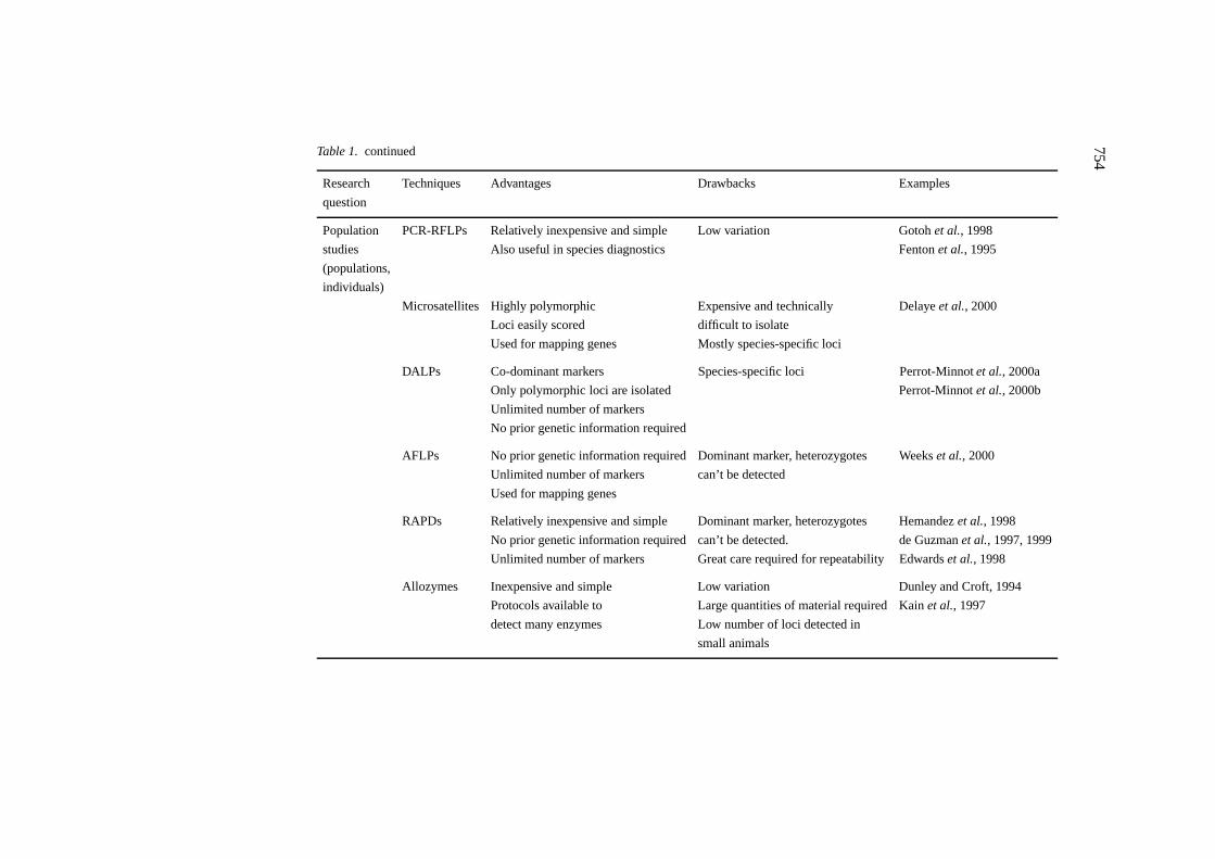
The development of markers is a prerequisite for studies of genetics. Since theappearance of one of the first compilations of molecular biology techniquesapplied to insects and mites (Hoy, 1994), the panel of available methodologieshas substantially increased. We describe here techniques that are currentlybeing used, or are of potential, in detecting genetic polymorphism (Table 1).These techniques can be further used in studies of molecular systematicsand evolutionary genetics (see Hilliset al. (1996) for an overview of DNAtechniques and data analysis applied to molecular systematics).
Allozymes
Protein electrophoresis has been an effective technique for the detection ofgenetic polymorphism for over three decades (see Pasteuret al. (1988) fordetailed information of methods). Enzymatic polymorphism detected by elec-trophoresis has been widely used on ticks and mites and an exhaustive re-view is not provided here. The most commonly studied enzymatic systemin mites is that of the esterases (for example Sula and Weyda, 1983; Go-toh and Takayama, 1992; Osakabe and Komazaki, 1996). Multilocus studies
753
Table 1. Common molecular techniques useful for evolutionary studies, and selected examples in the Acari
Research Techniques Advantages Drawbacks Examples
question
Phylogenetic rDNA sequencing Suited to resolve “deep phylogenies” Less informative for resolving Blacket al., 1997
No prior genetic information required can’t be detected. de Guzmanet al., 1997, 1999
Unlimited number of markers Great care required for repeatability Edwardset al., 1998
Allozymes Inexpensive and simple Low variation Dunley and Croft, 1994
Protocols available to Large quantities of material required Kainet al., 1997
detect many enzymes Low number of loci detected in
small animals
755
have been used to study the dispersion of the phytoseiidTyphlodromus pyriSheuten (Dunley and Croft, 1994), to trace the origin of a major pasture pestHalotydeus destructor(Tucker) from its putative source in South Africa toAustralia (Quin, 1997), for species identification in the Oribatida (Avanzatiet al., 1994) and to study the population structure of the spider miteTetra-nychus urticaeKoch in glasshouses (Tsagkarakouet al., 1999) and in openfields (Tsagkarakouet al., 1997, 1998). In ticks, several enzymatic systemscan be resolved from single individuals, however, diverse studies have repor-ted low polymorphism of the resolved loci (Delayeet al., 1997; Kainet al.,1997).
Although protein electrophoresis is still a useful method for studying ge-netic polymorphism (Lewontin, 1991), it has important limitations: (1) thebiological material must be kept alive or be frozen until used, and (2) at agiven locus the technique reveals only a fraction of the actual genetic vari-ation. Moreover, the small size of many mite species seriously limits thenumber of enzyme systems that can be detected per individual. Modificationfor increased efficiency and for work specifically with small organisms usingisoelectric focusing on cellulose acetate membranes has been described byKazmeret al. (1991) and adapted forT. urticaeby Tsagkarakouet al. (1996),making it possible to reveal up to five loci per individual in a single run. Thetechnique has been applied to investigate the persistence of a predatory miteNeoseiulus fallacis(Garman) introduced in apple orchards during biologicalcontrol programs in Canada (Navajaset al., 2000b).
Direct analysis of proteins
Highly sensitive techniques for separating proteins, such as two-dimensionalelectrophoresis (O’Farrell, 1975), have not yet been widely applied to studyticks and mites. However, there has been some use of SDS-PAGE in combin-ation with general protein staining to investigate quantitative differences inproteins between organophosphate resistant and sensitive strains ofBoophilusmicroplus(Canestrini) (Rosario-Cruzet al., 1997).
Amplification of DNA using PCR
Allozyme data are now increasingly replaced by several types of DNA-baseddata. The advent of the polymerase chain reaction (PCR) (Mullis and Faloona,1987; Saikiet al., 1988) made it possible to apply the DNA approach tosmall animals because only tiny amounts of initial material are necessary.Thus, most of the current techniques used to examine nucleotide variationsare based on PCR to amplify sufficient quantities of DNA. Starting fromDNA extracted by classic techniques (see Hubbardet al. (1995) for ticks pre-
756
served in alcohol), PCR uses the enzymeTaqDNA polymerase to synthesise acomplementary DNA sequence from a single stranded DNA oligonucleotide(primer) hybridised to a specific part (target) of the DNA strand. A PCRmachine (thermocycler) applies successive temperature cycles (typically 30–50 cycles) that include a denaturation cycle (typically 94◦C) followed by anannealing stage (typically 40–60◦C, but see also the ‘touchdown’ procedure(Don and Cox, 1991)) followed by a final extension stage under the effectof the polymerase (typically 72◦C). The DNA fragment flanked by the twoprimers is thus duplicated exponentially to produce sufficient DNA quantitiesfor further analytical procedures. Among the different techniques available,we describe those that are already used or are potentially of use in Acari.
Random amplified polymorphic DNA (RAPD)
The RAPD technique uses the PCR principle for random amplification ofDNA sequences (Williamset al., 1990). Amplification is performed usinga single primer with a very short sequence (8–10 base pairs) under temper-ature conditions (usually low) enhancing multiple binding at sites scatteredthroughout the genome. DNA molecules generated from the DNA of differentindividuals are then separated on an agarose gel matrix. Several DNA frag-ments are usually amplified and some of these may be present in a proportionof the individuals in a population. A large set of primers has the advantageof screening the entire genome. However, the interpretation of RAPD datais sometimes limited by poor repeatability of the results (Black, 1993), withthe problem aggravated in the case of small species in which the quantity ofDNA obtained per individual is reduced, thus preventing accurate assays ofDNA concentration. Another limit of these markers is that the RAPD patternsdisplay dominance, preventing identification of heterozygotes. Nevertheless,the RAPD technique is often used as the first source of information becauseresults are generated quickly and easily. By cloning and sequencing RAPDproducts it is also possible to design new more-specific primers which will bemore reliable (see Yli-Mattilaet al., 2000). In addition, if the necessary pre-cautions of verification of the heritability of the patterns obtained by RAPDare taken, the technique can be used successfully for the analysis of pedigreesand paternity exclusion (see Black (1993) for a review). Using the transmis-sion patterns of RAPD markers Perrot-Minnot and Navajas (1995) showedthat genetic material is transmitted from father to son in controlled crossesin a pseudo-arrhenotokous phytoseiid mite. Besides, the RAPD approach hasbeen used to compare strains ofTetranychussp. as a complement of fecundityand biometry studies (Henceet al., 1998). In ticks, Hernandezet al. (1998)used 120 RAPD primers to develop genomic probes for detection of acaricide
757
resistence in the cattle tickB. microplusand detected profiles differentiatingsusceptible and resistant strains.
Restriction fragment length polymorphism (RFLP)
Genome DNA regions isolated by PCR or by other means can be digested byrestriction enzymes that generate RFLP patterns. This technique was usedto differentiate between species of the genusPanonychus(Tetranychidae)(Osakabe and Sakagami, 1994) and to detect genomic variability in differ-ent populations ofB. microplus(Passoset al., 1999). RFLP patterns of theproducts of PCR are also used for species diagnosis purposes as describedbelow.
Microsatellites
Microsatellites are defined as short DNA fragments (≈100 bp) containingpatterns with two to six base-pairs repeated in tandem. During DNA replica-tion, repetition units are added to, or lost from, the microsatellite, causing therapid evolution of these regions. There is generally a high variability in thenumber of repetitions at a given locus. This makes a large number of allelesper microsatellite locus available for population analysis (see Goldstein andSchlötterer (1999) for a recent review). These are co-dominant markers andthe substantial polymorphism of these loci can be detected in a simple mannerby measuring the size of the PCR-amplified fragments. Microsatellite loci areavailable for an increasing number of organisms and this new type of datahas greatly stimulated the development of powerful statistical methods andcomputer programs for analyzing allele frequency data (Luikart and England,1999). Microsatellites are indeed excellent for studies of population genetics(Jarne and Lagoda, 1996; Goldstein and Schlötterer, 1999).
The isolation of microsatellite loci involves the construction of DNA ge-nomic libraries that are then screened with microsatellite-type repeated se-quence probes (Estoupet al., 1993). Once a locus has been identified it issequenced and primers are designed for the unique microsatellite flankingregions. This step can be omitted if primers known to work in one speciesamplify microsatellite regions in related species. Whereas microsatellite se-quences were considered to be present in all the eukaryote genomes (Hamadaand Kakunaga, 1982), Navajaset al. (1998) showed that they are extremelyrare in two screened mite species (T. urticaeand N. fallacis). Efforts havecontinued in the search for microsatellites forT. urticae, for which a lib-rary enriched in microsatellite sequences (Edwardset al., 1996) has beenmade, facilitating the isolation of several polymorphic microsatellites in mites(Navajas, unpublished data). Also the European tick,Ixodes ricinus(L.), has
758
been screened for microsatellite loci. Six polymorphic loci have been detec-ted, including PCR primers hybridising with other members of theI. ricinuscomplex (Delayeet al., 1998). In a recent publication Evans (2000) providesinformation on nine microsatellite loci in the honey bee parasitic miteVarroajacobsonioudemans.
Direct amplification of length polymorphism (DALP)
The DALP method involves the use of random PCR primers to generatemulti-band genome DNA patterns and enables the sequencing of polymorphicbands. PCR-specific bands are then defined and enable the study of distinctloci (Desmaraiset al., 1998). This strategy combines the advantage ofa prioridefinition of polymorphism and the reproducibility of the results by means ofthe use of specific primers. This new method has been applied to isolate fivepolymorphic loci in the phytoseiid miteNeoseiulus californicus(McGregor)as presented by Perrot-Minnotet al. (2000b). The Mendelian transmissionof alleles and their co-dominance were verified, thus making DALP markersuseful for genetic studies. The DALP technique has recently been used tostudy the pseudo-arrhenotokous reproduction system inN. californicus. Inthis study, the transmission patterns of parental alleles through two genera-tions are reported at five loci, providing evidence for the retention in the malesomatic tissues of most if not all the paternal chromosomes (Perrot-Minnotet al., 2000a).
Amplified fragment length polymorphim (AFLP)
The AFLP technique selects, by PCR amplification, restriction fragmentsgenerated from a total digest of genomic DNA (Voset al., 1995). Usingthis method, sets of restriction fragments can be visualised by PCR withoutprior knowledge of the genome of the target species. As for other fingerprint-ing based methods, such as DALP or RAPD, it gives access to an almostunlimited number of genetic markers. However, in the AFLP technique het-erozygote genotypes cannot be distinguished from homozygote genotypesand, as indicated previously for RAPD, this limits the use of these markersfor population genetic studies. The AFLP method has been used in mites forthe first time by Weekset al. (2000).
Sequencing of DNA fragments
The DNA fragment amplified by any PCR can be quickly analysed for poly-morphism either in the cleavage profile of restriction enzymes (RFLP), orsequenced by techniques that have now become routine and economical.
759
Obtaining the nucleotide sequence provides access to the ultimate detail ofvariation in the DNA. DNA sequences are collated and stored in data lib-raries (e.g. EMBL and GenBank) and are universally usable, powerful data.These also provide search facilities which allows unknown sequences to beidentified by similarity search engines such as BLAST (Karlin and Altschul,1993). Sequencing of the product can either be done directly (Navajaset al.,1998a) or after cloning into plasmid vectors (Fentonet al., 1997). The lattertechnique is more expensive, but is particularly important when investigat-ing the extent of intra-specific variation in ribosomal genes (see moleculardrive below). DNA sequences are used to assess the divergence between taxaor individuals and also enable the reconstruction of the phylogenetic rela-tions between the taxonomic entities studied. The inference of phylogeniesfrom molecular data requires the selection of the appropriate method fromthe many techniques that have been described (Swoffordet al., 1996). Theauthors provide a comprehensive description of the analytical methods andsummarise the different programs and software packages available for con-ducting phylogenetic and population analysis. In addition, several programsare devoted to multiple nucleotide sequences alignment, among these oneof the most popular is Clustal W (Thompsonet al., 1994). Alignment ofsequences may be difficult because of length variation resulting from inser-tions/deletions in the nucleotide sequences. Although in the case of ribosomalsequences, the analysis of secondary structure to identify homologous posi-tions may improve alignments (Kjer, 1995), structures may appear conservedirrespective of phylogenetic associations (Hancock and Vogler, 2000).
Different genomic regions can be analysed depending on the problemexamined. In particular, these regions may have different rates of evolutionand/or different modes of inheritance (maternal vs Mendelian). Rapidlyevolving genes are useful for comparisons of closely related taxa and slowlyevolving genes are useful for comparisons of distantly related taxa. Two ofthe most popular markers used in molecular evolution are mitochondrial DNA(mtDNA) and nuclear ribosomal DNA (rDNA). Information for Acari onboth genomic regions is beginning to accumulate in the gene data-bases. Theimpact that this and related data has, or should have, in acarology is discussednext.
Studies of Genome Characterisation
Mitochondrial DNA
The molecular biology and the patterns of evolution of animal mtDNA arewell understood (Wolstenholme, 1992; Avise, 1994; Simonet al., 1994). The
760
mitochondrial genome of two ticks,Ixodes hexagonusLeach andRhipiceph-alus sanguineusLatreille, has been sequenced entirely (Black and Roehrdanz,1998). The gene arrangements of these two species, together with that ofB.microplus(Campbell and Barker, 1999), have been established. The mtDNAof the spider miteT. urticae has also been characterised by a restrictionmap (Fournieret al., 1994). A study of 20 mite species belonging to theTetranychidae and Tenuipalpidae shows that the characteristics and mode ofevolution of the mtDNA are similar to those known for insects (genetic code,use of codons, base composition), and this is probably the result of ancestralcharacters shared between the two arthropod classes (Navajaset al., 1996). Asin insects, the mitochondrial sequences of mites are extremely A+T rich (av-erage 75%). However, variation in base composition has been shown betweenspecies within the same family (Navajaset al., 1996). This has important im-plications for the construction of phylogenies and requires the developmentof specific methods which take this type of variation into account (Galtierand Gouy, 1995). Several genes of the mitochondrial genome are increasinglyused to assess phylogenetic relationships among animal taxa (see Simonet al.(1994) for a review). Evolutionary studies on mites have mainly surveyed themitochondrial Cytochrome Oxidase subunit I gene (COI), whereas in studieson ticks mitochondrial ribosomal 16S is most popular.
Nuclear ribosomal DNA
Nuclear ribosomal DNA still provides one of the most complete tools fora multitude of molecular tasks. The main ribosomal locus in eukaryotic or-ganisms consists of three genes encoding the 18S, 5.8S and 28S subunitsof the ribosome. Between these genes are the internal transcribed spacers1 (ITS1, between the 18S and 5.8S gene) and 2 (ITS2, between the 5.8Sand 28S gene) (Hillis and Dixon, 1991). The three genes are reiterated intandem and between each group lies the intergenic spacer. There may be morethan one hundred copies of the ribosomal genes on a chromosome and theymay be found on more than one chromosome (Wenet al., 1974). Reitera-tion is essentially a pre-amplification step which makes detecting these genesconsiderably easier than for single copy genes.
In the conserved ribosomal genes, PCR primers can be defined that workin a wide range of species (Kaliszewskiet al., 1992). Sequences of the ampli-fied fragments are useful for comparison of phylogenetically distant taxa. Tocomplement this, the ITS regions are very useful for distinguishing betweenclosely related taxa as they evolve more rapidly than the coding regions (Hillisand Dixon, 1991). To characterize the ITS regions it is possible to design PCRprimers defined in the more conserved genes (28S, 5.8S and 18S) flanking theITS. While these gene sequences provide primer sites for use in amplifying
761
ITS regions from many arthropods, including several species of ticks (Zahleret al., 1997; Fukunagaet al., 2000) and different groups of mites such as Psor-optidae (Zahleret al., 1995b, 1998; Essiget al., 1999), Sarcoptidae (Zahleret al., 1999), Eriophyidae (Fentonet al., 1993), Tetranychidae (Navajaset al.,1992) and Phytoseiidae (Navajaset al., 1999b), it is still possible for smallchanges in one or two bases to prevent amplification, as has been found inphytoseiids (Yli-Mattilaet al., 2000) and eriophyids (Fentonet al., 1997).Once an ITS region has been successfully amplified it can then be analyzedby additional techniques.
The repetitive nature of ribosomal genes results in copies of ITS regionshaving the potential to vary within and between individuals. This can giverise to intra-specific diversity of ITS sequences, as has been found in someIxodesticks (McLainet al., 1995; Richet al., 1997). However, this tendencyto diversify is countered by a process known as molecular drive which ensuresthat the different copies of rDNA are the same (Dover, 1982; Polancoet al.,1998). In the phytophagous mites examined so far, this process appears to bevery effective as they exhibit very low levels of intra-specific polymorphism(Fentonet al., 1997; Navajaset al., 1998a). Thus, in the phytophagous mitesthese regions have no problems associated with intra-individual diversity andcan probably be widely applied.
It is a problem with many DNA techniques that a newly isolated markercould have come from a contamination event or a symbiotic organism andthis is particularly the case with PCR. If DNA sequences are being invest-igated then it may be possible to use conserved regions to investigate thesource of the sequence. The rDNA ITS regions are flanked by the 18S, 5.8Sand 28S genes. These gene regions are evolving slowly and they containphylogenetic information which can be used to search a sequence databasefor matches. Such a search will reveal sequences belonging to the taxonclosest to the isolated sequence and this then helps confirm the source. Asearch with an Acarine sequence might identify another Acarine sequence.However, such sequences are still not very frequent and it is more likelythat the conserved parts of the ribosomal genes will match other arthropodsequences. The methodology also facilitates the elimination of contaminatingsequences. For example, fungal sequences have been found in aphids (Fentonet al., 1994) and more recently in mites (Yli-Mattilaet al., 2000).
Other nuclear genes
The general characterisation of genes in the Acari is still in its infancy. How-ever, two genes have been isolated and analysed from the cattle tickB. mi-croplus. These genes encode the ecto-5′-nuclease and the octopamine-likeG-protein receptor (Liyouet al., 1999; Baxter and Barker, 1999). The ge-
762
nomic sequence of the first gene contained no significant introns. It is notclear yet if this will be generally applicable to the Acari. Introns have proveduseful in providing intra-specific markers in other organisms. The octopaminereceptor gene was sequenced in amitraz-resistant and sensitive strains and nodifferences were found. This suggested that the resistance was not based onmutational differences in this gene. A third gene, the elongation factor-1α, hasnow proved to have potential for phylogenetic analysis of the Mesostigmata(Klompen, 2000).
In a fundamental study of mite developmental biology conducted by Telfordand Thomas (1998), an oribatid mite was used to understand the evolutionof the homologous zerknült (zen) gene of insects and the Hox 3 genes ofvertebrates.
Overview of DNA Data
Phylogeny
Our knowledge of the phylogenetic relationships between organisms has re-cently greatly expanded through analysis of molecular data. DNA based phylo-genies have been used to examine major taxonomic levels (mainly Ixodidaeand Tetranychidae) that have challenged the established views of taxa rela-tionships.
In the Ixodidade, exhaustive phylogenetic analysis based on sequences ofthe mitochondrial 16S rDNA (Black and Piesman, 1994; Norriset al., 1999),variable regions of the 18S (V4) and 28S (D1) ribosomal genes (Cramptonet al., 1996) and the entire 18S region (Blacket al., 1997; Dobson and Barker,1999) covers several aspects of the evolution of the group. In a global ap-proach that integrates nuclear and mitochondrial rDNA and morphologicaldata sets, Klompenet al. (2000) present an evolutionary scenario of the re-lationships among tick genera and subfamilies. At a lower taxonomic level,the mitochondrial 16S sequences were used to examine the phylogenetic rela-tionships in the genusDermacentor(Crosbieet al., 1998) andRhipicephalus(Mangold et al., 1998). In a recent review mainly devoted to the molecu-lar detection of pathogen DNA in ticks, Sapaganoet al. (1999) present acompilation of the published PCR primers used to amplified ixodid DNA.
In a comprehensive study ofV. jacobsoniAnderson and Trueman (2000)used phenotypic and reproductive variation together with COI sequence poly-morphism to examine populations collected from bees distributed throughoutAsia. They demonstrated that this ectoparasitic mite is a complex of at leasttwo different species. The authors suggest the creation of a new species,V.destructorn. sp. which is mostly applicable to findings of past research onV.jacobsoni.
763
Using a fragment of the COI in 20 species of phytophagous mites fromnine genera and two families (Tetranychidae and Tenuipalpidae) to estimatephylogenetic relationships between taxa, Navajaset al. (1996) showed thatmolecular and morphological classifications are compatible as a whole, butsome minor taxonomic revisions are called for. The data suggest, for example,that the genusOligonychusis polyphyletic and enables better appraisal of thedifferent morphological characters conventionally used in systematics.
In the case of plant-feeding mites phylogenetic relationships have beencompared to molecular relationships of their host plants. In a recent study themolecular phylogeny of the highly host-specific phytophagousCecidophyop-sis gall-mite species was compared to that of their host plants. This foundthat the mite species had only very recently diverged onto their specialisthosts whereas the plants themselves had been separated considerably longer.This indicates that the host–plant interactions of these mites are not basedon extended periods of co-evolution. The mites were related according to thephysiological alterations they impose on the plant, i.e., mites which did notinduce galls were very closely related, despite the fact that they had hosts thathad been separated for millions of years (Fentonet al., 2000).
A phylogenetic analysis of a portion of the 28S rDNA from a group ofdermanyssine mites that contains arrhenotokous, peudo-arrhenotokous andancestrally diplodiploid members was used to shed light on the evolution-ary origins of haplodiploidy in this group of organisms (Cruickshank andThomas, 1999).
Phylogeography and the genetic structure of the species
Distinct genotypes ofV. jacobsonihave been described based on RAPD mark-ers (Kraus and Hunt, 1995; de Guzmanet al., 1999) and mitochondrial COIdata (Anderson and Fuchs, 1998; de Guzmanet al., 1998) and their geo-graphic distribution world-wide assessed (see de Guzman and Rinderer (1999)for a review). Work on COI sequences has shown the genetic structuring ofthe speciesT. urticae throughout its distribution area. The COI sequenceshave made it possible to make a distinction between two lineages, and theresults suggest on the one hand ancient colonisation of the Mediterraneanregion and on the other recent colonisation of the temperate regions in thenorthern hemisphere (Navajaset al., 1998a). The phylogenetic informationobtained for mitochondrial sequences of COI has not revealed divergencesthat can be correlated with ecological factors such as host plant or colour ofT. urticae (Navajas, 1998), which in the past have led to subdivisions intosubspecies or races (Gotohet al., 1993). A survey of allozymes togetherwith ribosomal ITS2 sequence variation in populations ofT. urticaeorigin-ating from the Mediterranean basin allowed an evaluation of the role of the
764
host plant and the geographical distance in the genetic differentiation process(Navajaset al., 2000c). Such processes potentially lead to speciation.
In addition to the COI gene, the Cytochrome Oxidase III (COIII) gene hasbeen used to assess population structure as demonstrated by the work by Kainet al. (1999) on populations of the tickI. pacificusfrom the USA. In a studyof the tick Ixodes scapularisSay throughout its range, Norriset al. (1996)adopted a novel strategy to detect genetic variation that involves the use ofsingle strand conformation polymorphism (SSCP) analysis. Using the SSCPapproach to detect variation in a region of the 16S mitochondrial ribosomalDNA, Norriset al. (1996) estimated the frequency of haplotypes in various re-gions of the United States and determined phylogenetic relationships amongthese haplotypes.
Species discrimination
The small size of most mite species implies that a limited number of mor-phological characters are available for systematics. In addition, it is commonthat intra-specific variability of these characters complicates the determin-ation of taxa. Molecular markers can help to discriminate between speciesthat are morphologically very close, as has been reported for the two siblingspeciesTetranychus pueraricolaEhara and Gotoh andT. urticae(Ehara andGotoh, 1996). Differences between the sequences of ITS2 of the two speciesconsolidated the sibling species status and contributed to their unambigu-ous identification (Gotohet al., 1998). The ITS2 sequence variation togetherwith a cross-breeding experiment have also been used to establish the syn-onymy between two spider mite species:Tetranychus kanzawaiKishida andT. hydrangeaePritchard and Baker (Navajaset al., 2000a). Edwardset al.(1998) examined variable RAPD markers in threeTyphlodromalusmites andfound several bands that could be used individually to distinguish phytoseiidspecies.
Evidence of the reproductive isolation ofDermacentor marginatus(Sul-cer) andD. reticulatus(Fabricius) was supported by ribosomal ITS2 sequencesas a complement to cross-breeding and morphological studies (Zahleret al.,1995a; Zahler and Gothe, 1997). These results were corroborated by a com-parative study of inter- and intra-specific polymorphism of both tick spe-cies that exhibit overlapping phenotypes (Zahleret al., 1995b). The sameribosomal region was used to establish the taxonomic status of species be-longing to theRhipicephalus sangineusgroup (Zahleret al., 1997) and thetaxonomic status of two ticks,Ixodes neotomaeCooley andI. spinipalpisHadwen and Nuttall (Norriset al., 1997). Recently a molecular key has beendescribed for 17Ixodesspecies in the United States (Poucher, 1999).
765
In the Oribatida, analysis of a fragment of the mitochondrial COI geneserved to establish the taxonomic status (synonymy) ofSteganacarus mag-nus (Nicolet) andS. anomalus(Berlese) (Salomoneet al., 1996). A COIfragment was also used to distinguish between four species ofTetranychusmites involved in quarantine problems associated with apple imports in NorthAmerica (Lee and Lee, 1997).
For eriophyid mites the problems associated with identification are par-ticularly acute (Boczek and Griffiths, 1994). For theCecidophyopsismitesan RFLP approach of the ITS regions was found to be initially successful(Fentonet al., 1995). However this has been superseded by the developmentof a multiplex PCR system which co-amplifies three bands from ITS1 whichall vary in length between different species (Kumaret al., 1999). These areresolved on high resolution acrylamide gels (Kumaret al., 1999). This tech-nique even works on single eriophyid mites which are the smallest of all themites (Fentonet al., 1997).
Tracing introduction of species to new geographical regions
The study of the genetic structure of species is of practical application inthe case of pests. An increasing number of pest species are introduced eachyear into new biogeographical areas. In some cases, it is important to de-termine the area of origin of these introductions, for example when predatorsare to be imported within the framework of biological control operations.The green cassava mite,Mononychellus progresivusDoreste, originated inthe neotropical region and was accidentally introduced into East Africa in the1970s. Study of the genetic diversity of the African and American populationsof this mite based on sequences of mitochondrial COI and ribosomal ITS2has shown thatM. progresivuswas probably introduced into Africa fromColumbian populations or from a region bordering Columbia (Navajaset al.,1994).
Another important case of pest introduction is that of a parasite of bees,the miteV. jacobsoniin the Americas. Using RAPD markers, de Guzmanet al. (1997) suggested a European origin forV. jacobsoniin the United States,whereas mites in Brazil and Puerto Rico are probably predominantly Japanesein origin.
Wolbachiaand reproductive incompatibilities
As a complement to historical and ecological factors, incompatibilities in re-production may play a role in the structuring of populations. In this context, itis important to note that a bacterial endosymbiont of theWolbachiatype wasdetected in several species of Tetranychidae and Phytoseiidae (Breeuwer and
766
Jacobs, 1996; Johanowicz and Hoy, 1996; Tsagkarakouet al., 1996a). Thesemicroorganisms are known to be involved in alterations in the reproductivesystem in arthropods (Stouthameret al., 1999) including mites (Breeuwer,1997; Johanowicz and Hoy, 1998), although it has no effect in some cases(Gotohet al., 1995, 1999; Gomiet al., 1997; Navajaset al., 1999a). Naturalpopulations ofT. urticaehave been found to be either infected or uninfected(Breeuwer and Jacobs, 1996). The dynamics ofWolbachiainfection is notfully understood yet, however Johanowicz and Hoy (1999) showed that theinfection did not spread rapidly through experimental laboratory populationsof the phytoseiidMetaseiulus occidentalis(Nesbitt). The detection ofWolba-chia infection in mites, as well as in a diverse array of arthropods, has beenbased in the PCR amplification of three genomic regions: a fragment of the16S rDNA and part of protein coding genes, including theftsZ, which is in-volved in cell division, and more recently thewspwhich encodes a major cellsurface coat protein (see Tsagkarakouet al. (1996) and Jeyaprakash and Hoy(2000) for PCR protocols). By removing theWolbachiafrom its host throughan antibiotic supplemented diet or heat treatment it is possible to study theeffects that the bacteria had on their host (Van Opijnen and Breeuwer, 1999).
Concluding Remarks
In this review we have briefly described the current use of genetic markers inthe study of ticks and mites. These markers have already been used to increasethe accuracy of species identification as well as in helping identify previouslyunrecognised mite species. There are many more mite species to be identified.Phylogenetic relationships between taxa are now precisely established on thebasis of DNA sequences, although there is still much to be learned. Detailedanalysis of population structures has also been possible. Many of the methodsdescribed here are dependant on screening large numbers of individuals tofully understand the natural variation within a population. Clearly, this is timeconsuming, however the techniques are becoming routine and economical.Other model arthropods, mainly insects, have shown major advances in theunderstanding of their genetics and population structures (e.g. Waltonet al.,1999). Much of this has been facilitated by the use of genetic manipulation tointroduce marker genes which can also be selected e.g., the eye-colour systemin Drosophila (Klemenzet al., 1987). These advances are slowly workingtheir way into other arthropods and transformation has now been carried outwith transgenic phytoseiid mites (Presnail and Hoy, 1992). This system maybecome available for the manipulation of other beneficial mites, increasingtheir effectiveness as natural predators.
767
Acknowledgements
We thank J. Gutierrez for helpful comments on the manuscript.
References
Anderson, D.L. and Fuchs, S. 1998. Two genetically distinct populations ofVarroa jacobsoniwith contrasting reproductive abilities onApis mellifera. J. Apic. Res. 37: 69–78.
Anderson, D.L. and Trueman, J.W.H. 2000.Varroa jacobsoni(Acari: Varroidae) is more thanone species. Exp. Appl. Acarol. 24: 165–189.
Avanzati, A.M., Baratti, M. and Bernini, F. 1994. Molecular and morphological differentiationbetween steganacarid mites (Acari:Oribatida). Biol. J. Linn. Soc. 52: 325–340.
Avise, J.C. 1994. Molecular Markers, Natural History and Evolution. Chapman and Hall, NewYork.
Baxter, G.D. and Barker, S.C. 1999. Isolation of a cDNA for an octopamine-like, G-proteincoupled receptor from the cattle tick,Boophilus microplus. Insect Biochem. Mol. Biol. 29:461–467.
Black, W.C., IV. 1993. PCR with arbitrary primers: approach with care. Insect Mol. Biol. 2:1–6.
Black, W.C., IV and Piesman, J. 1994. Phylogeny of hard and soft-tick taxa (Acari: Ixodida)based on mitochondrial 16S ribosomal DNA sequences. Proc. Natl. Acad. Sci. USA 91:10034–10038.
Black, W.C., IV, Klompen, J.S.H. and Keirans, J.E. 1997. Phylogenetic relationships amongtick subfamilies (Ixodida: Ixodidae: Argasidae) based on the 18S nuclear rDNA gene. Mol.Phylogenet. Evol. 7: 129–144.
Black, W.C., IV and Roehrdanz, R.L. 1998. Mitochondrial gene order in not conserved inArthropods: Prostriate and Metastriate tick mitochondrial genomes. Mol. Biol. Evol. 15:1772–1785.
Boczek, J. and Griffiths, D.A. 1994. Structure and systematics of eriophyid mites(Acari: Eriophyoidae) and their relationship to host plants. Syst. Asso. Spe. Vol. 49:119–129.
Breeuwer, J.A.J. and Jacobs, G. 1996.Wolbachia: intracellular manipulators of mite reproduc-tion. Exp. Appl. Acarol. 20: 421–434.
Breeuwer, J.A.J. 1997.Wolbachia and cytoplasmic incompatibility in the spider mitesTetranychus urticaeandT. turkestani. Heredity 79: 41–47.
Campbell, N.J.H. and Barker, S.C. 1999. The novel mitochondrial gene arrangement of thecattle tick,Boophilus microplus: fivefold tandem repetition of a coding region. Mol. Biol.Evol. 16: 732–740.
Crampton, A., McKay, I. and Barker, S.C. 1996. Phylogeny of ticks (Ixodiade) inferred fromnuclear ribosomal DNA. Int. J. Parasitol. 26: 511–517.
Crosbie, P.R., Boyce, W.M. and Rodwell, T.C. 1998. DNA sequence variation inDermacentorhunteri and estimated phylogenies ofDermacentorspp. (Acari: Ixodidae) in the NewWorld. J. Med. Entomol. 35: 277–288.
Cruickshank, R.H. and Thomas, R.H. 1999. Evolution of haplodiploidy in Dermanyssine mites(Acari: Mesostigmata). Evolution 53: 1796–1803.
de Guzman, L.I., Rinderer, T.E. and Stelzer, J.A. 1997. DNA evidence of the origin ofVarroajacobsoniOudemans in the Americas. Biochem. Genet. 35: 327–335.
768
de Guzman, L.I., Rinderer, T.E., Stelzer, J.A. and Anderson, D.L. 1998. Congruence of RAPDand mitochondrial DNA markers in assessingVarroa jacobsonigenotypes. J. Apic. Res.37: 49–51.
de Guzman, L.I. and Rinderer, T.E. 1999. Identification and comparison ofVarroa speciesinfesting honey bees. Apidologie 30: 85–95.
de Guzman, L.I., Rinderer, T.E. and Stelzer, J.A. 1999. Occurrence of two genotypes ofVarroajacobsoniOud. in North America. Apidologie 30: 31–36.
Delaye, C., Beati, L., Aeschimann, A., Renaud, F. and de Meeus, T. 1997. Population geneticstructure ofIxodes ricinusin Switzerland from allozymic data: no evidence of divergencebetween nearby sites. Int. J. Parasitol. 27: 769–773.
Delaye, C., Aeschlimann, A., Renaud, F., Rosenthal, B. and De Meeus, T. 1998. Isola-tion and characterization of microsatellite markers in theIxodes ricinuscomplex (Acari:Ixodidade). Mol. Ecol. 7: 357–363.
Desmarais, E., Lanneluc, I. and Lagnel, J. 1998. Direct amplification of length polymorphisms(DALP) or how to get and characterize new genetic markers in many species. NucleicAcids Res. 26: 1458–1465.
Dobson, S.J. and Barker, S.C. 1999. Phylogeny of the hard ticks (Ixodidae) inferred from18S rDNA indicates that the genusAponommais paraphyletic. Mol. Phylogenet. Evol. 11:288–295.
Don, R.H. and Cox, P.T. 1991. Touchdown PCR to circumvent spurious priming during geneamplification. Nucleic Acids Res. 19: 4008.
Dover, G. 1982. Molecular drive: a cohesive mode of species evolution. Nature 299:111–117.
Dunley, J.E. and Croft, B.A. 1994. Gene flow measured by allozymic analysis in pesticideresistantTyphlodromus pyrioccurring within and near apple orchards. Exp. Appl. Acarol.18: 201–211.
Edwards, K.G., Barkers, J.H.A., Dali, A., Johns, C. and Karp, A. 1996. Microsatellitelibraries enriched for several microsatellite sequences in plants. Biotechniques 20:758–760.
Edwards, O.R., Melo, E.L., Smith, L. and Hoy, M. 1998. Discrimination on threeTyph-lodromalus species (Acari: Phytoseiidae) using random amplified polymorphic DNAmarkers. Exp. Appl. Acarol. 22: 101–109.
Ehara, S. and Gotoh, T. 1996. Two new species of spider mites occuring in Japan (Acari,Tetranychidae). J. Acarol. Soc. Jpn. 5: 17–25.
Essig, A., Rinder, H., Gothe, R. and Zahler, M. 1999. Genetic differentiation of mites of thegenusChorioptes(Acari: Psoroptidae). Exp. Appl. Acarol. 23: 309–318.
Estoup, A., Solignac, M., Harry, M. and Cornuet, J.M. 1993. Characterization of (GT)n and(CT)n microsatellites in two insect species:Apis melliferaandBombus terrestris. NucleicAcids Res. 21: 1427–1431.
Evans, J.D. 2000. Microsatellite loci in the honey bee parasitic miteVarroa jacobsoni. Mol.Ecol. 9: 1433–1449.
Fenton, B., Malloch, G., Brennan, R., Jones, A.T., Gordon, S., McGavin, W. and Birch, A.N.E.1993. Taxonomic evaluation of the three reputed species ofCecidophyopsismite onRibes.Acta Horticulturae 352: 535–538.
Fenton, B., Birch, A.N.E., Malloch, G., Woodford, J.A.T. and Gonzalez, C. 1994. Molecularanalysis of ribosomal DNA from the aphidAmphorophora idaeiand an associated fungalorganism. Insect Mol. Biol. 3: 183–189.
idae) from differentRibesspecies and countries using molecular genetics. Mol. Ecol. 4:383–387.
Fenton, B., Malloch, G. and Moxey, E. 1997. Analysis of Eriophyid mite rDNA InternalTranscribed Spacer sequences reveals multiple simple sequence repeats. Insect Mol. Biol.6: 23–32.
Fenton, B., Birch, A.N.E., Malloch, G., Lanman, P.G. and Brennan, R.M. 2000. Gall mitemolecular phylogeny and its relationship to the evolution of plant host specificity. Exp.Appl. Acarol. 24: 831–861.
Fournier, D., Bride, J.M. and Navajas, M. 1994. Mitochondrial DNA from spider mites: isol-ation, restriction map and partial sequence of the Cytochrome Oxidase Subunit I gene.Genetica 94: 73–75.
Fukunaga, M., Yabuki, M., Hamase, A., Oliver, J.H. and Nakao, M. 2000. Molecular phylo-genetic analysis of ixodid ticks based on the ribosomal DNA spacer, internal transcribedspacer 2, sequences. J. Parasitol. 86: 38–43.
Galtier, N. and Gouy, M. 1995. Inferring phylogenies from DNA sequences of unequal basecomposition. Proc. Natl. Acad. Sci. USA 92: 11317–11321.
Goldstein, D.B. and Schlötterer, C. 1999. Microsatellites, evolution and applications. OxfordUniversity Press, New York.
Gomi, K., Gotoh, T. and Noda, H. 1997.Wolbachiahaving no effect on reproductive incom-patibility in Tetrancyhus kanzawaiKishida (Acari: Tetranychidae). Appl. Entomol. Zool.32: 485–490.
Gotoh, T. and Takayama, K. 1992. Developmental characteristics, genetic compatibility andesterase zymograms in three strains of the hawthorn spider mite,Tetranychus viennensisZacher (Acari: Tetranychidae). Ann. Missouri Bot. Gard. 1: 45–60.
Gotoh, T., Bruin, J., Sabelis, M.W. and Menken, S.B.J. 1993. Host race formation inTetra-nychus urticae: genetic differentiation, host plant preference, and mate choice in a tomatoand a cucumber strain. Entomol. Exp. Appl. 68: 171–178.
Gotoh, T., Oku, H., Moriya, K. and Odawara, M. 1995. Nucleus-cytoplasm interactions caus-ing reproductive incompatibility between two populations ofTetranychus quercivorusEhara et Gotoh (Acari:Tetranychidae). Heredity 74: 405–414.
Gotoh, T., Gutierrez, J. and Navajas, M. 1998. Molecular comparison of the sibling speciesTetranychus pueraricolaEhara & Gotoh andT. urticae Koch (Acari: Tetranychidae).Entomol. Sci. 1: 55–57.
Gotoh, T., Sugasawa, J. and Nagata, T. 1999. Reproductive compatibility of the two-spotted spider mite (Tetranychus urticae) infected with Wolbachia. Entomol. Sci. 2:289–295.
Hamada, H. and Kakunaga, T. 1982. Potential Z-DNA forming sequences are highly dispersedin the human genome. Nature 298: 396–398.
Hancock, J.M. and Vogler, A.P. 2000. How slippage-derived sequences are incorporated intorDNA variable-region secondary structure: Implications for phylogeny reconstruction.Mol. Phylogenet. Evol. 14: 366–374.
Hence, T., Neuberg, P. and Noèl-Lastelle, C. 1998. The use of fecundity, lobe biometry and theRAPD-PCR technique in order to compare strains ofTetranychussp. Exp. Appl. Acarol.22: 649–666.
Hernandez, R., Chen, A.C., Davey, R.B., Ivie, G.W., Wagner, G.G. and George, J.E. 1998.Comparison of genomic DNA in various strains ofBoophilus microplus(Acari: Ixodidae).J. Med. Entomol. 35: 895–900.
Hillis, D.M. and Dixon, M.T. 1991. Ribosomal DNA: Molecular evolution and phylogeneticinference. Quart. Rev. Biol. 66: 411–429.
770
Hillis, D.M., Moritz, C. and Mable, B. 1996. Molecular Systematics. Sinauer Associates, Inc.,Massachusetts, USA.
Hoy, M.A. 1994. Insect Molecular Genetics: an Introduction to Principles and Applications.Academic Press, San Diego.
Hubbard, M.J., Cann, K.J. and Wright, D.J.M. 1995. Validation and rapid extraction of nucleicacids from alcohol-preserved ticks. Exp. Appl. Acarol. 19: 473–478.
Jarne, P. and Lagoda, P. 1996. Microsatellites, from molecules to populations and back. TrendsEcol. Evol. 11: 424–429.
Jeyaprakash, A. and Hoy, M.A. 2000. Long PCR improvesWolbachia DNA amplifica-tion: wspsequences found in 76% of sixty-three arhropod species. Insect Mol. Biol. 9:393–405.
Johanowicz, D.L. and Hoy, M.A. 1996.Wolbachia in a predatory-prey system: 16S ri-bosomal DNA analysis of two phytoseiids (Acari: Phytoseiidae) and their prey (Acari:Tetranychidae). Ann. Entomol. Soc. Am. 89: 435–441.
Johanowicz, D.L. and Hoy, M.A. 1998. Experimental induction and termination of non-reciprocal reproductive incompatibilities in a parahaploid mite. Entomol. Exp. Appl. 87:51–58.
Johanowicz, D.L. and Hoy, M.A. 1999.Wolbachia infection dynamics in experimentallaboratory populations ofMetaseiulus occidentalis. Entomol. Exp. Appl. 93: 259–268.
Kain, D.E., Sperling, F.A. and Lane, R.S. 1997. Population genetic structure ofIxodespacificus(Acari: Ixodidae) using allozymes. J. Med. Entomol. 34: 441–450.
Kain, D.E., Sperling, F.A., Daly, H.V. and Lane, R.S. 1999. Mitochondrial DNA sequencevariation inIxodes pacificus(Acari: Ixodidae). Heredity 83: 378–386.
Kaliszewski, M.J., Tobolewski, J., Seyoum, S., Chojnacki, I., Kaliszewska, M.M., Stanton,D.J. and Colwell, R.K. 1992. The polymerase chain reaction and sequencing of mite DNA.Int. J. Acarol. 18: 231–239.
Karlin, S. and Altschul, S.A. 1993. Applications and statistics for multiple high-scoringsegments in molecular sequences. Proc. Natl. Acad. Sci. USA 90: 5873–5877.
Kazmer, D. 1991. Isoelectric focusing procedures for the analysis of allozymic variation inminute arthropods. Ann. Entomol. Soc. Am. 84: 332–339.
Kjer, K.M. 1995. Use of rRNA secondary structure in phylogenetic studies to identify ho-mologous positions: An example of alignment and data presentation from the frogs. Mol.Phylogenet. Evol. 4: 314–330.
Klemenz, R., Weber, U. and Gehring, W. 1987. Thewhitegene is a marker in a new P-elementvector for gene transfer inDrosophila. Nucleic Acids Res. 15: 3947–3959.
Klompen, J.S.H. 2000. A priliminary assessment if the utility of elongation factor-1α inelucidating relationships among basal Mesostigmata. Exp. Appl. Acarol. 24: 805–820.
Klompen, J.S.H., Black, W.C., IV, Keirans, J.E. and Norris, D.E. 2000. Systematics andbiogeography of hard ticks, a total evidence approach. Cladistics 16: 79–102.
Kraus, B. and Hunt, G. 1995. Differentiation ofVarroa jacobsoniOud. populations by randomamplification of polymorphic DNA (RAPD). Apidologie 26: 283–290.
Kumar, L., Fenton, B. and Jones, A.T. 1999. Identification ofCecidophyopsismites (Acari:Eriophyidae) based on variable simple sequence repeats of ribosomal DNA internaltranscribed spacer-1 sequences via multiplex PCR. Insect Mol. Biol. 8: 347–357.
Lee, M.L. and Lee, M.H. 1997. Amplified mitochondrial DNA identify four species ofTetranychusmites (Acarina: Tetranychidae) in Korea. Korean J. Appl. Entomol. 36:30–36.
Lewontin, R.C. 1991. Electrophoresis in the development of evolutionary genetics: milestoneor millstone. Genetics 128: 657–662.
771
Liyou, N., Hamilton, S., Elvin, C. and Willadsen, P. 1999. Cloning and expression ofecto 5′-nucleotidase from the cattle tickBoophilus microplus. Insect Mol. Biol. 8:257–266.
Luikart, G. and England, P.R. 1999. Statistical analysis of microsatellite DNA data. TrendsEcol. Evol. 14: 253–255.
Mangold, A.J., Bargues, M.D. and Mas-Coma, S. 1998. 18S rRNA gene sequences and phylo-genetic relationships of European hard-tick species (Acari:Ixodidae). Parasitol. Res. 84:31–37.
McLain, D.K., Wesson, D., Oliver, J.H. and Collins, F. 1995. Variation in ribosomal DNAinternal transcribed spacer I among eastern populations ofIxodes scapularis(Acari:Ixodidae). J. Med. Entomol. 32: 351–360.
Mullis, K.B. and Faloona, F.A. 1987. Specific synthesis of DNAin vitro via a polymerase-catalyzed chain reaction. Methods in Enzymol. 155: 335–350.
Navajas, M., Cotton, D., Kreiter, S. and Gutierrez, J. 1992. Molecular approach in spidermites (Acari: Tetranychidae): preliminary data on ribosomal DNA sequences. Exp. Appl.Acarol. 15: 211–218.
Navajas, M., Gutierrez, J., Bonato, O., Bolland, H.R. and Mapangou-Divassa, S. 1994.Intraspecific diversity of the cassava green miteMononychellus progresivus(Acari: Tetra-nychidae) using comparisons of mitochondrial and nuclear ribosomal DNA sequences andcross-breeding. Exp. Appl. Acarol. 18: 351–360.
Navajas, M., Fournier, D., Lagnel, J. and Boursot, P. 1996. Mitochondrial COI sequences inmites: evidence for variation in base composition. Insect Mol. Biol. 5: 1–5.
Navajas, M. 1998. Host plant associations in the spider miteTetranychus urticae(Acari:Tetranychidae): insights from molecular phylogeography. Exp. Appl. Acarol. 22:201–214.
Navajas, M., Lagnel, J., Gutierrez, J. and Boursot, P. 1998a. Species wide homogeneity ofnuclear ribosomal ITS2 sequences in the spider miteTetranychus urticaecontrasts withextensive mitochondrial COI polymorphism. Heredity 80: 742–752.
Navajas, M.J., Thistlewood, H.M.A., Lagnel, J. and Hughes, C. 1998b. Microsatellitesequences are under-represented in two mite genomes. Insect Mol. Biol. 7: 249–256.
Navajas, M., Gutierrez, J., Lagnel, J., Fauvel, G. and Gotoh, T. 1999a. DNA sequences andcross-breeding experiments in the hawthorn spider miteAmphitetranychus viennensisre-veal high genetic differentiation between Japanese and French populations. Entomol. Exp.Appl. 90: 113–122.
Navajas, M., Lagnel, J., Fauvel, G. and de Moraes, G. 1999b. Sequence variation of ribosomalinternal transcribed spacers (ITS) in commercially important Phytoseiidae mites. Exp.Appl. Acarol. 23: 851–859.
Navajas, M., Gutierrez, J., Williams, M. and Gotoh, T. 2000a. Synonymy between two spidermite species,Tetranychus kanzawaiandT. hydrangea,shown by ribosomal ITS2 sequencesand cross-breeding. Bull. Entomol. Res., in press.
Navajas, M., Thistlewood, H., Lagnel, J., Marshall, D., Tsagkarakou, A. and Pasteur, N. 2000b.Field releases of the predatory miteNeoseiulus fallacis(Acari: Phytoseiidae) in Canada,monitored by pyrethroid resistance and allozyme markers. Biol. Control, in press.
Navajas, M., Tsagkarakou, A., Lagnel, J. and Perrot-Minnot, M.J. 2000c. Genetic differenti-ation inTetranychus urticae(Acari: Tetranychidae): polymorphism, host races or siblingspecies? Exp. Appl. Acarol. 24: 365–376.
Norris, D.E., Klompen, J.S.H., Keirans, J.E. and Black IV, W.C. 1996. Population genetics ofIxodes scapularis(Acari: Ixodidae) based on mitochondrial 16S and 12S genes. J. Med.Entomol. 33: 78–89.
772
Norris, D.E., Klompen, J.S., Keirans, J.E., Lane, R.S., Piesman, J. and Black IV, W.C. 1997.Taxonomic status ofIxodes neotomaeand I. spinipalpis (Acari: Ixodidade) based onmitochondrial DNA evidence. J. Med. Entomol. 34: 696–703.
Norris, D.E., Klompen, J.S.H. and Black IV, W.C. 1999. Comparison of the mitochondrial 12Sand 16S ribosomal DNA genes in resolving phylogenetic relationships among hard-ticks(Acari: Ixodidae). Ann. Entomol. Soc. Am. 92: 117–129.
O’Farrell, P. 1975. High resolution two dimensional electrophoresis of proteins. J. Biol. Chem.250: 4007–4021.
Oliver, J.H. 1977. Cytogenetics of mites and ticks. Annu. Rev. Entomol. 22: 407–429.Osakabe, M. and Komazaki, S. 1996. Differences in esterase isozymes betweenPano-
nychus citripopulations infestingCitrus and Osmanthus. Exp. Appl. Acarol. 20: 113–119.
Osakabe, M. and Sakagami, Y. 1994. RFLP analysis of ribosomal DNA in sibling spe-cies of spider mite, genusPanonychus(Acari: Tetranychidae). Insect Mol. Biol. 3:63–66.
Palmer, M.J., Bantle, J.A., Guo, X. and Scott Fargo, W. 1994. Genome size and organizationin the ixodid tickAmblyomma americanum(L.). Insect Mol. Biol. 3: 57–62.
Passos, D.T., da Silva, S.S., Richter, M.F. and Ozaki, L.S. 1999. Detection of genomic variab-ility in different populations of the cattle tickBoophilus microplusin southern Brazil. Vet.Parasitol. 87: 83–92.
Pasteur, N., Pasteur, G., Catalan, J. and Bonhome, F. 1988. Practical Isozyme Genetics. EllisHorwood Ltd., Chichester, England.
Perrot-Minnot, M.J. and Navajas, M. 1995. Pseudo-arrhenotoky involves biparental inherit-ance of RAPD markers in males of the haplo-diploid miteTyphlodromus pyri. Genome38: 838–844.
Perrot-Minnot, M.J., Lagnel, J., Migeon, A. and Navajas, M. 2000a. Tracking paternal geneswith DALP markers in a pseudo-arrhenotokous reproductive system: biparental transmis-sion but haplodiploid-like inheritance in the miteNeoseiulus californicus. Heredity 84:702–709.
Perrot-Minnot, M.J., Lagnel, J., Desmarais, E. and Navajas, M. 2000b. Isolation and charac-terization by direct amplification of length polymorphism (DALP) of codominant geneticmarker with mendelian inheritance inNeoseiulus californicus(Acari: Phytoseiidae). Exp.Appl. Acarol. 24: 795–803.
Polanco, C., Gonzalez, A.I., de la Fuente, A. and Dover, G.A. 1998. Multigene family of ri-bosomal DNA inDrosophila melanogasterreveals contrasting patterns of homogenisationfor IGS and ITS spacer regions: A possible mechanism to resolve this paradox. Genetics149: 243–256.
Presnail, J.K. and Hoy, M.A. 1992. Stable genetic transformation of a beneficial arthropod,Metaseiulus occidentalis(Acari: Phytoseiidae), by microinjection technique. Rev. Agric.Entomol. 81.
Quin, T.K. 1997. Population genetics of redlegged earth mitesHalotydeus destructorandH.anthropus(Acarina: Penthaleidae) from Australia and/or South Africa. Bull. Entomol.Res. 87: 289–298.
Rich, S.M., Rosenthal, B.M., Telford, I.S.R., Spielman, A., Hartl, D.L. and Ayala, F.J. 1997.Heterogeneity of the internal transcribed spacer (ITS-2) region within individual deerticks. Insect Mol. Biol. 6: 123–129.
Rosario-Cruz, R., Miranda-Miranda, E. and Garcia-Vasquez, Z. 1997. Detection of esteraseactivity in susceptible and organophosphate resistant strains of the cattle tickBoophilusmicroplus(Acari: Ixodidae). Bull. Entomol. Res. 87: 197–202.
773
Saiki, R.K., Gelfand, D.H., Stoffel, S., Scharf, S.J., Higuchi, R., Horn, G.T., Mullis, K.B. andErlich, H.A. 1988. Primer-directed enzymatic amplification of DNA with a thermostableDNA polymerase. Science 239: 487–491.
Salomone, N., Frati, F. and Bernini, F. 1996. Investigation on the taxonomic status ofSteganacarus magnusandSteganacarus anomalus(Acari: Oribatida) using mitochondrialDNA sequences. Exp. Appl. Acarol. 20: 607–615.
Simon, C., Frati, F., Beckenbach, A., Crespi, B., Liu, H. and Flook, P. 1994. Evolution,weighting and phylogenetic utility of mitochondrial gene sequences and a compila-tion of conserved polymerase chain reaction primers. Ann. Entomol. Soc. Am. 87:651–701.
Sparagano, O.A.E., Allsopp, M.T.E.P., Manl, R.A., Rijkema, S.G.T., Figueroa, J.V. and Jonge-jan, F. 1999. Molecular detection of pathogen DNA in ticks (Acari: Ixodidae): A review.Exp. Appl. Acarol. 23: 929–960.
Stouthamer, R., Breeuwer, J.A.J. and Hurst, G.D.D. 1999.Wolbachia pipientis: Microbialmanipulator of Arthropod reproduction. Annu. Rev. Microbiol. 53: 71–102.
Sula, J. and Weyda, F. 1983. Esterase polymorphism in several populations of the two-spottedspider mite,Tetranychus urticaeKoch. Experientia 39: 78–79.
Swofford, D.L., Olsen, G.J., Waddell, P.J. and Hillis, D.M., 1996. Phylogenetic inference. In:Molecular Systematics, D. M. Hillis, C. Moritz and B. K. Mable (Eds), Sinauer Associates,Inc., Sunderland, MA, USA, pp. 407–514.
Telford, M.J. and Thomas, R.H. 1998. Of mites and zen: expression studies in a cheliceratearthropod confirm zen is a divergent Hox gene. Dev. Genes Evol. 208: 591–594.
Thompson, J.D., Higgins, D.G. and Gibson, T.J. 1994. CLUSTAL W: improving the sensitiv-ity of progressive multiple sequence alignment throught weighting, position-specific gappenalties and weight matrix. Nucleic Acids Res. 22: 4673–4680.
Toda, S., Osakabe, Mh. and Komazaki, S. 2000. Interspecific diversity of mitochondrial COIsequences in JapanesePanonychusspecies (Acari: Tetranychidae). Exp. Appl. Acarol. 24:821–829.
Tsagkarakou, A., Guillemaud, T., Rousset, F. and Navajas, M. 1996a. Molecular identifica-tion of aWolbachiaendosymbiont in aTetranychus urticaestrain (Acari: Tetranychidae).Insect Mol. Biol. 5: 217–221.
Tsagkarakou, A., Navajas, M., Lagnel, J., Gutierrez, J. and Pasteur, N. 1996b. Genetic vari-ability in Tetranychus urticae(Acari: Tetranychidae) from Greece: insecticide resistanceand isozymes. J. Econ. Entomol. 89: 1354–1358.
Tsagkarakou, A., Navajas, M., Lagnel, J. and Pasteur, N. 1997. Population structure in thespider miteTetranychus urticae(Acari: Tetranychidae) from Crete based on multipleallozymes. Heredity 78: 84–92.
Tsagkarakou, A., Navajas, M., Papaioannou-Souliotis, P. and Pasteur, N. 1998. Gene flowamongTetranychus urticae(Acari: Tetranychidae) populations in Greece. Mol. Ecol. 6:305–314.
Tsagkarakou, A., Navajas, M., Rousset, F. and Pasteur, N. 1999. Genetic differentiationin Tetranychus urticae(Acari: Tetranychidae) from greenhouses in France. Exp. Appl.Acarol. 23: 365–378.
Van Opijnen, T. and Breeuwer, J.A.J. 1999. High temperatures eliminateWolbachia, a cyto-plasmic incompatibility inducing endosymbiont, from the two-spotted spider mite. Exp.Appl. Acarol. 23: 871–881.
Vos, P., Hogers, R., Bleeker, M., Reijans, M., van de Lee, T., Hornes, M., Fritjters, A., Pot,J., Peleman, J., Kuiper, M. and Zabeau, M. 1995. AFLP: a new technique for DNAfirgerprinting. Nucleic Acids Res. 23: 4407–4414.
774
Walton, C., Sharpe, R.G., Pritchard, S.J., Thelwell, N.J. and Butlin, R.K. 1999. Molecularidentification of mosquito species. Biol. J. Linn. Soc. 68: 241–256.
Weeks, A.R., van Opijnen, T. and Breeuwer, J.A.J. 2000. AFLP fingerprinting for assessingintraspecific variation and genome mapping in mites. Exp. Appl. Acarol. 24: 775–793.
Wen, W., Leon, P.E. and Hague, D.R. 1974. Multiple gene sites for 5S and 18 + 28S RNA onchromosomes ofGlyptotendipes barbipes(Staeger). J. Cell Biol. 62: 132–144.
Williams, J.G.K., Kubelik, A.R., Livak, K.J., Rafalski, J.A. and Tingey, S.V. 1990. DNA poly-morphisms amplified by arbitrary primers are useful as genetic markers. Nucleic AcidsRes. 18: 6531–6535.
Yli-Mattila, T., Paavanen-Huhtala, S., Fenton, B. and Tuovinen, T. 2000. Species and strainidentification of the predatory miteEuseius finlandicusby RAPD-PCR and ITS sequences.Exp. Appl. Acarol. 24: 863–880.
Zahler, M., Gothe, R. and Rinder, H. 1995a. Diagnostic DNA amplification from individualtick eggs, larva and nymphs. Exp. Appl. Acarol. 19: 731–736.
Zahler, M., Gothe, R. and Rinder, H. 1995b. Genetic evidence against a morphologically sug-gestive conspecificity ofDermacentor reticulatusandD. marginatus(Acari: Ixodidae).Int. J. Parasitol. 25: 1413–1419.
Zahler, M., Filippova, N.A., Morel, P.C., Gothe, R. and Rinder, H. 1997. Relationshipsbetween species of theRhipicephalus sanguineausgoup: a molecular approach. J.Parasitol. 83: 302–306.
Zahler, M. and Gothe, R. 1997. Evidence for the reproductive isolation ofDermacentormarginatusandDermacentor reticulatus(Acari: Ixodidae) ticks based on cross-breeding,morphology and molecular studies. Exp. Appl. Acarol. 21: 685–696.
Zahler, M., Essig, A., Gothe, R. and Rinder, H. 1998. Genetic evidence suggests thatPsor-optesisolates of different phenotypes, hosts and geographic origins are conspecific. Int. J.Parasitol. 28: 1713–1719.
Zahler, M., Essig, A., Gothe, R. and Rinder, H. 1999. Molecular analyses suggest monospe-cificity of the genusSarcoptes(Acari:Sarcoptidae). Int. J. Parasitol. 29: 759–766.