The Aspergillus fumigatus Damage Resistance Protein FamilyCoordinately Regulates Ergosterol Biosynthesis and AzoleSusceptibility
Jinxing Song,a Pengfei Zhai,a Yuanwei Zhang,a Caiyun Zhang,b Hong Sang,b Guanzhu Han,a Nancy P. Keller,c Ling Lua
Jiangsu Key Laboratory for Microbes and Functional Genomics, Jiangsu Engineering and Technology Research Center for Microbiology, College of Life Sciences, NanjingNormal University, Nanjing, Chinaa; Department of Dermatology, Jinling Hospital, School of Medicine, Nanjing University, Nanjing, Chinab; Department of Bacteriologyand Department of Medical Microbiology and Immunology, UW-Madison, Madison, Wisconsin, USAc
ABSTRACT Ergosterol is a major and specific component of the fungal plasma membrane, and thus, the cytochrome P450 en-zymes (Erg proteins) that catalyze ergosterol synthesis have been selected as valuable targets of azole antifungals. However, theopportunistic pathogen Aspergillus fumigatus has developed worldwide resistance to azoles largely through mutations in thecytochrome P450 enzyme Cyp51 (Erg11). In this study, we demonstrate that a cytochrome b5-like heme-binding damage resis-tance protein (Dap) family, comprised of DapA, DapB, and DapC, coordinately regulates the functionality of cytochrome P450enzymes Erg5 and Erg11 and oppositely affects susceptibility to azoles. The expression of all three genes is induced in an azoleconcentration-dependent way, and the decreased susceptibility to azoles requires DapA stabilization of cytochrome P450 proteinactivity. In contrast, overexpression of DapB and DapC causes dysfunction of Erg5 and Erg11, resulting in abnormal accumula-tion of sterol intermediates and further accentuating the sensitivity of �dapA strains to azoles. The results of exogenous-heminrescue and heme-binding-site mutagenesis experiments demonstrate that the heme binding of DapA contributes the decreasedazole susceptibility, while DapB and -C are capable of reducing the activities of Erg5 and Erg11 through depletion of heme. Invivo data demonstrate that inactivated DapA combined with activated DapB yields an A. fumigatus mutant that is easily treat-able with azoles in an immunocompromised mouse model of invasive pulmonary aspergillosis. Compared to the single Dap pro-teins found in Saccharomyces cerevisiae and Schizosaccharomyces pombe, we suggest that this complex Dap family regulatorysystem emerged during the evolution of fungi as an adaptive means to regulate ergosterol synthesis in response to environmentalstimuli.
IMPORTANCE Knowledge of the ergosterol biosynthesis route in fungal pathogens is useful in the design of new antifungaldrugs and could aid in the study of antifungal-drug resistance mechanisms. In this study, we demonstrate that three cytochromeb5-like Dap proteins coordinately regulate the azole resistance and ergosterol biosynthesis catalyzed by cytochrome P450 pro-teins. Our new insights into the Dap regulatory system in fungal pathogens may have broad therapeutic ramifications beyondtheir usefulness for classic azole antifungals. Moreover, our elucidation of the molecular mechanism of Dap regulation of cyto-chrome P450 protein functionality through heme-binding activity may extend beyond the Kingdom Fungi with applicabilitytoward Dap protein regulation of mammalian sterol synthesis.
Received 19 November 2015 Accepted 14 January 2016 Published 23 February 2016
Citation Song J, Zhai P, Zhang Y, Zhang C, Sang H, Han G, Keller NP, Lu L. 2016. The Aspergillus fumigatus damage resistance protein family coordinately regulates ergosterolbiosynthesis and azole susceptibility. mBio 7(1):e01919-15. doi:10.1128/mBio.01919-15.
Editor John W. Taylor, University of California Invited Editor Gustavo H. Goldman, Universidade de Sao Paulo
Sterols are major components of most eukaryotic plasma mem-branes and have been shown to be responsible for a number of
biological functions, such as membrane fluidity and the functionsof integral membrane proteins (1–6). Eukaryotic kingdoms differin the precise structure of sterols such that animals synthesizecholesterol, plants synthesize sitosterol, campesterol, and stigmas-terol, and fungi synthesize mainly ergosterol. Common to all ste-rols is having the saturated bond at C-5,6 and the presence of thehydroxyl group at C-3. Ergosterol differs from cholesterol by thepresence of unsaturated bonds at C-7,8 in the ring structure andC-22 in the side chain and by the presence of a methyl group atC-24 on the side chain (7). Thus, ergosterol biosynthetic compo-
nents specific to fungi have been selected as the targets for most ofthe antifungal compounds currently used in agricultural settingsand in combating human infections.
A valuable class of antifungals, the azoles, specifically targetCyp51 (Erg11), a cytochrome P450 monooxygenase also knownas lanosterol demethylase, which is critical for ergosterol synthesis(8). Cytochrome P450 proteins are characterized by the spectralabsorbance of a cysteine-linked heme molecule at the active site(9, 10). The human pathogen Aspergillus fumigatus contains twoCyp51 (Erg11A and -B [Erg11A/B]) paralogs, encoded by erg11A(Afu4g06890) and erg11B (Afu7g03740), that are 60% homolo-gous with each other and act in a compensatory manner in the
ergosterol biosynthesis pathway (11). It is postulated that cyp51A(erg11A) may encode the major sterol 14-alpha-demethylase (12),with Cyp51B (Erg11B) either being functionally redundant orhaving an alternative function under particular unknown growthconditions (13, 14). Both Cyp51 paralogs locate to the endoplas-mic reticulum, and deletion of both genes is required for lethality(14). Over the last few decades, the most common mechanism ofresistance to azole antifungals observed in A. fumigatus samplesisolated in the clinical and environmental milieux is related topoint mutations in Cyp51A (Erg11A) (15, 16), called hot spots,that alter the drug-enzyme interaction. Mutations in Cyp51A(Erg11A) may directly restrict the docking of the drug, reduce theaffinity of the target by altering the structure of the opening ineach molecule, or change the position of the heme-binding mol-ecule (17). Therefore, itraconazole (ITZ) and voriconazole(VOR), commonly used as the first-choice therapies for acute al-lergic aspergillosis or invasive aspergillosis because of their limitedside effects in the host (18), are increasingly less efficacious fortreatment of A. fumigatus infections.
Dap1 (damage resistance protein 1) was first identified in thebudding yeast Saccharomyces cerevisiae in relation to DNA damageresistance (19–21). Dap1 is a predicted 25-kDa protein that iscomprised largely of a heme 1 domain and has significant homol-ogy with cytochrome b5, which positively regulates some reactionscatalyzed by cytochrome P450 proteins (22). Cytochrome b5 con-tains protoporphyrin IX, which possibly serves as an electrontransfer link between NADH and cytochrome c and is known to beinvolved as an electron transfer component in a number of oxida-tive reactions in biological tissues (23). Several studies have indi-cated that a Dap1 defect partially arrests sterol synthesis at thestage catalyzed by Erg11 (19). In addition, Dap1 predominantlylocalizes to vacuole membranes and endosomes and mediates afunctional link between sterol synthesis and iron homeostasis inyeasts (10). To date, the Dap1 homolog in the filamentous fungusA. fumigatus has not been identified. Based on a genome-scalehomologue search, we find that most fungal pathogen genomescontain three Dap proteins, but notably, the nonpathogenic yeastsSchizosaccharomyces pombe and Saccharomyces cerevisiae containonly one Dap protein. We ask what the significance of the Dapproteins is for sterol biosynthesis and azole susceptibility in patho-gens, including the three copies of Dap proteins. Answering thesequestions may yield promising and unexpected insights into theunderstanding of the molecular mechanisms involved in the reg-ulation of the ergosterol biosynthesis pathway and azole resis-tance.
Here, we find that in A. fumigatus, three members of the Dapfamily, DapA, DapB, and DapC, all share a conserved cytochromeb5-like heme-binding domain. DapA predominantly controls sus-ceptibility to azoles by stabilizing two cytochrome P450 ergosterolbiosynthesis enzymes, Erg11 and Erg5, while DapB and DapChave roles opposite to that of DapA through competitive hemebinding that leads to loss of Erg5 and Erg11 activity. Conse-quently, overexpression of DapB or DapC exacerbates azole sus-ceptibility and induces a severely abnormal ergosterol biosynthe-sis profile in the absence of DapA. Our in vivo data demonstratethat the Dap family proteins coordinately regulate azole suscepti-bility in an immunocompromised mouse model of invasive pul-monary aspergillosis.
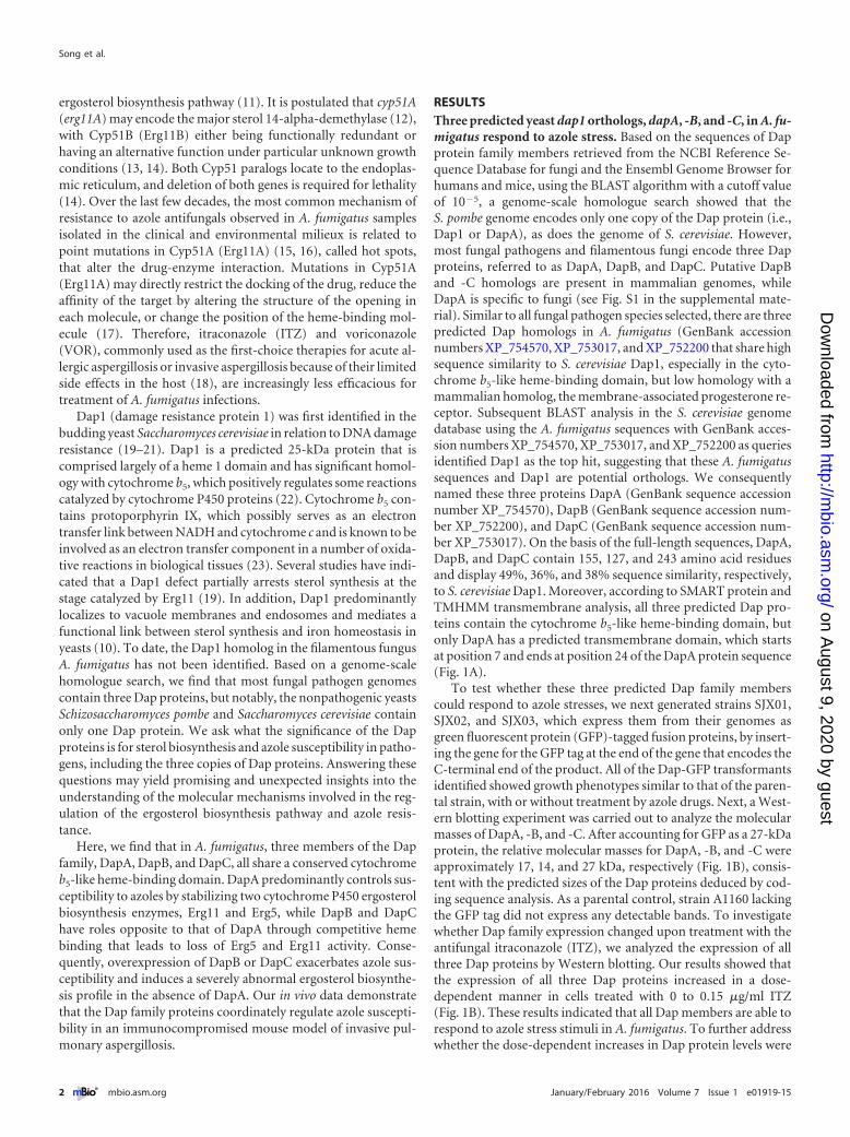
RESULTSThree predicted yeast dap1 orthologs, dapA, -B, and -C, in A. fu-migatus respond to azole stress. Based on the sequences of Dapprotein family members retrieved from the NCBI Reference Se-quence Database for fungi and the Ensembl Genome Browser forhumans and mice, using the BLAST algorithm with a cutoff valueof 10�5, a genome-scale homologue search showed that theS. pombe genome encodes only one copy of the Dap protein (i.e.,Dap1 or DapA), as does the genome of S. cerevisiae. However,most fungal pathogens and filamentous fungi encode three Dapproteins, referred to as DapA, DapB, and DapC. Putative DapBand -C homologs are present in mammalian genomes, whileDapA is specific to fungi (see Fig. S1 in the supplemental mate-rial). Similar to all fungal pathogen species selected, there are threepredicted Dap homologs in A. fumigatus (GenBank accessionnumbers XP_754570, XP_753017, and XP_752200 that share highsequence similarity to S. cerevisiae Dap1, especially in the cyto-chrome b5-like heme-binding domain, but low homology with amammalian homolog, the membrane-associated progesterone re-ceptor. Subsequent BLAST analysis in the S. cerevisiae genomedatabase using the A. fumigatus sequences with GenBank acces-sion numbers XP_754570, XP_753017, and XP_752200 as queriesidentified Dap1 as the top hit, suggesting that these A. fumigatussequences and Dap1 are potential orthologs. We consequentlynamed these three proteins DapA (GenBank sequence accessionnumber XP_754570), DapB (GenBank sequence accession num-ber XP_752200), and DapC (GenBank sequence accession num-ber XP_753017). On the basis of the full-length sequences, DapA,DapB, and DapC contain 155, 127, and 243 amino acid residuesand display 49%, 36%, and 38% sequence similarity, respectively,to S. cerevisiae Dap1. Moreover, according to SMART protein andTMHMM transmembrane analysis, all three predicted Dap pro-teins contain the cytochrome b5-like heme-binding domain, butonly DapA has a predicted transmembrane domain, which startsat position 7 and ends at position 24 of the DapA protein sequence(Fig. 1A).
To test whether these three predicted Dap family memberscould respond to azole stresses, we next generated strains SJX01,SJX02, and SJX03, which express them from their genomes asgreen fluorescent protein (GFP)-tagged fusion proteins, by insert-ing the gene for the GFP tag at the end of the gene that encodes theC-terminal end of the product. All of the Dap-GFP transformantsidentified showed growth phenotypes similar to that of the paren-tal strain, with or without treatment by azole drugs. Next, a West-ern blotting experiment was carried out to analyze the molecularmasses of DapA, -B, and -C. After accounting for GFP as a 27-kDaprotein, the relative molecular masses for DapA, -B, and -C wereapproximately 17, 14, and 27 kDa, respectively (Fig. 1B), consis-tent with the predicted sizes of the Dap proteins deduced by cod-ing sequence analysis. As a parental control, strain A1160 lackingthe GFP tag did not express any detectable bands. To investigatewhether Dap family expression changed upon treatment with theantifungal itraconazole (ITZ), we analyzed the expression of allthree Dap proteins by Western blotting. Our results showed thatthe expression of all three Dap proteins increased in a dose-dependent manner in cells treated with 0 to 0.15 �g/ml ITZ(Fig. 1B). These results indicated that all Dap members are able torespond to azole stress stimuli in A. fumigatus. To further addresswhether the dose-dependent increases in Dap protein levels were
mediated by increases in transcription or in protein stability, wecarried out quantitative reverse transcription (qRT)-PCR to testthe mRNA abundance in SJX01, SJX02, and SJX03 after exposureto azole treatment. The mRNA levels detected showed that thenative promoters of all three of the dap genes are able to sense andrespond to azole stress stimuli to some extent (see Fig. S2A in thesupplemental material). To further test whether the response toazole stress stimuli is related to the promoters of the three dapgenes, we generated strains SJX04, SJX05, and SJX06, expressingthe C-terminally GFP-tagged Dap proteins under the control ofthe constitutive promoter gpdA, originally obtained from Asper-gillus nidulans. Different from strains in which the Dap proteinswere controlled under the native promoter, no significant changesin Dap protein expression under the control of the gpdA promoterwere observed (see Fig. S2B), suggesting that only the dap nativepromoters are able to sense and respond to azole stress stimuli.
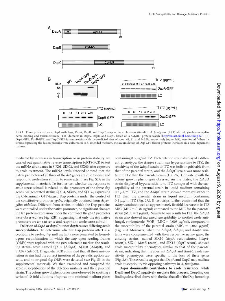
Deletion of dapA or dapC but not dapB causes differing azolesusceptibilities. To determine whether Dap proteins affect sus-ceptibility to azoles, dap null mutants were generated by homol-ogous recombination in which the dap open reading frames(ORFs) were replaced with the pyr4 selectable marker; the result-ing strains were named SJX07 (�dapA), SJX08 (�dapB), andSJX09 (�dapC). Diagnostic PCR confirmed that all three dap de-letion strains had the correct insertion of the pyr4 disruption cas-sette, and no original dap ORFs were detected (see Fig. S3 in thesupplemental material). We then examined and compared theazole susceptibilities of the deletion mutants and their parentalstrain. The colony growth phenotypes were observed by spotting aseries of 10-fold dilutions of spores onto minimal medium plates
containing 0.3 �g/ml ITZ. Each deletion strain displayed a differ-ent phenotype: the �dapA strain was hypersensitive to ITZ, thesensitivity of the �dapB strain to ITZ was indistinguishable fromthat of the parental strain, and the �dapC strain was more resis-tant to ITZ than the parental strain (Fig. 2A). Consistent with thecolony growth phenotypes observed on the plates, the �dapAstrain displayed hypersensitivity to ITZ compared with the sus-ceptibility of the parental strain in liquid medium containing0.2 �g/ml ITZ, and the �dapC strain showed more resistance toITZ than the parental strain in liquid medium containing0.4 �g/ml ITZ (Fig. 2A). E-test strips further confirmed that the�dapA strain showed an approximately fivefold decrease in its ITZMIC (MIC � 0.38 �g/ml) compared to the MIC for the parentalstrain (MIC � 2 �g/ml). Similar to our results for ITZ, the �dapAstrain also showed increased susceptibility to another azole anti-fungal, voriconazole (VOR) (MIC � 0.008 �g/ml), compared tothe susceptibility of the parental strain (MIC � 0.064 �g/ml)(Fig. 2B). Moreover, when the �dapA, �dapB, and �dapC mu-tants were complemented with their respective native gene, theensuing strains, named SJX10 (dapA reconstituted [dapA-recon]), SJX11 (dapB-recon), and SJX12 (dapC-recon), showedazole susceptibility phenotypes similar to that of the parentalstrain, indicating that the aberrant �dapA and �dapC azole sen-sitivity phenotypes were specific to the loss of these genes(Fig. 2A). These results suggest that DapA and DapC may mediateazole susceptibility via opposing pathways in A. fumigatus.
DapA dominantly contributes to azole resistance, whileDapB and DapC negatively mediate this process. Coupling ourfindings described above with the fact that all of the Dap homologs
FIG 1 Three predicted yeast Dap1 orthologs, DapA, DapB, and DapC, respond to azole stress stimuli in A. fumigatus. (A) Predicted cytochrome b5-likeheme-binding and transmembrane (TM) domains in DapA, DapB, and DapC, based on a SMART protein search (http://smart.embl-heidelberg.de/). (B)DapA-GFP, DapB-GFP, and DapC-GFP fusion proteins with the predicted sizes of about 44, 41, and 54 kDa, respectively (upper left), were found. When thestrains expressing the fusion proteins were cultured in ITZ-amended medium, the accumulation of Dap-GFP fusion proteins increased in a dose-dependentmanner.
Azole Susceptibility and Damage Resistance Proteins
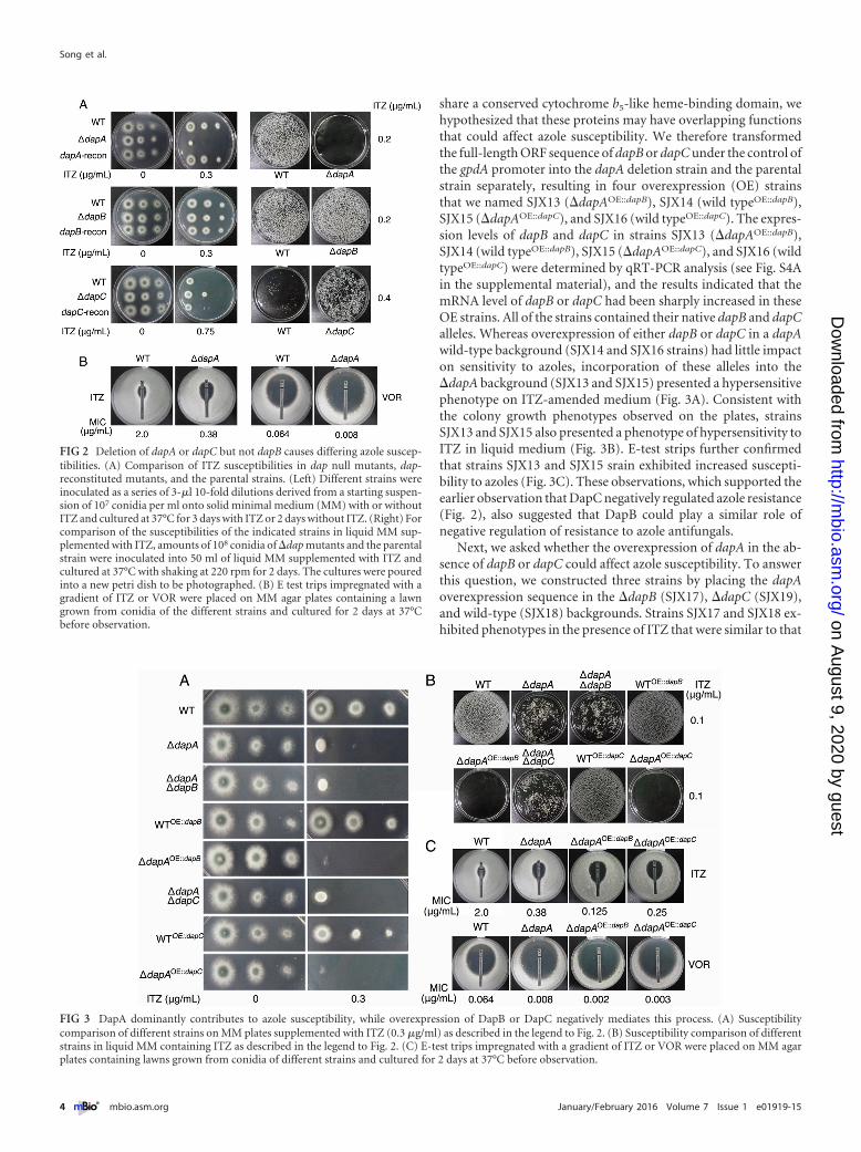
share a conserved cytochrome b5-like heme-binding domain, wehypothesized that these proteins may have overlapping functionsthat could affect azole susceptibility. We therefore transformedthe full-length ORF sequence of dapB or dapC under the control ofthe gpdA promoter into the dapA deletion strain and the parentalstrain separately, resulting in four overexpression (OE) strainsthat we named SJX13 (�dapAOE::dapB), SJX14 (wild typeOE::dapB),SJX15 (�dapAOE::dapC), and SJX16 (wild typeOE::dapC). The expres-sion levels of dapB and dapC in strains SJX13 (�dapAOE::dapB),SJX14 (wild typeOE::dapB), SJX15 (�dapAOE::dapC), and SJX16 (wildtypeOE::dapC) were determined by qRT-PCR analysis (see Fig. S4Ain the supplemental material), and the results indicated that themRNA level of dapB or dapC had been sharply increased in theseOE strains. All of the strains contained their native dapB and dapCalleles. Whereas overexpression of either dapB or dapC in a dapAwild-type background (SJX14 and SJX16 strains) had little impacton sensitivity to azoles, incorporation of these alleles into the�dapA background (SJX13 and SJX15) presented a hypersensitivephenotype on ITZ-amended medium (Fig. 3A). Consistent withthe colony growth phenotypes observed on the plates, strainsSJX13 and SJX15 also presented a phenotype of hypersensitivity toITZ in liquid medium (Fig. 3B). E-test strips further confirmedthat strains SJX13 and SJX15 srain exhibited increased suscepti-bility to azoles (Fig. 3C). These observations, which supported theearlier observation that DapC negatively regulated azole resistance(Fig. 2), also suggested that DapB could play a similar role ofnegative regulation of resistance to azole antifungals.
Next, we asked whether the overexpression of dapA in the ab-sence of dapB or dapC could affect azole susceptibility. To answerthis question, we constructed three strains by placing the dapAoverexpression sequence in the �dapB (SJX17), �dapC (SJX19),and wild-type (SJX18) backgrounds. Strains SJX17 and SJX18 ex-hibited phenotypes in the presence of ITZ that were similar to that
FIG 2 Deletion of dapA or dapC but not dapB causes differing azole suscep-tibilities. (A) Comparison of ITZ susceptibilities in dap null mutants, dap-reconstituted mutants, and the parental strains. (Left) Different strains wereinoculated as a series of 3-�l 10-fold dilutions derived from a starting suspen-sion of 107 conidia per ml onto solid minimal medium (MM) with or withoutITZ and cultured at 37°C for 3 days with ITZ or 2 days without ITZ. (Right) Forcomparison of the susceptibilities of the indicated strains in liquid MM sup-plemented with ITZ, amounts of 108 conidia of �dap mutants and the parentalstrain were inoculated into 50 ml of liquid MM supplemented with ITZ andcultured at 37°C with shaking at 220 rpm for 2 days. The cultures were pouredinto a new petri dish to be photographed. (B) E test trips impregnated with agradient of ITZ or VOR were placed on MM agar plates containing a lawngrown from conidia of the different strains and cultured for 2 days at 37°Cbefore observation.
FIG 3 DapA dominantly contributes to azole susceptibility, while overexpression of DapB or DapC negatively mediates this process. (A) Susceptibilitycomparison of different strains on MM plates supplemented with ITZ (0.3 �g/ml) as described in the legend to Fig. 2. (B) Susceptibility comparison of differentstrains in liquid MM containing ITZ as described in the legend to Fig. 2. (C) E-test trips impregnated with a gradient of ITZ or VOR were placed on MM agarplates containing lawns grown from conidia of different strains and cultured for 2 days at 37°C before observation.
of the �dapB deletion strain, which was equivalent to the wild-type phenotype (Fig. 2). In contrast, the overexpression of dapAcould partly rescue the resistance phenotype of the dapC deletionstrain (see Fig. S4B and C in the supplemental material). Thesefindings further strengthen the evidence for opposing activities ofDapA and DapC.
To further examine and compare the azole susceptibilities ofthe dap family, we next constructed �dapA �dapB and �dapA�dapC double deletion mutants through the deletion of dapB ordapC in the �dapA strain, yielding two strains named SJX20(�dapA �dapB) and SJX21 (�dapA �dapC). Strains SJX20 andSJX21 showed susceptibility profiles similar to that of the �dapAsingle mutant (Fig. 3A). These findings indicate that the gain ofazole resistance associated with a single dapC deletion required anactive DapA protein.
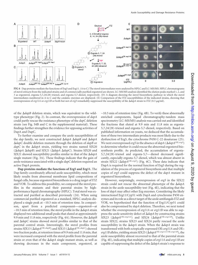
Dap proteins mediate the functions of Erg5 and Erg11. TheDap family coordinately affected azole susceptibility, which mostlikely results from abnormal membrane lipid compositions offungal cells, because ergosterol biosynthesis is a drug target of ITZand VOR. To address this possibility, we compared the sterol pro-files in the mutants and their parental strains by high-performance liquid chromatography (HPLC). Total sterol was ex-tracted and purified as described previously (24). When usingcommercial purified ergosterol as a standard, HPLC analysis dis-played a single peak at ~10.5 min of retention time. In compari-son, apart from a predicted component— ergosterol—at~10.5 min of retention time, the sterol profile in the �dapA straindisplayed two additional small peaks that eluted at approximately9.9 min and 11.8 min, respectively (Fig. 4A). However, the �dapBand �dapC strains showed sterol profiles similar to that of theparental control strain. Interestingly, the sterol profiles fromstrains SJX13 (�dapAOE::dapB) and SJX15 (�dapAOE::dapC) showedtwo fraction peaks, at retention times of 9.9 min and 11.8 min, thatwere increased compared with the sterol profile from the parentalstrain or even that of the �dapA single mutant strain, as well asshowing decreases in the main component, ergosterol, at
~10.5 min of retention time (Fig. 4B). To verify these abnormallyenriched components, liquid chromatography-tandem massspectrometry (LC-MS/MS) analysis was carried out and identifiedthe fractions that eluted at 9.9 min and 11.8 min as ergosta-5,7,24(28)-trienol and ergosta-5,7-dienol, respectively. Based onpublished information on yeasts, we deduced that the accumula-tion of these two intermediate products was most likely due to thedysfunction of Erg5, the cytochrome P450 C-22 desaturase (25).We next overexpressed erg5 in the absence of dapA (�dapAOE::erg5)to determine whether it could rescue the abnormal ergosterol bio-synthesis profile. As predicted, the accumulation of ergosta-5,7,24(28)-trienol and ergosta-5,7— dienol decreased signifi-cantly, especially ergosta-5,7-dienol, which was almost absent instrain SJX22 (�dapAOE::erg5) (Fig. 4C). These data indicate thatDapA is required for the normal function of Erg5 during the reg-ulation of the process of ergosterol biosynthesis and that multiplecopies of erg5 could suppress the defect of the dapA mutant inergosterol biosynthesis.
However, surprisingly, overexpression of erg5 in the SJX22strain could not rescue the abnormal phenotype of the �dapAstrain in the azole susceptibility test (Fig. 4E), indicating that theloss of dapA may affect other Erg enzymes. Considering the likelyinteractionof Erg11/Cyp51 with DapA and cytochrome P450 en-zymes and its role as a direct target of the azole antifungals ITZ andVOR, we hypothesized that the function of Erg11/Cyp51 couldalso be compromised by dapA deletion. Therefore, we next testedwhether the overexpression of erg11A or erg11B was able to sup-press the azole sensitivity defect of �dapA by constructing strainsSJX23 (�dapAOE::erg11A) and SJX24 (�dapAOE::erg11B). Unlikestrain SJX22, strains SJX23 and SJX24 partly rescued the azolesusceptibility in the �dapA strain. When the �dapA strain wastransformed with both ectopically expressed OE::erg11A and OE::erg11B alleles, yielding strain SJX25 (�dapAOE::erg11A OE::erg11B), theazole susceptibility almost recovered to that of the parental strain(Fig. 4E), indicating that multiple copies of erg11A and erg11B arecapable of suppressing the defect of the �dapA strain’s response to
FIG 4 Dap proteins mediate the functions of Erg5 and Erg11. (A to C) The sterol intermediates were analyzed by HPLC and LC-MS/MS. HPLC chromatogramsof sterol extracts from the indicated strains and of commercially purified ergosterol are shown. LC-MS/MS analysis identified the elution peaks marked 1, 2, and3 as ergosterol, ergosta-5,7,24(28)-trienol, and ergosta-5,7-dienol, respectively. (D) A diagram showing the sterol biosynthetic pathway in which the sterolintermediates numbered in A to C and the catalatic enzymes are displayed. (E) Comparison of the ITZ susceptibilities of the indicated strains, showing thatoverexpression of erg11A or erg11B or both but not of erg5 remarkably suppressed the susceptibility of the �dapA strain to ITZ (0.3 �g/ml).
Azole Susceptibility and Damage Resistance Proteins
azole antifungals. In comparison, overexpression of erg11A orerg11B in the parental wild-type background strain was unable tosignificantly increase azole resistance under the conditions tested(see Fig. S5A in the supplemental material). These data suggestthat DapA contributes to ergosterol synthesis and, hence, de-creased azole susceptibility by promoting Erg5 and Erg11 func-tions.
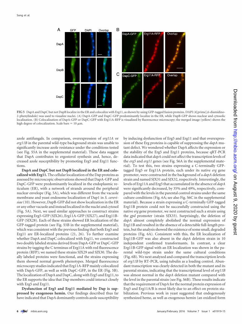
DapA and DapC but not DapB localized in the ER and colo-calized with Erg11. The cellular localization of the Dap proteins asassessed by microscopic observations showed that DapA-GFP andDapC-GFP were predominantly localized in the endoplasmic re-ticulum (ER), with a network of strands around the peripheralnuclear envelope (Fig. 5A), which was different from the vacuolemembrane and yeast endosome localization of Dap1 in S. cerevi-siae (10). However, DapB-GFP did not show localization in the ERor any other vacuole and instead localized in the nuclei and cytosol(Fig. 5A). Next, we used similar approaches to construct strainsexpressing Erg5-GFP (SJX26), Erg11A-GFP (SJX27), and Erg11B-GFP (SJX28). Each of these strains showed ER localization of theGFP-tagged protein (see Fig. S5B in the supplemental material),which was consistent with the previous finding that both Erg5 andErg11 are ER-localized proteins (21, 26). To further examinewhether DapA and DapC colocalized with Erg11, we constructedtwo doubly labeled strains derived from DapA-GFP or DapC-GFPstrains by tagging the C terminus of Erg11A with red fluorescenceprotein (RFP); we named these strains SJX29 and SJX30. The du-ally labeled proteins were functional, and the strains expressingthem showed normal growth phenotypes. Merged fluorescencemicroscopy studies indicated that Erg11A-RFP mainly colocalizedwith DapA-GFP, as well as with DapC-GFP, in the ER (Fig. 5B).The localization of DapA and DapC, along with Erg5 and Erg11, tothe ER supports the idea that Dap members could interact closelywith Erg5 and Erg11.
Dysfunction of Erg5 and Erg11 mediated by Dap is sup-pressed by exogenous hemin. Our findings described thus farhave indicated that DapA dominantly controls azole susceptibility
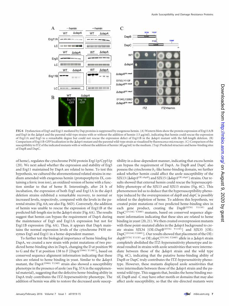
by inducing dysfunction of Erg5 and Erg11 and that overexpres-sion of these Erg proteins is capable of suppressing the dapA mu-tant defect. We wondered whether DapA affects the expression orthe stability of the Erg5 and Erg11 proteins, because qRT-PCRdata indicated that dapA could not affect the transcription levels ofthe erg5 and erg11 genes (see Fig. S6A in the supplemental mate-rial). To test this, two strains expressing a C-terminally GFP-tagged Erg5 or Erg11A protein, each under its native erg genepromoter, were constructed in the background of a dapA deletionstrain and named SJX31 and SJX32, respectively. Interestingly, thelevels of Erg11A and Erg5 that accumulated in the absence of dapAwere significantly decreased, by 35% and 49%, respectively, com-pared with the levels in the parental control strains under the sameculture conditions (Fig. 6A; see also Fig. S6C in the supplementalmaterial). Because a strain expressing a C-terminally GFP-taggedErg11B protein could not be successfully constructed using thenative erg gene promoter, we next constructed such a strain usingthe gpd promoter (strain SJX33). Surprisingly, the deletion ofdapA almost completely abolished the normal expression ofErg11B and resulted in the absence of a detectable full-length pro-tein, but the analysis showed the existence of some small, degradedproteins (Fig. 6A). Consistent with this, the ER localization ofErg11B-GFP was also absent in the dapA deletion strain in 10independent confirmed transformants. In contrast, a clearErg11B-GFP signal with an ER localization was shown in the pa-rental wild-type strain under the same cultural conditions(Fig. 6B). We next analyzed and compared the transcription levelsof erg11B by RT-PCR, using tubulin as a loading control. Abun-dant transcription was clearly detected in both the mutant and itsparental strains, indicating that the transcriptional level of erg11Bwas almost normal in the dapA deletion mutant compared withthe level in the parental strain (see Fig. S6B). These results indicatethat the requirement of DapA for the normal protein expression ofErg5 and Erg11A/B is most likely due to an effect on protein sta-bilization. Previous work in yeast suggested that endogenouslysynthesized heme, as well as exogenous hemin (an oxidized form
FIG 5 DapA and DapC but not DapB localize in the ER and colocalize with Erg11, as shown by using GFP-tagged fusion proteins. DAPI (4[prime],6-diamidino-2-phenylindole) was used to visualize nuclei. (A) DapA-GFP and DapC-GFP predominantly localize in the ER, while DapB-GFP shows nuclear and cytosoliclocalization. (B) Colocalization of DapA-GFP or DapC-GFP with Erg11A-RFP is visualized by fluorescence microscopy; the merged image (yellow) shows thehigh degree of colocalization. Scale bars � 10 �m.
of heme), regulates the cytochrome P450 protein Erg11p/Cyp51p(20). We next asked whether the expression and stability of Erg5and Erg11 maintained by DapA are related to heme. To test thishypothesis, we cultured the aforementioned related strains in me-dium amended with exogenous hemin (protoporphyrin IX, con-taining a ferric iron ion), an oxidized version of heme with a func-tion similar to that of heme B. Interestingly, after 24 h ofincubation, the expression of both Erg5 and Erg11A in the dapAdeletion strains exhibited a remarkable recovery, to normal orincreased levels, respectively, compared with the levels in the pa-rental strains (Fig. 6A; see also Fig. S6D). Conversely, the additionof hemin was unable to rescue the expression of Erg11B at thepredicted full-length size in the �dapA strain (Fig. 6A). The resultssuggest that hemin can bypass the requirement of DapA duringthe maintenance of Erg5 and Erg11A expression but not forErg11B expression (Fig. 6A). Thus, it appears that DapA main-tains the normal expression levels of the cytochrome P450 en-zymes Erg5 and Erg11 in a heme-dependent manner.
To further test the biological importance of heme binding forDapA, we created a new strain with point mutations of two pre-dicted heme-binding sites in DapA, changing the D at position 90to G and the Y at position 138 to F [DapA(D90G Y138F)], based onconserved sequence alignment information indicating that thesesites are related to heme binding in yeast. Similar to the �dapAmutant, the DapA(D90G Y138F) strain also showed a susceptibilityphenotype in the presence of azole (see Fig. S7A in the supplemen-tal material), suggesting that the defective heme-binding ability inDapA truly contributes the ITZ-hypersensitivity phenotype. Theaddition of hemin was able to restore the decreased azole suscep-
tibility in a dose-dependent manner, indicating that excess hemincan bypass the requirement of DapA. As DapB and DapC alsopossess the cytochrome b5-like heme-binding domain, we furtherasked whether hemin could affect the azole susceptibility of theSJX13 (�dapAOE::dapB) and SJX15 (�dapAOE::dapC) strains. Our re-sults showed that external hemin could rescue the hypersuscepti-bility phenotype of the SJX13 and SJX15 strains (Fig. 6C). Thisphenomenon led us to deduce that the hypersusceptibility pheno-type induced by the overexpression of dapB and dapC is possiblyrelated to the depletion of heme. To address this hypothesis, wecreated point mutations of two predicted heme-binding sites ineach gene product, creating the DapB(D75G Y115F) andDapC(D134G Y208F) mutants, based on conserved sequence align-ment information indicating that these sites are related to hemebinding in yeast (20, 21). We then created overexpression mutantswith these point-mutated alleles in the �dapA background to cre-ate strains SJX34 [OE::DapB(D75G Y115F)] and SJX35 [OE::DapC(D134G Y208F)]. Our results showed that placement of the OE::dapB(D75G Y115F) or OE::dapC(D134G Y208F) allele in a �dapA straincompletely abolished the ITZ-hypersensitivity phenotype and in-stead resulted in strains with azole sensitivities that were interme-diate between those of the �dapA strain and the wild type(Fig. 6C), indicating that the putative heme-binding ability ofDapB or DapC truly contributes the ITZ-hypersensitivity pheno-type. However, these mutants displayed azole sensitivities thatwere intermediate between those of the �dapA strain and the pa-rental wild type. This suggests that, besides the heme binding mo-tif, DapB and -C may have other motifs or domains that may alsoaffect azole susceptibility, so that the site-directed mutants were
FIG 6 Dysfunction of Erg5 and Erg11 mediated by Dap proteins is suppressed by exogenous hemin. (A) Western blots show the protein expression of Erg11A/Band Erg5 in the �dapA and the parental wild-type strains with or without the addition of hemin (13 �g/ml), indicating that hemin could rescue the expressionof Erg11A and Erg5 to a remarkable degree but could not rescue the expression defect of Erg11B in the �dapA mutant with the full-length deletion. (B)Comparison of Erg11B-GFP localization in the �dapA mutant and the parental wild-type strain as visualized by fluorescence microscopy. (C) Comparison of thesusceptibility to ITZ of the indicated mutants with or without the addition of hemin (48 �g/ml) to the medium. (Top) Predicted structure and heme-binding sitesof DapB and DapC.
Azole Susceptibility and Damage Resistance Proteins
unable to show exactly the same phenotypes as strains with dele-tions of the respective full-length proteins.
Next, we investigated and compared the dose-dependent im-pacts of hemin addition on the azole sensitivities of the �dapA,�dapAOE::dapB, and �dapAOE::dapC strains. Our results showed thatmore hemin is required to rescue the azole susceptibility pheno-type in strains SJX13 (�dapAOE::dapB) and SJX15 (�dapAOE::dapC),especially SJX13, than is required for rescue of the �dapA strain(see Fig. S7B in the supplemental material). These data suggestthat the ITZ-hypersensitivity phenotype induced by overexpres-sion of DapB or DapC is closely related to the depletion of hemin.Taken together, these results suggest that heme binding by DapBand DapC, in contrast to DapA heme binding, has a negative con-sequence for cytochrome P450 functionality, which likely explainsthe azole hypersensitivity of strains SJX13 (�dapAOE::dapB) andSJX15 (�dapAOE::dapC).
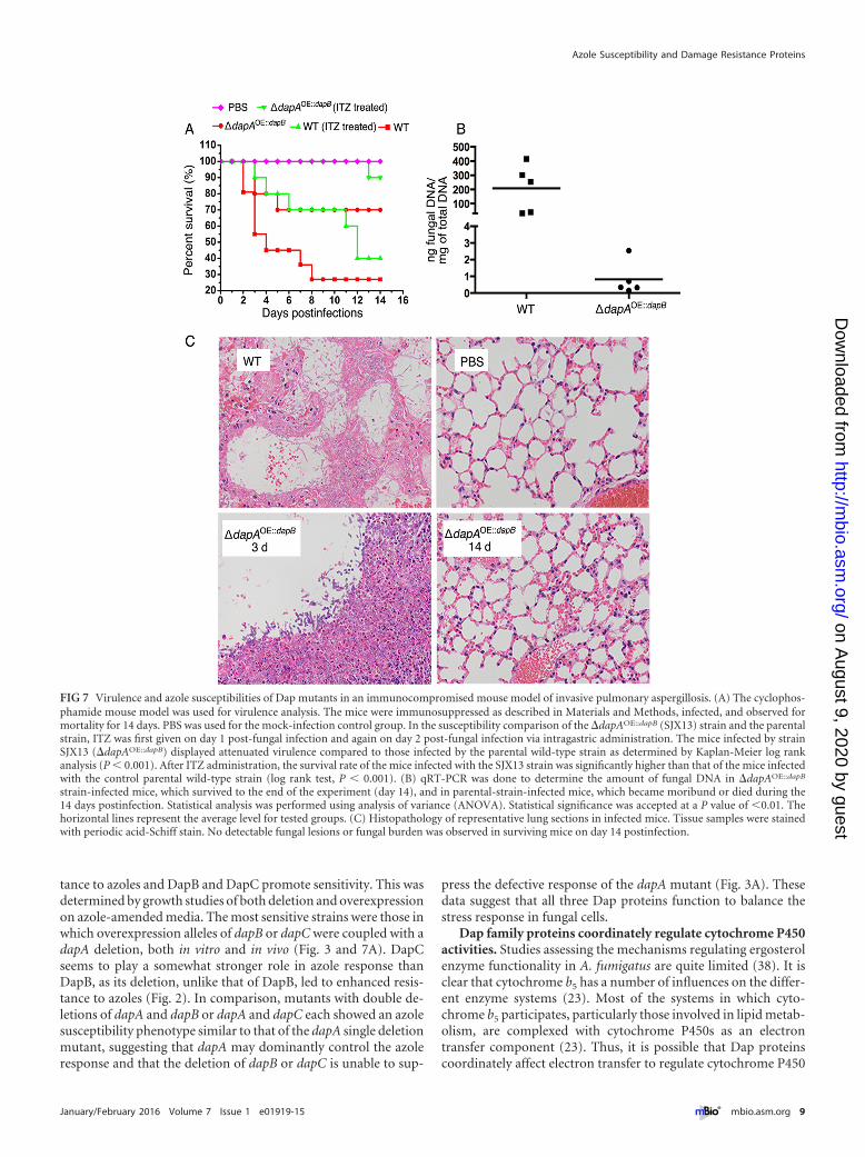
Virulence and the azole susceptibilities of Dap mutants in animmunocompromised mouse model of invasive pulmonaryaspergillosis. To assess any impact of Dap proteins in an in vivomurine model, we examined virulence and the efficacy of azoletherapy for selected Dap mutant strains, the �dapA and�dapAOE::dapB mutants, as representative of the azole-hypersensitivity phenotype. Conidia were inoculated into eachgroup of 10 immunocompromised mice, while the mice in thecontrol group were inoculated with saline solution. The groupinfected by the �dapA mutant showed a mortality rate similar tothose of the groups infected with the parental strain and the re-constituted strain (see Fig. S8A in the supplemental material). Thehistopathologic analysis of infected lungs from day 3 postinfectionwas consistent with these mortality data, showing no differencebetween strains (see Fig. S8B). However, the mice infected bystrain SJX13 (�dapAOE::dapB) displayed significantly attenuatedvirulence compared to those infected by the parental wild-typestrain according to Kaplan-Meier log rank analysis (P � 0.001)(Fig. 7A), suggesting that dapB overexpression contributes to thepathogenicity associated with the loss of dapA function. Throughhistopathologic analysis of infected lungs on day 3 postinfection,we found that mice infected with the parental strain A1160 hadexperienced extensive fungal growth. In contrast, only a smallamount of fungal growth was observed in the strain SJX13-infected mice at the same time point, with the tissue samples con-taining poorly germinated and ungerminated conidia (Fig. 7C).We next sacrificed all of the SJX13 strain-infected mice to analyzethe fungal burden measured by real-time PCR, which showed thatthe fungal load was significantly reduced (P � 0.01) in the strainSJX13-infected group compared to the load in the mice infectedwith the parental strain (Fig. 7B). Histopathological analyseson day 14 postinfection also confirmed that the fungal persistenceand inflammation in mice infected with the SJX13 strain was re-markably decreased compared to those in mice infected with theparental strain A1160 (Fig. 7C).
Next, we verified whether strain SJX13 also conferred hyper-sensitivity to azoles in an immunocompromised mouse model ofinvasive pulmonary aspergillosis. ITZ was given via intragastricadministration, first on day 1 post-fungal infection and again onday 2 post-fungal infection. Our results showed that ITZ admin-istration was capable of increasing the survival rate in mice com-pared with that of the group not given ITZ therapy, indicating thatITZ therapy in our immunocompromised mouse model was suc-cessful. Notably, in vivo data confirmed that strain SJX13 was eas-
ily treatable with azoles in an immunocompromised mouse modelof invasive pulmonary aspergillosis, resulting in survival of 9/10(90%) mice at 14 days postinfection (Fig. 7A). Kaplan-Meier logrank analysis showed there was a significant difference betweenthese two groups (P � 0.001), further suggesting that the survivalrate of mice infected with the SJX13 strain was significantly higherthan that of the mice infected with the control parental wild-typestrain after ITZ administration.
These in vivo data support a role for Dap proteins in mediatingazole susceptibility and virulence in A. fumigatus.
DISCUSSION
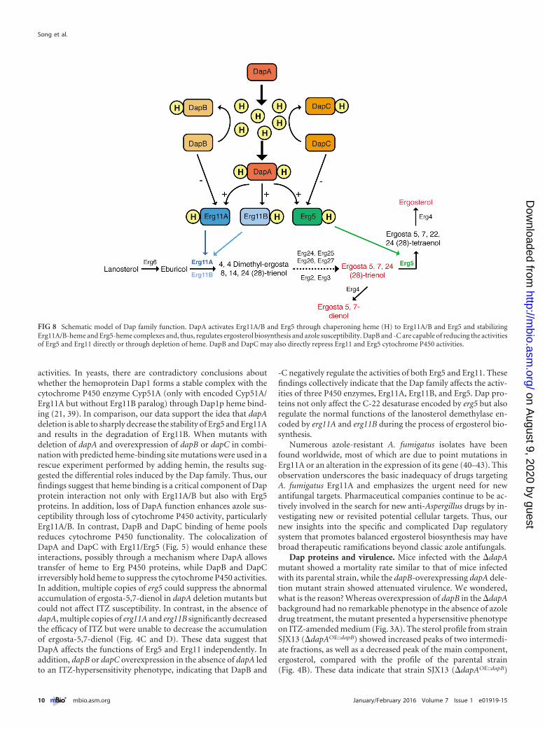
Dramatic increases in the incidence of aspergillosis caused pri-marily by A. fumigatus have occurred in recent years, primarilydue to an increase in the use of immunosuppressive therapies (27,28). Azole antifungals bind very weakly to mammalian cyto-chrome P450, which considerably reduces the toxicity of the drugin humans (29). Thus, to date, most antifungals are azole based,and azoles are currently the mainstay of antifungal treatment bothin agricultural and in clinical settings (30, 31). Many biochemistrystudies have verified that the free nitrogen of azole molecules isable to compete for oxygen with the catalytic heme of cytochromeP450 enzymes to inhibit the synthesis of ergosterol in fungal mem-branes (32). The lack of ergosterol alters membrane fluidity andsteric relationships for selected membrane-associated enzymesand results in the accumulation of phospholipids and unsaturatedfatty acids within the fungal cells (33–35). Although Aspergillusspecies are generally susceptible to azoles, intrinsic and acquiredresistance, particularly in the most common pathogenic species,A. fumigatus, is well documented and worrisomely on the rise (36,37). A thorough understanding of the drug resistance mechanismsof this fungus requires extensive exploration. In this study, weuncover a family of three conserved cytochrome b5-like heme-binding proteins, DapA, -B, and -C, that regulate the functionalityof three critical ergosterol-biosynthetic P450 enzymes, Erg5 andErg11A and -B. In contrast, overexpression of DapB and DapCcauses dysfunction of Erg5 and Erg11, resulting in abnormal ac-cumulation of sterol intermediates and further accentuating thesensitivity of �dapA mutants to azoles. Our studies reveal thatheme binding lies at the heart of this regulation, where DapApromotes Erg5 and Erg11 functionality, while DapB and DapCinhibit Erg5 and Erg11 activities. Thus, our finding provides thefirst characterization of the Dap family in A. fumigatus and alsogives insight to understand that the Dap family members act dif-ferently on sterol biosynthesis. A putative working model of thismechanism is presented in Fig. 8. The azole-sensitive phenotypesof Dap mutants revealed in this study yield possible avenues touncover alternative fungal-specific cellular targets.
Functions for multiple Dap homologs. Our genome-scale ho-mologue search shows that most fungal species encode three Dapproteins. Interestingly, the C. albicans genome contains four cop-ies (two DapC proteins), which is possibly due to a recent geneduplication event. The Kluyveromyces lactis and Ashbya gossypiigenomes possess two Dap protein copies (DapA and -C), while theS. cerevisiae, s. pombe, and Candida glabrata genomes only encodeDapA, which might be due to independent losses of DapB and -Cduring convergent evolution.
Although all three dap genes in A. fumigatus showed increasedexpression in an azole-dependent manner, the proteins exhibitedopposite roles in azole sensitivity, where DapA promotes resis-
tance to azoles and DapB and DapC promote sensitivity. This wasdetermined by growth studies of both deletion and overexpressionon azole-amended media. The most sensitive strains were those inwhich overexpression alleles of dapB or dapC were coupled with adapA deletion, both in vitro and in vivo (Fig. 3 and 7A). DapCseems to play a somewhat stronger role in azole response thanDapB, as its deletion, unlike that of DapB, led to enhanced resis-tance to azoles (Fig. 2). In comparison, mutants with double de-letions of dapA and dapB or dapA and dapC each showed an azolesusceptibility phenotype similar to that of the dapA single deletionmutant, suggesting that dapA may dominantly control the azoleresponse and that the deletion of dapB or dapC is unable to sup-
press the defective response of the dapA mutant (Fig. 3A). Thesedata suggest that all three Dap proteins function to balance thestress response in fungal cells.
Dap family proteins coordinately regulate cytochrome P450activities. Studies assessing the mechanisms regulating ergosterolenzyme functionality in A. fumigatus are quite limited (38). It isclear that cytochrome b5 has a number of influences on the differ-ent enzyme systems (23). Most of the systems in which cyto-chrome b5 participates, particularly those involved in lipid metab-olism, are complexed with cytochrome P450s as an electrontransfer component (23). Thus, it is possible that Dap proteinscoordinately affect electron transfer to regulate cytochrome P450
FIG 7 Virulence and azole susceptibilities of Dap mutants in an immunocompromised mouse model of invasive pulmonary aspergillosis. (A) The cyclophos-phamide mouse model was used for virulence analysis. The mice were immunosuppressed as described in Materials and Methods, infected, and observed formortality for 14 days. PBS was used for the mock-infection control group. In the susceptibility comparison of the �dapAOE::dapB (SJX13) strain and the parentalstrain, ITZ was first given on day 1 post-fungal infection and again on day 2 post-fungal infection via intragastric administration. The mice infected by strainSJX13 (�dapAOE::dapB) displayed attenuated virulence compared to those infected by the parental wild-type strain as determined by Kaplan-Meier log rankanalysis (P � 0.001). After ITZ administration, the survival rate of the mice infected with the SJX13 strain was significantly higher than that of the mice infectedwith the control parental wild-type strain (log rank test, P � 0.001). (B) qRT-PCR was done to determine the amount of fungal DNA in �dapAOE::dapB
strain-infected mice, which survived to the end of the experiment (day 14), and in parental-strain-infected mice, which became moribund or died during the14 days postinfection. Statistical analysis was performed using analysis of variance (ANOVA). Statistical significance was accepted at a P value of �0.01. Thehorizontal lines represent the average level for tested groups. (C) Histopathology of representative lung sections in infected mice. Tissue samples were stainedwith periodic acid-Schiff stain. No detectable fungal lesions or fungal burden was observed in surviving mice on day 14 postinfection.
Azole Susceptibility and Damage Resistance Proteins
activities. In yeasts, there are contradictory conclusions aboutwhether the hemoprotein Dap1 forms a stable complex with thecytochrome P450 enzyme Cyp51A (only with encoded Cyp51A/Erg11A but without Erg11B paralog) through Dap1p heme bind-ing (21, 39). In comparison, our data support the idea that dapAdeletion is able to sharply decrease the stability of Erg5 and Erg11Aand results in the degradation of Erg11B. When mutants withdeletion of dapA and overexpression of dapB or dapC in combi-nation with predicted heme-binding site mutations were used in arescue experiment performed by adding hemin, the results sug-gested the differential roles induced by the Dap family. Thus, ourfindings suggest that heme binding is a critical component of Dapprotein interaction not only with Erg11A/B but also with Erg5proteins. In addition, loss of DapA function enhances azole sus-ceptibility through loss of cytochrome P450 activity, particularlyErg11A/B. In contrast, DapB and DapC binding of heme poolsreduces cytochrome P450 functionality. The colocalization ofDapA and DapC with Erg11/Erg5 (Fig. 5) would enhance theseinteractions, possibly through a mechanism where DapA allowstransfer of heme to Erg P450 proteins, while DapB and DapCirreversibly hold heme to suppress the cytochrome P450 activities.In addition, multiple copies of erg5 could suppress the abnormalaccumulation of ergosta-5,7-dienol in dapA deletion mutants butcould not affect ITZ susceptibility. In contrast, in the absence ofdapA, multiple copies of erg11A and erg11B significantly decreasedthe efficacy of ITZ but were unable to decrease the accumulationof ergosta-5,7-dienol (Fig. 4C and D). These data suggest thatDapA affects the functions of Erg5 and Erg11 independently. Inaddition, dapB or dapC overexpression in the absence of dapA ledto an ITZ-hypersensitivity phenotype, indicating that DapB and
-C negatively regulate the activities of both Erg5 and Erg11. Thesefindings collectively indicate that the Dap family affects the activ-ities of three P450 enzymes, Erg11A, Erg11B, and Erg5. Dap pro-teins not only affect the C-22 desaturase encoded by erg5 but alsoregulate the normal functions of the lanosterol demethylase en-coded by erg11A and erg11B during the process of ergosterol bio-synthesis.
Numerous azole-resistant A. fumigatus isolates have beenfound worldwide, most of which are due to point mutations inErg11A or an alteration in the expression of its gene (40–43). Thisobservation underscores the basic inadequacy of drugs targetingA. fumigatus Erg11A and emphasizes the urgent need for newantifungal targets. Pharmaceutical companies continue to be ac-tively involved in the search for new anti-Aspergillus drugs by in-vestigating new or revisited potential cellular targets. Thus, ournew insights into the specific and complicated Dap regulatorysystem that promotes balanced ergosterol biosynthesis may havebroad therapeutic ramifications beyond classic azole antifungals.
Dap proteins and virulence. Mice infected with the �dapAmutant showed a mortality rate similar to that of mice infectedwith its parental strain, while the dapB-overexpressing dapA dele-tion mutant strain showed attenuated virulence. We wondered,what is the reason? Whereas overexpression of dapB in the �dapAbackground had no remarkable phenotype in the absence of azoledrug treatment, the mutant presented a hypersensitive phenotypeon ITZ-amended medium (Fig. 3A). The sterol profile from strainSJX13 (�dapAOE::dapB) showed increased peaks of two intermedi-ate fractions, as well as a decreased peak of the main component,ergosterol, compared with the profile of the parental strain(Fig. 4B). These data indicate that strain SJX13 (�dapAOE::dapB)
FIG 8 Schematic model of Dap family function. DapA activates Erg11A/B and Erg5 through chaperoning heme (H) to Erg11A/B and Erg5 and stabilizingErg11A/B-heme and Erg5-heme complexes and, thus, regulates ergosterol biosynthesis and azole susceptibility. DapB and -C are capable of reducing the activitiesof Erg5 and Erg11 directly or through depletion of heme. DapB and DapC may also directly repress Erg11 and Erg5 cytochrome P450 activities.
has an abnormal or defective ergosterol biosynthesis pathway.Previous studies have verified that there might be a tight link be-tween ergosterol biosynthesis and iron acquisition ability, which isa major factor in host defense against the pathogen (44–46). Totest whether the dapB overexpression in the dapA deletionmutant could result in a defect in iron acquisition ability, the�dapAOE::dapB mutant was inoculated onto minimal medium. Itappeared to have robust hyphal growth and conidiation eventhough it probably displayed a slightly smaller colony size than theparental strain. In comparison, when inoculated onto minimalmedium containing the iron chelator bathophenanthroline disul-fonate (BPS; 200 �M), the mutant displayed very severe growthdefects compared to the growth of the parental strain (see Fig. S8Cin the supplemental material), indicating that the �dapAOE::dapB
mutant had an impaired iron acquisition ability, especially underthe low-iron cultural condition, while there was no detectabledifference between the �dapA mutant and its parental strain un-der the iron-limited cultural condition. These data also suggest thepossibility that the main reason for the attenuated virulence in themouse model is the damaged iron acquisition in the �dapAOE::dapB
mutant. It seems probable that deprivation of hemin by overex-pression of DapB leads to dysfunction of cellular mechanismsother than DapA function. Moreover, we found that inactivatedDapA combined with activated DapB yields an A. fumigatus mu-tant that is easily treatable with azoles in an immunocompromisedmouse model of invasive pulmonary aspergillosis.
MATERIALS AND METHODSAdditional details on the materials and methods used can be found inText S1 in the supplemental material.
Strains, media, and culture conditions. The A. fumigatus strains usedin this study are listed in Table S1 in the supplemental material. Mutantstrains were constructed as described in detail in Text S1.
Antifungal susceptibility testing. Susceptibility testing of all dap mu-tants and the parental strains against the antifungal drugs ITZ and VORwas performed using solid and liquid media. MIC values are based onE-test strip assays (47).
Microscopy and live-cell imaging. Fluorescence localization and im-age processing for fungi expressing DapA-GFP, DapB-GFP, and DapC-GFP and Erg11-RFP were performed as described in reference 48.
Western blotting. The expression levels of GFP and RFP fusion pro-teins were determined by probing Western blots with the respective anti-body (Roche) and developing them by enhanced chemiluminescence(ECL; Amersham) as described previously (49). Signal intensity was cal-culated by using a gel imaging system, and statistical analysis was per-formed using analysis of variance (ANOVA). Statistical significance isaccepted at a P value of �0.05.
Total sterol extraction and HPLC-MS analysis. The sterols extractedfrom A. fumigatus strains were analyzed using HPLC (Agilent Technolo-gies) and detected at 280 nm on a AQ C18 column (250 mm by 4.6 mmwith a 5-�m particle size). HPLC-MS/MS was carried out using a Fouriertransform ion cyclotron resonance (FT-ICR) mass spectrometer with areverse-phase C18 column (AQ C18 column, 4.6 mm by 250 mm with a5-�m particle size).
RNA preparation. Total RNA was isolated using TRIzol (catalognumber 15596-025; Invitrogen), following the manufacturer’s instruc-tions.
Murine virulence and the azole susceptibility assays. The immuno-compromised mouse model for invasive pulmonary aspergillosis was de-scribed previously (50). Briefly, white female ICR mice (6 to 8 weeks old,22 to 25 g) were given intraperitoneal injections of cyclophosphamide(150 mg/kg of body weight) on days 3 and 1 relative to infection and asubcutaneous injection of hydrocortisone acetate (40 mg/kg of body
weight) on day 1. Statistical analysis of survival was performed usingKaplan-Meier log rank analysis. Statistical significance is accepted at aP value of �0.01. All animal experiments in this study were performedaccording to the Guide for the Care and Use of Laboratory Animals of theU.S. National Institutes of Health (51). The animal experimental protocolwas approved by the Animal Care and Use Committee of Nanjing NormalUniversity, China (permit no. 2090658) according to the governmentalguidelines for animal care.
SUPPLEMENTAL MATERIALSupplemental material for this article may be found at http://mbio.asm.org/lookup/suppl/doi:10.1128/mBio.01919-15/-/DCSupplemental.
This work was financially supported by grants from the National NaturalScience Foundation of China (NSFC81330035) to L.L., the Special Fundfor the Doctoral Program of Higher Education of China (no.20123207110012) to L.L., Innovation Project for College Graduates ofJiangsu Province (grant no. CXZZ13_0415), Excellent Doctoral Disserta-tion Special Fund of Nanjing Normal University (grant no.1812000002140) to J.S., the Priority Academic Program Development(PAPD) of Jiangsu Higher Education Institutions, and the NIH R01Al065728-01 to N.P.K.
A. fumigatus strain Af1160 was obtained from FGSC (http://www.fg-sc.net).
FUNDING INFORMATIONThe Special Fund for the Doctoral Program of Higher Education of Chinaprovided funding to Ling Lu under grant number No. 20123207110012.Innovation Project for College Graduates of Jiangsu Province providedfunding to Jinxing Song under grant number No. CXZZ13_0415. Excel-lent Doctoral Dissertation Special Fund of Nanjing Normal Universityprovided funding to Jinxing Song under grant number No.1812000002140.
This work was funded by National Natural Science Foundation of China(NSFC) under grant NSFC81330035 and by NIH R01 Al065728-01 toN.P.K. The funders had no role in study design, data collection and inter-pretation, or the decision to submit the work for publication.
REFERENCES1. Caspeta L, Chen Y, Ghiaci P, Feizi A, Buskov S, Hallström BM,
2. Sáenz JP, Sezgin E, Schwille P, Simons K. 2012. Functional convergenceof hopanoids and sterols in membrane ordering. Proc Natl Acad Sci U S A109:14236 –14240. http://dx.doi.org/10.1073/pnas.1212141109.
3. Hodzic A, Rappolt M, Amenitsch H, Laggner P, Pabst G. 2008. Differ-ential modulation of membrane structure and fluctuations by plant sterolsand cholesterol. Biophys J 94:3935–3944. http://dx.doi.org/10.1529/biophysj.107.123224.
4. Dupont S, Beney L, Ferreira T, Gervais P. 2011. Nature of sterols affectsplasma membrane behavior and yeast survival during dehydration.Biochim Biophys Acta 1808:1520 –1528. http://dx.doi.org/10.1016/j.bbamem.2010.11.012.
RD, Ntambi JM, Nett JE, Mitchell AP, Andes DR. 2014. Novel entries ina fungal biofilm matrix encyclopedia. mBio 5(4):e01333-14. http://dx.doi.org/10.1128/mBio.01333-14.
6. Mor V, Rella A, Farnoud AM, Singh A, Munshi M, Bryan A, Naseem S,Konopka JB, Ojima I, Bullesbach E, Ashbaugh A, Linke MJ, Cushion M,Collins M, Ananthula HK, Sallans L, Desai PB, Wiederhold NP, Fo-thergill AW, Kirkpatrick WR, Patterson T, Wong LH, Sinha S, GiaeverG, Nislow C, Flaherty P, Pan XW, Cesar GV, Tavares PD, Frases S,Miranda K, Rodrigues ML, Luberto C, Nimrichter L, Del Poeta M.2015. Identification of a new class of antifungals targeting the synthesis offungal sphingolipids. mBio 6:e00647-15. http://dx.doi.org/10.1128/mBio.00647-15.
7. Matyash V, Entchev EV, Mende F, Wilsch-Bräuninger M, Thiele C,Schmidt AW, Knölker HJ, Ward S, Kurzchalia TV. 2004. Sterol-derivedhormone(s) controls entry into diapause in Caenorhabditis elegans byconsecutive activation of DAF-12 and DAF-16. PLoS Biol 2:e280. http://dx.doi.org/10.1371/journal.pbio.0020280.
8. Yoshida Y, Aoyama Y. 1987. Interaction of azole antifungal agents withcytochrome P-45014DM purified from Saccharomyces cerevisiae micro-somes. Biochem Pharmacol 36:229 –235. http://dx.doi.org/10.1016/0006-2952(87)90694-0.
9. Hannemann F, Bichet A, Ewen KM, Bernhardt R. 2007. CytochromeP450 systems— biological variations of electron transport chains.Biochim Biophys Acta 1770:330 –344. http://dx.doi.org/10.1016/j.bbagen.2006.07.017.
10. Craven RJ, Mallory JC, Hand RA. 2007. Regulation of iron homeostasismediated by the heme-binding protein Dap1 (damage resistance protein1) via the p450 protein Erg11/Cyp51. J Biol Chem 282:36543–36551.http://dx.doi.org/10.1074/jbc.M706770200.
11. Mellado E, Diaz-Guerra TM, Cuenca-Estrella M, Rodriguez-Tudela JL.2001. Identification of two different 14-alpha sterol demethylase-relatedgenes (cyp51A and cyp51B) in Aspergillus fumigatus and other Aspergil-lus species. J Clin Microbiol 39:2431–2438. http://dx.doi.org/10.1128/JCM.39.7.2431-2438.2001.
12. Mellado E, Garcia-Effron G, Buitrago MJ, Alcazar-Fuoli L, Cuenca-Estrella M, Rodriguez-Tudela JL. 2005. Targeted gene disruption of the14-alpha sterol demethylase (cyp51A) in Aspergillus fumigatus and its rolein azole drug susceptibility. Antimicrob Agents Chemother 49:2536 –2538. http://dx.doi.org/10.1128/AAC.49.6.2536-2538.2005.
13. Warrilow AG, Melo N, Martel CM, Parker JE, Nes WD, Kelly SL, KellyDE. 2010. Expression, purification, and characterization of Aspergillusfumigatus sterol 14-alpha demethylase (CYP51) isoenzymes A and B. An-timicrob Agents Chemother 54:4225– 4234. http://dx.doi.org/10.1128/AAC.00316-10.
14. Hu W, Sillaots S, Lemieux S, Davison J, Kauffman S, Breton A, LinteauA, Xin C, Bowman J, Becker J, Jiang B, Roemer T. 2007. Essential geneidentification and drug target prioritization in Aspergillus fumigatus.PLoS Pathog 3:e24. http://dx.doi.org/10.1371/journal.ppat.0030024.
15. Snelders E, Camps SM, Karawajczyk A, Rijs AJ, Zoll J, Verweij PE,Melchers WJ. 2015. Genotype–phenotype complexity of the TR 46/Y121F/T289A cyp51A azole resistance mechanism in Aspergillus fumiga-tus. Fungal Genet Biol 82:129 –135. http://dx.doi.org/10.1016/j.fgb.2015.06.001.
17. Gollapudy R, Ajmani S, Kulkarni SA. 2004. Modeling and interactions ofAspergillus fumigatus lanosterol 14-alpha demethylase “A” with azole an-tifungals. Bioorg Med Chem 12:2937–2950. http://dx.doi.org/10.1016/j.bmc.2004.03.034.
18. Onyewu C, Blankenship JR, Del Poeta M, Heitman J. 2003. Ergosterolbiosynthesis inhibitors become fungicidal when combined with calcineu-rin inhibitors against Candida albicans, Candida glabrata, and Candidakrusei. Antimicrob Agents Chemother 47:956 –964. http://dx.doi.org/10.1128/AAC.47.3.956-964.2003.
19. Hand RA, Jia N, Bard M, Craven RJ. 2003. Saccharomyces cerevisiaeDap1p, a novel DNA damage response protein related to the mammalianmembrane-associated progesterone receptor. Eukaryot Cell 2:306 –317.http://dx.doi.org/10.1128/EC.2.2.306-317.2003.
20. Mallory JC, Crudden G, Johnson BL, Mo C, Pierson CA, Bard M,Craven RJ. 2005. Dap1p, a heme-binding protein that regulates the cyto-
chrome P450 protein Erg11p/Cyp51p in Saccharomyces cerevisiae. MolCell Biol 25:1669 –1679. http://dx.doi.org/10.1128/MCB.25.5.1669-1679.2005.
21. Hughes AL, Powell DW, Bard M, Eckstein J, Barbuch R, Link AJ,Espenshade PJ. 2007. Dap1/PGRMC1 binds and regulates cytochromeP450 enzymes. Cell Metab 5:143–149. http://dx.doi.org/10.1016/j.cmet.2006.12.009.
22. Meyer C, Schmid R, Scriba PC, Wehling M. 1996. Purification andpartial sequencing of high-affinity progesterone-binding site(s) from por-cine liver membranes. Eur J Biochem 239:726 –731. http://dx.doi.org/10.1111/j.1432-1033.1996.0726u.x.
23. Schenkman JB, Jansson I. 2003. The many roles of cytochrome b(5).Pharmacol Ther 97:139 –152. http://dx.doi.org/10.1016/S0163-7258(02)00327-3.
24. Blosser SJ, Cramer RA. 2012. SREBP-dependent triazole susceptibility inAspergillus fumigatus is mediated through direct transcriptional regula-tion of erg11A (cyp51A). Antimicrob Agents Chemother 56:248 –257.http://dx.doi.org/10.1128/AAC.05027-11.
25. Sun X, Wang W, Wang K, Yu X, Liu J, Zhou F, Xie B, Li S. 2013. SterolC-22 desaturase ERG5 mediates the sensitivity to antifungal azoles in Neu-rospora crassa and Fusarium verticillioides. Front Microbiol 4:127. http://dx.doi.org/10.3389/fmicb.2013.00127.
26. Homma K, Yoshida Y, Nakano A. 2000. Evidence for recycling of cyto-chrome P450 sterol 14-demethylase from the cis-Golgi compartment tothe endoplasmic reticulum (ER) upon saturation of the ER-retentionmechanism. J Biochem 127:747–754. http://dx.doi.org/10.1093/oxfordjournals.jbchem.a022666.
27. Wingard JR. 2005. The changing face of invasive fungal infections inhematopoietic cell transplant recipients. Curr Opin Oncol 17:89 –92.http://dx.doi.org/10.1097/01.cco.0000152975.65477.7c.
28. Ascioglu S, Rex JH, de Pauw B, Bennett JE, Bille J, Crokaert F, DenningDW, Donnelly JP, Edwards JE, Erjavec Z, Fiere D, Lortholary O,Maertens J, Meis JF, Patterson TF, Ritter J, Selleslag D, Shah PM,Stevens DA, Walsh TJ, Coopera IFI. 2002. Defining opportunistic inva-sive fungal infections in immunocompromised patients with cancer andhematopoietic stem cell transplants: an international consensus. Clin In-fect Dis 34:7–14. http://dx.doi.org/10.1086/323335.
29. Georgopapadakou NH. 1998. Antifungals: mechanism of action and re-sistance, established and novel drugs. Curr Opin Microbiol 1:547–557.http://dx.doi.org/10.1016/S1369-5274(98)80087-8.
30. Lass-Flörl C. 2011. Triazole antifungal agents in invasive fungalinfections: a comparative review. Drugs 71:2405–2419. http://dx.doi.org/10.2165/11596540-000000000-00000.
31. Verweij PE, Kema GH, Zwaan B, Melchers WJ. 2013. Triazole fungicidesand the selection of resistance to medical triazoles in the opportunisticmould Aspergillus fumigatus. Pest Manag Sci 69:165–170. http://dx.doi.org/10.1002/ps.3390.
32. White TC, Marr KA, Bowden RA. 1998. Clinical, cellular, and molecularfactors that contribute to antifungal drug resistance. Clin Microbiol Rev11:382– 402.
33. Mo C, Valachovic M, Randall SK, Nickels JT, Bard M. 2002. Protein-protein interactions among C-4 demethylation enzymes involved in yeaststerol biosynthesis. Proc Natl Acad Sci U S A 99:9739 –9744. http://dx.doi.org/10.1073/pnas.112202799.
34. Pichler H, Gaigg B, Hrastnik C, Achleitner G, Kohlwein SD, Zellnig G,Perktold A, Daum G. 2001. A subfraction of the yeast endoplasmic retic-ulum associates with the plasma membrane and has a high capacity tosynthesize lipids. Eur J Biochem 268:2351–2361. http://dx.doi.org/10.1046/j.1432-1327.2001.02116.x.
35. Laude AJ, Prior IA. 2004. Plasma membrane microdomains: organiza-tion, function and trafficking. Mol Membr Biol 21:193–205. http://dx.doi.org/10.1080/09687680410001700517.
36. Arendrup MC. 2014. Update on antifungal resistance in Aspergillus andCandida. Clin Microbiol Infect 20(Suppl 6):42– 48. http://dx.doi.org/10.1111/1469-0691.12513.
37. Chowdhary A, Sharma C, Hagen F, Meis JF. 2014. Exploring azoleantifungal drug resistance in Aspergillus fumigatus with special referenceto resistance mechanisms. Future Microbiol 9:697–711. http://dx.doi.org/10.2217/fmb.14.27.
38. Ferreira ME, Colombo AL, Paulsen I, Ren Q, Wortman J, Huang J,Goldman MH, Goldman GH. 2005. The ergosterol biosynthesis pathway,transporter genes, and azole resistance in Aspergillus fumigatus. Med My-col 43(Suppl 1):S313–S319.
39. Thompson AM, Reddi AR, Shi X, Goldbeck RA, Moënne-Loccoz P,Gibney BR, Holman TR. 2007. Measurement of the heme affinity foryeast Dap1p, and its importance in cellular function. Biochemistry 46:14629 –14637. http://dx.doi.org/10.1021/bi7013739.
40. Howard SJ, Webster I, Moore CB, Gardiner RE, Park S, Perlin DS,Denning DW. 2006. Multi-azole resistance in Aspergillus fumigatus. Int JAntimicrob Agents 28:450 – 453. http://dx.doi.org/10.1016/j.ijantimicag.2006.08.017.
41. Mann PA, Parmegiani RM, Wei S-Q, Mendrick CA, Li X, LoebenbergD, DiDomenico B, Hare RS, Walker SS, McNicholas PM. 2003. Muta-tions in Aspergillus fumigatus resulting in reduced susceptibility to po-saconazole appear to be restricted to a single amino acid in the cytochromeP450 14 alpha-demethylase. Antimicrob Agents Chemother 47:577–581.http://dx.doi.org/10.1128/AAC.47.2.577-581.2003.
42. Mellado E, Garcia-Effron G, Alcázar-Fuoli L, Melchers WJ, Verweij PE,Cuenca-Estrella M, Rodríguez-Tudela JL. 2007. A new Aspergillus fumigatusresistance mechanism conferring in vitro cross-resistance to azole antifungalsinvolves a combination of cyp51A alterations. Antimicrob Agents Chemother51:1897–1904. http://dx.doi.org/10.1128/AAC.01092-06.
43. Chen J, Li H, Li R, Bu D, Wan Z. 2005. Mutations in the cyp51A gene andsusceptibility to itraconazole in Aspergillus fumigatus serially isolatedfrom a patient with lung aspergilloma. J Antimicrob Chemother 55:31–37.http://dx.doi.org/10.1093/jac/dkh507.
44. Blatzer M, Barker BM, Willger SD, Beckmann N, Blosser SJ, CornishEJ, Mazurie A, Grahl N, Haas H, Cramer RA. 2011. SREBP coordinatesiron and ergosterol homeostasis to mediate triazole drug and hypoxiaresponses in the human fungal pathogen Aspergillus fumigatus. PLoSGenet 7:e1002374. http://dx.doi.org/10.1371/journal.pgen.1002374.
45. Marvig RL, Damkiær S, Khademi SM, Markussen TM, Molin S, JelsbakL. 2014. Within-host evolution of Pseudomonas aeruginosa reveals adap-tation toward iron acquisition from hemoglobin. mBio 5:e00966-14.http://dx.doi.org/10.1128/mBio.00966-14.
46. Hu G, Chen SH, Qiu J, Bennett JE, Myers TG, Williamson PR. 2014.Microevolution during serial mouse passage demonstrates FRE3 as a vir-ulence adaptation gene in Cryptococcus neoformans. mBio 5:e00941-14.http://dx.doi.org/10.1128/mBio.00941-14.
47. Liu F-F, Pu L, Zheng Q-Q, Zhang Y-W, Gao R-S, Xu X-S, Zhang S-Z,Lu L. 2015. Calcium signaling mediates antifungal activity of triazoledrugs in the aspergilli. Fungal Genet Biol 81:182–190. http://dx.doi.org/10.1016/j.fgb.2014.12.005.
48. Zhong G, Wei W, Guan Q, Ma Z, Wei H, Xu X, Zhang S, Lu L. 2012.Phosphoribosyl pyrophosphate synthetase, as a suppressor of the sepHmutation in Aspergillus nidulans, is required for the proper timing ofseptation. Mol Microbiol 86:894 –907. http://dx.doi.org/10.1111/mmi.12026.
49. Zhong GW, Jiang P, Qiao WR, Zhang YW, Wei WF, Lu L. 2014. Proteinphosphatase 2A (PP2A) regulatory subunits para and PabA orchestrateseptation and conidiation and are essential for PP2A activity in Aspergillusnidulans. Eukaryot Cell 13:1494 –1506. http://dx.doi.org/10.1128/EC.00201-14.
50. Jiang H, Shen Y, Liu W, Lu L. 2014. Deletion of the putative stretch-activated ion channel Mid1 is hypervirulent in Aspergillus fumigatus.Fungal Genet Biol 62:62–70. http://dx.doi.org/10.1016/j.fgb.2013.11.003.
51. National Research Council. 1996. Guide for the Care and Use of Labora-tory Animals. National Academies Press, Washington, DC.
Azole Susceptibility and Damage Resistance Proteins


![(ALDER(GREY, ALMOND, ASPERGILLUS FUMIGATUS, BAHIA … · 2019-11-06 · [ ]adult comprehensive allergy panel (alder(grey, almond, aspergillus fumigatus, bahia grass, bermuda grass,](https://static.documents.pub/doc/80x56/5fb12d57752ad135660a561b/aldergrey-almond-aspergillus-fumigatus-bahia-2019-11-06-adult-comprehensive.jpg)


