Gazi University, Faculty of Medicine, Department of Gastroenterology, 06520 Besevler, Ankara, TurkeyGazi University, Faculty of Medicine, Department of Pathology, 06520 Besevler, Ankara, Turkey
r t i c l e i n f o
rticle history:eceived 10 November 2013eceived in revised form 18 January 2014ccepted 1 April 2014
eywords:ilirubinhronic hepatitis Civer fibrosis
a b s t r a c t
We proposed to evaluate the association between serum indirect bilirubin levels and liver fibrosis inpatients with chronic hepatitis C (CHC) genotype 1b. Biopsy proven CHC genotype 1b patients’ demo-graphics, clinical and histopathological characteristics were evaluated. Logistic regression analysis wasdone to evaluate the clinical, laboratory and demographic features of the histologically proven liver fibro-sis in CHC patients. A total of 112 biopsy proven CHC genotype 1b patients were enrolled into the study.Liver fibrosis scores were measured by using Ishak fibrosis scores and were divided into two groups;fibrosis scores ≤2 were categorized as mild fibrosis, 82 patients (73.2%), whereas fibrosis scores >2 werecategorized as advanced fibrosis group, 30 patiens (26.8%). Patients with advanced fibrosis had lower
indirect bilirubin levels than the mild fibrosis group (0.28 ± 0.02 mg/dl vs. 0.44 ± 0.032 mg/dl, p < 0.001,respectively). Indirect bilirubin level was negatively correlated with advanced fibrosis scores (r = −0.416and p < 0.001). In multivariate logistic regression analysis, low indirect bilirubin level was an independentpredicting factor of advanced liver fibrosis (OR: 0.001, 95% CI: 0.0–0.005, p < 0.001). There is an inverserelationship between indirect bilirubin levels and advanced liver fibrosis caused by CHC genotype 1b.
Chronic hepatitis C (CHC) is usually an asymptomatic viral infec-ion that may progress to liver fibrosis, cirrhosis, hepatocellulararcinoma or liver failure. Fibrosis becomes obvious in the coursef years in many patients. Advanced fibrosis may progress to liverailure [1]. Evaluation of liver fibrosis caused by CHC is importantn the assessement of prognosis, selection of antiviral therapy andlinical course of the disease.
Liver biopsy is considered the gold standard method for thevaluation of liver fibrosis. Nevertheless, liver biopsy has vari-us disadvantages such as inter-observer variability and samplingrrors, invasiveness and complications such as bleeding [2]. Theost significant benefit of biopsy is the increased decision mak-
Please cite this article in press as: M. Cengiz, et al., The association bhepatitis C virus infection, Pathol. – Res. Pract (2014), http://dx.doi.or
ng ability of treatment and predicting the clinical course based onhe histopathological features. Expertise in liver biopsy is crucial toecide on judgments for antiviral treatment [3].
∗ Corresponding author at: Gazi University, Faculty of Medicine, Department ofastroenterology, Besevler/Cankaya, 06520 Ankara, Turkey. Tel.: +90 312 20227586;
It has been hypothesized that oxidative stress may play a rolein liver damage caused by CHC through various biologic path-ways [4,5]. Indirect bilirubin is a metabolic end product of hemebreakdown in the reticular-endothelial system [6], and recognizedas a powerful antioxidant cytoprotectant [7]. Bilirubin protectsagainst oxidative stress by inhibiting the action of NADPH oxidasethat increases superoxide production [8,9]. Moreover, bilirubincan easily clear up peroxyl radicals, a singlet oxygen, hydroxylradicals reactive nitrogen varieties [10,11], and minimize the alpha-tocopheroxyl radical that promotes recycling in association withvitamin E [12]. In inclusion, bilirubin may have anti-inflammatoryattribution, and work as the major antifibrogenic agent throughheme oxygenase-1 (HMOX1) [13]. Additionally, there is a pow-erful cytoprotective association with indirect bilirubin as seenin Gilbert’s syndrome. It simply shows that indirect hyper-bilirubinemia is related to reduced likelihood of coronary arterydisease, carotid stenosis and also cancer [11,14–17]. It is possiblethat indirect bilirubin may play a role in reducing the oxida-tive stress, inflammation and preventing the progression of liver
etween indirect bilirubin levels and liver fibrosis due to chronicg/10.1016/j.prp.2014.04.001
fibrosis.We proposed to evaluate the association between indirect
bilirubin levels and liver fibrosis in a well characterized group ofpatients with biopsy proven CHC genotype 1b.
The study was performed in Gazi University Hospital Depart-ent of Gastroenterology, a tertiary reference center (Ankara,
urkey), between October 2009 and May 2013. Biopsy proven CHCenotype 1b patients were enrolled in the study. Patients whoad consistently elevated serum transaminase levels for no lesshan six months and liver biopsy specimens compatible with CHC;s well as positive for both anti-HCV antibody and HCV RNA-ositive serum (at least >50 IU/ml) according to real time (RT) –CR (Cobas Amplicor CHC v2. 0, Roche Molecular Systems) werencluded. Patients who had autoimmune hepatitis, primary bil-ary cirrhosis, excessive alcohol consumption (alcohol intake above0 g/day for women and 20 g/day for men), presence of hepatitis Burface antigen, anti-human immunodeficiency virus antibody orepatocellular carcinoma, CHC other than genotype 1b, past his-ory of venous thrombosis, severe cardiac and/or renal diseases,se of immunosuppressive agents, any hepatotoxic medication andntioxidant treatment were excluded. Patients who had clinicalr imaging evidence of decompensated cirrhosis such as portalystemic encephalopathy, variceal bleeding or ascites were alsoxcluded from the study. Reticulocyte count and peripheral bloodlm examination were assessed in order to exclude the hemolysismong patients who had hyperbilurubinemia. Sustained virologi-al response (SVR) was defined as undetectable serum HCV RNA at4 weeks after the end of treatment.
aboratory tests
Venous blood samples were obtained on the morning of liveriopsy after an overnight fast of at least 10–12 h. HemoglobinHB), platelet (PLT), leukocyte and protrombin time were measuredy Beckman Coulter Gen-S automated analyzer (High Wycombe,K). The calculation of the international normalized ratio (INR)as made by dividing the prothrombin time of the blood sample
o the prothrombin time of normal blood sample. Serum alanineminotransferase (ALT), aspartate aminotransferase (AST), alkalinehosphatase (ALP), gamma glutamyltransferase (GGT), albumin,otal cholesterol, triglyceride, high density lipoprotein (HDL), lowensity lipoprotein (LDL), total bilirubin and direct bilirubin lev-ls were determined by automated techniques Cobas Integra 800oche Modular System (Indianapolis, US). Indirect bilirubin wasalculated as total bilirubin − conjugated bilirubin.
Demographical, clinical and laboratory data were collected andegistered in a database by an uninformed clinician to prevent bias.
istopathological evaluation of the liver
Percutaneous liver biopsies were performed by using disposableenghini (Hepafix 16 gauge; Braun Melsungen AG, Melsungen,ermany) type needles by an experienced clinician. The biopsypecimens of liver tissues more than 20 mm in length and includingt least 12 portal triads were fixed in formalin buffer, inserted inaraffin and stained with Hematoxylin and Eosin (H&E), Masson’soldner, Masson’s trichrome and reticulin.
Grading as well as staging scores were determined in accordanceith the approach of Ishak et al. [18]. The patients were divided into
wo groups depending on the presence of bridging fibrosis histolog-
Please cite this article in press as: M. Cengiz, et al., The association bhepatitis C virus infection, Pathol. – Res. Pract (2014), http://dx.doi.or
cally; fibrosis scores ≤2 were grouped as mild fibrosis group andbrosis scores ≥2 were grouped as advanced fibrosis group. Signif-
cant fibrosis was defined as Ishak score of 3 or more (presence ofridging fibrosis) [19]. All histopathological evaluations of the liver
PRESSand Practice xxx (2014) xxx–xxx
were carried out by the same experienced hepatopathologist in ablind manner.
Ethics
The study was in accordance with the ethical guidelines of theDeclaration of Helsinki and was approved by the local ethics com-mittee.
Statistical analysis
Statistical analysis was performed using the SPSS softwareversion 18 (SPSS Inc., Chicago, IL, USA) and MedCalc version12. Normally distributed continuous variables were expressedas mean ± standard deviation, whereas skewed distributed con-tinuous variables were expressed as median ± standard error;normality of variables distribution was investigated usingvisual (histogram, probability plots) and analytical methods(Kolmogorov–Smirnov/Shapiro–Wilk’s test). Categorical variableswere presented as frequencies and percentages. In order to assesscorrelations between bilirubin levels and histological features,Spearman’s correlation coefficients were used. The univariateanalysis to identify variables associated with liver fibrosis wereinvestigated using Chi-square, Fisher exact, Student’s t andMann–Whitney U tests where appropriate. In the multivariate anal-ysis, the possible factors identified with univariate analysis werefurther entered into the multivariate logistic regression analysisto determine independent predictors of liver fibrosis by avoidingthe multicollinearity. Hosmer–Lemeshow goodness of fit statisticswas used to evaluate the model fit. The diagnostic performance ofindirect bilirubin level was evaluated by using receiver operatingcharacteristic (ROC) curves and compared by using the area underthe ROC curve (AUROC). The sensitivity and specificity for variousvalues in the final model were calculated to determine the best cutoff points that would predict advanced liver fibrosis. A 5% type-Ierror level was used to infer statistical significance.
Results
A total of 112 biopsy proven CHC genotype 1b patients all ofwhom were received the same standard antiviral therapy proto-col (PEGIFN�2 + ribavirin) were enrolled in the study. Among thepatients 60.7% were female and the median age was 55. Indirecthyperbilirubinemia was present in 14.2% of the CHC cohort study.The median age of patients with indirect hyperbilirubinemia andwithout was 51 (20–74) vs. 55 (18–80), respectively. There was nostatistically significant difference between the ages of the studyparticipants. From the patients with indirect hyperbilirubinemia62% were female while 59.2% of patients without indirect hyper-bilirubinemia were female. There was no statistically significantdifference by gender.
According to the liver histopathological features, the mild fibro-sis group was consisted of 82 (73.2%) patients, while there were30 (26.8%) patients in the advanced fibrosis group. There were 44(39.3%) patients with fibrosis score 1, 38 (33.9%) patients with fibro-sis score 2, 16 (14.3%) patients with fibrosis score 3, 7 (6.3%) patientswith fibrosis score 4, 5 (4.5%) patients with fibrosis score 5 and 2(1.8%) patients with fibrosis score 6. In the advanced fibrosis group,the patients were older than the mild fibrosis group (59 ± 7.94)vs. (54 ± 12.69), respectively, p < 0.05. While SVR, indirect biliru-bin levels, direct bilirubin levels and platelet counts were lowerin the advanced fibrosis group, the levels of ALT, AST, ALP and
etween indirect bilirubin levels and liver fibrosis due to chronicg/10.1016/j.prp.2014.04.001
GGT were lower in the mild fibrosis group. There was a negativecorrelation between serum indirect bilirubin levels and necroin-flammatory activity (NIA) scores of patients (r = −0.204, p = 0.03).The median NIA scores of mild and advanced fibrosis groups were
M. Cengiz et al. / Pathology – Research and Practice xxx (2014) xxx–xxx 3
n leve
6caarl
TD
SfgiD
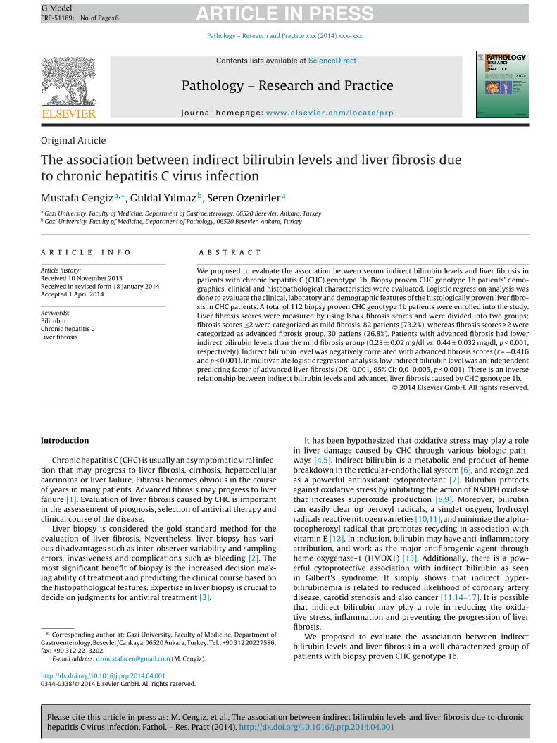
Fig. 1. The comparison of serum indirect bilirubi
± 0.23 vs. 7.5 ± 0.3, respectively, p ≤ 0.001. The demographics,linical and laboratory data of mild and advanced fibrosis groups
Please cite this article in press as: M. Cengiz, et al., The association bhepatitis C virus infection, Pathol. – Res. Pract (2014), http://dx.doi.or
re summarized in Table 1. The indirect bilirubin levels of mildnd advanced fibrosis groups were (0.44 ± 0.032) vs. (0.28 ± 0.02),espectively p < 0.001. The comparison of serum indirect bilirubinevels between the two groups is shown in Fig. 1.
able 1emographics and laboratory data of the fibrosis stage groups.
VR, sustained virologic response; NS, not significant; ALT, alanine aminotrans-erase; AST, aspartate aminotransferase; ALP, alkaline phosphatase; GGT, gammalutamyltransferase; LDL, low-density lipoprotein; NIA, necroinflammatory activ-ty.ata are presented as mean ± SD.a Variables are skewed distributed and expressed as median ± SE.* Mild fibrosis is defined according to Ishak staging score ≤2.
** Advanced fibrosis is defined according to Ishak staging score >2.
ls between mild and andvanced fibrosis groups.
Table 2 shows the analysis of the correlations between demo-graphics, laboratory tests and fibrosis stages of the groups.Advanced fibrosis correlated significantly with ALT, AST levels andINR. However fibrosis correlated positively with age and genderbut there was a significant negative correlation between indirectbilirubin levels and fibrosis.
ROC curve analysis for the identification of advanced fibrosisdemonstrated AUC: 0.771, 95% CI (0.682–0.845), sensitivity: 76.7%,specificity: 69.5% in the indirect bilirubin at the criterion: ≤0.32,p < 0.001. ROC curve of indirect bilirubin levels in the identificationof patients with advanced fibrosis is shown in Fig. 2.
Univariate analysis for the presence of advanced fibrosis indi-
etween indirect bilirubin levels and liver fibrosis due to chronicg/10.1016/j.prp.2014.04.001
cated that SVR, age, gender, platelet counts, ALT, AST and indirectbilirubin levels were statistically significant predictor factors. Theresults of univariate analysis are shown in Table 3.
Table 2The correlations between demographics, laboratory data and liver fibrosis.
I, 95% confidence interval; SVR, sustained virological response; NS, not signifi-ant; ALT, alanine aminotransferase; AST, aspartat aminotransferase; GGT, gammalutamyltransferase.
Those variables that were statistically significant in univariateogistic regression analysis were further evaluated in multivariate
Please cite this article in press as: M. Cengiz, et al., The association bhepatitis C virus infection, Pathol. – Res. Pract (2014), http://dx.doi.or
ogistic regression analysis, and indirect bilirubin continued to betatistically significant and an independent predictor of advancedbrosis (OR: 0.001, 95% CI: 0.000–0.005, p < 0.001), as seen inable 4.
able 4ultivariate analyses of fibrosis in patients with CHC genotype 1b.
The principal result of our study is the inverse relationshipbetween serum indirect bilirubin levels and the severity of liverfibrosis in CHC genotype 1b patients. Serum bilirubin levels werenegatively correlated with the progression of liver fibrosis. Wedemonstrated by logistic regression analysis that low indirectbilirubin level was independently associated with the predictionof advanced fibrosis in CHC genotype 1b patients. To our knowl-edge, this is the first study that shows a negative relationshipbetween indirect bilirubin levels and liver fibrosis in CHC genotype1b patients. It has been found that higher serum bilirubin levelswere significantly related with a lower disease progression in non-alcoholic fatty liver disease (NAFLD) patients in Korean population[20]. But it was not evaluated according to fibrotic stages of liver andthe disease progression. We also showed that lower indirect biliru-bin levels may predict high fibrotic stages in a well-characterizedpatient population of CHC.
In a large retrospective study it has been shown that the lowerlevels of indirect bilirubin were inversely correlated with fibrosisin biopsy proven NASH patients [21]. Indirect hyperbilirubinemiais known to be associated with lower prevalence of hypertension,lower risk of cardiac disease and atherosclerotic vascular disease[11,14,15,22]. It is possible that indirect hyperbilirubinemia mayprovide a protective effect against liver fibrosis but the underlyingmechanisms remain unclear. Recently Hjelkrem et al. [21] foundan association between the severity of liver injury due to NASHand indirect hyperbilirubinemia. Kumar et al. [23] as a result oftheir study found an association between histopathologically lessadvanced liver disease and indirect hyperbilirubinemia.
The relationship between serum indirect bilirubin levels andliver fibrosis in CHC patients is not well identified. The role of indi-rect bilirubin in inhibition of pathogenesis of liver fibrosis in CHC
etween indirect bilirubin levels and liver fibrosis due to chronicg/10.1016/j.prp.2014.04.001
depending on the effective antioxidant, anti-inflammatory and alsoanti-fibrogenic effects are still uncertain. The anti-inflammatoryactivity of bilirubin has been analyzed by Keshavan et al. [24]who discovered that bilirubin prevents lymphocyte migration and
educes the entire leukocyte numbers and inhibits eosinophil andymphocyte infiltration within murine lung parenchyma. Li et al.13] found the key hepatic antifibrogenic components attributedo bilirubin and its particular mediation involving HMOX1. Ele-ated serum indirect bilirubin levels and HMOX1 are related toeduction of breast cancer [25] and oral squamous malignancy [26].ubhanova et al. [27] in a recent study concluded an upregulation ofiliverdin reductase (BLVRA) in peripheral blood leukocytes (PBL)
n CHC. They also demonstrated that the lack of overexpressionf BLVRA was associated with non-responsiveness in CHC patientsho were on standard antiviral treatment.
Indirect bilirubin is known as a powerful endogenous antioxi-ant in various tissues and organs for so many years [12]. The actualntioxidant activity of bilirubin was initially recognized by Stockert al. in an in vitro test in which bilirubin was able to scavenge per-xyl radicals in liposomes much more effective than �-tocopherol,he best recognized chemical counteracting lipid peroxidation [10].esearches revealed that lipophilic bilirubin works more effec-ively than protecting lipids through oxidation when comparedith water-soluble anti-oxidants for example glutathione that pro-
ects protein from oxidation [28]. Indirect bilirubin is directlyssociated with the entire serum antioxidant capability in humaneings [29].
Recently Salomone et al. [30] showed that low indirect biliru-in level was an independent predictor of advanced inflammationnd fibrosis in European NASH patients and they indicated thatndirect bilirubin as an antioxidant; might have protective effectsgainst progression of liver injury. Also Kwak et al. [31] found annverse relationship between serum indirect bilirubin levels andAFLD independent of metabolic risk factors.
Our study has some limitations including the fact that our resultsay not be generalized to different populations and races. Genetic
nalysis defining elevated indirect bilirubin levels such as Gilbert’syndrome (UGT1A1 gene) [32] was not investigated. In the future,tudies defining whether there is an association between indirectilirubin due to the UGT1A1 gene expression and liver fibrosis and itsontribution to CHC should be designed. Because of the retrospec-ive design and the small number of patients; low indirect bilirubinevels cannot be considered as the only cause of liver fibrosis androspective randomized controlled clinical trials should be carriedut to verify these results.
In conclusion, our study showed that indirect bilirubin levelsre inversely associated with the severity of liver fibrosis in CHCenotype 1b patients. Because indirect bilirubin may be a pri-ary endogenous lipid antioxidant, our results may give a feasibleolecular system for the advanced association of liver fibrosis in
HC genotype 1b patients. In addition to this, the prediction of liverbrosis by indirect bilirubin levels in this population of patientsay lead to new evaluation and treatment modalities. Treatment
ptions increasing indirect bilirubin levels may be beneficial inatients at advanced fibrosis stages in CHC genotype 1b patients.arge population based randomized controlled clinical trials areeeded to confirm the association between serum indirect bilirubin
evels and liver fibrosis.
onflict of interest
There is no potential conflict of interest.
eferences
Please cite this article in press as: M. Cengiz, et al., The association bhepatitis C virus infection, Pathol. – Res. Pract (2014), http://dx.doi.or
[1] J.D. Scott, D.R. Gretch, Molecular diagnostics of hepatitis C virus infection: asystematic review, JAMA 297 (7) (2007) 724–732.
[2] S.M. Martinez, G. Crespo, M. Navasa, X. Forns, Noninvasive assessment of liverfibrosis, Hepatology 53 (1) (2011) 325–335.
[
PRESSand Practice xxx (2014) xxx–xxx 5
[3] G.M. Lauer, B.D. Walker, Hepatitis C virus infection, N. Engl. J. Med. 345 (1)(2001) 41–52.
[4] E. Peterhans, Reactive oxygen species and nitric oxide in viral diseases, Biol.Trace Elem. Res. 56 (1) (1997) 107–116.
[5] M. Okuda, K. Li, M.R. Beard, L.A. Showalter, F. Scholle, S.M. Lemon, S.A. Wein-man, Mitochondrial injury, oxidative stress, and antioxidant gene expressionare induced by hepatitis C virus core protein, Gastroenterology 122 (2) (2002)366–375.
[6] S.W. Ryter, J. Alam, A.M. Choi, Heme oxygenase-1/carbon monoxide: from basicscience to therapeutic applications, Physiol. Rev. 86 (2) (2006) 583–650.
[7] D.E. Baranano, M. Rao, C.D. Ferris, S.H. Snyder, Biliverdin reductase: amajor physiologic cytoprotectant, Proc. Natl. Acad. Sci. USA 99 (25) (2002)16093–16098.
[8] T. Inoguchi, P. Li, F. Umeda, H.Y. Yu, M. Kakimoto, M. Imamura, T. Aoki, T. Etoh,T. Hashimoto, M. Naruse, H. Sano, H. Utsumi, H. Nawata, High glucose level andfree fatty acid stimulate reactive oxygen species production through proteinkinase C-dependent activation of NAD (P) H oxidase in cultured vascular cells,Diabetes 49 (11) (2000) 1939–1945.
[9] S. Lanone, S. Bloc, R. Foresti, A. Almoki, C. Taielle, J. Callebert, M. Conti, D.Goven, M. Aubier, B. Dureuil, J. El-Benna, R. Motterlini, J. Boczkowski, Bilirubindecreases nos2 expression via inhibition of NAD (P) H oxidase: implicationsfor protection against endotoxic shock in rats, FASEB J. 19 (13) (2005) 1890–1892.
10] R. Stocker, Y. Yamamoto, A.F. McDonagh, A.N. Glazer, B.N. Ames, Bilirubin is anantioxidant of possible physiological importance, Science 235 (4792) (1987)1043–1046.
11] H.A. Schwertner, L. Vitek, Gilbert syndrome, UGT1A1*28 allele, and cardiovas-cular disease risk: possible protective effects and therapeutic applications ofbilirubin, Atherosclerosis 198 (1) (2008) 1–11.
12] R. Stocker, Antioxidant activities of bile pigments, Antioxid. Redox. Signal. 6 (5)(2004) 841–849.
13] L. Li, P. Grenard, J.T. Nhieu, B. Julien, A. Mallat, A. Habib, S. Lotersztain, Hemeoxygenase-1 is an antifibrogenic protein in human hepatic myofibroblasts,Gastroenterology 125 (2) (2003) 460–469.
14] L. Vitek, L. Novotny, M. Sperl, R. Holaj, J. Spacil, The inverse association ofelevated serum bilirubin levels with subclinical carotid atherosclerosis, Cere-brovasc. Dis. 21 (5–6) (2006) 408–414.
15] N. Ishizaka, Y. Ishizaka, E. Takahashi, M. Yamakado, H. Hashimoto, High serumbilirubin level is inversely associated with the presence of carotid plaque,Stroke 32 (2) (2001) 580–583.
16] A. Jiráskova, J. Novotny, L. Novotny, P. Vodicka, B. Pardini, A. Naccarati,H.A. Schwertner, J.A. Hubácek, L. Puncochárova, Z. Smerhovsky, L. Vítek,Association of serum bilirubin and promoter variations in HMOX1 andUGT1A1 genes with sporadic colorectal cancer, Int. J. Cancer 131 (7) (2012)1549–1555.
17] S.D. Zucker, P.S. Horn, K.E. Sherman, Serum bilirubin levels in the U.S. popula-tion: gender effect and inverse correlation with colorectal cancer, Hepatology40 (4) (2004) 827–835.
18] K. Ishak, A. Baptista, L. Bianchi, F. Callea, J. De Groote, F. Gudat, H. Denk, V.Desmet, G. Korb, R.N. MacSween, Histological grading and staging of chronichepatitis, J. Hepatol. 22 (6) (1995) 696–699.
19] C.T. Wai, J.K. Greenson, R.J. Fontana, J.D. Kalbfleisch, J.A. Marrero, H.S. Conjee-varam, A.S. Lok, A simple noninvasive index can predict both significant fibrosisand cirrhosis in patients with chronic hepatitis C, Hepatology 38 (2) (2003)518–526.
20] Y. Chang, S. Ryu, Y. Zhang, H.J. Son, J.Y. Kim, J. Cho, E. Gualler, A cohort study ofserum bilirubin levels and incident non-alcoholic fatty liver disease in middleaged Korean workers, PLoS ONE 7 (5) (2012) e37241.
21] M. Hjelkrem, A. Morales, C.D. Williams, S.A. Harrison, Unconjugated hyper-bilirubinemia is inversely associated with non-alcoholic steatohepatitis(NASH), Aliment. Pharmacol. Ther. 35 (12) (2012) 1416–1423.
22] L. Vítek, M. Jirsa, M. Brodanova, M. Kalab, Z. Marecek, V. Danziq, L. Novotny,P. Kotal, Gilbert syndrome and ischemic heart disease: a protective effect ofelevated bilirubin levels, Atherosclerosis 160 (2) (2002) 449–456.
23] R. Kumar, A. Rastogi, J.S. Maras, S.K. Sarin, Unconjugated hyperbilirubinemiain patients with non-alcoholic fatty liver disease: a favorable endogenousresponse, Clin. Biochem. 45 (3) (2012) 272–274.
25] S. Ching, D. Ingram, R. Hahnel, J. Beilby, E. Rossi, Serum levels of micronutrients,antioxidants and total antioxidant status predict risk of breast cancer in a casecontrol study, J. Nutr. 132 (2) (2002) 303–306.
26] K.W. Chang, T.C. Lee, W.I. Yeh, M.Y. Chung, C.J. Liu, l.Y. Chi, S.C. Lin, Polymor-phism in heme oxygenase-1 (HO-1) promoter is related to the risk of oralsquamous cell carcinoma occurring on male areca chewers, Br. J. Cancer 91(8) (2004) 1551–1555.
27] I. Subhanova, L. Muchova, M. Lenicek, H.J. Vreman, O. Luksan, K. Kubickova, M.Kreidlova, T. Zima, L. Vitek, P. Urbanek, Expression of biliverdin reductase A
etween indirect bilirubin levels and liver fibrosis due to chronicg/10.1016/j.prp.2014.04.001
in peripheral blood leukocytes is associated with treatment response in HCV-infected patients, PLoS ONE 8 (3) (2013) e57555.
28] T.W. Sedlak, M. Saleh, D.S. Hiqqinson, B.D. Paul, K.R. Juluri, S.H. Sneyder, Biliru-bin and glutathione have complementary antioxidant and cytoprotective roles,Proc. Natl. Acad. Sci. USA 106 (113) (2009) 5171–5176.
[Serum bilirubin levels are inversely associated with nonalcoholic fatty liverdisease, Clin. Mol. Hepatol. 18 (4) (2012) 383–390.
ARTICLERP-51189; No. of Pages 6
M. Cengiz et al. / Pathology – Res
29] Z. Yesilova, M. Serdar, C.N. Ercin, A. Gunay, G. Kilciler, A. Has imi, A. Uygun, I. Kurt,M.K. Erbil, K. Dagalp, Decreased oxidation susceptibility of plasma low densitylipoproteins in patients with Gilbert’s syndrome, J. Gastroenterol. Hepatol. 23
Please cite this article in press as: M. Cengiz, et al., The association bhepatitis C virus infection, Pathol. – Res. Pract (2014), http://dx.doi.or
(10) (2008) 1556–1560.30] F. Salomone, G. Li Volti, C. Rosso, G. Grosso, E. Buqianesi, Unconjugated bilirubin,
a potent endogenous antioxidant, is decreased in patients with non-alcoholicsteatohepatitis and advanced fibrosis, J. Gastroenterol. Hepatol. 28 (7) (2013)1202–1208.
[
PRESSand Practice xxx (2014) xxx–xxx
31] M.S. Kwak, D. Kim, G.E. Chung, S.J. Kang, M.J. Park, Y.J. Kim, J.H. Yoon, H.S. Lee,
etween indirect bilirubin levels and liver fibrosis due to chronicg/10.1016/j.prp.2014.04.001
32] Y.C. Lin, P.F. Chang, F.C. Hu, M.H. Chang, Y.H. Ni, Variants in the UGT1A1 gene andthe risk of pediatric nonalcoholic fatty liver disease, Pediatrics 124 (6) (2009)e1221–e1227.