138
The bacterial membrane insertase YidC: In vivo studies of substrate binding and membrane insertion Christian Daniel Klenner

The bacterial membrane insertase YidC: In vivo studies of substrate binding and membrane insertion Christian Daniel Klenner

The bacterial membrane insertase YidC: In vivo studies of substrate binding and membrane insertion
Dissertation zur Erlangung
des Doktorgrades der Naturwissenschaften
(Dr. rer. nat.)
Fakultät Naturwissenschaften
Universität Hohenheim
Institut für Mikrobiologie und Molekularbiologie
vorgelegt von
Christian Daniel Klenner
aus Ostfildern
2015
Dekan: Prof. Dr. Heinz Breer 1. berichtende Person: Prof. Dr. Andreas Kuhn 2. berichtende Person: Prof. Dr. Heinz Breer 3. Prüfer: Prof. Dr. Wolfgang R.L. Hanke Mündliche Prüfung am: 11. Februar 2016

To myself, for hanging in there

i
Contents List of publications included in this thesis ........................................................................................... iii
Abstract ............................................................................................................................................... iv
Abbreviations ....................................................................................................................................... v
Chapter 1 ................................................................................................................................................. 1
General Introduction ........................................................................................................................... 1
1.1 BIOLOGICAL MEMBRANES .......................................................................................... 1
1.1.1 FEATURES OF BIOLOGICAL MEMBRANES .................................................................... 1
1.1.2 LIPIDS AND MEMBRANE COMPOSITION OF BACTERIA AND EUKARYOTES ................ 2
1.2 MEMBRANE PROTEINS ............................................................................................... 4
1.2.1 PERIPHERAL MEMBRANE PROTEINS ........................................................................... 4
1.2.2 AMPHITROPIC PROTEINS ............................................................................................. 5
1.2.3 INTEGRAL MEMBRANE PROTEINS ............................................................................... 6
1.2.4 PROTEIN-LIPID INTERACTION ...................................................................................... 8
1.3 BIOGENESIS OF BACTERIAL α-HELICAL MEMBRANE PROTEINS ..................................... 9
1.3.1 PROTEIN TARGETING: HOW PROTEINS FIND THEIR FINAL DESTINATION................. 10
1.3.1.1 The signal sequence of secretory preproteins .......................................................... 10
1.3.1.2 The post-translational SecB-pathway........................................................................ 11
1.3.1.3 Twin-arginine translocation (Tat)-pathway: Targeting of folded proteins ................ 11
1.3.1.4 SRP-pathway: The main pathway for inner membrane proteins .............................. 11
1.3.1.5 Non-classical targeting pathways .............................................................................. 14
1.3.2 TOPOLOGY OF MEMBRANE PROTEINS ...................................................................... 15
1.3.2.1 Topogenic signals define topology of membrane proteins ....................................... 16
1.3.2.2 The universal positive-inside rule .............................................................................. 18
1.3.2.3 Topological determinants .......................................................................................... 19
1.3.3 INSERTION OF α-HELICAL MEMBRANE PROTEINS.................................................... 20
1.3.3.1 The Sec translocon: A protein-conducting channel for secretion and insertion ....... 20
1.3.3.2 Accessory components of the Sec translocon and their function ............................. 21
1.3.3.3 Structure of SecYEG ................................................................................................... 21
1.3.3.4 The Sec translocon in action: seemingly contrary functions ..................................... 23
1.3.3.5 Translocation of secretory proteins across the membrane ...................................... 24
1.3.3.6 Insertion of membrane proteins into the lipid bilayer via the Sec translocon ......... 24
1.3.4 FOLDING OF α-HELICAL MEMBRANE PROTEINS ....................................................... 26
1.3.4.1 The two-stage model for membrane protein folding ............................................... 26

ii
1.3.4.2 Early stages of membrane protein folding ................................................................ 26
1.3.4.3 Formation of an α-helical bundle .............................................................................. 27
1.3.4.4 Role of lipids in protein folding ................................................................................. 28
1.4 THE MEMBRANE INSERTASE YidC ............................................................................. 28
1.4.1 YidC/Oxa1/Alb3 PROTEIN FAMILY AND FUNCTIONAL CONSERVATION AMONG HOMOLOGS ............................................................................................................................ 29
1.4.1.1 Oxa proteins in the inner membrane of mitochondria ............................................. 30
1.4.1.2 Alb proteins in the thylakoid membrane of chloroplasts .......................................... 31
1.4.2 X-RAY STRUCTURE OF YidC2 FROM BACILLUS HALODURANS ................................... 31
1.4.3 THE MEMBRANE INSERTASE YidC FROM ESCHERICHIA COLI .................................... 35
1.4.3.1 Crystal structure of Escherichia coli YidC .................................................................. 37
1.4.3.2 YidC and the ribosome .............................................................................................. 41
1.4.3.3 Functionally important regions of Escherichia coli YidC ........................................... 41
1.4.3.4 Cellular response upon YidC depletion ..................................................................... 43
1.4.4 YidC MEDIATES MEMBRANE PROTEIN INSERTION AND ASSISTS IN FOLDING AND ASSEMBLY .............................................................................................................................. 44
1.4.4.1 Various pathways enable targeting to the YidC insertase ........................................ 46
1.4.4.2 Pf3 coat protein is a model substrate for YidC mediated insertion .......................... 47
1.4.4.3 YidC in action: Molecular mechanism of membrane insertion ................................. 48
1.4.4.4 YidC cooperates with the Sec translocon .................................................................. 51
1.4.4.5 YidC as a membrane embedded molecular chaperone ............................................ 53
1.4.4.6 YidC assists in the assembly of highly ordered membrane protein complexes ........ 54
Objectives of this thesis ..................................................................................................................... 55
Chapter 2 ............................................................................................................................................... 57
The Pf3 coat protein contacts TM1 and TM3 of YidC during membrane biogenesis ....................... 57
Chapter 3 ............................................................................................................................................... 71
Dynamic disulfide scanning of the membrane-inserting Pf3 coat protein reveals multiple YidC substrate contacts ............................................................................................................................. 71
Chapter 4 ............................................................................................................................................... 91
Summary ........................................................................................................................................... 91
Zusammenfassung ............................................................................................................................. 93
Concluding remarks and outlook ...................................................................................................... 95
Acknowledgements ........................................................................................................................... 99
Presentations at national and international conferences ............................................................... 101
References ....................................................................................................................................... 103
iii
List of publications included in this thesis I Christian Klenner, Jijun Yuan, Ross E. Dalbey and Andreas Kuhn (2008)
The Pf3 coat protein contacts TM1 and TM3 of YidC during membrane biogenesis.
FEBS Lett 582, 3967-72 II Christian Klenner and Andreas Kuhn (2012)
Dynamic disulfide scanning of the membrane-inserting Pf3 coat protein reveals multiple YidC substrate contacts. J Biol Chem 287, 3769-76
Other publications
III Lu Zhu, Christian Klenner, Andreas Kuhn and Ross E. Dalbey (2012) Both YidC and SecYEG are required for translocation of the periplasmic loops 1 and 2 of the multispanning membrane protein TatC. J Mol Biol 424, 354-67.

iv
Abstract
Membrane proteins play a key role in many cellular processes. As a prerequisite for
proper function, these proteins have to be inserted into biological membranes. The
insertion process involves highly conserved translocation machineries – the translocons.
In bacteria, YidC acts in cooperation with the Sec translocon, the main insertion site for
membrane proteins. In addition, YidC can function independently of the Sec translocon,
e.g. facilitating biogenesis of respiratory complexes and the F1F0-ATPase. This is most
likely a reason why YidC is an essential protein in Escherichia coli.
At the time this project was initiated high resolution 3D structures of YidC were limited
to the non-functional large periplasmic loop and it was largely unclear how YidC
substrates are inserted into the membrane. This thesis aims to identify YidC-substrate
contacts during membrane biogenesis and to investigate the molecular mechanism
underlying YidC mediated insertion.
For capturing protein-protein interactions we have established an in vivo cross-linking
assay using a set of single cysteine mutants of YidC and the inserting small phage protein
Pf3 coat representing a model substrate of the YidC insertion pathway. We found that
YidC contacts Pf3 coat protein with various regions of the conserved transmembrane
(TM) core domains, which had been shown to be critical for function. An expressed Pf3
mutant with a defect in membrane insertion was unable to contact TM residues of YidC
facing the periplasmic leaflet, whereas residues at the cytoplasmic leaflet were still
contacted. We therefore suggest that the YidC mediated insertion is a dynamic process
with early binding followed by the translocation and insertion of substrate proteins.
v
Abbreviations Adenosine triphosphate ATP Amino acids AAs Amino (terminus) N Alb Albino Alkaline phosphatase PhoA Angstrom Å Arabidopsis thaliana A. thaliana Bacillus halodurans B. halodurans Bacillus subtilis B. subtilis Blue native BN Cardiolipin CL Carboxy (terminus) C Cytochrome c oxidase Cox Cytoplasmic domain C Debye–Waller factor B-factor Deoxyribonucleic acid DNA Electron microscopy EM Endoplasmic reticulum ER Escherichia coli E. coli Fluorescence cross- FCCS correlation spectroscopy Green fluorescent protein GFP Gene product gp Guanosine triphosphate GTP Hemoglobin protease Hbp Inner membrane IM Inner membrane proteins IMPs Kilodalton kDa Lactose permease LacY Light-harvesting chlorophyll- LHCPs binding proteins Membrane protein MP Messenger ribonucleic mRNA acid Micrometer µm
Nanometer nm Nicotinamide adenine NADH dinucleotide (oxidized) Outer membrane OM Oxidase assembly 1 Oxa1 Oxidase assembly 2 Oxa2 Periplasmic domain P Phage shock protein A PspA Phosphatidylcholine PC Phoshatidylethanolamine PE Phosphatidylglycerol PG Phosphatidylinositol PI Polyacrylamide gel PAGE electrophoresis Proton motive force pmf Ribosomal ribonucleic acid rRNA Ribosome nascent chain RNC Saccharomyces cerevisiae S. cerevisiae Secretory Sec Signal recognition particle SRP Single-stranded ssDNA deoxyribonucleic acid Stimulated emission STED depletion Tail-anchored membrane TAMPs proteins Three-dimensional 3D Translocase of the inner TIM23 membrane Trigger factor TF Twin arginine Tat translocation Two-dimensional 2D Transmembrane TM

vi
Amino acids 3 - letters 1 - letter
Alanine Ala A
Asparagine Asn N
Arginine Arg R
Aspartic acid Asp D
Cysteine Cys C
Glutamic acid Glu E
Glutamine Gln Q
Glycine Gly G
Histidine His H
Isoleucine Ile I
Leucine Leu L
Lysine Lys K
Methionine Met M
Phenyalanine Phe F
Proline Pro P
Serine Ser S
Threonine Thr T
Tryptophan Trp W
Tyrosine Tyr Y
Valine Val V

vii
1
Chapter 1 General Introduction
1.1 BIOLOGICAL MEMBRANES
All living cells are surrounded by at least one membrane. A cell is defined as a living
unit and is separated thereby from its neighboring cellular environment. In eukaryotic
cells, membranes of the endoplasmic reticulum (ER), the Golgi apparatus, mitochondria,
chloroplasts and other membrane enclosed organelles specify characteristic differences
in content and function of these diverse cell organelles and the cytoplasm. In bacteria a
distinction is made between Gram-positive and Gram-negative bacteria based on
membrane architecture. In contrast to Gram-positive bacteria, which have a single
membrane, Gram-negative bacteria possess two membranes, an inner and an outer
membrane.
1.1.1 FEATURES OF BIOLOGICAL MEMBRANES
Even though each membrane exhibits unique functions, most membranes show
correlated major features: (i) building of physical borders to maintain specific
compositions and efficient control of biochemical processes in different membrane
enclosed organelles, (ii) transport of a restricted class of molecules through the lipid
bilayer – this is called the semipermeable character of membranes, (iii) acting as
interfaces to transduce signals between different cell compartments, and (iv)
maintaining essential cellular functions by providing an ideal environment for the
activity of enzymes, ion pumps or receptors which are linked to functions (ii) and (iii).
Biological membranes are mainly consisting of amphipathic lipids and proteins with a
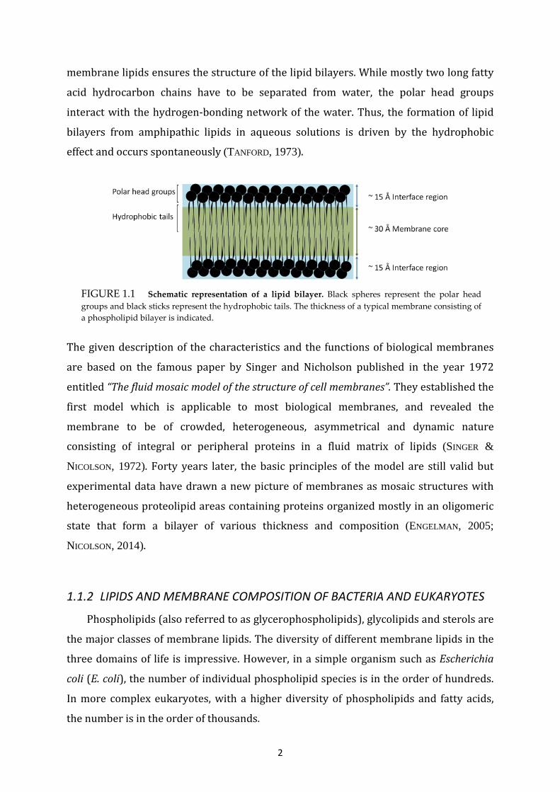
variable amount of carbohydrates. They are represented by bilayers of lipids, which are
organized in approximately 60 Ångstrom (Å) leaflets with their polar head groups facing
the two surfaces (approximately 15 Å each) and the nonpolar hydrocarbon chains
forming the hydrophobic core region (30 Å) (Figure 1.1). The chemistry of the
2
membrane lipids ensures the structure of the lipid bilayers. While mostly two long fatty
acid hydrocarbon chains have to be separated from water, the polar head groups
interact with the hydrogen-bonding network of the water. Thus, the formation of lipid
bilayers from amphipathic lipids in aqueous solutions is driven by the hydrophobic
effect and occurs spontaneously (TANFORD, 1973).
The given description of the characteristics and the functions of biological membranes
are based on the famous paper by Singer and Nicholson published in the year 1972
entitled “The fluid mosaic model of the structure of cell membranes”. They established the
first model which is applicable to most biological membranes, and revealed the
membrane to be of crowded, heterogeneous, asymmetrical and dynamic nature
consisting of integral or peripheral proteins in a fluid matrix of lipids (SINGER &
NICOLSON, 1972). Forty years later, the basic principles of the model are still valid but
experimental data have drawn a new picture of membranes as mosaic structures with
heterogeneous proteolipid areas containing proteins organized mostly in an oligomeric
state that form a bilayer of various thickness and composition (ENGELMAN, 2005;
NICOLSON, 2014).
1.1.2 LIPIDS AND MEMBRANE COMPOSITION OF BACTERIA AND EUKARYOTES
Phospholipids (also referred to as glycerophospholipids), glycolipids and sterols are
the major classes of membrane lipids. The diversity of different membrane lipids in the
three domains of life is impressive. However, in a simple organism such as Escherichia
coli (E. coli), the number of individual phospholipid species is in the order of hundreds.
In more complex eukaryotes, with a higher diversity of phospholipids and fatty acids,
the number is in the order of thousands.
FIGURE 1.1 Schematic representation of a lipid bilayer. Black spheres represent the polar head groups and black sticks represent the hydrophobic tails. The thickness of a typical membrane consisting of a phospholipid bilayer is indicated.

3
Biosynthesis of membrane lipids occurs at the cytoplasmic leaflet of the inner
membrane (IM) in bacteria and mainly at the ER in eukaryotes by a set of catalytic
membrane bound or cytosolic enzymes. The expression of coding genes, involved in
initial carboxylation steps (acc genes), fatty acid biogenesis (fab genes) and
phospholipid synthesis (pls genes) is strictly controlled. For example, many Gram-
positives express FapR, a global transcriptional factor, to regulate all the genes involved
in lipid metabolism (SCHUJMAN et al., 2003). Membrane lipid homeostasis is challenging
for all organisms and they have to adjust lipid composition in response to a changing
environment constantly.
The composition of different membrane systems in archaea, bacteria and eukarya varies
tremendously. Archaeal membranes and cell surfaces are structurally incomparable to
other membranes and consist of unique mono- or bilayer forming lipids. In E. coli, a well-
known model organism and representative of Gram-negative bacteria, the phospholipids
phosphatidylethanolamine (PE), phosphatidylglycerol (PG) and cardiolipin (CL, also
called diphosphatidylglycerol) are the major head groups of membrane lipids (AMES,
1968). The backbone is occupied predominantly by palmitic acid (16:0) and
monounsaturated fatty acids palmitoleic (16:1) and cis-vaccenic acids (18:1). The
composition of phospholipid head groups in the inner leaflet of the outer membrane
(OM) and the IM in E. coli is identical: 70 to 80 % of PE, 20 to 25 % of PG and 5 % or less
of CL (DOWHAN, 1997). In the outer monolayer of the OM another type of phospholipid
unique to Gram-negative bacteria is present: the glucosamine- and lipid A-based
lipopolysaccharide also known as endotoxin because of its toxic effects during Gram-
negative infections (RAETZ, 1990). Many Gram-positive bacteria lack the zwitterionic PE
but contain derivatives of anionic PG which are either zwitterionic or net positively
charged.
The mass ratio of protein to lipid in bacterial membranes is approximately 3:1. For
comparison, the simplest biological membrane system - vertebrate myelin - has a ratio
of approximately 1:4, because myelin mainly acts as an insulator with no enzymatic
function (GUIDOTTI, 1972).
In eukaryotic cells, the ER is the main organelle involved in phospholipid and cholesterol
synthesis. Other places for phospholipid synthesis are mitochondria and the Golgi
apparatus. Late endosomes and the plasma membrane are responsible for synthesis of
4
minor phospholipids like phosphatidylinositol (PI) derivates and of sphingosines. Both
are signaling lipids involved in signal transduction. With respect to the cellular function,
each organellar membrane is packed with various lipids. The ER membrane is loosely
packed with phosphatidylcholine (PC), PE and PI (VAN MEER et al., 2008) to allow proper
function as an organelle for insertion and transport of newly synthesized proteins and
lipids. Sterols synthesized at the ER are rapidly transported to the plasma membrane
and to endosomes. Together with sphingolipids, cholesterol is then packed at high
density in the plasma and endosomal membranes to resist mechanical stress and
osmotic pressure. The lipid composition of mitochondrial IMs is similar to their bacterial
ancestors (VAN MEER & DE KROON, 2011; HORVATH & DAUM, 2013). They are enriched of
PC, PE and CL and synthesize PG as a precursor of CL. The protein/lipid ratio of the IM of
mitochondria is 4:1 and thus very high (LUCKEY, 2008) and comparable to the ratio
found in bacteria. In general, a high enzymatic activity of an organelle correlates with
high protein content. Thus, rough ER, chloroplasts and nuclear membranes have higher
amounts of protein than for example myelin, smooth ER and Golgi membranes.
1.2 MEMBRANE PROTEINS
Representative biological membranes contain many types of proteins. These
membrane proteins (MPs) maintain essential cellular functions and processes such as
signaling, biogenesis, ion and nutrient transport and metabolism. A common distinction
is made between peripheral (extrinsic) and integral (intrinsic) membrane proteins
(SINGER & NICOLSON, 1972). Members of a third type of membrane proteins are called
amphitropic proteins (JOHNSON & CORNELL, 1999).
1.2.1 PERIPHERAL MEMBRANE PROTEINS
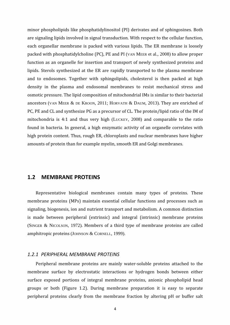
Peripheral membrane proteins are mainly water-soluble proteins attached to the
membrane surface by electrostatic interactions or hydrogen bonds between either
surface exposed portions of integral membrane proteins, anionic phospholipid head
groups or both (Figure 1.2). During membrane preparation it is easy to separate
peripheral proteins clearly from the membrane fraction by altering pH or buffer salt
5
FIGURE 1.2 Electrostatic interactions of peripheral proteins with integral proteins (left) or anionic phospholipids (right).
concentration. An extensive systematic analysis of the E. coli peripheral IM proteome by
efficient subfractionation experiments revealed that approximately 17 % of the basal
proteome are peripheral IM proteins (PAPANASTASIOU, 2013).
A typical example of a peripheral protein is the human cytochrome c, a 12 kilodalton
(kDa) small heme c containing protein of the intermembrane space of mitochondria. It
binds to cytochrome c oxidase mainly via a cluster of carboxy-terminal (C-terminal)
arginine and lysine residues (NICHOLLS, 1974). Furthermore, electrostatic interactions
with anionic phospholipids have been shown for cytochrome c in circular dichroism and
surface plasmon resonance studies (DE JONG & DE KRUIJFF, 1990; STEPANOV et al., 2009).
In bacteria, the SRP receptor FtsY is attached to the membrane surface via anionic
phospholipids (DE LEEUW et al., 2000) and interacts directly with the integral multi
subunit Sec translocon (ANGELINI et al., 2005).
1.2.2 AMPHITROPIC PROTEINS
The class of amphitropic proteins is a special group of peripheral proteins. These
proteins have two obvious localizations: one form is located in an aqueous environment
and one form is located at the membrane (JOHNSON & CORNELL, 1999). The reversible
binding to the membrane regulates the function of these proteins in various cellular
processes. Three different principles explain how interactions with the lipid bilayer are
achieved: (i) binding by ‘lipid clamps’ structures; (ii) attachment of lipid anchors for
transient membrane insertion, often combined with exposed positive residues for
electrostatic interactions; (iii) partitioning of an amphipathic alpha-helix (α-helix) into

6
the membrane bilayer. The bacterial protein SecA is a famous representative of
amphitropic membrane proteins, as its ATPase activity is regulated by anionic
phospholipid binding (LILL et al., 1990). Further cellular functions of the remarkable
nanomachine will be discussed later.
1.2.3 INTEGRAL MEMBRANE PROTEINS
By far the largest class of membrane proteins is the class of integral (intrinsic)
membrane proteins. They mediate plenty of cellular processes in transport, metabolism,
biogenesis and signalling. In contrast to peripheral membrane proteins, integral ones
are embedded firmly into the bilayer by hydrophobic interactions between the lipid
hydrocarbon core and hydrophobic stretches of the proteins, and can only be removed
by the use of detergents (amphipathic surfactants that disrupt the interactions between
the hydrophobic domain of the protein and the lipid hydrocarbon core).
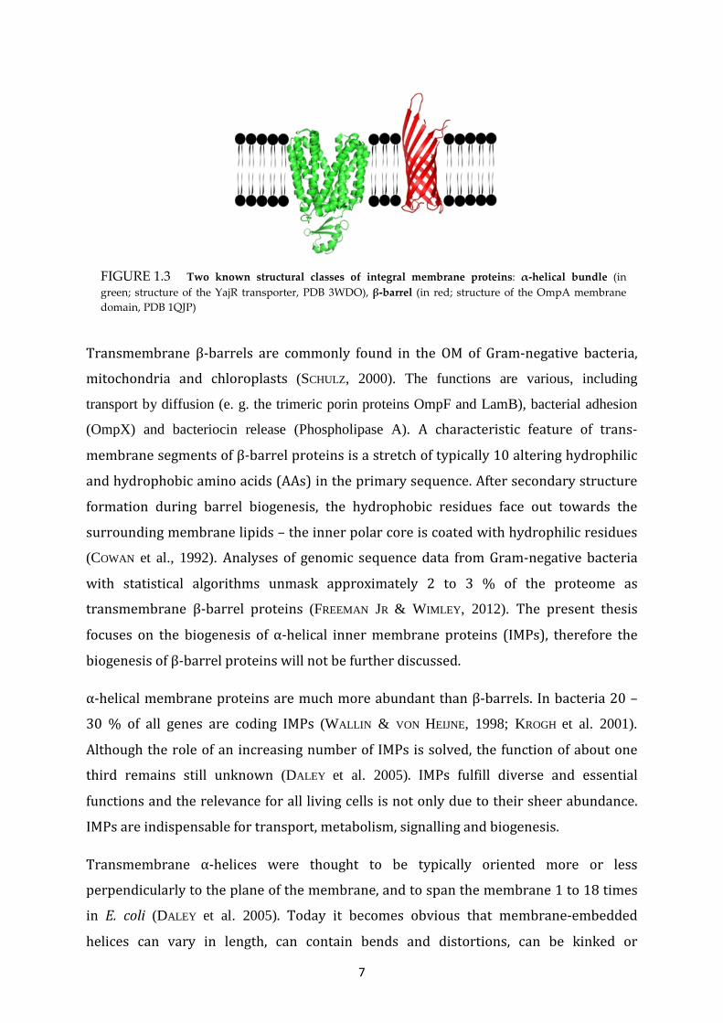
There are two model-like types of integral MPs which differ in their secondary structure
and localization (Figure 1.3). Data from known structures clearly illustrate that the most
common structural motifs in the transmembrane part of integral membrane proteins are
α-helical bundles and β-barrels. During biogenesis α-helical MPs are folded into more or
less complex bundles, predominantly perpendicular to the membrane. In general, they
have longer and more hydrophobic transmembrane (TM) segments than the β-barrels.
Transmembrane β-barrels are formed by up to 22 antiparallel, tilted β-strands (like e.g.
in the iron-siderophore transporter FhuA). The first and last β-strands close the barrel
upon interaction. The differences in those two structural motifs are dictated by the
encoded sequence and the biogenesis of proteins in the lipid bilayer. Here it is important
to consider that a polypeptide chain obtains the most stable conformation by formation
of interchain peptide backbone hydrogen bonds (H-bonds) in the lipid bilayer (VON
HEIJNE, 1994).
7
Transmembrane β-barrels are commonly found in the OM of Gram-negative bacteria,
mitochondria and chloroplasts (SCHULZ, 2000). The functions are various, including
transport by diffusion (e. g. the trimeric porin proteins OmpF and LamB), bacterial adhesion
(OmpX) and bacteriocin release (Phospholipase A). A characteristic feature of trans-
membrane segments of β-barrel proteins is a stretch of typically 10 altering hydrophilic
and hydrophobic amino acids (AAs) in the primary sequence. After secondary structure
formation during barrel biogenesis, the hydrophobic residues face out towards the
surrounding membrane lipids – the inner polar core is coated with hydrophilic residues
(COWAN et al., 1992). Analyses of genomic sequence data from Gram-negative bacteria
with statistical algorithms unmask approximately 2 to 3 % of the proteome as
transmembrane β-barrel proteins (FREEMAN JR & WIMLEY, 2012). The present thesis
focuses on the biogenesis of α-helical inner membrane proteins (IMPs), therefore the
biogenesis of β-barrel proteins will not be further discussed.
α-helical membrane proteins are much more abundant than β-barrels. In bacteria 20 –
30 % of all genes are coding IMPs (WALLIN & VON HEIJNE, 1998; KROGH et al. 2001).
Although the role of an increasing number of IMPs is solved, the function of about one
third remains still unknown (DALEY et al. 2005). IMPs fulfill diverse and essential
functions and the relevance for all living cells is not only due to their sheer abundance.
IMPs are indispensable for transport, metabolism, signalling and biogenesis.
Transmembrane α-helices were thought to be typically oriented more or less
perpendicularly to the plane of the membrane, and to span the membrane 1 to 18 times
in E. coli (DALEY et al. 2005). Today it becomes obvious that membrane-embedded
helices can vary in length, can contain bends and distortions, can be kinked or
FIGURE 1.3 Two known structural classes of integral membrane proteins: α-helical bundle (in green; structure of the YajR transporter, PDB 3WDO), β-barrel (in red; structure of the OmpA membrane domain, PDB 1QJP)
8
interrupted in the middle of the membrane and even can span only a part of the
membrane and then turn back (VON HEIJNE, 2006). The membrane embedded portion of
IMPs consists of helical stretches with 15 to 30 largely hydrophobic AAs to span the
hydrophobic core region of approximately 30 Å. Interestingly, charged AAs like lysine,
arginine, aspartic acid and glutamic acid are frequently found within membrane helices
although this is energetically not favorable. Often but not necessarily, such polar AAs
within hydrophobic domains have a central role in protein function.
1.2.4 PROTEIN-LIPID INTERACTION
The lipid bilayer does not only function as a diffusion barrier and a simple matrix for
integral or associated membrane proteins. Although bilayer forming and non-bilayer
forming lipids (such as PE and CL, respectively) have no catalytic function, they can
affect insertion, folding and assembly of MPs enormously. Patchy microdomains within
the bilayer, based on defined lipid species compositions, enable the organization of large
functional protein complexes.
In general, three types of binding modes for lipid interactions with MPs can be
distinguished: (i) an annular shell of lipids bound to the protein surface, (ii) non-annular
surface lipids immersed in cavities and clefts of the protein surface – this is found in
multi-subunit complexes, (iii) lipids residing within a membrane protein or a membrane
protein complex (PALSDOTTIR & HUNTE, 2004).
Specific protein–lipid interactions depend on the chemical and structural architecture of
lipids. Protein function and membrane integrity also depend on common properties of
lipids like self-association, shape and/or fluidity, as many membrane proteins undergo
conformational changes during activity. For example, the MscL protein is allowed to
properly open its large water filled pore only in response to lipid bilayer deformations,
because the opening process needs transmembrane helical movements within the
protein structure (PEROZO et al., 2002). Frequently, partial delipidation of membranes
leads to a decrease in protein activity (DOWHAN, 1997). The proper function of some
proteins strictly depends on the interaction with defined phospholipid species. For
example the function of cytochrome bc1 complex is strongly coupled to a firm
association of cardiolipin (SCHÄGGER et al., 1990; GOMEZ JR & ROBINSON, 1999). It has
been shown in vitro that anionic PG and non-bilayer lipids stimulate protein

9
FIGURE 1.4 Biogenesis of α-helical membrane proteins in E. coli (see text for details).
translocation mediated by the Sec translocon (VAN DER DOES et al., 2000). In addition,
cardiolipin tightly associated with the Sec translocon, promotes an efficient binding of
SecA and stimulates ATP hydrolysis (GOLD et al., 2010).
1.3 BIOGENESIS OF BACTERIAL α-HELICAL MEMBRANE PROTEINS
The biogenesis of a membrane protein can be divided into a few distinct steps
(Figure 1.4). After gene transcription, the messenger ribonucleic acid (mRNA) is
decoded by ribosomes in the cytoplasm. Then, the polypeptide is targeted to the
cytoplasmic membrane and inserted into the membrane. This occurs co-translationally
in most cases. The final destination of each protein is in general encoded in the amino-
terminal (N-terminal) sequence of the nascent amino acid chain. The membrane
insertion is catalyzed by the interaction of inserting membrane proteins with the Sec
translocon or the YidC membrane insertase, or occurs spontaneously for very small
hydrophobic proteins. During the membrane insertion process the topology is
determined and a membrane protein adopts its secondary structure. After or during
insertion the protein begins to fold properly into its native conformation. Following
folding, many α-helical membrane proteins interact with other membrane or soluble
proteins to form functional multi-subunit complexes.

10
1.3.1 PROTEIN TARGETING: HOW PROTEINS FIND THEIR FINAL DESTINATION
A challenging task for every cell is to ensure the correct transport of newly
synthesized proteins to their final destination. Günter Blobel discovered in the 1970s
that proteins carry discrete sequences, named signal sequences or topogenic sequences
(BLOBEL & SABATINI, 1971; BLOBEL & DOBBERSTEIN, 1975; BLOBEL, 1980). In general, signal
sequences are attachments at the N-terminus of the polypeptide chain that are decoded
by cytoplasmic or membrane bound receptor proteins. In concert with different
translocons and soluble or membrane bound protein factors, these targeting sequences
pave the way to sort various polypeptide chains from each other. In bacteria, newly
synthesized proteins need to be correctly localized and inserted into the IM and need to
be exported to the extracellular place or, for Gram-negative, to the periplasmic space
and the outer membrane.
In E. coli two major targeting pathways rule the direction of proteins: (i) post-
translational pathways, in which the majority of periplasmic, OM and secretory proteins
are targeted to the cytoplasmic membrane after protein synthesis, and (ii) the co-
translational pathway, in which most α-helical IMPs are targeted to the membrane
during the ongoing synthesis by the ribosome (SARAOGI & SHAN, 2013). In the following,
the targeting of proteins destined for crossing the IM will be discussed briefly, and then
the targeting of IMPs will be explained in more detail.
1.3.1.1 The signal sequence of secretory preproteins Proteins, which are synthesized as preproteins (or precursors) and destined to locate in
the periplasm, at the outer membrane or extracellular (in the following referred to as
secretory proteins), usually have a cleavable signal sequence with a typical size of
approximately 20 to 30 residues. The signal sequence can be divided into three different
domains: (i) the ‘N-domain’ with a positive net charge, (ii) an ‘H-domain’ of
approximately 7 to 13 mainly hydrophobic residues, and (iii) the slightly polar ‘C-
domain’ that contains the cleavage site for the signal peptidase (VON HEIJNE, 1985). After
the preprotein is translocated across the membrane, it is processed into its mature form
by cleavage of the signal sequence by an externally signal peptidase (PAETZEL et al.,
2002).

11
1.3.1.2 The post-translational SecB-pathway In bacteria, many secretory proteins as well as periplasmic and outer membrane
proteins are targeted to the cytoplasmic membrane post-translationally by SecB and are
mostly transported across the membrane via the Sec translocon. Once the signal
sequence exits the tunnel of a translating ribosome, a cytoplasmic chaperone, called
trigger factor, conducts nascent preproteins into the post-translationally SecB pathway
by preventing the interaction between the signal recognition particle (SRP) and the
signal peptide (BECK et al., 2000). The molecular chaperone SecB captures nascent
preproteins and keeps them in a loosely folded and non-aggregated state by binding the
mature region of the preprotein (HARTL et al., 1990). SecB then targets the translocation-
competent protein to the SecA ATPase which is tightly associated with the Sec
translocon at the cytoplasmic face of the inner membrane (HARDY & RANDALL, 1993;
FEKKES et al., 1998).
1.3.1.3 Twin-arginine translocation (Tat)-pathway: Targeting of folded proteins Some secretory proteins are using an alternative targeting route. They are translocated
in an entirely folded state via the Tat translocon, which is present in bacteria and the
chloroplasts of plants. Tat substrate proteins typically bind metal cofactors and contain
a specific signal sequence with the twin arginine (RR) consensus motif S-R-R-x-F-L-K
located in the ‘N-domain’ (BERKS, 1996). Presumably cytosolic chaperones like DnaK
(PEREZ-RODRIGUEZ et al., 2007) are involved in membrane targeting of Tat dependent
proteins, but so far no specific targeting factor for Tat signal sequences (like SRP) has
been uncovered. Interestingly, for a polytopic Rieske iron sulfur membrane protein from
Streptomyces coelicolor expressed in E. coli, it has been shown that both the Sec and the
Tat translocons are co-operating in membrane integration (KELLER et al., 2012). In
contrast, some Tat substrates from E. coli, containing hydrophobic C-terminal
transmembrane helices, are integrated into the lipid bilayer solely by the Tat translocon
(HATZIXANTHIS et al., 2003).
1.3.1.4 SRP-pathway: The main pathway for inner membrane proteins Most proteins that are intended to reside in the IM of E. coli are generally targeted to the
membrane by the ubiquitous co-translational SRP-targeting pathway (DE GIER et al.,
1996; ULBRANDT et al., 1997). The universally conserved ribonucleoprotein complex SRP

12
is present in all three kingdoms of life. Compared to its mammalian homolog, E. coli SRP
is relatively simple and is comprised of the 48 kDa GTPase Ffh (a homolog of SRP54) and
a small 4.5S RNA with 114 bases in length. Ffh consists of two domains: (i) the ‘NG-
domain’ which is located at the N-terminus containing a GTP-binding site, and (ii) the C-
terminal methionine rich ‘M-domain’. The prokaryotic homolog of the SRP receptor SRα
is the peripheral membrane protein FtsY. The receptor FtsY contains a similar NG
domain like Ffh, which is located at the C-terminus, as well as a highly positively charged
N-terminal ‘A-domain’. Despite its simplicity, bacteria SRP can substitute eukaryotic
homologs to promote efficient targeting to the ER membrane (BERNSTEIN et al., 1993).
The SRP targeting pathway is initiated when SRP detects an N-terminal sequence
presented on polypeptide chains once they emerge from the translating ribosome
(LUIRINK et al., 1992). Unlike signal sequences of secretory or Tat client proteins, signal
sequences of IMPs which are recognized by SRP mostly have no cleavage site and no
defined recognition feature. These targeting sequences are in general enhanced
hydrophobic α-helical transmembrane domains and called signal anchor sequences.
Proverbially, they anchor inserting membrane proteins permanently into the lipid
bilayer with a NinCout orientation. Thus, the first hydrophobic TM segment of an α-helical
membrane protein often serves as a signal for membrane targeting (ZERIAL et al., 1986).
SRP binds the signal anchor sequence of a ribosome nascent chain (RNC) in a deep
groove of the ‘M domain’, shaped by mainly hydrophobic residues, including the
conserved methionine residues (KEENAN et al., 1998). Since a conserved domain of the
4.5S RNA binds to the ‘M-domain’ close to the groove, both protein and RNA most likely
provide the signal sequence binding site. Structures obtained by cryo-electron
microscopy (EM) of an RNC complex with E. coli SRP show that the distinct presentation
of the signal sequence at the ribosomal tunnel exit allows an efficient slide of the signal
sequence into the hydrophobic groove of the SRP ‘M-domain’ (SCHAFFITZEL et al., 2006).
Therefore, specific binding of SRP to the ribosome tunnel exit is an important
precondition. This is achieved by binding of the helical ‘N-domain’ to the ribosomal
proteins L23 and L29 (HALIC et al., 2006; GU et al., 2003).
The membrane targeting of SRP bound RNC complexes (RNC-SRP) to the cytoplasmic
side of the membrane is achieved by the interaction of the two ‘NG domains’ of SRP and
its receptor. The receptor FtsY exists both in a soluble (LUIRINK et al., 1994) and a
13
membrane associated form, the latter most likely being preferred (ANGELINI et al., 2005).
It has been shown that only membrane bound FtsY molecules are capable to promote
dissociation of SRP from the RNC (LAM et al., 2010, VALENT et al., 1998). Altogether, the
membrane association of FtsY is highly dynamic and it is discussed controversially,
whether soluble FtsY is able to bind translating ribosomes aside the membrane
(HERSKOVITS & BIBI, 2000). In order to finalize membrane targeting, the RNC-SRP-FtsY
complex is anchored to the membrane by the positively charged α-helical ‘A-domain’ of
FtsY (PARLITZ et al., 2007). At the membrane, GTP hydrolysis of both SRP and the
receptor FtsY (in the GTPase G-domains of the proteins) results in the dissociation of the
RNC-SRP-FtsY complex (CONNOLLY et al., 1991) and the recycling of SRP and FtsY into the
cytosol for upcoming targeting events. In the most common cases the RNCs are released
to the Sec translocon which facilitates the insertion of targeted membrane proteins
alone or in cooperation with the YidC insertase (XIE & DALBEY 2008). Both genetic and
structural studies showed clearly that basic residues of cytosolic SecY loops interact
with ribosomal proteins L23 and L29 at the ribosomal tunnel exit (CHENG et al., 2005;
MENETRET et al., 2007; BECKMANN et al., 2001; FRAUENFELD et al., 2011). As mentioned
above, SRP binds to the ribosomal proteins L23 and L29 during the targeting process.
Thus, a stable RNC-SecYEG complex formation requires the detachment of SRP from the
RNC. As the translation resumes on Sec-associated ribosomes, the nascent polypeptide
chain slips into the aqueous translocation channel or directly into the lateral gate region.
During translocation, transmembrane segments exit the channel laterally into the lipid
bilayer (VAN DEN BERG et al., 2004).
Besides the Sec translocon, the YidC insertase receives a small number of SRP substrates
and mediates their membrane insertion. This has been shown for the mechanosensitive
channel protein MscL and - to a certain degree - for Foc, the subunit c of the ATPase
(FACEY et al., 2007; VAN BLOOIS et al., 2004; YI et al., 2004). In addition, it has been shown
that Ffh and FtsY directly contact YidC (WELTE et al., 2012).
The depletion of SRP (Ffh) in E. coli leads to global kinetic defects in the biogenesis and
localization of IMPs, resulting in increased protein aggregation in the cytoplasm and
finally to cell death. To overcome the protein aggregation, the cells show a strong σ32
response which leads to upregulated expression levels of molecular chaperones in the
cytoplasm (WICKSTRÖM et al., 2011a). Another study revealed that SRP depletion has no
significant negative influence on the steady-state level or distribution of most inner
14
membrane proteins. On the other hand, SRP depletion leads to an immediate reduction
of the proton motive force (pmf) (ZHANG et al., 2012). These observations show clearly
that SRP is essential for global cell integrity, but they also suggest that alternative (SRP-
independent) targeting pathways for inner membrane proteins exist in E. coli.
1.3.1.5 Non-classical targeting pathways The affinity of ribosomes for the Sec translocon is highly conserved (PRINZ et al., 2000).
Therefore, it is obvious that ribosomes might support co-translational targeting
independently of SRP. In eukaryotes, ribosomes remain associated at the ER membrane
after co-translational targeting and can be primed by an mRNA encoding a membrane
protein (POTTER & NICCHITTA, 2002). In bacteria, it has been shown that mRNAs coding
for inner membrane proteins are targeted to the membrane in a translation independent
mechanism (NEVO-DINUR et al., 2011). Most likely there is a correlation between the
uracil content and the localization of mRNAs, as mRNAs of membrane proteins have
significantly higher uracil content (PRILUSKY & BIBI, 2009).
Tail-anchored membrane proteins (TAMPs) are a small, heterogeneous class of proteins
in E. coli which are anchored to the membrane with a C-terminal transmembrane
segment and consequently contain no N-terminally located hydrophobic signal sequence
(BORGESE & RIGHI, 2010; CRANEY et al., 2011). Together with a few small < 50 AAs single
spanning membrane proteins in E. coli the TAMPs have to be targeted and inserted into
the membrane independently of SRP and the Sec translocon. Factors promoting
targeting and insertion of TAMPs as found in eukaryotes (Get pathway) and archaea
(ArsA homolog) have not been identified so far. The detailed mechanisms of the
biogenesis of these proteins remain to be investigated. Recently it has been shown that
the TAMP TssL, a component of the Typ VI secretion system of enteroaggregative E. coli,
requires the membrane insertase YidC and with some limitations the molecular
chaperone DnaK for membrane biogenesis, but not the Sec-system (SOUSSOULA & KUHN,
unpublished data; ASCHTGEN et al., 2012).
M13 procoat protein, the coat protein of filamentous E. coli phage M13, is targeted to the
membrane in a passive mode, without any targeting factors. Here, the targeting is
promoted by electrostatic interactions between positively charged AAs at the C- and N-
termini and the negatively charged head groups of membrane phospholipids (GALLUSSER

15
& KUHN, 1990). There is evidence that Pf3 coat protein of Pseudomonas aeruginosa phage
Pf3 is targeted to the membrane in the same way, since two positively charged residues
are present at the C-terminus of the protein.
The role of “classical” chaperones like Trigger factor (TF), DnaK and GroEL in an
alternative targeting pathway is not clear despite their expression being up-regulated in
cells depleted of SRP (WICKSTRÖM et al., 2011a; ZHANG et al., 2012). However, there is
evidence from in vitro studies, that the chaperone GroEL might mediates post-
translational targeting of the polytopic membrane protein lactose permease (LacY) and
bacteriorhodopsin (BOCHKAREVA et al. 1996; DEATON et al., 2004).
1.3.2 TOPOLOGY OF MEMBRANE PROTEINS
A fundamental aspect in the biogenesis of membrane proteins is the question how a
given polypeptide chain is oriented in the lipid environment during insertion. The
topology of a membrane protein can be considered as a 2D representation of the protein
and is defined by the number of transmembrane helices and the orientation of the N-
and C-terminus relative to the lipid bilayer (Figure 1.5). Topology maps can be predicted
theoretically by using algorithm based prediction programs or experimentally by using
terminal tagging with alkaline phosphatase (PhoA) and green fluorescent protein (GFP)
(DALEY et al., 2005).
Well defined topogenic signals reside in the protein sequence. Topogenic signals are
recognized and decoded by the translocation and insertion machineries after targeting
FIGURE 1.5 Topology of a membrane protein with five transmembrane helices (colored). The amino-
terminus (N) is located in the periplasm (referred to as ‘outside’) and the carboxy-terminus (C) is located in the cytoplasm (referred to as ‘inside’). The transmembrane helices (colored) are connected via short loops.
16
to the membrane (BLOBEL, 1980). The proper orientation of TM segments is an absolute
precondition for effective folding into the native 3D conformation.
1.3.2.1 Topogenic signals define topology of membrane proteins The variety of topogenic signals and the resulting topologies of different model proteins
are summarized and illustrated in Figure 1.6. There are two classes of single-spanning
membrane proteins (Figure 1.6A), commonly called type I and type II membrane
proteins (VON HEIJNE & GAVEL, 1988). Type I membrane proteins contain a reverse signal
anchor (also called type I signal anchor) which facilitates the translocation of the polar
N-terminus and anchors the protein into the membrane with an Noutside-Cinside
orientation. An example of such a protein is Pf3 coat (ROHRER & KUHN, 1990). Other type
I proteins are synthesized as precursor proteins with a cleavable signal sequence and a
stop transfer sequence as topogenic elements. The cleavage by signal peptidase after
insertion results in type I orientation of the mature protein. For the phage protein M13
gp3, the signal sequence initiates translocation of the hydrophilic domain. The mature C-
terminal transmembrane segment contains a stop transfer signal to terminate
translocation. In contrast, M13 procoat protein (gp8) is inserted by a different
mechanism, yet still adopting the same topology as gp3 does. Here, the insertion signals
are located in the signal sequence and the membrane segment, both elements forming a
topogenic element called ‘helical hairpin’ to translocate the polar domain (KUHN et al.,
1986; ENGELMAN & STEITZ, 1981). Type II membrane proteins (with an Ninside-Coutside
orientation) possess an uncleaved signal anchor (or type II signal anchor) that initiates
translocation of the C-terminus across the membrane. For example, the cell division
protein FtsQ is a type II membrane protein (CARSON et al., 1991).
The topologies of double- and multi-spanning membrane proteins are conducted by
discrete hydrophobic transmembrane segments and the interplay of the various
topogenic signals described above. Examples for these complex membrane proteins are
illustrated in Figure 1.6B and C.

17
FIGURE 1.6 Topology of membrane proteins and topogenic signals. (A) Single-spanning membrane proteins of type I (Pf3 coat, M13 procoat and M13 gp3) and type II (FtsQ). (B) Double-spanning membrane proteins. (C) Multi-spanning membrane proteins. The arrowhead symbols indicate cleavage by signal peptidase after insertion.
The topogenesis of multi-spanning membrane proteins can be considered as the
consecutive insertion of alternating start- and stop-transfer sequences or helical hairpin
loops. In the simplest model the first transmembrane segment, i. e. the initial topogenic
signal, defines the orientation of itself and the alternate orientation of following
transmembrane segments. The insertion mechanism is not that strict for all multi-

18
spanning proteins, what has been shown for the MalF protein. Most likely the signals for
proper topogenesis are located throughout the whole MalF protein, since depletion of
the second transmembrane segment does not alter orientation of downstream segments
(MCGOVERN et al., 1991).
The hydrophobicity of transmembrane segments is crucial for their function in serving
as topogenic sequences. Interestingly, sequence-comparison of the different
hydrophobic topogenic sequences did not show any significant deviation in the AAs
composition but a clear difference between the downstream and upstream polar
flanking regions (VON HEIJNE & GAVEL, 1988).
1.3.2.2 The universal positive-inside rule The positive-inside rule by Gunnar von Heijne is postulating that membrane protein
topology is primarily determined by charged residues in cytoplasmic domains flanking
hydrophobic transmembrane segments. In general, the positively charged residues
arginine and lysine (Arg and Lys) are up to 4-times more prevalent in cytoplasmic
domains compared to the ‘outside’ (VON HEIJNE, 1986). The hypothesis derived from
statistical analysis of E. coli membrane proteins was confirmed experimentally; showing
that leader peptidase from E. coli (Figure 1.6B) reverses its topology when additional
lysine residues are placed at the N-terminus (VON HEIJNE, 1989). Genome-wide analysis
of the membrane proteome in all domains of life revealed the universality of the
positive-inside rule (NILSSON et al., 2005).
A comparable significant enrichment of negatively charged residues has not been
detected in any extramembrane domains (GRANSETH et al., 2005; NILSSON et al., 2005).
Yet, there is evidence that the negatively charged AAs glutamic acid or aspartic acid (Glu
and Asp) also direct helix orientation (NILSSON et al., 1990; DELGADO-PARTIN & DALBEY,
1998). For the biogenesis of Pf3 coat protein rather negatively charged residues than
positively charged residues appear to be topogenic (KIEFER et al., 1997).
How discrete topological signals are interpreted by cell components is not fully
understood. It is assumed that a complex interplay of the translocation and insertion
machineries, helix characteristics and interactions within the protein, as well as the final
membrane localization, which is mostly defined by the lipid composition, decode

19
topogenic signals. In the following I will describe factors that contribute to the
determination of membrane topology.
1.3.2.3 Topological determinants The lipid composition of the target membrane is one factor involved in guiding
membrane topology. The negatively charged phospholipids PG and CL, being among the
most abundant phospholipids in the inner E. coli membrane, direct positively charged
protein domains to remain in the cytoplasm in accordance with the positive-inside rule
(VAN KLOMPENBURG et al., 1997). In addition, neutral lipids like PE reduce the potential of
negatively charged residues to serve as topogenic signals (DOWHAN & BOGDANOV, 2009).
Alteration of lipid composition can lead to reversible orientations of multi-spanning
membrane proteins such as lactose permease LacY and phenylalanine permease PheP
(BOGDANOV et al., 2002; ZHANG et al., 2003).
The hydrophilic channel of the Sec translocon synchronizes the insertion of hydrophobic
transmembrane segments and the translocation of flanking domains with different net
charges to the ‘outside’. In the yeast channel subunit Sec61p (homolog of prokaryotic
SecY) conserved charged residues are located at the cytoplasmic (E382) and the
‘outside’ (R67, R74) facing end of the channel. These charged residues are suggested to
contribute for orienting topogenic signals. Mutations of the charged residues affect the
orientation of a model protein and reduce the influence of the positive-inside rule
(GODER et al., 2004).
The pmf is another factor which presumably is involved in interpretation of topogenic
signals in accordance with the positive-inside rule. In most bacteria, chemiosmosis leads
to a positive outside and negative cytoplasm. This charge difference may prevent
translocation of positively charged polypeptide domains and support translocation of
negatively charged domains (CAO & DALBEY, 1994; ANDERSSON & HEIJNE, 1994; CAO et al.,
1995; KIEFER et al., 1997). However, the retention of positively charged AAs cannot be
exclusively determined by the pmf, since obligate acidophilic archaea with an inverted
membrane potential show the same distribution of Arg and Lys residues in integral
membrane proteins as neutrophilic bacteria (VAN DE VOSSENBERG, 1998). This fact
underlines that electrostatic interaction between negatively charged phospholipid head

20
groups and positively charged residues is a much more relevant topological
determinant.
Taken together, how and when topogenesis of membrane proteins is determined seems
to be a complex event which is still very puzzling, because the majority of membrane
proteins do not insert spontaneously into the membrane and require precise working
translocases or insertases to orchestrate insertion.
1.3.3 INSERTION OF α-HELICAL MEMBRANE PROTEINS
Newly synthesized α-helical membrane proteins are primarily targeted to two
different insertion sites: (i) to the Sec translocon/YidC and (ii) to the insertase YidC that
is not associated with the Sec translocon. The Sec translocon is a protein-conducting
channel present in all domains of life which is required for the translocation of secretory
proteins and the insertion of α-helical membrane proteins. The membrane insertase
YidC is also highly conserved but homologous proteins are missing in the phylum
Crenarchaeota and in ER membranes (POHLSCHRÖDER et al., 2005). YidC promotes
insertion, folding and assembly of α-helical membrane proteins both in cooperation with
the Sec translocon and as an autonomous insertion site. YidC mediated biogenesis of
membrane proteins will be discussed in detail in chapter 1.4 to underline the central
significance of YidC for this thesis.
1.3.3.1 The Sec translocon: A protein-conducting channel for secretion and insertion In bacteria, the Sec translocon is composed of a heterooligomeric complex of integral
membrane proteins and the peripheral associated component SecA. The aqueous
protein-conducting channel is formed by the essential core proteins SecY and SecE
(homologs of the eukaryotic core components Sec61α and Sec61γ, respectively) and a
distinct protein, SecG, which is not essential for cell viability (HANADA et al., 1994;
HARTMANN et al., 1994). SecG shows no obvious homology to the corresponding β-
subunits in eukaryotes and archaea. The accessory components SecDFYajC and YidC
complete the membrane-embedded portion of the Sec translocon in some cases. As
mentioned above, SecA is associated to the translocon as a peripheral membrane
protein, which is, like SecY and SecE, essential for cell viability and has so far been only
found in bacteria and chloroplasts (SARDIS & ECONOMOU, 2010). In addition, ribosomal

21
proteins and RNA contact the Sec translocon at multiple sites of cytosolic SecY domains
(CHENG et al., 2005; KUHN et al., 2011; FRAUENFELD et al., 2011).
1.3.3.2 Accessory components of the Sec translocon and their function SecD and SecF are polytopic membrane proteins with large periplasmic domains.
Together with the single spanning YajC, SecDF forms a heterotrimeric complex
associated to the SecYEG channel (DUONG & WICKNER, 1997). It is assumed, that
SecDFYajC is not essential for the insertion, yet the complex most likely enhances the
efficiency of this process (BRUNDAGE et al., 1990; HANADA et al., 1994; POGLIANO &
BECKWITH, 1994; TSUKAZAKI et al., 2011). Structural analysis and biochemical data suggest
that SecDF utilizes the pmf to complete the translocation of substrates (NOUWEN et al.,
2005; TSUKAZAKI et al., 2011). YidC associates directly to SecYEG or to SecDF and
functions in concert with the SecYEG channel (SCOTTI et al., 2000; NOUWEN & DRIESSEN;
2002; XIE et al., 2006; SACHELARU et al., 2013). Recently an intact and active complex of
SecYEG, SecDFYajC and YidC (also known as the holotranslocon) was successfully
overexpressed, purified and reconstituted for functional analysis of protein insertion
and translocation (SCHULZE et al., 2014).
1.3.3.3 Structure of SecYEG The X-ray crystallographic structure of the SecYEG/β complex from Methanococcus
jannashii together with the cryo-EM structure of E. coli SecYEG, bound to an RNC,
remarked a breakthrough in the field, providing first insights into the molecular
organization and the detailed structure of the protein-conducting channel (VAN DEN BERG
et al., 2004; MITRA et al., 2005). SecY consists of ten TM segments. The X-ray structure
revealed that SecY forms an hourglass channel consisting of two clamshell-like domains
of SecY TM domains 1-5 and TM domains 6-10, which are connected by a periplasmic
loop between TM5 and TM6 (Figure 1.7). The center of the two halves represents the
protein-conducting pore as two hydrophilic funnel-like cavities, which are open towards
the cytoplasm and periplasm, respectively.
22
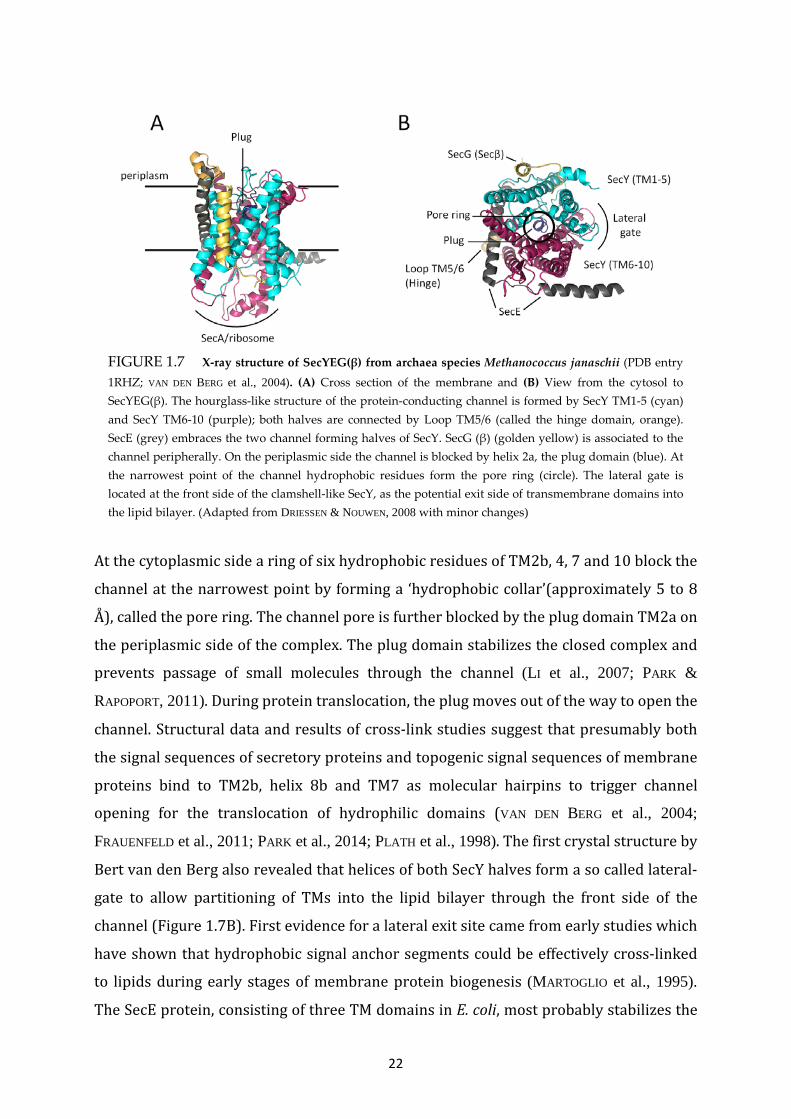
FIGURE 1.7 X-ray structure of SecYEG(β) from archaea species Methanococcus janaschii (PDB entry 1RHZ; VAN DEN BERG et al., 2004). (A) Cross section of the membrane and (B) View from the cytosol to SecYEG(β). The hourglass-like structure of the protein-conducting channel is formed by SecY TM1-5 (cyan) and SecY TM6-10 (purple); both halves are connected by Loop TM5/6 (called the hinge domain, orange). SecE (grey) embraces the two channel forming halves of SecY. SecG (β) (golden yellow) is associated to the channel peripherally. On the periplasmic side the channel is blocked by helix 2a, the plug domain (blue). At the narrowest point of the channel hydrophobic residues form the pore ring (circle). The lateral gate is located at the front side of the clamshell-like SecY, as the potential exit side of transmembrane domains into the lipid bilayer. (Adapted from DRIESSEN & NOUWEN, 2008 with minor changes)
At the cytoplasmic side a ring of six hydrophobic residues of TM2b, 4, 7 and 10 block the
channel at the narrowest point by forming a ‘hydrophobic collar’(approximately 5 to 8
Å), called the pore ring. The channel pore is further blocked by the plug domain TM2a on
the periplasmic side of the complex. The plug domain stabilizes the closed complex and
prevents passage of small molecules through the channel (LI et al., 2007; PARK &
RAPOPORT, 2011). During protein translocation, the plug moves out of the way to open the
channel. Structural data and results of cross-link studies suggest that presumably both
the signal sequences of secretory proteins and topogenic signal sequences of membrane
proteins bind to TM2b, helix 8b and TM7 as molecular hairpins to trigger channel
opening for the translocation of hydrophilic domains (VAN DEN BERG et al., 2004;
FRAUENFELD et al., 2011; PARK et al., 2014; PLATH et al., 1998). The first crystal structure by
Bert van den Berg also revealed that helices of both SecY halves form a so called lateral-
gate to allow partitioning of TMs into the lipid bilayer through the front side of the
channel (Figure 1.7B). First evidence for a lateral exit site came from early studies which
have shown that hydrophobic signal anchor segments could be effectively cross-linked
to lipids during early stages of membrane protein biogenesis (MARTOGLIO et al., 1995).
The SecE protein, consisting of three TM domains in E. coli, most probably stabilizes the

23
channel by embracing the two SecY domains on the back side. SecG/β is located at the
outside of the channel and shows only weak association to SecY.
It is still controversially discussed, whether SecY functions as a monomer or as a dimer
to facilitate translocation and insertion. Evidence for a SecY dimer as the functional state
were coming from studies in which both ‘back-to-back’ and ‘front-to-front’ dimeric
forms of SecY in crystals have been observed (BREYTON et al., 2002; DALAL et al., 2012;
MITRA et al., 2006). However, several crystal structures of SecY complexes from bacteria
and archaea indicate that the active channel is formed by one copy of SecY (VAN DEN
BERG et al. 2004; TSUKAZAKI et al., 2008; ZIMMER et al., 2008; EGEA & STROUD, 2010). In
addition, cross-linking studies have shown that a single copy is sufficient to promote
protein translocation, although both forms of SecY dimers have been found in vivo (PARK
& RAPOPORT 2012). The latter is consistent with data that suggest that in a dimeric state
of the translocon the nontranslocating complex can contribute to SecA binding and
stimulation of its ATPase activity while the other copy promotes translocation of
substrate proteins (OSBORNE & RAPOPORT, 2007; DALAL et al., 2012). Studies of protein
secretion and insertion with the successfully reconstituted holotranslocon (with a
monomeric stoichiometry) and results from single molecule experiments further
suggest that a single complex is sufficient for function (SCHULZE et al., 2014; KEDROV et
al., 2013). Recently obtained cryo-EM structures of ribosome bound Sec translocons in
an active state revealed that a single SecYEG and Sec61 complex is associated with the
ribosome during co-translational translocation (PARK et al., 2014; GOGALA et al., 2014).
Both studies present sensational insights into conformational states within the active
Sec translocon during translocation for the first time.
1.3.3.4 The Sec translocon in action: seemingly contrary functions An exceptional feature of the Sec translocon is its capability to promote three essential
cell processes: (i) the secretion of unfolded preproteins from the cytoplasm into the
periplasm through the channel, (ii) the translocation of polar domains of membrane
proteins across the membrane, and (iii) the insertion of hydrophobic transmembrane
segments into the phospholipid bilayer (DRIESSEN & NOUWEN, 2008).

24
1.3.3.5 Translocation of secretory proteins across the membrane The translocation of preproteins generally occurs in a post-translational route after
targeting to the Sec translocon - mostly via the SecB pathway (HARTL et al., 1990). The
energy for preprotein translocation is provided by ATP hydrolysis at SecA and by the
pmf (DRIESSEN, 1992; VAN DALEN et al., 1999). The SecA protein is found predominantly in
the cytoplasm, where it binds to translated SecB/preprotein complexes and at the
membrane, where it is associated with the Sec translocon (HUBER et al., 2011; ZIMMER et
al., 2008). For the translocation of a polypeptide chain SecA interacts with cytoplasmic
loops of SecY and SecEG (MITRA et al., 2005; VAN DER SLUIS et al., 2006; ZIMMER et al., 2008;
NAGAMORI et al., 2002). It is assumed, that the binding of SecA to SecYEG induces the plug
displacement in order to open the channel (ZIMMER et al., 2008). The binding of ATP to
SecA at the Sec translocon initiates translocation and allows the binding of the N-
terminal signal sequence into the SecYEG channel. At next, multiple cycles of ATP
binding and hydrolysis at SecA, that lead to repeated binding and release of the
preprotein, cause the stepwise translocation of the polypeptide chain through the Sec
translocon (ECONOMOU & WICKNER, 1994; VAN DER WOLK et al., 1997). The pmf is capable
to drive translocation after ATP hydrolysis has lead to dissociation of the polypeptide
chain from SecA (SCHIEBEL et al., 1991). How exactly the pmf acts as a driving force for
translocation is not clear. It is conceivable that an electrophoretic mechanism drive the
translocation of negatively charged residues. After translocation across the membrane
has been completed, the preprotein is cleaved by a signal peptidase to its mature form,
thus obtaining its native conformation (PAETZEL et al., 2002).
1.3.3.6 Insertion of membrane proteins into the lipid bilayer via the Sec translocon The insertion of the majority of α-helical membrane proteins is catalyzed by the Sec
translocon and occurs mostly co-translationally. The energy for this process is provided
presumably by the ongoing translation at the ribosome and the pmf. The Sec translocon
catalyzes the translocation of certain hydrophilic domains across the membrane and the
insertion of hydrophobic transmembrane segments into the lipid bilayer. The
translocation of large hydrophilic domains requires energy provided by ATP hydrolysis
at SecA, whereas a variety of membrane proteins with shorter, less hydrophilic domains
is inserted into the membrane independently of SecA (KUHN, 1988; WERNER et al., 1992;
SAAF et al., 1995). The detailed molecular mechanism by which hydrophobic
transmembrane segments move out of the aqueous channel to enter the hydrophobic

25
core region of the phospholipid bilayer still remains largely unresolved. The decision if
translocation arrests and hydrophobic segments are released into the lipid bilayer is
most likely made in the translocon channel or the lateral gate by interpretation of
distinct topological signals (described in the previous chapter 3.2). In general, the
efficiency of the insertion process depends on the hydrophobicity and the length of the
TM segments as well as the distribution of charged residues (XIE et al., 2007; HESSA et al.,
2005 and 2007). Studies of the thermodynamics of membrane insertion by Tara Hessa
and colleagues suggest that the insertion is driven by protein-lipid interactions at the
translocon-bilayer interface (HESSA et al., 2005). During biogenesis of membrane
proteins, the recognition of hydrophobic regions, with sufficient hydrophobicity to serve
as stop-transfer signals, leads to an arrest in translocation and to the insertion of the
respective TM segment (SAAF et al., 1998b; DUONG & WICKNER, 1998). The putative lateral
gate, through which TM segments move out of the channel into the lipid environment of
the membrane, is located at the front side of SecY between TM2b/3 and TM7/8 (Figure
1.7B) (VAN DEN BERG et al., 2004; TSUKAZAKI et al., 2008; DU PLESSIS et al., 2009; EGEA &
STROUD, 2010; FRAUENFELD et al., 2011; GOGALA et al., 2014; PARK et al., 2014). The
transition from a closed to an open state of the lateral gate is most likely the result of a
large rotation in the N-terminal half of SecY and resulting movements of SecE and SecG
(PARK et al., 2014). Recently obtained cryo-EM structures of a ribosome-SecY complex
with an insertion intermediate of nascent proteorhodopsin showed the localization of
inserted TM domains outside of the SecY channel in direct vicinity to the potential
lateral gate for the first time (BISCHOFF et al., 2014). For efficient insertion into the
bilayer, the local lipid environment next to the lateral gate might be influenced by
interactions between rRNA helix 59 of the ribosome and phospholipid head groups
(FRAUENFELD et al., 2011). Although this is speculative, the local disorder of the bilayer
next to the lateral gate may pave the way for the insertion of IMPs.
Regarding polytopic membrane proteins, cross-linking studies suggested that TM
segments of polytopic membrane proteins are released through the lateral gate into the
bilayer in a sequential mode, one by one or even in pairs (BECK et al., 2001; SADLISH et al.,
2005; SKACH, 2009). A predominant role in the Sec-dependent insertion of polytopic
membrane proteins was assigned to YidC, which will be discussed in a following chapter.

26
1.3.4 FOLDING OF α-HELICAL MEMBRANE PROTEINS
Following Anfinsen’s dogma, the three-dimensional (3D) structure of a protein is
determined by the primary sequence of the polypeptide chain (ANFINSEN, 1973). At least,
this is true for small globular proteins. Membrane proteins require another dimension,
the specific environment of the lipid bilayer, and most of them have to be inserted into
the membrane by protein factors, to acquire their native structures.
1.3.4.1 The two-stage model for membrane protein folding The widely accepted two-stage model by Popot and Engelmann postulates that folding of
membrane proteins occurs in two fundamental stages: insertion and folding (POPOT &
ENGELMAN, 1990) (Figure 1.8). In the first stage, individual TM segments are inserted
into the bilayer. This process can be both coordinated and driven by a translocon
complex or by the membrane insertase YidC. Helix formation is determined by the
primary sequence, is driven by the hydrophobic effect and stabilized by hydrogen
bonding between the polar groups of the peptide backbone. Thus, the first stage of the
model results in the thermodynamic equilibrium of individual TM helices within the
lipid bilayer. In the second stage, TM helices interact with each other to form helical
bundles. The second stage may include rearrangements and reorientations of TM
segments that lead to higher order structures. The two-stage model was later improved
to establish a three-stage model. The additional stage describes how helical bundles
create a less hydrophobic interior space to incorporate prosthetic groups and additional
polypeptides such as coil domains or helices of short length (ENGELMAN et al., 2003).
Experimental evidence for this third stage came from folding studies with
bacteriorhodopsin protein fragments. Kinetic analysis show that retinal binds after
association of the two fragments (POPOT et al., 1987).
1.3.4.2 Early stages of membrane protein folding The initial formation of helical secondary structures occurs in the ribosome tunnel in a
co-translational manner (WOOLHEAD et al., 2004; LU & DEUTSCH, 2005; LIN et al., 2012).
Further steps in early folding of membrane proteins are controlled presumably by the
translocation and insertion machineries before or after individual transmembrane
segments are released into the lipid bilayer.

27
FIGURE 1.8 Two-stage model for membrane protein folding. In the first stage, transmembrane segments are inserted into the bilayer. This can be catalyzed by the Sec translocon or YidC (not shown). In the second stage, transmembrane segments interact with each other to form helical bundles.
For the eukaryotic channel forming Sec61α and the bacterial Sec translocon, it has been
shown that helices are released sequentially, pair wise or even as a bundle of helices
(SKACH, 2009; BECK et al., 2001). For the bacterial Sec translocon early steps in folding of
polytopic proteins were proven using a recently developed co-translational in vivo assay,
in which a pulling force on nascent chains are measured indirectly using a translational
arrest peptide (ISMAIL et al., 2012). It has been shown that C-terminal transmembrane
segments interact with more N-terminally helices at an early stage when the C-terminal
helix portions are released from the Sec channel into the membrane (CYMER & VON
HEIJNE, 2013). This suggests that early tertiary interactions occur co-translationally and
assign a function for the Sec translocon in early helix packing and/or formation of helical
bundles during insertion.
1.3.4.3 Formation of an α-helical bundle Folding of polytopic α-helical membrane proteins can be considered simply as pairs of
interacting helices which form the bulky native structure. High resolution structures of
membrane proteins show that helix-helix interactions occur by hydrogen bonds, van der
Waals’ interactions and salt bridges between neighboring side chains of individual
helices. There are several motifs encoded in the primary sequence which determine
helix-helix interactions. The best characterized motif is the GXXXG motif (X stands for
any AA) in which small glycine residues mediate close approach of helices (LEMMON et

28
al., 1992). Another motif commonly found in membrane proteins and associated with
helix packing is the glycine zipper motif (GXXXGXXXG) (KIM et al., 2005). These motifs
are often found in homo-oligomeric channel proteins. For example, the first TM of the
pentameric MscL protein in E. coli possesses such a glycine zipper which mediates
channel formation by helix interactions. In addition, a repeated heptad motif within the
sequence leads to the well known ‘knobs-into-holes’ interaction between helices
(LANGOSCH & HERINGA, 1998). Although the presence of polar residues in TM regions is
very rare, it has been shown that even single AAs Gln, Glu, Asn and Asp can mediate
helix-packing of artificial polyleucine helices (ZHOU et al., 2001).
1.3.4.4 Role of lipids in protein folding Membrane proteins fold and function in the membrane and there are strong evidences
which also support a role of lipids in the folding process. In concert with the Sec
translocon, anionic phospholipids assist early folding stages during insertion (VAN
KLOMPENBURG et al., 1997; DE VRIJE et al., 1988). Lipids directly involved in folding of
membrane proteins are termed ‘lipochaperones’ as they assist folding like molecular
chaperones do. Extensive studies of folding and assembly of LacY suggest that
phospholipid PE acts as a lipochaperone (BOGDANOV & DOWHAN, 1999). In addition, the
overall lipid composition of the membrane and the thickness, asymmetry and fluidity of
the lipid bilayer might influence folding of α-helical membrane proteins (CYMER et al.,
2012).
1.4 THE MEMBRANE INSERTASE YidC
The membrane bound YidC protein is presumably the simplest translocon for
biogenesis of inner membrane proteins in E. coli. While the vast majority of inner
membrane proteins are inserted via the Sec translocon, a not negligible number of
membrane proteins use YidC to achieve membrane insertion. For the first time, this has
been shown by Samuelson et al. in 2000, when they uncovered the central role of YidC
for insertion of the small phage protein M13 procoat (SAMUELSON et al., 2000). In
addition, in the same year YidC has been shown to be a component of the Sec translocon
and that it can be cross-linked to Sec-dependent membrane proteins (SCOTTI et al., 2000).
29
Since then, when these first exciting evidences for a new membrane insertion pathway
have been found, a lot of work has been done to draw a more and more precise picture
of YidC mediated membrane insertion and the cooperation of YidC with the Sec
translocon. YidC seems to work additionally as a membrane bound chaperone and to
assist in folding of inner membrane proteins. Furthermore, it functions in supporting the
assembly of a variety of oligomeric membrane protein complexes (DALBEY et al., 2014).
1.4.1 YidC/Oxa1/Alb3 PROTEIN FAMILY AND FUNCTIONAL CONSERVATION
AMONG HOMOLOGS
E. coli YidC is a member of the YidC/Oxa1/Alb3 protein family that is widely spread
in all domains of life (Figure 1.9). Members of the protein family have been identified in
overall genome analyses upon the discovery, that Oxa1 (oxidase assembly 1) protein in
yeast mitochondria is involved in biogenesis of cytochrome oxidase (BONNEFOY et al.,
1994; BAUER et al., 1994). Bacteria, eukaryotes and archaea (i. e. Euryarchaeota)
comprise at least one evolutionary conserved YidC protein involved in the biogenesis of
membrane proteins in the cytoplasmic membrane of bacteria and archaea, the inner
membrane of mitochondria (Oxa proteins) and the thylakoid membrane of chloroplasts
(Alb proteins), respectively (LUIRINK et al., 2001; YEN et al., 2001; ZHANG et al., 2009;
FUNES et al., 2009; FUNES et al., 2011). The conserved catalytic core region of the
homologous proteins is represented by the five C-terminal TM domains (YEN et al., 2001)
(Figure 1.9). The highest sequence identity among bacterial and eukaryotic homologs has
been found in TM2 and TM3 and the overall sequence identity of YidC to Oxa1 (from
Saccharomyces. cerevisiae) and to Alb3 (from Arabidopsis thaliana) amounts moderate
20 % and 30 %, respectively (KIEFER & KUHN, 2007). In contrast to E. coli YidC, the
eukaryotic homologs Oxa1, Alb3, Alb4, as well as YidC2 of Gram-positive bacteria
contain extended positively charged C-terminal sequences of variable length with a
predicted coiled-coil structure, which is proposed to bind RNCs and ribosomes during
co-translational insertion.
The function of YidC-like proteins is conserved among the family members. They play a
central role in the biogenesis of some subunits of large energy-transducing complexes.
Nevertheless, it is surprising that the YidC-like proteins are exchangeable among
different species and domains. For example, respiratory defects in S. cerevisiae caused by
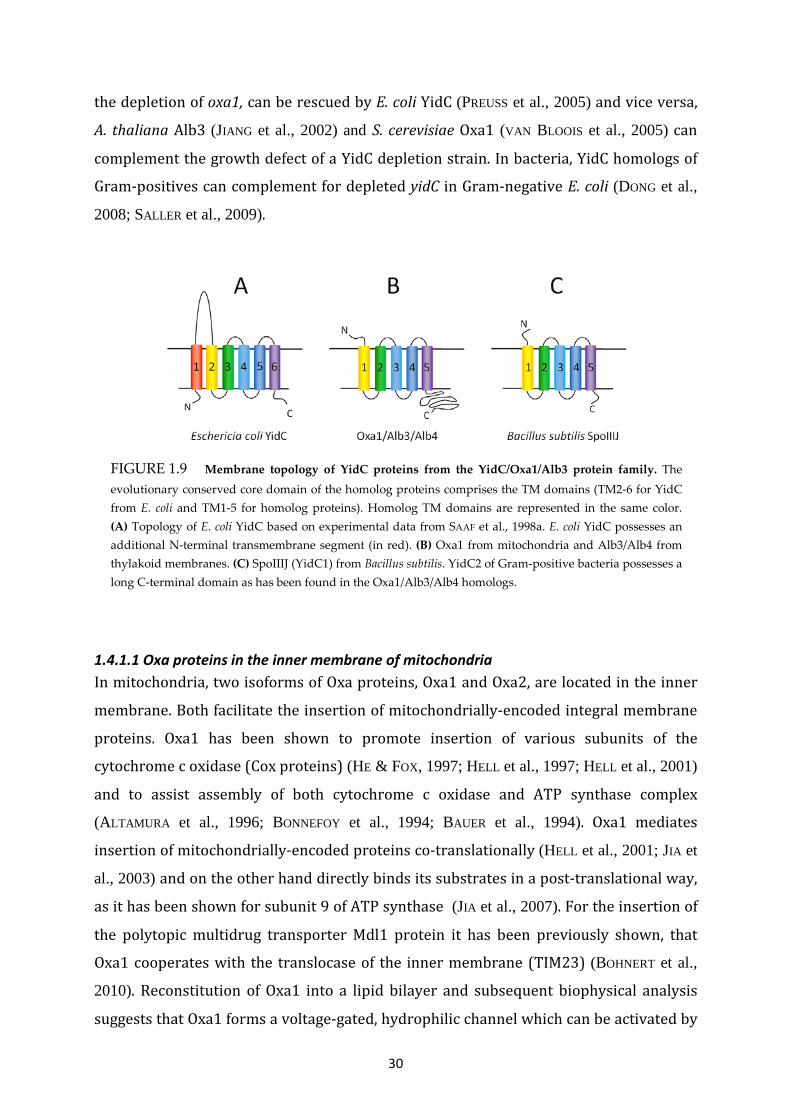
30
FIGURE 1.9 Membrane topology of YidC proteins from the YidC/Oxa1/Alb3 protein family. The evolutionary conserved core domain of the homolog proteins comprises the TM domains (TM2-6 for YidC from E. coli and TM1-5 for homolog proteins). Homolog TM domains are represented in the same color. (A) Topology of E. coli YidC based on experimental data from SAAF et al., 1998a. E. coli YidC possesses an additional N-terminal transmembrane segment (in red). (B) Oxa1 from mitochondria and Alb3/Alb4 from thylakoid membranes. (C) SpoIIIJ (YidC1) from Bacillus subtilis. YidC2 of Gram-positive bacteria possesses a long C-terminal domain as has been found in the Oxa1/Alb3/Alb4 homologs.
the depletion of oxa1, can be rescued by E. coli YidC (PREUSS et al., 2005) and vice versa,
A. thaliana Alb3 (JIANG et al., 2002) and S. cerevisiae Oxa1 (VAN BLOOIS et al., 2005) can
complement the growth defect of a YidC depletion strain. In bacteria, YidC homologs of
Gram-positives can complement for depleted yidC in Gram-negative E. coli (DONG et al.,
2008; SALLER et al., 2009).
1.4.1.1 Oxa proteins in the inner membrane of mitochondria In mitochondria, two isoforms of Oxa proteins, Oxa1 and Oxa2, are located in the inner
membrane. Both facilitate the insertion of mitochondrially-encoded integral membrane
proteins. Oxa1 has been shown to promote insertion of various subunits of the
cytochrome c oxidase (Cox proteins) (HE & FOX, 1997; HELL et al., 1997; HELL et al., 2001)
and to assist assembly of both cytochrome c oxidase and ATP synthase complex
(ALTAMURA et al., 1996; BONNEFOY et al., 1994; BAUER et al., 1994). Oxa1 mediates
insertion of mitochondrially-encoded proteins co-translationally (HELL et al., 2001; JIA et
al., 2003) and on the other hand directly binds its substrates in a post-translational way,
as it has been shown for subunit 9 of ATP synthase (JIA et al., 2007). For the insertion of
the polytopic multidrug transporter Mdl1 protein it has been previously shown, that
Oxa1 cooperates with the translocase of the inner membrane (TIM23) (BOHNERT et al.,
2010). Reconstitution of Oxa1 into a lipid bilayer and subsequent biophysical analysis
suggests that Oxa1 forms a voltage-gated, hydrophilic channel which can be activated by

31
the binding of small peptides of natural substrate proteins (KRÜGER et al., 2012). The
second isoform, Oxa2 (also called Cox) was initially identified in a screen of mutants
impaired in activity of the cytochrome c oxidase (TZAGOLOFF et al., 1990). Oxa2/Cox is
required for the post-translational insertion of the C-terminal part of Cox2p (SARACCO &
FOX, 2002). Here, Oxa2 cooperates with Oxa1, which promotes insertion of the N-
terminal region of Cox2p (HE & FOX, 1997; HELL et al., 1997).
1.4.1.2 Alb proteins in the thylakoid membrane of chloroplasts The thylakoid-localized membrane protein Alb3 was designated to be the homolog of
yeast mitochondria Oxa1 with a similar, essential insertase activity in chloroplasts
thylakoid membranes (SUNDBERG et al., 1997). So far, the only substrates, destined for the
Alb3 mediated insertion pathway, are nuclear-encoded light-harvesting chlorophyll-
binding proteins (LHCPs) of the photosystems I and II (MOORE et al., 2000). To exert
membrane biogenesis of LHCPs, Alb3 cooperates in a post-translational way with
chaperones of the stroma, the SRP system of chloroplasts (cpSRP43, cpSRP54), and its
receptor FtsY. Furthermore, Alb3 cooperates most likely with the cpSec translocase after
import of LHCPs into the chloroplast stroma has occurred (reviewed in ANKELE et al.,
2012). Another, less important isoform found in thylakoid membranes, is Alb4. Alb4 exhibits
a less pronounced phenotype than the Alb3 albino phenotype does (GERDES et al., 2006;
BENZ et al., 2009). Unlike Alb3, Alb4 does not function as an insertase but rather acts as a
stabilization and assembly factor of chloroplast ATP synthase (BENZ et al., 2009).
1.4.2 X-RAY STRUCTURE OF YidC2 FROM BACILLUS HALODURANS
Like many other Gram-positive bacteria, the genomes of Bacillus species contain two
yidc genes, encoding for the membrane proteins YidC1 (SpoIIIJ) and YidC2 (YqjG) (FUNES
et al., 2009). Knock-out studies have shown, that at least one isoform is sufficient for cell
viability, whereas the inactivation of both genes is lethal (MURAKAMI et al., 2002). Both
isoforms are synthesized with an N-terminal cleavable signal sequence and contain the
conserved core-region of five TM domains, whereas only YidC2 comprises an extended
C-terminal tail like Oxa1 and Alb3 (TJALSMA et al., 2003; FUNES et al., 2009).

32
The recently obtained high resolution x-ray structure of B. halodurans YidC2 at 2.4 Å,
obtained by KUMAZAKI et al., constitutes a major breakthrough in the field of YidC
mediated membrane insertion. The structure reveals a hydrophilic groove, formed by
the five TM domains, which is open towards both the hydrophobic core of the lipid
bilayer and the cytoplasm, but closed at the periplasmic side by tightly packed
hydrophobic side chains of the TM helices and the connecting periplasmic loops
(KUMAZAKI et al., 2014a).
KUMAZAKI et al. presented the nearly identical structures of two YidC2 constructs (PDB
entries: 3WO6(residues 27-266) and 3WO7(residues 27-267)). Both are lacking the N-terminal
signal sequence and most of the C-terminal sequences. In the following I will refer to the
structural details of YidC2(residues 27-266) as represented in Figure 1.10. The obtained
structure shows the detailed organization of the periplasmic domains, the TM helices of
the core region (TM1-5) and the cytoplasmic domains. The P1 domain is shown as an
amphipathic helix lying nearly in parallel to the membrane interface which is consistent
with the x-ray structure of the corresponding region in E. coli YidC (OLIVER & PAETZEL,
2008; RAVAUD et al., 2008). The TM1 helix is relatively long, kinked at conserved proline
residues (P74, P98) and protrudes into the C1 domain, consisting of two α-helices which
form a helical hairpin of mostly hydrophilic residues at the cytoplasmic surface of the
membrane. The cytoplasmic halves of the core domain TM1-5 form the hydrophilic
groove which is coated by conserved hydrophilic residues, distributed in all five TM
domains. Interestingly, a highly conserved arginine residue in TM1 (R72) is located in
the central region of the hydrophilic cavity (shown as sticks and labeled in Figure 1.10A
and C).
The hydrophobic core of YidC2 consists of the tightly packed periplasmic halves of TM1-
5 and the P2 domain, both capping the hydrophilic cavity towards the periplasm (Figure
1.10 C). The hydrophobic gasket has been shown to be impermeable for ions and water
molecules (KUMAZAKI et al., 2014a). However, this finding contradicts recently obtained
experimental data on the reconstituted YidC homolog Oxa1 from yeast mitochondria,
suggesting the formation of a voltage-gated hydrophilic channel pore which is
permeable for cations (KRÜGER et al., 2012).

33
The local crystallographic B-factor (Debye-Waller factor) indicates static and dynamic
disorder of atoms in the protein crystal. Together with molecular dynamics simulations
the overall B-factor assign the highest flexibility of YidC2 to the C1 region and the
cytoplasmic halves of the TM regions. However, the hydrophobic core shows a lower B-
factor and seems to be the rigid part of the YidC2 protein (Figure 1.10B). In addition, a
superimposition of the two obtained structures, YidC(residues 27-266) and YidC(residues 27-267)
shows a structural flexibility of the hydrophilic groove and the C1 region (KUMAZAKI et
al., 2014a).
Based on the solved crystal structure, KUMAZAKI et al. used site directed mutagenesis of
the B. subtilis orthologue SpoIIIJ for the determination of functionally important regions.
First of all, single point mutations of the conserved charged R73 residue in TM1
FIGURE 1.10 Structure of Bacillus halodurans YidC2 (PDB entry 3WO6). (A) Cartoon representation viewed in the plane of the membrane, looking into the hydrophilic groove (left). TM domains and connecting loops are numbered. The conserved Arg residue R72 is labeled and shown as sticks. (B) Surface representation orientated as shown in (A), looking directly into the groove. The crystallographic B-factor of the overall structure which reflects mobility and flexibility of structural elements is rainbow colored from red (highest B-factor – reflecting highest mobility and flexibility) to blue (lowest B-factor). (C) View from the periplasm, showing that the hydrophilic groove is closed towards the periplasm. (Adapted from

34
(corresponding to R72 in YidC2) to M, A, Q, E or D significantly decreased the insertion
activity of SpoIIIJ and resulted in a lethal phenotype as observed in a complementation
assay. Solely the substitution of R73 by a positively charged lysine can partially rescue
the insertion of MifM, which has been used in this assay as a model substrate of SpoIIIJ
(CHIBA et al., 2009). Other single residues of the hydrophilic groove do not seem to be as
important for activity, since ten of the polar residues coating the groove can be replaced
by the non-polar amino acid alanine. KUMAZAKI et al. then investigated the importance of
the amphipathic helix P1, the groove-capping P2 domain and the highly flexible C1
region. The results show clearly that the partial deletion of distinct parts of the tested
domains impaired both the insertion of MifM and cell viability. This is mostly consistent
with data from experiments assessing the functional important regions of E. coli YidC
(JIANG et al., 2003).
In summary, the first high resolution structure of a YidC-like protein remarks a
milestone in discovering the molecular organization and function of YidC family
proteins. The structure reveals a remarkable tertiary arrangement of a protein inside
the membrane, since the functional important hydrophilic groove is open towards the
lipid bilayer and the cytoplasm but closed to the periplasm (KUMAZAKI et al., 2014a).
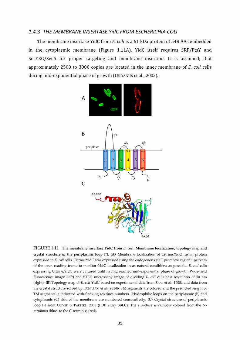
35
1.4.3 THE MEMBRANE INSERTASE YidC FROM ESCHERICHIA COLI
The membrane insertase YidC from E. coli is a 61 kDa protein of 548 AAs embedded
in the cytoplasmic membrane (Figure 1.11A). YidC itself requires SRP/FtsY and
SecYEG/SecA for proper targeting and membrane insertion. It is assumed, that
approximately 2500 to 3000 copies are located in the inner membrane of E. coli cells
during mid-exponential phase of growth (URBANUS et al., 2002).
FIGURE 1.11 The membrane insertase YidC from E. coli: Membrane localization, topology map and crystal structure of the periplasmic loop P1. (A) Membrane localization of Citrine:YidC fusion protein expressed in E. coli cells. Citrine:YidC was expressed using the endogenous yidC promotor region upstream of the open reading frame to monitor YidC localization in as natural conditions as possible. E. coli cells expressing Citrine:YidC were cultured until having reached mid-exponential phase of growth. Wide-field fluorescence image (left) and STED microscopy image of dividing E. coli cells at a resolution of 50 nm (right). (B) Topology map of E. coli YidC based on experimental data from SAAF et al., 1998a and data from the crystal structure solved by KUMAZAKI et al., 2014b. TM segments are colored and the predicted length of TM segments is indicated with flanking residues numbers. Hydrophilic loops on the periplasmic (P) and cytoplasmic (C) side of the membrane are numbered consecutively. (C) Crystal structure of periplasmic loop P1 from OLIVER & PAETZEL, 2008 (PDB entry 3BLC). The structure is rainbow colored from the N-terminus (blue) to the C-terminus (red).

36
Although a local accumulation of YidC proteins at the cell poles was proposed (URBANUS
et al., 2002), data obtained from high resolution fluorescence microscopy suggest that
YidC is rather distributed in the whole cell (KLENNER, GERKEN & KUHN, unpublished data)
(Figure 1.11A). Hydrophobicity and PhoA fusion analyses suggest a membrane topology
of six TM domains with an Ninside-Cinside orientation (Figure 1.11B) (SÄÄF et al., 1998a).
The N-terminal TM domain serves as an uncleaved signal anchor sequence and is not present
in other members of the protein family (SÄÄF et al., 1998a). TM1 and TM2 are connected by
a large hydrophilic domain, called loop P1, of approximately 320 AAs. Until last,
structural information of E. coli YidC was limited to high resolution structures from the
loop P1, and a low resolution projection map of reconstituted YidC from cryo-EM
analysis of 2D crystals (OLIVER & PAETZEL, 2008; RAVAUD et al., 2008; LOTZ et al., 2008).
Recently, the crystal structure of E. coli YidC was refined at 3.2 Å resolution (KUMAZAKI et
al., 2014b). The characteristics of the structure will be described in detail in the following
chapter 1.4.3.1.
The loop P1 of E. coli YidC consists primarily of a large β-supersandwich with a short α-
helical domain within and at the edge of the sandwich, respectively, and two α-helices at
the very C-terminus, which seem to be a flexible linker between the P1 loop and the
downstream core domain (OLIVER & PAETZEL, 2008; RAVAUD et al., 2008; KUMAZAKI et al.,
2014b) (Figure 1.11C). Surprisingly, approximately 92 % of the large periplasmic domain
P1 can be deleted without affecting YidC activity (JIANG et al., 2003). However, it has been
found that regions of the loop P1 mediate interaction with SecF (XIE et al., 2006).
Recently obtained data from cross-linking studies by HENNON & DALBEY gathered first
insights into the orientation of the C-terminal transmembrane domains TM2-6
constituting the core region of YidC. Their results suggest that TM3, TM4, TM5 and TM6
each have a helical face towards TM2 (HENNON & DALBEY, 2014). In addition, the
cytoplasmic regions of TM3-TM6 seem to be very flexible, based on the stable
intermolecular cross-links between TM2 and a range of residues in TM3-TM6, that have
been observed with bifunctional cross-linkers of different spanner length (HENNON &
DALBEY, 2014). The spatial arrangement of the TM helices as well as the flexibility of the
cytoplasmic halves of the TM helices obtained by HENNON & DALBEY seem to be partially
consistent with the recently obtained crystal structure of B. halodurans YidC2 and E. coli
YidC (KUMAZAKI et al., 2014a; KUMAZAKI et al., 2014b). The C-terminal end of E. coli YidC

37
(16 AAs) is short and moderately positively charged compared to the corresponding
regions in various homologs from other species (KIEFER & KUHN, 2007; SEITL et al., 2013).
In the past, it has been discussed controversially, whether YidC acts as a monomer or in
an oligomeric state. Evidence that YidC is mostly present in a dimeric form has come
from blue native polyacrylamide gel electrophoresis (BN-PAGE) experiments (BOY &
KOCH, 2009). In addition, the placement of a single cysteine residue in TM1 converts
overexpressed YidC almost completely into the dimeric form (KLENNER, unpublished
data). A 2D projection map of reconstituted, detergent-solubilized E. coli YidC suggests a
dimeric state with an anti-parallel face-to-face orientation (LOTZ et al., 2008). The low
resolution cryo-EM structures of both a RNC-YidC and RNC-Oxa1 complex have drawn a
similar picture: Here, YidC and Oxa1 were modeled as dimers bound to the translating
ribosome (KOHLER et al., 2009). Oxa1 dimeric and tetrameric state has been suggested to
be the active unit (NARGANG et al., 2002; KRÜGER et al., 2012). SpoIIIJ from B. subtilis is
able to form dimers presumably stabilized by a disulfide bound between a conserved
cysteine in TM2 of each monomer (CÔRTE et al., 2014). Interestingly, sporulation is
blocked when the cysteine is mutated, thus the active state of SpoIIIJ seems to be the
dimeric form. On the other hand, recent studies propose a monomeric state of E. coli
YidC to be the functional unit. First of all, the crystal structure of YidC2 and E. coli YidC
clearly shows YidC as a monomer (KUMAZAKI et al., 2014a; KUMAZAKI et al., 2014b). In
addition, two cryo-EM structures have shown a single copy of YidC bound to an active
ribosome (SEITL et al., 2013; WICKLES et al., 2014). Furthermore, fluorescence cross-
correlation spectroscopy (FCCS) studies revealed that a single copy of YidC is sufficient
to bind an active ribosome (KEDROV et al., 2013). Examination of the overexpressed and
purified SecYEG-SecDF-YajC-YidC holocomplex by BN-PAGE shows, that YidC is present
within the complex as a monomer (SCHULZE et al., 2014).
Taken together, YidC-like proteins seemingly tend to dimerize and oligomerize. The
physiological relevance of this observation is not clear in all cases. It has been shown
that E. coli YidC is fully active in a monomeric state.
1.4.3.1 Crystal structure of Escherichia coli YidC Recently, KUMAZAKI et al. have published the structure of E. coli YidC at 3.2 Å
resolution (KUMAZAKI et al., 2014b). The structure was determined by using structural
data of the P1 domain (PDB entry 3BS6; RAVAUD et al., 2008) and the TM regions of B.

38
FIGURE 1.12 Crystal structure of Escherichia coli YidC (PDB entry 3WVF). (A) Cartoon representation viewed in the plane of the membrane, looking into the hydrophilic groove (left) and viewed from the side (right). The conserved Arg residue R366 in the center of the hydrophilic groove is shown as sticks and indicated with a black arrowhead (left). (B) Representation of local B-factors orientated as shown in (A), looking directly into the groove. The crystallographic B-factor of the overall structure which reflects mobility and flexibility of structural elements is rainbow colored from red in thick lines (highest B-factor – reflecting highest mobility and flexibility) to blue in thin lines (lowest B-factor).
halodurans YidC2 (PDB entry 3WO6; KUMAZAKI et al., 2014a) for molecular replacement.
The structure comprises the TM domains 2-6 and the P1 and C1 regions (Figure 1.12
and Figure 1.13). The first TM domain (TM1) as well as the C2, C3 and the tail regions is
structurally disordered.

39
The arrangement of the five TM domains is similar to that of B. halodurans: a hydrophilic
groove is formed by loosely packed portions of TM α-helices 1-5 at the cytoplasmic
leaflet and tightly packed domains of the α-helices in the periplasmic leaflet, capping the
groove towards the periplasm. The groove is coated by conserved hydrophilic residues
distributed in TM domains 2-6 and opened towards the cytoplasmic side and the
membrane interior by a gap between TM3 and TM5 (KUMAZAKI et al., 2014b) (Figure
1.12) . In the central region of the hydrophilic cavity, a highly conserved residue of TM2,
i.e. R366, is located at exactly the same position as the corresponding residue R72 in the
structure of B. halodurans YidC2 (Figure 1.10 and Figure 1.12).
In the E. coli YidC structure the large P1 domain is identical to the previously published
structures of the P1 domains (OLIVER & PAETZEL, 2008; RAVAUD et al., 2008; KUMAZAKI et
al., 2014b). The extracellular P1 domain is linked to TM2 via a short membrane
embedded α-helix (Figure 1.12A). Residues of the P1 domain forms several hydrogen
bonds with residues of the P2 domain (connecting TM3 and TM4) and TM3, suggesting
that the spatial orientation of the P1 domain is stabilized by these means and in addition
by interactions with membrane lipids (KUMAZAKI et al., 2014b; RAVAUD et al., 2008).
It is consistent with the B. halodurans structure that the most flexible region of E. coli
YidC was assigned to the C1 region, consisting of two antiparallel α-helices at the
cytoplasmic leaflet of the membrane and to the N-terminal region of TM3(Figure 1.12B).
This suggests that the flexibility of the C1 domain and TM3 is a conserved structural
feature which is important for YidC function.
In summary, the first crystal structure of E. coli YidC reveals that the formation of a
hydrophilic groove by the transmembrane core domains TM2-6 is the structural basis
for YidC-mediated insertion. The sequence alignment of the YidC/Oxa1/Alb3 family
members shows clearly a high conservation of the positively charged residue which is
located in the center of the hydrophilic groove (Figure 1.13).
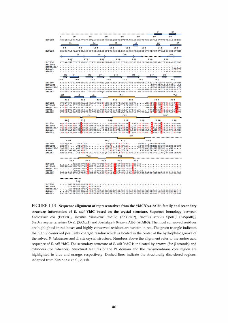
40
FIGURE 1.13 Sequence alignment of representatives from the YidC/Oxa1/Alb3 family and secondary structure information of E. coli YidC based on the crystal structure. Sequence homology between Escherichia coli (EcYidC), Bacillus halodurans YidC2, (BhYidC2), Bacillus subtilis SpoIIIJ (BsSpoIIIJ), Saccharomyces cerevisiae Oxa1 (ScOxa1) and Arabidopsis thaliana Alb3 (AtAlb3). The most conserved residues are highlighted in red boxes and highly conserved residues are written in red. The green triangle indicates the highly conserved positively charged residue which is located in the center of the hydrophilic groove of the solved B. halodurans and E. coli crystal structure. Numbers above the alignment refer to the amino acid sequence of E. coli YidC. The secondary structure of E. coli YidC is indicated by arrows (for β-strands) and cylinders (for α-helices). Structural features of the P1 domain and the transmembrane core region are highlighted in blue and orange, respectively. Dashed lines indicate the structurally disordered regions. Adapted from KUMAZAKI et al., 2014b.
41
1.4.3.2 YidC and the ribosome The long positively charged C-terminus of many YidC homologs was assigned to enable
an electrostatic interaction with ribosomes during co-translational insertion (JIA et al.,
2003; PREUSS et al., 2005; KOHLER et al., 2009; GRUSCHKE et al., 2010; HAQUE et al., 2010;
PALMER et al., 2012; SEITL et al., 2013). The C-terminal end of YidC is short (approximately
18 AAs) compared to the homologs (approximately 100) and less positively charged (+7
compared to ±20) (SEITL et al., 2014). An efficient association of E. coli YidC with
ribosomes has been shown by using YidC with a C-terminal hexa-histidine tag
(protonated at acidic pH) (KOHLER et al., 2009) and a chimera YidC with the fused C-
terminus of planctomycete Rhodopirellula baltica (SEITL et al., 2013; WICKLES et al.,
2014). A recent study revealed, that the binding of detergent–solubilized histidine tagged
YidC to the ribosomes is significantly decreased under physiological conditions, when
histidine residues are not protonated (KEDROV et al., 2013). However, the reconstitution
of YidC into lipid-nanodiscs leads to an efficient ribosome binding at the physiological
condition at pH 7.4. Interestingly, the interaction is increased, when the ribosome is
loaded with an emerging nascent chain of a substrate protein (KEDROV et al., 2013).
Although YidC seems to bind not only to the exit tunnel (WELTE et al., 2012), ribosome
binding sites for YidC were designated to the ribosomal proteins L23, L24, L29 and to
RNA helix 59, located in the vicinity of the ribosomal tunnel exit (KOHLER et al., 2009;
SEITL et al., 2013; WICKLES et al., 2014).
1.4.3.3 Functionally important regions of Escherichia coli YidC Functionally critical regions of E. coli YidC have been defined in a detailed study using
mutagenesis and subsequent complementation and insertion assays (JIANG et al., 2003).
The results, summarized in Figure 1.14, indicate that the C-terminal residues of loop P1,
in the crystal structure seen as an amphipathic helix, and the hydrophilic loops P2 and
P3 are functional domains. In addition, JIANG et al. found that the core region of YidC,
comprising TM2-6, is critical for YidC activity. However, TM4 and TM5 can be replaced
by non-homologous TM domains of leader peptidase without loss of function. In TM2,
TM3 and TM6 single point mutations of I361, L436 and Y516 to the residue serine lead
to inactivation of YidC. Recently, reinvestigation of the importance of the C1 domain has
shown that deletion of the entire C1 domain (residues 371 to 416) leads to a lethal
phenotype, when the expression of the chromosomal encoded yidC gene was inhibited
(CHEN et al., 2014).

42
FIGURE 1.14 Functionally important regions of E. coli YidC. Several deletions or even single point mutations in distinct domains lead to a loss of YidC activity (indicated by symbols in the topology model of YidC on the left and described in the legend on the right). The illustration summarizes results from JIANG et al. 2003, YUAN et al. 2007, WICKLES et al. 2014 and CHEN et al., 2014.
Another study has shown, that single point mutations of two residues located in TM3
lead to a cold-sensitive phenotype (P431L, C423R; Figure 1.14) (YUAN et al., 2007). In
addition, a suppressor mutation of C423R, to rescue the lethal phenotype at the
nonpermissive temperature, has been found in TM2 at residue T362 when mutated to
glutamic acid (T362E). Further evidence for functionally important residues has been
adduced recently by WICKLES et al. Based on their structural model of E. coli YidC, two
single residues which are suggested to be important for strong helix-helix interactions to
stabilize the core helix bundle were mutated. Consequently, the mutation of T362 (TM2)
and Y517 (TM6) to the non-polar residue alanine completely inactivated YidC (WICKLES
et al., 2014). In addition, deletion or mutation of residues identified to bind translating
ribosomes leads to a lethal phenotype, i. e. Δ487-489 and D488K/A (C2 loop) and the
double mutation Y370A/ Y377A (C1 loop).
Based on the recently obtained crystal structure of B. halodurans YidC2, mutational
analysis in B. subilis SpoIIIJ revealed a functionally critical role for the conserved
arginine residue (R72) located in the center of the hydrophilic groove (KUMAZAKI et al.,
2014a). In E. coli YidC, the serial mutation of five residues in the corresponding region to
serine (R366-GIM-Y370 in TM2) did not have an effect on the insertion of a model
substrate (JIANG et al., 2003) but the single point mutations R366E and R366D
43
inactivated YidC, since the insertion of Pf3-Lep was blocked (CHEN et al., 2014). Further
growth complementation assays with E. coli YidC variants with changes at residue 366
showed, that the conserved R366 residue is not that strictly required for the function of
E. coli YidC as found for B. halodurans YidC2 (KUMAZAKI et al., 2014a; KUMAZAKI et al.,
2014b).
1.4.3.4 Cellular response upon YidC depletion Quantitative analyses of both changes in gene expression and the constitution of the
inner membrane proteome upon YidC depletion provide a detailed insight into the
reaction of cells upon the depletion of YidC and underline the physiological importance
of YidC for essential cellular processes (PRICE et al., 2010; WANG et al., 2010; WICKSTRÖM
et al., 2011b). YidC depletion results in growth defects after 5 hours (h) - YidC depleted
cells are significantly increased in length compared to wild type cells. Most likely, this is
a result of the loss of membrane integrity upon YidC depletion. Additionally, the
depletion leads to a significantly decreased motility (WANG et al., 2010). A typical effect
of YidC depletion is the overexpression of the phage shock protein A (PspA), which is
thought to be induced by conditions affecting the pmf to maintain integrity of the
cytoplasmic membrane (VAN DER LAAN et al., 2003; JONES et al., 2003; DARWIN et al., 2005).
In case of YidC depletion, defects in the assembly of cytochrome o oxidase and the FoF1-
ATP synthase lead to a reduction of the pmf (VAN DER LAAN et al., 2003). Interestingly,
transcriptome studies revealed that the level of pspA transcripts is 15–fold higher in
YidC depleted cells, compared to the induction caused by disrupting the pmf with the
protonophore CCCP (carbonyl cyanide m-chlorophenylhydrazone) (WANG et al., 2010).
This suggests that the strong PspA response is not only coupled to the disruption of the
pmf. The activation of the Cpx stress pathway, which is generally induced after
alteration of inner membrane integrity (reviewed in RAIVIO, 2014) has also been observed
in YidC depleted cells (WANG et al., 2010; SHIMOHATA et al., 2007). Another effect
observed upon YidC depletion is the upregulation of cytoplasmic chaperones, like DnaK,
DnaJ, GroEL, and ClpB (PRICE et al., 2010). Analysis of the inner membrane proteome using
two-dimensional blue native/SDS-PAGE (2D BN/SDS-PAGE) has shown that - in addition to
DnaK and GroEL - the expression of periplasmic and membrane embedded chaperones like
PpiD (induced by the Cpx stress pathway), OppA, FtsH, HflC, Hflk is highly upregulated in
YidC depleted cells (WICKSTRÖM et al., 2011b).

44
FIGURE 1.15 YidC substrates and their membrane topology. YidC mediates the insertion of small phage coat proteins Pf3 coat and M13 procoat (left side, in blue). M13 procoat is synthesized with an N-terminal cleavable signal sequence (processing by leader peptidase after insertion is indicated with an arrowhead). In addition, YidC mediates the insertion of endogenous E. coli proteins MscL, possessing a cytoplasmic α-helical domain (in red), subunit c of the ATPase (Foc) and the TAMP protein TssL, a component of the Type IV secretion system of enteroaggregative E. coli (right side, in green).
1.4.4 YidC MEDIATES MEMBRANE PROTEIN INSERTION AND ASSISTS IN
FOLDING AND ASSEMBLY
So far, the number of proteins destined to use solely YidC for membrane integration
is relatively limited (Figure 1.15). The function of YidC as a Sec-independent working
membrane insertase has been initially shown for the insertion of small phage coat
proteins M13 procoat and Pf3 coat, both of which are sec-independent proteins (WOLFE
et al., 1985; ROHRER & KUHN, 1990). M13 procoat consists of 79 residues and comprises
one TM domain and an N-terminal cleavable signal sequence. Pf3 coat is a protein of 44
residues and consists of one TM domain. It has been shown that membrane insertion of
both proteins was highly impaired in cells depleted of YidC (SAMUELSON et al., 2000;
SAMUELSON et al., 2001; CHEN et al., 2002). That the observed insertion deficiency was not
substantiated by a pleiotropic effect as a result of YidC depletion has been shown for
both phage proteins by the observation of an efficient insertion into proteoliposomes
containing YidC as the unique proteinous factor (SEREK et al., 2004; STIEGLER et al., 2011).
In addition, a contact of inserting Pf3 coat protein to YidC has been demonstrated in
extensive studies (CHEN et al., 2002; YU et al., 2008; GERKEN et al., 2008; WINTERFELD et al.,
2009; IMHOF et al., 2011; ERNST et al., 2011; WINTERFELD et al., 2013).
45
The first endogenous E. coli substrate has been identified to be the subunit c of the F1Fo-
ATP synthase (Foc), a protein consisting of 79 residues and two TM domains connected
by a cytoplasmic loop (Figure 1.15). Membrane-inserted Foc protein oligomerizes to a
decamer forming the proton-conducting c-ring of the membrane embedded Fo part of
the ATP synthase. Similar to the phage proteins, membrane insertion of Foc was
decreased in cells depleted of YidC (VAN DER LAAN et al., 2003). Later it has been shown
in detail that the insertion of Foc strictly depends on YidC (YI et al., 2003; VAN BLOOIS et
al., 2004; VAN DER LAAN et al., 2004). The second endogenous YidC substrate is
presumably the mechanosensitive channel protein MscL (FACEY et al., 2007; PRICE et al.,
2011; NEUGEBAUER et al., 2012). MscL is made up of 136 residues and comprises two TM
domains, interconnected by a periplasmic loop, and possesses a short α-helical domain
inside the cytoplasm (CHANG et al., 1998). The identification of MscL protein to be a YidC
substrate and the requirement of YidC for the assembly of the pentameric channel is
discussed controversially (POP et al., 2009; PRICE et al., 2011; BERRIER et al., 2011;
NEUGEBAUER et al., 2012). Recently, a YidC dependency on the membrane insertion of
TssL, a tail-anchored membrane protein and component of the TypIV secretion system
of enteroaggregative E. coli (EAEC), has been reported (ASCHTGEN et al., 2012; SOUSSOULA
& KUHN, unpublished data).
A comparison of the so far identified YidC substrates reveals commonalities which seem
to be general criteria for YidC dependency. All proteins are relatively small
(approximately 10 kDa), not exceeding two TM domains and with short hydrophilic
domains. A global approach to analyze the inner membrane proteome upon YidC
depletion clearly has shown that only the biogenesis of membrane proteins with soluble
domains smaller than 100 residues has been impaired (WICKSTRÖM et al., 2011b). A
second specific characteristic of YidC substrates is most likely the distribution of
negative charges in the periplasmic TM flanking regions or even in the TM domains itself
(DALBEY et al., 2014). Pf3 coat protein carries two negative charged Asp residues at
position 7 and 18 in the N-terminal region which is translocated into the periplasm
during insertion. The same applies for Foc; in this case the N-terminus destined to be
located in the periplasm possesses single Glu and Asp residues. In addition, the
periplasmic loops of M13 procoat and MscL are net negatively charged (-3 and -1,
respectively). Regarding the insertion of an artificial model protein which does not
require YidC and the Sec translocon it has been shown clearly that the addition of
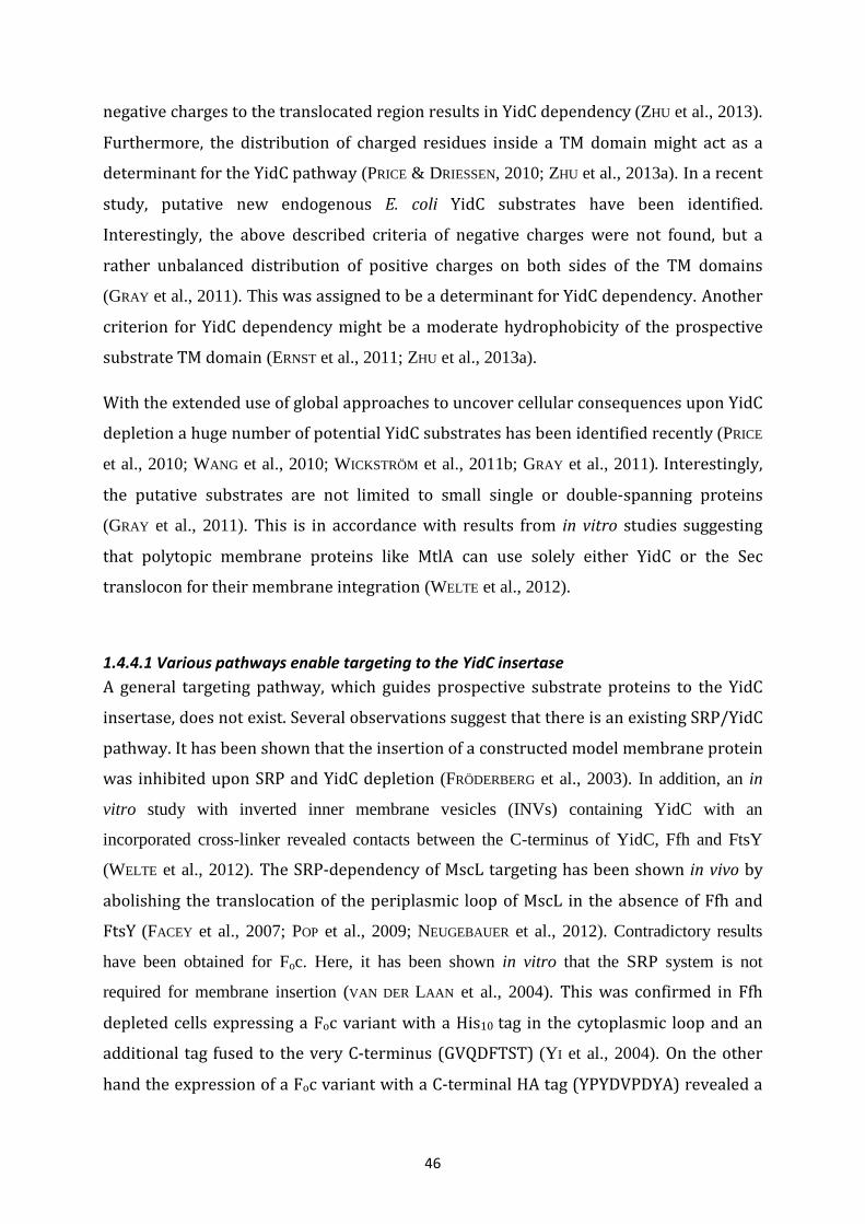
46
negative charges to the translocated region results in YidC dependency (ZHU et al., 2013).
Furthermore, the distribution of charged residues inside a TM domain might act as a
determinant for the YidC pathway (PRICE & DRIESSEN, 2010; ZHU et al., 2013a). In a recent
study, putative new endogenous E. coli YidC substrates have been identified.
Interestingly, the above described criteria of negative charges were not found, but a
rather unbalanced distribution of positive charges on both sides of the TM domains
(GRAY et al., 2011). This was assigned to be a determinant for YidC dependency. Another
criterion for YidC dependency might be a moderate hydrophobicity of the prospective
substrate TM domain (ERNST et al., 2011; ZHU et al., 2013a).
With the extended use of global approaches to uncover cellular consequences upon YidC
depletion a huge number of potential YidC substrates has been identified recently (PRICE
et al., 2010; WANG et al., 2010; WICKSTRÖM et al., 2011b; GRAY et al., 2011). Interestingly,
the putative substrates are not limited to small single or double-spanning proteins
(GRAY et al., 2011). This is in accordance with results from in vitro studies suggesting
that polytopic membrane proteins like MtlA can use solely either YidC or the Sec
translocon for their membrane integration (WELTE et al., 2012).
1.4.4.1 Various pathways enable targeting to the YidC insertase A general targeting pathway, which guides prospective substrate proteins to the YidC
insertase, does not exist. Several observations suggest that there is an existing SRP/YidC
pathway. It has been shown that the insertion of a constructed model membrane protein
was inhibited upon SRP and YidC depletion (FRÖDERBERG et al., 2003). In addition, an in
vitro study with inverted inner membrane vesicles (INVs) containing YidC with an
incorporated cross-linker revealed contacts between the C-terminus of YidC, Ffh and FtsY
(WELTE et al., 2012). The SRP-dependency of MscL targeting has been shown in vivo by
abolishing the translocation of the periplasmic loop of MscL in the absence of Ffh and
FtsY (FACEY et al., 2007; POP et al., 2009; NEUGEBAUER et al., 2012). Contradictory results
have been obtained for Foc. Here, it has been shown in vitro that the SRP system is not
required for membrane insertion (VAN DER LAAN et al., 2004). This was confirmed in Ffh
depleted cells expressing a Foc variant with a His10 tag in the cytoplasmic loop and an
additional tag fused to the very C-terminus (GVQDFTST) (YI et al., 2004). On the other
hand the expression of a Foc variant with a C-terminal HA tag (YPYDVPDYA) revealed a
47
SRP dependency of Foc insertion (VAN BLOOIS et al., 2004). Furthermore, a contact of
nascent TM1 to Ffh has been detected in this study.
It is generally accepted that targeting of the phage proteins Pf3 coat and M13 procoat
occurs independently of the SRP system (CHEN et al., 2002; DE GIER et al., 1998), although
Pf3Lep (a Pf3 coat variant with the P2 domain of leader peptidase fused to the C-
terminus) and M13 procoat could be cross-linked to Ffh (CHEN et al., 2002; VALENT et al.,
1995) (Figure 1.17). For both phage proteins targeting based on electrostatic
interactions with the negatively charged phospholipid head groups is proposed. This has
directly been shown for M13 procoat which exposes positively charged residues at the
N- and C-Terminus (GALLUSER & KUHN, 1990). Pf3 coat protein contains two positive
residues at its C-terminus (Figure 1.16). Mutation of these residues to negatively
charged residues led to strongly reduced membrane association (KUHN, 1995). Thus, a
similar targeting of Pf3 coat protein based on electrostatic interactions is assumed.
In addition, it is possible that cytosolic domains of YidC recognize and directly bind
positively charged residues of substrate proteins (Figure 1.17). This has been shown for
Foc in co-sedimentation studies under YidC depletion conditions, suggesting only weak
interactions of Foc with negatively charged phospholipid head groups (KOL et al., 2008).
1.4.4.2 Pf3 coat protein is a model substrate for YidC mediated insertion Pf3 coat protein is the major coat of filamentous bacteriophage Pf3 infecting Gram-
negative Pseudomonas aeruginosa (STANISICH et al., 1974). Approximately 2500 copies of
Pf3 coat together with minor coat proteins at the tips form a helical capsid of
approximately 6 nm in diameter and up to 1 µm in length to cover the single-stranded
DNA (ssDNA) of 5833 bases (WELSH et al., 1998). After insertion of Pf3 coat into the host
membrane, the monomers oligomerize to encircle the ssDNA, which extrudes out of the
cell as it is proposed for filamentous phage assembly (RUSSEL, 1991). Prior to the
assembly, the secondary structure of Pf3 coat is approximately 40-75 % α-helical and
folds almost completely to an α-helical structure onto phage progeny (AISENBREY et al.,
2006; WELSH et al., 1998).
Pf3 coat protein is a type I membrane protein consisting of 44 AAs. The hydrophobic
transmembrane region (18 AAs) is flanked by a negatively charged N-terminal region
(18 AAs) and a short positively charged C-terminal tail of 8 AAs (Figure 1.16). As
48
FIGURE 1.16 Pf3 coat protein - a model substrate for YidC-mediated insertion. Schematic re-presentation, amino acid sequence and charge distribution of Pf3 coat.
mentioned above the insertion of Pf3 coat does not require SecA and SecYEG but strictly
depends on the YidC insertase (ROHRER & KUHN, 1990; CHEN et al., 2002).
1.4.4.3 YidC in action: Molecular mechanism of membrane insertion The existence of both post-translational and co-translational YidC insertion pathways
has been suggested (Figure 1.17). A post-translational insertion is proposed for M13
procoat and Pf3 coat due to their small size (DE GIER et al., 1998; CHEN et al., 2002). The
cryo-EM structure of YidC bound to a translating ribosome (KOHLER et al., 2009) together
with observed cross-links between ribosomes, Ffh and FtsY (WELTE et al., 2012) in an in
vitro approach support a co-translational mechanism as it has been shown in yeast
mitochondria by Oxa1 (JIA et al., 2003) and for a chimera YidC protein (SEITL et al., 2013;
WICKLES et al., 2014).
The membrane biogenesis of Pf3 coat protein has been studied extensively to uncover
the underlying molecular mechanism of YidC mediated insertion. It has been shown that
during insertion YidC meets its substrates in the conserved core region (CHEN et al.,
2002; YU et al., 2008; NEUGEBAUER et al., 2012). So far, direct substrate contacts to the
non-conserved cytoplasmic regions of YidC have not been observed. Based on
tryptophan fluorescence it has been shown that binding of Pf3 coat to YidC induces
conformational changes within the periplasmic domains (WINTERFELD et al., 2009; IMHOF
et al., 2011). Structural flexibility of the conserved periplasmic loops P2 and P3 is most
likely a prerequisite for proper insertion of substrates. An exception is the P1 loop,
which can be deleted almost completely without loss of function (JIANG et al., 2003).

49
FIGURE 1.17 Targeting of proteins to the YidC insertase and two modes of insertion. (A) Post-translational insertion of substrate proteins - this has been shown for M13 procoat and Pf3 coat (DE GIER et al., 1998; CHEN et al., 2002). Most likely both proteins are targeted to YidC by electrostatic interactions between positively charged residues at the N- or/and the C-terminal end (black circles) and with negatively charged phospholipid head groups (red circles) (GALLUSER & KUHN, 1990; KUHN, 1995). In addition, a direct electrostatic attraction to the cytoplasmic regions of YidC is conceivable (blue circles). The insertion of both proteins requires the proton motive force (pmf) (KUHN et al., 1990; ROHRER & KUHN, 1990). (B) Co-translational insertion and targeting via SRP. RNCs of substrates are targeted to YidC by the SRP system, the insertion occurs in a co-translational way. One example is most likely MscL protein (FACEY et al., 2007). (C) The C-terminus of Rhodopirellula baltica YidC (RbYidC) fused to E. coli YidC can partially substitute for the SRP receptor FtsY and enables a direct contact to a translating ribosome (SEITL et al., 2013) as it was shown for the YidC homolog Oxa1 (JIA et al., 2003).
It was suggested that the translocation of the hydrophilic N-terminus of the Pf3 coat
protein and the insertion of the transmembrane segment is driven by hydrophobic
forces; based on the observation that altering the pH value during fluorescence
measurements did not influence substrate binding (GERKEN et al., 2008). For
translocation of highly negatively charged substrate domains the pmf is required to
support YidC mediated insertion, as it has been found for M13 procoat and Pf3 coat
(KUHN et al., 1990; ROHRER & KUHN, 1990; KIEFER et al., 1997). The insertion of MscL, Foc
and CyoA does not require the pmf (FACEY et al., 2007; VAN DER LAAN et al., 2004; CELEBI
et al., 2006).
The conserved TM3 of YidC seems to play a predominant role in the insertion
mechanism because various proteins, including Pf3 coat, M13 procoat, MscL and FtsQ,
contact TM3 during their membrane insertion (CHEN et al., 2002; NEUGEBAUER et al.,
50
2012; WICKLES et al., 2014). In addition, the transmembrane segment of inserting FtsQ
has been most likely found as an electron density in a cryo-EM structure of YidC next to
TM3 (WICKLES et al., 2014).
A breakthrough in understanding the molecular mechanism of YidC mediated insertion
was achieved recently by solving the x-ray structure of Bacillus halodurans YidC2
(KUMAZAKI et al., 2014a) and the creation of an accurate structural model of E. coli YidC
(WICKLES et al., 2014). Based on the crystal structure and biochemical data it is suggested
that a conserved positively charged arginine residue inside the discovered hydrophilic
groove attracts negatively charged regions of substrate proteins. This allows an initial,
partial insertion of the TM domain into the lipid bilayer. Indeed, the membrane protein
insertion and folding monitor protein MifM (CHIBA et al., 2009), a single spanning
substrate protein of YidC2, was successfully cross-linked into the hydrophilic groove
(KUMAZAKI et al., 2014a). A step-wise neutralization of the acidic N-terminal MifM region
(net charge -3 in the wild type) led to a decreased insertion efficiency until insertion was
almost completely abolished (charge 0). Interestingly, when the acidic residues of a Pf3
coat/MifM fusion protein were mutated, the effect was less pronounced. The crucial role
of the charged N-terminal domain for Pf3 coat insertion has already been shown (KIEFER
et al., 1997). It is proposed that the pmf and/or hydrophobic interactions between the
TM domain and the membrane core facilitate the translocation of the hydrophilic
domain after substrate binding to the hydrophilic cleft. How this exactly happens is still
not clear, because the hydrophilic groove penetrates deep into the YidC core domain and
is closed towards the periplasm by the most rigid parts of the TM domains.
The mechanism described above fits well to single-spanning membrane proteins with a
negatively charged N-terminus. However, the mechanism is based on a conserved
arginine residue within the hydrophilic groove. In E. coli YidC, the corresponding residue
R366 is not essential for function (JIANG et al., 2003) and only substitution to negatively
charged residues inhibit the insertion of Pf3-Lep and to some extent of a M13 procoat
variant (CHEN et al., 2014) . In addition, it remains unclear how double-spanning or even
polytopic membrane proteins are inserted via YidC. Thus, it is conceivable that the
insertion mechanism slightly differs among the YidC homologs and that some YidC
substrates need other requirements for an efficient insertion.
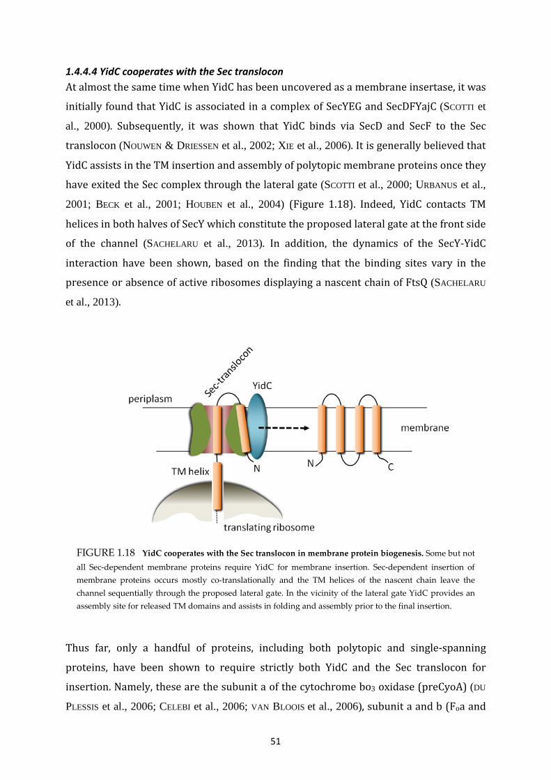
51
FIGURE 1.18 YidC cooperates with the Sec translocon in membrane protein biogenesis. Some but not all Sec-dependent membrane proteins require YidC for membrane insertion. Sec-dependent insertion of membrane proteins occurs mostly co-translationally and the TM helices of the nascent chain leave the channel sequentially through the proposed lateral gate. In the vicinity of the lateral gate YidC provides an assembly site for released TM domains and assists in folding and assembly prior to the final insertion.
1.4.4.4 YidC cooperates with the Sec translocon At almost the same time when YidC has been uncovered as a membrane insertase, it was
initially found that YidC is associated in a complex of SecYEG and SecDFYajC (SCOTTI et
al., 2000). Subsequently, it was shown that YidC binds via SecD and SecF to the Sec
translocon (NOUWEN & DRIESSEN et al., 2002; XIE et al., 2006). It is generally believed that
YidC assists in the TM insertion and assembly of polytopic membrane proteins once they
have exited the Sec complex through the lateral gate (SCOTTI et al., 2000; URBANUS et al.,
2001; BECK et al., 2001; HOUBEN et al., 2004) (Figure 1.18). Indeed, YidC contacts TM
helices in both halves of SecY which constitute the proposed lateral gate at the front side
of the channel (SACHELARU et al., 2013). In addition, the dynamics of the SecY-YidC
interaction have been shown, based on the finding that the binding sites vary in the
presence or absence of active ribosomes displaying a nascent chain of FtsQ (SACHELARU
et al., 2013).
Thus far, only a handful of proteins, including both polytopic and single-spanning
proteins, have been shown to require strictly both YidC and the Sec translocon for
insertion. Namely, these are the subunit a of the cytochrome bo3 oxidase (preCyoA) (DU
PLESSIS et al., 2006; CELEBI et al., 2006; VAN BLOOIS et al., 2006), subunit a and b (Foa and

52
FIGURE 1.19 Topology of membrane proteins that require both YidC and the Sec translocon. YidC cooperates with the Sec translocon for the insertion of polytopic and single-spanning membrane proteins. PreCyoA (subunit a of the cytochrome bo3 oxidase) is synthesized with an N-terminal signal sequence which is cleaved after insertion (the black arrowhead indicates the cleavage site).
Fob) of the F1Fo-ATP synthase (KOL et al., 2009; YI et al., 2003; YI et al., 2004), and the
subunit K of the NADH:ubiquione oxidoreductase (NuoK) (PRICE & DRIESSEN, 2010)
(Figure 1.19). For the membrane biogenesis of preCyoA YidC acts upstream of the Sec
translocon and facilitates insertion of the helical hairpin consisting of the signal
sequence and the first TM domain. For the insertion of the second TM domain and the
large periplasmic domain a coordinated action of SecYEG and SecA is required (CELEBI et
al., 2006; VAN BLOOIS et al., 2006). A downstream action of YidC is conceivable in two
different ways. Firstly, YidC is believed to provide an assembly platform prior to the
sequentially release of TM segments into the bilayer, as it was shown for leader
peptidase (HOUBEN et al., 2004). Secondly, YidC assists in the assembly of released TM
domains to helical bundles prior to the insertion into the membrane (BECK et al., 2001;
WAGNER et al., 2008).
For many Sec-dependent membrane proteins the orchestrated action of SecY and YidC
seems not to be essentially required although a close proximity of YidC and the inserting
polypeptides has been reported (WELTE et al., 2012; VAN DER LAAN et al., 2004). This
suggests that YidC has an effect on the kinetics of the lateral release of polar domains out
of the channel and might be required for efficient assembly and folding of the released
substrates.
It is not clear how it is decided whether a Sec-dependent protein requires the function of
YidC or not. However, it is suggested that the hydrophobicity of the TM domains and the
distribution of charged residues within the hydrophobic domains or the periplasmic
53
regions determine the YidC dependency (PRICE & DRIESSEN, 2010; NEUGEBAUER et al.,
2012; ZHU et al., 2013a; SOMAN et al., 2014).
1.4.4.5 YidC as a membrane embedded molecular chaperone There are strong hints supporting the idea that YidC might function as a membrane
embedded chaperone to assist folding of polytopic membrane proteins into their final
tertiary conformation (DALBEY & KUHN, 2004). Lactose permease (LacY), consisting of 12
transmembrane helices in E. coli, recruites SRP/FtsY and the Sec translocon for
membrane targeting and insertion (MACFARLANE & MÜLLER, 1995; SELUANOV & BIBI,
1991; ITO & AKIYAMA, 1991). Although YidC is not required for membrane insertion per
se, a direct role of YidC in folding of LacY has been shown (NAGAMORI et al., 2004).
Folding of LacY in YidC depleted membrane vesicles leads to structural alterations
which were indirectly detected with conformational monoclonal antibodies, binding
native folded epitopes on both sides of the membrane (NAGAMORI et al., 2004). In
addition, in vivo studies suggest, that periplasmic loops and helix packing of LacY both
are disordered in the absence of YidC (ZHU et al., 2013b). Similar results have been found
for the protein MalF, a subunit of the MalFGK maltose transporter. Here, the stability of
MalF and even the assembly of the entire MalFGK complex are significantly reduced in
cells depleted of YidC (WAGNER et al., 2008). The upregulation of chaperones as a cellular
response to overcome YidC depletion further support the role of YidC as a chaperone
involved in the folding and assembly of TM domains and folding of hydrophilic loops.
YidC might also be involved in the degradation machinery to remove misfolded proteins
from the membrane, as YidC was successful cross-linked to a large complex of
membrane bound chaperons and proteases FtsH, HflC and HflK (VAN BLOOIS et al., 2008).
Interestingly, YidC appeared to be important for the biogenesis of Sec-dependent
secretory proteins and lipoproteins as well. For the autotransporter hemoglobin
protease (Hbp), a virulence factor secreted by Gram-negative bacteria, it was shown that
the Hbp signal peptide contacts YidC during early biogenesis. It is suggested that YidC
chaperones Hbp into a secretion competent state in the periplasm after its translocation
(JONG et al., 2010). The specific role of YidC in the biogenesis of the lipoproteins Lpp
(murein lipoprotein) and BRP (bacteriocin release protein) remains unclear but it is
assumed that YidC is required for the insertion of the signal peptides and/or fot
chaperoning Lpp and BRP to the Lol-system (FRÖDERBERG et al., 2004).
54
1.4.4.6 YidC assists in the assembly of highly ordered membrane protein complexes The function of YidC to work as an insertase and molecular chaperon is related to its
involvement in the assembly of membrane protein complexes. In comparative analyses
of oligomeric membrane protein complex levels in E. coli cells containing YidC or not, it
was clearly shown that YidC depletion leads to reduced levels of these complexes
(WICKSTRÖM et al., 2011). Specifically, the assembly of the F1Fo-ATP synthase, the
cytochrome bo3 oxidase and the NADH dehydrogenase was reduced by approximately
60 %, approximately 50 % and approximately 30 %, respectively (WICKSTRÖM et al.,
2011). As mentioned above, YidC is crucial for the insertion of subunits of all these
membrane protein complexes. This implies a related function of YidC for the assembly of
respiratory chain and energy-transducing complexes as it was found in yeast
mitochondria for the F1Fo-ATP synthase (JIA et al., 2007). The co-purification of YidC and
subunits of the NADH dehydrogenase, the cytochrome bo3 oxidase and Foc from the
F1Fo-ATP synthase (PRICE et al., 2010; KOL et al., 2008) and the fact that SpoIIIJ and YqjG,
YidC homologs from Bacillus subtilis, copurify with the F1Fo ATP synthase complex,
further support this idea (SALLER et al., 2009).
55
Objectives of this thesis
The membrane insertase YidC inserts newly synthesized proteins into the plasma
membrane. YidC protein of Escherichia coli is a six-spanning protein of 548 amino acid
residues with a large periplasmic domain between the first two TM regions. YidC is
required for the biogenesis of respiratory complexes, ATP synthase and for example the
mechanosensitive channel protein MscL. Also, the coat proteins of filamentous phages
Pf3 and M13 strictly require YidC for membrane insertion.
The best studied YidC substrate is the 44 amino acid residues long Pf3 coat protein of
the phage Pf3, infecting Pseudomonas aeruginosa. During the insertion process, the Pf3
coat protein binds reversibly to YidC and adopts finally a transmembrane conformation.
The aims of this study were the identification of YidC-substrate contacts during
membrane biogenesis of the inserting Pf3 coat protein and the investigation of the
molecular mechanism and the dynamics underlying YidC-mediated insertion. In order
for this to be achieved, a set of single cysteine mutants of YidC and the inserting Pf3 coat
protein were prepared. Next, an in vivo cross-linking assay was established to monitor
YidC-substrate complex formation during the insertion process. Attempts were made to
detect substrate binding in distinct regions of the YidC TM domains.
In addition, this study aims to characterize the insertion process of translocation
deficient Pf3 coat mutants. The translocation of these Pf3 coat variants was blocked due
to charge changes in the N-terminal segment flanking the transmembrane domain.
Dynamic insertion studies allow determining whether these mutants still contact YidC in
an initial state of the partial membrane insertion.

56

57
Chapter 2
The Pf3 coat protein contacts TM1 and TM3 of YidC during membrane biogenesis
CHRISTIAN KLENNERa, JIJUN YUANb, ROSS E. DALBEYb, ANDREAS KUHNa,* a Institute of Microbiology and Molecular Biology, University of Hohenheim, 70599 Stuttgart, Germany b Ohio State University, Department of Chemistry, 100 West 18th Avenue, Columbus, OH 43210, USA Keywords: Membrane insertase; Disulfide mapping; Translocase; Transmembrane helix; Membrane biogenesis; Helix–helix contact ABSTRACT
The coat protein of bacteriophage Pf3 is inserted into the plasma membrane of
Escherichia coli by the insertase YidC. To identify which of the six transmembrane
regions of YidC bind the single-spanning Pf3 coat protein during membrane protein
biogenesis, we used the disulfide cross-linking approach. We generated single cysteines
in each of the transmembrane regions of YidC and in the center of the hydrophobic
region of Pf3 coat protein. We found that the substrate Pf3 coat contacts the first and
third transmembrane segment (TM) of YidC as cross-links between these two proteins
can be formed in vivo during membrane biogenesis. A detailed disulfide-mapping study
revealed that one face of TM3 of YidC makes contact with the Pf3 protein.
INTRODUCTION In bacteria, membrane proteins are inserted into the membrane by two unrelated
evolutionarily conserved pathways. The main pathway requires the protein-conducting
SecYEG channel and accessory components SecDFYajC, YidC, and SecA for membrane
protein insertion (for review see [1,2]). SecY and SecE are related to Sec61a and Sec61c
in the ER system. The other pathway requires the membrane protein insertase YidC for
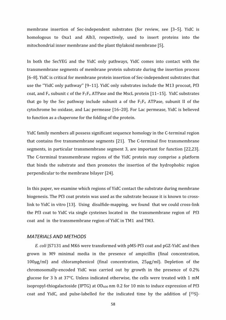
58
membrane insertion of Sec-independent substrates (for review, see [3–5]. YidC is
homologous to Oxa1 and Alb3, respectively, used to insert proteins into the
mitochondrial inner membrane and the plant thylakoid membrane [5].
In both the SecYEG and the YidC only pathways, YidC comes into contact with the
transmembrane segments of membrane protein substrate during the insertion process
[6–8]. YidC is critical for membrane protein insertion of Sec-independent substrates that
use the ‘‘YidC only pathway’’ [9–11]. YidC only substrates include the M13 procoat, Pf3
coat, and Fo subunit c of the F1Fo ATPase and the MscL protein [11–15]. YidC substrates
that go by the Sec pathway include subunit a of the F1Fo ATPase, subunit II of the
cytochrome bo oxidase, and Lac permease [16–20]. For Lac permease, YidC is believed
to function as a chaperone for the folding of the protein.
YidC family members all possess significant sequence homology in the C-terminal region
that contains five transmembrane segments [21]. The C-terminal five transmembrane
segments, in particular transmembrane segment 3, are important for function [22,23].
The C-terminal transmembrane regions of the YidC protein may comprise a platform
that binds the substrate and then promotes the insertion of the hydrophobic region
perpendicular to the membrane bilayer [24].
In this paper, we examine which regions of YidC contact the substrate during membrane
biogenesis. The Pf3 coat protein was used as the substrate because it is known to cross-
link to YidC in vitro [13]. Using disulfide-mapping, we found that we could cross-link
the Pf3 coat to YidC via single cysteines located in the transmembrane region of Pf3
coat and in the transmembrane region of YidC in TM1 and TM3.
MATERIALS AND METHODS
E. coli JS7131 and MK6 were transformed with pMS-Pf3 coat and pGZ-YidC and then
grown in M9 minimal media in the presence of ampicillin (final concentration,
100µg/ml) and chloramphenicol (final concentration, 25µg/ml). Depletion of the
chromosomally-encoded YidC was carried out by growth in the presence of 0.2%
glucose for 3 h at 37°C. Unless indicated otherwise, the cells were treated with 1 mM
isopropyl-thiogalactoside (IPTG) at OD600 nm 0.2 for 10 min to induce expression of Pf3
coat and YidC, and pulse-labelled for the indicated time by the addition of [35S]-
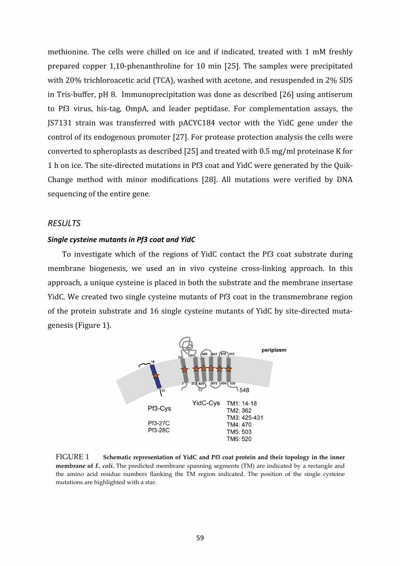
59
FIGURE 1 Schematic representation of YidC and Pf3 coat protein and their topology in the inner membrane of E. coli. The predicted membrane spanning segments (TM) are indicated by a rectangle and the amino acid residue numbers flanking the TM region indicated. The position of the single cysteine mutations are highlighted with a star.
methionine. The cells were chilled on ice and if indicated, treated with 1 mM freshly
prepared copper 1,10-phenanthroline for 10 min [25]. The samples were precipitated
with 20% trichloroacetic acid (TCA), washed with acetone, and resuspended in 2% SDS
in Tris-buffer, pH 8. Immunoprecipitation was done as described [26] using antiserum
to Pf3 virus, his-tag, OmpA, and leader peptidase. For complementation assays, the
JS7131 strain was transferred with pACYC184 vector with the YidC gene under the
control of its endogenous promoter [27]. For protease protection analysis the cells were
converted to spheroplasts as described [25] and treated with 0.5 mg/ml proteinase K for
1 h on ice. The site-directed mutations in Pf3 coat and YidC were generated by the Quik-
Change method with minor modifications [28]. All mutations were verified by DNA
sequencing of the entire gene.
RESULTS
Single cysteine mutants in Pf3 coat and YidC
To investigate which of the regions of YidC contact the Pf3 coat substrate during
membrane biogenesis, we used an in vivo cysteine cross-linking approach. In this
approach, a unique cysteine is placed in both the substrate and the membrane insertase
YidC. We created two single cysteine mutants of Pf3 coat in the transmembrane region
of the protein substrate and 16 single cysteine mutants of YidC by site-directed muta-
genesis (Figure 1).
60
The Pf3 mutations replaced the isoleucine and valine at positions 27 and 28,
respectively. Expression of these Pf3 coat mutants was inducible by adding IPTG to the
growth medium, since the Pf3 genes were cloned into the plasmid pMS119HE [29]. In
YidC, a cysteine residue was placed into the center of each of the six transmembrane
regions at positions 16, 362, 430, 470, 503 and 520 starting with a protein that lacks the
endogenous cysteine at position 423 (cysteine-less background). The YidC mutant genes
were cloned either into pACYC184 (for growth complementation) or the pGZ119EH
plasmid (for membrane protein insertion assay). Both of these plasmids are compatible
with the pMS plasmids [29].
Functionality of the YidC mutants with single cysteines
The functionality of the YidC mutants was determined by testing whether the single
cysteine YidC mutants could complement the YidC depletion strain JS7131. JS7131 has
the YidC gene under the control of the araBAD promoter cloned into the lambda
attachment site and the endogenous YidC gene was inactivated by a deletion [9]. This
strain is arabinose dependent for growth. JS7131 was transformed with the various YidC
mutants in pACYC184 and the strain streaked onto a LB agar plate containing either
0.2% arabinose (Figure 2A, Ara) or 0.2% glucose (Figure 2A, Glc). Figure 2A shows that
all the YidC cysteine mutants could complement the growth defect of the JS7131 strain
under glucose conditions where the chromosomal YidC expression is repressed. Only the
negative control where the JS7131 strain was transformed with the pMS119 plasmid
could not grow on LB agar plates with 0.2% glucose.
Protease mapping studies also verified that the single cysteine YidC mutants were fully
functional as a membrane protein insertase. For these studies, we transformed the YidC
mutants into the arabinose-dependent MK6 strain where, like the JS7131 strain, the
expression of the chromosomal yidC is under the control of the araBAD promoter but the
gene is left at its original position in the chromosome. The YidC cysteine mutants were
coexpressed with the Pf3-P2 coat protein that encodes an extended version of the Pf3
coat protein at the C-terminus to allow immunoprecipitation of the proteinase K
protected fragment (Figure 2B). MK6 cells with both plasmids were grown for 3 h in
medium containing 0.2% glucose to deplete the chromosomal encoded YidC protein.
Radiolabelled cells expressing the YidC mutants and Pf3 coat were subjected to
proteinase K mapping (see Figure 2 legend for details). Significantly, when the single

61
FIGURE 2 Functionality of the YidC cysteine mutants. (A) Complementation of growth of the YidC depletion strain MK6 transformed with plasmids pACYC184 (vector without insert), or with plasmid expressing the YidC423S (cysteine-less YidC), YidC16C, YidC326C, YidC430C, YidC470C, YidC503C, YidC520C mutants, where the number indicates the position of the single cysteine. (B) Membrane insertion of Pf3-P2 in MK6 cells bearing the IPTG inducible pMS119 vector containing Pf3-P2 and pGZ119 vector containing the single cysteine YidC mutant. The cells were induced for 10 min with 1 mM IPTG to express Pf3-P2 and the YidC cysteine mutants followed by pulse-labelling with [35S]-methionine for 1 min, then chased with non-labelled methionine for 1 min and converted to spheroplasts. One aliquot was treated with proteinase K for 60 min on ice (+PK, even lanes), whereas another aliquot was untreated (-PK, odd lanes). The cells were TCA-precipitated and analyzed by SDS–PAGE and phosphorimaging. The percentage of digested Pf3-P2 coat protein was determined. The cells without a YidC encoding plasmid were grown in 0.2% arabinose (lanes 1 and 2) or 0.2% glucose (lanes 3 and 4). Cells bearing the plasmids encoding for the respective YidC mutant were grown in 0.2% glucose (5–16).
cysteine mutants were expressed, efficient insertion of Pf3-P2 does occur as Pf3-P2 is
converted to the shifted form by protease cleavage (see lanes 5–16), but not when cells
without plasmid were depleted of YidC (lane 4; non-digested form of Pf3-P2
accumulated). These studies verified that the YidC mutant proteins are functional.
Interaction between the Pf3 coat protein and YidC
To identify the transmembrane domain of YidC that contacts the Pf3 coat substrate
during membrane biogenesis, we used disulfide-mapping. In this approach, a stable
disulfide-linked YidC-substrate complex will form if the cysteines are in close proximity
under oxidizing conditions. Coexpression of the Pf3 coat and YidC containing single

62
FIGURE 3 The TM1 and TM3 of YidC contact the Pf3 coat protein. MK6 cells bearing the plasmid
pMS-Pf3-27C (lanes 1–14) or pMS-Pf3-28C (lanes 15 and 16) and a second plasmid, pGZ119 encoding the respective single cysteine YidC mutant were grown in the presence of 0.2% glucose to deplete the cells from the chromosomally-encoded YidC. The cells were induced with 1 mM IPTG for 10 min and pulse-labelled with [35S]-methionine for 1 min. One aliquot was put on ice (-CuP, odd lanes), whereas the other aliquot was treated with 1 mM copper phenanthroline (+CuP, even lanes) for 10 min. The cells were TCA-precipitated, immunoprecipitated with antibody to the Pf3 virus, and analyzed on a non-reducing SDS–PAGE and phosphorimaging.
cysteines were analyzed under oxidizing conditions to test whether transmembrane
segments of the substrate and YidC contact each other at the cysteine positions under
oxidizing conditions (Figure 3). MK6 cells harbouring a plasmid coding for Pf3-27C and a
plasmid coding for a single cysteine YidC mutant were grown at 37 °C for 3 h in the
presence of glucose. The proteins were expressed by the addition of 1 mM IPTG for 10
min and then labeled by the addition of [35S]-methionine for 1 min. Then, one aliquot
was incubated with 1 mM copper phenanthroline (CuP) for 10 min at 0 °C (+CuP lanes),
and another aliquot was placed on ice (-CuP). The samples were TCA-precipitated,
resuspended in SDS, and immunoprecipitated with antiserum to Pf3 phage. The samples
were then treated with DTT (-CuP) or not (+CuP) and analyzed on a non-reducing SDS–
PAGE gel. As expected, the addition of the oxidizing agent CuP increases the amount of
Pf3 coat dimer with a concomitant decrease in the amount of the Pf3 coat monomer
(Figure 3; compare even and odd lanes). In addition, in the cells that express Pf3-27C
coat and YidC with one cysteine in the trans- membrane region TM1 or TM3 we
observed an additional band at about 65 kDa which was more intense under oxidizing
conditions (lanes 4 and 8) compared to the reduced conditions (lanes 3 and 7). Under
the reduced condition (lane 3) we observed a slightly lower band at about 60 kDa that
probably corresponds to YidC that dissociated from Pf3 coat protein on the SDS–PAGE
gel after remaining non-covalently bound during the immunoprecipitation. YidC-Pf3
cross-linking at 16 C and 430 C was also observed when Pf3-28C coat was coexpressed
(lanes 15 and 16).

63
To verify that cross-linking occurs between Pf3 coat and certain YidC cysteine mutants,
we tested whether the 65 kDa band is immunoprecipitated with an antibody directed to
the 10his-tag located at the C-terminus of the YidC-Cys mutant proteins (Figure 4A,
lanes 2 and 4). Figure 4A shows with the Pf3 coat 27C and the YidC430C mutant that a
65 kDa band is detected with Pf3 coat antiserum (lane 3) and with the his antiserum
(lane 4). The 65 kDa band that is recognized by the Pf3 and YidC antibodies has a slightly
higher molecular weight than the 10hisYidC alone (lane 2). As expected, no Pf3 coat
monomers or dimers are observed with the his antiserum (lane 4) while they are
detected with Pf3 antiserum (lanes 1 and 3).
FIGURE 4 Analysis of the Pf3-YidC cross-linked products. (A) The cross-linked Pf3-27C – YidC430C is recognized by both antibodies and can be reduced. MK6 cells with the plasmids encoding Pf3-27C and YidC430C were pulse-labelled with [35S]-methionine for 1 min, treated with copper phenanthroline for 10 min (+CuP, lanes 3–5) or were left untreated (-CuP, lanes 1 and 2). One oxidized aliquot was reduced by 1 mM DTT and 1 mM EDTA (lane 5). The samples were immunoprecipitated with antibodies to the Pf3 virus (P, lanes 1, 3, 5, and 6), and to the his10-tag (H, lanes 2, 4, 7, 8, and M). For a control, cells with only the plasmid encoding Pf3-27C (lanes 6 and 7) and cells with only the plasmid encoding YidC430C (lanes 8 and M) are shown. They were grown either in the presence of IPTG or without induction (lane 8). The sample in lane M was precipitated with antibodies to the histag (H) and to OmpA (O), showing YidC430C (Y) and OmpA (O) as size markers. (B) Cross-linking of YidC to Pf3-P2 that has an extended carboxyl-terminal domain. MK6 cells expressing YidC-430C was coexpressed with Pf3-P2 that had a cysteine at position 28. After pulselabelling for 1 min, the cells were chilled on ice and an aliquot was oxidized with CuP for 10 min (+CuP, lanes 3–6). Lanes 1 and 2 show the samples under non-oxidized conditions (-CuP). For the samples in lanes 5 and 6 the cells were converted to spheroplasts and 0.5 mg/ml proteinase K was added for 1 h (lane 6). The samples were TCA-precipitated and immunoprecipitated with antiserum to leader peptidase (L, lanes 1, 3, 5, and 6) and to his-tag (H, lanes M, 2 and 4). YidC has a his10-tag at the C-terminus. As a size marker, YidC430C is shown in lane M.

64
The oxidized samples were reduced by the addition of 1 mM DTT and 1 mM EDTA
resulting in the dissociation of the YidC–Pf3 complex (lane 5). No YidC is detected with
the Pf3 coat antiserum in this sample. As a control, cells expressing the Pf3-27C without
a YidC-Cys mutant were analyzed (lanes 6 and 7). No protein was detectable at the
position of 65 kDa. As a control, we show cells with only the YidC430C plasmid after
induction with IPTG (lane M) and the non-induced cells (lane 8). For a size reference, the
sample shown in lane M was precipitated with OmpA antiserum together with the
antiserum to YidC-his.
Importantly, the substrate that is cross-linked to YidC during a 1 min pulse is not
accessible to protease. We showed this using a Pf3 coat that was extended at the C-
terminus by the P2 domain of leader peptidase. Figure 4B indicates that due to this
extension the Pf3-YidC cross-linked product is now ~ 75 kDa in size (Figure 4B, lane 3,
arrowhead). After oxidation, the cells were converted to spheroplasts and treated with
proteinase K. The Pf3-P2 dimers were clipped by the proteinase K and converted to a
protease-protected fragment that was recognized by the antibody to leader peptidase
(Figure 4B, lane 6), demonstrating that the dimers were inserted across the membrane.
The cross-linked Pf3-P2-YidC was largely resistant to the protease (Figure 4B, lane 6)
suggesting that the N-terminal tail of Pf3-P2 was not fully accessible from the
periplasmic side of the membrane.
To examine how the YidC TM1 and TM3 regions contact the Pf3 coat substrate,
additional residues within TM1 were changed to a cysteine at positions 14–18 and in
TM3 from residues 425 to 431. Coexpression with Pf3-27C coat protein showed that the
interaction with YidC17C (Figure 5A) and with YidC430C (Figure 5B) is very specific. As
a control, Pf3-16C, which has a single cysteine in the amino-terminal hydrophilic region,
did not cross-link to YidC16C (Figure 5A; lane 6). Also, no cross-link was observed with
Pf3-27C when YidC without a cysteine was coexpressed (lane 7). We verified that Pf3
coat and YidC-Cys mutants were expressed at a comparable level (data not shown).
DISKUSSION
In this paper, we show that TM1 and TM3 of YidC make contact with the Pf3 coat
protein during membrane biogenesis. These contacts presumably occur in the substrate
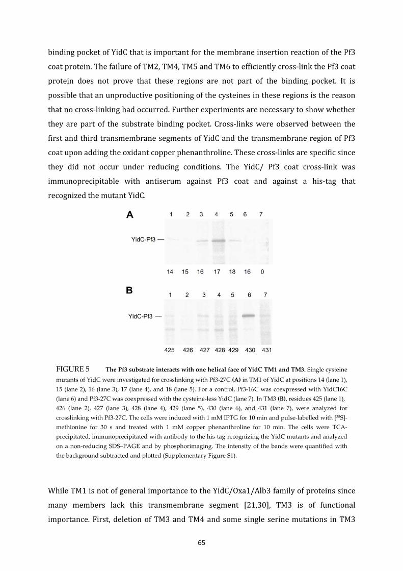
65
binding pocket of YidC that is important for the membrane insertion reaction of the Pf3
coat protein. The failure of TM2, TM4, TM5 and TM6 to efficiently cross-link the Pf3 coat
protein does not prove that these regions are not part of the binding pocket. It is
possible that an unproductive positioning of the cysteines in these regions is the reason
that no cross-linking had occurred. Further experiments are necessary to show whether
they are part of the substrate binding pocket. Cross-links were observed between the
first and third transmembrane segments of YidC and the transmembrane region of Pf3
coat upon adding the oxidant copper phenanthroline. These cross-links are specific since
they did not occur under reducing conditions. The YidC/ Pf3 coat cross-link was
immunoprecipitable with antiserum against Pf3 coat and against a his-tag that
recognized the mutant YidC.
While TM1 is not of general importance to the YidC/Oxa1/Alb3 family of proteins since
many members lack this transmembrane segment [21,30], TM3 is of functional
importance. First, deletion of TM3 and TM4 and some single serine mutations in TM3
FIGURE 5 The Pf3 substrate interacts with one helical face of YidC TM1 and TM3. Single cysteine mutants of YidC were investigated for crosslinking with Pf3-27C (A) in TM1 of YidC at positions 14 (lane 1), 15 (lane 2), 16 (lane 3), 17 (lane 4), and 18 (lane 5). For a control, Pf3-16C was coexpressed with YidC16C (lane 6) and Pf3-27C was coexpressed with the cysteine-less YidC (lane 7). In TM3 (B), residues 425 (lane 1), 426 (lane 2), 427 (lane 3), 428 (lane 4), 429 (lane 5), 430 (lane 6), and 431 (lane 7), were analyzed for crosslinking with Pf3-27C. The cells were induced with 1 mM IPTG for 10 min and pulse-labelled with [35S]-methionine for 30 s and treated with 1 mM copper phenanthroline for 10 min. The cells were TCA-precipitated, immunoprecipitated with antibody to the his-tag recognizing the YidC mutants and analyzed on a non-reducing SDS–PAGE and by phosphorimaging. The intensity of the bands were quantified with the background subtracted and plotted (Supplementary Figure S1).
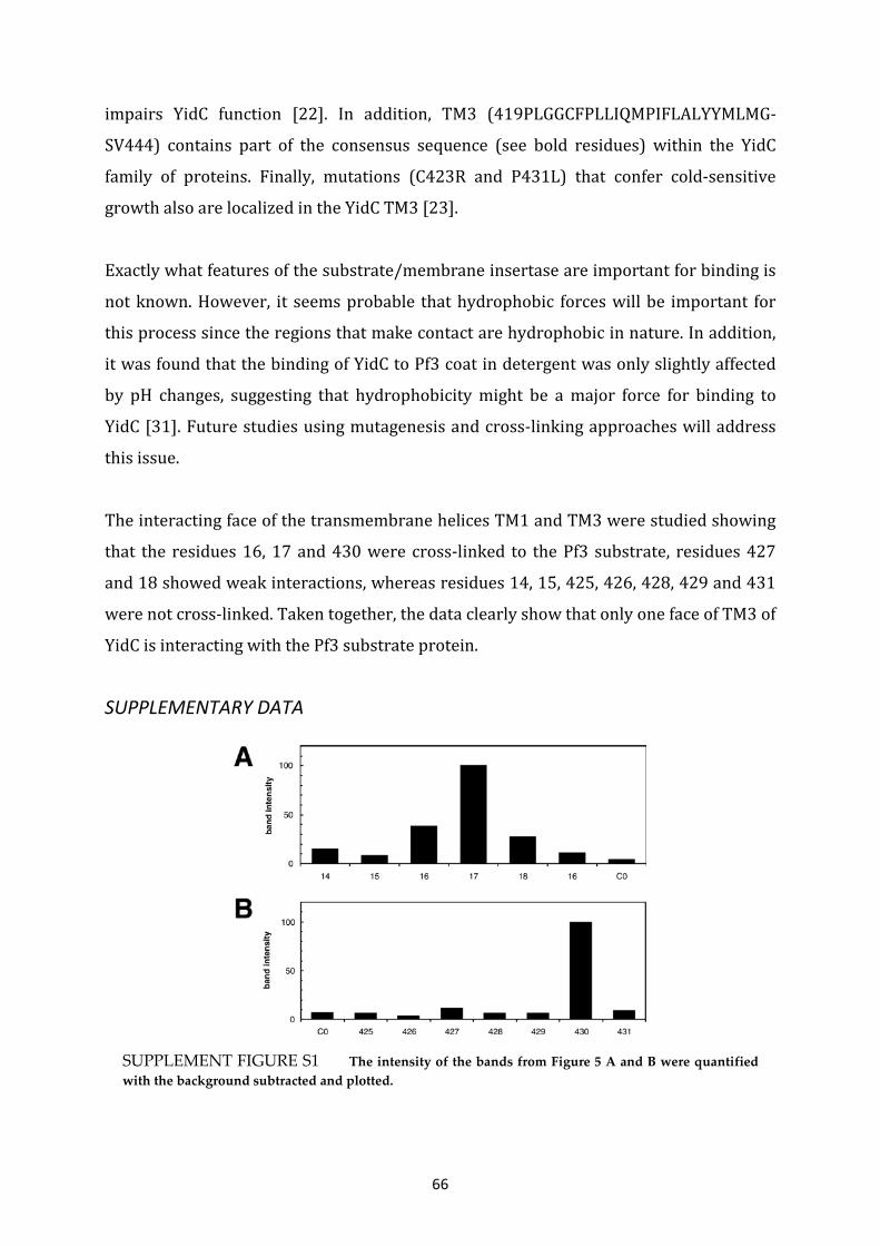
66
impairs YidC function [22]. In addition, TM3 (419PLGGCFPLLIQMPIFLALYYMLMG-
SV444) contains part of the consensus sequence (see bold residues) within the YidC
family of proteins. Finally, mutations (C423R and P431L) that confer cold-sensitive
growth also are localized in the YidC TM3 [23].
Exactly what features of the substrate/membrane insertase are important for binding is
not known. However, it seems probable that hydrophobic forces will be important for
this process since the regions that make contact are hydrophobic in nature. In addition,
it was found that the binding of YidC to Pf3 coat in detergent was only slightly affected
by pH changes, suggesting that hydrophobicity might be a major force for binding to
YidC [31]. Future studies using mutagenesis and cross-linking approaches will address
this issue.
The interacting face of the transmembrane helices TM1 and TM3 were studied showing
that the residues 16, 17 and 430 were cross-linked to the Pf3 substrate, residues 427
and 18 showed weak interactions, whereas residues 14, 15, 425, 426, 428, 429 and 431
were not cross-linked. Taken together, the data clearly show that only one face of TM3 of
YidC is interacting with the Pf3 substrate protein.
SUPPLEMENTARY DATA
SUPPLEMENT FIGURE S1 The intensity of the bands from Figure 5 A and B were quantified with the background subtracted and plotted.

67
ACKNOWLEDGEMENTS
This work was supported by a DFG Grant Ku749/3-4 to A.K. and an National Institute of
Health Grant (GM63862-05) to R.E.D.
CONTRUBTION TO THIS MANUSCRIPT
C.K. performed experiments uncovering YidC-Pf3 contacts. J.Y. performed experiments
to verify the functionality of the YidC cysteine mutants. C.K., J.Y. and A.K prepared the
figures. C.K., R.E.D. and A.K. wrote the manuscript. R.E.D. and A.K. supervised and
directed the research.
REFERENCES
[1] Xie, K. and Dalbey, R.E. (2008) Inserting proteins into the bacterial cytoplasmic
membrane using the Sec and YidC translocases. Nat. Rev. Microbiol. 6, 234–244.
[2] Veenendaal, A.K., van der Does, C. and Driessen, A.J. (2004) The protein-conducting
channel SecYEG. Biochim. Biophys. Acta 1694, 81–95.
[3] Kiefer, D. and Kuhn, A. (2007) YidC as an essential and multifunctional component in
membrane protein assembly. Int. Rev. Cytol. 259, 113–138.
[4] Luirink, J., Heijne, G.v., Houben, E. and Gier, J.-W.d. (2005) Biogenesis of inner
membrane proteins in Escherichia coli. Ann. Rev. Microbiol. 59, 329–355.
[5] Dalbey, R.E. and Kuhn, A. (2004) YidC family members are involved in the membrane
insertion, lateral integration, folding, and assembly of membrane proteins. J. Cell Biol.
166, 769–774.
[6] Scotti, P.A. et al. (2000) YidC, the Escherichia coli homologue of mitochondrial Oxa1p,
is a component of the Sec translocase. EMBO J. 19, 542–549.
[7] Urbanus, M.L. et al. (2001) Sec-dependent membrane protein insertion: sequential
interaction of nascent FtsQ with SecY and YidC. EMBO Rep. 2, 524–529.
[8] Beck, K., Eisner, G., Trescher, D., Dalbey, R.E., Brunner, J. and Müller, M. (2001) YidC,
an assembly site for polytopic Escherichia coli membrane proteins located in immediate
proximity to the SecYE translocon and lipids. EMBO Rep. 2, 709–714.
[9] Samuelson, J.C., Chen, M., Jiang, F., Moller, I., Wiedmann, M., Kuhn, A., Phillips, G.J. and
Dalbey, R.E. (2000) YidC mediates membrane protein insertion in bacteria. Nature 406,
637–641.

68
[10] Serek, J., Bauer-Manz, G., Struhalla, G., Van Den Berg, L., Kiefer, D., Dalbey, R. and
Kuhn, A. (2004) Escherichia coli YidC is a membrane insertase for Sec-independent
proteins. EMBO J. 23, 294–301.
[11] Van Der Laan, M., Bechtluft, P., Kol, S., Nouwen, N. and Driessen, A.J. (2004) F1F0
ATP synthase subunit c is a substrate of the novel YidC pathway for membrane protein
biogenesis. J. Cell Biol. 165, 213–222.
[12] Samuelson, J.C., Jiang, F., Yi, L., Chen, M., de Gier, J.W., Kuhn, A. and Dalbey, R.E.
(2001) Function of YidC for the insertion of M13 procoat protein in E. coli: translocation
of mutants that show differences in their membrane potential dependence and Sec-
requirement. J. Biol. Chem. 16, 16.
[13] Chen, M., Samuelson, J.C., Jiang, F., Muller, M., Kuhn, A. and Dalbey, R.E. (2002)
Direct interaction of YidC with the Sec-independent Pf3 coat protein during its
membrane protein insertion. J. Biol. Chem. 277, 7670–7675.
[14] Van Bloois, E., Jan Haan, G., de Gier, J.W., Oudega, B. and Luirink, J. (2004) F(1)F(0)
ATP synthase subunit c is targeted by the SRP to YidC in the E. coli inner membrane.
FEBS Lett. 576, 97–100.
[15] Facey, S.J., Neugebauer, S.A., Krauss, S. and Kuhn, A. (2007) The mechanosensitive
channel protein MscL is targeted by the SRP to the novel YidC membrane insertion
pathway of Escherichia coli. J. Mol. Biol. 365, 995–1004.
[16] Yi, L., Celebi, N., Chen, M. and Dalbey, R.E. (2004) Sec/SRP requirements and
energetics of membrane insertion of subunits a, b, and c of the Escherichia coli F1F0 ATP
synthase. J. Biol. Chem. 279, 39260–39267.
[17] Van Bloois, E., Haan, G.J., de Gier, J.W., Oudega, B. and Luirink, J. (2006) Distinct
requirements for translocation of the N-tail and C-tail of the Escherichia coli inner
membrane protein CyoA. J. Biol. Chem. 281, 10002–10009.
[18] Celebi, N., Yi, L., Facey, S.J., Kuhn, A. and Dalbey, R.E. (2006) Membrane biogenesis
of subunit II of cytochrome bo oxidase: contrasting requirements for insertion of N-
terminal and C-terminal domains. J. Mol. Biol. 357, 1428–1436.
[19] du Plessis, D.J., Nouwen, N. and Driessen, A.J. (2006) Subunit a of cytochrome o
oxidase requires both YidC and SecYEG for membrane insertion. J. Biol. Chem. 281,
12248–12252.
[20] Nagamori, S., Smirnova, I.N. and Kaback, H.R. (2004) Role of YidC in folding of
polytopic membrane proteins. J. Cell Biol. 165, 53–62.
69
[21] Yen, M.R., Harley, K.T., Tseng, Y.H. and Saier Jr., M.H. (2001) Phylogenetic and
structural analyses of the oxa1 family of protein translocases. FEMS Microbiol. Lett. 204,
223–231.
[22] Jiang, F., Chen, M., Yi, L., de Gier, J.W., Kuhn, A. and Dalbey, R.E. (2003) Defining the
regions of Escherichia coli YidC that contribute to activity. J. Biol. Chem. 278, 48965–
48972.
[23] Yuan, J., Phillips, G.J. and Dalbey, R.E. (2007) Isolation of coldsensitive yidC mutants
provides insights into the substrate profile of the YidC insertase and the importance of
transmembrane 3 in YidC function. J. Bacteriol. 189, 8961–8972.
[24] Kuhn, A., Stuart, R., Henry, R. and Dalbey, R.E. (2003) The Alb3/Oxa1/YidC protein
family: membrane-localized chaperones facilitating membrane protein insertion?
Trends Cell Biol. 13, 510–516.
[25] Nagler, C., Nagler, G. and Kuhn, A. (2007) Cysteine residues in the transmembrane
regions of M13 procoat protein suggest that oligomeric coat proteins assemble onto
phage progeny. J. Bacteriol. 189, 2897–2905.
[26] Kuhn, A. and Wickner, W. (1985) Isolation of mutants in M13 coat protein that
affect its synthesis, processing, and assembly into phage. J. Biol. Chem. 260, 15907–
15913.
[27] Jiang, F., Yi, L., Moore, M., Chen, M., Rohl, T., van Wijk, K.J., de Gier, J.W., Henry, R. and
Dalbey, R.E. (2002) Chloroplast YidC homolog Albino3 can functionally complement the
bacterial YidC depletion strain and promote membrane insertion of both bacterial and
chloroplast thylakoid proteins. J. Biol. Chem. 277, 19281–19288.
[28] Kiefer, D. and Kuhn, A. (1999) Hydrophobic forces drive spontaneous membrane
insertion of the bacteriophage Pf3 coat protein without topological control. EMBO J. 18,
6299–6306.
[29] Lessl, M., Balzer, D., Lurz, R., Waters, V.L., Guiney, D.G. and Lanka, E. (1992)
Dissection of IncP conjugative plasmid transfer: definition of the transfer region Tra2 by
mobilization of the Tra1 region in trans. J. Bacteriol. 174, 2493–2500.
[30] Luirink, J., Samuelsson, T. and de Gier, J.W. (2001) YidC/Oxa1p/Alb3: evolutionarily
conserved mediators of membrane protein assembly. FEBS Lett. 501, 1–5.
[31] Gerken, U., Erhardt, D., Bär, G., Ghosh, R. and Kuhn, A. (2008) Initial binding process
of the membrane insertase YidC with its substrate Pf3 coat protein is reversible.
Biochemistry 47, 6052–6058.

70

71
Chapter 3
Dynamic disulfide scanning of the membrane-inserting Pf3 coat protein reveals multiple YidC substrate contacts
CHRISTIAN KLENNERa & ANDREAS KUHNa
a Institute of Microbiology and Molecular Biology, University of Hohenheim, 70599 Stuttgart, Germany ABSTRACT
The membrane insertase YidC inserts newly synthesized proteins into the plasma
membrane. While defects in YidC homologs in animals and plants cause diseases, YidC in
bacteria is essential for life. Membrane insertion and assembly of ATP synthase and
respiratory complexes is catalyzed by YidC. To investigate how YidC interacts with
membrane-inserting proteins, we generated single cysteine mutants in YidC and in the
model substrate Pf3 coat protein. The single cysteine mutants were expressed and
analyzed for disulfide formation during 30 s of synthesis. The results show that the
substrate contacts different YidC residues in four of the six transmembrane regions. The
residues are located either in the region of the inner leaflet, in the center, as well as in
the periplasmic leaflet, consistent with the hypothesis that YidC presents a hydrophobic
platform for inserting membrane proteins. In a YidC mutant where most of the
contacting residues were mutated to serines, YidC function was severely disturbed and
no longer active in a complementation test, suggesting that the residues are important
for function. In addition, a Pf3 mutant with a defect in membrane insertion was deficient
to contact the periplasmic residues of YidC.
INTRODUCTION
The membrane insertase YidC of Escherichia coli is required for the biogenesis of
respiratory complexes, ATP synthase, and other membrane proteins (1, 2). When YidC is
depleted from bacterial cells, growth ceases, and the cells die, revealing that it is an

72
essential protein (3, 4). In the absence of YidC, the subunit c protein of the Fo part of the
ATP synthase has been shown to accumulate in the cytoplasm because its membrane
insertion is blocked (5, 6). Also a number of other proteins, such as the CyoA protein of
the terminal oxidase complex (7), the mechanosensitive channel protein MscL (8), and
the coat proteins of filamentous phage Pf3 and M13 require YidC for their membrane
insertion. All these substrate proteins comprise less than 200 amino acids and contain
periplasmic regions of less than 30 residues. The best studied substrate is the 44 amino
acid-long Pf3 coat protein. After its synthesis, it binds reversibly to YidC and is
membrane-inserted within a few minutes. The binding of purified Pf3 coat protein has
been investigated by its ability to quench the 1-anilinonaphthalene-8-sulfonate (ANS)-
labeled YidC protein in detergent (9) and also to quench the membrane-integrated
insertase (10). The insertion of the Pf3 coat protein had been studied in a reconstituted
system with proteoliposomes that only contained YidC (11). When limiting amounts of
YidC were present in the proteoliposomes, up to 150 Pf3 coat proteins were inserted per
YidC molecule, suggesting that YidC is catalytically driving the membrane insertion of
proteins.
YidC is a six-spanning membrane protein of 548 amino acids (12). The structure of the
large periplasmic domain between the first two transmembrane (TM) regions has
recently been solved to high resolution (Figure 1) (13, 14). The homologues found in
mitochondria and chloroplasts lack the first transmembrane region and most of the first
periplasmic domain of 330 amino acid residues. Accordingly, the deletion of 300
residues from the periplasmic domain of the YidC from E. coli retained a still functional
YidC mutant deficient for the interaction with SecF (15). When the first transmembrane
region of YidC was replaced by a cleavable signal peptide, it maintained activity (16).
However, the transmembrane regions were shown to be essential for the function of
YidC. Deletions of TM3/4 and TM5/6 resulted in inactive YidC (17). Also, the
replacement of residues 418 to 425 by serines did not retain activity. Surprisingly, a
number of substitutions of residues in the transmembrane regions with serines or
alanines had no noticeable effect (17). Presumably, a number of residues in the
hydrophobic transmembrane regions of YidC are involved in substrate binding and are
required during the insertion process.
73
The transmembrane regions are the most conserved parts of the YidC protein.
Particularly, this was observed for the sequence ofTM2 andTM3 throughout and for the
periplasmic portions of TM5 and TM6 (12). Single cysteines in TM3 were shown to be
involved in substrate contacts in vivo (18) and in vitro (19). Here, we have extended this
study and systematically scanned all six transmembrane regions of YidC for contacts
with a substrate by single cysteine mutants. The results show that TM1, TM3, and TM4
provide nine major contacting residues. Three additional contacts are located in the
periplasmic half of TM5. A substrate mutant that is deficient for membrane insertion
was able to contact the YidC residues close to the cytoplasm but failed to contact the
residues in the periplasmic half of the transmembrane region.
EXPERIMENTAL PROCEDURES
Bacterial Growth, Pulse Labeling, and Disulfide Formation
E. coli MK6 (18) was transformed with pMS-Pf3 coat and pACYC-YidC and then
grown in M9 minimal media in the presence of ampicillin (final concentration, 100
µg/ml) and chloramphenicol (final concentration, 25 µg/ml). In the MK6 strain, the
promoter of the yidC gene had been exchanged with the araC-araBAD promoter cassette.
Depletion of the chromosomally encoded YidC was achieved by growth in the presence
of 0.2% glucose for 3 h at 37 °C. Unless indicated otherwise, the cells were treated with
1mM isopropyl 1-thio-D-galactopyranoside (IPTG) at A600 nm 0.2 for 10 min to induce
expression of the Pf3 coat and pulse-labeled for the indicated time by the addition of
[35S]methionine. The cells were chilled on ice and, if indicated, treated with 1mM freshly
prepared copper 1,10-phenanthroline for 3 min (20). The samples were precipitated
with 20% TCA, washed with acetone, and resuspended in 2% SDS in Tris buffer (pH 8).
Immunoprecipitation was done as described (21) using antiserum to Pf3 phage or to
YidC. The samples were analyzed by SDS-PAGE and phosphor imaging.
Generation of Single Cysteine Mutants
The site-directed mutations in the Pf3 coat and YidC were generated by the
QuikChange method with minor modifications (22). All mutations were verified by DNA
sequencing of the entire gene.
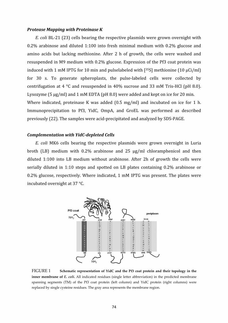
74
FIGURE 1 Schematic representation of YidC and the Pf3 coat protein and their topology in the inner membrane of E. coli. All indicated residues (single letter abbreviation) in the predicted membrane spanning segments (TM) of the Pf3 coat protein (left column) and YidC protein (right columns) were replaced by single cysteine residues. The gray area represents the membrane region.
Protease Mapping with Proteinase K
E. coli BL-21 (23) cells bearing the respective plasmids were grown overnight with
0.2% arabinose and diluted 1:100 into fresh minimal medium with 0.2% glucose and
amino acids but lacking methionine. After 2 h of growth, the cells were washed and
resuspended in M9 medium with 0.2% glucose. Expression of the Pf3 coat protein was
induced with 1 mM IPTG for 10 min and pulselabeled with [35S] methionine (10 µCi/ml)
for 30 s. To generate spheroplasts, the pulse-labeled cells were collected by
centrifugation at 4 °C and resuspended in 40% sucrose and 33 mM Tris-HCl (pH 8.0).
Lysozyme (5 µg/ml) and 1 mM EDTA (pH 8.0) were added and kept on ice for 20 min.
Where indicated, proteinase K was added (0.5 mg/ml) and incubated on ice for 1 h.
Immunoprecipitation to Pf3, YidC, OmpA, and GroEL was performed as described
previously (22). The samples were acid-precipitated and analyzed by SDS-PAGE.
Complementation with YidC-depleted Cells
E. coli MK6 cells bearing the respective plasmids were grown overnight in Luria
broth (LB) medium with 0.2% arabinose and 25 µg/ml chloramphenicol and then
diluted 1:100 into LB medium without arabinose. After 2h of growth the cells were
serially diluted in 1:10 steps and spotted on LB plates containing 0.2% arabinose or
0.2% glucose, respectively. Where indicated, 1 mM IPTG was present. The plates were
incubated overnight at 37 °C.
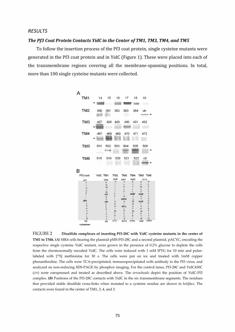
75
FIGURE 2 Disulfide complexes of inserting Pf3-28C with YidC cysteine mutants in the center of TM1 to TM6. (A) MK6 cells bearing the plasmid pMS-Pf3-28C and a second plasmid, pACYC, encoding the respective single cysteine YidC mutant, were grown in the presence of 0.2% glucose to deplete the cells from the chromosomally encoded YidC. The cells were induced with 1 mM IPTG for 10 min and pulse-labeled with [35S] methionine for 30 s. The cells were put on ice and treated with 1mM copper phenanthroline. The cells were TCA-precipitated, immunoprecipitated with antibody to the Pf3 virus, and analyzed on non-reducing SDS-PAGE by phosphor imaging. For the control lanes, Pf3-28C and YidC430C (ctr) were coexpressed and treated as described above. The arrowheads depict the position of YidC-Pf3 complex. (B) Positions of the Pf3-28C contacts with YidC in the six transmembrane segments. The residues that provided stable disulfide cross-links when mutated to a cysteine residue are shown in boldface. The contacts were found in the center of TM1, 3, 4, and 5.
RESULTS
The Pf3 Coat Protein Contacts YidC in the Center of TM1, TM3, TM4, and TM5
To follow the insertion process of the Pf3 coat protein, single cysteine mutants were
generated in the Pf3 coat protein and in YidC (Figure 1). These were placed into each of
the transmembrane regions covering all the membrane-spanning positions. In total,
more than 100 single cysteine mutants were collected.

76
E. coli MK6 cells were transformed with a plasmid coding for one of the Pf3 coat protein
single cysteine mutants and with a second plasmid coding for one of the YidC single
cysteine mutants. The MK6 cells that have the chromosomal yidC under control of the
araBAD promoter (18) were grown in glucose media to deplete the YidC wild-type and
allow the plasmid-borne expression of YidC. All mutant proteins showed a comparable
expression level. To map the contact sites during the insertion event, the two proteins
were coexpressed in E. coli MK6 cells and pulse-labeled with [35S] methionine for 30 s
and treated with oxidant for 3 min. Then, the proteins were acid-precipitated and
immunoprecipitated with an antibody to Pf3 coat protein. When the single cysteine
residue was at position 28 of the Pf3 coat protein, which is in the center of the TM
region, cross-linked YidC-Pf3 complexes were detected with certain cysteine mutants of
YidC (Figure 2). These mutants had their cysteine residue in the center of TM1 at
position 14 and 17, in TM3 at position 427 and 430, in TM4 at position 468, and in TM5
at positions 502 and 504/ 505. The neighboring positions showed significantly weaker
or no cross-links. No strong contacts were detected in TM2 and TM6. To evaluate exactly
the level of cross-linking, a positive control with cells coexpressing YidC 430C and Pf3–
28C was added and loaded on the same gel.
The Pf3 coat protein contacts with YidC430 and 505 were analyzed in more detail
(Figure 3). The pulse-labeled samples were immunoprecipitated either with Pf3 (Figure
3A, lanes 1–5) or with YidC antiserum (lanes 6–10) and analyzed by polyacrylamide
electrophoresis, respectively. The contacts only occurred under oxidative conditions and
when a cysteine is present on both protein partners (Figure 3, A and C). Under oxidative
conditions, most of the Pf3 coat protein was present as dimers, but some monomers
were also detectable. In addition, a significant proportion was found at about 65 kDa
corresponding to a Pf3-YidC cross-link product. Clearly, the Pf3-YidC cross-link product
was immunoprecipitated with both antibodies and migrated a little slower (Figure 3C,
lanes 2-4) than the cysteine-less YidC on SDS-PAGE (lane 5). When the cysteine-less YidC
was coexpressed, no YidC-cross-linked product was detectable (Figure 3A, lane 5).
However, the formation of Pf3 coat protein dimers was unaffected. The contacts we
observed with Pf3–28C were all at positions of the YidC protein that were predicted, on
the basis of the hydrophobicity profile, to be in the center of the bilayer (Figure 2, lower
panel), in TM1, 3, 4, and 5.

77
FIGURE 3 Pf3-YidC disulfide complexes are recognized by antibodies to Pf3 and to YidC. (A) Coexpression of Pf3-28C and YidC with a single cysteine at residue 430 (lanes 1, 2, 6, and 7) and 505 (lanes 3, 4, 8, and 9) were analyzed as described for Figure 2, except that in lanes 1, 3, 6, and 8 no copper phenanthroline was added. Lanes 1-5 were immunoprecipitated with an antibody to Pf3. Lanes 6-10 were immunoprecipitated with an antibody to YidC. As a control, the cysteine-less YidC mutant (lanes 5 and 10) was expressed. The Pf3 coat protein was found as a monomer, dimer, and cross-linked with YidC. Molecular weight markers are indicated in kDa at the left margin (laneM). (B) E. coli MK6 cells expressing the wild-type Pf3 coat protein (lanes 1 and 2) and Pf3–28C (lanes 3 and 4) were pulse-labeled as described above and treated with copper phenanthroline (lanes 2 and 4). The empty vector is shown as a control (lane 5). (C) For better separation, lanes 6-10 of Figure 3A were applied to a long PAGE.
Pf3 Contacts in the YidC Region Located in the Inner and Periplasmic Leaflet
We then tested which YidC residues were contacted by a Pf3 coat protein that had
its single cysteine at position 33, predicted to localize in the inner leaflet of the
membrane (Figure 4). MK6 cells bearing the plasmids for expressing a single cysteine
mutant of YidC and a second plasmid expressing the Pf3 coat protein with a cysteine at
position 33, respectively, were pulse-labeled for 30 s and analyzed for disulfide
formation. YidC-Pf3 cross-link products were found to the YidC-TM1 (position 9), to
TM3 (positions 424 and 427), and TM4 (position 476). Weak contacts were observed in
TM3 at positions 425 and 428 that are next to the strong cross-linking signals and also in
TM2 at positions 367 and 370. All these contacts are located in YidC at the predicted
inner leaflet of the membrane bilayer (Figure 4, lower panel). However, no strong
contacts were observed with TM5 and TM6 in the region of the inner leaflet on the basis
of the comparison of the background signals with a control culture (coexpression of Pf3–
28C and YidC-505C).

78
Next, the YidC residues predicted to be located in the periplasmic leaflet of the
transmembrane regions were investigated. When a Pf3 coat protein with a cysteine
residue at position 24 was coexpressed, contacts were observed for YidC-TM3 at
position 435 and for YidC-TM5 at 504 and 508 (Figure 5A). No major contacts were
found with TM2 and TM4, as compared with the background. Weak interactions were
detected with the YidC residue 18, 19 (in TM1) and 514 (in TM6). We noticed that in
general, the YidC contacts to Pf3–24C were weaker than to Pf3–33C and Pf3–28C,
presumably because the periplasmic contacts may occur for a shorter time.
FIGURE 4 Inserting Pf3-33C coat protein contacts YidC transmembrane residues located in the inner leaflet. (A) E. coli MK6 cells bearing the plasmid pMS-Pf3-33C and a second plasmid encoding the respective single cysteine YidC mutant were grown and treated as described in the legend to Fig. 2. Cross-links were detected by immunoprecipitations with an antibody to Pf3. For the control lanes, Pf3-28C and YidC505C (ctr) were coexpressed and treated as described above. The arrowheads depict the position of the YidC-Pf3 complex. (B) Positions of the Pf3-33C contacts with YidC in the six transmembrane segments. The residues that provided stable disulfide cross-links when mutated to a cysteine residue are shown in boldface. The contacts were found in the inner half of TM1, 3, and 4.

79
Taken together, we have observed 12 strong contact sites in TM1, 3, 4, and 5. They were
often flanked by residues that also showed a contact to the substrate but, mostly, much
weaker. This was the case for the flanking residues of residues 9, 17, 424, 427, 468, 477,
and 505. The residues 504 and 505 gave about the same signal to Pf3–24C and Pf3–28C
(Figs. 2 and 5), suggesting that both residues are equally contacting the substrate.
Contacts of Insertion-deficient Pf3 Coat Protein Mutants with YidC
To study the different stages of YidC-substrate interaction, experiments were
performed with Pf3 coat protein mutants that are deficient for membrane insertion (24).
FIGURE 5 Inserting Pf3-24C coat protein contacts YidC in the transmembrane residues of the outer leaflet. (A) E. coli MK6 cells bearing the plasmid pMS-Pf3-24C and a second plasmid encoding the respective single cysteine YidC mutant were grown and treated as described in the legend to Fig. 2. Cross-links were detected by immunoprecipitations with an antibody to Pf3. For the control lanes, Pf3-28C and YidC505C (ctr) were coexpressed and treated as described above. The arrowheads depict the position of the YidC-Pf3 complex. (B) Positions of the Pf3-24C contacts with YidC in the six transmembrane segments. The residues that provided stable disulfide cross-links when mutated to a cysteine residue are shown in boldface. The contacts were found in the outer half of TM 3 and 5.

80
FIGURE 6 Membrane insertion of Pf3-RS, Pf3-RR, and Pf3-3R is inhibited. E. coli BL-21 cells expressing Pf3, Pf3-24C, Pf3-24C-RS, Pf3-24C-RR, or Pf3-24C-3R, respectively, were pulse-labeled for 3 min with [35S] methionine. The outer membranes of the cells were opened by osmotic shock, and proteinase K (+ PK lanes) was added to the outside of the cells, and they were incubated on ice for 1 h. The samples were immunoprecipitated with antiserum to Pf3, GroEL, or OmpA, respectively, and analyzed by PAGE and phosphor imaging.
The Pf3-RR mutant has two arginines substituted for the amino acids at positions 17 and
18, whereas the Pf3-RS mutant has an arginine and a serine at these positions,
respectively. A third mutant, Pf3-3R, is identical to Pf3-RR but has an additional arginine
at position 7 replacing a glutamate. Single cysteines were introduced in all these
mutants at position 24 to study their interaction with YidC and their insertion into the
membrane. First, the membrane insertion of the mutants was studied in vivo by
proteinase mapping (Fig. 6). E. coli BL-21 cells expressing the respective Pf3 coat protein
mutant were grown in minimal medium lacking methionine to a density of 2 x 10⁸
cells/ml. [35S] methionine was added for 3 min, and the cells were analyzed for Pf3 coat
protein insertion. Although the wild- type Pf3 coat was accessible to the protease added
to the periplasmic side (lanes 2 and 4), the mutants Pf3-RS, Pf3-RR, and Pf3-3R were not
digested by the protease demonstrating that they are inhibited for membrane insertion.
For a control, the cytoplasmic protein GroEL and OmpA were immunoprecipitated, and
we verified that the cells remained intact and the periplasm was accessible to the
outside-added protease.
The Pf3-RR mutant was then coexpressed in E. coli MK6 with a single cysteine mutant of
YidC and analyzed for contacts to YidC at the respective positions (Fig. 7). When the
cysteine at position 24 in the Pf3 wild-type (Fig. 7A) was compared with Pf3-RR-24C (B),
the contacts to the YidC residues at 435 and 508 were substantially reduced in the
insertion-deficient Pf3-RR mutant. These residues are in the periplasmic membrane

81
FIGURE 7 Pf3-RR is blocked for contacting the YidC residues in the outer leaflet. E. coli MK6 cells expressing Pf3-24C (A), Pf3-24C-RR (B), Pf3-28C (C), Pf3-28C-RR (D), Pf3-33C (E), or Pf3-33C-RR (F) were coexpressed with the respective YidC mutants to analyze close contacts by disulfide crosslinking. The cells were labeled with [35S] methionine for 30 s and immunoprecipitated with antiserum to Pf3. As a control, the cysteine-less YidC (Co) was coexpressed. The samples were analyzed by PAGE and phosphor imaging.
portion of YidC. Similarly, the residues in the center of the membrane at 430, 468, and
505 were not or only weakly cross-linked with Pf3-RR with a cysteine at position 28
(Fig. 7D). However, the YidC residues 424, 427, and 476 located in the inner leaflet were
not inhibited for contacting Pf3-RR-33C (compare Fig. 7, E and F). The Pf3–3R mutant
containing three arginine residues in the amino terminal domain was also tested for
YidC interaction (Fig. 8). When this Pf3-3R mutant had a cysteine at position 24, 28, or
33, it was inhibited to interact with the residues 508, 505, and 476 of YidC located in the
outer, central, and inner leaflet, respectively (Fig. 8A). When the samples were
immunoprecipitated with antibodies to YidC, the shift of the band caused by a bound Pf3
protein was not observed when the 3R mutations were present in Pf3 (Fig. 8B),
indicating that this Pf3 mutant is unable to interact with the YidC insertase.
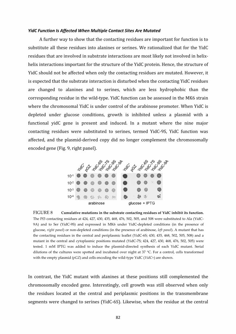
82
FIGURE 8 Cumulative mutations in the substrate contacting residues of YidC inhibit its function. The Pf3 contacting residues at 424, 427, 430, 435, 468, 476, 502, 505, and 508 were substituted to Ala (YidC-9A) and to Ser (YidC-9S) and expressed in MK6 under YidC-depleted conditions (in the presence of glucose, right panel) or non-depleted conditions (in the presence of arabinose, left panel). A mutant that has the contacting residues in the central and periplasmic leaflet (YidC-6S; 430, 435, 468, 502, 505, 508) and a mutant in the central and cytoplasmic positions mutated (YidC-7S; 424, 427, 430, 468, 476, 502, 505) were tested. 1 mM IPTG was added to induce the plasmid-directed synthesis of each YidC mutant. Serial dilutions of the cultures were spotted and incubated over night at 37 °C. For a control, cells transformed with the empty plasmid (pGZ) and cells encoding the wild-type YidC (YidC+) are shown.
YidC Function Is Affected When Multiple Contact Sites Are Mutated
A further way to show that the contacting residues are important for function is to
substitute all these residues into alanines or serines. We rationalized that for the YidC
residues that are involved in substrate interactions are most likely not involved in helix-
helix interactions important for the structure of the YidC protein. Hence, the structure of
YidC should not be affected when only the contacting residues are mutated. However, it
is expected that the substrate interaction is disturbed when the contacting YidC residues
are changed to alanines and to serines, which are less hydrophobic than the
corresponding residue in the wild-type. YidC function can be assessed in the MK6 strain
where the chromosomal YidC is under control of the arabinose promoter. When YidC is
depleted under glucose conditions, growth is inhibited unless a plasmid with a
functional yidC gene is present and induced. In a mutant where the nine major
contacting residues were substituted to serines, termed YidC-9S, YidC function was
affected, and the plasmid-derived copy did no longer complement the chromosomally
encoded gene (Fig. 9, right panel).
In contrast, the YidC mutant with alanines at these positions still complemented the
chromosomally encoded gene. Interestingly, cell growth was still observed when only
the residues located at the central and periplasmic positions in the transmembrane
segments were changed to serines (YidC-6S). Likewise, when the residue at the central
83
and cytoplasmic positions was changed to serine (YidC-7S), growth was not affected.
Taken together, we conclude that YidC function is only distorted when multiple
contacting residues along the entire membrane region were changed.
DISCUSSION
The membrane insertase YidC catalyzes the insertion of the Pf3 coat protein into the
inner membrane of E. coli (11). During this process, the Pf3 protein binds to YidC mainly
by hydrophobic interactions (9) and induces a conformational change within YidC (10,
25). As a result, the Pf3 coat protein adopts a transmembrane conformation, exposing its
N-terminal domain to the periplasm. Although the substrate binding step most likely
involves the cytoplasmic residues of YidC, insertion of the TM region and translocation
of the N-terminal region are likely to require other contacting residues of YidC. The
molecular interactions of newly synthesized Pf3 coat protein were analyzed during 30 s
after the onset of its synthesis. The data presented here show that multiple YidC-
substrate contacts are observed involving the participation of most of the
transmembrane segments of YidC. Only strong disulfide signals were considered. The
weak signals were mostly observed at positions flanking the strong signals. Weak signals
at other positions (e.g. at 358, 360, 370, and 514) may arise from a structural flexibility
of YidC. Although TM3 showed contacts all across the predicted membrane-spanning α-
helix, TM5 has three positions (502, 505, and 508), located in the central and outer
leaflet, that make a contact with the newly expressed Pf3 coat protein. These contacts
reveal a first three-dimensional picture of how the substrate is possibly binding YidC
(Fig. 10). In the cytoplasmic leaflet, the substrate contacts TM1, 3, and 4 of YidC,
whereas in the periplasmic leaflet, TM3 and 5 are contacted. This suggests that the
residues at positions 424, 427, and 476 are primarily involved in the first substrate
binding step, whereas residues 430, 435, 468, 502, 505, and 508 are required for
translocation. The data that we have obtained with the substrate mutant Pf3-RR that is
defective for membrane insertion are in support of this model. The insertion-deficient
mutant RR-Pf3 was contacting 424, 427, and 476 in the cytoplasmic leaflet. However, the
contacts to the central and periplasmic residues 430, 435, 468, 505 and 508 were
substantially reduced (Fig. 7).
84
Intriguingly, the residues of YidC that are most conserved among the bacterial
homologues are present in TM2, TM3, and in the C1 loop (12). Also, the periplasmic
region of TM5 shows a high degree of homology (26). In these conserved TM regions of
YidC we found seven major contacting residues. Surprisingly, several contacts were
found within TM1, which is less conserved and is missing in Gram-positive bacteria as
well as in the mitochondrial and thylakoidal homologues. This suggests that TM1
contributes to the YidC function, although it is not essential in the YidC family of
proteins. The importance of the other TM regions for the function of YidC has been
implicated from a number of site-directed mutants (17). The deletion of these TM
regions inactivates the protein. Single residue substitutions had shown that only a few
residues within the transmembrane regions are sensitive. Most of these locate in TM3,
corroborating its central role for the insertase activity (17). Interestingly, the critical
residues are not at the exact positions where our substrate contacts are localized but
adjacent to them. Most likely, these residues are not involved in substrate interaction
but rather in helix-helix contacts within YidC. One such contact was proposed for
residue 423 in TM3 with TM2 (27). When the cysteine at 423 was substituted for an
arginine, a cold-sensitive phenotype was observed. Second-site suppressors identified
residue T362E forming a possible charge pair with Arg-423, suggesting
thatTM2andTM3are close together. A further suppressor mutation of Arg-423 within
TM3 was found at L426E in the same TM, indicating that residues 362, 423, and 426 are
involved in helix-helix interaction and not in substrate binding. In accordance with this,
we did not find Pf3-YidC contacts at 362, 423, or 426.
A previous study (19) had also identified TM3 of YidC as the substrate contact site.
However, different residues were found to form disulfide contacts to the in vitro
synthesized substrates, possibly because the cysteine mutants at positions 427 and 428
were not available. The Sec-dependent FtsQ protein was found to contact residues 425
and 426. With residue 425, we also found a weak interaction with Pf3, but residue 426
did not form a disulfide with Pf3. Another difference between that study and ours is that
they had a nascent chain-arrested ribosome interacting with YidC in membrane vesicles
in vitro. It is possible that the nascent chain could bind to the outside of YidC. Our
experimental approach was in vivo, and to detect the formed disulfides we looked
shortly (30-s pulse) after the addition of [35S] methionine. In this time span we expected
to observe the naturally occurring contacts. In our results, the transmembrane segment
85
TM3 showed four contacting residues at 424, 427, 430, and 435, corroborating that it is
the most important YidC helix involved in substrate binding. When the residues are
projected as a helical wheel, the contacting residues are on the same helical face (Fig.
10B). A similar result was obtained for TM1 and TM5, where more than one contact was
observed. All substrate-contacting residues are hydrophobic. In particular, the
extremely hydrophobic phenylalanine was found in six different contact sites. Because
these phenylalanine residues are located in four different transmembrane helices, they
together may constitute a stacking-like arrangement over the entire membrane-
spanning region. In conclusion, the substrate-interacting surface of YidC is hydrophobic
across the entire membrane span. We propose that the contacting residues form a
hydrophobic platform that supports the transmembrane alignment of an incoming α-
helix of the substrate. This proposed mechanism differs from the one of SecY, which is
mostly hydrophilic with a small hydrophobic pore ring in its center (28).
One important question is whether the observed contacting residues are indeed of
functional importance for membrane insertion. To answer this question, we studied Pf3
mutants that were deficient for membrane insertion. These mutants should fail to
contact YidC at least at the periplasmic positions. The Pf3-RR mutant has an alanine
residue at position 17 and an aspartic acid residue at position 18 substituted with
arginines. Pf3-RR was not found to contact YidC at the periplasmic and central positions.
For the Pf3-3R mutant, which had the aspartic acid at position 7 substituted to an
arginine, in addition to the two arginines at 17 and 18, showed no contacts to YidC, not
even to the cytoplasmic residue at 476 (Fig. 8). These data suggest that the Pf3-RR
mutant protein still binds to the cytoplasmic side of YidC but cannot reach out to contact
the more periplasmically located residues of YidC. Therefore, the translocation process
of Pf3-RR across the membrane was perturbed, and the Pf3-3R mutant appeared to be
blocked for both binding and translocation.
Previously, the TM regions of YidC had been analyzed by serine scanning, and thereby
most of the contacting residues had been mutated to serine individually (17). All of these
mutants turned out to be functional. However, in our study we found that when we
combined the serine mutations of most major contacting sites, YidC function was
severely affected (Fig. 9).

86
FIGURE 10 Model of the YidC hydrophobic protein binding platform. (A) The major contact sites in TM1, 3, 4, and 5 are depicted by a numbered circle referring to the amino acid residue position. (B) Helical wheel projection to illustrate the substrate contact sites of the YidC TM regions. The major contact sites are highlighted as dark circles with the respective position. Both ends of each TM segment are labeled with the position number outside the circle. Because TM2 and TM6 showed no strong contacts, they are not included in the model.
Taken together, our data show that YidC contacts its substrate Pf3 at multiple sites
within the transmembrane domain (Fig. 10). This is in line with the proposal that YidC
provides a hydrophobic platform supporting membrane insertion of proteins (29). The
contacts are required for the insertase activity because the Pf3 cysteine mutants that are
defective for membrane insertion were impaired to contact YidC.

87
ACKNOWLEDGEMENTS
This work was supported by Deutsche Forschungsgemeinschaft grant Ku749/6- (to A.
K.).
CONTRUBTION TO THIS MANUSCRIPT
C.K. and A.K. designed the experiments. C.K. performed all experiments. C.K and A.K.
prepared the figures. C.K. and A.K. wrote the manuscript. A.K. supervised and directed
the research.
REFERENCES
1. Facey, S. J., and Kuhn, A. (2010) Biogenesis of bacterial inner-membrane proteins. Cell.
Mol. Life Sci. 67, 2343–2362
2. Price, C. E., and Driessen, A. J. (2010) Biogenesis of membrane bound respiratory
complexes in Escherichia coli. Biochim. Biophys. Acta 1803, 748–766
3. Samuelson, J. C., Chen, M., Jiang, F., Möller, I., Wiedmann, M., Kuhn, A., Phillips, G. J., and
Dalbey, R. E. (2000) YidC mediates membrane protein insertion in bacteria. Nature 406,
637–641
4. Wang, P., Kuhn, A., and Dalbey, R. E. (2010) Global change of gene expression and cell
physiology in YidC-depleted Escherichia coli. J. Bacteriol. 192, 2193–2209
5. Yi, L., Celebi, N., Chen, M., and Dalbey, R. E. (2004) Sec/SRP requirements and
energetics of membrane insertion of subunits a, b, and c of the Escherichia coli F1F0 ATP
synthase. J. Biol. Chem. 279, 39260–39267
6. van der Laan, M., Bechtluft, P., Kol, S., Nouwen, N., and Driessen, A. J. (2004) F1F0 ATP
synthase subunit c is a substrate of the novel YidC pathway for membrane protein
biogenesis. J. Cell Biol. 165, 213–222
7. Celebi, N., Yi, L., Facey, S. J., Kuhn, A., and Dalbey, R. E. (2006) Membrane biogenesis of
subunit II of cytochrome bo oxidase. Contrasting requirements for insertion of N-
terminal and C-terminal domains. J. Mol. Biol. 357, 1428–1436
8. Facey, S. J., Neugebauer, S. A., Krauss, S., and Kuhn, A. (2007) The mechanosensitive
channel protein MscL is targeted by the SRP to the novel YidC membrane insertion
pathway of Escherichia coli. J. Mol. Biol. 365, 995–1004

88
9. Gerken, U., Erhardt, D., Schubert, A., Bär, G., Ghosh, R., and Kuhn, A. (2008) Initial
binding process of the membrane insertase YidC with its substrate Pf3 coat protein is
reversible. Biochemistry 47, 6052–6058
10. Winterfeld, S., Imhof, N., Roos, T., Bär, G., Kuhn, A., and Gerken, U. (2009) Substrate-
induced conformational change of the Escherichia coli membrane insertase YidC.
Biochemistry 48, 6684–6691
11. Serek, J., Bauer-Manz, G., Struhalla, G., van den Berg, L., Kiefer, D., Dalbey, R., and
Kuhn, A. (2004) Escherichia coli YidC is a membrane insertase for Sec-independent
proteins. EMBO J. 23, 294–301
12. Kiefer, D., and Kuhn, A. (2007) YidC as an essential and multifunctional component in
membrane protein assembly. Int. Rev. Cytology 259, 113–138
13. Oliver D. C., and Paetzel M. (2008) Crystal structure of the major periplasmic domain
of the bacterial membrane protein assembly facilitator YidC. J. Biol. Chem. 283, 5208–
5216
14. Ravaud S., Stjepanovic G., Wild K., Sinning I. (2008) The crystal structure of the
periplasmic domain of the Escherichia coli membrane protein insertase YidC contains a
substrate binding cleft. J. Biol. Chem. 283, 9350–9358
15. Xie, K., Kiefer, D., Nagler, G., Dalbey, R. E., and Kuhn, A. (2006) Different regions of the
nonconserved large periplasmic domain of Escherichia coli YidC are involved in the SecF
interaction and membrane insertase activity. Biochemistry 45, 13401–13408
16. Jiang, F., Yi, L., Moore, M., Chen, M., Rohl, T., Van Wijk, K. J., De Gier, J. W., Henry, R.,
and Dalbey, R. E. (2002) Chloroplast YidC homolog Albino3 can functionally complement
the bacterial YidC depletion strain and promote membrane insertion of both bacterial
and chloroplast thylakoid proteins. J. Biol. Chem. 277, 19281–19288
17. Jiang, F., Chen, M., Yi, L., de Gier, J. W., Kuhn, A., and Dalbey, R. E. (2003) Defining the
regions of Escherichia coli YidC that contribute to activity. J. Biol. Chem. 278, 48965–
48972
18. Klenner, C., Yuan, J., Dalbey, R. E., and Kuhn, A. (2008) The Pf3 coat protein contacts
TM1 and TM3 of YidC during membrane biogenesis. FEBS Letters 582, 3967–3972
19. Yu, Z., Koningstein, G., Pop, A., and Luirink, J. (2008) The conserved third
transmembrane segment of YidC contacts nascent Escherichia coli inner membrane
proteins. J. Biol. Chem. 283, 34635–34642
20. Nagler, C., Nagler, G., and Kuhn, A. (2007) Cysteine residues in the transmembrane

89
regions of M13 procoat protein suggest that oligomeric coat proteins assemble onto
phage progeny. J. Bacteriol. 189, 2897–2905
21. Kuhn, A., and Wickner, W. (1985) Isolation of mutants in M13 coat protein that affect
its synthesis, processing, and assembly into phage. J. Biol. Chem. 260, 15907–15913
22. Kiefer, D., and Kuhn, A. (1999) Hydrophobic forces drive spontaneous membrane
insertion of the bacteriophage Pf3 coat protein without topological control. EMBO J. 18,
6299–6306
23. Studier, F. W., and Moffatt, B. A. (1986) Use of bacteriophage T7 RNA polymerase to
direct selective high-level expression of cloned genes. J. Mol. Biol. 189, 113–130
24. Kiefer, D., Hu, X., Dalbey, R., and Kuhn, A. (1997) Negatively charged amino acid
residues play an active role in orienting the Sec-independent Pf3 coat protein in the
Escherichia coli inner membrane. EMBO J. 16, 2197–2204
25. Imhof, N., Kuhn, A., and Gerken, U. (2011) Substrate-dependent conformational
dynamics of the Escherichia coli membrane insertase YidC. Biochemistry 50, 3229–3239
26. Kuhn, A., Stuart, R., Henry, R., and Dalbey, R. E. (2003) The Alb3/Oxa1/YidC protein
family. Membrane-localized chaperones facilitating membrane protein insertion?
Trends Cell Biol. 13, 510–516
27. Yuan, J., Phillips, G. J., and Dalbey, R. E. (2007) Isolation of cold-sensitive yidC
mutants provides insights into the substrate profile of the YidC insertase and the
importance of transmembrane 3 in YidC function. J. Bacteriol. 189, 8961–8972
28. Van den Berg, B., Clemons, W. M., Jr., Collinson, I., Modis, Y., Hartmann, E., Harrison, S.
C., and Rapoport, T. A. (2004) X-ray structure of a proteinconducting channel. Nature
427, 36–44
29. Dalbey, R. E., and Kuhn, A. (2004) YidC family members are involved in the
membrane insertion, lateral integration, folding, and assembly of membrane proteins. J.
Cell Biol. 166, 769–774

90
91
Chapter 4 Summary
YidC of Escherichia coli belongs to the evolutionarily conserved proteins of the
Oxa1/YidC/Alb3 insertase family. The transmembrane regions of the core domain,
comprising of TM2-6, are the most conserved parts among the homologs and are crucial
for the function as a membrane insertase. This is particularly true for the TM2, TM3 and
TM5 (KUHN et al., 2003; KIEFER & KUHN, 2007). In bacteria, YidC acts as an independently
working membrane insertase and, as well, in cooperation with the Sec translocon for the
biogenesis of various membrane proteins. YidC is required for the biogenesis of
respiratory complexes, ATP synthase and for example the mechanosensitive channel
protein MscL. Also, the coat proteins of filamentous phage Pf3 and M13 require YidC for
membrane insertion. The best studied substrate is the Pf3 coat protein of phage Pf3
infecting Pseudomonas aeruginosa – i.e. a small protein of 44 amino acids in length.
In the context of this thesis, the YidC-dependent biogenesis of Pf3 coat was analyzed to
gain better insight into the entire insertion process. In doing so, a set of more than 100
single cysteine mutants in distinct domains of YidC and Pf3 coat were generated. To
study the insertion of Pf3 coat under physiological conditions, an in vivo cross-linking
assay was established for capturing YidC-Pf3 interactions within a short period of time
after the onset of synthesis (1 minute) using 35S-Met pulse-labelling methods.
YidC binds inserting Pf3 coat protein in distinct regions of the highly conserved TM
domains involving four of the six TM helices. It was verified that TM3 is indispensable
for the function of YidC since four contacting residues were found in this TM helix. A
helical wheel projection of substrate binding helices reveals the localization of the
contacting residues of each TM segment on one helical face. This implies a helix
arrangement of the transmembrane core domain which enables binding of inserting
substrate proteins and interactions with transmembrane domains over the entire
membrane-spanning part of YidC. The serial mutation of nine from twelve contacting
residues, which are strongly hydrophobic in most cases, to serines impaired the function
of YidC, whereas the single mutations had no effect.

92
Additionally, the insertion process of translocation deficient Pf3 coat mutants was
analyzed for intermediate states of the insertion process. It has been shown that the
insertion deficient Pf3 coat mutants are inhibited at a late step of membrane insertion,
i.e. forming the YidC contacts in the periplasmic leaflet.
Based on this work, further studies confirmed that the identified substrate contacting
regions of YidC play a key role in YidC-mediated insertion. The mechanosensitive
channel protein MscL, M13 procoat, nascent Foc and the polytopic membrane protein
LacY contact YidC at exactly the same positions (NEUGEBAUER et al., 2012; SPANN & KUHN,
unpublished results; WICKLES et al., 2014; ZHU et al., 2013b).
93
Zusammenfassung
YidC von Escherichia coli gehört zu den evolutionär konservierten Proteine der
Oxa1/ YidC/Alb3 Insertase Familie. Die Transmembranbereiche (TM) der Kerndomäne,
die die TM2-6 umfasst, sind die konserviertesten Bereiche unter den Homologen und
sind entscheidend für ihre Funktion als Membraninsertase. Dies gilt insbesondere für
die TM2, TM3 und TM5 (KUHN et al., 2003; KIEFER & KUHN, 2007). In Prokaryonten
wirkt YidC als unabhängig arbeitende Membraninsertase und in Zusammenarbeit mit
dem Sec-Translokon in Prozessen der Biogenese von Membranproteinen. YidC wird für
die Assemblierung von Komplexen der Atmungskette, der ATP-Synthase und
beispielsweise für die Biogenese des mechanosensitiven Kanalproteins MscL benötigt.
Außerdem benötigen die Hüllproteine der fadenförmigen Phagen Pf3 und M13 YidC für
die Membraninsertion. Das am besten untersuchte Substrat ist das Hüllprotein des
Phagen Pf3, der Pseudomonas aeruginosa infiziert – es ist ein kleines Protein mit einer
Länge von 44 Aminosäuren.
Im Rahmen dieser Arbeit wurde die YidC-abhängige Biogenese des Pf3 coat Proteins
untersucht, um einen besseren Einblick in den Mechanismus des gesamten
Insertionsprozesses zu gewinnen. Dabei wurden mehr als 100 Einzelcysteinmutanten in
verschiedenen Domänen des YidC und Pf3 coat Proteins erzeugt. Für die Untersuchung
der Pf3 coat Insertion unter physiologischen Bedingungen wurde eine in vivo
Quervernetzungsmethode etabliert, die es ermöglichte YidC-Pf3 Interaktionen inner-
halb der Dauer von einer Minute nach Synthesebeginn in „35S-Met-Pulse-labelling“
Experimenten zu untersuchen.
YidC bindet inserierendes Pf3 coat Protein in unterschiedlichen Bereichen der hoch-
konservierten Transmembrandomänen. Die Bindung erfolgt an vier der sechs
Transmembrandomänen. Die Wichtigkeit der TM3 für die Funktion von YidC wurde
bestätigt. In dieser TM wurden vier substratbindende Aminosäuren identifiziert. Die
„helical-wheel projection“ der substratbindenden Helices zeigte, dass die kontaktie-
renden Aminosäuren jeder Transmembrandomäne auf einer Seite der jeweiligen Helix
lokalisieren. Dies impliziert eine Anordnung der membranständigen Kerndomäne, die
eine Bindung des inserierenden Substratproteins und Wechselwirkungen zwischen
Transmembrandomänen über den gesamten membranspannenden Teil von YidC

94
ermöglicht. Die gleichzeitige Mutation von neun der zwölf kontaktierenden, meist stark
hydrophoben Aminosäuren zu der leicht polaren Aminosäure Serin beeinträchtigte die
Funktion von YidC, während die Einzelmutationen keinen Effekt zeigten.
Zusätzlich wurde der Insertionsprozess von translokationsinhibierten Pf3 coat
Mutanten untersucht. Es wurde gezeigt, dass die Bindung von insertionsdefizienten Pf3
coat Proteinen an YidC in einem späteren Schritt der Membraninsertion gehemmt ist.
Auf der Grundlage dieser Arbeit, konnten weitere unabhängige Studien bestätigen, dass
die identifizierten substratbindenden Bereiche von YidC eine universelle Funktion in der
YidC-abhängigen Insertion besitzen. Es wurde gezeigt, dass das mechanosensitive
Protein MscL, M13 procoat, naszierendes Foc Protein und das polytope Membranprotein
LacY YidC an genau den gleichen Positionen kontaktiert (NEUGEBAUER et al. 2012; SPANN
& KUHN, unveröffentlichte Ergebnisse; WICKLES et al., 2014; ZHU et al., 2013).

95
Concluding remarks and outlook
The recent publication of the crystal structures of Bacillus halodurans YidC2 and
Escherichia coli YidC is a breakthrough in the field of YidC-mediated insertion (KUMAZAKI
et al., 2014a; KUMAZAKI et al., 2014b). Most likely, the hydrophilic groove and the
flexibility of the C1 region and the attached TM3 represent the fundamental structural
features of YidC, necessary to promote membrane insertion of substrate proteins. Both
YidC structures suggest an insertion mechanism in which newly synthesized substrates
are guided to the highly flexible C1 region as a first step (Figure 1.21A). The polar region
of the inserting substrate protein is then attracted by residues coating the interior of the
hydrophilic groove which is open towards the cytoplasm and the membrane (Figure
1.21B). By this means, the polar region of the inserting substrate is allowed to penetrate
into the middle of the lipid bilayer within the hydrophilic groove during initial events in
the insertion process. Consequently, YidC seems to lower the energy barrier which has
to overcome for the partial integration of substrate TM domains into the membrane. A
prominent role in this mechanism was designated to the conserved single arginine
residue which is located in the center of the hydrophilic groove in both structures.
FIGURE 1.22 Proposed stages of YidC mediated membrane insertion. The model is based on the combination of data from the recently published crystal structure of Escherichia coli YidC and data from the present thesis. The transmembrane domain of Pf3 coat protein is colored in red. Flanking hydrophilic regions are colored in blue. The hydrophilic cavity of YidC is colored in blue as well as the positively charged C1 region. Hydrophobic faces of transmembrane helices flanking the hydrophilic cavity are shown as parallel stripes on both sides of the cavity and colored in salmon. (A-D) Four proposed stages of YidC mediated membrane insertion (see text for details).

96
However, this seems to be only true for SpoIIIJ of B. subtilis and Streptococcus mutans
YidC2 but not for E. coli YidC since there a substitution mutant to alanine was fully
functional (KUMAZAKI et al., 2014a; KUMAZAKI et al., 2014b; CHEN et al., 2014). In the next
step of insertion, the polar substrate region is released from the hydrophilic groove to
transit the membrane core region (Figure 1.21C). One possibility would be that the
membrane potential attracts negatively charged residues located in regions which are
destined to reside in the periplasm. This mechanism might be supported by the
hydrophobic interaction of the TM regions of YidC and the TM regions of the substrate.
In the present thesis, strong evidences for the importance of hydrophobic interactions
between YidC and inserting substrates in order to facilitate the final insertion of the
substrates transmembrane regions were gained. Multiple substrate contacting residues
were found in the highly conserved transmembrane segments TM3 and TM5 which
constitute the border TM helices of the hydrophilic groove opening (KUMAZAKI et al.,
2014b; KLENNER & KUHN, 2012). The crystal structure clearly confirms that the substrate
contacting residues are located mainly on one helical face of the respective TM domain
(Figure 1.22).
FIGURE 1.22 Localization of substrate-contacting residues in the vicinity of the hydrophilic groove. Cross section of the membrane - looking into the hydrophilic groove. The residues which were successfully cross-linked to inserting Pf3 coat protein are highlighted by numbers in the crystal structure (PDB entry 3WVF). The residues are indicated by sticks and labeled in colored circles.

97
Since most of the substrate contacting residues are strong hydrophobic (KLENNER &
KUHN, 2012) it thus stands to reason that the hydrophobic parts of TM helices flanking
the cavity enable a “hydrophobic slide” of substrate TM segments along YidC during
translocation of the hydrophilic parts across the periplasmic leaflet (DALBEY & KUHN,
2014) (Figure 1.21C). Finally, the substrate adopts its transmembrane conformation and
is released from YidC (Figure 1.21D).
Thus, it is conceivable that hydrophobic interactions between YidC TM segments facing
the core region of the membrane and substrate TM segments lead to the final insertion
into the bilayer. Interestingly, the YidC-independent proteins FtsQ and LacY contact
similar regions of YidC during membrane biogenesis. This suggests that YidC TM3 and
TM5 helices are important for the proposed activity of YidC as a molecular chaperon.
Based on the present data of the detailed cysteine cross-linking studies and the crystal
structure of E. coli YidC further experiments will elucidate YidC mediated insertion in
even more detail. Future questions will address how YidC handles different substrates
and how the insertase cooperates with the Sec translocon. Especially the purification
and crystallization of insertion intermediates of different substrates bound to YidC could
be helpful to fully elucidate distinct steps in the insertion mechanism.

98

99
Acknowledgements
At last!
Without all the people I have interacted and worked with during my scientific education, I would not have been able to mature as a person and as a scientist. Thanks to all of you, with special thanks to;
Prof. Dr. Andreas Kuhn, you guided, supported and supervised me throughout my diploma and PhD studies. You always shared your knowledge and interest in science with me and your door has always been open - I really appreciate that.
Prof. Dr. Ross Dalbey and his group at Ohio State University for the productive collaboration during the past years.
Dr. Tilmann Roos, for pushing me into science. Your calmness in the lab an in life should be an inspiration to everyone.
Present and former Postdocs Dr. Sebastian Leptihn, Dr. Domenico Lupo, Dr. Uwe Gerken, Dr. Sandy Facey, Dr. Friederike Hörmann, scientific staff member Dr. Dorothee Kiefer and fledgling Postdoc Dr. Ines Seitl for sharing your outstanding scientific and methodical skills with me, critical discussions and much fun in the lab.
Gisela Nagler, for everything you have done for me in the lab. Preparation of competent cells of unreached quality, DNA-Sequencing, Mutagenesis ... of course I am not going to list all of them here. We had so much fun working together in your! lab 333.
The Aso-Lab members, Lutz Teßmer and Damir Opacak, for having unforgettable times - it was a pleasure for me and I really appreciate our friendships. Dirk Spann, I think we have to include you into the “hall of lab”. Thanks for adopting my x-linking baby and your work in the field of YidC mediated insertion. My bachelor and diploma students Eva and Lavinia, for your great work on various YidC projects and for having good times in the lab. All the past and present members of the Institute of Microbiology, for being so kind and helpful.

100
Curriculum vitae
Personal details
Name: Christian Daniel Klenner
Date and place of birth: 01.01.1978, Ostfildern
Nationality: German
Scientific Education
09/2008 – 12/2013 Phd student at the University of Hohenheim, Stuttgart,
Institute for Microbiology and Molecular Biology
11/2007 – 07/2008 Diploma thesis at the University of Hohenheim, Stuttgart,
Institute for Microbiology and Molecular Biology
10/2002 – 07/2008 Studies in Biology at the University of Hohenheim,
Stuttgart; Major subjects: Microbiology, medical
microbiology and zoology
Employment
01/2013 – to date Teacher at the vocational college for Laboratory Assistants
in Biotechnology (BTA), Landwirtschaftliche Schule
Hohenheim, Stuttgart
03/2015 – 09/2015 Laboratory Coordinator, CureVac AG, Tübingen
09/2008 – 12/2013 Employee in the group of Prof. Dr. Andreas Kuhn at the
University of Hohenheim, Stuttgart, Institute for
Microbiology and Molecular Biology
101
Publications
Christian Klenner, Jijun Yuan, Ross E. Dalbey and Andreas Kuhn (2008) The Pf3 coat protein
contacts TM1 and TM3 of YidC during membrane biogenesis. FEBS Lett 582, 3967-72
Christian Klenner and Andreas Kuhn (2012) Dynamic disulfide scanning of the membrane -
inserting Pf3 coat protein reveals multiple YidC substrate contacts. J Biol Chem 287, 3769-76
Lu Zhu, Christian Klenner, Andreas Kuhn and Ross E. Dalbey (2012) Both YidC and SecYEG are
required for translocation of the periplasmic loops 1 and 2 of the multispanning membrane
protein TatC. J Mol Biol 424, 354-67.
Presentations at national and international conferences
2013 64. Mosbacher Kolloquium; Membranes in Motion
Mosbach, Germany, 2013.
2012 Gordon research conferences;
Protein Transport Across Cell Membranes
Galveston, Texas, 2012.
2011 Embo conference on protein transport systems
Santa Margeritha di Pula, Italien, 2011.

102
103
References
AISENBREY, C., HARZER, U., BAUER-MANZ, G., BAR, G., CHOTIMAH, I. N., BERTANI, P., SIZUN, C., KUHN, A. & BECHINGER, B. (2006). Proton-decoupled 15N and 31P solid-state NMR investigations of the Pf3 coat protein in oriented phospholipid bilayers. FEBS J 273, 817-28.
ALTAMURA, N., CAPITANIO, N., BONNEFOY, N., PAPA, S. & DUJARDIN, G. (1996). The Saccharomyces cerevisiae OXA1 gene is required for the correct assembly of cytochrome c oxidase and oligomycin-sensitive ATP synthase. FEBS Lett 382, 111-5.
AMES, G. F. (1968). Lipids of Salmonella typhimurium and Escherichia coli: structure and metabolism. J Bacteriol 95, 833-43.
ANDERSSON, H. & VON HEIJNE, G. (1994). Membrane protein topology: effects of delta mu H+ on the translocation of charged residues explain the 'positive inside' rule. EMBO J 13, 2267-72.
ANFINSEN, C. B. (1973). Principles that govern the folding of protein chains. Science 181, 223-30.
ANGELINI, S., DEITERMANN, S. & KOCH, H. G. (2005). FtsY, the bacterial signal-recognition particle receptor, interacts functionally and physically with the SecYEG translocon. EMBO Rep 6, 476-81.
ANKELE, E., NICK, S., BENZ, M. & SOLL, J. (2012). The Alb3/Oxa1/YidC family members of Arabidopsis thaliana: membrane-localized insertases essential for cell survival. Journal of Endocytobiosis and Cell Research, 69-76
ASCHTGEN, M. S., ZOUED, A., LLOUBES, R., JOURNET, L. & CASCALES, E. (2012). The C-tail anchored TssL subunit, an essential protein of the enteroaggregative Escherichia coli Sci-1 Type VI secretion system, is inserted by YidC. Microbiologyopen 1, 71-82.
BAUER, M., BEHRENS, M., ESSER, K., MICHAELIS, G. & PRATJE, E. (1994). PET1402, a nuclear gene required for proteolytic processing of cytochrome oxidase subunit 2 in yeast. Mol Gen Genet 245, 272-8.
BECK, K., EISNER, G., TRESCHER, D., DALBEY, R. E., BRUNNER, J. & MULLER, M. (2001). YidC, an assembly site for polytopic Escherichia coli membrane proteins located in immediate proximity to the SecYE translocon and lipids. EMBO Rep 2, 709-14.
BECK, K., WU, L. F., BRUNNER, J. & MULLER, M. (2000). Discrimination between SRP- and SecA/SecB-dependent substrates involves selective recognition of nascent chains by SRP and trigger factor. EMBO J 19, 134-43.
BECKMANN, R., SPAHN, C. M., ESWAR, N., HELMERS, J., PENCZEK, P. A., SALI, A., FRANK, J. &
BLOBEL, G. (2001). Architecture of the protein-conducting channel associated with the translating 80S ribosome. Cell 107, 361-72.

104
BENZ, M., BALS, T., GUGEL, I. L., PIOTROWSKI, M., KUHN, A., SCHUNEMANN, D., SOLL, J. &
ANKELE, E. (2009). Alb4 of Arabidopsis promotes assembly and stabilization of a non chlorophyll-binding photosynthetic complex, the CF1CF0-ATP synthase. Mol Plant 2, 1410-24.
BERRIER, C., GUILVOUT, I., BAYAN, N., PARK, K. H., MESNEAU, A., CHAMI, M., PUGSLEY, A. P. & GHAZI, A. (2011). Coupled cell-free synthesis and lipid vesicle insertion of a functional oligomeric channel MscL MscL does not need the insertase YidC for insertion in vitro. Biochim Biophys Acta 1808, 41-6.
BERKS, B. C. (1996). A common export pathway for proteins binding complex redox cofactors? Mol Microbiol 22, 393-404.
BERNSTEIN, H. D., ZOPF, D., FREYMANN, D. M. & WALTER, P. (1993). Functional substitution of the signal recognition particle 54-kDa subunit by its Escherichia coli homolog. Proc Natl Acad Sci U S A 90, 5229-33.
BISCHOFF, L., WICKLES, S., BERNINGHAUSEN, O., VAN DER SLUIS, E. O. & BECKMANN, R. (2014). Visualization of a polytopic membrane protein during SecY-mediated membrane insertion. Nat Commun 5, 4103.
BLOBEL, G. (1980). Intracellular protein topogenesis. Proc Natl Acad Sci U S A 77, 1496-500.
BLOBEL, G. & SABATINI, D. D. (1971). Ribosome-membrane interaction in eukaryotic cells. Biomembranes 2, 193-195. BLOBEL, G. & DOBBERSTEIN, B. (1975). Transfer of proteins across membranes. I. Presence of proteolytically processed and unprocessed nascent immunoglobulin light chains on membrane bound ribosomes of murine myeloma. J Cell Biol 67, 835-51. BOCHKAREVA, E., SELUANOV, A., BIBI, E. & GIRSHOVICH, A. (1996). Chaperonin-promoted post-translational membrane insertion of a multispanning membrane protein lactose permease. J Biol Chem 271, 22256-61.
BOGDANOV, M. & DOWHAN, W. (1999). Lipid-assisted protein folding. J Biol Chem 274, 36827-30.
BOGDANOV, M., HEACOCK, P. N. & DOWHAN, W. (2002). A polytopic membrane protein displays a reversible topology dependent on membrane lipid composition. EMBO J 21, 2107-16.
BOHNERT, M., REHLING, P., GUIARD, B., HERRMANN, J. M., PFANNER, N. & VAN DER LAAN, M. (2010). Cooperation of stop-transfer and conservative sorting mechanisms in mitochondrial protein transport. Curr Biol 20, 1227-32.
BONNEFOY, N., CHALVET, F., HAMEL, P., SLONIMSKI, P. P. & DUJARDIN, G. (1994). OXA1, a Saccharomyces cerevisiae nuclear gene whose sequence is conserved from prokaryotes to eukaryotes controls cytochrome oxidase biogenesis. J Mol Biol 239, 201-12.
105
BORGESE, N. & RIGHI, M. (2010). Remote origins of tail-anchored proteins. Traffic 11, 877-85.
BOY, D. & KOCH, H. G. (2009). Visualization of distinct entities of the SecYEG translocon during translocation and integration of bacterial proteins. Mol Biol Cell 20, 1804-15.
BREYTON, C., HAASE, W., RAPOPORT, T. A., KUHLBRANDT, W. & COLLINSON, I. (2002). Three-dimensional structure of the bacterial protein-translocation complex SecYEG. Nature 418, 662-5.
BRUNDAGE, L., HENDRICK, J. P., SCHIEBEL, E., DRIESSEN, A. J. & WICKNER, W. (1990). The purified E. coli integral membrane protein SecY/E is sufficient for reconstitution of SecA-dependent precursor protein translocation. Cell 62, 649-57.
CAO, G. & DALBEY, R. E. (1994). Translocation of N-terminal tails across the plasma membrane. EMBO J 13, 4662-9.
CAO, G., KUHN, A. & DALBEY, R. E. (1995). The translocation of negatively charged residues across the membrane is driven by the electrochemical potential: evidence for an electrophoresis-like membrane transfer mechanism. EMBO J 14, 866-75.
CARSON, M. J., BARONDESS, J. & BECKWITH, J. (1991). The FtsQ protein of Escherichia coli: membrane topology, abundance, and cell division phenotypes due to overproduction and insertion mutations. J Bacteriol 173, 2187-95.
CELEBI, N., YI, L., FACEY, S. J., KUHN, A. & DALBEY, R. E. (2006). Membrane biogenesis of subunit II of cytochrome bo oxidase: contrasting requirements for insertion of N-terminal and C-terminal domains. J Mol Biol 357, 1428-36.
CHANG, G., SPENCER, R. H., LEE, A. T., BARCLAY, M. T. & REES, D. C. (1998). Structure of the MscL homolog from Mycobacterium tuberculosis: a gated mechanosensitive ion channel. Science 282, 2220-6.
CHEN, M., SAMUELSON, J. C., JIANG, F., MULLER, M., KUHN, A. & DALBEY, R. E. (2002). Direct interaction of YidC with the Sec-independent Pf3 coat protein during its membrane protein insertion. J Biol Chem 277, 7670-5.
CHEN, Y., SOMAN, R., SHANMUGAM, S. K., KUHN, A. & DALBEY, R. E. (2014). The role of the strictly conserved positively charged residue differs among the Gram-positive, Gram-negative and chloroplast YidC homologs. J Biol Chem.
CHENG, Z., JIANG, Y., MANDON, E. C. & GILMORE, R. (2005). Identification of cytoplasmic residues of Sec61p involved in ribosome binding and cotranslational translocation. J Cell Biol 168, 67-77.
CHIBA, S., LAMSA, A. & POGLIANO, K. (2009). A ribosome-nascent chain sensor of membrane protein biogenesis in Bacillus subtilis. EMBO J 28, 3461-75.

106
CONNOLLY, T., RAPIEJKO, P. J. & GILMORE, R. (1991). Requirement of GTP hydrolysis for dissociation of the signal recognition particle from its receptor. Science 252, 1171-3.
CORTE, L., VALENTE, F., SERRANO, M., GOMES, C. M., MORAN, C. P., JR. & HENRIQUES, A. O. (2014). A conserved cysteine residue of bacillus subtilis SpoIIIJ is important for endospore development. PLoS One 9, e99811.
COWAN, S. W., SCHIRMER, T., RUMMEL, G., STEIERT, M., GHOSH, R., PAUPTIT, R. A., JANSONIUS, J. N. & ROSENBUSCH, J. P. (1992). Crystal structures explain functional properties of two E. coli porins. Nature 358, 727-33.
CRANEY, A., TAHLAN, K., ANDREWS, D. & NODWELL, J. (2011). Bacterial transmembrane proteins that lack N-terminal signal sequences. PLoS One 6, e19421.
CYMER, F., VEERAPPAN, A. & SCHNEIDER, D. (2012). Transmembrane helix-helix interactions are modulated by the sequence context and by lipid bilayer properties. Biochim Biophys Acta 1818, 963-73.
CYMER, F. & VON HEIJNE, G. (2013). Cotranslational folding of membrane proteins probed by arrest-peptide-mediated force measurements. Proc Natl Acad Sci U S A 110, 14640-5.
DALAL, K., CHAN, C. S., SLIGAR, S. G. & DUONG, F. (2012). Two copies of the SecY channel and acidic lipids are necessary to activate the SecA translocation ATPase. Proc Natl Acad Sci U S A 109, 4104-9.
DALBEY, R. E. & KUHN, A. (2004). YidC family members are involved in the membrane insertion, lateral integration, folding, and assembly of membrane proteins. J Cell Biol 166, 769-74.
DALBEY, R. E. & KUHN, A. (2014). How YidC inserts and folds proteins across a membrane. Nat Struct Mol Biol 21, 435-6.
DALBEY, R. E., KUHN, A., ZHU, L. & KIEFER, D. (2014). The membrane insertase YidC. Biochim Biophys Acta 1843, 1489-96.
DALBEY, R. E., WANG, P. & KUHN, A. (2011). Assembly of bacterial inner membrane proteins. Annu Rev Biochem 80, 161-87.
DALEY, D. O., RAPP, M., GRANSETH, E., MELEN, K., DREW, D. & VON HEIJNE, G. (2005). Global topology analysis of the Escherichia coli inner membrane proteome. Science 308, 1321-3.
DARWIN, A. J. (2005). The phage-shock-protein response. Mol Microbiol 57, 621-8.
DE GIER, J. W., MANSOURNIA, P., VALENT, Q. A., PHILLIPS, G. J., LUIRINK, J. & VON HEIJNE, G. (1996). Assembly of a cytoplasmic membrane protein in Escherichia coli is dependent on the signal recognition particle. FEBS Lett 399, 307-9.
DE GIER, J. W., SCOTTI, P. A., SAAF, A., VALENT, Q. A., KUHN, A., LUIRINK, J. & VON HEIJNE, G. (1998). Differential use of the signal recognition particle translocase targeting pathway for
107
inner membrane protein assembly in Escherichia coli. Proc Natl Acad Sci U S A 95, 14646-51.
DE JONGH, H. H. & DE KRUIJFF, B. (1990). The conformational changes of apocytochrome c upon binding to phospholipid vesicles and micelles of phospholipid based detergents: a circular dichroism study. Biochim Biophys Acta 1029, 105-12.
DE LEEUW, E., TE KAAT, K., MOSER, C., MENESTRINA, G., DEMEL, R., DE KRUIJFF, B., OUDEGA, B., LUIRINK, J. & SINNING, I. (2000). Anionic phospholipids are involved in membrane association of FtsY and stimulate its GTPase activity. EMBO J 19, 531-41.
DE VRIJE, T., DE SWART, R. L., DOWHAN, W., TOMMASSEN, J. & DE KRUIJFF, B. (1988). Phosphatidylglycerol is involved in protein translocation across Escherichia coli inner membranes. Nature 334, 173-5.
DEATON, J., SUN, J., HOLZENBURG, A., STRUCK, D. K., BERRY, J. & YOUNG, R. (2004). Functional bacteriorhodopsin is efficiently solubilized and delivered to membranes by the chaperonin GroEL. Proc Natl Acad Sci U S A 101, 2281-6.
DELGADO-PARTIN, V. M. & DALBEY, R. E. (1998). The proton motive force, acting on acidic residues, promotes translocation of amino-terminal domains of membrane proteins when the hydrophobicity of the translocation signal is low. J Biol Chem 273, 9927-34.
DONG, Y., PALMER, S. R., HASONA, A., NAGAMORI, S., KABACK, H. R., DALBEY, R. E. &
BRADY, L. J. (2008). Functional overlap but lack of complete cross-complementation of Streptococcus mutans and Escherichia coli YidC orthologs. J Bacteriol 190, 2458-69.
DOWHAN, W. (1997). Molecular basis for membrane phospholipid diversity: why are there so many lipids? Annu Rev Biochem 66, 199-232.
DOWHAN, W. & BOGDANOV, M. (2009). Lipid-dependent membrane protein topogenesis. Annu Rev Biochem 78, 515-40.
DRIESSEN, A. J. (1992). Precursor protein translocation by the Escherichia coli translocase is directed by the protonmotive force. EMBO J 11, 847-53.
DRIESSEN, A. J. & NOUWEN, N. (2008). Protein translocation across the bacterial cytoplasmic membrane. Annu Rev Biochem 77, 643-67.
DU PLESSIS, D. J., BERRELKAMP, G., NOUWEN, N. & DRIESSEN, A. J. (2009). The lateral gate of SecYEG opens during protein translocation. J Biol Chem 284, 15805-14.
DU PLESSIS, D. J., NOUWEN, N. & DRIESSEN, A. J. (2006). Subunit a of cytochrome o oxidase requires both YidC and SecYEG for membrane insertion. J Biol Chem 281, 12248-52.
DUONG, F. & WICKNER, W. (1997). Distinct catalytic roles of the SecYE, SecG and SecDFyajC subunits of preprotein translocase holoenzyme. EMBO J 16, 2756-68.

108
DUONG, F. & WICKNER, W. (1998). Sec-dependent membrane protein biogenesis: SecYEG, preprotein hydrophobicity and translocation kinetics control the stop-transfer function. EMBO J 17, 696-705.
ECONOMOU, A. & WICKNER, W. (1994). SecA promotes preprotein translocation by undergoing ATP-driven cycles of membrane insertion and deinsertion. Cell 78, 835-43.
EGEA, P. F. & STROUD, R. M. (2010). Lateral opening of a translocon upon entry of protein suggests the mechanism of insertion into membranes. Proc Natl Acad Sci U S A 107, 17182-7.
ENGELMAN, D. M. (2005). Membranes are more mosaic than fluid. Nature 438, 578-80.
ENGELMAN, D. M., CHEN, Y., CHIN, C. N., CURRAN, A. R., DIXON, A. M., DUPUY, A. D., LEE, A. S., LEHNERT, U., MATTHEWS, E. E., RESHETNYAK, Y. K., SENES, A. & POPOT, J. L. (2003). Membrane protein folding: beyond the two stage model. FEBS Lett 555, 122-5.
ENGELMAN, D. M. & STEITZ, T. A. (1981). The spontaneous insertion of proteins into and across membranes: the helical hairpin hypothesis. Cell 23, 411-22.
ERNST, S., SCHONBAUER, A. K., BAR, G., BORSCH, M. & KUHN, A. (2011). YidC-driven membrane insertion of single fluorescent Pf3 coat proteins. J Mol Biol 412, 165-75.
FACEY, S. J., NEUGEBAUER, S. A., KRAUSS, S. & KUHN, A. (2007). The mechanosensitive channel protein MscL is targeted by the SRP to the novel YidC membrane insertion pathway of Escherichia coli. J Mol Biol 365, 995-1004.
FEKKES, P., DE WIT, J. G., VAN DER WOLK, J. P., KIMSEY, H. H., KUMAMOTO, C. A. &
DRIESSEN, A. J. (1998). Preprotein transfer to the Escherichia coli translocase requires the co-operative binding of SecB and the signal sequence to SecA. Mol Microbiol 29, 1179-90.
FRAUENFELD, J., GUMBART, J., SLUIS, E. O., FUNES, S., GARTMANN, M., BEATRIX, B., MIELKE, T., BERNINGHAUSEN, O., BECKER, T., SCHULTEN, K. & BECKMANN, R. (2011). Cryo-EM structure of the ribosome-SecYE complex in the membrane environment. Nat Struct Mol Biol 18, 614-21.
FREEMAN, T. C., JR. & WIMLEY, W. C. (2012). TMBB-DB: a transmembrane beta-barrel proteome database. Bioinformatics 28, 2425-30.
FRODERBERG, L., HOUBEN, E., SAMUELSON, J. C., CHEN, M., PARK, S. K., PHILLIPS, G. J., DALBEY, R., LUIRINK, J. & DE GIER, J. W. (2003). Versatility of inner membrane protein biogenesis in Escherichia coli. Mol Microbiol 47, 1015-27.
FRODERBERG, L., HOUBEN, E. N., BAARS, L., LUIRINK, J. & DE GIER, J. W. (2004). Targeting and translocation of two lipoproteins in Escherichia coli via the SRP/Sec/YidC pathway. J Biol Chem 279, 31026-32.
FUNES, S., HASONA, A., BAUERSCHMITT, H., GRUBBAUER, C., KAUFF, F., COLLINS, R., CROWLEY, P. J., PALMER, S. R., BRADY, L. J. & HERRMANN, J. M. (2009). Independent gene
109
duplications of the YidC/Oxa/Alb3 family enabled a specialized cotranslational function. Proc Natl Acad Sci U S A 106, 6656-61.
FUNES, S., KAUFF, F., VAN DER SLUIS, E. O., OTT, M. & HERRMANN, J. M. (2011). Evolution of YidC/Oxa1/Alb3 insertases: three independent gene duplications followed by functional specialization in bacteria, mitochondria and chloroplasts. Biol Chem 392, 13-9.
GALLUSSER, A. & KUHN, A. (1990). Initial steps in protein membrane insertion. Bacteriophage M13 procoat protein binds to the membrane surface by electrostatic interaction. EMBO J 9, 2723-9.
GERDES, L., BALS, T., KLOSTERMANN, E., KARL, M., PHILIPPAR, K., HUNKEN, M., SOLL, J. &
SCHUNEMANN, D. (2006). A second thylakoid membrane-localized Alb3/OxaI/YidC homologue is involved in proper chloroplast biogenesis in Arabidopsis thaliana. J Biol Chem 281, 16632-42.
GERKEN, U., ERHARDT, D., BAR, G., GHOSH, R. & KUHN, A. (2008). Initial binding process of the membrane insertase YidC with its substrate Pf3 coat protein is reversible. Biochemistry 47, 6052-8.
GODER, V., JUNNE, T. & SPIESS, M. (2004). Sec61p contributes to signal sequence orientation according to the positive-inside rule. Mol Biol Cell 15, 1470-8.
GOGALA, M., BECKER, T., BEATRIX, B., ARMACHE, J. P., BARRIO-GARCIA, C., BERNINGHAUSEN, O. & BECKMANN, R. (2014). Structures of the Sec61 complex engaged in nascent peptide translocation or membrane insertion. Nature 506, 107-10.
GOLD, V. A., ROBSON, A., BAO, H., ROMANTSOV, T., DUONG, F. & COLLINSON, I. (2010). The action of cardiolipin on the bacterial translocon. Proc Natl Acad Sci U S A 107, 10044-9.
GOMEZ, B., JR. & ROBINSON, N. C. (1999). Phospholipase digestion of bound cardiolipin reversibly inactivates bovine cytochrome bc1. Biochemistry 38, 9031-8.
GRANSETH, E., DALEY, D. O., RAPP, M., MELEN, K. & VON HEIJNE, G. (2005). Experimentally constrained topology models for 51,208 bacterial inner membrane proteins. J Mol Biol 352, 489-94.
GRAY, A. N., HENDERSON-FROST, J. M., BOYD, D., SHARAFI, S., NIKI, H. & GOLDBERG, M. B. (2011). Unbalanced charge distribution as a determinant for dependence of a subset of Escherichia coli membrane proteins on the membrane insertase YidC. MBio 2.
GRUSCHKE, S., GRONE, K., HEUBLEIN, M., HOLZ, S., ISRAEL, L., IMHOF, A., HERRMANN, J. M. & OTT, M. (2010). Proteins at the polypeptide tunnel exit of the yeast mitochondrial ribosome. J Biol Chem 285, 19022-8.
GU, S. Q., PESKE, F., WIEDEN, H. J., RODNINA, M. V. & WINTERMEYER, W. (2003). The signal recognition particle binds to protein L23 at the peptide exit of the Escherichia coli ribosome. RNA 9, 566-73.
110
GUIDOTTI, G. (1972). The composition of biological membranes. Arch Intern Med 129, 194-201.
HALIC, M., BLAU, M., BECKER, T., MIELKE, T., POOL, M. R., WILD, K., SINNING, I. &
BECKMANN, R. (2006). Following the signal sequence from ribosomal tunnel exit to signal recognition particle. Nature 444, 507-11.
HANADA, M., NISHIYAMA, K. I., MIZUSHIMA, S. & TOKUDA, H. (1994). Reconstitution of an efficient protein translocation machinery comprising SecA and the three membrane proteins, SecY, SecE, and SecG (p12). J Biol Chem 269, 23625-31.
HAQUE, M. E., ELMORE, K. B., TRIPATHY, A., KOC, H., KOC, E. C. & SPREMULLI, L. L. (2010). Properties of the C-terminal tail of human mitochondrial inner membrane protein Oxa1L and its interactions with mammalian mitochondrial ribosomes. J Biol Chem 285, 28353-62.
HARDY, S. J. & RANDALL, L. L. (1993). Recognition of ligands by SecB, a molecular chaperone involved in bacterial protein export. Philos Trans R Soc Lond B Biol Sci 339, 343-52; discussion 352-4.
HARTL, F. U., LECKER, S., SCHIEBEL, E., HENDRICK, J. P. & WICKNER, W. (1990). The binding cascade of SecB to SecA to SecY/E mediates preprotein targeting to the E. coli plasma membrane. Cell 63, 269-79.
HARTMANN, E., SOMMER, T., PREHN, S., GORLICH, D., JENTSCH, S. & RAPOPORT, T. A. (1994). Evolutionary conservation of components of the protein translocation complex. Nature 367, 654-7.
HATZIXANTHIS, K., PALMER, T. & SARGENT, F. (2003). A subset of bacterial inner membrane proteins integrated by the twin-arginine translocase. Mol Microbiol 49, 1377-90.
HE, S. & FOX, T. D. (1997). Membrane translocation of mitochondrially coded Cox2p: distinct requirements for export of N and C termini and dependence on the conserved protein Oxa1p. Mol Biol Cell 8, 1449-60.
HELL, K., HERRMANN, J., PRATJE, E., NEUPERT, W. & STUART, R. A. (1997). Oxa1p mediates the export of the N- and C-termini of pCoxII from the mitochondrial matrix to the intermembrane space. FEBS Lett 418, 367-70.
HELL, K., NEUPERT, W. & STUART, R. A. (2001). Oxa1p acts as a general membrane insertion machinery for proteins encoded by mitochondrial DNA. EMBO J 20, 1281-8.
HENNON, S. W. & DALBEY, R. E. (2014). Cross-linking-based flexibility and proximity relationships between the TM segments of the Escherichia coli YidC. Biochemistry 53, 3278-86.
HERSKOVITS, A. A. & BIBI, E. (2000). Association of Escherichia coli ribosomes with the inner membrane requires the signal recognition particle receptor but is independent of the signal recognition particle. Proc Natl Acad Sci U S A 97, 4621-6.
111
HESSA, T., KIM, H., BIHLMAIER, K., LUNDIN, C., BOEKEL, J., ANDERSSON, H., NILSSON, I., WHITE, S. H. & VON HEIJNE, G. (2005). Recognition of transmembrane helices by the endoplasmic reticulum translocon. Nature 433, 377-81.
HESSA, T., MEINDL-BEINKER, N. M., BERNSEL, A., KIM, H., SATO, Y., LERCH-BADER, M., NILSSON, I., WHITE, S. H. & VON HEIJNE, G. (2007). Molecular code for transmembrane-helix recognition by the Sec61 translocon. Nature 450, 1026-30.
HORVATH, S. E. & DAUM, G. (2013). Lipids of mitochondria. Prog Lipid Res 52, 590-614.
HOUBEN, E. N., TEN HAGEN-JONGMAN, C. M., BRUNNER, J., OUDEGA, B. & LUIRINK, J. (2004). The two membrane segments of leader peptidase partition one by one into the lipid bilayer via a Sec/YidC interface. EMBO Rep 5, 970-5.
HUBER, D., RAJAGOPALAN, N., PREISSLER, S., ROCCO, M. A., MERZ, F., KRAMER, G. &
BUKAU, B. (2011). SecA interacts with ribosomes in order to facilitate posttranslational translocation in bacteria. Mol Cell 41, 343-53.
IMHOF, N., KUHN, A. & GERKEN, U. (2011). Substrate-dependent conformational dynamics of the Escherichia coli membrane insertase YidC. Biochemistry 50, 3229-39.
ISMAIL, N., HEDMAN, R., SCHILLER, N. & VON HEIJNE, G. (2012). A biphasic pulling force acts on transmembrane helices during translocon-mediated membrane integration. Nat Struct Mol Biol 19, 1018-22.
ITO, K. & AKIYAMA, Y. (1991). In vivo analysis of integration of membrane proteins in Escherichia coli. Mol Microbiol 5, 2243-53.
JIA, L., DIENHART, M., SCHRAMP, M., MCCAULEY, M., HELL, K. & STUART, R. A. (2003). Yeast Oxa1 interacts with mitochondrial ribosomes: the importance of the C-terminal region of Oxa1. EMBO J 22, 6438-47.
JIA, L., DIENHART, M. K. & STUART, R. A. (2007). Oxa1 directly interacts with Atp9 and mediates its assembly into the mitochondrial F1Fo-ATP synthase complex. Mol Biol Cell 18, 1897-908.
JIANG, F., CHEN, M., YI, L., DE GIER, J. W., KUHN, A. & DALBEY, R. E. (2003). Defining the regions of Escherichia coli YidC that contribute to activity. J Biol Chem 278, 48965-72.
JIANG, F., YI, L., MOORE, M., CHEN, M., ROHL, T., VAN WIJK, K. J., DE GIER, J. W., HENRY, R. & DALBEY, R. E. (2002). Chloroplast YidC homolog Albino3 can functionally complement the bacterial YidC depletion strain and promote membrane insertion of both bacterial and chloroplast thylakoid proteins. J Biol Chem 277, 19281-8.
JOHNSON, J. E. & CORNELL, R. B. (1999). Amphitropic proteins: regulation by reversible membrane interactions (review). Mol Membr Biol 16, 217-35.
JONES, S. E., LLOYD, L. J., TAN, K. K. & BUCK, M. (2003). Secretion defects that activate the phage shock response of Escherichia coli. J Bacteriol 185, 6707-11.

112
JONG, W. S., TEN HAGEN-JONGMAN, C. M., RUIJTER, E., ORRU, R. V., GENEVAUX, P. &
LUIRINK, J. (2010). YidC is involved in the biogenesis of the secreted autotransporter hemoglobin protease. J Biol Chem 285, 39682-90.
KEDROV, A., SUSTARSIC, M., DE KEYZER, J., CAUMANNS, J. J., WU, Z. C. & DRIESSEN, A. J. (2013). Elucidating the native architecture of the YidC: ribosome complex. J Mol Biol 425, 4112-24.
KEENAN, R. J., FREYMANN, D. M., WALTER, P. & STROUD, R. M. (1998). Crystal structure of the signal sequence binding subunit of the signal recognition particle. Cell 94, 181-91.
KELLER, R., DE KEYZER, J., DRIESSEN, A. J. & PALMER, T. (2012). Co-operation between different targeting pathways during integration of a membrane protein. J Cell Biol 199, 303-15.
KIEFER, D., HU, X., DALBEY, R. & KUHN, A. (1997). Negatively charged amino acid residues play an active role in orienting the Sec-independent Pf3 coat protein in the Escherichia coli inner membrane. EMBO J 16, 2197-204.
KIEFER, D. & KUHN, A. (2007). YidC as an essential and multifunctional component in membrane protein assembly. Int Rev Cytol 259, 113-38.
KIM, S., JEON, T. J., OBERAI, A., YANG, D., SCHMIDT, J. J. & BOWIE, J. U. (2005). Transmembrane glycine zippers: physiological and pathological roles in membrane proteins. Proc Natl Acad Sci U S A 102, 14278-83.
KLENNER, C. & KUHN, A. (2012). Dynamic disulfide scanning of the membrane-inserting Pf3 coat protein reveals multiple YidC substrate contacts. J Biol Chem 287, 3769-76.
KLENNER, C., YUAN, J., DALBEY, R. E. & KUHN, A. (2008). The Pf3 coat protein contacts TM1 and TM3 of YidC during membrane biogenesis. FEBS Lett 582, 3967-72.
KOHLER, R., BOEHRINGER, D., GREBER, B., BINGEL-ERLENMEYER, R., COLLINSON, I., SCHAFFITZEL, C. & BAN, N. (2009). YidC and Oxa1 form dimeric insertion pores on the translating ribosome. Mol Cell 34, 344-53.
KOL, S., MAJCZAK, W., HEERLIEN, R., VAN DER BERG, J. P., NOUWEN, N. & DRIESSEN, A. J. (2009). Subunit a of the F(1)F(0) ATP synthase requires YidC and SecYEG for membrane insertion. J Mol Biol 390, 893-901.
KOL, S., NOUWEN, N. & DRIESSEN, A. J. (2008). The charge distribution in the cytoplasmic loop of subunit C of the F1F0 ATPase is a determinant for YidC targeting. J Biol Chem 283, 9871-7.
KROGH, A., LARSSON, B., VON HEIJNE, G. & SONNHAMMER, E. L. (2001). Predicting transmembrane protein topology with a hidden Markov model: application to complete genomes. J Mol Biol 305, 567-80.
113
KRUGER, V., DECKERS, M., HILDENBEUTEL, M., VAN DER LAAN, M., HELLMERS, M., DREKER, C., PREUSS, M., HERRMANN, J. M., REHLING, P., WAGNER, R. & MEINECKE, M. (2012). The mitochondrial oxidase assembly protein1 (Oxa1) insertase forms a membrane pore in lipid bilayers. J Biol Chem 287, 33314-26.
KUHN, A. (1988). Alterations in the extracellular domain of M13 procoat protein make its membrane insertion dependent on secA and secY. Eur J Biochem 177, 267-71.
KUHN, A. (1995). Major coat proteins of bacteriophage Pf3 and M13 as model systems for Sec-independent protein transport. FEMS Microbiol Rev 17, 185-90.
KUHN, A., KREIL, G. & WICKNER, W. (1986). Both hydrophobic domains of M13 procoat are required to initiate membrane insertion. EMBO J 5, 3681-5.
KUHN, A., STUART, R., HENRY, R. & DALBEY, R. E. (2003). The Alb3/Oxa1/YidC protein family: membrane-localized chaperones facilitating membrane protein insertion? Trends Cell Biol 13, 510-6.
KUHN, A., WICKNER, W. & KREIL, G. (1986). The cytoplasmic carboxy terminus of M13 procoat is required for the membrane insertion of its central domain. Nature 322, 335-9.
KUHN, A., ZHU, H. Y. & DALBEY, R. E. (1990). Efficient translocation of positively charged residues of M13 procoat protein across the membrane excludes electrophoresis as the primary force for membrane insertion. EMBO J 9, 2385-9.
KUHN, P., WEICHE, B., STURM, L., SOMMER, E., DREPPER, F., WARSCHEID, B., SOURJIK, V. &
KOCH, H. G. (2011). The bacterial SRP receptor, SecA and the ribosome use overlapping binding sites on the SecY translocon. Traffic 12, 563-78.
KUMAZAKI, K., CHIBA, S., TAKEMOTO, M., FURUKAWA, A., NISHIYAMA, K., SUGANO, Y., MORI, T., DOHMAE, N., HIRATA, K., NAKADA-NAKURA, Y., MATURANA, A. D., TANAKA, Y., MORI, H., SUGITA, Y., ARISAKA, F., ITO, K., ISHITANI, R., TSUKAZAKI, T. & NUREKI, O. (2014a). Structural basis of Sec-independent membrane protein insertion by YidC. Nature 509, 516-20.
KUMAZAKI, K., KISHIMOTO, T., FURUKAWA, A., MORI, H., TANAKA, Y., DOHMAE, N., ISHITANI, R., TSUKAZAKI, T. & NUREKI, O. (2014b). Crystal structure of Escherichia coli YidC, a membrane protein chaperone and insertase. Sci Rep 4, 7299.
LAM, V. Q., AKOPIAN, D., ROME, M., HENNINGSEN, D. & SHAN, S. O. (2010). Lipid activation of the signal recognition particle receptor provides spatial coordination of protein targeting. J Cell Biol 190, 623-35.
LANGOSCH, D. & HERINGA, J. (1998). Interaction of transmembrane helices by a knobs-into-holes packing characteristic of soluble coiled coils. Proteins 31, 150-9.
LEMMON, M. A., FLANAGAN, J. M., HUNT, J. F., ADAIR, B. D., BORMANN, B. J., DEMPSEY, C. E. & ENGELMAN, D. M. (1992). Glycophorin A dimerization is driven by specific interactions between transmembrane alpha-helices. J Biol Chem 267, 7683-9.
114
LI, W., SCHULMAN, S., BOYD, D., ERLANDSON, K., BECKWITH, J. & RAPOPORT, T. A. (2007). The plug domain of the SecY protein stabilizes the closed state of the translocation channel and maintains a membrane seal. Mol Cell 26, 511-21.
LILL, R., DOWHAN, W. & WICKNER, W. (1990). The ATPase activity of SecA is regulated by acidic phospholipids, SecY, and the leader and mature domains of precursor proteins. Cell 60, 271-80.
LIN, K. F., SUN, C. S., HUANG, Y. C., CHAN, S. I., KOUBEK, J., WU, T. H. & HUANG, J. J. (2012). Cotranslational protein folding within the ribosome tunnel influences trigger-factor recruitment. Biophys J 102, 2818-27.
LOTZ, M., HAASE, W., KUHLBRANDT, W. & COLLINSON, I. (2008). Projection structure of yidC: a conserved mediator of membrane protein assembly. J Mol Biol 375, 901-7.
LU, J. & DEUTSCH, C. (2005). Folding zones inside the ribosomal exit tunnel. Nat Struct Mol Biol 12, 1123-9.
LUCKEY, M. (2008) Membrane structural biology: with biochemical and biophysical foundations. Cambridge University Press: Cambridge, UK, 2008 LUIRINK, J., HIGH, S., WOOD, H., GINER, A., TOLLERVEY, D. & DOBBERSTEIN, B. (1992). Signal-sequence recognition by an Escherichia coli ribonucleoprotein complex. Nature 359, 741-3. LUIRINK, J., SAMUELSSON, T. & DE GIER, J. W. (2001). YidC/Oxa1p/Alb3: evolutionarily conserved mediators of membrane protein assembly. FEBS Lett 501, 1-5.
LUIRINK, J., TEN HAGEN-JONGMAN, C. M., VAN DER WEIJDEN, C. C., OUDEGA, B., HIGH, S., DOBBERSTEIN, B. & KUSTERS, R. (1994). An alternative protein targeting pathway in Escherichia coli: studies on the role of FtsY. EMBO J 13, 2289-96.
MACFARLANE, J. & MULLER, M. (1995). The functional integration of a polytopic membrane protein of Escherichia coli is dependent on the bacterial signal-recognition particle. Eur J Biochem 233, 766-71.
MARTOGLIO, B., HOFMANN, M. W., BRUNNER, J. & DOBBERSTEIN, B. (1995). The protein-conducting channel in the membrane of the endoplasmic reticulum is open laterally toward the lipid bilayer. Cell 81, 207-14.
MCGOVERN, K., EHRMANN, M. & BECKWITH, J. (1991). Decoding signals for membrane protein assembly using alkaline phosphatase fusions. EMBO J 10, 2773-82.
MENETRET, J. F., SCHALETZKY, J., CLEMONS, W. M., JR., OSBORNE, A. R., SKANLAND, S. S., DENISON, C., GYGI, S. P., KIRKPATRICK, D. S., PARK, E., LUDTKE, S. J., RAPOPORT, T. A. &
AKEY, C. W. (2007). Ribosome binding of a single copy of the SecY complex: implications for protein translocation. Mol Cell 28, 1083-92.

115
MITRA, K., FRANK, J. & DRIESSEN, A. (2006). Co- and post-translational translocation through the protein-conducting channel: analogous mechanisms at work? Nat Struct Mol Biol 13, 957-64.
MITRA, K., SCHAFFITZEL, C., SHAIKH, T., TAMA, F., JENNI, S., BROOKS, C. L., 3RD, BAN, N. &
FRANK, J. (2005). Structure of the E. coli protein-conducting channel bound to a translating ribosome. Nature 438, 318-24.
MOORE, M., HARRISON, M. S., PETERSON, E. C. & HENRY, R. (2000). Chloroplast Oxa1p homolog albino3 is required for post-translational integration of the light harvesting chlorophyll-binding protein into thylakoid membranes. J Biol Chem 275, 1529-32.
MURAKAMI, T., HAGA, K., TAKEUCHI, M. & SATO, T. (2002). Analysis of the Bacillus subtilis spoIIIJ gene and its Paralogue gene, yqjG. J Bacteriol 184, 1998-2004.
NAGAMORI, S., NISHIYAMA, K. & TOKUDA, H. (2002). Membrane topology inversion of SecG detected by labeling with a membrane-impermeable sulfhydryl reagent that causes a close association of SecG with SecA. J Biochem 132, 629-34.
NAGAMORI, S., SMIRNOVA, I. N. & KABACK, H. R. (2004). Role of YidC in folding of polytopic membrane proteins. J Cell Biol 165, 53-62.
NAGAMORI, S., VAZQUEZ-IBAR, J. L., WEINGLASS, A. B. & KABACK, H. R. (2003). In vitro synthesis of lactose permease to probe the mechanism of membrane insertion and folding. J Biol Chem 278, 14820-6.
NARGANG, F. E., PREUSS, M., NEUPERT, W. & HERRMANN, J. M. (2002). The Oxa1 protein forms a homooligomeric complex and is an essential part of the mitochondrial export translocase in Neurospora crassa. J Biol Chem 277, 12846-53.
NEUGEBAUER, S. A., BAULIG, A., KUHN, A. & FACEY, S. J. (2012). Membrane protein insertion of variant MscL proteins occurs at YidC and SecYEG of Escherichia coli. J Mol Biol 417, 375-86.
NEVO-DINUR, K., NUSSBAUM-SHOCHAT, A., BEN-YEHUDA, S. & AMSTER-CHODER, O. (2011). Translation-independent localization of mRNA in E. coli. Science 331, 1081-4.
NICHOLLS, P. (1974). Cytochrome c binding to enzymes and membranes. Biochim Biophys Acta 346, 261-310.
NICOLSON, G. L. (2014). The Fluid-Mosaic Model of Membrane Structure: still relevant to understanding the structure, function and dynamics of biological membranes after more than 40 years. Biochim Biophys Acta 1838, 1451-66.
NILSSON, I. & VON HEIJNE, G. (1990). Fine-tuning the topology of a polytopic membrane protein: role of positively and negatively charged amino acids. Cell 62, 1135-41.
NILSSON, J., PERSSON, B. & VON HEIJNE, G. (2005). Comparative analysis of amino acid distributions in integral membrane proteins from 107 genomes. Proteins 60, 606-16.

116
NOUWEN, N. & DRIESSEN, A. J. (2002). SecDFyajC forms a heterotetrameric complex with YidC. Mol Microbiol 44, 1397-405.
NOUWEN, N., PIWOWAREK, M., BERRELKAMP, G. & DRIESSEN, A. J. (2005). The large first periplasmic loop of SecD and SecF plays an important role in SecDF functioning. J Bacteriol 187, 5857-60.
OLIVER, D. C. & PAETZEL, M. (2008). Crystal structure of the major periplasmic domain of the bacterial membrane protein assembly facilitator YidC. J Biol Chem 283, 5208-16.
OSBORNE, A. R. & RAPOPORT, T. A. (2007). Protein translocation is mediated by oligomers of the SecY complex with one SecY copy forming the channel. Cell 129, 97-110.
PAETZEL, M., KARLA, A., STRYNADKA, N. C. & DALBEY, R. E. (2002). Signal peptidases. Chem Rev 102, 4549-80.
PALMER, S. R., CROWLEY, P. J., OLI, M. W., RUELF, M. A., MICHALEK, S. M. & BRADY, L. J. (2012). YidC1 and YidC2 are functionally distinct proteins involved in protein secretion, biofilm formation and cariogenicity of Streptococcus mutans. Microbiology 158, 1702-12.
PALSDOTTIR, H. & HUNTE, C. (2004). Lipids in membrane protein structures. Biochim Biophys Acta 1666, 2-18.
PAPANASTASIOU, M., ORFANOUDAKI, G., KOUKAKI, M., KOUNTOURAKIS, N., SARDIS, M. F., AIVALIOTIS, M., KARAMANOU, S. & ECONOMOU, A. (2013). The Escherichia coli peripheral inner membrane proteome. Mol Cell Proteomics 12, 599-610.
PARK, E., MENETRET, J. F., GUMBART, J. C., LUDTKE, S. J., LI, W., WHYNOT, A., RAPOPORT, T. A. & AKEY, C. W. (2014). Structure of the SecY channel during initiation of protein translocation. Nature 506, 102-6.
PARK, E. & RAPOPORT, T. A. (2011). Preserving the membrane barrier for small molecules during bacterial protein translocation. Nature 473, 239-42.
PARK, E. & RAPOPORT, T. A. (2012). Bacterial protein translocation requires only one copy of the SecY complex in vivo. J Cell Biol 198, 881-93.
PARLITZ, R., EITAN, A., STJEPANOVIC, G., BAHARI, L., BANGE, G., BIBI, E. & SINNING, I. (2007). Escherichia coli signal recognition particle receptor FtsY contains an essential and autonomous membrane-binding amphipathic helix. J Biol Chem 282, 32176-84.
PEREZ-RODRIGUEZ, R., FISHER, A. C., PERLMUTTER, J. D., HICKS, M. G., CHANAL, A., SANTINI, C. L., WU, L. F., PALMER, T. & DELISA, M. P. (2007). An essential role for the DnaK molecular chaperone in stabilizing over-expressed substrate proteins of the bacterial twin-arginine translocation pathway. J Mol Biol 367, 715-30.
PEROZO, E., KLODA, A., CORTES, D. M. & MARTINAC, B. (2002). Physical principles underlying the transduction of bilayer deformation forces during mechanosensitive channel gating. Nat Struct Biol 9, 696-703.

117
PLATH, K., MOTHES, W., WILKINSON, B. M., STIRLING, C. J. & RAPOPORT, T. A. (1998). Signal sequence recognition in posttranslational protein transport across the yeast ER membrane. Cell 94, 795-807.
POGLIANO, K. J. & BECKWITH, J. (1994). Genetic and molecular characterization of the Escherichia coli secD operon and its products. J Bacteriol 176, 804-14.
POHLSCHRODER, M., HARTMANN, E., HAND, N. J., DILKS, K. & HADDAD, A. (2005). Diversity and evolution of protein translocation. Annu Rev Microbiol 59, 91-111.
POP, O. I., SOPROVA, Z., KONINGSTEIN, G., SCHEFFERS, D. J., VAN ULSEN, P., WICKSTROM, D., DE GIER, J. W. & LUIRINK, J. (2009). YidC is required for the assembly of the MscL homopentameric pore. FEBS J 276, 4891-9.
POPOT, J. L. & ENGELMAN, D. M. (1990). Membrane protein folding and oligomerization: the two-stage model. Biochemistry 29, 4031-7.
POPOT, J. L., GERCHMAN, S. E. & ENGELMAN, D. M. (1987). Refolding of bacteriorhodopsin in lipid bilayers. A thermodynamically controlled two-stage process. J Mol Biol 198, 655-76.
POTTER, M. D. & NICCHITTA, C. V. (2002). Endoplasmic reticulum-bound ribosomes reside in stable association with the translocon following termination of protein synthesis. J Biol Chem 277, 23314-20.
PREUSS, M., OTT, M., FUNES, S., LUIRINK, J. & HERRMANN, J. M. (2005). Evolution of mitochondrial oxa proteins from bacterial YidC. Inherited and acquired functions of a conserved protein insertion machinery. J Biol Chem 280, 13004-11.
PRICE, C. E. & DRIESSEN, A. J. (2010). Conserved negative charges in the transmembrane segments of subunit K of the NADH:ubiquinone oxidoreductase determine its dependence on YidC for membrane insertion. J Biol Chem 285, 3575-81.
PRICE, C. E., KOCER, A., KOL, S., VAN DER BERG, J. P. & DRIESSEN, A. J. (2011). In vitro synthesis and oligomerization of the mechanosensitive channel of large conductance, MscL, into a functional ion channel. FEBS Lett 585, 249-54.
PRICE, C. E., OTTO, A., FUSETTI, F., BECHER, D., HECKER, M. & DRIESSEN, A. J. (2010). Differential effect of YidC depletion on the membrane proteome of Escherichia coli under aerobic and anaerobic growth conditions. Proteomics 10, 3235-47.
PRILUSKY, J. & BIBI, E. (2009). Studying membrane proteins through the eyes of the genetic code revealed a strong uracil bias in their coding mRNAs. Proc Natl Acad Sci U S A 106, 6662-6.
PRINZ, A., HARTMANN, E. & KALIES, K. U. (2000). Sec61p is the main ribosome receptor in the endoplasmic reticulum of Saccharomyces cerevisiae. Biol Chem 381, 1025-9.
RAETZ, C. R. (1990). Biochemistry of endotoxins. Annu Rev Biochem 59, 129-70.

118
RAIVIO, T. L. (2014). Everything old is new again: an update on current research on the Cpx envelope stress response. Biochim Biophys Acta 1843, 1529-41.
RAVAUD, S., STJEPANOVIC, G., WILD, K. & SINNING, I. (2008). The crystal structure of the periplasmic domain of the Escherichia coli membrane protein insertase YidC contains a substrate binding cleft. J Biol Chem 283, 9350-8.
ROHRER, J. & KUHN, A. (1990). The function of a leader peptide in translocating charged amino acyl residues across a membrane. Science 250, 1418-21.
RUSSEL, M. (1991). Filamentous phage assembly. Mol Microbiol 5, 1607-13.
SAAF, A., ANDERSSON, H., GAFVELIN, G. & VON HEIJNE, G. (1995). SecA-dependence of the translocation of a large periplasmic loop in the Escherichia coli MalF inner membrane protein is a function of sequence context. Mol Membr Biol 12, 209-15.
SAAF, A., MONNE, M., DE GIER, J. W. & VON HEIJNE, G. (1998). Membrane topology of the 60-kDa Oxa1p homologue from Escherichia coli. J Biol Chem 273, 30415-8.
SAAF, A., WALLIN, E. & VON HEIJNE, G. (1998). Stop-transfer function of pseudo-random amino acid segments during translocation across prokaryotic and eukaryotic membranes. Eur J Biochem 251, 821-9.
SACHELARU, I., PETRIMAN, N. A., KUDVA, R., KUHN, P., WELTE, T., KNAPP, B., DREPPER, F., WARSCHEID, B. & KOCH, H. G. (2013). YidC occupies the lateral gate of the SecYEG translocon and is sequentially displaced by a nascent membrane protein. J Biol Chem 288, 16295-307.
SADLISH, H., PITONZO, D., JOHNSON, A. E. & SKACH, W. R. (2005). Sequential triage of transmembrane segments by Sec61alpha during biogenesis of a native multispanning membrane protein. Nat Struct Mol Biol 12, 870-8.
SALLER, M. J., FUSETTI, F. & DRIESSEN, A. J. (2009). Bacillus subtilis SpoIIIJ and YqjG function in membrane protein biogenesis. J Bacteriol 191, 6749-57.
SAMUELSON, J. C., CHEN, M., JIANG, F., MOLLER, I., WIEDMANN, M., KUHN, A., PHILLIPS, G. J. & DALBEY, R. E. (2000). YidC mediates membrane protein insertion in bacteria. Nature 406, 637-41.
SAMUELSON, J. C., JIANG, F., YI, L., CHEN, M., DE GIER, J. W., KUHN, A. & DALBEY, R. E. (2001). Function of YidC for the insertion of M13 procoat protein in Escherichia coli: translocation of mutants that show differences in their membrane potential dependence and Sec requirement. J Biol Chem 276, 34847-52.
SARACCO, S. A. & FOX, T. D. (2002). Cox18p is required for export of the mitochondrially encoded Saccharomyces cerevisiae Cox2p C-tail and interacts with Pnt1p and Mss2p in the inner membrane. Mol Biol Cell 13, 1122-31.

119
SARAOGI, I. & SHAN, S. O. (2013). Co-translational protein targeting to the bacterial membrane. Biochim Biophys Acta 1843, 1433-41.
SARDIS, M. F. & ECONOMOU, A. (2010). SecA: a tale of two protomers. Mol Microbiol 76, 1070-81.
SCHAFFITZEL, C., OSWALD, M., BERGER, I., ISHIKAWA, T., ABRAHAMS, J. P., KOERTEN, H. K., KONING, R. I. & BAN, N. (2006). Structure of the E. coli signal recognition particle bound to a translating ribosome. Nature 444, 503-6.
SCHÄGGER, H., HAGEN, T., ROTH, B., BRANDT, U., LINK, T. A. & VON JAGOW, G. (1990). Phospholipid specificity of bovine heart bc1 complex. Eur J Biochem 190, 123-30.
SCHIEBEL, E., DRIESSEN, A. J., HARTL, F. U. & WICKNER, W. (1991). Delta mu H+ and ATP function at different steps of the catalytic cycle of preprotein translocase. Cell 64, 927-39.
SCHUJMAN, G. E., PAOLETTI, L., GROSSMAN, A. D. & DE MENDOZA, D. (2003). FapR, a bacterial transcription factor involved in global regulation of membrane lipid biosynthesis. Dev Cell 4, 663-72.
SCHULZ, G. E. (2000). beta-Barrel membrane proteins. Curr Opin Struct Biol 10, 443-7.
SCHULZE, R. J., KOMAR, J., BOTTE, M., ALLEN, W. J., WHITEHOUSE, S., GOLD, V. A., LYCKLAMA, A. N. J. A., HUARD, K., BERGER, I., SCHAFFITZEL, C. & COLLINSON, I. (2014). Membrane protein insertion and proton-motive-force-dependent secretion through the bacterial holo-translocon SecYEG-SecDF-YajC-YidC. Proc Natl Acad Sci U S A 111, 4844-9.
SCOTTI, P. A., URBANUS, M. L., BRUNNER, J., DE GIER, J. W., VON HEIJNE, G., VAN DER DOES, C., DRIESSEN, A. J., OUDEGA, B. & LUIRINK, J. (2000). YidC, the Escherichia coli homologue of mitochondrial Oxa1p, is a component of the Sec translocase. EMBO J 19, 542-9.
SEITL, I., WICKLES, S., BECKMANN, R., KUHN, A. & KIEFER, D. (2013). The C-terminal regions of YidC from Rhodopirellula baltica and Oceanicaulis alexandrii bind to ribosomes and partially substitute for SRP receptor function in Escherichia coli. Mol Microbiol 91, 408-21.
SELUANOV, A. & BIBI, E. (1997). FtsY, the prokaryotic signal recognition particle receptor homologue, is essential for biogenesis of membrane proteins. J Biol Chem 272, 2053-5.
SEREK, J., BAUER-MANZ, G., STRUHALLA, G., VAN DEN BERG, L., KIEFER, D., DALBEY, R. &
KUHN, A. (2004). Escherichia coli YidC is a membrane insertase for Sec-independent proteins. EMBO J 23, 294-301.
SHIMOHATA, N., NAGAMORI, S., AKIYAMA, Y., KABACK, H. R. & ITO, K. (2007). SecY alterations that impair membrane protein folding and generate a membrane stress. J Cell Biol 176, 307-17.
SINGER, S. J. & NICOLSON, G. L. (1972). The fluid mosaic model of the structure of cell membranes. Science 175, 720-31.

120
SKACH, W. R. (2009). Cellular mechanisms of membrane protein folding. Nat Struct Mol Biol 16, 606-12.
SOMAN, R., YUAN, J., KUHN, A. & DALBEY, R. E. (2014). Polarity and charge of the periplasmic loop determine the YidC and sec translocase requirement for the M13 procoat lep protein. J Biol Chem 289, 1023-32.
STANISICH, V. A. (1974). The properties and host range of male-specific bacteriophages of Pseudomonas aeruginosa. J Gen Microbiol 84, 332-42.
STEPANOV, G., GNEDENKO, O., MOL'NAR, A., IVANOV, A., VLADIMIROV, Y. & OSIPOV, A. (2009). Evaluation of cytochrome c affinity to anionic phospholipids by means of surface plasmon resonance. FEBS Lett 583, 97-100.
STIEGLER, N., DALBEY, R. E. & KUHN, A. (2011). M13 procoat protein insertion into YidC and SecYEG proteoliposomes and liposomes. J Mol Biol 406, 362-70.
SUNDBERG, E., SLAGTER, J. G., FRIDBORG, I., CLEARY, S. P., ROBINSON, C. & COUPLAND, G. (1997). ALBINO3, an Arabidopsis nuclear gene essential for chloroplast differentiation, encodes a chloroplast protein that shows homology to proteins present in bacterial membranes and yeast mitochondria. Plant Cell 9, 717-30.
TANFORD, C (1973). The hydrophobic effect: Formation of micelles and biological membranes. Wiley New York
TJALSMA, H., BRON, S. & VAN DIJL, J. M. (2003). Complementary impact of paralogous Oxa1-like proteins of Bacillus subtilis on post-translocational stages in protein secretion. J Biol Chem 278, 15622-32.
TSUKAZAKI, T., MORI, H., ECHIZEN, Y., ISHITANI, R., FUKAI, S., TANAKA, T., PEREDERINA, A., VASSYLYEV, D. G., KOHNO, T., MATURANA, A. D., ITO, K. & NUREKI, O. (2011). Structure and function of a membrane component SecDF that enhances protein export. Nature 474, 235-8.
TSUKAZAKI, T., MORI, H., FUKAI, S., ISHITANI, R., MORI, T., DOHMAE, N., PEREDERINA, A., SUGITA, Y., VASSYLYEV, D. G., ITO, K. & NUREKI, O. (2008). Conformational transition of Sec machinery inferred from bacterial SecYE structures. Nature 455, 988-91.
TZAGOLOFF, A., CAPITANIO, N., NOBREGA, M. P. & GATTI, D. (1990). Cytochrome oxidase assembly in yeast requires the product of COX11, a homolog of the P. denitrificans protein encoded by ORF3. EMBO J 9, 2759-64.
ULBRANDT, N. D., NEWITT, J. A. & BERNSTEIN, H. D. (1997). The E. coli signal recognition particle is required for the insertion of a subset of inner membrane proteins. Cell 88, 187-96.
URBANUS, M. L., FRODERBERG, L., DREW, D., BJORK, P., DE GIER, J. W., BRUNNER, J., OUDEGA, B. & LUIRINK, J. (2002). Targeting, insertion, and localization of Escherichia coli YidC. J Biol Chem 277, 12718-23.

121
URBANUS, M. L., SCOTTI, P. A., FRODERBERG, L., SAAF, A., DE GIER, J. W., BRUNNER, J., SAMUELSON, J. C., DALBEY, R. E., OUDEGA, B. & LUIRINK, J. (2001). Sec-dependent membrane protein insertion: sequential interaction of nascent FtsQ with SecY and YidC. EMBO Rep 2, 524-9.
VALENT, Q. A., KENDALL, D. A., HIGH, S., KUSTERS, R., OUDEGA, B. & LUIRINK, J. (1995). Early events in preprotein recognition in E. coli: interaction of SRP and trigger factor with nascent polypeptides. EMBO J 14, 5494-505.
VALENT, Q. A., SCOTTI, P. A., HIGH, S., DE GIER, J. W., VON HEIJNE, G., LENTZEN, G., WINTERMEYER, W., OUDEGA, B. & LUIRINK, J. (1998). The Escherichia coli SRP and SecB targeting pathways converge at the translocon. EMBO J 17, 2504-12.
VAN BLOOIS, E., DEKKER, H. L., FRODERBERG, L., HOUBEN, E. N., URBANUS, M. L., DE
KOSTER, C. G., DE GIER, J. W. & LUIRINK, J. (2008). Detection of crosslinks between FtsH, YidC, HflK/C suggests a linked role for these proteins in quality control upon insertion of bacterial inner membrane proteins. FEBS Lett 582, 1419-24.
VAN BLOOIS, E., HAAN, G. J., DE GIER, J. W., OUDEGA, B. & LUIRINK, J. (2006). Distinct requirements for translocation of the N-tail and C-tail of the Escherichia coli inner membrane protein CyoA. J Biol Chem 281, 10002-9.
VAN BLOOIS, E., JAN HAAN, G., DE GIER, J. W., OUDEGA, B. & LUIRINK, J. (2004). F(1)F(0) ATP synthase subunit c is targeted by the SRP to YidC in the E. coli inner membrane. FEBS Lett 576, 97-100.
VAN BLOOIS, E., NAGAMORI, S., KONINGSTEIN, G., ULLERS, R. S., PREUSS, M., OUDEGA, B., HARMS, N., KABACK, H. R., HERRMANN, J. M. & LUIRINK, J. (2005). The Sec-independent function of Escherichia coli YidC is evolutionary-conserved and essential. J Biol Chem 280, 12996-3003.
VAN DALEN, A., KILLIAN, A. & DE KRUIJFF, B. (1999). Delta psi stimulates membrane translocation of the C-terminal part of a signal sequence. J Biol Chem 274, 19913-8.
VAN DE VOSSENBERG, J. L., ALBERS, S. V., VAN DER DOES, C., DRIESSEN, A. J. & VAN
KLOMPENBURG, W. (1998). The positive inside rule is not determined by the polarity of the delta psi (transmembrane electrical potential). Mol Microbiol 29, 1125-7.
VAN DEN BERG, B., CLEMONS, W. M., JR., COLLINSON, I., MODIS, Y., HARTMANN, E., HARRISON, S. C. & RAPOPORT, T. A. (2004). X-ray structure of a protein-conducting channel. Nature 427, 36-44.
VAN DER DOES, C., SWAVING, J., VAN KLOMPENBURG, W. & DRIESSEN, A. J. (2000). Non-bilayer lipids stimulate the activity of the reconstituted bacterial protein translocase. J Biol Chem 275, 2472-8.
VAN DER LAAN, M., BECHTLUFT, P., KOL, S., NOUWEN, N. & DRIESSEN, A. J. (2004). F1F0 ATP synthase subunit c is a substrate of the novel YidC pathway for membrane protein biogenesis. J Cell Biol 165, 213-22.
122
VAN DER LAAN, M., URBANUS, M. L., TEN HAGEN-JONGMAN, C. M., NOUWEN, N., OUDEGA, B., HARMS, N., DRIESSEN, A. J. & LUIRINK, J. (2003). A conserved function of YidC in the biogenesis of respiratory chain complexes. Proc Natl Acad Sci U S A 100, 5801-6.
VAN DER SLUIS, E. O., NOUWEN, N., KOCH, J., DE KEYZER, J., VAN DER DOES, C., TAMPE, R. &
DRIESSEN, A. J. (2006). Identification of two interaction sites in SecY that are important for the functional interaction with SecA. J Mol Biol 361, 839-49.
VAN DER WOLK, J. P., DE WIT, J. G. & DRIESSEN, A. J. (1997). The catalytic cycle of the escherichia coli SecA ATPase comprises two distinct preprotein translocation events. EMBO J 16, 7297-304.
VAN KLOMPENBURG, W., NILSSON, I., VON HEIJNE, G. & DE KRUIJFF, B. (1997). Anionic phospholipids are determinants of membrane protein topology. EMBO J 16, 4261-6.
VAN MEER, G. & DE KROON, A. I. (2011). Lipid map of the mammalian cell. J Cell Sci 124, 5-8.
VAN MEER, G., VOELKER, D. R. & FEIGENSON, G. W. (2008). Membrane lipids: where they are and how they behave. Nat Rev Mol Cell Biol 9, 112-24.
VON HEIJNE, G. (1985). Signal sequences. The limits of variation. J Mol Biol 184, 99-105.
VON HEIJNE, G. (1986). The distribution of positively charged residues in bacterial inner membrane proteins correlates with the trans-membrane topology. EMBO J 5, 3021-7.
VON HEIJNE, G. (1989). Control of topology and mode of assembly of a polytopic membrane protein by positively charged residues. Nature 341, 456-8.
VON HEIJNE, G. (1994). Membrane proteins: From sequence to structure. Annu Rev Biophys Biomol Struct 23, 167-192.
VON HEIJNE, G. (2006). Membrane-protein topology. Nat Rev Mol Cell Biol 7, 909-18.
VON HEIJNE, G. & GAVEL, Y. (1988). Topogenic signals in integral membrane proteins. Eur J Biochem 174, 671-8.
WAGNER, S., POP, O. I., HAAN, G. J., BAARS, L., KONINGSTEIN, G., KLEPSCH, M. M., GENEVAUX, P., LUIRINK, J. & DE GIER, J. W. (2008). Biogenesis of MalF and the MalFGK(2) maltose transport complex in Escherichia coli requires YidC. J Biol Chem 283, 17881-90.
WALLIN, E. & VON HEIJNE, G. (1998). Genome-wide analysis of integral membrane proteins from eubacterial, archaean, and eukaryotic organisms. Protein Sci 7, 1029-38.
WANG, P., KUHN, A. & DALBEY, R. E. (2010). Global change of gene expression and cell physiology in YidC-depleted Escherichia coli. J Bacteriol 192, 2193-209.
WELSH, L. C., SYMMONS, M. F., STURTEVANT, J. M., MARVIN, D. A. & PERHAM, R. N. (1998). Structure of the capsid of Pf3 filamentous phage determined from X-ray fibre diffraction data at 3.1 A resolution. J Mol Biol 283, 155-77.

123
WELTE, T., KUDVA, R., KUHN, P., STURM, L., BRAIG, D., MULLER, M., WARSCHEID, B., DREPPER, F. & KOCH, H. G. (2012). Promiscuous targeting of polytopic membrane proteins to SecYEG or YidC by the Escherichia coli signal recognition particle. Mol Biol Cell 23, 464-79.
WERNER, P. K., SAIER, M. H., JR. & MULLER, M. (1992). Membrane insertion of the mannitol permease of Escherichia coli occurs under conditions of impaired SecA function. J Biol Chem 267, 24523-32.
WICKLES, S., SINGHAROY, A., ANDREANI, J., SEEMAYER, S., BISCHOFF, L., BERNINGHAUSEN, O., SOEDING, J., SCHULTEN, K., VAN DER SLUIS, E. O. & BECKMANN, R. (2014). A structural model of the active ribosome-bound membrane protein insertase YidC. Elife 3, e03035.
WICKSTROM, D., WAGNER, S., BAARS, L., YTTERBERG, A. J., KLEPSCH, M., VAN WIJK, K. J., LUIRINK, J. & DE GIER, J. W. (2011). Consequences of depletion of the signal recognition particle in Escherichia coli. J Biol Chem 286, 4598-609.
WICKSTROM, D., WAGNER, S., SIMONSSON, P., POP, O., BAARS, L., YTTERBERG, A. J., VAN
WIJK, K. J., LUIRINK, J. & DE GIER, J. W. (2011). Characterization of the consequences of YidC depletion on the inner membrane proteome of E. coli using 2D blue native/SDS-PAGE. J Mol Biol 409, 124-35.
WINTERFELD, S., ERNST, S., BORSCH, M., GERKEN, U. & KUHN, A. (2013). Real time observation of single membrane protein insertion events by the Escherichia coli insertase YidC. PLoS One 8, e59023.
WINTERFELD, S., IMHOF, N., ROOS, T., BAR, G., KUHN, A. & GERKEN, U. (2009). Substrate-induced conformational change of the Escherichia coli membrane insertase YidC. Biochemistry 48, 6684-91.
WOLFE, P. B., RICE, M. & WICKNER, W. (1985). Effects of two sec genes on protein assembly into the plasma membrane of Escherichia coli. J Biol Chem 260, 1836-41.
WOOLHEAD, C. A., MCCORMICK, P. J. & JOHNSON, A. E. (2004). Nascent membrane and secretory proteins differ in FRET-detected folding far inside the ribosome and in their exposure to ribosomal proteins. Cell 116, 725-36.
XIE, K. AND DALBEY, R.E. (2008) Inserting proteins into the bacterial cytoplasmic membrane using the Sec and YidC translocases. Nat Rev Microbiol. 6, 234–244.
XIE, K., HESSA, T., SEPPALA, S., RAPP, M., VON HEIJNE, G. & DALBEY, R. E. (2007). Features of transmembrane segments that promote the lateral release from the translocase into the lipid phase. Biochemistry 46, 15153-61.
XIE, K., KIEFER, D., NAGLER, G., DALBEY, R. E. & KUHN, A. (2006). Different regions of the nonconserved large periplasmic domain of Escherichia coli YidC are involved in the SecF interaction and membrane insertase activity. Biochemistry 45, 13401-8.

124
YEN, M. R., HARLEY, K. T., TSENG, Y. H. & SAIER, M. H., JR. (2001). Phylogenetic and structural analyses of the oxa1 family of protein translocases. FEMS Microbiol Lett 204, 223-31.
YI, L., CELEBI, N., CHEN, M. & DALBEY, R. E. (2004). Sec/SRP requirements and energetics of membrane insertion of subunits a, b, and c of the Escherichia coli F1F0 ATP synthase. J Biol Chem 279, 39260-7.
YI, L., JIANG, F., CHEN, M., CAIN, B., BOLHUIS, A. & DALBEY, R. E. (2003). YidC is strictly required for membrane insertion of subunits a and c of the F(1)F(0)ATP synthase and SecE of the SecYEG translocase. Biochemistry 42, 10537-44.
YU, Z., KONINGSTEIN, G., POP, A. & LUIRINK, J. (2008). The conserved third transmembrane segment of YidC contacts nascent Escherichia coli inner membrane proteins. J Biol Chem 283, 34635-42.
YUAN, J., PHILLIPS, G. J. & DALBEY, R. E. (2007). Isolation of cold-sensitive yidC mutants provides insights into the substrate profile of the YidC insertase and the importance of transmembrane 3 in YidC function. J Bacteriol 189, 8961-72.
ZERIAL, M., MELANCON, P., SCHNEIDER, C. & GAROFF, H. (1986). The transmembrane segment of the human transferrin receptor functions as a signal peptide. EMBO J 5, 1543-50.
ZHANG, D., SWEREDOSKI, M. J., GRAHAM, R. L., HESS, S. & SHAN, S. O. (2012). Novel proteomic tools reveal essential roles of SRP and importance of proper membrane protein biogenesis. Mol Cell Proteomics 11, M111 011585.
ZHANG, W., BOGDANOV, M., PI, J., PITTARD, A. J. & DOWHAN, W. (2003). Reversible topological organization within a polytopic membrane protein is governed by a change in membrane phospholipid composition. J Biol Chem 278, 50128-35.
ZHANG, Y. J., TIAN, H. F. & WEN, J. F. (2009). The evolution of YidC/Oxa/Alb3 family in the three domains of life: a phylogenomic analysis. BMC Evol Biol 9, 137.
ZHOU, F. X., MERIANOS, H. J., BRUNGER, A. T. & ENGELMAN, D. M. (2001). Polar residues drive association of polyleucine transmembrane helices. Proc Natl Acad Sci U S A 98, 2250-5.
ZHU, L., KABACK, H. R. & DALBEY, R. E. (2013). YidC protein, a molecular chaperone for LacY protein folding via the SecYEG protein machinery. J Biol Chem 288, 28180-94.
ZHU, L., KLENNER, C., KUHN, A. & DALBEY, R. E. (2012). Both YidC and SecYEG are required for translocation of the periplasmic loops 1 and 2 of the multispanning membrane protein TatC. J Mol Biol 424, 354-67.
ZHU, L., WASEY, A., WHITE, S. H. & DALBEY, R. E. (2013). Charge composition features of model single-span membrane proteins that determine selection of YidC and SecYEG translocase pathways in Escherichia coli. J Biol Chem 288, 7704-16.

125
ZIMMER, J., NAM, Y. & RAPOPORT, T. A. (2008). Structure of a complex of the ATPase SecA and the protein-translocation channel. Nature 455, 936-43.

126
127
Eidesstattliche Versicherung gemäß § 7 Absatz 7 der Promotionsordnung der Universität Hohenheim zum Dr. rer. nat.
1. Bei der eingereichten Dissertation zum Thema
..................................................................................................................................
..................................................................................................................................
handelt es sich um meine eigenständig erbrachte Leistung.
2. Ich habe nur die angegebenen Quellen und Hilfsmittel benutzt und mich keiner unzulässigen Hilfe Dritter bedient. Insbesondere habe ich wörtlich oder sinngemäß aus anderen Werken übernommene Inhalte als solche kenntlich gemacht.
3. Ich habe nicht die Hilfe einer kommerziellen Promotionsvermittlung oder - beratung in Anspruch genommen.
4. Die Bedeutung der eidesstattlichen Versicherung und der strafrechtlichen Folgen
einer unrichtigen oder unvollständigen eidesstattlichen Versicherung sind mir bekannt.
Die Richtigkeit der vorstehenden Erklärung bestätige ich: Ich versichere an Eides Statt, dass ich nach bestem Wissen die reine Wahrheit erklärt und nichts verschwiegen habe.
_________________________ _________________________ Ort und Datum Unterschrift


