71
The Blood & Lymphatic System
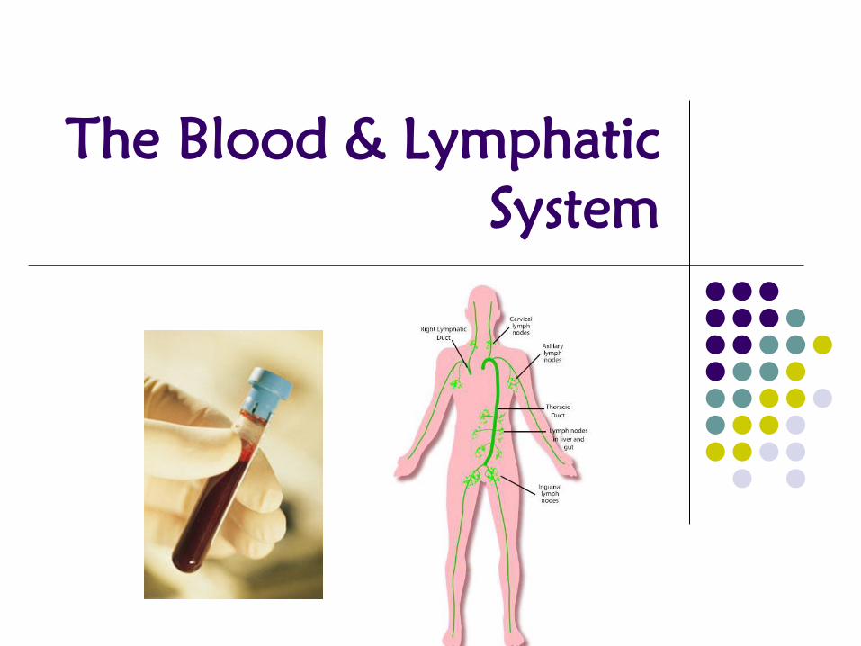
The Blood & Lymphatic
System
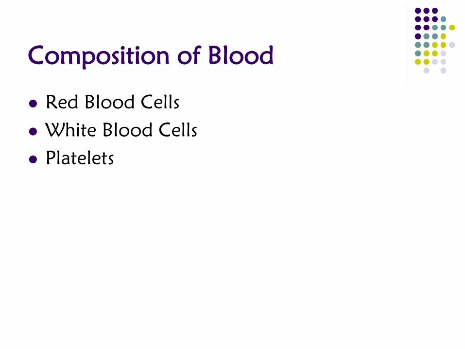
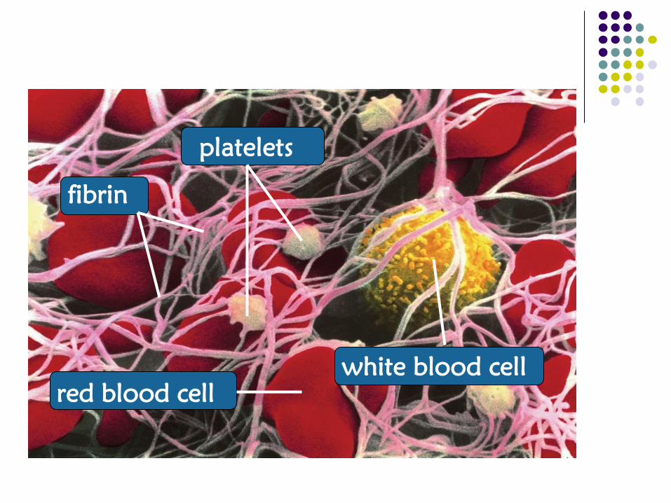
Composition of Blood
Red Blood Cells
White Blood Cells
Platelets


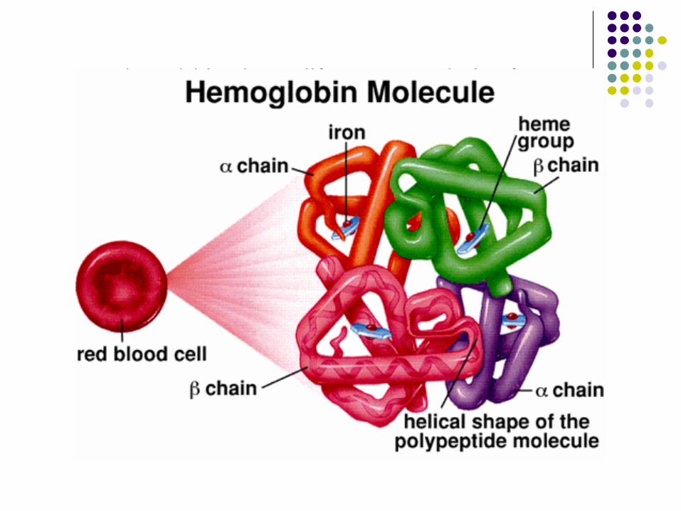
Red Blood Cells (aka erythrocytes)
Structure
Do not have a nucleus
Contain specialized proteins called
hemoglobin
Hemoglobin contains iron which
facilitates the transport of oxygen and
carbon dioxide

How healthy are your RBCs?
Perform a Complete Blood Count or Full Blood
Count
Take different measurements and compare them to the
rest of the hematocrit
Mean Corpuscular Volume (MCV): measures the
average volume of cytoplasm in a RBC
Red Blood Cells

Mean Corpuscular Hemoglobin (MCH): measures
the mass of hemoglobin in each RBC
Red Blood Cells
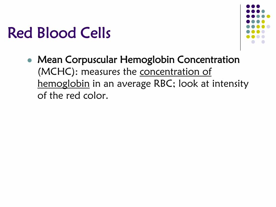
Mean Corpuscular Hemoglobin Concentration
(MCHC): measures the concentration of
hemoglobin in an average RBC; look at intensity
of the red color.
Red Blood Cells

Blood Type
Different blood types exist because there are
different proteins on the blood cells called
agglutinogens; can be classified as either A, B, and
D.
ABO Blood Group System classifies people with
varying proteins (A or B) on their red blood cells
Red Blood Cells

Blood Type
Type A has the A protein on the blood cell; also have B
antibodies
Red Blood Cells

Blood Type
Type B has the B protein on the blood cell; also have A
antibodies
Red Blood Cells

Blood Type
Type AB has both A and B protein on the blood cell; has
not antibodies
Red Blood Cells

Blood Type
Type O has neither the A or B protein on the blood cell;
have both A & B antibodies
Red Blood Cells

The D protein is called the Rh factor (or Rhesus
factor)
Rh-positive people HAVE the D protein on their blood
cells
Rh-negative people DO NOT HAVE the D protein on
their blood cells
Red Blood Cells

Successful blood transfusions depend on the
combination from donor to recipient.
Example: Recipient with blood type A can only receive
blood from a donor that is either blood type A or type
O. If they don’t, the blood will agglutinate, or clump
up together! Here’s why:
Red Blood Cells

Donor:
Type B
Recipient
So… the recipient’s immune system attacks the “foreign blood”!

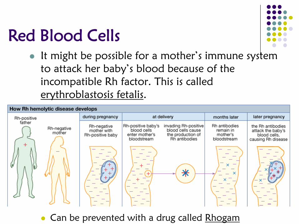
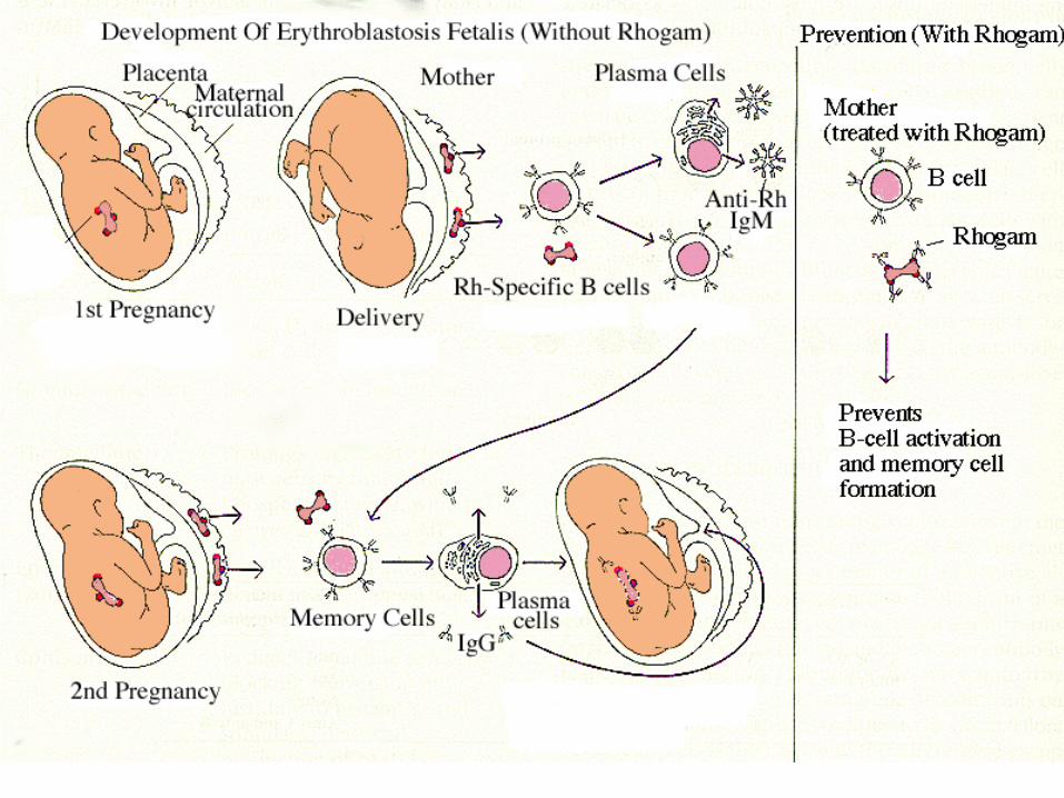
It might be possible for a mother’s immune system
to attack her baby’s blood because of the
incompatible Rh factor. This is called
erythroblastosis fetalis.
Can be prevented with a drug called Rhogam
Red Blood Cells

It might be possible for a mother’s immune system
to attack her baby’s blood because of the
incompatible Rh factor. This is called
erythroblastosis fetalis.
Can be prevented with a drug called Rhogam
Red Blood Cells

It might be possible for a mother’s immune system
to attack her baby’s blood because of the
incompatible Rh factor. This is called
erythroblastosis fetalis.
Can be prevented with a drug called Rhogam
Red Blood Cells

It might be possible for a mother’s immune system
to attack her baby’s blood because of the
incompatible Rh factor. This is called
erythroblastosis fetalis.
Can be prevented with a drug called Rhogam
Red Blood Cells
It might be possible for a mother’s immune system
to attack her baby’s blood because of the
incompatible Rh factor. This is called
erythroblastosis fetalis.
Can be prevented with a drug called Rhogam
Red Blood Cells

Function: carry oxygen throughout the body
This is possible because of hemoglobin which can carry
up to 4 O2 molecules at a time.
The oxygen-carrying efficiency of hemoglobin is
measured as percent saturation. Depending on the
pressure, hemoglobin will keep or release its oxygen.
Red Blood Cells

Partial pressure (mmHg)
Percent
Saturation
(Greater the
number, the
better
hemoglobin is
at getting O2)
Maternal hemoglobin
Fetal hemoglobin
Llama hemoglobin
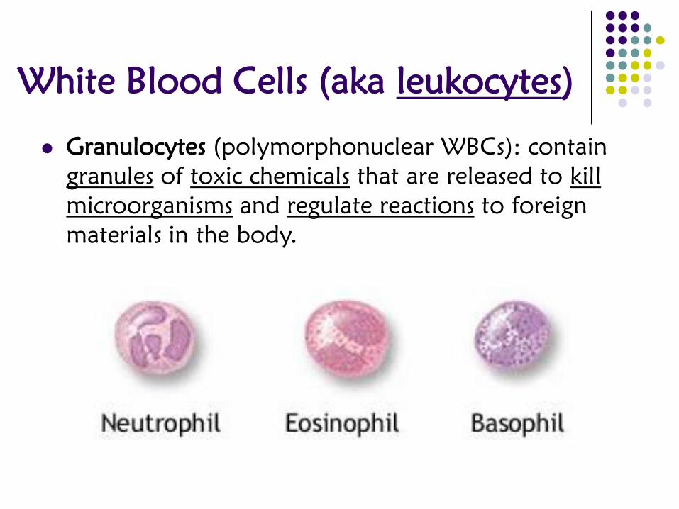
White Blood Cells (aka leukocytes)
Granulocytes (polymorphonuclear WBCs): contain
granules of toxic chemicals that are released to kill
microorganisms and regulate reactions to foreign
materials in the body.

Neutrophils (40-70%): most common type of
white blood cell; secrete antibiotics and eat the
remains of bacteria and damaged cells
antibiotic: chemical that harms or kills bacteria
White Blood Cells (aka leukocytes)
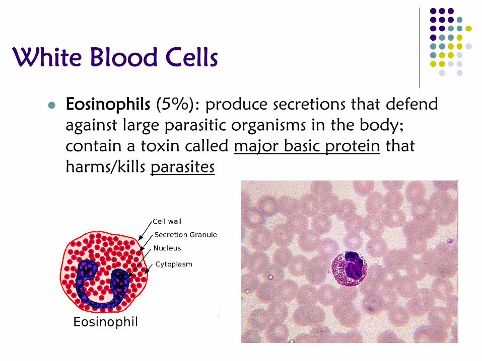
Eosinophils (5%): produce secretions that defend
against large parasitic organisms in the body;
contain a toxin called major basic protein that
harms/kills parasites
White Blood Cells

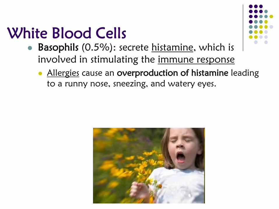
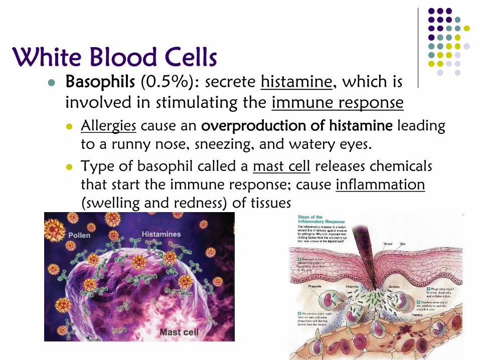
Basophils (0.5%): secrete histamine, which is
involved in stimulating the immune response
White Blood Cells
Basophils (0.5%): secrete histamine, which is
involved in stimulating the immune response
Allergies cause an overproduction of histamine leading
to a runny nose, sneezing, and watery eyes.
White Blood Cells
Basophils (0.5%): secrete histamine, which is
involved in stimulating the immune response
Allergies cause an overproduction of histamine leading
to a runny nose, sneezing, and watery eyes.
Type of basophil called a mast cell releases chemicals
that start the immune response; cause inflammation
(swelling and redness) of tissues
White Blood Cells

Agranulocytes (mononuclear WBCs): lack
granules
Monocytes (1-5%): develop into two types of
macrophages
White Blood Cells

Agranulocytes (mononuclear WBCs): lack
granules
Monocytes (1-5%): develop into two types of
macrophages
Circulating monocytes: detect infectious agents (called
pathogens) traveling through in the blood; also
involved in bone growth and maintenance
Tissue monocytes: remove dead cells and attack
microorganisms that are more difficult to kill like fungi
White Blood Cells

Lymphocytes (20-50%): present in the blood and lymphatic
system
White Blood Cells

Lymphocytes (20-50%): present in the blood and lymphatic
system
B lymphocytes: assists with the immune response by making
antibodies; produced and mature in the bone marrow
T lymphocytes: responsible for stimulating the immune
response and killing infected cells; produced in the bone
marrow and mature in the thymus gland
White Blood Cells

What are they?
Not true cells, but are cell fragments that come
from larger cells called megakaryocytes
Covered with many proteins that allow the
platelets to stick to a variety of materials
Stickiness helps to reduce blood flow to a damaged
area during injuries that break the blood vessels
Platelets (aka thrombocytes)

Blood Clotting process; generally a two-
step process
Platelets adhere to an injured area
Activation of the clot formation
When blood vessels are intact, they produce
prostacyclin which prevents platelet activation.
Platelets

blood vessel
clot

Platelets
Clotting Cascade: a reaction that occurs
when there is damage to a blood vessel or
body tissue
Damaged tissue releases various tissue
components into the blood
Collagen and clotting factors alert other parts of
the body that there is damaged tissue
Platelets adhere to the damaged tissue and to
each other

Platelets
Prothrombin (secreted by platelets) is converted
into thrombin
Thrombin catalyzes the reaction to convert
fibrinogen into fibrin
Platelets
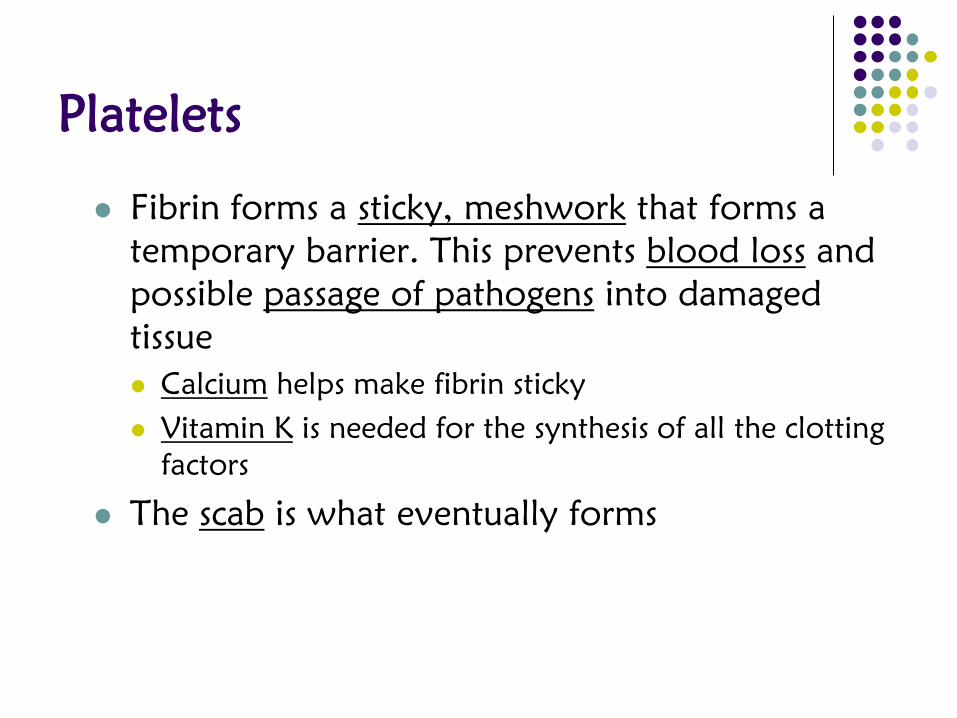
Fibrin forms a sticky, meshwork that forms a
temporary barrier. This prevents blood loss and
possible passage of pathogens into damaged
tissue
Calcium helps make fibrin sticky
Vitamin K is needed for the synthesis of all the clotting
factors
The scab is what eventually forms
platelets
fibrin
white blood cell
red blood cell

Platelets
Since clots are temporary structures, and
scabs don’t last forever, healthy cells near
the clot release Tissue Plasminogen
Activator (TPA).
Initiates the conversion of plasminogen into
plasmin, which digests fibrin and thereby
dissolves the clot; reduces damage to
cardiovascular diseases caused by blood clots.

Lympatic System: Structures
Lymphatic vessels:
thin ducts that carry
a clear fluid called
lymph; drain excess
fluids that
accumulates in
tissues which
prevents edema
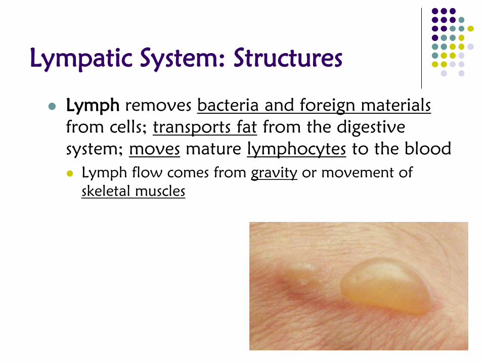
Lympatic System: Structures
Lymph removes bacteria and foreign materials
from cells; transports fat from the digestive
system; moves mature lymphocytes to the blood
Lymph flow comes from gravity or movement of
skeletal muscles

Lymphatic System: Structures
lymphatic trunks: division of the lymphatic
vessels; drain lymph from larger areas of the
body; has collections of small regions of
lymphatic tissue
tonsils: lymphoid tissue found in the throat
Peyer’s Patches: lymphoid tissue found in the digestive
system

Lymphatic System: Structures
lymph nodes (lymph glands): eliminate antigens from
the lymph; have 4 components

Lymphatic System: Structures
blood vessels: enter and exit the lymph
node through the hilum
lymphatic sinuses: fluid-filled sack

Lymphatic System: Structures
filler tissue (stroma): divided into regions by walls
in the capsule called trabeculae
lymphocytes: B and T lymphocytes are found in
the stroma; help with the immune response

Lymphatic System: Structures
Lymph Glands:
spleen: located in the upper left region of the
abdomen near the stomach; divided into two
regions

Lymphatic System: Structures
Lymph Glands:
spleen: located in the upper left region of the
abdomen near the stomach; divided into two
regions
red pulp: storage area for red blood cells; responsible
for removing old or damaged RBCs from the
circulations
white pulp: contain B-lymphocytes and T-lymphocytes
Lymphatic System: Structures
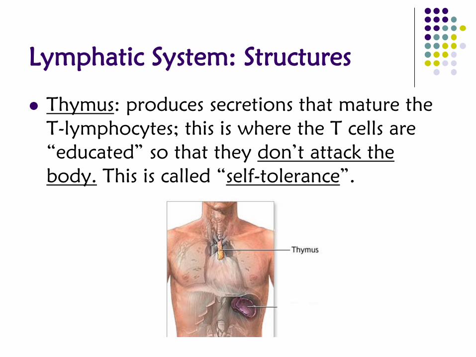
Thymus: produces secretions that mature the
T-lymphocytes; this is where the T cells are
“educated” so that they don’t attack the
body. This is called “self-tolerance”.
Immune Response
Immune Response: triggered by the presence
of a foreign antigen
Innate Immunity: aka nonspecific immunity; uses
barriers that block a variety of infections to
protect the body
Physical Barriers: block virtually all kinds of
pathogens
Skin: blocks microorganisms from entering tissues;
constant shedding removes microbes; skin secretions
prevent microbes from overgrowing
Mucous membranes: catch incoming pathogens before
they can enter the bloodstream
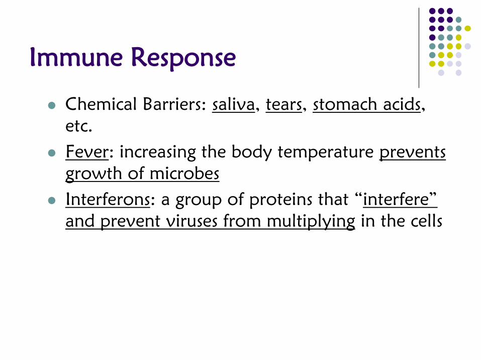
Immune Response
Chemical Barriers: saliva, tears, stomach acids,
etc.
Fever: increasing the body temperature prevents
growth of microbes
Interferons: a group of proteins that “interfere”
and prevent viruses from multiplying in the cells

Immune Response
Acquired Immunity: occurs when the body
adapts to specific infections; it’s like the body
learns about the “invader” and kills it as
effectively as possible.

Immune Response
Macrophages engulf (eat) the invaders and
present the antigen on its surface
A helper T-cell attaches to the macrophage and
receives information. The helper T-cell activates
B cells and other T cells and the immune system
responds in 2 ways:

Immune Response
Cell-mediated immunity: the T cells that are
activated seek out infected cells and kill them

Immune Response
Humoral immunity: the B cells that are activated
divide into plasma cells (make lots of antibodies)
and memory cells (stores information about the
pathogen and initiates a faster immune response
the second time around)

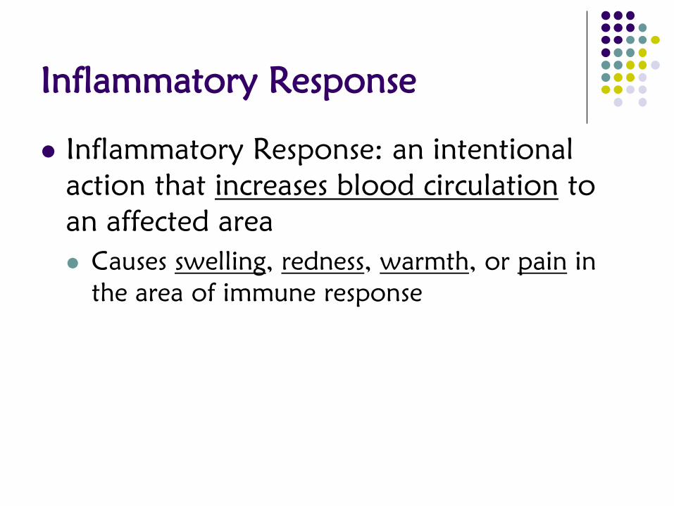
Inflammatory Response
Inflammatory Response: an intentional
action that increases blood circulation to
an affected area
Causes swelling, redness, warmth, or pain in
the area of immune response


Immunization & Vaccination
Natural immunity: fighting the disease the
old-fashioned way (getting sick and fighting it
yourself)
Artificial immunity: faking out your immune
system to prepare it for a real attack;
example: vaccines

Immunization & Vaccination
Immunity against a disease
can be acquired through
active or passive immunity
Active: exposing antigens to
your immune system cells;
either naturally or artificially
Passive: when the antibodies
are basically given to you;
example would be breast milk
Anemia

Hemophilia
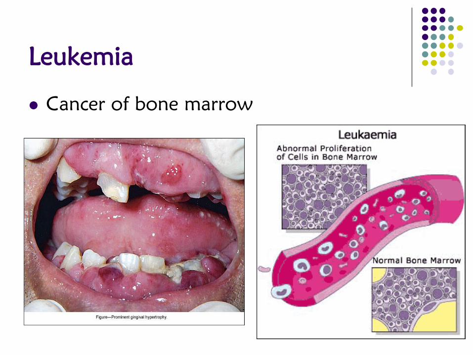
Leukemia
Cancer of bone marrow

Hodgkins Lymphoma

Lymphedema
Build up of fluids

Elephantiasis
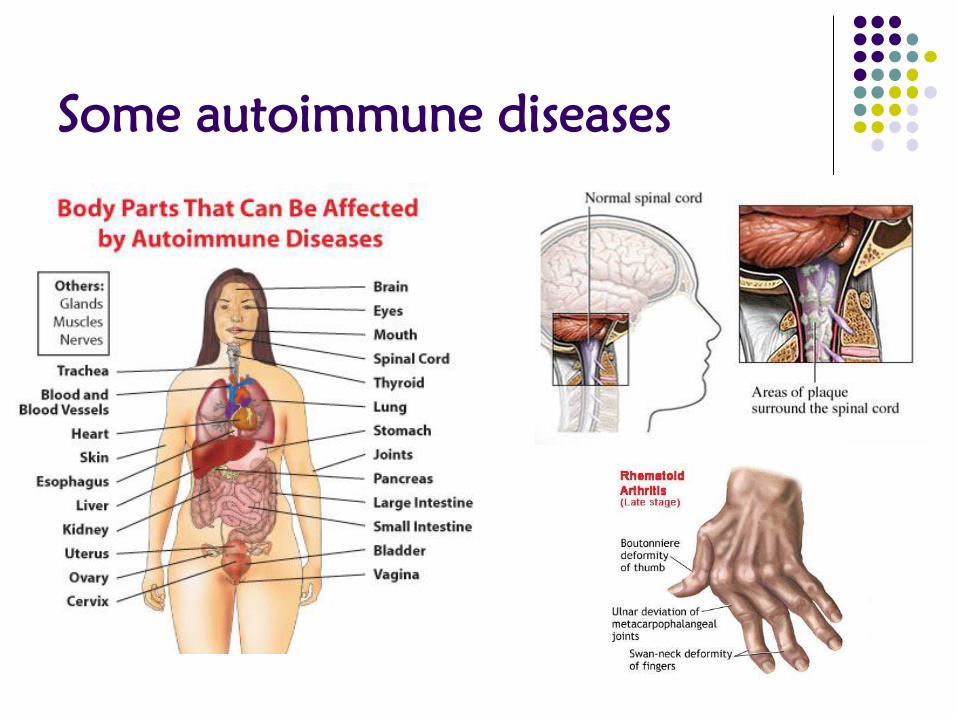
Some autoimmune diseases


