843 PROF. J. ROSE BRADFORD ON THE PATHOLOGY OF THE KIDNEYS. regards abdominal neurosis, in a paper recently read before the Medical Society of London.3 The partial removal of the thyroid gland in the acute stage of the disease is attended with very considerable danger and only very inadequate success. The best cases were those of Mr. Paul of Liverpool, but in six cases recorded by him all except one were of more than two years’ standing. Abadie and Faure have had successes from section or ablation of the cervical sympathetic, but of Faure’s three cases one was of twelve years’ standing and in the others the duration is not stated. Doubtless the section of the sympathetic will slow the heart, but it has not yet been satisfactorily shown to influence the -disease, and one would regard with some misgivings the future of a heart deprived of due sympathetic innervation, whilst it is clinically remarkable how well the heart comes <out of the long struggle but little damaged when the patient has been carefully steered through by judicious handling. it sometimes happens that a disfiguring enlargement or a more serious distortion of the thyroid remains after the tacbycardial symptoms have subsided and in one instance I have seen this condition dealt with very successfully by Temoval. I can but repeat that in our present knowledge of pathology and therapeutics bearing upon tachycardial condi- tions, including exophthalmic goitre, all remedial measures, however valuable some of them may be on proper occasions and at proper times, are absolutely futile unless they be sub- ordinated to the essential treatment by graduated rest and isolation from all worry, fatigue, and excitement. Of the opposite more rare form of cardiac neurosis, bradycardia, I need not say much, for the cases require but little treatment. One form is that which sometimes follows apon the rapid heart of exophthalmic goitre. I have {recently had a remarkable example of this in my ward at the Middlesex Hospital. The patient was a woman, aged sixty years, who twenty years ago was operated upon by Dr. Qxalabin at Gay’s Hospital for ovarian disease and both ovaries were removed. She almost immediately became the subject of acute Graves’s disease. Ten years later she came under the observation of Dr. Pringle as an out-patient at the Middlesex Hospital still presenting all the symptoms of active exophthalmic goitre, proptosis, enlarged and pulsatile thyroid, a pulse of 136, agitations, tremors, sweatings, &c. ’She could not come into the hospital but continued her tattendance, coming periodically from the east side of London for many years, in the course of which, chiefly under small doses of aconite, belladonna, and bromide, her thyroid gradually dwindled to an atrophic state, her pulse slowed to 0, and her manner changed from one of excitement and nervous agitation to a quiescent, almost an apathetic, demeanour. She still preserved her proptosis which she has i at the present time, and at no time did Dr. Pringle or I Dr. Pasteur, under whom she had more recently been as ,out-patient, regard her case as presenting the distinct signs ,,3f myxoedema. She had no albuminuria, but latterly her Aegs became somewhat cedematous. Her heart has slowed down to 45 and 30, there has been for some time a basic aystolic murmur, and the left ventricle is decidedly hyper- !trophied and somewhat dilated. The signs are rather those ’of degenerative roughening, however, than of constriction of the aorta, for the pulse is of fairly full volume and regular, although very slow. After a few weeks’ rest in bed and some strychnine tonic she has gone to the convalescent home. This seems to be a case analogous with regard to the myxcedemic state to those cases of tachycardia I have spoken of as not being distinctly associated with ’Graves’s malady and yet approximating to it. Bradycardia us very uncommonly associated with well-marked myxoedema ;and may continue notwithstanding the disease has been - cured or held in check by thyroid treatment. I have seen it in epileptics, in which case the rhythm is not only slow but generally irregular. As a temporary condition bradycardia is not infrequently met with as a sequel to influenza and ,also in association wlth the stage of depressed temperature that frequently follows upon other fevers. In the influenzal oeases that I have seen it has been associated with a very high density of urine with great surcharge of urea, as though there had been some previous accumulation, the urine becoming solid with crystals on the addition of cold nitric acid. Spurious anguinal seizures are sometimes observed under these circumstances. I have never known .snch to prove fatal, although of course in the presence 3 THE LANCET, March 5th, 1898. 4 Brit. Med. Jour., vol. ii., p. 6, 1897. 5 Progrès Médical, 1897. of any previous heart disease they might readily prove so. Chronic high arterial tension is generally associated with a slow, sometimes a very slow, pulse, whereas in acutely raised tension the heart’s action is, as I have pointed out, generally quickened. In chronic bradycardia, a con- dition that tends to remain permanent and does not necessarily shorten life, an occasional twenty-four hours’ rest in bed should be enjoined and for mental work the recumbent posture should be preferred. In cases of a more temporary kind the combination of strychnine with an alkali or iodide of potassium (the two drugs being kept in separate bottles and only mixed at the time of taking) is a very useful one. Caffein is also very useful, especially where the urine is scanty. A five minutes’ whiff of oxygen three or four times in the twenty-four hours is a valuable cardiac stimulant. In cases of mygoedema of course thyroid extract will be given, but it is not wise to push it to the production of any excite- ment of circulation. The Boulstonian Lectures: OBSERVATIONS ON THE PATHOLOGY OF THE KIDNEYS. Delivered before the Royal College of Physicians of London on March 15th, 17th, and 22nd, 1898, BY JOHN ROSE BRADFORD, M.D., D.SC. LOND., F.R.S., FELLOW OF THE COLLEGE; PHYSICIAN TO UNIVERSITY COLLEGE HOS- PITAL; PROFESSOR OF MATERIA MEDICA AND THERAPEUTICS IN UNIVERSITY COLLEGE; PROFESSOR SUPERINTENDENT OF THE BROWN INSTITUTION. (From the Laboratory of the Brown Institution.) ! LECTURE 11. Delivered on March 7M. , Ms. PRESIDENT AND B’ELLOwa,—In my last lecture I detailed at some length the results following partial nephrectomies (of varying degrees of severity) both on the composition of the urine and on the general metabolism of the body, and I trust that I showed that whereas all partial nephrectomies increased the flow of urinary water only the more severe ones increased the excretion and production of urea. We will now consider some of the results following complete double nephrectomy. Experimentally the phenomena following the cessation of the renal functions may be studied by three different methods of procedure: (1) ligature of the renal arteries ; (2) complete removal of the kidneys ; and (3) the ligature of the ureters. The first method is on the whole un- suitable, since there is always a certain amount of doubt as to the extent of the collateral blood supply, and therefore in all my observations either both kidneys were removed or both ureters ligatured. When the kidneys were removed this was done in two operations in order to minimise the effect of shock, &c., and this was more especially done in order that an accurate comparison might be made between the effects of double nephrectomy on the one hand, and the effect of double ligature of the ureters on the other. The ligature of both ureters from an operative point of view is a trivial procedure, especially if they are both ligatured through an incision near the pubes. On the other hand, the removal of both kidneys at once, whether through one incision or by two lumbar incisions, is a very severe operation and thus it is misleading to compare the immediate effects of the two proceedings. The re- moval of one kidney in the dog is an operation which does not cause much shock and with due care hasmorrhage may be avoided. Even when the second nephrectomy is performed and the only kidney present is removed it is astonishingt o see the small amount of shock that is produced. The animal is in no way collapsed and immediately after recovery from the anaesthetic may run about and behave very much like a normal animal, so that, provided the kidneys be Hmoved like this, in two stage?, there is no objection to contra&bgr;tilJg 1 Lecture I. was published in THE LANCET of March 19th, 1898.
Transcript
843PROF. J. ROSE BRADFORD ON THE PATHOLOGY OF THE KIDNEYS.
regards abdominal neurosis, in a paper recently read beforethe Medical Society of London.3 The partial removal of thethyroid gland in the acute stage of the disease is attendedwith very considerable danger and only very inadequatesuccess. The best cases were those of Mr. Paul of Liverpool,but in six cases recorded by him all except one were ofmore than two years’ standing. Abadie and Faure havehad successes from section or ablation of the cervicalsympathetic, but of Faure’s three cases one was of twelveyears’ standing and in the others the duration is not stated.Doubtless the section of the sympathetic will slow the heart,but it has not yet been satisfactorily shown to influence the-disease, and one would regard with some misgivings thefuture of a heart deprived of due sympathetic innervation,whilst it is clinically remarkable how well the heart comes<out of the long struggle but little damaged when the patienthas been carefully steered through by judicious handling.it sometimes happens that a disfiguring enlargement or amore serious distortion of the thyroid remains after thetacbycardial symptoms have subsided and in one instance Ihave seen this condition dealt with very successfully byTemoval. I can but repeat that in our present knowledge ofpathology and therapeutics bearing upon tachycardial condi-tions, including exophthalmic goitre, all remedial measures,however valuable some of them may be on proper occasionsand at proper times, are absolutely futile unless they be sub-ordinated to the essential treatment by graduated rest andisolation from all worry, fatigue, and excitement.
Of the opposite more rare form of cardiac neurosis,bradycardia, I need not say much, for the cases require butlittle treatment. One form is that which sometimes followsapon the rapid heart of exophthalmic goitre. I have{recently had a remarkable example of this in my ward at theMiddlesex Hospital. The patient was a woman, aged sixtyyears, who twenty years ago was operated upon by Dr.Qxalabin at Gay’s Hospital for ovarian disease and bothovaries were removed. She almost immediately became thesubject of acute Graves’s disease. Ten years later she cameunder the observation of Dr. Pringle as an out-patient at theMiddlesex Hospital still presenting all the symptoms ofactive exophthalmic goitre, proptosis, enlarged and pulsatilethyroid, a pulse of 136, agitations, tremors, sweatings, &c.’She could not come into the hospital but continued hertattendance, coming periodically from the east side of Londonfor many years, in the course of which, chiefly under smalldoses of aconite, belladonna, and bromide, her thyroidgradually dwindled to an atrophic state, her pulse slowed to0, and her manner changed from one of excitement andnervous agitation to a quiescent, almost an apathetic,demeanour. She still preserved her proptosis which she has iat the present time, and at no time did Dr. Pringle or IDr. Pasteur, under whom she had more recently been as,out-patient, regard her case as presenting the distinct signs,,3f myxoedema. She had no albuminuria, but latterly herAegs became somewhat cedematous. Her heart has sloweddown to 45 and 30, there has been for some time a basicaystolic murmur, and the left ventricle is decidedly hyper-!trophied and somewhat dilated. The signs are rather those’of degenerative roughening, however, than of constrictionof the aorta, for the pulse is of fairly full volume andregular, although very slow. After a few weeks’ rest in bedand some strychnine tonic she has gone to the convalescenthome. This seems to be a case analogous with regardto the myxcedemic state to those cases of tachycardia Ihave spoken of as not being distinctly associated with’Graves’s malady and yet approximating to it. Bradycardiaus very uncommonly associated with well-marked myxoedema;and may continue notwithstanding the disease has been- cured or held in check by thyroid treatment. I have seen itin epileptics, in which case the rhythm is not only slow butgenerally irregular. As a temporary condition bradycardiais not infrequently met with as a sequel to influenza and,also in association wlth the stage of depressed temperaturethat frequently follows upon other fevers. In the influenzaloeases that I have seen it has been associated with a veryhigh density of urine with great surcharge of urea, as
though there had been some previous accumulation, theurine becoming solid with crystals on the addition of coldnitric acid. Spurious anguinal seizures are sometimesobserved under these circumstances. I have never known.snch to prove fatal, although of course in the presence
3 THE LANCET, March 5th, 1898.4 Brit. Med. Jour., vol. ii., p. 6, 1897. 5 Progrès Médical, 1897.
of any previous heart disease they might readily proveso. Chronic high arterial tension is generally associatedwith a slow, sometimes a very slow, pulse, whereas inacutely raised tension the heart’s action is, as I have pointedout, generally quickened. In chronic bradycardia, a con-dition that tends to remain permanent and does not
necessarily shorten life, an occasional twenty-four hours’rest in bed should be enjoined and for mental work therecumbent posture should be preferred. In cases of a moretemporary kind the combination of strychnine with an alkalior iodide of potassium (the two drugs being kept in separatebottles and only mixed at the time of taking) is a very usefulone. Caffein is also very useful, especially where the urineis scanty. A five minutes’ whiff of oxygen three or four timesin the twenty-four hours is a valuable cardiac stimulant. Incases of mygoedema of course thyroid extract will be given,but it is not wise to push it to the production of any excite-ment of circulation.
The Boulstonian Lectures:OBSERVATIONS ON THE PATHOLOGY OF
THE KIDNEYS.Delivered before the Royal College of Physicians of London
on March 15th, 17th, and 22nd, 1898,
BY JOHN ROSE BRADFORD, M.D.,D.SC. LOND., F.R.S.,
FELLOW OF THE COLLEGE; PHYSICIAN TO UNIVERSITY COLLEGE HOS-PITAL; PROFESSOR OF MATERIA MEDICA AND THERAPEUTICS IN
UNIVERSITY COLLEGE; PROFESSOR SUPERINTENDENTOF THE BROWN INSTITUTION.
(From the Laboratory of the Brown Institution.)
! LECTURE 11.Delivered on March 7M.
, Ms. PRESIDENT AND B’ELLOwa,—In my last lecture I
detailed at some length the results following partialnephrectomies (of varying degrees of severity) both on thecomposition of the urine and on the general metabolism ofthe body, and I trust that I showed that whereas all partialnephrectomies increased the flow of urinary water only themore severe ones increased the excretion and production ofurea. We will now consider some of the results followingcomplete double nephrectomy.Experimentally the phenomena following the cessation
of the renal functions may be studied by three differentmethods of procedure: (1) ligature of the renal arteries ;(2) complete removal of the kidneys ; and (3) the ligatureof the ureters. The first method is on the whole un-suitable, since there is always a certain amount of doubtas to the extent of the collateral blood supply, and thereforein all my observations either both kidneys were removed orboth ureters ligatured. When the kidneys were removed thiswas done in two operations in order to minimise the effect ofshock, &c., and this was more especially done in order thatan accurate comparison might be made between the effectsof double nephrectomy on the one hand, and the effectof double ligature of the ureters on the other. The
ligature of both ureters from an operative point of viewis a trivial procedure, especially if they are both ligaturedthrough an incision near the pubes. On the otherhand, the removal of both kidneys at once, whetherthrough one incision or by two lumbar incisions, is a
very severe operation and thus it is misleading to comparethe immediate effects of the two proceedings. The re-
moval of one kidney in the dog is an operation which doesnot cause much shock and with due care hasmorrhage may beavoided. Even when the second nephrectomy is performedand the only kidney present is removed it is astonishingt osee the small amount of shock that is produced. The animalis in no way collapsed and immediately after recovery fromthe anaesthetic may run about and behave very much like anormal animal, so that, provided the kidneys be Hmovedlike this, in two stage?, there is no objection to contra&bgr;tilJg
1 Lecture I. was published in THE LANCET of March 19th, 1898.
844 PROF. J. ROSE BRADFORD ON THE PATHOLOGY OF THE KIDNEYS.
the effects following the operation with those seen afterligature of both ureters.
In all the experiments on this question care was takenthat full antiseptic precautions were used and in but onecase was there any peritonitis either local or general. Thisis a point of very great importance and it is not improbablethat many of the symptoms described formerly as followingcomplete nephrectomy were really dependent upon thepresence of septic peritonitis and not upon the absenceof the kidneys.The duration of life in dogs after complete nephrectomy
carried out, as above described, in two stages is usually fromthree to five days, the animals in no case living beyond thefifth day and more usually dying on the fourth day. Duringthe first two days very little amiss is to be noted about theanimal except that the appetite is poor and that after thefirst day all food is liable to be refused. Muscular weaknessbegins to show itself on the second day, is well marked onthe third day, and progresses steadily, but the nephrectomisedanimal is usually able to run about even on the third andperhaps on the fourth day. Thirst is sometimes present, butnot as a rule to a marked extent. Wasting is very markedand progressive ; for instance, one dog weighing six kilo-grammes lost three-quarters of a kilogramme in from three tofive days. The most characteristic feature apart from theweakness and wasting is, however, the course of the bodytemperature. At the time of the operation the rectal tem-perature is probably between 101° F. and 102° F. Withintwenty-four hours of the operation it falls to 100°, on thesecond day after the operation the temperature had probablyfallen to 99°, and on the third day to 98° or even to 970.The fall is a continuous and progressive one and theanimal remains active and lively until the temperaturereaches from 98° to 970. When the temperature falls to 960the animal is really in a moribund condition although it mayreach 950 before death actually occurs. The course of thetemperature chart is the best indication of the probableduration of life. These are the most prominent symptomsseen after double nephrectomy and it is most important tobear in mind that certainly for three days and sometimesfor longer the animal not only remains conscious but isactive and lively and able to run about. As deathapproaches the weakness increases and there may beslight drowsiness, but this is never a prominent symptom,and personally I have never seen in nephrectomised dogsanything approaching to coma. Vomiting is also not a con-spicuous symptom and in this my experience is contrary tothat of the older observers and I cannot help thinking thatthe vomiting observed and made so much of’ by Bernardand others was really due to septic complications such asperitonitis. Vomiting, as just mentioned, is not a con-
spicuous symptom, and sometimes it is completely absent.In others it is present towards the end after the third day.Convulsions I have never seen, and that very constant ursemicsymptom, dyspnoea, is also not conspicuous. Slight diarrhoeamay be present. It will be seen from the above sketchof the symptoms following complete double nephrectomythat they only very distantly resemble the clinical pictureof acute urasmia or at any rate of the acute uraemia seen inBright’s disease.The results following the ligature of both ureters are
very similar if not identical with those seen after completenephrectomy, and I have been quite unable as yet todifferentiate in any way between the symptoms produced bythose two procedures. The duration of life after ligature ofboth ureters was if anything rather shorter than after com-plete nephrectomy, but this may have been due, althoughimprobably, to both ureters having been tied at once. Theanimals usually died in from three to four, instead of fromthree to five. days, otherwise the symptoms were practicallyidentical. Vomiting was observed in one case especiallyafter drinking, and in this case some local peritonitis wasfound, the ureters having been ligatured by two separateincisions in the lumbar region. The vomited fluid yieldedno gas by the hypobromite method, and I think it most
probable that the vomiting arose from the peritonitis, as
this was the only instance in the series where this symptomwas at all marked.The fact that whether the renal functions be abrogated
by the complete removal of the kidneys or by ligature ofboth ureters the resulting symptoms are practically identicalis, I think, a factor of some importance with regard tothe nature of ursemia. Ligature of the ureter experiment-ally invariably leads to the production of hydronephrosis,
and in no case was the obstruction of the ureter followedby that complete suppression that has been observed inman after complete obstruction of the ureter. InaEmuchas the ligature of the ureter is always followed by hydro-nephrosis it might be argued that the results following thisoperation ought not to be similar to those seen after com-plete double nephrectomy. That this is so, however, canbe shown not only by the facts already detailed but also bythe fact that after the removal of a large portion of onekidney the disturbance of nutrition and the alteration inthe urine described in my last lecture are seen to followequally well whether (a) the opposite kidney be excised or(b) whether the ureter of this kidney be tied. Not only arethe same effects produced but the duration of life with thefragment of one kidney is in no way prolonged by the factthat the ureter of the opposite kidney is tied instead of thatkidney being removed. A further and perhaps even moreconclusive proof of the same fact is the following:ligature of the ureter causes hydronephrosis, if after theproduction of this the ureter be cut down upon andopened and the hydronephrosis drained the kidney returnsto its former shape but not to its former size. It isto the eye apparently a normal but small kidney. Onmicroscopic examination such a kidney shows that theepithelium of the tubules has undergone a peculiar change,the cells shrinking and losing their granules and becomingglass-like in appearance; the nuclei, however, stain, althoughthe protoplasm of the cells is quite clear. These appearancessuggest that these are resting cells not capable of performingtheir usual functions. Such a kidney secretes a clear acidfluid containing little if any urea. Although it secretes sucha fluid comparatively freely, such a kidney is, however,quite useless to the economy, and if the opposite healthykidney be removed, all the phenomena characteristic ofdouble complete nephrectomy are seen. This experi-ment shows two things-in the first place that ligatureof the ureter permanently damages the epithelial cellstructures of the kidney, for these are more affectedthan the glomeruli (and there is no overgrowth of fibroustissue), and in the second place it affords another demon-stration that the essential portion of the kidney is the
epithelium lining the convoluted tubules. This experimentis in striking contrat to those described in the firstlecture, where a small fragment of normal kidney was shownto be capable of maintaining the renal functions for an indefi-nite period. An entire kidney that has been exposed foreven a short time-i.e., fourteen to twenty-one days-to theeffects of the complete obstruction of the ureter with con.sequent hydronephrosis is unable when that obstruction isremoved by draining the distended ureter to perform renalfunctions that are of any avail in prolonging life. Such akidney is practically useless to the animal, although, as men.tioned above, it is able to secrete an acid, clear liquid. It isprobable that this is a glomerular secretion, modified it maybe by the amount of damage that has been inflicted on theglomerulus by the increased pressure that has been pro-duced in the glomerular chamber by the ligature of theureter. At any rate, the ligature of the ureter leads tc’much more pronounced and permanent lesions in theepithelium of the tubules than it does in the glome-ruli, and it seems to me that by this method oftying the ureter and subsequently draining the hydro-nephrosis so produced we may be able to differen-tiate in the mammal the functions of the glomerularchamber from those of the convoluted tubules. I have withthis object in view some observations in progress at thepresent time. As ligature of the ureter causes these changesin the epithelial structures of the kidney very rapidly it isnot very surprising that ligatures of the ureters and removalof both kidneys should produce practically identical effects.This fact is an argument against the existence of an
internal renal secretion, since in other cases where glandshave an external and an internal secretion the ligature of theexcretory duct does not produce the effect seen after theremoval of the gland. This is undoubtedly the case withthe pancreas and also with the liver, since in the lattercase it is fair to look upon the glycogenic function ofthe liver as an internal secretion of a kind, and theligature of the bile duct does not materially interfere as faras is known with the other and metabolic functions of theliver. Although it may be argued that the ligature of theureter very soon destroys the activity of the cells of theconvoluted tubules, yet the production of the hydro-nephrosis and the fairly free secretion that continues after
845PROF. J. ROSE BRADFORD ON THE PATHOLOGY OF THE KIDNEYS.
draining the distended ureter show that the secretoryactivity of the glomeruli at any rate persists, so that it
,cannot be said that ligature of the ureters is theoreticallyequivalent to the removal of the kidney. Some of the viewsbeld on the nature of the different forms of uraemia havebeen based on the idea that the results following completenephrectomy and those following ligature of the ureters weredifferent, and this, I think, is untenable from an experimentalpoint of view, and I hope to adduce evidence later that it is-equally untenable in the human subject.
After ligature of both ureters and after double nephrec-tomy there is of necessity a great accumulation of nitro-;genous extractive matters in the blood and tissues and, asmentioned above, the increase in the muscles is not only.
very great but, unlike that in the blood, it is notconfined to the nitrogenous extractives, such as urea, that.are soluble in absolute alcohol, but those like creatin (in-soluble in absolute alcohol) are also increased. The increaseof urea in the blood after double nephrectomy or after liga-ture of both ureters may amount to some twenty times thenormal 0’3 per cent., and even more is by no means excep-tional. This same great increase is seen in the muscles,<where normally there is no urea, the small quantities detectedby ordinary procedures being in all probability dependent- upon contamination with blood. The amount of other ex-tractives in muscles after nephrectomy may be nearly trebled,and seeing that normally there is as much as 0’2 per cent.of creatin this means that very large quantities of creatin:and such like substances accumulate in the muscles afterdouble nephrectomy. Similar large quantities of these ex-tractives accumulate in the liver and brain. I have notbeen able to satisfy myself that there is any essential differ-"enoe in these results whether the kidneys have been removedor the ureters tied.The quantities of extractives in the blood are so large that
the question naturally presents itself, and more especially inthe light of the experiments on partial nephrectomy detailed’sn my first lecture, whether these quantities can be accountedfor on the hypothesis that they are simply the products that,cannot be excreted of a normal metabolism. If the viewsadvanced in the first lecture are correct it is clear that aftercomplete nephrectomy as after partial nephrectomy thereshould be increased disintegration of proteid material leadingto the production of nitrogenous extractives in excessiveamount. In order to test this theory experiments were per-formed to determine the distribution of urea in the bloodand tissues after its intravenous injections in large quantities,the ureters being ligatured a few minutes before the ureawas injected into the jugular vein. The animals wereanaesthetised with chloroform and morphia and killed bybleeding at periods varying from a few minutes to an’hour and a half after the injection. The distribution- of urea in the blood and tissues was very similar to that!seen after partial or double nephrectomy-that is to say, themuscles contained a greater percentage than the liver-butthe very large quantities seen in the muscles after doubleQephrectomy were not seen after the injection of urea unless,,huge quantities were injected. Further, as the amount ofurea normally excreted by the dogs before nephrectomy<could be readily determined and the duration of life after1ihe complete nephrectomy was known it was quite possibleto calculate how much urea the nephrectomised dog shouldY1ave produced but was unable to excrete owing to theabsence of the kidneys. This quantity of urea was then Iinjected into a dog of the same weight as the nephrec-’iiomiaed animal and the results of the analysis of the Itissues were compared in the two cases. In all the cases,four in number, where this was done the amounts I"of urea, &c., as determined in the nephrectomised 1animal were greater than in the control animal which 1had received the amount of urea that theoretically should 1have been found in the nephrectomised animal. In these 1observations the other extractives were neglected but, 1as pointed out above, after nephrectomy these are also (
Increased, so that I think there is clear evidence that after e’complete double nephrectomy the amounts of nitrogenous aextractives in the blood and tissues are greater than can (
be accounted for on any hypothesis of simple retention. I Ithink that the results following double nephrectomy are c
quite in accordance with those described above as seen after tsevere partial nephrectomy (removals of three-quarters of jthe total kidney weight) and that we must admit that twhether the kidney be entirely removed or greatly diminished c
in amount in both cases there is a sudden and great t
disintegration of the proteid tissues of the bedy leadingt to the production of this large excess of nitrogenous7 extractives. After double nephrectomy practically no food; is eaten and there is rapid and great wasting, so that these} extractives have a tissue origin and, as insisted on above, the: same is the case after the more severe partial nephrectomies.! I have described at some length the results obtained
Iby experimentally investigating the effects following thes operations of nephrectomy partial and complete. Now I
propose to consider shortly some results obtained clinically-by the investigation of cases of chronic renal disease, moreespecially with reference to the influence of this disease on5the formation and the excretion of urea. Speaking broadly,the tendency of chronic renal disease is to cause a diminution; in the excretion of urea, or at least this is the common; teaching on the subject, and no doubt it is to a great
, extent true, but when this diminished excretion of urea isi referred entirely to a supposed incapacity of the damaged
kidney to excrete such bodies the basis of this view is notso sound. It is irrational to expect a patient suffering fromchronic renal disease to excrete quantities of urea com-
parable to the normal for the following reasons: the appetiteis poor and frequently a considerable amount of whatfood is taken is rejected by vomiting, diarrhoea is alsofrequently present, and all these factors will seriouslydiminish the amount of proteid available for disintegrationinto urea. Albuminuria is often present and frequently isexcessive in amount, and perhaps this loss is greatest whereit is not always suspected-namely, in persons with chronicrenal disease passing a considerable quantity of urine witha moderate or even a low percentage, such as a third or aquarter, of albumin. The greatest losses of albumin do notoccur in cases where the percentage amount of albumin ishighest, since in such cases the quantity of urine passed isusually greatly diminished. A daily loss of 20 grammesof dry proteid is by no means exceptional in renal diseaseand in some cases the amount rises to as much as 40grammes. Lastly, in dropsical patients a considerablequantity of urea and other extractives is retained in thedropsical effusions. All these causes operate in tending todiminish the amount of urea that can be excreted by thekidneys, and it is most misleading to compare the ureaexcretion of such a patient with the normal text-bookamount of 30 grammes per diem excreted by a healthyperson on a diet sufficient to maintain the body weight andto argue that an excretion of 20 grammes per diem or less isdependent entirely and directly upon the renal lesion. Inmany cases of chronic renal disease quantities of urea notgreatly below the normal amount may be excreted even at atime when the patient is very seriously ill and even in a verydangerous condition, and this remark applies more especiallyto the cases where dropsy is absent. It is more especiallytrue in the cases of what is called the "contracted whitekidney," a grave form of Bright’s disease in which thequantity of urine is considerably increased and contains 1 oreven 2 per cent. of albumin. No doubt in the cases ofchronic Bright’s disease associated with dropsy and also wherecardiac lesions such as nntral disease are present the urea ex-cretion undergoes a great diminution. Personally I considerthat a better notion of the state of the kidney lesion can beformed by observing the quantity and specific gravity of theurine than by observations on the amount of urea. Thelatter are often misleading for the reasons mentioned above.The quantity of water excreted, particularly if it is controlledby other observations, gives most reliable and valuable infor-mation ; copious dilute urine, if not due to the presence ofwaxy disease (by no means so common now), is a matter ofgrave significance not only as indicative of the probablepresence of what is ordinarily spoken of as the granularkidney but also as indicating the existence of other destruc-tive diseases of the renal tissue such as the so-called con-tracted white kidney, cystic kidneys, &c. It is customaryto consider that the excretion of this dilute urine is depen-dent upon the heightened blood-pressure that is so oftenseen in these kidney diseases, but the character of the urineapproximates very closely with that described above asoccurring after partial and more especially after doublepartial nephrectomy. If this comparison be fair thequantity of dilute urine excreted may afford an idea ofthe extent of the destruction of renal tissue in disease,just as it does in the nephrectomy experiments, the greaterthe hydruria the less the available kidney substance. The
comparison instituted between the experimental results andthe results of disease is, however, fallacious ni one respect.
846 PROF. J. ROSE BRADFORD ON THE PATHOLOGY OF THE KIDNEYS. I
Whereas in both the quantity of kidney is diminished in theexperimental case what renal tissue remains is healthy,whereas this is not so in most diseased conditions even whenthe incidence of the disease is not absolutely uniform. Incystic disease of the kidneys, however, for a time at anyrate, the remaining kidney tissue is practically normal, andthere is, I think, a fairly close parallel here between theresults of disease and the phenomena of the laboratory. Inboth a dilute urine is excreted, in both the renal tissue isgreatly diminished, and in neither do cardio - vascularchanges necessarily ensue. If the amount of waterexcreted can be correlated to the amount of kidney tissueleft and so the. experimental facts and the clinicalfacts be brought into harmony this cannot be done withregard to the urea excretion. Although it is a veryfamiliar fact that patients with renal disease, and moreespecially perhaps patients with the more insidious andchronic forms of renal disease, such as contracted whitekidney, granular kidney, &c., suffer from great weaknessand wasting and the body temperature is low, yet the onestriking difference between the two sets of results clinicallyand experimentally lies undoubtedly in the fact that whereasthe urea excretion is increased in the latter it is apparentlynot in the former. I believe this discrepancy is due to thefact that in renal diseases the epithelium of the tubules isdiseased and unable to excrete urea freely and in this respectit resembles the kind of epithelium described above asproduced by ligature of the ureter. It will be rememberedthat the atrophied kidney that has been produced by ligatureof the ureter is quite unable to excrete urea and that theepithelium of the tubules of such a kidney is glass-likeand devoid of all the granules and striations that are
so characteristic of renal epithelium. The other most
striking difference between the effects of disease and the
results of experiment is that whereas complete suppressionis quite a common clinical occurrence as a result of obstruc-tion of an ureter, yet it is not possible to produce thiscondition in the laboratory.
Extraetives in the blood and tissues in renal diseases.-Ihave examined the blood and tissues in a considerablenumber of cases of chronic renal disease fatal from ur2emiaand in addition to this I have made a few analyses of the ’blood and of the dropsical exudations in cases that werenot urasmic at the time the analysis was made. In a fewinstances subsequent analyses were made in the same caseswhen death had occurred from ursemia. In addition thereis a series of analyses in cases where complete and fatalsuppression resulted from a variety of causes. In all casesthe blood was received into an excess of absolute alcohol,this was evaporated, the residue dissolved in absolutealcohol, again evaporated, and extracted with water. Thefiltered watery extract was divided into two portions ; inthe one the amount of nitrogen liberated by the Dupr6process (hypobromide of soda) was measured and in theother half the total nitrogen present was determined by theKjeldhal process. In this way a control on the Dupr6 esti-mate was obtained. The blood was obtained usually withintwelve hours of death ; in some cases where venesection hadbeen employed in the treatment of ursemia the blood wasreceived fresh.
Five cases of complete suppression were examined. Inthree of these the suppression arose as the result of calculousobstruction. The first case was a remarkable one of doublecalculous anuria; in the other two the ureter of the onlyefficient kidney was blocked by a stone. The other twocases were patients in whom suppression resulted, in the onecase apparently reflexly as the result of latent peritonitisset up by the perforation of a duodenal ulcer, and the fifthcase was a very remarkable if not an unique one whereendarteritia and thrombosis of all the small arteries of thekidney led to complete necrosis of the cortex of both kidneysand caused complete suppression of urine.2 2
All these patients presented clinically the phenomenadescribed by Sir William Roberts as characteristic ofcalculous suppression-that is to say, that until shortlybefore death there were none of the most typical andcharacteristic phenomena of uraemia, such as coma, con-
vulsions, dyspnoea, &c. The following conclusions can hedrawn from these cases. There is an enormous increase inthe amount of urea in the blood and far greater than isusually supposed: thus 0-015 per cent. may probably be taken
2 Published in detail in the forthcoming number of the Journal ofPathology.
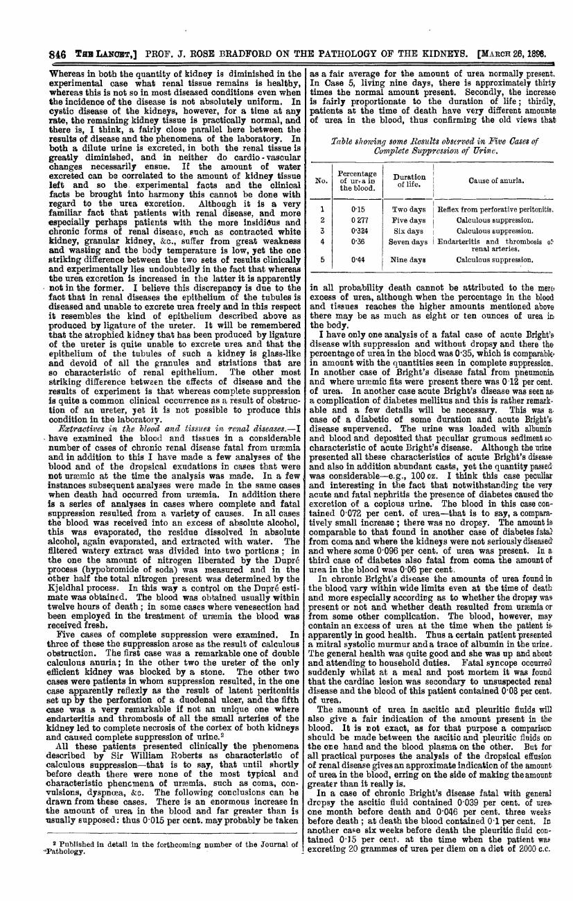
as a fair average for the amount of urea normally presentIn Case 5, living nine days, there is approximately thirtytimes the normal amount present. Secondly, the increaseis fairly proportionate to the duration of life ; thirdly,patients at the time of death have very different amountsof urea in the blood, thus confirming the old views that
Table showing some Res1Ûts observed in Five Cases ofComplete Suppression of Ilrine.
in all probability death cannot be attributed to the mereexcess of urea, although when the percentage in the bloodand tissues reaches the higher amounts mentioned abovethere may be as much as eight or ten ounces of urea mthe body.
I have only one analysis of a fatal case of acute Bright’e.disease with suppression and without dropsy and there thepercentage of urea in the blood was 0’35, which is comparable;in amount with the quantities seen in complete suppression.In another case of Bright’s disease fatal from pneumoniaand where ursemic fits were present there was 012 per cent.of urea. In another case acute Bright’s disease was seen a&a complication of diabetes mellitus and this is rather remark-able and a few details will be necessary. This was a,
case of a diabetic of some duration and acute Bnght’sdisease supervened. The urine was loaded with albuminand blood and deposited that peculiar grumous sediment socharacteristic of acute Bright’s disease. Although the urinepresented all these characteristics of acute Bright’s diseaseand also in addition abundant casts, yet the quantity passedwas considerable-e.g., 100 oz. I think this case peculiarand interesting in the fact that notwithstanding the veryacute and fatal nephritis the presence of diabetes caused theexcretion of a copious urine. The blood in this case contained 0’072 per cent. of urea-that is to say, a compara-tively small increase ; there was no dropsy. The amount is
comparable to that found in another case of diabetes fatatfrom coma and where the kidneys were not seriously diseasedand where some 0’096 per cent. of urea was present. In athird case of diabetes also fatal from coma the amount ofurea in the blood was 0’06 per cent.
In chronic Bright’s disease the amounts of urea found inthe blood vary within wide limits even at the time of deathand more especially according as to whether the dropsy waspresent or not and whether death resulted from ursemia orfrom some other complication. The blood, however, maycontain an excess of urea at the time when the patient is,
apparently in good health. Thus a certain patient presenteda mitral systolic murmur and a trace of albumin in the urine.The general health was quite good and she was up and aboutand attending to household duties. Fatal syncope occurredsuddenly whilst at a meal and post mortem it was foundthat the cardiac lesion was secondary to unsuspected renaldisease and the blood of this patient contained 0’08 per cent.of urea.The amount of urea in ascitic and pleuritic fluids will
also give a fair indication of the amount present in theblood. It is not exact, as for that purpose a comparisonshould be made between the ascitic and pleuritic fluids onthe one hand and the blood plasma on the other. But forall practical purposes the analysis of the dropsical effusionof renal disease gives an approximate indication of the amountof urea in the blood, erring on the side of making the amountgreater than it really is.
In a case of chronic Bright’s disease fatal with generaldropsy the ascitic fluid contained 0’039 per cent. of urea.one month before death and 0’046 per cent. three weeksbefore death ; at death the blood contained 0’1 per cent. Inanother case six weeks before death the pleuritic fluid COD-tained 0’)5 per cent. at the time when the patient wasexcreting 20 grammes of urea per diem on a diet of 2000 c.c.
847DR. BUZZARD: THE INFECTIOUS ORIGIN OF INFANTILE PARALYSIS.
of milk. This patient, however, died from ursmia and thepercentage in the blood at death was 0-44 per cent. Twcother cases of chronic Bright’s disease, fatal and accom-panied with general dropsy, showed that the blood at thetime of death contained 0 16 per cent. and 0’07 per cent.of urea respectively. All these observations and others thatI need not quote show that with chronic Bright’s disease,with and without general dropsy, there is an increase ofarea in the blood even when the patient is fairly well andthat this increases at death, as would be expected, but theincrease is not so great as that seen in suppression unlessarasmia occurs.
It is, however, in uraemia that the greatest increase isfound and the following statements are based on the
-
analysis of the blood in twenty-five cases of fatal ursemiaunaccompanied with dropsy. The highest percentage ofarea in the blood was 0’5 per cent. This was observedtwice. In one case the amount was 0’46 per cent., in nineother cases the amounts varied between 0-3 per cent. and0.4 per cent., so that in half the cases investigated the per-centage of urea in the blood was between 0’3 and 05. Inall the other cases the quantity varied between 02 per cent.and 03 per cent. and in no case of chronic Bright’a disease,fatal from ursemia and unaccompanied by dropsy or someinflammatory complication, did the percentage sink below0.2 and, in fact, this smaller amount was only seen in twocases. The quantities of urea found in the cases ofBright’s disease, fatal from uraemia, are comparable with thequantities found in the cases of complete suppression thathved approximately one week from the time of the onsetof the suppression. There are, however, at least two
points of difference between the two series of cases. Inthe suppression series there was always complete suppres-sion of urine and this in my experience is uncommonin the acute anaemia complicating chronic renal diseaseunassociated with dropsy. In the second place, the dura-tion of life after complete suppression was at least sixdays when the percentage of urea reached 0’3, whereasin acute ursemia life was not prolonged for more thanfrom two to three days from the onset of acute symptoms inmost cases. One case was exceptional and presented acutearaemic symptoms for more than a week and it is of suffi-- cieBt interest to quote in detail. The patient was bled with
I
,great relief on account of the onset of urasmic symptoms onFeb. 25th and the average of three determinations of theamount of urea in the blood yielded 0-13 per cent. Thevenesection was followed by considerable improvement, butthe next day the patient again became comatose and asecond venesection was performed with very beneficialresults. This second specimen of blood yielded 0’24 percent. of urea. Daring these two days the urine was passedunconsciously and could not be collected. From Feb. 28th toMarch 3rd it could be collected in part and the daily averagefor these days was at least twelve grammes and certainlymuch more was passed as the patient passed a considerablequantity unconsciously and also some with the stools.The benefit of the venesection was temporary, the uraemicsymptoms returned, and the patient died on March 3rd,and the blood contained 0’3 per cent. of urea. This caseillustrates several points, and more especially shows the- onset of ursemic symptoms when the percentage of urea inthe blood was still low, and the further point that a very.great increase in the amount of the extractives in the bloodmay take place whilst there is still a considerable excretionof urea, since it must be borne in mind that, in the firstplace, all the urea was not recovered, and, in the secondplace, that little or no food was taken, and hence it will beseen that the urea excretion in this case was by no means sosmall as it appears to be at first sight. This case wasinvestigated more thoroughly than any other, but in anothercase where there was 0’38 per cent. of urea in the bloodsome 8 grammes of urea were recovered from the urine inthe preceding twenty-four hours notwithstanding the loss ofsome urine and the presence of severe diarrhoea and persis-tent vomiting.All these observations tend to the same conclusion, that in
Qrmmia there is a very great excess of urea in the blood, even I
greater than in most cases of complete suppression, andthere is not only not necessarily complete suppression of urine,but quantities of urea may be passed in the urine that arenot very inadequate considering the condition of the patient. iWe have as yet only considered the presence of extractives ]
111 the blood, but there is a similar, and in fact a greater, 1 Iincrease in the tissuee, and, as in the experimental cases, this 1
increase is especially marked in the muscles. In the musclesthere is not only an incteabe of urea approximately equal inpercentage amount to that seen in the blood, but in additionthere is a corresponding or even greater increase in the otherextractives belonging to what may be called the creatin
group. These are not present to any great extent in theblood even in urasmia, but the excess in the muscles is verynotable. I have been able to examine the blood in two casesof eclampsia and here the percentage of urea in one case was0-06 per cent. ar d in the other 0036 per cent., confirmingformer observations that in this malady there is a strik-ing difference between the state of the blood and thatfound in uraemia. In one of the fatal cases of eclampsia itis of some interest that the urine was highly albuminous andcontained considerable quantities of blood ; in point of factthere was nephritis, but notwithstanding this the percentageof urea was but 0 036. Thus the greatest excess seen ineclampsia was only four times the normal.The conclusions drawn from the examination of the blood
and tissues in renal disease may be shortly summarised asfollows : (1) in suppression there is a great increase in thenitrogenous extractives in the blood and tissues ; (2) in acuteuraemia without dropsy there may a be still greater increase ;(3) in chronic renal disease fatal from other causes there issome increase, but not comparable with the above ; (4) inrenal disease even in apparent good health there is a certainsmall excess; and (5) in eclampsia the excess is trivial inamount.
ABSTRACT OF
A Clinical LectureON
CASES ILLUSTRATING THE INFECTIVEORIGIN OF INFANTILE PARALYSIS.
Delivered at the National Hospital for the Paralysed andEpileptic, Queen-square, on Feb. 8th, 1898,
BY THOMAS BUZZARD, M.D., F.R.C.P. LOND.,PHYSICIAN TO THE HOSPITAL.
GENTLEMEN,-The two patients exhibited to-day are
typical examples of acute anterior poliomyelitis. Their casesare both of comparatively recent date, the first having beenattacked eleven weeks and the second nine months ago. Ineach the principal incidence of the disorder has been uponthe lower extremities.The patient first shown is a boy, aged fourteen years,
who was admitted under my care a month ago suffer ngfrom paralysis of both lower extremities. From notes taken
by Dr. Collier, resident medical officer, it appears thatthe boy’s ordinary health had been good with the exceptionof three attacks of influenza, the last of which imme-
diately preceded the paralysis. Seven weeks before admis-sion (i e., about eleven weeks ago) the patient was saidto have had influenza. He felt sick, had headache anda stiff neck, with fever lasting three days. The highesttemperature was 102° F., which declined next day to 101°.He got about again and went to school, but thinks he gota chill and the next day he stayed in bed feeling onlyslightly ill, but with some headache. Two days later, in themorning, he found he could not uee the right leg and at thesame time there was retention of urine and incontinence offasces. There was no pain anywhere. After a week thetrouble with the sphincters passed off. He thinks the legswere at their worst four days after power had begun to belost, and after that they slightly improved. At the com-mencement of the illness be had some little aching in thearms. On examination it was found that the power ofextending the elbow-joint was decidedly weak on both sides,and the erector spinae and abdominal muscles were alsowanting in strength, but no localised weakness could bepicked out in them. Both lower limbs were much wasted ;the feet were cold and somewhat cyanosed. He could notraise the right leg or extend or flex the right knee, and failedto resist passive flexion and extension of the knee. He cannow do all this. Abduction and adduction of the right