1674 The Bradshaw Lecture ON THE RESULTS OF BRONCHIAL OBSTRUCTION. Delivered before the Royal College of Physicians of London on Nov. 1st, 1910, BY G. NEWTON PITT, M.D.CANTAB., F.R.C.P. LOND., PHYSICIAN TO GUY’S HOSPITAL AND LECTURER ON MEDICINE. MR. PRESIDENT AND GENTLEMEN,-My first duty is to thank your predecessor in the chair for the honour he has done me in nominating me for this lectureship to-day, and to express the diffidence I feel in choosing a subject on which it mav seem there is not much that is new to be said. We are met to-day to do honour to the memory of Dr. William Wood Bradshaw, who, it would appear, was a man of wide interests ; he contributed papers on such subjects as the use of cod-liver oil in chronic rheumatism, on narcotics, on abdominal abscess, and other non-professional articles to the Misoellany and elsewhere. Born at Bristol in 1801, according both to Foster’s Alumni Oxiensis " and to the inscription on his grave, he was educated at the Westminster and Middlesex Hospitals, taking his M.R.C.8. in 1833. In the early part of the same year he became M.D. Erlangen. He became an Extra Licentiate of this College in 1841, after examination by the President and the "Elects," which entitled him to practise beyond seven miles from London. In 1854 he was elected a Fellow of the Royal College of Surgeons of England. In 1859 Dr. Coplans Hawkins and Dr. R. P. Smith nominated him as a Member of this College, which nomination was approved. At ’the mature age of 43 he matriculated at Newton Hall, Oxford, and was granted a degree in 1847 without any examination. He married the widow of a wealthy jeweller at Andover, and, according to Mr. Rickman Godlee, to whom I am indebted for his portrait and for much of this information, her money was to be left away in case she re-married ; but they bought the reversion, married, and settled down at Reading. He seems to have been a cultivated, refined, somewhat eccentric man with a rather theatrical manner, and never did much practice. He died in 1866 ; his widow founded a lectureship at each of the Colleges in order to perpetuate his memory. THE RESULTS OF COMPRESSION OF A BRONCHUS. The results of compression of a bronchus may be the following. 1. The lung may be slightly reduced in size. There may be definite diminution in the entry of air, but the resonance may be unaltered, and even post mortem beyond an increase in the amount of mucoid contents, and a diminution in the amount of air in the lung, this may be all that can be observed even when there has been definite pressure on the tube. 2. The chest may be hyper-resonant, the lung over-dis- tended, and the diaphragm displaced down abnormally. This occurs only with partial obstruction of the tube when respiration is vigorous, and will be discussed under the results produced by aneurysm. 3. The lung may be reduced in size, airless and solid from collapse. The chest will be dull on percussion. This results when the obstruction to the tube is complete or when respiration is feeble. It may occur within a few hours, and was studied by Lichtheim by observing the effects of intro- ducing foreign bodies into the bronchi so as to completely bleck them, but practically complete obstruction is generally due to other causes and rarely to the presence of a foreign body. In the early stage the bronchi beyond the obstruction are empty. The lungs in the first and second conditions also tend gradually to become more or less airless. 4. The bronchi become filled with retained secretions, at first chiefly mucus, but catarrhal changes soon ensue, cells - accumulate, together with purulent and inflammatory products. 5. Secondary inflammatory changes take place, due partly to the entry of secretions into the alveoli, so that what has been called a retention pneumonia ensues ; the alveoli fill with catarrhal cells and giant cells appear, although they are unconnected with tubercle, as has been shown by inoculating animals with the products. When alveolar walls remain long in contact, organisation with obliteration takes place and new connective tissue is formed. The bronchi normally often contain a few staphylococci and streptococci, which when retained in suitable media begin to develop, with the result that pyrexia and a more or less acute pneumonic change develops. The changes that have taken place so far may all pass away and leave but little damage ; but two further changes soon take place-a general fibrosis of the lung beyond the obstruction and a bronchiectasis. These ultimately develop, whatever be the initial process, if the obstruction persist. 6. Dilatation of the tubes. This does not take place equally in all the tubes obstructed, and its distribution is largely determined by local causes. When only one branch of a bronchus has been pressed upon, the dilatation is limited to the tubes in its area. Generally the tubes at the posterior and lower part of the lung are the most affected, probably because the secretion tends to stagnate there and set up local destructive changes as the patient is in the semi. recumbent position. Normally there is a constant secretion of mucus in the bronchi, which passes imperceptibly out into the larger tubes and thence to the trachea, but we are quite ignorant what is the amount normally secreted in 24 hours. When a bronchus is occluded the mucus at once begins to accumulate and is capable of being secreted under pressure, so that as with an obstructed gall-bladder the tubes become over-distended. Now bronchial contents cannot pass through the bronchial wall on account of the basement membrane ; they can only pass out by the bronchus, or be sucked back by inspiration into the alveoli which remain patent, and so on into the lymphatics and the glands. Normally the contents pass almost entirely by the bronchus, because the initial act of expiration consists in the contraction of the lung by its own elasticity, which as the alveoli empty drives the secretion out of the smaller bronchioles into the larger tubes, to be then carried on by the forcible muscular expiration. With a collapsed lung this initial mechanism fails. As the alveoli become blocked, there is a greater suction on those that remain, and this leads to further occlusion of the alveoli, but the movement of the chest on the affected side is always more or less feeble. The result is that, whether the lung is distended or collapsed, the bronchi become distended with mucoid or muco-purulent contents under high pressure, and hence the walls yield. Owing to the interference with the circulation, which is largely dependent on the suction of the alveoli as they open up with inspiration, the arteries being on the proximal and the veins on the distal side, the tissues " are insufficiently nourished, and this, added to the local irritation, causes the bronchial walls to deteriorate, so that the elastic and muscle fibres tend to disappear and local dilatations occur. It is an old controversy as to whether inspiratory or expiratory efforts are the more efficient in the production of bronchiectases, and I am sorry to say that at the present day the latter theory seems to have the more advocates. In the cases under consideration it is difficult to see how violent expiration, compressing the lung forcibly, as with a cough, can throw any greater strain on the inside of the bronchi than it does on the outside. If the same pressure is applied all round the wall of a bronchus it cannot tend to dilate it. The increased pressure of the contained air is the result of the pressure applied to the outer surface of the bronchus. On the other hand, if it is borne in mind that aspiration is applied to the surface of the lung by inspira- tion, which tends to open it up, and if the alveoli fail to respond, strain is thrown on the deeper parts such as the bronchi ; if there is no obstruction they open up and fill with air from the trachea, but when the bronchus is obstructed they must be subjected to strain which tends to dilate them. The feebleness of the respiratory movements spares them greatly, but the contracted condition of the lung tends to set up an inspiratory dyspnoea, and paroxysms occur from time to time, especially with exertion, which must aspirate the tubes and gradually make them dilate. Consequently we do not find marked dilatation except when there is also in these cases definite fibrosis. The slighter degrees of dilatation are associated with but slight con- solidation of the lung. The presence of pleuritic effusion
Transcript
1674
The Bradshaw LectureON
THE RESULTS OF BRONCHIALOBSTRUCTION.
Delivered before the Royal College of Physicians of London onNov. 1st, 1910,
BY G. NEWTON PITT, M.D.CANTAB.,F.R.C.P. LOND.,
PHYSICIAN TO GUY’S HOSPITAL AND LECTURER ON MEDICINE.
MR. PRESIDENT AND GENTLEMEN,-My first duty is tothank your predecessor in the chair for the honour he hasdone me in nominating me for this lectureship to-day, and toexpress the diffidence I feel in choosing a subject on which itmav seem there is not much that is new to be said.We are met to-day to do honour to the memory of Dr.
William Wood Bradshaw, who, it would appear, was a manof wide interests ; he contributed papers on such subjects asthe use of cod-liver oil in chronic rheumatism, on narcotics,on abdominal abscess, and other non-professional articles to theMisoellany and elsewhere. Born at Bristol in 1801, accordingboth to Foster’s Alumni Oxiensis " and to the inscription onhis grave, he was educated at the Westminster and MiddlesexHospitals, taking his M.R.C.8. in 1833. In the early partof the same year he became M.D. Erlangen. He became anExtra Licentiate of this College in 1841, after examinationby the President and the "Elects," which entitled him topractise beyond seven miles from London. In 1854 he waselected a Fellow of the Royal College of Surgeons of
England. In 1859 Dr. Coplans Hawkins and Dr. R. P.Smith nominated him as a Member of this College, whichnomination was approved. At ’the mature age of 43 hematriculated at Newton Hall, Oxford, and was granted adegree in 1847 without any examination. He married thewidow of a wealthy jeweller at Andover, and, according toMr. Rickman Godlee, to whom I am indebted for his portraitand for much of this information, her money was to be leftaway in case she re-married ; but they bought the reversion,married, and settled down at Reading. He seems to havebeen a cultivated, refined, somewhat eccentric man with arather theatrical manner, and never did much practice. Hedied in 1866 ; his widow founded a lectureship at each ofthe Colleges in order to perpetuate his memory.
THE RESULTS OF COMPRESSION OF A BRONCHUS.The results of compression of a bronchus may be the
following.1. The lung may be slightly reduced in size. There may
be definite diminution in the entry of air, but the resonancemay be unaltered, and even post mortem beyond an increasein the amount of mucoid contents, and a diminution in theamount of air in the lung, this may be all that can beobserved even when there has been definite pressure on thetube.
2. The chest may be hyper-resonant, the lung over-dis-tended, and the diaphragm displaced down abnormally.This occurs only with partial obstruction of the tube whenrespiration is vigorous, and will be discussed under theresults produced by aneurysm.
3. The lung may be reduced in size, airless and solid fromcollapse. The chest will be dull on percussion. Thisresults when the obstruction to the tube is complete or whenrespiration is feeble. It may occur within a few hours, andwas studied by Lichtheim by observing the effects of intro-ducing foreign bodies into the bronchi so as to completelybleck them, but practically complete obstruction is generallydue to other causes and rarely to the presence of a foreignbody. In the early stage the bronchi beyond the obstructionare empty. The lungs in the first and second conditions alsotend gradually to become more or less airless.
4. The bronchi become filled with retained secretions, atfirst chiefly mucus, but catarrhal changes soon ensue, cells- accumulate, together with purulent and inflammatoryproducts.
5. Secondary inflammatory changes take place, due partlyto the entry of secretions into the alveoli, so that what hasbeen called a retention pneumonia ensues ; the alveoli
fill with catarrhal cells and giant cells appear, althoughthey are unconnected with tubercle, as has been shown byinoculating animals with the products. When alveolar wallsremain long in contact, organisation with obliteration takesplace and new connective tissue is formed. The bronchinormally often contain a few staphylococci and streptococci,which when retained in suitable media begin to develop, withthe result that pyrexia and a more or less acute pneumonicchange develops.The changes that have taken place so far may all pass
away and leave but little damage ; but two further changessoon take place-a general fibrosis of the lung beyond theobstruction and a bronchiectasis. These ultimately develop,whatever be the initial process, if the obstruction persist.
6. Dilatation of the tubes. This does not take placeequally in all the tubes obstructed, and its distribution is
largely determined by local causes. When only one branchof a bronchus has been pressed upon, the dilatation islimited to the tubes in its area. Generally the tubes at theposterior and lower part of the lung are the most affected,probably because the secretion tends to stagnate there andset up local destructive changes as the patient is in the semi.recumbent position.Normally there is a constant secretion of mucus in thebronchi, which passes imperceptibly out into the larger tubesand thence to the trachea, but we are quite ignorant what isthe amount normally secreted in 24 hours. When a bronchusis occluded the mucus at once begins to accumulate and iscapable of being secreted under pressure, so that as with anobstructed gall-bladder the tubes become over-distended.Now bronchial contents cannot pass through the bronchialwall on account of the basement membrane ; they can onlypass out by the bronchus, or be sucked back by inspirationinto the alveoli which remain patent, and so on into thelymphatics and the glands. Normally the contents passalmost entirely by the bronchus, because the initial act ofexpiration consists in the contraction of the lung by its ownelasticity, which as the alveoli empty drives the secretion outof the smaller bronchioles into the larger tubes, to be thencarried on by the forcible muscular expiration. With a
collapsed lung this initial mechanism fails. As the alveolibecome blocked, there is a greater suction on those thatremain, and this leads to further occlusion of the alveoli, butthe movement of the chest on the affected side is alwaysmore or less feeble. The result is that, whether the lung isdistended or collapsed, the bronchi become distended withmucoid or muco-purulent contents under high pressure, andhence the walls yield. Owing to the interference with thecirculation, which is largely dependent on the suction of thealveoli as they open up with inspiration, the arteries beingon the proximal and the veins on the distal side, the tissues "
are insufficiently nourished, and this, added to the localirritation, causes the bronchial walls to deteriorate, so
that the elastic and muscle fibres tend to disappear andlocal dilatations occur.
It is an old controversy as to whether inspiratory orexpiratory efforts are the more efficient in the production ofbronchiectases, and I am sorry to say that at the presentday the latter theory seems to have the more advocates. Inthe cases under consideration it is difficult to see howviolent expiration, compressing the lung forcibly, as with acough, can throw any greater strain on the inside of thebronchi than it does on the outside. If the same pressure is
applied all round the wall of a bronchus it cannot tend todilate it. The increased pressure of the contained air is theresult of the pressure applied to the outer surface of thebronchus. On the other hand, if it is borne in mind thataspiration is applied to the surface of the lung by inspira-tion, which tends to open it up, and if the alveoli fail torespond, strain is thrown on the deeper parts such as thebronchi ; if there is no obstruction they open up and fill withair from the trachea, but when the bronchus is obstructedthey must be subjected to strain which tends to dilate them.The feebleness of the respiratory movements spares themgreatly, but the contracted condition of the lung tends toset up an inspiratory dyspnoea, and paroxysms occur fromtime to time, especially with exertion, which must aspiratethe tubes and gradually make them dilate.
Consequently we do not find marked dilatation except whenthere is also in these cases definite fibrosis. The slighterdegrees of dilatation are associated with but slight con-solidation of the lung. The presence of pleuritic effusion
1675
usually inhibits, or entirely prevents, the dilatation o[ the
bronchi, because the lung not being in contact with thepleura is no longer held up by capillary attraction betweenthe two surfaces, and therefore is not subjected to so muchaspiration. There is no evidence that connective tissue in
contracting is capable of producing traction on the tubes atall comparable with that exercised by the powerful suctionaction of inspiration. It was noted many years ago by Dr.Charlton Bastian that in one-fifth of all cases of cirrhosisof the lung the tubes are not at all dilated. This is what
might naturally be expected, as respiration would probablybe feeble in many, but if the fibroid change were theessential cause of bronchiectasis it would be inexplicable.
It has been suggested that the accumulated weight of thecontained secretions will suffice to distend the terminal tubeswhen a bronchus is compressed by an aneurysm. Theterminal tubules are not the ones that are especially dilated,and the tubules lie in a closed box, the chest, and aresupported outside by the same pressure as that inside them.The pressure inside the chest may be slightly increased, butthere is no difference between the pressures inside and out-side the bronchi, which is the important factor.The essential factors which lead to dilatation of the tubes
would therefore appear to be local changes in the walls, theaccumulation of secretion under tension, and the effects ofinspiration on a lung which is not capable of expandingsatisfactorily.
7. Fibroid thickening of the lung. There are severalfactors which conduce to this result : inflammatory andorganising connective tissue changes result (a) fromthe retention of the secretions, (b) from the prolongedcollapse of the alveoli, (c) from the broncho-pneumonia,(d) from the peri-bronchial changes which follow on thedestruction of the bronchial epithelium, which generallytakes place, and finally (e) from the interference with thepulmonary and lymphatic circulations which normally dependon the opening up and the contraction of the alveoli.Moreover, when we bear in mind that in health the alveolarsurface of each lung amounts to 100 square metres, each aircell being bathed in a film of blood 10µ in thickness, we canrealise what a difference it must make to the nutrition of thetissues when the alveoli fail to open and the amount of thecirculating blood is enormously reduced; hence retrogradefibroid changes must gradually develop. The fibroid changeis therefore also a natural sequence of obstruction, but thecauses are not quite the same as those which produce thebronchiectasis.
8. Secondary infections producing a septic pneumoniabreaking down into abscesses are not infrequent and varywith the cause ; in some they are essential and early, inothers a late and accidental development.
9. Gangrene. This especially occurs with malignantdisease and when the pulmonary artery is occluded. Thecommunication between the bronchial and pulmonaryarteries is a free one, but varies in different cases, andhence gangrene does not always occur even when the
pulmonary artery is compressed. The condition may alsoresult from a very virulent infection.
10. Putrefactive changes. Although the bronchi are oftendilated, the contents are not always foul. The change is
always a serious one, as it accelerates the destructive changesin the lung and introduces a severe toxic poison into thesystem ; the organisms are usually anaerobic, and hence arebut rarely cultivated.
11. As a secondary result of the bronchiectasis, especiallyif foul, there may be a pleurisy or an empyema.Although stridor is a symptom that at once attracts atten-
tion, it is to be noted that it was present in but a very smallproportion of the cases examined ; it is marked when thetrachea is involved, but otherwise was noticeable in only twoor three of the cases of growth and aneurysm, yet severedyspnœa was common.
CAUSES OF BRONCHIAL OBSTRUCTION.Bronchial obstruction may arise from causes without or
Within the bronchus. Usually only one bronchus is affected,but in some cases the trachea is involved, as well as one orboth bronchi. The external causes producing compressionare :-
1. Aneurysm of the aorta (35).2. Malignant disease (56).
(a) Bronchial glands-(1) Primary, usually sarcoma 6):(2) Secondary to growth at a cistance (6).
(b) (Esophagus, epithelicma (16).
3. Enlarged caseous or suppurating glands (12).4. Gummata (2).5. Abscess (3).6. Dilated left auricle (1).
The internal causes are :—
1. Bronchiolitis.2. Syphilitic ulceration with scarring (5).3. Primary malignant disease of a bronchus (usually columnar-celled’
carcinoma) (8).4. Foreign bodies in the bronchus (8).The material I propose to discuss is collected from our
records at Guy’s Hospital, and I desire to thank my colleaguesfor their kind permission to refer to cases under their care.I shall only discuss the general results and give a fewillustrative cases.
ANEURYSM OF THE AORTA.
During the period taken, out of 11,000 post-mortemexaminations there have been 140 aneurysms of the thoracicaorta. Thirty compressed the left bronchus, and 16 of theseeroded into it ; five compressed the right bronchus and noneeroded it. To contrast the results of compression producedby the various causes it may be noted that in this series, 16cases-i.e., one-half-there was bronchiectasis beyond theobstruction, but in only eight was it associated with a markedfibroid thickening of the connective tissue and increased pig-mentation of the lung with a thickened pleura. In several (6)there was also an acute terminal pneumonia, and in four anacute septic pneumonia ; in 3 there was an acute gan-grene, and sometimes there was an acute pleurisy, but onlyonce was there a pleuritic effusion. When there was nodefinite dilatation of the tubes the lungs generally showedbronchi full of muco-purulent secretion, often with collapseand a more or less airless lung.The dilatation of the tubes takes place only in the part of
the lung beyond the obstruction and is irregularly distributed,being generally least marked in the anterior part. Theamount varies greatly ; in some cases the distension of thetubes is but slight, when the lung may be also collapsed orpneumonic. Marked increase in the fibrous tissue, as it occurs
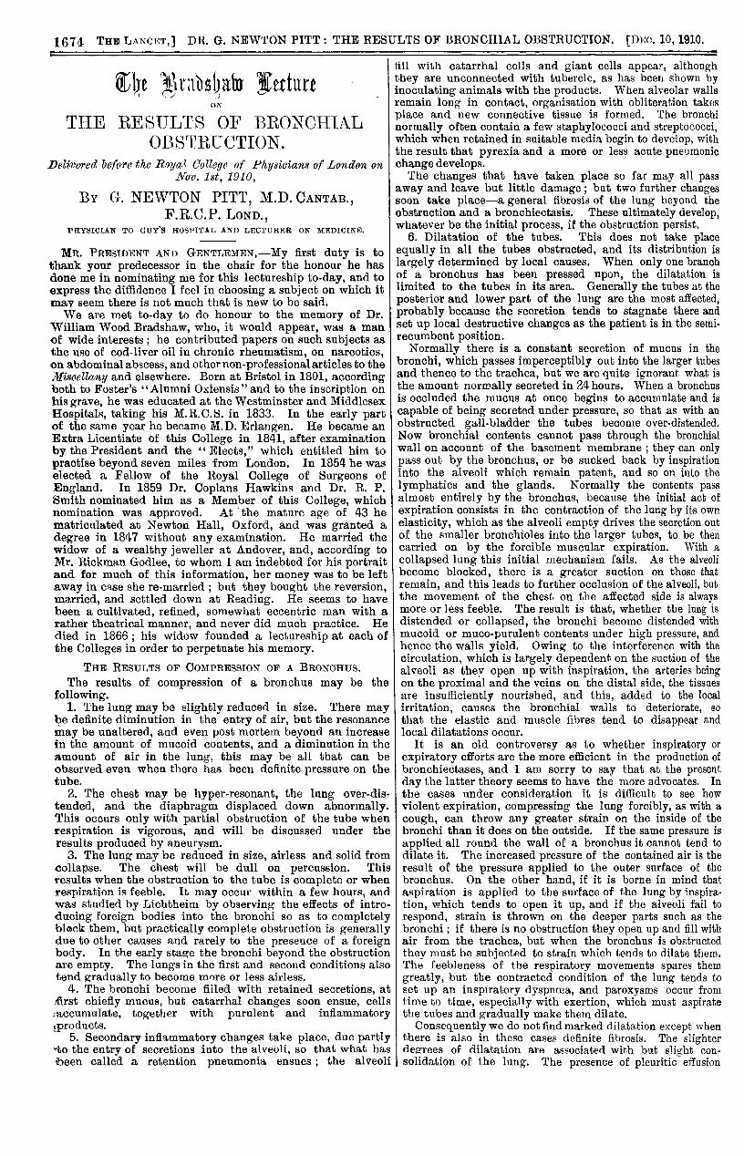
FIG. 1.
Trachea and bronchi opened from behind to show that ananeurysm of the transverse part of the aorta presses on theanterior and outer part of the left bronchus. In this case theaneurysm is just beyond the transverse arch and has rupturedinto the bronchus at A.
less frequently, appears to be to some extent a secondarychange and to be partly set up by the absorption of theirritating contents. In advanced cases the fibrous trabeculæ-stand out, the lung is very tough, and may be riddled withinter-communicating cavities. In only one in 12 was anacute gangrenous condition noted, although in many thecontents were very foul. The bronchiectasis requires at leasttwo or three months in which to develop, but occasionally ittook a year or more, and it is very remarkable how verylatent the constitutional symptoms may be so long as thechanges are limited to the fibroid lm g beyond the obstruc-tion. When much foul secretion is expectorated the healthypart of the lung is apt to become infected, and if an acute-
1676
septic pneumonia develop the disturbance to health isintense.The ascending area is not in very close relationship with
the right bronchus, and only occasionally will an aneurysmof this part (usually a saccularone) compress the bronchus,and then on its anterior surface;not infrequently it will alsoinvolve the trachea; in noneof the cases did it rupture intothe bronchus. Occasionally theazygos vein, which is in closerelation to the right bronchus,may also be compressed, andthis may be the explanation ofthe only case with pleuriticeffusion. In none of the right-sided cases did the breathsounds entirely disappear, nordid a local bronchiectasis de-velop.A sac connected with the
latter part of the transversearch may compress more or less
completely the left bronchus onits outer and anterior surface,and often the trachea as well,and very rarely the left pul-monary artery which lies abovethe bronchus (Fig. 1). This
it by capillary action follows, and the air is sucked past !theobstruction as the lung attempts to open. With expirationthere is a contraction of the lung due to its elasticity, whichis feeble but sufficient for ordinary purposes. This drives
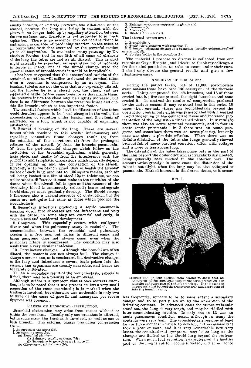
FiG. 2.
arterial stenosis probablyaccelerates retrograde changesin the lung and the develop-ment of the fibroid thickening,
A similar specimen with an aneurysm of the descending aorta;laid open, this is seen to lie on the posterior surface of theleft bronchus which it compresses.
the air from the smaller tothe larger bronchi, because thepressure in the former is thegreater. When forcible expira-tion is applied, the bronchi aswell as the lung tissue are
compressed with an equal pres-sure ; if there is no obstructionthe air passes on and thepressure therefore falls in thelarger bronchi. When an ob-struction exists, however, thismechanism fails, the air hasgreater difficulty in escapingthan it had in entering, andthe lung becomes over-dis-tended. The pressure may beso much raised that thediaphragm may be displacedtwo inches and the heartpushed out of position.
This condition graduallydevelops and persists for a
certain time, but by degreesthe bronchi fill with secre-
tion, thus increasing the diffi-culties of the ingress of air,and as the contained air isnot replaced, and is subjectedto high pressure, it becomes
but it did not in any case lead to gangrene. In no casedid an infarct form, and in only one did an aneurysmrupture into the pulmonary artery. When the aneurysm- comes off lower down it will compress the bronchus frombehind and on
absorbed by the pulmonary blood, while the lung be.comes solid, airless, and dull on percussion. Lichtheimhas shown experimentally that when the bronchus is
completely occluded this airless condition develops almost
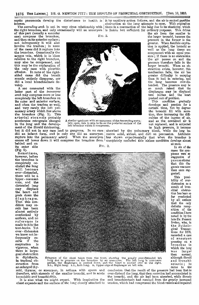
FIG. 3.its outer side(Fig. 2).Sometimes,
however, beforethe bronchus iscompletely oc-
cluded the lungwill becomeover - distended,there will be ahyper - resonantnote, and thedistended lungmay displacethe heart and
push down thediaphragm.That this con-dition may re-
sult has beenalmost entirelyoverlooked byauthors, and noreference ismade to it in thetext-books. Yetover - distensionwe know not in-
frequently re-
sults if the
respiration is
vigorous, whenthere is laryn-gealobstructionin diphtheria,in tracheal ob-struction from
an enlarged thy-
Skiagram of the chest taken from the front, showing the greatly over-distended leftlung due to pressure on the bronchus by an aneurysm. The left lung is over-trans-parent, the diaphragm is pushed down, and the heart is pushed over to the right.R.L., Right lung. L.L., Left lung. D, Upper edge of diaphragm on left side.
roid, thymus, or aneurysm, in asthma with spasm and
therefore, with stenosis of the smaller bronchi, and in acutebronchitis and bronchiolitis.
It is also what we might expect. With inspiration thechest expands and the surface of the lung closely attached to
at once.In six of the
cases the sym.ptoms were so
suggestive of
pneumothoraxthat this dia-gnosis was morethan once sug-gested.
This possi-bility of over-
distension as a
result of bron-chial obstruc-tion has been sofar overlookedby all authorsthat the onlydefinite recog-nition of thecondition I havenoted is by thelate Dr. PearsonIrvine, who, inthe Patholo-gical Transac-tions for 1878,recorded a caseof aneurysmpressing on a
bronchus inwhich the lung
! was largerthan normal,although fibroidand bronchi-ectasic ; hecame to the
conclusion that the result of the pressure had been first toover-distend the lung, that then secretion had accumulated inthe bronchi, and the air had been absorbed, while fibrosisand bronchiectasis had resulted from the greatly increasedtension, which had compressed the blood-vessels and impaired
1677
the nutrition of the tissues. C. Jacobsohn has also morethan once noted that the position of the dulness producedby a growth at the root of the lung may vary with respirationowing to the over-distension of the affected lung, which isbest shown by the X rays.CASE 1. Saccular thorcceie aneurysm compressing the left
.bronchus and producing an over-distended lung.—By some itwas thought that the case was one of pneumothorax. The
patient was a man, aged 29 years, admitted in November,1905. He had been in the army and had had syphilis. Coughhad been present for a year and had been worse for the lastthree months, with dyspnœa and pain in the left chest. He.had no great distress while in bed ; the left chest moved
,badly; there was no tactile vocal fremitus, and no voice norbreath sounds on the left side. On percussion, the note was’hyper-resonant. There was no cardiac dulness on the left,and on the right there was dulness, which varied with respi-ration, extending from the fourth rib out to beyond the rightnipple and down to the liver. There was marked epigastricpulsation, the heart sounds were faint, and no bruit was.audible. There was compensatory breathing over the right.chest. There was considerable uncertainty with regard tothe diagnosis, but it was thought that most probably the con-dition was due to an aneurysm pressing on the left bronchus.The patient was examined with the X rays. The whole of.the left side of the chest was remarkably transparent com-,pared with the right ; no foreign body was visible ; the heartwas displaced nearly 2 inches to the right of the sternum"with deep inspiration, and back again behind the sternum with expiration. The diaphragm was depressed on the left side, but no trace of any aneurysm could be seen. (Fig. 3.) ’’The temperature was normal and the pulse 72. There were no stridor, no evidence of any syphilitic ulceration of the tracheaand.bronchi, and no expectoration. It was elicited that three
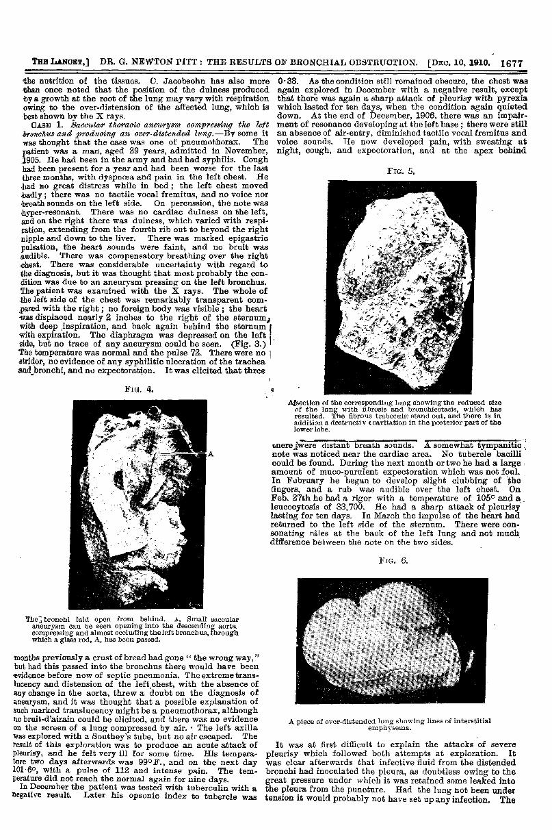
Fia. 4.
The bronchi laid open from behind. A, Small saccularaneurysm can be seen opening into the descending aortacompressing and almost occluding the left bronchus, throughwhich a glass rod, A, has been passed.
months previously a crust of bread had gone the wrong way," but had this passed into the bronchus there would have beenevidence before now of septic pneumonia. The extreme trans-lucency and distension of the left.chest, with the absence ofany change in the aorta, threw a doubt on the diagnosis ofaneurysm, and it was thought that a possible explanation ofsuch marked translucency might be a pneumothorax, althoughno brnit-d’airain could be elicited, and there was no evidenceon the screen of a lung compressed by air. The left axillawas explored with a Southey’s tube, but no air escaped. Theresult of this exploration was to produce an acute attack ofpleurisy, and he felt very ill for some time. His tempera-ture two days afterwards was 99°F., and on the next day101 6°, with a pulse of 112 and intense pain. The tem-perature did not reach the normal again for nine days.
In December the patient was tested with tuberculin with anegative result. Later his opsonic index to tubercle was
0-38. As the condition still remained obscure, the chest wasagain explored in December with a negative result, exceptthat there was again a sharp attack of pleurisy with pyrexiawhich lasted for ten days, when the condition again quieteddown. At the end of December, 1906, there was an impair-ment of resonance developing at the left base ; there were stillan absence of air-entry, diminished tactile vocal fremitus andvoice sounds. He now developed pain, with sweating atnight, cough, and expectoration, and at the apex behind
FiG. 5.
AEsection of the corresponding lung showing the reduced sizeof the lung with fibrosis and bronchiectasis, which hasresulted. The fibrous trabeculæ stand out, and there is inaddition a destructiv tcavitation in the posterior part of thelower lobe.
there were distant breath sounds. A somewhat tympaniticnote was noticed near the cardiac area. No tubercle bacillicould be found. During the next month or two he had a largeamount of muco-purulent expectoration which was not foul.In February he began to develop slight clubbing of thefingers, and a rub was audible over the left chest. OnFeb. 27th he had a rigor with a temperature of 1050 and aleucocytosis of 33,700. He had a sharp attack of pleurisylasting for ten days. In March the impulse of the heart hadreturned to the left side of the sternum. There were con-sonating rites at the back of the left lung and not muchdifference between the note on the two sides.
FIG. 6.
A piece of over-distended lung showing lines of interstitialemphysema.
It was at first difficult to explain the attacks of severepleurisy which followed both attempts at exploration. Itwas clear afterwards that infective fluid from the distendedbronchi had inoculated the pleura, as doubtless owing to thegreat pressure under which it was retained some leaked intothe pleura from the puncture. Had the lung not been undertension it would probably not have set up any infection. The
1678
obstruction to the bronchus was not complete as there was Iexpectoration, although the movement was so feeble thatwe could not hear air being sucked up to the surface ofthe lung. It is noticeable that so long as the lung remainedquiet, and before it became fibroid, so that the contents didnot pass into the lymphatics but remained in the lung, orwere expectorated, the patient remained free from fever, yetthe faintest trace in the pleura set up a vi )lent reaction.
His symptoms gradually quieted down, and he con-
tinued fairly well. I did not see him from April, 1906,to the end of February, 1907, when he was seized with
general pains, pyrexia, and troublesome cough. Soonthe expectoration became abundant and extremely foul,so that he vomited frequently. On readmission he was veryill and feeble. The left chest was contracted; the movementwas deficient; there was an impaired note at the apex andbelow, and also in the axilla it was dull. Over the upper spacesthe air-entry was deficient and broncho-vesicular; belowit was lost. Numerous
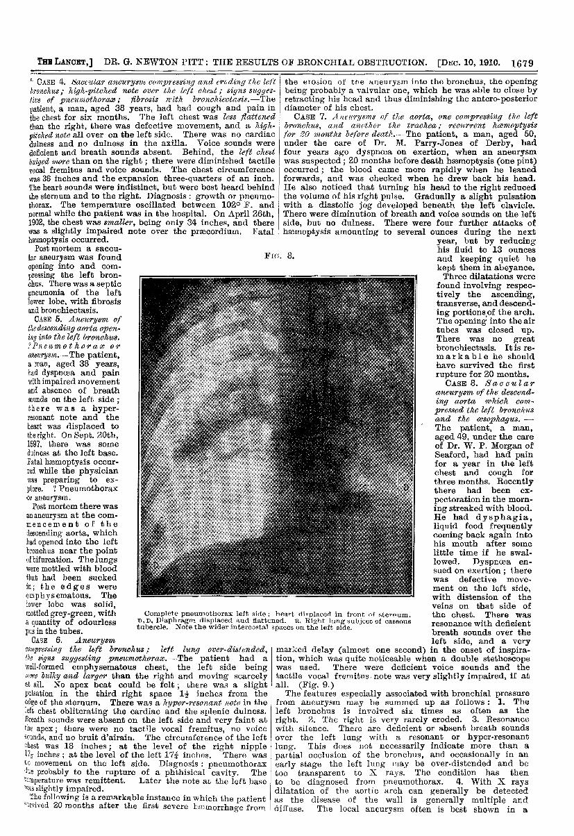
over-distended lung. This will be at once apparent it wecontrast it with an equally good skiagram of a pneumothorax,also taken so long ago as 1902 by him. (Fig. 8.) Herethe air space is more transparent than was the over-distendedlung, but the lung, shrunk np near its root, is obvious. dueto a ruptured phthisical cavity above this, which was.
wanting in the other picture. The heart and diaphragmare more displaced, and the latter has a much shareroutline.
[Since the lecture Dr. Martin has been able to obtain me
the following notes of the case, which was in Victoria ParkHospital in 1902. There was pulsation over the upper part ofthe sternum, with a dull note. The note over the left chest wasresonant; no cardiac dulness ; apex beat was one inch to theleft of the sternum in the fifth space ; the heart sounds werevery feeble, but best heard on the right of the sternum;extremely feeble breath sounds all over the left chest, it
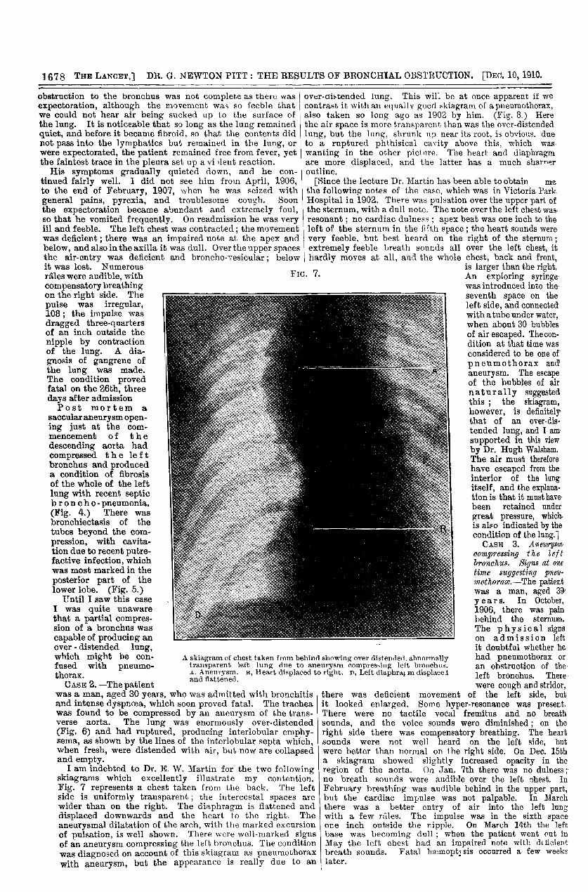
hardly moves at all, and the whole chest, back and front,is larger than the right.FiG. 7.râles were audible, with
compensatory breathingon the right side. The
pulse was irregular,108; the impulse wasdragged three-quartersof an inch outside the
nipple by contractionof the lung. A dia-
gnosis of gangrene ofthe lung was made.The condition provedfatal on the 26th, threedays after admissionPost mortem a
saccular aneurysm open-ing just at the com-
mencement of f the edescending aorta hadcompressed the e le f tbronchus and produceda condition of fibrosisof the whole of the leftlung with recent septicbroncho- pneumonia.(Fig. 4.) There wasbronchiectasis of thetubes beyond the com-pression, with cavita-tion due to recent putre-factive infection, whichwas most marked in the
posterior part of thelower lobe. (Fig. 5.)
Until I saw this caseI was quite unaware
that a partial compres-sion of a bronchus wascapable of producing an over - distended lung, which might be con- A skiagram of chest taken from behind showing over distended, abnormallyfused with pneumo- transparent left lung due to aneurysm compres’ing left bronchus.
thorax A, Aneurysm. R, Heart displaced to right. D, Left diaphragm displacedand Rattened.
An exploring syringewas introduced into theseventh space on theleft side, and connectedwith a tube under water,when about 30 bubblesof air escaped. The con-dition at that time wasconsidered to be one ofpneumothorax and
aneurysm. The escapeof the bubbles of air
naturally suggestedthis ; the skiagram,however, is definitelythat of an over-dis-tended lung, and I amsupported in this viewby Dr. Hugh Walsbam.The air must thereforehave escaped from theinterior of the lungitself, and the explana-tion is that it must havebeen retained undergreat pressure, which,is also indicated by thecondition of the lung.]CASE 3. Aneurysm
compressing the left tbronchus. Signs at onetime suggesting pneu-mothorax.—The patientwas a man, aged 39years. In October,1906, there was painbehind the sternum.The physical 1 signs
! on admission leftit doubtful whether hehad pneumothorax or
an obstruction of theleft bronchus. There
CASE 2.—The patientwas a man, aged 30 years, who was admitted with bronchitisand intense dyspnoea, which soon proved fatal. The tracheawas found to be compressed by an aneurysm of the trans-verse aorta. The lung was enormously over-distended(Fig. 6) and had ruptured, producing interlobular emphy-sema, as shown by the lines of the interlobular septa which,when fresh, were distended with air, but now are collapsedand empty.
I am indebted to Dr. E. W. Martin for the two followingskiagrams which excellently illustrate my contention.Fig. 7 represents a chest taken from the back. The leftside is uniformly transparent ; the intercostal spaces are
wider than on the right. The diaphragm is flattened and
displaced downwards and the heart to the right. The
aneurysmal dilatation of the arch, with the marked excursionof pulsation, is well shown. There were well-marked signsof an aneurysm compressing the left bronchus. The conditionwas diagnosed on account of this skiagram as pneumothoraxwith aneurysm, but the appearance is really due to an
were cough and stridor,there was deficient movement of the left side, butit looked enlarged. Some hyper-resonance was present.There were no tactile vocal fremitus and no breathsounds, and the voice sounds were diminished ; on theright side there was compensatory breathing. The heartsounds were not well heard on the left side, butwere better than normal on the right side. On Dec. 15tha skiagram showed slightly increased opacity in theregion of the aorta. On Jan. 7th there was no dulness; 5no breath sounds were audible over the left nbest. InFebruary breathing was audible behind in the upper part,but the cardiac impulse was not palpable. In Marchthere was a better entry of air into the left lung with
a few nîles. The impulse was in the sixth space
one inch outside the nipple. On March 14th the leftbase was becoming dull ; when the patient went out inMay the left chest had an impaired note with deficientbreath sounds. Fatal hæmoptysis occurred a few weekslater.
1679
" CASE 4. 8aactttar aneurysm aúmp’ressing and trúdi’llfJ the lett Ibronchus,. high-pitched note over the left chest; signs sngges-five of pneumothorax,. fibrosis lâth bronchiectasis.-The
patient, a man, aged 38 years, had had cough and pain inthe chest for six months. The left chest was less flattenedthan the right, there was defective movement, and a high-pitched note all over on the left side. There was no cardiacdulness and no dulness in the axilla. Voice sounds were
deficient and breath sounds absent. Behind, the left chestbulged more than on the right ; there were diminished tactilevocal fremitus and voice sounds. The chest circumferencewas 36 inches and the expansion three-quarters of an inch.The heart sounds were indistinct, but were best heard behindthe sternum and to the right. Diagnosis : growth or pneumo-thorax. The temperature oscillated between 102° F. andnormal while the patient was in the hospital. On April 26th,1902, the chest was smaller, being only 34 inches, and therewas a slightly impaired note over the prsecordium. Fatal
the eicion of the aneurysm into the bronchus, the openingbeing probably a valvular one, which he was able to close byretracting his head and thus diminishing the antero-posteriordiameter of his chest.CASE 7. Anwrysms of the aorta, one compressing the left
bronelzecs, and anrthe?’ the trachea,. reatlrrent haemoptysisfor 20 months before deatt’.-The patient, a man, aged 50,under the care of Dr. M. Parry-Jones of Derby, hadfour years ago dyspnoea on exertion, when an aneurysmwas suspected ; 20 months before death hemoptysis (one pint)occurred; the blood came more rapidly when he leanedforwards, and was checked when he drew back his head.He also noticed that turning his head to the right reducedthe volume of his right pulse. Gradually a slight pulsationwith a diastolic jog developed beneath the left clavicle.There were diminution of breath and voice sounds on the leftside, but no dulness. There were four further attacks ofhemoptysis amounting to several ounces during the next
Fia. 8.
hemoptysis occurred.Post mortem a saecu-
lar aneurysm was found
opening into and com-pressing the left bron-chus. There was a septicpneumonia of the left tlower lobe, with fibrosisand bronchiectasis.CASE 5. Aneitrysm of
the descending aorta open-ing into the left bronohus.’>Pneltmothoraae or
awitrysm.-The patient,a man, aged 38 years,had dyspncea and painwith impaired movementand absence of breathsounds on the left side ; ;there was a hyper-resonant note and theheart was displaced to
the right. On Sept. 20th,1897, there was some
dulness at the left base.Fatal hemoptysis occur-red while the physicianwas preparing to ex-
plore. ? Pneumothoraxor aneurysm.Post mortem there was
an aneurysm at the com-mencement .of the
descending aorta, whichhad opened into the leftbronchus near the pointof bifurcation. The lungswere mottled with bloodthat had been suckedin; the edges were
emphysematous. The
Complete pneumothorax left side ; heart displaced in front of sternum.D, D. Diaphragm displaced and flattened. R, Right lung subject of caseoustubercle. Note the wider intercostal spaces on the left side.
lower lobe was solid,mottled grey-green, witha quantity of odourlesspus in the tubes.CASE 6. Aneurysm
compressing the left bronchus ; left lung over. distended,the signs suggesting pne2cmotlzora.-The patient had a
well-formed emphysematous chest, the left side beingmore bulky and larger than the right and moving scarcelyat all. No apex beat could be felt ; there was a slightpulsation in the third right space 1 inches from theedge of the sternum. There was a hyper-resonant lwte in theleft chest obliterating the cardiac and the splenic dulness.Breath sounds were absent on the left side and very faint atthe apex; there were no tactile vocal fremitus, no voicesounds, and no bruit d’airain. The circumference of the leftchest was 18 inches; at the level of the right nipple172 inches ; at the level of the left In inches. There wasto movement on the left side. Diagnosis : pneumothoraxdue probably to the rupture of a phthisical cavity. Thetemperature was remittent. Later the note at the left base’as slightly impaired.The following is a remarkable instance in which the patient
>ôrvived 20 months after the first severe hemorrhage from
year, but by reducinghis fluid to 13 ounces
and keeping quiet hekept them in abeyance.Three dilatations were
found involving respec-tively the ascending,transverse, and descend-ing portions of the arch.The opening into the airtubes was closed up.There was no greatbronchiectasis. It is re-markable he shouldhave survived the first
rupture for 20 months.CASE 8. Saccular
aneurysm of the desoend-ing aorta which oom-
pressed the left bronchusand the dMAaM. -The patient, a man,
I aged 49, under the careof Dr. W. P. Morgan of
I. Seaford, had had painfor a year in the left
! chest and cough for! three months. Recently
there had been ex-
pectoration in the morn-‘ ing streaked with blood.
He had dysphagia,liquid food frequentlycoming back again intohis mouth after somelittle time if he swal-
i lowed. Dyspnoea en-
sued on exertion ; therewas defective move-
ment on the left side,with distension of theveins on that side ofthe chest. There wasresonance with deficientbreath sounds over theleft side, and a very
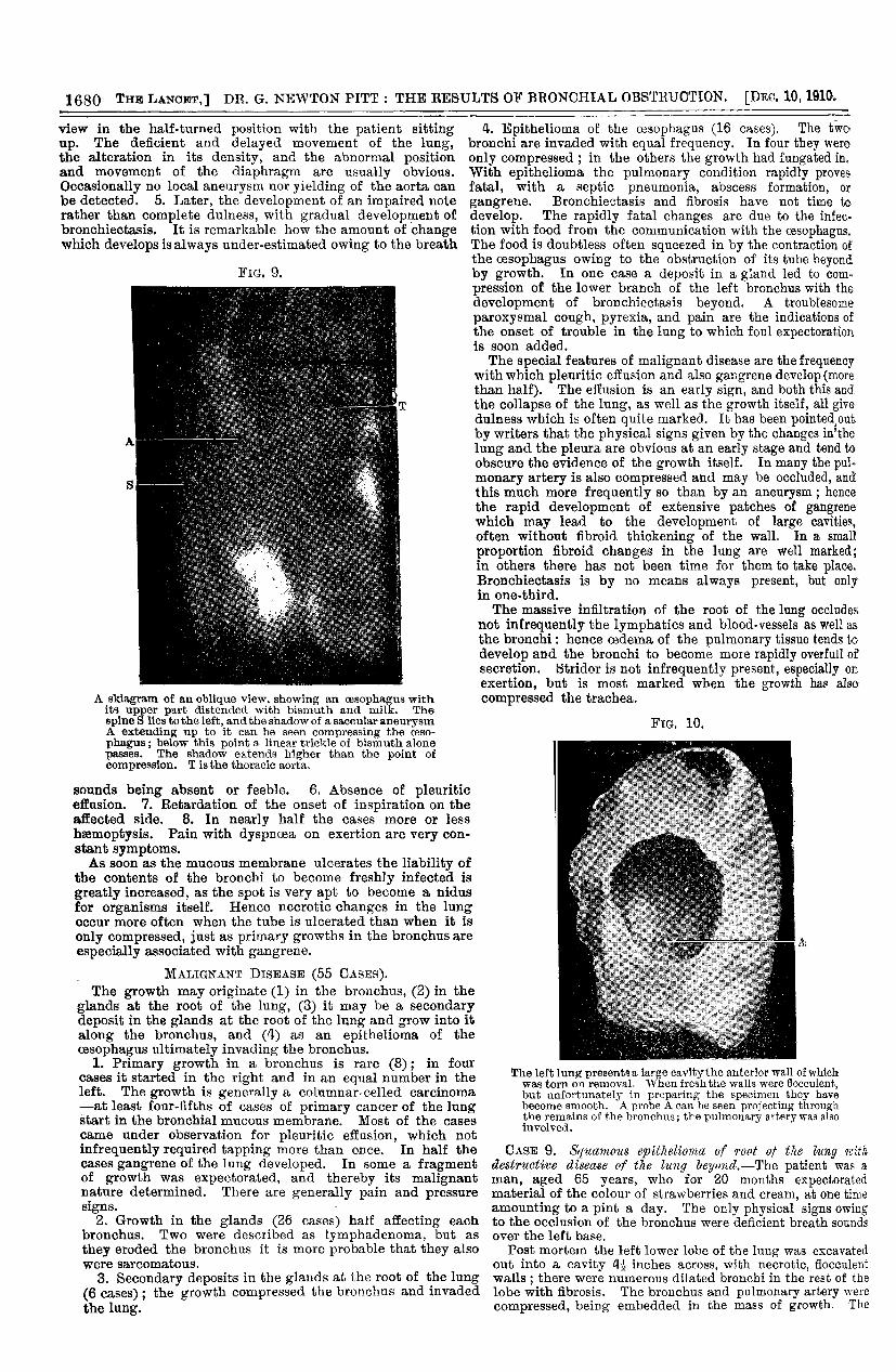
marked delay (almost one second) in the onset of inspira-tion, which was quite noticeable when a double stethoscopewas used. There were deficient voice sounds and thetactile vocal fremitus. note was very slightly impaired, if atall. (Fig. 9.)The features especially associated with bronchial pressure
from aneurysm may be summed up as follows : 1. Theleft bronchus is involved six times as often as the
right. 2. The right is very rarely eroded. 3. Resonancewith silence. There are deficient or absent breath soundsover the left lung with a resonant or hyper-resonantlung. This does not necessarily indicate more than a
partial occlusion of the bronchus, and occasionally in anearly stage the left lung may be over-distended and betoo transparent to X rays. The condition has thento be diagnosed from pneumothorax. 4. With X raysdilatation of the aortic arch can generally be detectedas the disease of the wall is generally multiple andaiffuse. The local aneurysm often is best shown in a
1680
view in the half-turned position with the patient sittingup. The deficient and delayed movement of the lung,the alteration in its density, and the abnormal positionand movement of the diaphragm are usually obvious.
Occasionally no local aneurysm nor yielding of the aorta canbe detected. 5. Later, the development of an impaired noterather than complete dulness, with gradual development ofbronchiectasis. It is remarkable how the amount of changewhich develops is always under-estimated owing to the breath
FIG. 9.
A skiagram of an oblique view, showing an oesophagus with Cits upper part distended with bismuth and milk. Thespine S lies to the left, and the shadow of a saccular aneurysmA extending up to it can be seen compressing the ceso-phagus ; below this point a linear trickle of bismuth alonepasses. The shadow extends higher than the point ofcompression. T is the thoracic aorta.
sounds being absent or feeble. 6. Absence of pleuriticeffusion. 7. Retardation of the onset of inspiration on theaffected side. 8. In nearly half the cases more or less
haemoptysis. Pain with dyspncea on exertion are very con-stant symptoms.As soon as the mucous membrane ulcerates the liability of
the contents of the bronchi to become freshly infected isgreatly increased, as the spot is very apt to become a nidusfor organisms itself. Hence necrotic changes in the lungoccur more often when the tube is ulcerated than when it is
only compressed, just as primary growths in the bronchus areespecially associated with gangrene.
MALIGNANT DISEASE (55 CASES).The growth may originate (1) in the bronchus, (2) in the
glands at the root of the lung, (3) it may be a secondarydeposit in the glands at the root of the lung and grow into italong the bronchus, and (4) as an epithelioma of theoesophagus ultimately invading the bronchus.
1. Primary growth in a bronchus is rare (8); in fourcases it started in the right and in an equal number in theleft. The growth is generally a columnar-celled carcinoma-at least four-fifths of cases of primary cancer of the lungstart in the bronchial mucous membrane. Most of the casescame under observation for pleuritic effusion, which notinfrequently required tapping more than once. In half thecases gangrene of the lung developed. In some a fragmentof growth was expectorated, and thereby its malignantnature determined. There are generally pain and pressuresigns. -
2. Growth in the glands (26 cases) half affecting eachbronchus. Two were described as lymphadenoma, but as
they eroded the bronchus it is more probable that they alsowere sarcomatous.
3. Secondary deposits in the glands at the root of the lung(6 cases) ; the growth compressed the bronchus and invadedthe lung.
4. Epithelioma of the oesophagus (16 cases). The twobronchi are invaded with equal frequency. In four they wereonly compressed ; in the others the growth had fungated in.With epithelioma the pulmonary condition rapidly provesfatal, with a septic pneumonia, abscess formation, or
gangrene. Bronchiectasis and fibrosis have not time todevelop. The rapidly fatal changes are due to the infec-tion with food from the communication with the oesophagus.The food is doubtless often squeezed in by the contraction ofthe oesophagus owing to the obstruction of its tube beyondby growth. In one case a deposit in a gland led to com-pression of the lower branch of the left bronchus with thedevelopment of bronchiectasis beyond. A troublesomeparoxysmal cough, pyrexia, and pain are the indications ofthe onset of trouble in the lung to which foul expectorationis soon added.The special features of malignant disease are the frequency
with which pleuritic effusion and also gangrene develop (morethan half). The effusion is an early sign, and both this andthe collapse of the lung, as well as the growth itself, all givedulness which is often quite marked. It has been pointed.outby writers that the physical signs given by the changes in’thelung and the pleura are obvious at an early stage and tend toobscure the evidence of the growth itself. In many the pul-monary artery is also compressed and may be occluded, andthis much more frequently so than by an aneurysm; hencethe rapid development of extensive patches of gangrenewhich may lead to the development of large cavities,often without fibroid thickening of the wall. In a small
proportion fibroid changes in the lung are well marked;in others there has not been time for them to take place.Bronchiectasis is by no means always present, but onlyin one-third.The massive infiltration of the root of the lung occludes
not infrequently the lymphatics and blood-vessels as well asthe bronchi: hence oedema of the pulmonary tissue tends todevelop and the bronchi to become more rapidly overfull ofsecretion. Stridor is not infrequently present, especially onexertion, but is most marked when the growth has also
compressed the trachea.
FM. 10.
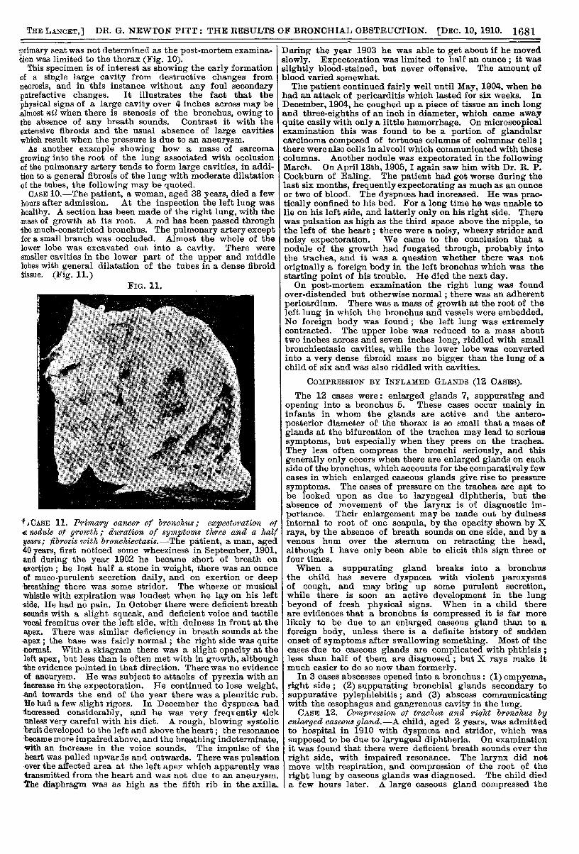
The left lung presents a large cavity the anterior wall of whichwas torn on removal. When fresh the walls were flocculent,but unfortunately in preparing the specimen they havebecome smooth. A probe A can be seen projecting throughthe remains of the bronchus; the pulmonary artery was alsoinvolved.
CASE 9. Syzzramo2zs epithelioma of root of the ltlng nitiadestructive disease of the lung beyond.-Tbe patient was aman, aged 65 years, who for 20 months expectoratedmaterial of the colour of strawberries and cream, at one timeamounting to a pint a day. The only physical signs owingto the occlusion of the bronchus were deficient breath soundsover the left base.
Post mortem the left lower lobe of the lung was excavatedout into a cavity 4-2L inches across, with necrotic, flocculentwalls ; there were numerous dilated bronchi in the rest of thelobe with fibrosis. The bronchus and pulmonary artery werecompressed, being embedded in the mass of growth. The
1681
primary seat was not determined as the post-mortem examina-tion was limited to the thorax (Fig. 10).
This specimen is of interest as showing the early formationof a single large cavity from destructive changes fromnecrosis, and in this instance without any foul secondaryputrefactive changes. It illustrates the fact that the
physical signs of a large cavity over 4 inches across may bealmost *il when there is stenosis of the bronchus, owing tothe absence of any breath sounds. Contrast it with the Iextensive fibrosis and the usual absence of large cavitieswhich result when the pressure is due to an aneurysm.As another example showing how a mass of sarcoma
growing into the root of the lung associated with occlusionof the pulmonary artery tends to form large cavities, in addi-tion to a general fibrosis of the lung with moderate dilatationof the tubes, the following may be quoted.CASE 10.-The patient, a woman, aged 38 years, died a few
hours after admission. At the inspection the left lung washealthy. A section has been made of the right lung, with themass of growth at its root. A rod has been passed throughthe much-constricted bronchus. The pulmonary artery exceptfor a small branch was occluded. Almost the whole of thelower lobe was excavated out into a cavity. There weresmaller cavities in the lower part of the upper and middlelobes with general dilatation of the tubes in a dense fibroidtissue. (Fig. 11.)
FIG. 11.
’fiCASE 11. Primary cancer of broncaits; expectoration ofdnodule ot growth; dttration of symptoms three and a halfyears; fibrosis with bronchiectasis.-The patient, a man, aged40 years, first noticed some wheeziness in September, 1901,-and during the year 1902 he became short of breath onexertion ; he lost half a stone in weight, there was an ounceof muco-purulent secretion daily, and on exertion or deep’breathing there was some stridor. The wheeze or musicalwhistle with expiration was loudest when he lay on his leftside. He had no pain. In October there were deficient breathsounds with a slight squeak, and deficient voice and tactilevocal fremitus over the left side. with dulness in front at theapex. There was similar deficiency in breath sounds at the;apex; the base was fairly normal; the right side was quitenormal. With a skiagram there was a slight opacity at theleft apex, but less than is often met with in growth, although-the evidence pointed in that direction. There was no evidenceof aneurysm. He was subject to attacks of pyrexia with anincrease in the expectoration. He continued to lose weight,and towards the end of the year there was a pleuritic rub.He had a few slight rigors. In December the dyspncea had’increased considerably, and he was very frequently sickunless very careful with his diet. A rough, blowing systolic-bruit developed to the left and above the heart ; the resonance’became more impaired above, and the breathing indeterminate,with an increase in the voice sounds. The impulse of theheart was pulled upwarjs and outwards. There was pulsationover the affected area at the left apex which apparently wastransmitted from the heart and was not due to an aneurysm.’The diaphragm was as high as the fifth rib in the axilla.
During the year 1903 he was able to get about if he movedslowly. Expectoration was limited to half an ounce ; it wasslightly blood-stained, but never offensive. The amount ofblood varied somewhat.The patient continued fairly well until May, 1904, when he
had an attack of pericarditis which lasted for six weeks. InDecember, 1904, he coughed up a piece of tissue an inch longand three-eighths of an inch in diameter, which came awayquite easily with only a little hasmorrhage. On microscopicalexamination this was found to be a portion of glandularcarcinoma composed of tortuous columns of columnar cells ;there were also cells in alveoli which communicated with thesecolumns. Another nodule was expectorated in the followingMarch. On April 18th, 1905, again saw him with Dr. R. P.Cockburn of Ealing. The patient had got worse during thelast six months, frequently expectorating as much as an ounceor two of blood. The dyspncea had increased. He was prac-tically confined to his bed. For a long time he was unable tolie on his left side, and latterly only on his right side. Therewas pulsation as high as the third space above the nipple, tothe left of the heart; there were a noisy, wheezy stridor andnoisy expectoration. We came to the conclusion that anodule of the growth had fungated through, probably intothe trachea, and it was a question whether there was notoriginally a foreign body in the left bronchus which was thestarting point of his trouble. He died the next day.On post-mortem examination the right lung was found
over-distended but otherwise normal ; there was an adherentpericardium. There was a mass of growth at the root of theleft lung in which the bronchus and vessels were embedded.No foreign body was found; the left lung was extremelycontracted. The upper lobe was reduced to a mass abouttwo inches across and seven inches long, riddled with smallbronchiectasic cavities, while the lower lobe was convertedinto a very dense fibroid mass no bigger than the lung of achild of six and was also riddled with cavities.
COMPRESSION BY INFLAMED GLANDS (12 CASES).The 12 cases were: enlarged glands 7, suppurating and
opening into a bronchus 5. These cases occur mainly ininfants in whom the glands are active and the antero-
posterior diameter of the thorax is so small that a mass ofglands at the bifurcation of the trachea may lead to serioussymptoms, but especially when they press on the trachea.They less often compress the bronchi seriously, and thisgenerally only occurs when there are enlarged glands on eachside of the bronchus, which accounts for the comparatively fewcases in which enlarged caseous glands give rise to pressuresymptoms. The cases of pressure on the trachea are apt tobe looked upon as due to laryngeal diphtheria, but theabsence of movement of the larynx is of diagnostic im-portance. Their enlargement may be made out by dulnessinternal to root of one scapula, by the opacity shown by Xrays, by the absence of breath sounds on one side, and by avenous hum over the sternum on retracting the head,although I have only been able to elicit this sign three orfour times.When a suppurating gland breaks into a bronchus
the child has severe dyspnoea with violent paroxysmsof cough, and may bring up some purulent secretion,while there is soon an active development in the lungbeyond of fresh physical signs. When in a child thereare evidences that a bronchus is compressed it is far morelikely to be due to an enlarged caseous gland than to aforeign body, unless there is a definite history of suddenonset of symptoms after swallowing something. Most of thecases due to caseous glands are complicated with phthisis ;less than half of them are diagnosed ; but X rays make itmuch easier to do so now than formerly.
In 3 cases abscesses opened into a bronchus : (1) empyema,right side ; (2) suppurating bronchial glands secondary tosuppurative pylephlebitis; and (3) abscess communicatingwith the oesophagus and gangrenous cavity in the lung.CASE 12. Compression ot trachea and right bro1WhtlS by
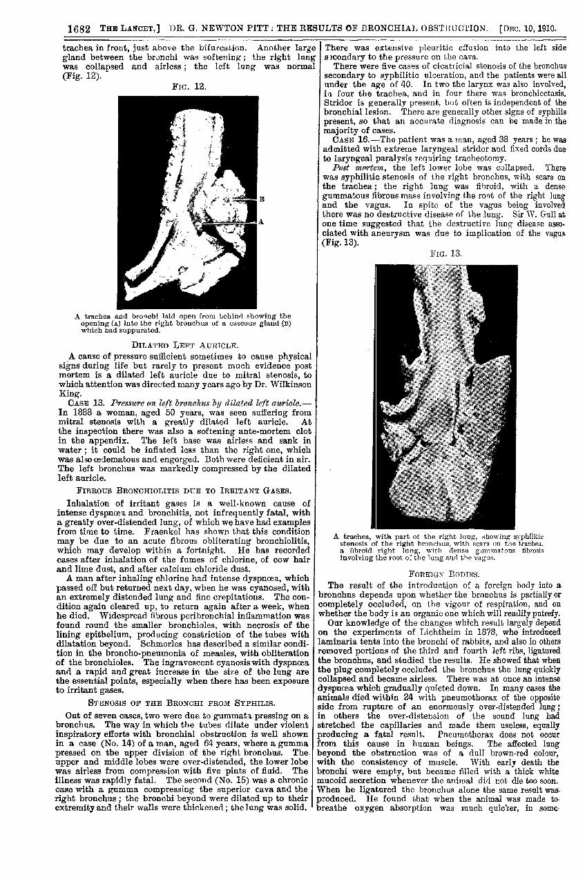
enlarged eccseozts gland.-A child, aged 2 years, was admittedto hospital in 1910 with dyspncea and stridor, which wassupposed to be due to laryngeal diphtheria. On examinationit was found that there were deficient breath sounds over theright side, with impaired resonance. The larynx did notmove with respiration, and compression of the root of the
right lung by caseous glands was diagnosed. The child dieda few hours later. A large caseous gland compressed the
1682
trachea in front, just above the bifurcation. Another largegland between the bronchi was softening ; the right lungwas collapsed and airless ; the left lung was normal
(Fig. 12).FiG. 12.
A trachea and bronchi laid open from behind showing theopening (A) into the right bronchus of a caseous gland (B)which had suppurated.
DILATED LEFT AURICLE.
A cause of pressure sufficient sometimes to cause physicalsigns during life but rarely to present much evidence postmortem is a dilated left auricle due to mitral stenosis, towhich attention was directed many years ago by Dr. WilkinsonKing.CASE 13. Presszcre (In left broneltits by dilated left auriole.-
In 1888 a woman, aged 50 years, was seen suffering frommitral stenosis with a greatly dilated left auricle. Atthe inspection there was also a softening ante-mortem clotin the appendix. The left base was airless and sank in
water ; it could be inflated less than the right one, which Iwas alsocedematous and engorged. Both were deficient in air.The left bronchus was markedly compressed by the dilatedleft auricle.
FIBROUS BRONCHIOLITIS DUE TO IRRITANT GASES.
Inhalation of irritant gases is a well-known cause ofintense dyspnoea, and bronchitis, not infrequently fatal, witha greatly over-distended lung, of which w have had examplesfrom time to time. Fraenkel has shown that this conditionmay be due to an acute fibrous obliterating bronchiolitis,which may develop within a fortnight. He has recordedcases after inhalation of the fumes of chlorine, of cow hairand lime dust, and after calcium chloride dust.A man after inhaling chlorine had intense dyspnoea, which
passed off but returned next day, when he was cyanosed, withan extremely distended lung and fine crepitations. The con-dition again cleared up, to return again after a week, whenhe died. Widespread fibrous peribronchial inflammation wasfound round the smaller bronchioles, with necrosis of thelining epithelium, producing constriction of the tubes withdilatation beyond. Schmorles has described a similar condi-tion in the broncho-pneumonia of measles, with obliterationof the bronchioles. The ingravescent cyanosis with dyspnoeaand a rapid and great increase in the size of the lung arethe essential points, especially when there has been exposureto irritant gases.
STENOSIS OF THE BROXCHI FROM SYPHILIS.
Out of seven cases, two were due to gummata, pressing on abronchus. The way in which the tubes dilate under violent
inspiratory efforts with bronchial obstruction is well shownin a case (No. 14) of a man, aged 64 years, where a gummapressed on the upper division of the right bronchus. The.
upper and middle lobes were over-distended, the lower lobewas airless from compression with five pints of fluid. Theillness was rapidly fatal. The second (No. 15) was a chroniccase with a gumma compressing the superior cava and theright bronchus ; the bronchi beyond were dilated up to theirextremity and their walls were thickened; the lung was solid.
There was extensive pleuritic effusion into the left side
secondary to the pressure on the cava.There were five cases of cicatricial stenosis of the bronchus
secondary to syphilitic ulceration, and the patients were allunder the age of 40. In two the larynx was also involved,ill four the trachea, and in four there was bronchiectasis.Stridor is generally present, but often is independent of thebronchial lesion. There are generally other signs of syphilispresent, so that an accurate diagnosis can be made in themajority of cases.CASE 16.-The patient was a man, aged 38 years ; he was
admitted with extreme laryngeal stridor and fixed cords dueto laryngeal paralysis requiring tracheotomy.
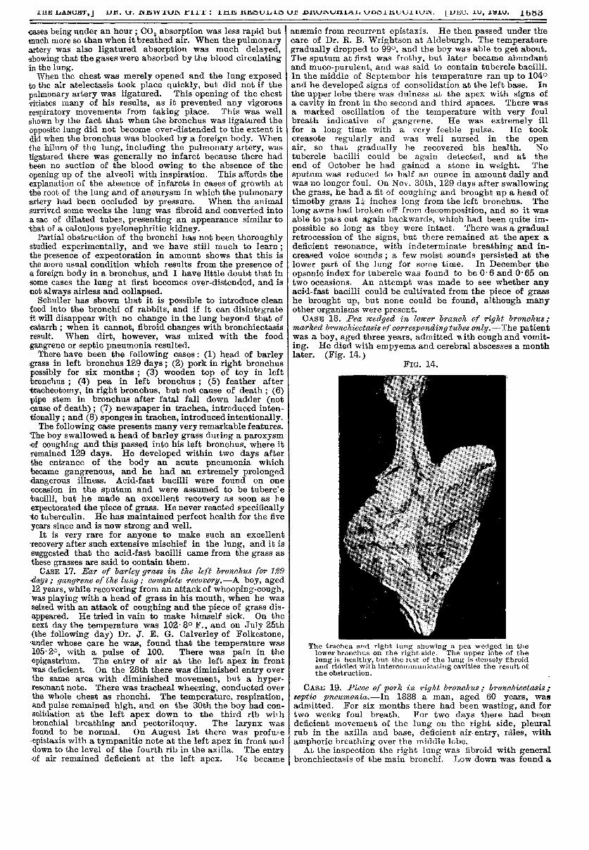
Post mortem, the left lower lobe was collapsed. Therewas syphilitic stenosis of the right bronchus, with scars onthe trachea; the right lung was fibroid, with a densegummatous fibrous mass involving the root of the right lungand the vagus. In spite of the vagus being involvedthere was no destructive disease of the lung. Sir NV. Gull atone time suggested that the destructive lung disease asso-ciated with aneurysm was due to implication of the vagus(Fig. 13).
FiG. 13.
A trachea, with part of the right lung, showing syphiliticstenosis of the right bronchus, with scars on the trachea.a fibroid right lung, with dense gummatous fibrosisinvolving the root of the lung and the vagus.
FOREIGN BODIES.The result of the introduction of a foreign body into a.
bronchus depends upon whether the bronchus is partially or-completely occluded, on the vigour of respiration, and onwhether the body is an organic one which will readily putrefy.Our knowledge of the changes which result largely depend
on the experiments of Lichtheim in 1878, who introducedlaminaria tents into the bronchi of rabbits, and also in othersremoved portions of the third and fourth left ribs, ligaturedthe bronchus, and studied the results. He showed that whenthe plug completely occluded the bronchus the lung quicklycollapsed and became airless. There was at once an intense
dyspnoea which gradually quieted down. In many cases theanimals died within 24 with pneumothorax of the oppositeside from rupture of an enormously over-distended lung;in others the over-distension of the sound lung hadstretched the capillaries and made them useless, equallyproducing a fatal result. Pneumothorax does not occur
from this cause in human beings. The affected lungbeyond the obstruction was of a dull brown-red colour,with the consistency of muscle. With early death thebronchi were empty, but became filled with a thick whitemucoid secretion whenever the animal did not die too soon.When he ligatured the bronchus alone the same result was-produced. He found that when the animal was made tobreathe oxygen absorption was much quic’ier, in some-
1683
cases being under an hour ; CO2 absorption was less rapid butmuch more so than when it breathed air. When the pulmonaryartery was also ligatured absorption was much delayed,showing that the gases were absorbed by the blood circulatingin the lung.When the chest was merely opened and the lung exposed
to the air atelectasis took place quickly, but did not if thepulmonary artery was ligatured. This opening of the chestvitiates many of his results, as it prevented any vigorousrespiratory movements from taking place. This was wellshown by the fact that when the bronchus was ligatured theopposite lung did not become over-distended to the extent itdid when the bronchus was blocked by a foreign body. Whenthe hilum of the lung, including the pulmonary artery, wasligatured there was generally no infarct because there hadbeen no suction of the blood owing to the absence of theopening up of the alveoli with inspiration. This affords the
explanation of the absence of infarcts in cases of growth atthe root of the lung and of aneurysm in which the pulmonaryartery had been occluded by pressure. When the animalsurvived some weeks the lung was fibroid and converted intoa sac of dilated tubes, presenting an appearance similar tothat of a calculous pyelonephritic kidney.
Partial obstruction of the bronchi has not been thoroughlystudied experimentally, and we have still much to learn ;the presence of expectoration in amount shows that this isthe more usual condition which results from the presence ofa foreign body in a bronchus, and I have little doubt that insome cases the lung at first becomes over-distended, and isnot always airless and collapsed.
Schuller has shown that it is possible to introduce cleanfood into the bronchi of rabbits, and if it can disintegrateit will disappear with no change in the lung beyond that ofcatarrh ; when it cannot, fibroid changes with bronchiectasisresult. When dirt, however, was mixed with the foodgangrene or septic pneumonia resulted.There have been the following cases: (1) head of barley
grass in left bronchus 129 days ; (2) pork in right bronchuspossibly for six months ; (3) wooden top of toy in left Ibronchus ; (4) pea in left bronchus ; (5) feather aftertracheotomy, in right bronchus, but not cause of death ; (6)pipe stem in bronchus after fatal fall down ladder (notcause of death) ; (7) newspaper in trachea, introduced inten-tionally ; and (8) sponges in trachea, introduced intentionally.The following case presents many very remarkable features.
The boy swallowed a head of barley grass during a paroxysmof coughing and this passed into his left bronchus, where itremained 129 days. He developed within two days afterthe entrance of the body an acute pneumonia whichbecame gangrenous, and he had an extremely prolongeddangerous illness. Acid-fast bacilli were found on one
occasion in the sputum and were assumed to be tuberc’ebacilli, but he made an excellent recovery as soon as heexpectorated the piece of grass. He never reacted specificallyto tuberculin. He has maintained perfect health for the fiveyears since and is now strong and well.
It is very rare for anyone to make such an excellent
recovery after such extensive mischief in the lung, and it issuggested that the acid-fast bacilli came from the grass asthese grasses are said to contain them.CASE 17. Ear of barley grass in the left bronchus for 129
4ays; gangrene of the lung,. cumplete reoovery.-A boy, aged12 years, while recovering from an attack of whooping-cough,was playing with a head of grass in his mouth, when he wasseized with an attack of coughing and the piece of grass dis-appeared. He tried in vain to make himself sick. On thenext day the temperature was 102’8° F., and on July 25th(the following day) Dr. J. E. G. Calverley of Folkestone,’under whose care he was, found that the temperature was105.2°, with a pulse of 100. There was pain in the
epigastrium. The entry of air at the left apex in frontwas deficient. On the 28th there was diminished entry overthe same area with diminished movement, but a hyper-resonant note. There was tracheal wheezing, conducted overthe whole chest as rhonchi. The temperature, respiration,and pulse remained high, and on the 30th the boy had con-solidation at the left apex down to the third rib withbronchial breathing and pectoriloquy. The larynx wasfound to be normal. On August 1st there was profuse-epistaxis with a tympanitic note at the left apex in front anddown to the level of the fourth rib in the axilla. The entry-of air remained deficient at the left apex. He became
anaemic from recurrent epistaxis. He then passed under thecare of Dr. R. B. Wrightson at Aldeburgh. The temperaturegradually dropped to 990, and the boy was able to get about.The sputum at first was frothy, but later became abundantand muco-purulent, and was said to contain tubercle bacilli.In the middle of September his temperature ran up to 104°and he developed signs of consolidation at the left base. Inthe upper lobe there was dulness at the apex with signs ofa cavity in front in the second and third spaces. There wasa marked oscillation of the temperature with very foulbreath indicative of gangrene. He was extremely illfor a long time with a very feeble pulse. He tookcreasote regularly and was well nursed in the openair, so that gradually he recovered his health. Notubercle bacilli could be again detected, and at theend of October he had gained a stone in weight. The
sputum was reduced to half an ounce in amount daily andwas no longer foul. On Nov. 30th, 129 days after swallowingthe grass, he had a fit of coughing and brought up a head oftimothy grass 1 inches long from the left bronchus. The
long awns had broken off from decomposition, and so it wasable to pafs out again backwards, which had been quite im-possible so long as they were intact. There was a gradualretrocession of the signs, but there remained at the apex adeficient resonance, with indeterminate breathing and in-creased voice sounds ; a few moist sounds persisted at thelower part of the lung for some time. In December the
opsonic index for tubercle was found to be 0-6 and 0-65 ontwo occasions. An attempt was made to see whether anyacid-fast bacilli could be cultivated from the piece of grasshe brought up, but none could be found, although manyother organisms were present.CASE 18. Pea wedged in lower branch of right bronchus;
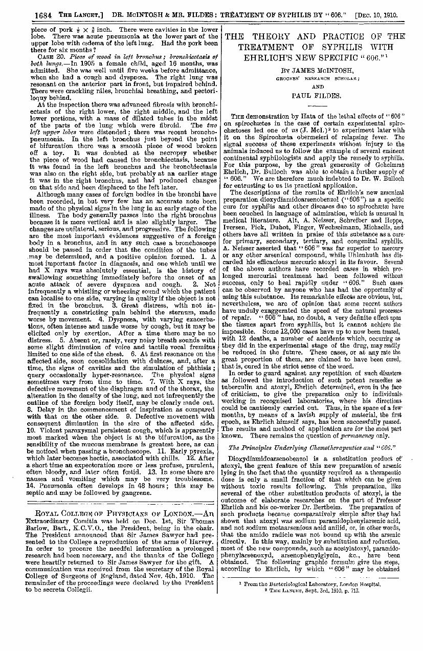
marked bronchiectasis of corresponding tubes only.-The patientwas a boy, aged three years, admitted BB ith cough and vomit-ing. He died with empyema and cerebral abscesses a monthlater. (Fig. 14.)
FiG. 14.
The trachea and right lung showing a pea wedged in thelower bronchus on the right side. The upper lobe of thelung is healthy, but the rest of the lung is densely fibroidand riddled with intercommunicating cavities the result ofthe obstruction.
CASE 19. Pieoe of pork in right bronoh2cs; bronchiectasis;septic pneumonia.—In 1888 a man, aged 60 years, wasadmitted. For six months there had been wasting, and fortwo weeks foul breath. For two days there had beendeficient movement of the lung on the right side, pleuralrub in the axilla and base, deficient air-entry, râles, withamphoric breathing over the middle Icbe.At the inspection the right lung was fibroid with general
bronchiectasis of the main bronchi. Low down was found a
1684
piece of pork 2 X i inch. There were cavities in the lowerlobe. There was acute pneumonia at the lower part of theupper lobe with oedema of the left lung. Had the pork beenthere for six months ? CASE 20. Pieoe of wood in lett bronchus ; bronchiectasis of
both lungs.-In 1905 a female child, aged 16 months, wasadmitted. She was well until five weeks before admittance,when she had a cough and dyspnoea. The right lung wasresonant on the anterior part in front, but impaired behind.There were crackling rales, bronchial breathing, and pectori-loquy behind.At the inspection there was advanced fibrosis with bronchi-
ectasis of the right lower, the right middle, and the leftlower portions, with a mass of dilated tubes in the midstof the parts of the lung which were fibroid. The twoleft upper lobes were distended ; there was recent broncho-pneumonia. In the left bronchus just beyond the pointof bifurcation there was a smooth piece of wood brokenoff a toy. It was doubted at the necropsy whetherthe piece of wood had caused the bronchiectasis, becauseit was found in the left bronchus and the bronchiectasiswas also on the right side, but probably at an earlier stageit was in the right bronchus, and had produced changeson that side and been displaced to the left later.Although many cases of foreign bodies in the bronchi have
been recorded, in but very few has an accurate note beenmade of the physical signs in the lung in an early stage of theillness. The body generally passes into the right bronchusbecause it is more vertical and is also slightly larger. The
changes are unilateral, serious, and progressive. The followingare the most important evidences suggestive of a foreignbody in a bronchus, and in any such case a bronchoscopeshould be passed in order that the condition of the tubesmay be determined, and a positive opinion formed. 1. Amost important factor in diagnosis, and one which until wehad X rays was absolutely essential, is the history ofswallowing something immediately before the onset of anacute attack of severe dyspnoea and cough. 2. Not
infrequently a whistling or wheezing sound which the patientcan localise to one side, varying in quality if the object is notfixed in the bronchus. 3. Great distress, with not in-
frequently a constricting pain behind the sternum, madeworse by movement. 4. Dyspnoea, with varying exacerba-tions, often intense and made worse by cough, but it may beelicited only by exertion. After a time there may be nodistress. 5. Absent or, rarely, very noisy breath sounds withsome slight diminution of voice and tactile vocal fremituslimited to one side of the chest. 6. At first resonance on theaffected side, soon consolidation with dulness, and, after atime, the signs of cavities and the simulation of phthisis ;query occasionally hyper-resonance. The physical signssometimes vary from time to time. 7. With X rays, thedefective movement of the diaphragm and of the thorax, thealteration in the density of the lung, and not infrequently theoutline of the foreign body itself, may be clearly made out.8. Delay in the commencement of inspiration as comparedwith that on the other side. 9. Defective movement withconsequent diminution in the size of the affected side.10. Violent paroxysmal persistent cough, which is apparentlymost marked when the object is at the bifurcation, as the sensibility of the mucous membrane is greatest here, as can Ibe noticed when passing a bronchoscope. 11. Early pyrexia,which later becomes hectic, associated with chills. 12. Aftera short time an expectoration more or less profuse, purulent,often bloody, and later often foetid. 13. In some there arenausea and vomiting which may be very troublesome.14. Pneumonia often develops in 48 hours ; this may be
septic and may be followed by gangrene.
ROYAL COLLEGE OF PHYSICIANS OF LONDON.-AnExtraordinary Comitia was held on Dec. 1st, Sir ThomasBarlow, Bart., K.C.V.O., the President, being in the chair.The President announced that Sir James Sawyer had pre-sented to the College a reproduction of the arms of Harvey.In order to procure the needful information a prolongedresearch had been necessary, and the thanks of the Collegewere heartily returned to Sir James Sawyer for the gift. Acommunication was received from the secretary of the RoyalCollege of Surgeons of England, dated Nov. 4th, 1910. Theremainder of the proceedings were declared by the Presidentto be secreta Collegii.
THE THEORY AND PRACTICE OF THE
TREATMENT OF SYPHILIS WITH
EHRLICH’S NEW SPECIFIC "606."1
BY JAMES McINTOSH,GROCERS’ RESEARCH SCHOLAR;
AND
PAUL FILDES.
THE demonstration by Hata of the lethal effects of ° 606 "on spirochastes in the case of certain experimental spiro.chastoses led one of us (J. McI.)2 to experiment later withit on the Spirochasta obermeieri of relapsing fever. Thesignal success of these experiments without injury to theanimals induced us to follow the example of several eminentcontinental syphilologists and apply the remedy to syphilis.For this purpose, by the great generosity of GeheimratEhrlich, Dr. Bulloch was able to obtain a further supply of" 606." We are therefore much indebted to Dr. W. Bulloch
for entrusting to us its practical application.The descriptions of the results of Ehrlich’s new arsenical
preparation dioxydiamidoarsenobenzol (" 606") as a specifiecure for syphilis and other diseases due to spirochætæ havebeen couched in language of admiration, which is unusual inmedical literature. Alt, A. Neisser, Schreiber and Hoppe,Iversen, Pick, Duhot, Finger, Wechselmann, Michaelis, andothers have all written in praise of this substance as a curefor primary, secondary, tertiary, and congenital syphilis,A. Neisser asserted that "606" " was far superior to mercuryor any other arsenical compound, while Uhlenhuth has dis-carded his efficacious mercuric atoxyl in its favour. Severalof the above authors have recorded cases in which pro-longed mercurial treatment had been followed withoutsuccess, only to heal rapidly under "606." Such cases.
can be observed by anyone who has had the opportunity of-using this substance. Its remarkable effects are obvious, but,.nevertheless, we are of opinion that some recent authorshave unduly exaggerated the speed of the natural processes-of repair. " 606 " has, no doubt, a very definite effect uponthe tissues apart from syphilis, but it cannot achieve the
impossible. Some 12,000 cases have up to now been treated,with 12 deaths, a number of accidents which, occurring asthey did in the experimental stage of the drug, may readilybe reduced in the future. These cases, or at any rate the
great proportion of them, are claimed to have been cured,-that is, cured in the strict sense of the word.
In order to guard against any repetition of such disasters.as followed the introduction of such potent remedies as-
tuberculin and atoxyl, Ehrlich determined, even in the faceof criticism, to give the preparation only to individuals-
working in recognised laboratories, where his directions-could be cautiously carried out. Thus, in the space of a fewmonths, by means of a lavish supply of material, the first
epoch, as Ehrlich himself says, has been successfully passed.The results and method of application are for the most partknown. There remains the question of permanency only.
The Principles Tlnderlying Chemotherapeutics and "606."
Dioxydiamidoarsenobenzol is a substitution product of .
atoxyl, the great feature of this new preparation of arseniclying in the fact that the quantity required as a therapeuticdose is only a small fraction of that which can be givenwithout toxic results following. This preparation, likeseveral of the other substitution products of atoxyl, is theoutcome of elaborate researches on the part of ProfessorEhrlich and his co-worker Dr. Bertheim. The preparation ofsuch products became comparatively simple after they hadshown that atoxyl was sodium paramidophenylarsenic acid,and not sodium metaarsenious acid anilid, or, in other words,that the amido radicle was not bound up with the arsenicdirectly. In this way, mainly by substitution and reduction,most of the new compounds, such as acetylatoxyl, paramido-phenylarsenoxyd, arsenopbenylglycin, &c., have beenobtained. The following graphic formulas give the steps,according to Ehrlich, by which "606 " may be obtained
1 From the Bacteriological Laboratory, London Hospital.2 THE LANCET, Sept. 3rd, 1910, p. 713.