547 Introduction 548 An Overview of the Cardiovascular System 548 The Pericardium 548 Structure of the Heart Wall 550 Orientation and Superficial Anatomy of the Heart 552 Internal Anatomy and Organization of the Heart 554 The Cardiac Cycle 561 CHAPTER OBJECTIVES The Cardiovascular System The Heart 1. Describe the basic design of the cardiovascular system and the function of the heart. 2. Describe the structure of the subdivisions of the pericardium and discuss its functions. 3. Identify and describe the epicardium, myocardium, and endocardium of the heart. 4. Identify important differences between cardiac muscle tissue and skeletal muscle tissue. 5. Discuss the structure and function of the fibrous skeleton of the heart. 6. Identify and describe the external form and surface features of the heart. 7. Describe the structural and functional specializations of each chamber of the heart. 8. Identify the major arteries and veins of the pulmonary and systemic circuits that are connected to the heart. 9. Trace the path of blood flow through the heart. 10. Describe the structure and function of each of the heart valves. 11. Locate the coronary blood vessels and identify their origins and major branches. 12. Name and trace the components of the conduction pathway of the heart. 13. Describe the function of the conduction pathway. 14. Discuss the events that take place during the cardiac cycle. 15. Describe the cardiac centers and discuss their functions in regulating the heart. 21
Transcript
547
Introduction 548
An Overview of the Cardiovascular System 548
The Pericardium 548
Structure of the Heart Wall 550
Orientation and Superficial Anatomy of the Heart 552
Internal Anatomy and Organization of the Heart 554
The Cardiac Cycle 561
C H A P T E R O B J E C T I V E S
The CardiovascularSystemThe Heart
1. Describe the basic design of thecardiovascular system and the functionof the heart.
2. Describe the structure of thesubdivisions of the pericardium anddiscuss its functions.
3. Identify and describe the epicardium,myocardium, and endocardium of theheart.
4. Identify important differences betweencardiac muscle tissue and skeletalmuscle tissue.
5. Discuss the structure and function ofthe fibrous skeleton of the heart.
6. Identify and describe the external formand surface features of the heart.
7. Describe the structural and functionalspecializations of each chamber of theheart.
8. Identify the major arteries and veins ofthe pulmonary and systemic circuitsthat are connected to the heart.
9. Trace the path of blood flow throughthe heart.
10. Describe the structure and function ofeach of the heart valves.
11. Locate the coronary blood vessels andidentify their origins and majorbranches.
12. Name and trace the components of theconduction pathway of the heart.
13. Describe the function of theconduction pathway.
14. Discuss the events that take placeduring the cardiac cycle.
15. Describe the cardiac centers anddiscuss their functions in regulating theheart.
21
548 THE CARDIOVASCULAR SYSTEM
Right atrium
Systemic arteries
Systemic veinsPulmonary veins
Pulmonary arteries
Rightventricle
Capillariesin lungs
Left atrium
Capillaries inhead, neck,upper limbs
Leftventricle
Capillaries intrunk and
lower limbs
PULMONARY CIRCUIT SYSTEMIC CIRCUIT
Figure 21.1 A Generalized View of the Pulmonary and Systemic CircuitsBlood flows through separate pulmonary and systemic circuits, driven by thepumping of the heart. Each circuit begins and ends at the heart and containsarteries, capillaries, and veins. Arrows indicate the direction of blood flow withineach circuit.
Every living cell relies on the surrounding interstitial fluid as a source ofoxygen and nutrients and as a place for the disposal of wastes. Levels ofgases, nutrients, and waste products in the interstitial fluid are kept stablethrough continuous exchange between the interstitial fluid and the circulat-ing blood. The blood must stay in motion to maintain homeostasis. If bloodstops flowing through a tissue, its oxygen and nutrient supplies are ex-hausted quickly, its capacity to absorb wastes is soon reached, and neitherhormones nor white blood cells can get to their intended targets. Thus, allof the functions of the cardiovascular system ultimately depend on theheart, because it is the heart that keeps blood moving. This muscular organbeats approximately 100,000 times each day, propelling blood through theblood vessels. Each year the heart pumps more than 1.5 million gallons ofblood, enough to fill 200 train tank cars.
For a practical demonstration of the heart’s pumping abilities, turnon the faucet in the kitchen and open it all the way. To deliver an amountof water equal to the volume of blood pumped by the heart in an averagelifetime, that faucet would have to be left on for at least 45 years. Equallyremarkable, the volume of blood pumped by the heart can vary widely, be-tween 5 and 30 liters per minute. The performance of the heart is closelymonitored and finely regulated by the nervous system to ensure that gas,nutrient, and waste levels in the peripheral tissues remain within normallimits, whether one is sleeping peacefully, reading a book, or involved in avigorous racquetball game.
We begin this chapter by examining the structural features that enablethe heart to perform so reliably, even in the face of widely varying physicaldemands. We will then consider the mechanisms that regulate cardiac activ-ity to meet the body’s ever-changing needs.
An Overview of the CardiovascularSystem [Figure 21.1]Despite its impressive workload, the heart is a small organ; your heart isroughly the size of your clenched fist. The heart’s four muscular chambers,the right and left atria (A-tre-a; singular, atrium; “chamber”) and right andleft ventricles (VEN-tri-kls; “little belly”), work together to pump bloodthrough a network of blood vessels between the heart and the peripheraltissues. The network can be subdivided into two circuits: the pulmonarycircuit, which carries carbon dioxide-rich blood from the heart to the gas-exchange surfaces of the lungs and returns oxygen-rich blood to the heart;and the systemic circuit, which transports oxygen-rich blood from theheart to the rest of the body’s cells, returning carbon dioxide-rich bloodback to the heart. The right atrium receives blood from the systemic cir-cuit, and the right ventricle discharges blood into the pulmonary circuit.The left atrium collects blood from the pulmonary circuit, and the left ven-tricle ejects blood into the systemic circuit. When the heart beats, the atriacontract first, followed by the ventricles. The two ventricles contract at thesame time and eject equal volumes of blood into the pulmonary and sys-temic circuits.
Each circuit begins and ends at the heart. Arteries transport bloodaway from the heart; veins return blood to the heart (Figure 21.1). Bloodtravels through these circuits in sequence. For example, blood returning tothe heart in the systemic veins must complete the pulmonary circuit beforereentering the systemic arteries. Capillaries are small, thin-walled vesselsthat interconnect the smallest arteries and veins. Capillaries are calledexchange vessels because their thin walls permit exchange of nutrients,dissolved gases, and waste products between the blood and surroundingtissues.
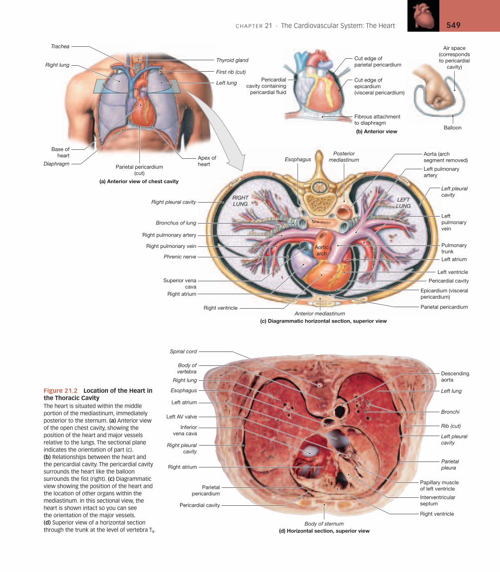
The Pericardium [Figure 21.2]The heart is located near the anterior chest wall (Figure 21.2a), directly pos-terior to the sternum in the pericardial (per-i-KAR-de-al) cavity, a portionof the ventral body cavity. The pericardial cavity is situated between thepleural cavities, in the mediastinum, which also contains the thymus,esophagus, and trachea.l p. 19 The position of the heart relative to otherstructures in the mediastinum is shown in Figure 21.2c,d.
The pericardium is the serous membrane lining the pericardial cavity.To visualize the relationship between the heart and the pericardial cavity,imagine pushing your fist toward the center of a large balloon (Figure 21.2b).The wall of the balloon corresponds to the pericardium, and your fist is theheart. The pericardium is divided into the visceral pericardium (the part ofthe balloon in contact with your fist) and the parietal pericardium (the restof the balloon). Your wrist, where the balloon folds back upon itself, corre-sponds to the base of the heart (so named because it is where the heart is at-tached to the major vessels and bound to the mediastinum).
The loose connective tissue of the visceral pericardium, or epicardium,is bound to the cardiac muscle tissue of the heart. The serous membrane ofthe parietal pericardium is reinforced by an outer layer of dense, irregularconnective tissue containing abundant collagen fibers. This reinforcinglayer is known as the fibrous pericardium. Together, the parietal peri-cardium and the fibrous pericardium form the tough pericardial sac. Atthe base of the heart, the collagen fibers of the fibrous pericardium stabilize
C H A P T E R 21 . The Cardiovascular System: The Heart 549
Aorticarch
Pulmonary trunk
Apex ofheartParietal pericardium
(cut)
Base ofheart
Right pleural cavity
Right pulmonary artery
Right pulmonary vein
Phrenic nerve
Superior venacava
Right atrium
Right ventricle
Epicardium (visceralpericardium)
Pericardial cavity
Left ventricle
Left atrium
Leftpulmonaryvein
Left pleuralcavity
Left pulmonary artery
Aorta (arch segment removed)
Balloon
Air space(correspondsto pericardial
cavity)
PosteriormediastinumEsophagus
Bronchus of lung
RIGHTLUNG
LEFTLUNG
(b) Anterior view
(c) Diagrammatic horizontal section, superior view
Pericardialcavity containing
pericardial fluid
Fibrous attachmentto diaphragm
Cut edge of epicardium (visceral pericardium)
Cut edge ofparietal pericardium
Rib (cut)
Bronchi
Left pleuralcavity
Parietal pleura
Left atrium
Left AV valve
Papillary muscleof left ventricle
Interventricularseptum
Right ventricle
Body of sternum
Pericardial cavity
Parietalpericardium
Right atrium
Inferiorvena cava
Right pleuralcavity
Right lung
Esophagus
Spinal cord
Body ofvertebra Descending
aorta
Left lung
(d) Horizontal section, superior view
Parietal pericardiumAnterior mediastinum
Trachea
First rib (cut)
Left lung
Diaphragm
Right lungThyroid gland
(a) Anterior view of chest cavity
Figure 21.2 Location of the Heart inthe Thoracic CavityThe heart is situated within the middleportion of the mediastinum, immediatelyposterior to the sternum. (a) Anterior viewof the open chest cavity, showing theposition of the heart and major vesselsrelative to the lungs. The sectional planeindicates the orientation of part (c).(b) Relationships between the heart andthe pericardial cavity. The pericardial cavitysurrounds the heart like the balloonsurrounds the fist (right). (c) Diagrammaticview showing the position of the heart andthe location of other organs within themediastinum. In this sectional view, theheart is shown intact so you can see the orientation of the major vessels.(d) Superior view of a horizontal sectionthrough the trunk at the level of vertebra T8.
550 THE CARDIOVASCULAR SYSTEM
the positions of the pericardium, heart, and associated vessels in the medi-astinum. The slender gap between the opposing parietal and visceral sur-faces is the pericardial cavity. This cavity normally contains 10–20 ml ofpericardial fluid secreted by the pericardial membranes. Pericardial fluidacts as a lubricant, reducing friction between the opposing surfaces. Themoist pericardial lining prevents friction as the heart beats, and the collagenfibers binding the base of the heart to the mediastinum limit movement ofthe major vessels during a contraction.
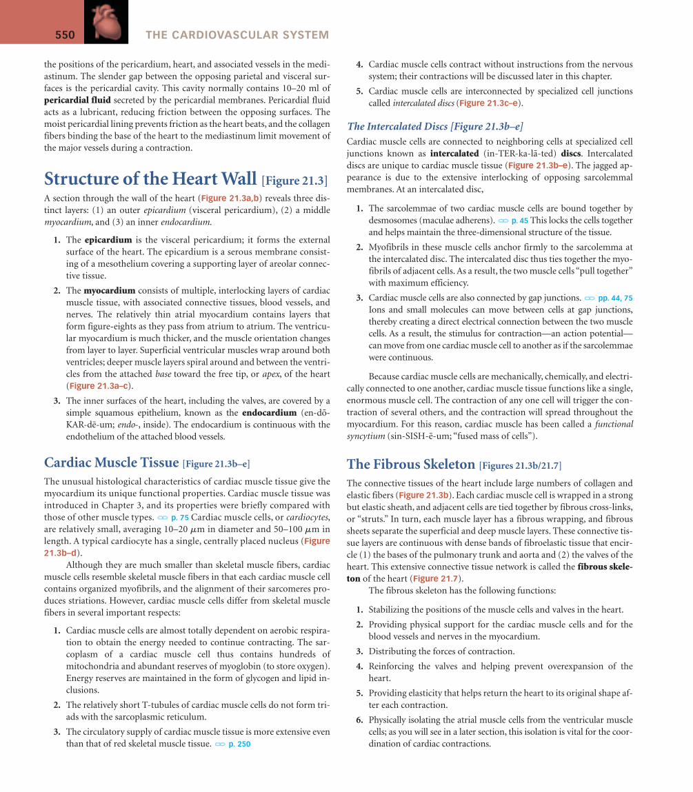
Structure of the Heart Wall [Figure 21.3]A section through the wall of the heart (Figure 21.3a,b) reveals three dis-tinct layers: (1) an outer epicardium (visceral pericardium), (2) a middlemyocardium, and (3) an inner endocardium.
1. The epicardium is the visceral pericardium; it forms the externalsurface of the heart. The epicardium is a serous membrane consist-ing of a mesothelium covering a supporting layer of areolar connec-tive tissue.
2. The myocardium consists of multiple, interlocking layers of cardiacmuscle tissue, with associated connective tissues, blood vessels, andnerves. The relatively thin atrial myocardium contains layers thatform figure-eights as they pass from atrium to atrium. The ventricu-lar myocardium is much thicker, and the muscle orientation changesfrom layer to layer. Superficial ventricular muscles wrap around bothventricles; deeper muscle layers spiral around and between the ventri-cles from the attached base toward the free tip, or apex, of the heart(Figure 21.3a–c).
3. The inner surfaces of the heart, including the valves, are covered by asimple squamous epithelium, known as the endocardium (en-do-KAR-de-um; endo-, inside). The endocardium is continuous with theendothelium of the attached blood vessels.
Cardiac Muscle Tissue [Figure 21.3b–e]
The unusual histological characteristics of cardiac muscle tissue give themyocardium its unique functional properties. Cardiac muscle tissue wasintroduced in Chapter 3, and its properties were briefly compared withthose of other muscle types.l p. 75 Cardiac muscle cells, or cardiocytes,are relatively small, averaging 10–20 mm in diameter and 50–100 mm inlength. A typical cardiocyte has a single, centrally placed nucleus (Figure21.3b–d).
Although they are much smaller than skeletal muscle fibers, cardiacmuscle cells resemble skeletal muscle fibers in that each cardiac muscle cellcontains organized myofibrils, and the alignment of their sarcomeres pro-duces striations. However, cardiac muscle cells differ from skeletal musclefibers in several important respects:
1. Cardiac muscle cells are almost totally dependent on aerobic respira-tion to obtain the energy needed to continue contracting. The sar-coplasm of a cardiac muscle cell thus contains hundreds ofmitochondria and abundant reserves of myoglobin (to store oxygen).Energy reserves are maintained in the form of glycogen and lipid in-clusions.
2. The relatively short T-tubules of cardiac muscle cells do not form tri-ads with the sarcoplasmic reticulum.
3. The circulatory supply of cardiac muscle tissue is more extensive eventhan that of red skeletal muscle tissue.l p. 250
4. Cardiac muscle cells contract without instructions from the nervoussystem; their contractions will be discussed later in this chapter.
5. Cardiac muscle cells are interconnected by specialized cell junctionscalled intercalated discs (Figure 21.3c–e).
The Intercalated Discs [Figure 21.3b–e]Cardiac muscle cells are connected to neighboring cells at specialized celljunctions known as intercalated (in-TER-ka-la-ted) discs. Intercalateddiscs are unique to cardiac muscle tissue (Figure 21.3b–e). The jagged ap-pearance is due to the extensive interlocking of opposing sarcolemmalmembranes. At an intercalated disc,
1. The sarcolemmae of two cardiac muscle cells are bound together bydesmosomes (maculae adherens).l p. 45 This locks the cells togetherand helps maintain the three-dimensional structure of the tissue.
2. Myofibrils in these muscle cells anchor firmly to the sarcolemma atthe intercalated disc. The intercalated disc thus ties together the myo-fibrils of adjacent cells. As a result, the two muscle cells “pull together”with maximum efficiency.
3. Cardiac muscle cells are also connected by gap junctions.l pp. 44, 75
Ions and small molecules can move between cells at gap junctions,thereby creating a direct electrical connection between the two musclecells. As a result, the stimulus for contraction—an action potential—can move from one cardiac muscle cell to another as if the sarcolemmaewere continuous.
Because cardiac muscle cells are mechanically, chemically, and electri-cally connected to one another, cardiac muscle tissue functions like a single,enormous muscle cell. The contraction of any one cell will trigger the con-traction of several others, and the contraction will spread throughout themyocardium. For this reason, cardiac muscle has been called a functionalsyncytium (sin-SISH-e-um; “fused mass of cells”).
The Fibrous Skeleton [Figures 21.3b/21.7]
The connective tissues of the heart include large numbers of collagen andelastic fibers (Figure 21.3b). Each cardiac muscle cell is wrapped in a strongbut elastic sheath, and adjacent cells are tied together by fibrous cross-links,or “struts.” In turn, each muscle layer has a fibrous wrapping, and fibroussheets separate the superficial and deep muscle layers. These connective tis-sue layers are continuous with dense bands of fibroelastic tissue that encir-cle (1) the bases of the pulmonary trunk and aorta and (2) the valves of theheart. This extensive connective tissue network is called the fibrous skele-ton of the heart (Figure 21.7).
The fibrous skeleton has the following functions:
1. Stabilizing the positions of the muscle cells and valves in the heart.
2. Providing physical support for the cardiac muscle cells and for theblood vessels and nerves in the myocardium.
3. Distributing the forces of contraction.
4. Reinforcing the valves and helping prevent overexpansion of theheart.
5. Providing elasticity that helps return the heart to its original shape af-ter each contraction.
6. Physically isolating the atrial muscle cells from the ventricular musclecells; as you will see in a later section, this isolation is vital for the coor-dination of cardiac contractions.
C H A P T E R 21 . The Cardiovascular System: The Heart 551
(a) Anterior view
(b) Sectional view
Pericardial cavity
Base of heart
Pericardialcavity
Cut edge of pericardium
Apex of heart
MYOCARDIUM(cardiac muscle tissue)
Areolar tissue
Mesothelium
Parietalpericardium
Connective tissues
Artery
Vein
Mesothelium
Areolar tissueEPICARDIUM(visceral pericardium)
Areolarconnective
tissue
Endothelium
ENDOCARDIUMHeart wall
Intercalateddisc (sectioned)
Intercalateddisc
Intercalated disc
Intercalateddisc
Nucleus
Cardiac musclecell (sectioned)
Mitochondria
Bundles ofmyofibrils
Gap junction
Z lines boundto opposing cell
membranes
Desmosomes
(c) Cardiac muscle tissue (LM � 575)
(d) Cardiac muscle cells
(e) Structure of an intercalated disc
Cardiac muscle cell
Dense fibrous layer
Figure 21.3 Histological Organization of Muscle Tissue in the Heart Wall(a) Anterior view of the heart showing several important landmarks.(b) A diagrammatic section through the heart wall showing the structure of the epicardium, myocardium, and endocardium. (c) and (d) Histological anddiagrammatic views of cardiac muscle tissue. Distinguishing characteristics ofcardiac muscle cells include (1) small size; (2) a single, centrally placed nucleus;(3) branching interconnections between cells; and (4) the presence of intercalateddiscs. (e) The structure of an intercalated disc.
1. How could you distinguish a sample of cardiac muscle tissue from asample of skeletal muscle tissue?
2. What is the pericardial cavity?3. How are cardiac muscle cells connected to their neighbors?4. Why is cardiac muscle called a functional syncytium?
See blue “Answers” tab at back of book.
Orientation and SuperficialAnatomy of the Heart[Figures 21.2b/21.4/21.5]Although advertisements and cartoons often show the heart at the center ofthe chest, a midsagittal section would not cut the heart in half. This is be-cause the heart (1) lies slightly to the left of the midline, (2) sits at an angleto the longitudinal axis of the body, and (3) is rotated toward the left side.
1. The heart lies slightly to the left of the midline: The heart is locatedwithin the mediastinum, between the two lungs. Because the heart liesslightly to the left of the midline, the notch within the medial surfaceof the left lung is considerably deeper than the corresponding notchin the medial surface of the right lung. The base is the broad superiorportion of the heart, where the heart is attached to the major arteriesand veins of the systemic and pulmonary circuits. The base of theheart includes both the origins of the major vessels and the superiorsurfaces of the two atria. In terms of our balloon analogy, the base cor-responds to the wrist (Figure 21.2b). The base sits posterior to thesternum at the level of the third costal cartilage, centered about 1.2 cm(0.5 in.) to the left side (Figure 21.4). The apex (A-peks) is the infe-rior, rounded tip of the heart, which points laterally at an oblique an-
gle. A typical adult heart measures approximately 12.5 cm (5 in.) fromthe attached base to the apex. The apex reaches the fifth intercostalspace approximately 7.5 cm (3 in.) to the left of the midline.
2. The heart sits at an oblique angle to the longitudinal axis of the body: Thebase forms the superior border of the heart. The right border of theheart is formed by the right atrium; the left border is formed by the leftventricle and a small portion of the left atrium. The left border extendsto the apex, where it meets the inferior border. The inferior border isformed mainly by the inferior wall of the right ventricle.
3. The heart is rotated slightly toward the left: As a result of this rotation,the anterior surface, or sternocostal (ster-no-KOS-tal) surface, con-sists primarily of the right atrium and right ventricle (Figure 21.5a).The posterior and inferior wall of the left ventricle forms much of thesloping posterior surface, or diaphragmatic surface, that extends be-tween the base and the apex of the heart (Figure 21.5b).
The four internal chambers of the heart are associated with grooves orsulci visible on its external surface (Figure 21.5). A shallow interatrial grooveseparates the two atria, while the deeper coronary sulcus marks the borderbetween the atria and the ventricles. The division between the left and rightventricles is indicated by linear depressions on the anterior surface (theanterior interventricular sulcus) and the posterior surface (the posteriorinterventricular sulcus). The connective tissue of the epicardium at thecoronary and interventricular sulci usually contains substantial amounts ofadipose tissue that must be removed to expose the underlying grooves.These sulci also contain the arteries and veins that supply blood to the car-diac muscle of the heart.
The atria and the ventricles have very different functions—the atria re-ceive venous blood that must continue on to the ventricles, whereas the ven-tricles must propel blood around the systemic and pulmonary circuits. Thesefunctional differences are of course linked to external and internal structuraldifferences. Examine Figure 21.5, which details the superficial anatomy of theheart, and note the distinguishing characteristics of the atria and ventricles.
552 THE CARDIOVASCULAR SYSTEM
Superiorborder
Inferior border
Rightborder
Base ofheart
Ribs
Apex ofheart
1 1
2 2
3 3
4 4
55
6 6
77
889 9
10 10
Leftborder
Figure 21.4 Position and Orientation of the HeartThe location of the heart within the thoracic cavity and the borders of the heart.
C H A P T E R 21 . The Cardiovascular System: The Heart 553
RIGHT ATRIUM
RIGHT VENTRICLE
LEFTVENTRICLE
Arch of aorta
Left pulmonary veins
Left pulmonary artery
Coronarysinus
Fat in posteriorinterventricular sulcus
LEFTATRIUM
Fat incoronary
sulcus
Right pulmonaryartery
Superiorvena cava
Rightpulmonaryveins (superior and inferior)
Inferiorvena cava
(b) Posterior (diaphragmatic) surface
RIGHTVENTRICLE
LEFT ATRIUM
LEFTVENTRICLE
Coronary sinus
Inferior venacava
Rightpulmonaryveins (superiorand inferior)
Superiorvena cava
Auricle ofleft atrium
Great cardiacvein (blue) and
circumflex branchof left coronary
artery (red)
Right pulmonaryartery
Left pulmonaryartery
Left pulmonaryveins (superior
and inferior)
RIGHT ATRIUM
(a) Anterior (sternocostal) surface
Arch of aorta
Left subclavian arteryLeft common carotid artery
Brachiocephalic trunk
Left pulmonaryartery
Ligamentumarteriosum
Pulmonarytrunk
Descendingaorta
Auricleof leftatrium
Auricle ofright
atrium
Superiorvena cava
Ascendingaorta
RIGHTATRIUM
RIGHTVENTRICLE LEFT
VENTRICLEFat in
coronarysulcus
Fat inanteriorinterventricularsulcus
Parietal pericardiumfused to diaphragm
Superiorvena cava
Auricle ofright atrium
RIGHT ATRIUM
Coronary sulcus
Marginal branchof right
coronary artery
Right coronaryartery
RIGHTVENTRICLE
Anteriorinterventricular
sulcus
LEFTVENTRICLE
Auricle ofleft atrium
Ascendingaorta
Pulmonary trunk
Fibrouspericardium
Parietalpericardium
Figure 21.5 Superficial Anatomy of the Heart(a) Anterior view of the heart and great vessels. In the photo, the pericardial sac has been cut and reflected to expose theheart and great vessels. (b) Posterior view of the heart and great vessels; the coronary vessels have been injected withcolored latex (see Figure 21.8).
554 THE CARDIOVASCULAR SYSTEM
The right atrium is situated anterior, inferior, and to the right of theleft atrium. The left atrium extends posterior to the right atrium; it formsmost of the posterior surface of the heart superior to the coronary sulcus.Both atria have relatively thin muscular walls and, as a result, they are highlydistensible. When not filled with blood, the outer portion of each atriumdeflates and becomes a rather lumpy and wrinkled flap. This expandable ex-tension of an atrium is called an auricle (AW-ri-kel; auris, ear) because itreminded early anatomists of the external ear. The auricle is also known asan atrial appendage.
The ventricles lie inferior to the coronary sulcus (Figure 21.5). Theright ventricle makes up a large percentage of the sternocostal surface of theheart. The left ventricle extends from the coronary sulcus to the apex or tipof the heart, forming the left and diaphragmatic surfaces of the heart.
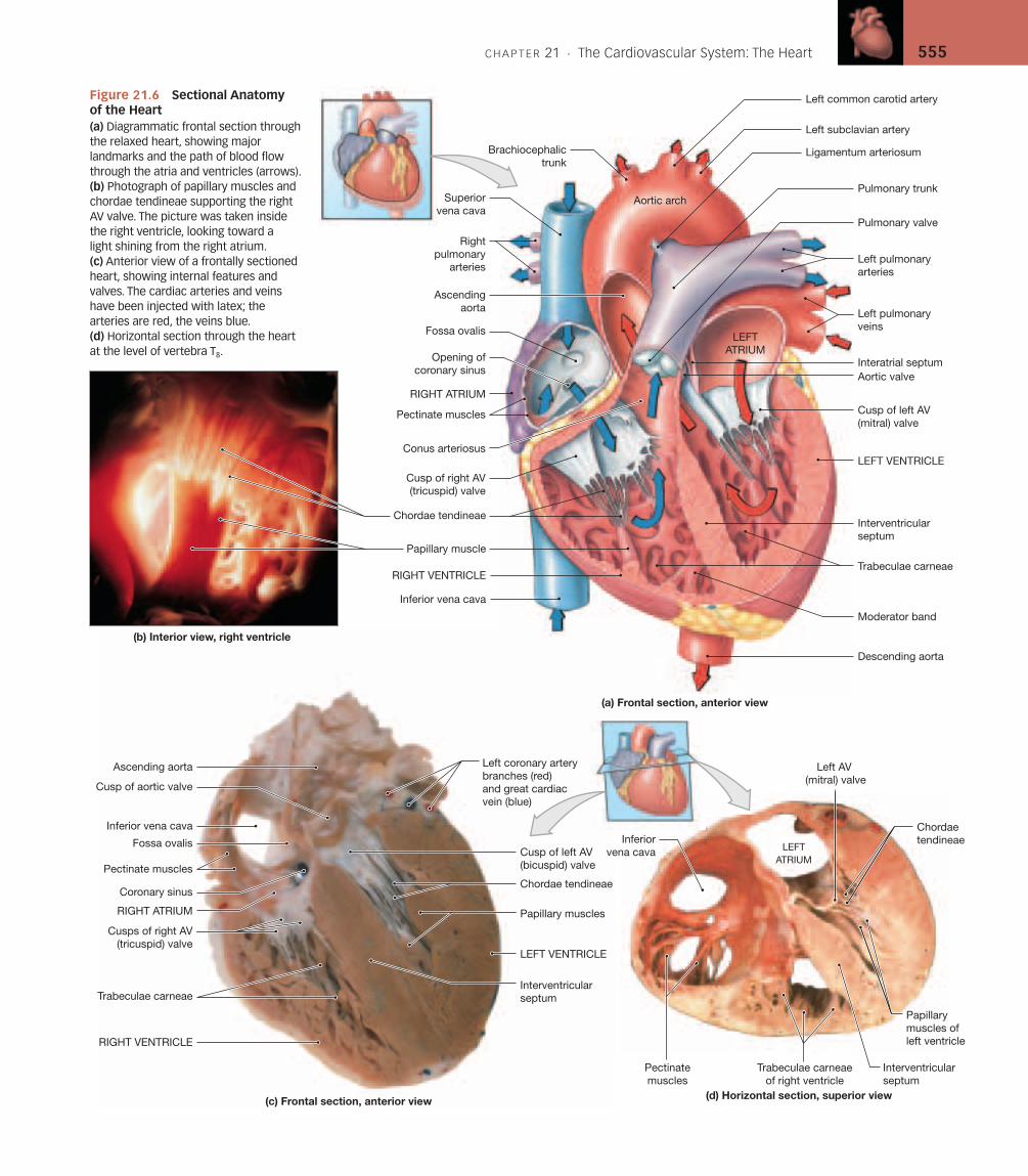
Internal Anatomy and Organizationof the Heart [Figure 21.6]Figure 21.6 details the internal anatomy and functional organization of theatria and ventricles. The atria are separated by the interatrial septum(septum, a wall), and the interventricular septum separates the ventricles(Figure 21.6a,c). Each atrium communicates with the ventricle of the sameside. Valves are folds of endocardium that extend into the openings be-tween the atria and ventricles. These valves open and close to prevent back-flow, thereby maintaining a one-way flow of blood from the atria into theventricles. (Valve structure and function will be described under a separateheading.)
An atrium functions to collect blood returning to the heart and de-liver it to the attached ventricle. The functional demands placed on the rightand left atria are very similar, and the two chambers look almost identical.The demands placed on the right and left ventricles are very different, andthere are significant structural differences between the two.
The Right Atrium [Figures 21.5/21.6a,c]
The right atrium receives oxygen-poor venous blood from the systemic cir-cuit through the superior vena cava (VE-na CA-va) and the inferior venacava (Figures 21.5 and 21.6a,c). The superior vena cava, which opens intothe posterior, superior portion of the right atrium, delivers venous bloodfrom the head, neck, upper limbs, and chest. The inferior vena cava, whichopens into the posterior and inferior portion of the right atrium, delivers ve-nous blood from the tissues and organs of the abdominal and pelvic cavities,and the lower limbs. The veins of the heart itself, called coronary veins, collectblood from the heart wall and deliver it to the coronary sinus (Figure 21.5b).This collecting vessel opens into the posterior wall of the right atrium, infe-rior to the opening of the inferior vena cava. (The coronary blood vessels willbe described under a separate heading.)
Prominent muscular ridges, the pectinate muscles (pectin, comb), ormusculi pectinati, extend along the inner surface of the right auricle andacross the adjacent anterior atrial wall. The interatrial septum separatesthe right and left atria. From the fifth week of embryonic development un-til birth, there is an oval opening, the foramen ovale, in this septum. (SeeEmbryology Summaries in Chapter 28.) The foramen ovale permits bloodflow directly from the right atrium to the left atrium while the lungs are de-veloping and nonfunctional. At birth the lungs begin functioning and theforamen ovale closes; after 48 hours it is permanently sealed. A small de-pression, the fossa ovalis, persists at this site in the adult heart. Occasion-ally the foramen ovale does not close, and it remains patent (open). As aresult, blood recirculates into the pulmonary circuit, reducing the efficiencyof systemic circulation and elevating blood pressure in the pulmonary ves-sels. This can lead to cardiac enlargement, fluid buildup in the lungs, andeventual heart failure.
The Right Ventricle [Figures 21.5/21.6]
Oxygen-poor venous blood travels from the right atrium into the right ven-tricle through a broad opening bounded by three fibrous flaps. These flaps,or cusps, form the right atrioventricular (AV) valve, or tricuspid valve (tri-KUS-pid; tri, three) (Figure 21.6). The free edges of the cusps are attachedto bundles of collagen fibers, the chordae tendineae (KOR-de TEN-di-ne-e;“tendinous cords”). These bundles arise from the papillary (PAP-i-ler-e)muscles, cone-shaped muscular projections of the inner ventricular sur-face. The chordae tendineae limit the movement of the cusps and preventbackflow of blood from the right ventricle into the right atrium; the mech-anism will be detailed in a later section.
The internal surface of the ventricle contains a series of irregular muscu-lar folds, the trabeculae carneae (tra-BEK-u-le CAR-ne-e; carneus, fleshy).The moderator band is a band of ventricular muscle that extends from theinterventricular septum, a thick, muscular partition that separates the twoventricles, to the anterior wall of the right ventricle and the bases of the papil-lary muscles.
The superior end of the right ventricle tapers to a smooth-walled, cone-shaped pouch, the conus arteriosus, which ends at the pulmonary valve(pulmonary semilunar valve). This valve consists of three thick semilunar (halfmoon–shaped) cusps. As blood is ejected from the right ventricle, it passes
Clinical Note
Infection and Inflammation of the Heart Many different mi-croorganisms may infect heart tissue, leading to serious cardiac ab-normalities. Carditis (kar-DI-tis) is a general term for inflammation ofthe heart. Clinical conditions resulting from cardiac infection are usu-ally identified by the primary site of the infection. For example, infec-tions that affect the endocardium produce symptoms of endocarditis,a condition that damages primarily the chordae tendineae and heartvalves; the mortality rate may reach 21–35 percent. The most severecomplications of endocarditis result from the formation of blood clotson the damaged surfaces. These clots subsequently break free, enter-ing the bloodstream as drifting emboli that may cause strokes, heartattacks, or kidney failure. The destruction of heart valves by the infec-tion may lead to valve leakage, heart failure, and death.
Myocarditis, inflammation of the heart muscle, can be caused bybacteria, viruses, protozoans, or fungal pathogens that either attackthe myocardium directly or produce toxins that damage the myo-cardium. The sarcolemma of infected heart muscle cells become facili-tated, and the heart rate may rise dramatically. Over time, abnormalcontractions may appear and the heart muscle weakens; these prob-lems may eventually prove fatal.
If the pericardium becomes inflamed or infected, fluid may accu-mulate around the heart (cardiac tamponade), or the elasticity of thepericardium may be reduced (constrictive pericarditis). In both condi-tions, the expansion of the heart is restricted and cardiac output is re-duced. Treatment includes draining the excess fluid or cutting awindow in the pericardial sac.
C H A P T E R 21 . The Cardiovascular System: The Heart 555
Superiorvena cava
Rightpulmonary
arteries
Fossa ovalis
Ascendingaorta
Pectinate muscles
Conus arteriosus
Opening ofcoronary sinus
RIGHT ATRIUM
Cusp of right AV(tricuspid) valve
Trabeculae carneae
Inferior vena cava
RIGHT VENTRICLE
Moderator band
Descending aorta
Interventricularseptum
LEFT VENTRICLE
Papillary muscle
Chordae tendineae
Cusp of left AV(mitral) valve
Interatrial septum Aortic valve
Left pulmonaryveins
LEFTATRIUM
Left pulmonaryarteries
Pulmonary valve
Pulmonary trunkAortic arch
Ligamentum arteriosum Brachiocephalictrunk
Left subclavian artery
Left common carotid artery
(a) Frontal section, anterior view
(b) Interior view, right ventricle
Cusp of left AV(bicuspid) valve
Chordae tendineae
Papillary muscles
LEFT VENTRICLE
Interventricularseptum
RIGHT VENTRICLE
Trabeculae carneae
Cusps of right AV (tricuspid) valve
RIGHT ATRIUM
Coronary sinus
Pectinate muscles
Ascending aorta
Cusp of aortic valve
Fossa ovalis
Inferior vena cava
(c) Frontal section, anterior view
Interventricularseptum
Pectinatemuscles
Inferiorvena cava LEFT
ATRIUM
Trabeculae carneaeof right ventricle
Papillary muscles of left ventricle
Left AV (mitral) valve
(d) Horizontal section, superior view
Chordae tendineae
Left coronary artery branches (red)and great cardiac vein (blue)
Figure 21.6 Sectional Anatomy of the Heart(a) Diagrammatic frontal section throughthe relaxed heart, showing majorlandmarks and the path of blood flowthrough the atria and ventricles (arrows).(b) Photograph of papillary muscles andchordae tendineae supporting the rightAV valve. The picture was taken insidethe right ventricle, looking toward a light shining from the right atrium.(c) Anterior view of a frontally sectionedheart, showing internal features andvalves. The cardiac arteries and veinshave been injected with latex; thearteries are red, the veins blue.(d) Horizontal section through the heartat the level of vertebra T8.
556 THE CARDIOVASCULAR SYSTEM
through this valve to enter the pulmonary trunk, the start of the pulmonarycircuit. The arrangement of cusps in this valve prevents the backflow of bloodinto the right ventricle when that chamber relaxes. From the pulmonary trunk,blood flows into both the left and right pulmonary arteries (Figures 21.5and 21.6). These vessels branch repeatedly within the lungs before supplyingthe pulmonary capillaries, where gas exchange occurs.
The Left Atrium [Figures 21.5/21.6a]
From the pulmonary capillaries, the blood, now oxygen-rich, flows intosmall veins that ultimately unite to form four pulmonary veins, two fromeach lung. These left and right pulmonary veins empty into the posteriorportion of the left atrium (Figures 21.5 and 21.6a). The left atrium lackspectinate muscles, but it has an auricle. Blood flowing from the left atriuminto the left ventricle passes through the left atrioventricular (AV) valve,also known as the mitral (MI-tral; mitre, a bishop’s hat) valve or the bicuspid(bi-KUS-pid) valve. As the name bicuspid implies, this valve contains a pairof cusps (bi-, two) rather than a trio (tri-, three). The left atrioventricularvalve permits the flow of oxygen-rich blood from the left atrium into the leftventricle, but prevents blood flow in the reverse direction.
The Left Ventricle [Figures 21.5/21.6a,c,d]
The left ventricle has the thickest wall of any heart chamber. The extra-thickmyocardium enables it to develop enough pressure to force blood aroundthe entire systemic circuit; by comparison the right ventricle, which has arelatively thin wall, must push blood to the lungs and then back to the heart,a total distance of only about 30 cm (1 ft). The internal organization of theleft ventricle resembles that of the right ventricle (Figure 21.6a,c,d). How-ever, the trabeculae carneae are more prominent than they are in the rightventricle, there is no moderator band, and since the mitral valve has twocusps, there are two large papillary muscles rather than three.
Blood leaves the left ventricle by passing through the aortic valve(aortic semilunar valve) into the ascending aorta. The arrangement ofcusps in the aortic valve is the same as in the pulmonary semilunar valve.Saclike dilations of the base of the ascending aorta occur adjacent to eachcusp. These sacs, called aortic sinuses, prevent the individual cusps fromsticking to the wall of the aorta when the valve opens. The right and left coro-nary arteries, which deliver blood to the myocardium, originate at the aor-tic sinus. The aortic valve prevents the backflow of blood into the leftventricle once it has been pumped out of the heart and into the systemic cir-cuit. From the ascending aorta, blood flows on through the aortic arch andinto the descending aorta (Figures 21.5 and 21.6a). The pulmonary trunkis attached to the aortic arch by the ligamentum arteriosum, a fibrous bandthat is the remnant of an important fetal blood vessel. Cardiovascularchanges that occur at birth are described in Chapter 22.
Structural Differences between the Left and Right Ventricles [Figure 21.6a,c,d]
Anatomical differences between the left and right ventricles are best seen inthree-dimensional or sectional views (Figure 21.6a,c,d). The lungs partiallyenclose the pericardial cavity, and the base of the heart lies between the leftand right lungs. As a result, the pulmonary arteries and veins are relativelyshort and wide, and the right ventricle normally does not need to push veryhard to propel blood through the pulmonary circuit. The wall of the rightventricle is relatively thin, and in sectional view it resembles a pouch attachedto the massive wall of the left ventricle. When the right ventricle contracts, it
moves toward the wall of the left ventricle. This compresses the blood withinthe right ventricle, and the rising pressure forces the blood through the pul-monary valve and into the pulmonary trunk. This mechanism moves bloodvery efficiently at relatively low pressures, which are all that one needs tomove blood around the pulmonary circuit. Higher pressures would actuallybe dangerous, because the pulmonary capillaries are very delicate. Pressuresas high as those found in systemic capillaries would both damage the pul-monary vessels and force fluid into the alveoli of the lungs.
A comparable pumping arrangement would not be suitable for theleft ventricle, because six to seven times as much force must be exerted topropel blood through the systemic circuit. The left ventricle, which has anextremely thick muscular wall, is round in cross section. When the left ven-tricle contracts, two things happen: The distance between the base and apexdecreases, and the diameter of the ventricular chamber decreases. If youimagine the effects of simultaneously squeezing and rolling up the end of atoothpaste tube, you will get the idea. The forces generated are quite pow-erful, more than enough to force open the aortic valve and eject blood intothe ascending aorta. As the powerful left ventricle contracts, it also bulgesinto the right ventricular cavity. This intrusion improves the efficiency ofthe right ventricle’s efforts. Individuals whose right ventricular musculaturehas been severely damaged may continue to survive because of the extrapush provided by the contraction of the left ventricle.
1. What is the name of the groove separating the atria from theventricles?
2. What are some external characteristics that distinguish the atria fromthe ventricles?
See blue “Answers” tab at back of book.
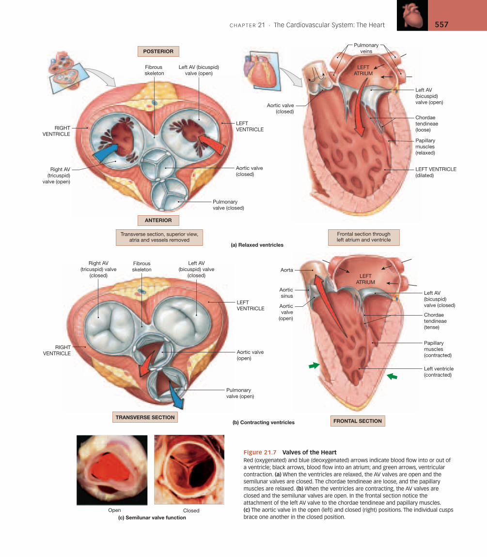
The Structure and Function of HeartValves [Figures 21.6/21.7]
Details of the structure and function of the four heart valves are shown inFigures 21.6 and 21.7.
The atrioventricular (AV) valves are situated between the atria and theventricles. Each AV valve has four components: (1) a ring of connective tis-sue that attaches to the fibrous skeleton of the heart; (2) connective tissuecusps, which function to close the opening between the heart chambers; and(3) chordae tendineae that attach the margins of the cusps to (4) thepapillary muscles of the heart wall.
There are two semilunar valves guarding the outflow from the twoventricles. These valves get their names from the shape of their threevalvules or cusps, which resemble half moon–shaped pockets. Thepulmonary valve is found at the exit of the pulmonary trunk from the rightventricle, while the aortic valve is found at the exit of the aorta from the leftventricle.
Valve Function during the Cardiac CycleThe chordae tendineae and papillary muscles associated with the AV valvesplay an important role in the normal function of the AV valves during thecardiac cycle. During the period of ventricular relaxation (ventricular dias-tole) the ventricles are filling with blood, the papillary muscles are relaxed,and the open AV valve offers no resistance to the flow of blood from atriumto ventricle. Over this period the semilunar valves are closed; the semilunarvalves do not need chordae tendineae because the relative positions of the
C H A P T E R 21 . The Cardiovascular System: The Heart 557
Right AV(tricuspid) valve
(closed)
(c) Semilunar valve function
Right AV(tricuspid)
valve (open)
Fibrousskeleton
Left AV(bicuspid) valve
(closed)
Pulmonaryvalve (closed)
Left AV (bicuspid)valve (open)
Fibrous skeleton
Open Closed
Aorta
Aorticsinus Left AV
(bicuspid)valve (closed)
Papillarymuscles(contracted)
Left ventricle(contracted)
Left AV (bicuspid)valve (open)
Pulmonaryveins
Papillarymuscles(relaxed)
Chordaetendineae(tense)
Chordaetendineae(loose)
(b) Contracting ventricles
(a) Relaxed ventricles
Aortic valve(closed)
Aortic valve(closed)
Aortic valve(open)
Pulmonaryvalve (open)
Aorticvalve
(open)
Transverse section, superior view,atria and vessels removed
Frontal section throughleft atrium and ventricle
TRANSVERSE SECTIONFRONTAL SECTION
LEFTVENTRICLE
LEFTVENTRICLERIGHT
VENTRICLE
RIGHTVENTRICLE
LEFT VENTRICLE(dilated)
LEFTATRIUM
LEFTATRIUM
ANTERIOR
POSTERIOR
Figure 21.7 Valves of the HeartRed (oxygenated) and blue (deoxygenated) arrows indicate blood flow into or out of a ventricle; black arrows, blood flow into an atrium; and green arrows, ventricularcontraction. (a) When the ventricles are relaxed, the AV valves are open and thesemilunar valves are closed. The chordae tendineae are loose, and the papillarymuscles are relaxed. (b) When the ventricles are contracting, the AV valves areclosed and the semilunar valves are open. In the frontal section notice theattachment of the left AV valve to the chordae tendineae and papillary muscles.(c) The aortic valve in the open (left) and closed (right) positions. The individual cuspsbrace one another in the closed position.
558 THE CARDIOVASCULAR SYSTEM
Clinical Note
Mitral Valve Prolapse Minor abnormalities in valve shape are rela-tively common. For example, an estimated 10 percent of normal indi-viduals age 14–30 have some degree of mitral valve prolapse (MVP).In this condition the mitral valve cusps do not close properly. Theproblem may involve abnormally long (or short) chordae tendineae ormalfunctioning papillary muscles. Because the valve does not workperfectly, some regurgitation may occur during left ventricular systole.The surges, swirls, and eddies that occur during regurgitation create arushing, gurgling sound known as a heart murmur. Most of these in-dividuals are completely asymptomatic, and they live normal, healthylives unaware of any circulatory malfunction. However, regurgitationmay increase the risk of valve infection after dental (or some medical)procedures.
cusps are stable, and the three symmetrical cusps support one another likethe legs of a tripod.
When the period of ventricular contraction (ventricular systole) be-gins, blood leaving the ventricles opens the semilunar valves, while bloodmoving back toward the atria swings the cusps of the AV valves together.Tension in the papillary muscles and chordae tendineae keeps the cuspsfrom swinging farther and opening into the atria. Thus, the chordaetendineae and papillary muscles are essential to prevent the backflow, orregurgitation, of blood into the atria each time the ventricles contract.
Serious valvular abnormalities can interfere with cardiac function; thetiming and intensity of the related heart sounds can provide useful diagnos-tic information. Physicians use an instrument called a stethoscope(STETH-o-scop) to listen to normal and abnormal heart sounds. Valvesounds may be muffled as they pass through the pericardium, surroundingtissues, and the chest wall. As a result, the stethoscope placement does notalways correspond to the position of the valve under review.
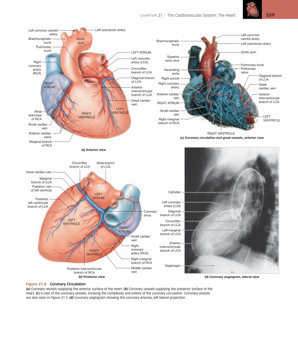
Coronary Blood Vessels [Figure 21.8]
The heart works continuously, and cardiac muscle cells require reliable sup-plies of oxygen and nutrients. The coronary circulation supplies blood tothe muscle tissue of the heart. During maximum exertion, the oxygen de-mand rises considerably, and the blood flow to the heart may increase tonine times that of resting levels.
The coronary circulation (Figure 21.8) includes an extensive networkof coronary blood vessels. The left and right coronary arteries originate atthe base of the ascending aorta, within the aortic sinus, as the first branchesof this vessel. Blood pressure here is the highest found anywhere in the sys-temic circuit, and this pressure ensures a continuous flow of blood to meetthe demands of active cardiac muscle tissue.
The Right Coronary Artery [Figure 21.8]The right coronary artery (RCA) branches off the ascending aorta, turns tothe right, and passes between the right auricle and the pulmonary trunk. Itthen continues within the coronary sulcus. Although many variations mayoccur, the branches of the right coronary artery typically supply blood to (1)the right atrium, (2) a portion of the left atrium, (3) the interatrial septum,
(4) the entire right ventricle, (5) a variable portion of the left ventricle, (6)the posteroinferior one-third of the interventricular septum, and (7) por-tions of the conducting system of the heart. The major branches are shownin Figure 21.8.
1. Atrial branches: As it curves across the anterior surface of the heart,the right coronary artery gives rise to atrial branches that supply themyocardium of the right atrium and a portion of the left atrium.
2. Ventricular branches: Near the right border of the heart, the rightcoronary artery usually gives rise to the right marginal branch thatextends toward the apex along the anterior surface of the right ventri-cle. It then continues across the posterior surface of the heart, supply-ing the posterior interventricular branch, or posterior descendingartery, which runs toward the apex within the posterior interventric-ular sulcus. This branch supplies blood to the interventricular septumand adjacent portions of the ventricles.
3. Branches to the conducting system: A small branch near the base of theright coronary artery penetrates the atrial wall to reach the sinoatrial(SA) node, also known as the cardiac pacemaker. A small branch to theatrioventricular (AV) node, another part of the conducting system of theheart, originates from the right coronary artery near the posterior inter-ventricular branch. These nodes and their role in the regulation of theheartbeat will be the topic of a later section.
The Left Coronary Artery [Figure 21.8]The left coronary artery commonly supplies blood to (1) most of the leftventricle, (2) a narrow slip of the right ventricle, (3) most of the leftatrium, and (4) the anterior two-thirds of the interventricular septum. Asit reaches the anterior surface of the heart, it gives rise to a circumflexbranch and an anterior interventricular branch (Figure 21.8). Thecircumflex branch curves to the left within the coronary sulcus, givingrise to one or more diagonal branches as it curves toward the posteriorsurface of the heart. It usually gives rise to a left marginal branch, and onreaching the posterior surface of the heart it forms a posterior left ven-tricular branch. The distal portions of the circumflex artery often meetand fuse with small branches of the right coronary artery. The muchlarger anterior interventricular branch, or left anterior descendingbranch, runs along the anterior surface within the anterior interventricu-lar sulcus. This artery supplies the anterior ventricular myocardium andthe anterior two-thirds of the interventricular septum. Small branchesfrom the anterior interventricular branch of the left coronary artery arecontinuous with those of the posterior interventricular branch of theright coronary artery. Such interconnections between arteries are calledanastomoses (a-nas-to-MO-ses; anastomosis, outlet). Because the arter-ies are interconnected in this way, the blood supply to the ventricularmuscle remains relatively constant, regardless of pressure fluctuationswithin the left and right coronary arteries.
The Cardiac Veins [Figures 21.5b/21.8a,b]The great cardiac vein and middle cardiac vein collect blood from smallerveins draining the myocardial capillaries; they deliver this venous blood tothe coronary sinus, a large thin-walled vein that lies in the posterior por-tion of the coronary sulcus (Figures 21.5b and 21.8a,b). As noted earlier inthe chapter, the coronary sinus drains into the right atrium inferior to theopening of the inferior vena cava.
C H A P T E R 21 . The Cardiovascular System: The Heart 559
Pulmonarytrunk
Brachiocephalictrunk
Left common carotidartery
Left coronaryartery (LCA)
Circumflexbranch of LCA
Diagonal branchof LCA
Anteriorinterventricularbranch of LCA
Great cardiacvein
Marginal branchof RCA
Anterior cardiacveins
Small cardiacvein
Rightcoronary
artery(RCA)
Aorticarch
Great cardiac vein
Circumflex branch of LCA
Posterior veinof left ventricle
Marginalbranch of LCA
Posterior left ventricularbranch of LCA
Coronarysinus
Small cardiacvein
Rightcoronaryartery (RCA)
Right marginalbranch of RCA
Middle cardiac vein
Posterior interventricularbranch of RCA
(a) Anterior view
(b) Posterior view
Catheter
Left coronaryartery (LCA)
Diagonalbranch of LCA
Circumflexbranch of LCA
Left marginalbranch of LCA
Anteriorinterventricularbranch of LCA
Diaphragm
(d) Coronary angiogram, lateral view
Superiorvena cava
Ascendingaorta
Right auricle
Right coronaryartery
Anterior cardiacvein
RIGHT ATRIUM
Right marginalbranch of RCA
Small cardiacvein
(c) Coronary circulation and great vessels, anterior view
Left subclavian artery
Aortic arch
Pulmonary trunkPulmonaryvalve
Greatcardiac vein
Diagonal branchof LCA
Brachiocephalictrunk
Left commoncarotid artery
Anteriorinterventricularbranch of LCA
LEFT ATRIUM
RIGHTVENTRICLE
LEFTVENTRICLE
RIGHTATRIUM
LEFTATRIUM
LEFTVENTRICLE
RIGHTVENTRICLE
RIGHTATRIUM
RIGHT VENTRICLE
LEFTVENTRICLE
Atrialbranches
of RCA
Atrial branchof LCA
Left subclavian artery
Figure 21.8 Coronary Circulation(a) Coronary vessels supplying the anterior surface of the heart. (b) Coronary vessels supplying the posterior surface of theheart. (c) A cast of the coronary vessels, showing the complexity and extent of the coronary circulation. Coronary vesselsare also seen in Figure 21.5. (d) Coronary angiogram showing the coronary arteries, left lateral projection.
560 THE CARDIOVASCULAR SYSTEM
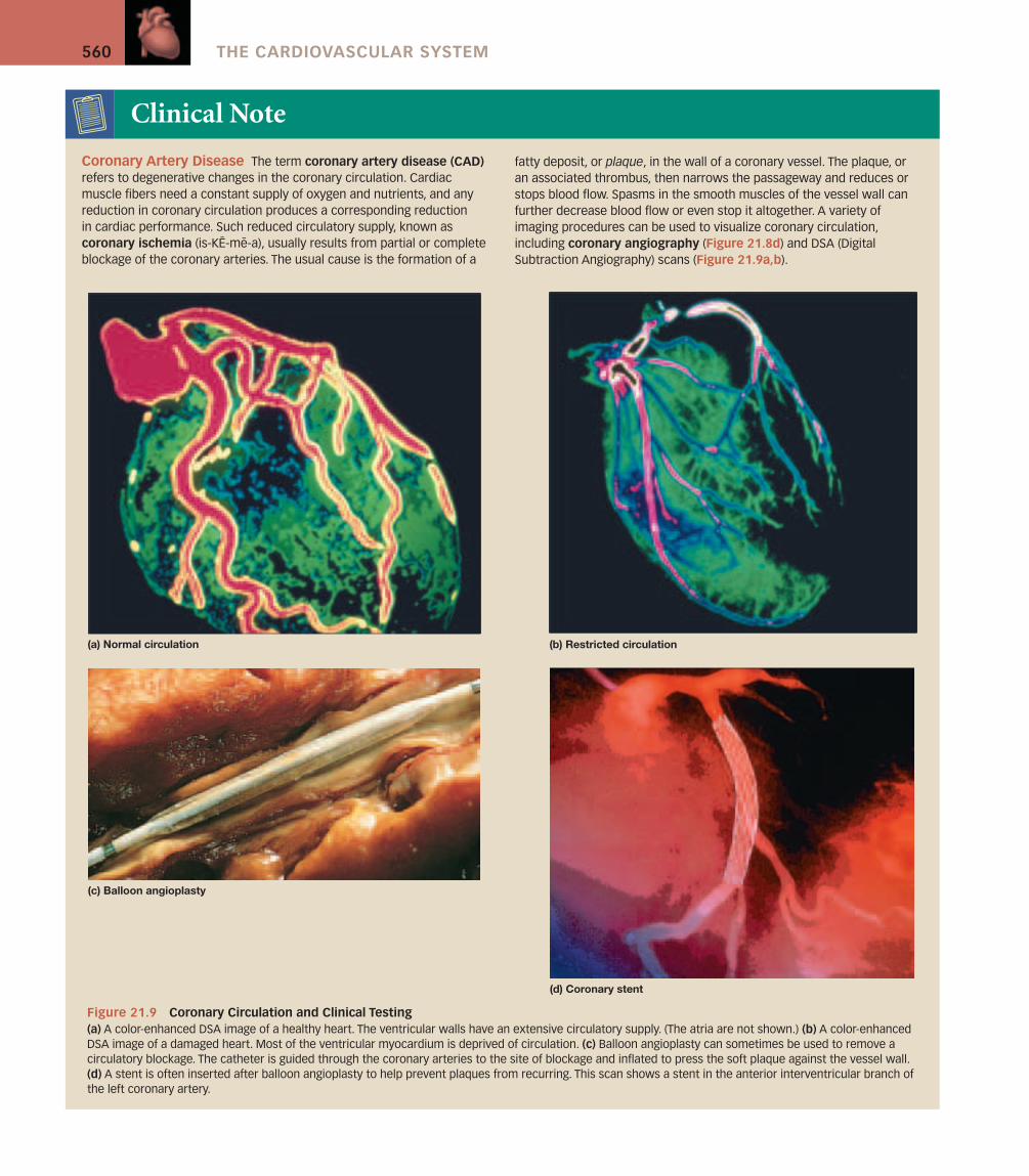
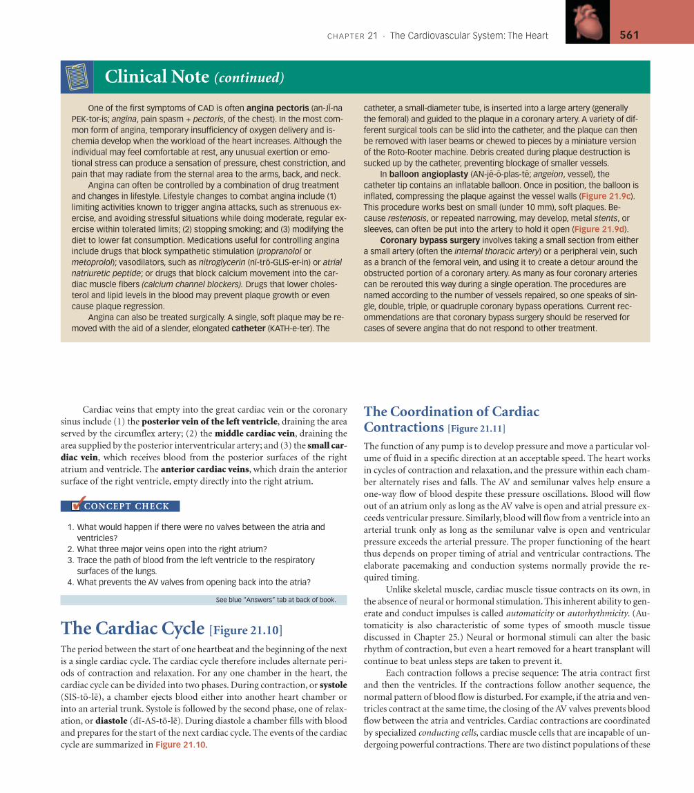
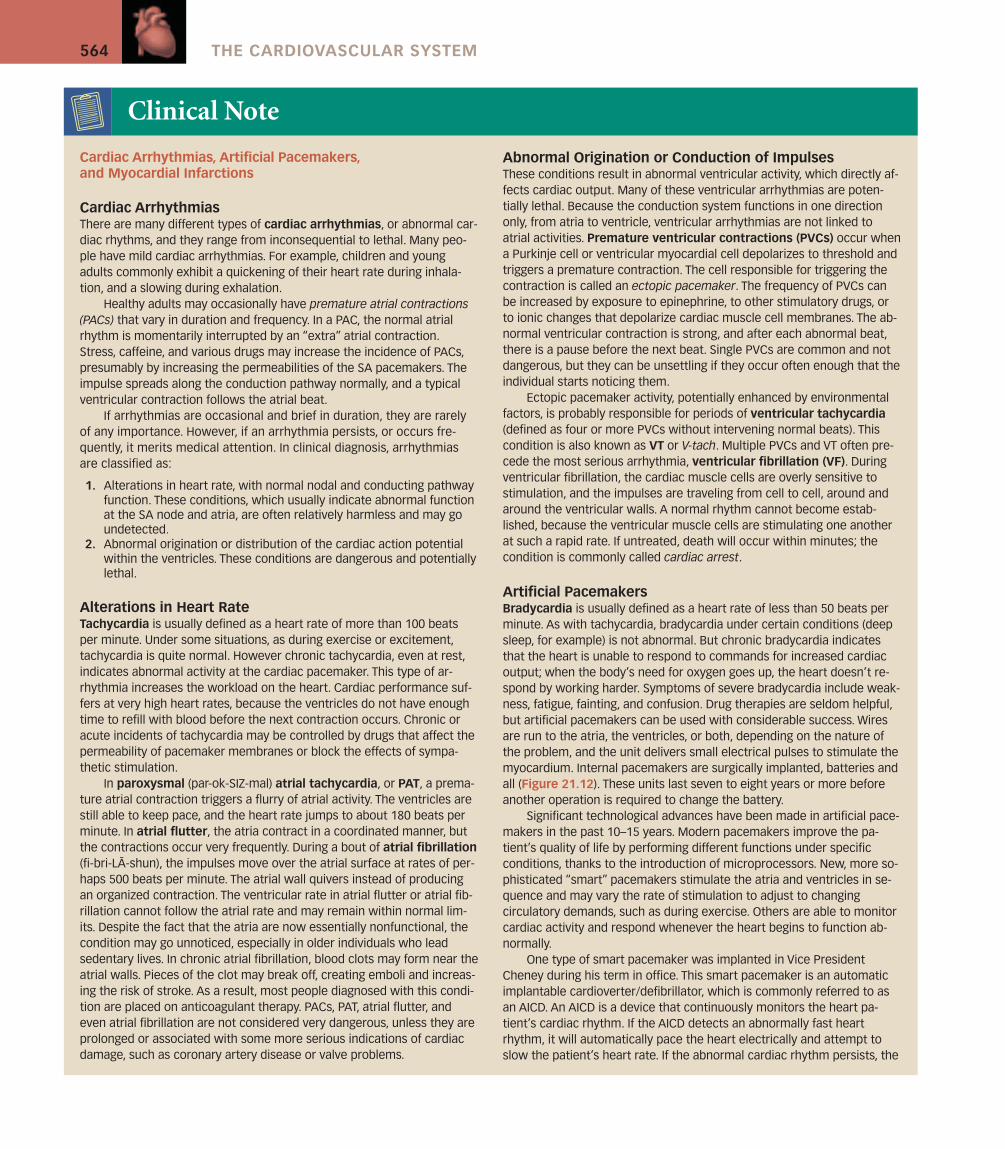
Clinical Note
Coronary Artery Disease The term coronary artery disease (CAD)refers to degenerative changes in the coronary circulation. Cardiacmuscle fibers need a constant supply of oxygen and nutrients, and anyreduction in coronary circulation produces a corresponding reduction in cardiac performance. Such reduced circulatory supply, known ascoronary ischemia (is-KE-me-a), usually results from partial or completeblockage of the coronary arteries. The usual cause is the formation of a
(d) Coronary stent
(a) Normal circulation (b) Restricted circulation
(c) Balloon angioplasty
Figure 21.9 Coronary Circulation and Clinical Testing(a) A color-enhanced DSA image of a healthy heart. The ventricular walls have an extensive circulatory supply. (The atria are not shown.) (b) A color-enhancedDSA image of a damaged heart. Most of the ventricular myocardium is deprived of circulation. (c) Balloon angioplasty can sometimes be used to remove acirculatory blockage. The catheter is guided through the coronary arteries to the site of blockage and inflated to press the soft plaque against the vessel wall.(d) A stent is often inserted after balloon angioplasty to help prevent plaques from recurring. This scan shows a stent in the anterior interventricular branch ofthe left coronary artery.
fatty deposit, or plaque, in the wall of a coronary vessel. The plaque, oran associated thrombus, then narrows the passageway and reduces orstops blood flow. Spasms in the smooth muscles of the vessel wall canfurther decrease blood flow or even stop it altogether. A variety ofimaging procedures can be used to visualize coronary circulation,including coronary angiography (Figure 21.8d) and DSA (DigitalSubtraction Angiography) scans (Figure 21.9a,b).
C H A P T E R 21 . The Cardiovascular System: The Heart 561
Cardiac veins that empty into the great cardiac vein or the coronarysinus include (1) the posterior vein of the left ventricle, draining the areaserved by the circumflex artery; (2) the middle cardiac vein, draining thearea supplied by the posterior interventricular artery; and (3) the small car-diac vein, which receives blood from the posterior surfaces of the rightatrium and ventricle. The anterior cardiac veins, which drain the anteriorsurface of the right ventricle, empty directly into the right atrium.
1. What would happen if there were no valves between the atria andventricles?
2. What three major veins open into the right atrium?3. Trace the path of blood from the left ventricle to the respiratory
surfaces of the lungs.4. What prevents the AV valves from opening back into the atria?
See blue “Answers” tab at back of book.
The Cardiac Cycle [Figure 21.10]
The period between the start of one heartbeat and the beginning of the nextis a single cardiac cycle. The cardiac cycle therefore includes alternate peri-ods of contraction and relaxation. For any one chamber in the heart, thecardiac cycle can be divided into two phases. During contraction, or systole(SIS-to-le), a chamber ejects blood either into another heart chamber orinto an arterial trunk. Systole is followed by the second phase, one of relax-ation, or diastole (di-AS-to-le). During diastole a chamber fills with bloodand prepares for the start of the next cardiac cycle. The events of the cardiaccycle are summarized in Figure 21.10.
The Coordination of CardiacContractions [Figure 21.11]
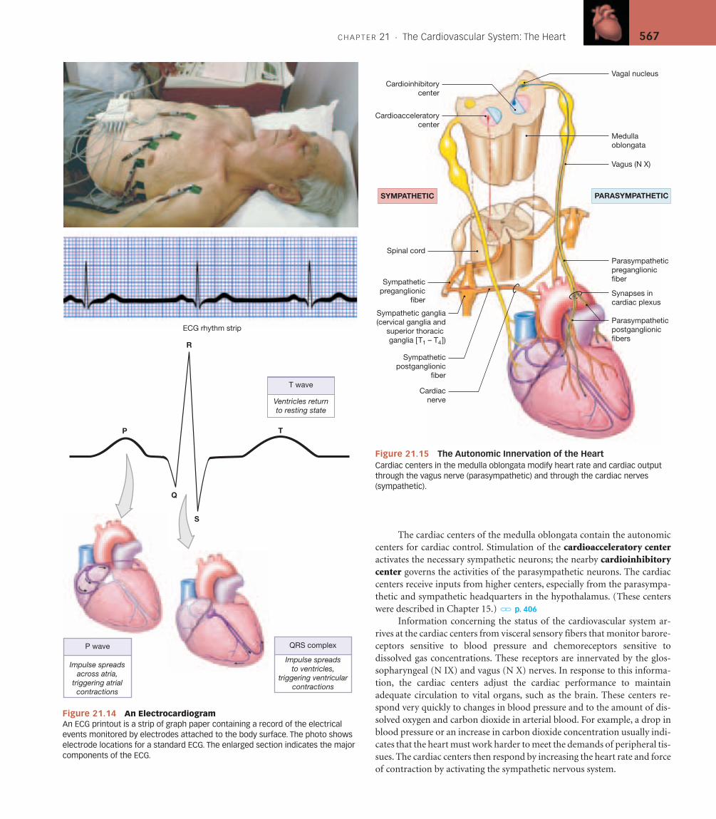
The function of any pump is to develop pressure and move a particular vol-ume of fluid in a specific direction at an acceptable speed. The heart worksin cycles of contraction and relaxation, and the pressure within each cham-ber alternately rises and falls. The AV and semilunar valves help ensure aone-way flow of blood despite these pressure oscillations. Blood will flowout of an atrium only as long as the AV valve is open and atrial pressure ex-ceeds ventricular pressure. Similarly, blood will flow from a ventricle into anarterial trunk only as long as the semilunar valve is open and ventricularpressure exceeds the arterial pressure. The proper functioning of the heartthus depends on proper timing of atrial and ventricular contractions. Theelaborate pacemaking and conduction systems normally provide the re-quired timing.
Unlike skeletal muscle, cardiac muscle tissue contracts on its own, inthe absence of neural or hormonal stimulation. This inherent ability to gen-erate and conduct impulses is called automaticity or autorhythmicity. (Au-tomaticity is also characteristic of some types of smooth muscle tissuediscussed in Chapter 25.) Neural or hormonal stimuli can alter the basicrhythm of contraction, but even a heart removed for a heart transplant willcontinue to beat unless steps are taken to prevent it.
Each contraction follows a precise sequence: The atria contract firstand then the ventricles. If the contractions follow another sequence, thenormal pattern of blood flow is disturbed. For example, if the atria and ven-tricles contract at the same time, the closing of the AV valves prevents bloodflow between the atria and ventricles. Cardiac contractions are coordinatedby specialized conducting cells, cardiac muscle cells that are incapable of un-dergoing powerful contractions. There are two distinct populations of these
One of the first symptoms of CAD is often angina pectoris (an-JI-naPEK-tor-is; angina, pain spasm + pectoris, of the chest). In the most com-mon form of angina, temporary insufficiency of oxygen delivery and is-chemia develop when the workload of the heart increases. Although theindividual may feel comfortable at rest, any unusual exertion or emo-tional stress can produce a sensation of pressure, chest constriction, andpain that may radiate from the sternal area to the arms, back, and neck.
Angina can often be controlled by a combination of drug treatmentand changes in lifestyle. Lifestyle changes to combat angina include (1)limiting activities known to trigger angina attacks, such as strenuous ex-ercise, and avoiding stressful situations while doing moderate, regular ex-ercise within tolerated limits; (2) stopping smoking; and (3) modifying thediet to lower fat consumption. Medications useful for controlling anginainclude drugs that block sympathetic stimulation (propranolol ormetoprolol); vasodilators, such as nitroglycerin (ni-tro-GLIS-er-in) or atrialnatriuretic peptide; or drugs that block calcium movement into the car-diac muscle fibers (calcium channel blockers). Drugs that lower choles-terol and lipid levels in the blood may prevent plaque growth or evencause plaque regression.
Angina can also be treated surgically. A single, soft plaque may be re-moved with the aid of a slender, elongated catheter (KATH-e-ter). The
catheter, a small-diameter tube, is inserted into a large artery (generallythe femoral) and guided to the plaque in a coronary artery. A variety of dif-ferent surgical tools can be slid into the catheter, and the plaque can thenbe removed with laser beams or chewed to pieces by a miniature versionof the Roto-Rooter machine. Debris created during plaque destruction issucked up by the catheter, preventing blockage of smaller vessels.
In balloon angioplasty (AN-je-o-plas-te; angeion, vessel), thecatheter tip contains an inflatable balloon. Once in position, the balloon isinflated, compressing the plaque against the vessel walls (Figure 21.9c).This procedure works best on small (under 10 mm), soft plaques. Be-cause restenosis, or repeated narrowing, may develop, metal stents, orsleeves, can often be put into the artery to hold it open (Figure 21.9d).
Coronary bypass surgery involves taking a small section from eithera small artery (often the internal thoracic artery) or a peripheral vein, suchas a branch of the femoral vein, and using it to create a detour around theobstructed portion of a coronary artery. As many as four coronary arteriescan be rerouted this way during a single operation. The procedures arenamed according to the number of vessels repaired, so one speaks of sin-gle, double, triple, or quadruple coronary bypass operations. Current rec-ommendations are that coronary bypass surgery should be reserved forcases of severe angina that do not respond to other treatment.
Clinical Note (continued)
562 THE CARDIOVASCULAR SYSTEM
cells. Nodal cells are responsible for establishing the rate of cardiac contrac-tion, and conducting fibers distribute the contractile stimulus to the gen-eral myocardium (Figure 21.11).
The Sinoatrial and AtrioventricularNodes [Figure 21.11]
Nodal cells are unusual because their cell membranes spontaneously depo-larize to threshold. Nodal cells are electrically coupled to one another, toconducting fibers, and to normal cardiac muscle cells. As a result, when anaction potential appears in a nodal cell, it sweeps through the conductingsystem, reaching all of the cardiac muscle tissue and causing a contraction.In this way, nodal cells determine the heart rate.
Not all nodal cells depolarize at the same rate, and the normal rate ofcontraction is established by the nodal cells that reach threshold first; theimpulse they produce will bring all other nodal cells to threshold. Theserapidly depolarizing cells are called pacemaker cells. They are found in thesinoatrial (si-no-A-tre-al) node (SA node), or cardiac pacemaker. TheSA node is embedded in the posterior wall of the right atrium, near the en-trance of the superior vena cava (Figure 21.11). Isolated pacemaker cellsdepolarize rapidly and spontaneously, generating 80–100 action potentialsper minute.
Each time the SA node generates an impulse, it produces a heartbeat,so theoretically the resting heart rate would be 80–100 beats per minute
(bpm). However, any factor that changes either the resting potential or therate of spontaneous depolarization at the SA node will alter the heart rate.For example, nodal cell activity is affected by the activity of the autonomicnervous system. When acetylcholine (ACh) is released by parasympatheticneurons, it slows the rate of spontaneous depolarization and lowers theheart rate. In contrast, when norepinephrine (NE) is released by sympa-thetic neurons, the rate of depolarization increases, and the heart rate accel-erates. Under normal resting conditions, parasympathetic activity reducesthe heart rate from the inherent nodal rate of 80–100 impulses per minuteto a more leisurely 70–80 beats per minute.
A number of clinical problems are the result of abnormal pacemakerfunction. Bradycardia (brad-e-KAR-de-a; bradys, slow) is the term used toindicate a heart rate that is slower than normal, whereas a faster-than-normal heart rate is termed tachycardia (tak-e-KAR-de-a; tachys, swift).Both terms are relative, and in clinical practice the definition varies depend-ing on the normal resting heart rate and conditioning of the individual.
The Conducting System of the Heart [Figure 21.11]The cells of the SA node are electrically connected to those of the largeratrioventricular (a-tre-o-ven-TRIK-u-lar) node (AV node) through con-ducting fibers in the atrial walls (Figure 21.11). As the signal for contrac-tion passes from the SA node to the AV node via the internodal pathways,the conducting fibers also pass the contractile stimulus to cardiac musclecells of both atria. The action potential then spreads across the atrial sur-
(f) Ventricular diastole—late: All chambers are relaxed. Ventricles fill passively.
(a) Atrial systole begins: Atrial contraction forces a small amount of additional blood into relaxed ventricles.
(c) Ventricular systole— first phase: Ventricular contraction pushes AV valves closed but does not create enough pressure to open semilunar valves.
(b) Atrial systole ends, atrial diastole begins
(d) Ventricular systole— second phase: As ventricular pressure rises and exceeds pressure in the arteries, the semilunar valves open and blood is ejected.
(e) Ventricular diastole—early: As ventricles relax, pressure in ventricles drops; blood flows back against cusps of semilunar valves and forces them closed. Blood flows into the relaxed atria.
Vent
ricul
ar d
iast
ole
Ven
tric
ular
sys
tole
Atrial systole
Atrial diastole
START
Cardiaccycle
800msec
0msec
100msec
370msec
Figure 21.10 The Cardiac CycleBlack arrows indicate movement of blood or valves;green arrows indicate myocardial contraction.
C H A P T E R 21 . The Cardiovascular System: The Heart 563
Internodalpathways
Purkinje fibers
Left bundle branch
AV bundle
Atrioventricular(AV) node
Right bundle branch
(a) Nodes and conducting fibers
Sinoatrial(SA) node
Moderator band
Stimulus spreads across theatrial surfaces and reachesthe AV node.
Elapsed time = 50 msec
SA node activity andatrial activation begin.
Time = 0
There is a 100-msec delayat the AV node. Atrialcontraction begins.
Elapsed time = 150 msec
The impulse travels along theinterventricular septum withinthe AV bundle and the bundlebranches to the Purkinje fibersand, via the moderator band, to the papillary muscles of theright ventricle.
Elapsed time = 175 msec
The impulse is distributed byPurkinje fibers and relayedthroughout the ventricularmyocardium. Atrial contractionis completed, and ventricularcontraction begins.
Elapsed time = 225 msec
SA node
AV node
Bundlebranches
Moderatorband
Purkinje fibers
AVbundle
(b)
STEP 5
STEP 4
STEP 3
STEP 2
STEP 1
Figure 21.11 The Conducting System of the Heart(a) The stimulus for contraction is generated by pacemaker cells at the SA node.From there, impulses follow three different paths through the atrial walls toreach the AV node. After a brief delay, the impulses are conducted to the bundleof His (AV bundle), and then on to the bundle branches, the Purkinje cells, andthe ventricular myocardial cells. (b) The movement of the contractile stimulusthrough the heart is shown in STEPS 1–5.
faces through cell-to-cell contact. The stimulus affects only the atria, be-cause the fibrous skeleton electrically isolates the atrial myocardium fromthe ventricular myocardium.
The AV node sits within the floor of the right atrium near the open-ing of the coronary sinus. Due to differences in the shape of the nodal cells,the impulse slows as it passes through the AV node. From there, the impulsetravels to the AV bundle, also known as the bundle of His (HISS). This rathermassive bundle of conducting fibers travels along the interventricular sep-tum a short distance before dividing into a right bundle branch and a leftbundle branch that extend toward the apex and then radiate across the in-ner surfaces of both ventricles. At this point, Purkinje (pur-KIN-je) cells(Purkinje fibers) convey the impulses very rapidly to the contractile cells ofthe ventricular myocardium. The conducting fibers of the moderator bandrelay the stimulus to the papillary muscles, which tense the chordaetendineae before the ventricles contract.
The stimulus for a contraction is generated at the SA node, and theanatomical relationships among the contracting cells, the nodal cells, andthe conducting fibers distribute the impulse so that (1) the atria contract to-gether, before the ventricles, and (2) the ventricles contract together in awave that begins at the apex and spreads toward the base. When the ventri-cles contract in this way, blood is pushed toward the base of the heart andout into the aortic and pulmonary trunks.
Embryology SummaryFor a summary of the development of the cardiovascular system, seeChapter 28 (Embryology and Human Development).
Cardiac ArrhythmiasThere are many different types of cardiac arrhythmias, or abnormal car-diac rhythms, and they range from inconsequential to lethal. Many peo-ple have mild cardiac arrhythmias. For example, children and youngadults commonly exhibit a quickening of their heart rate during inhala-tion, and a slowing during exhalation.
Healthy adults may occasionally have premature atrial contractions(PACs) that vary in duration and frequency. In a PAC, the normal atrialrhythm is momentarily interrupted by an “extra” atrial contraction.Stress, caffeine, and various drugs may increase the incidence of PACs,presumably by increasing the permeabilities of the SA pacemakers. Theimpulse spreads along the conduction pathway normally, and a typicalventricular contraction follows the atrial beat.
If arrhythmias are occasional and brief in duration, they are rarelyof any importance. However, if an arrhythmia persists, or occurs fre-quently, it merits medical attention. In clinical diagnosis, arrhythmiasare classified as:
1. Alterations in heart rate, with normal nodal and conducting pathwayfunction. These conditions, which usually indicate abnormal functionat the SA node and atria, are often relatively harmless and may goundetected.
2. Abnormal origination or distribution of the cardiac action potentialwithin the ventricles. These conditions are dangerous and potentiallylethal.
Alterations in Heart RateTachycardia is usually defined as a heart rate of more than 100 beatsper minute. Under some situations, as during exercise or excitement,tachycardia is quite normal. However chronic tachycardia, even at rest,indicates abnormal activity at the cardiac pacemaker. This type of ar-rhythmia increases the workload on the heart. Cardiac performance suf-fers at very high heart rates, because the ventricles do not have enoughtime to refill with blood before the next contraction occurs. Chronic oracute incidents of tachycardia may be controlled by drugs that affect thepermeability of pacemaker membranes or block the effects of sympa-thetic stimulation.
In paroxysmal (par-ok-SIZ-mal) atrial tachycardia, or PAT, a prema-ture atrial contraction triggers a flurry of atrial activity. The ventricles arestill able to keep pace, and the heart rate jumps to about 180 beats perminute. In atrial flutter, the atria contract in a coordinated manner, butthe contractions occur very frequently. During a bout of atrial fibrillation(fi-bri-LA-shun), the impulses move over the atrial surface at rates of per-haps 500 beats per minute. The atrial wall quivers instead of producingan organized contraction. The ventricular rate in atrial flutter or atrial fib-rillation cannot follow the atrial rate and may remain within normal lim-its. Despite the fact that the atria are now essentially nonfunctional, thecondition may go unnoticed, especially in older individuals who leadsedentary lives. In chronic atrial fibrillation, blood clots may form near theatrial walls. Pieces of the clot may break off, creating emboli and increas-ing the risk of stroke. As a result, most people diagnosed with this condi-tion are placed on anticoagulant therapy. PACs, PAT, atrial flutter, andeven atrial fibrillation are not considered very dangerous, unless they areprolonged or associated with some more serious indications of cardiacdamage, such as coronary artery disease or valve problems.
Abnormal Origination or Conduction of ImpulsesThese conditions result in abnormal ventricular activity, which directly af-fects cardiac output. Many of these ventricular arrhythmias are poten-tially lethal. Because the conduction system functions in one directiononly, from atria to ventricle, ventricular arrhythmias are not linked toatrial activities. Premature ventricular contractions (PVCs) occur whena Purkinje cell or ventricular myocardial cell depolarizes to threshold andtriggers a premature contraction. The cell responsible for triggering thecontraction is called an ectopic pacemaker. The frequency of PVCs canbe increased by exposure to epinephrine, to other stimulatory drugs, orto ionic changes that depolarize cardiac muscle cell membranes. The ab-normal ventricular contraction is strong, and after each abnormal beat,there is a pause before the next beat. Single PVCs are common and notdangerous, but they can be unsettling if they occur often enough that theindividual starts noticing them.
Ectopic pacemaker activity, potentially enhanced by environmentalfactors, is probably responsible for periods of ventricular tachycardia(defined as four or more PVCs without intervening normal beats). Thiscondition is also known as VT or V-tach. Multiple PVCs and VT often pre-cede the most serious arrhythmia, ventricular fibrillation (VF). Duringventricular fibrillation, the cardiac muscle cells are overly sensitive tostimulation, and the impulses are traveling from cell to cell, around andaround the ventricular walls. A normal rhythm cannot become estab-lished, because the ventricular muscle cells are stimulating one anotherat such a rapid rate. If untreated, death will occur within minutes; thecondition is commonly called cardiac arrest.
Artificial PacemakersBradycardia is usually defined as a heart rate of less than 50 beats perminute. As with tachycardia, bradycardia under certain conditions (deepsleep, for example) is not abnormal. But chronic bradycardia indicatesthat the heart is unable to respond to commands for increased cardiacoutput; when the body’s need for oxygen goes up, the heart doesn’t re-spond by working harder. Symptoms of severe bradycardia include weak-ness, fatigue, fainting, and confusion. Drug therapies are seldom helpful,but artificial pacemakers can be used with considerable success. Wiresare run to the atria, the ventricles, or both, depending on the nature ofthe problem, and the unit delivers small electrical pulses to stimulate themyocardium. Internal pacemakers are surgically implanted, batteries andall (Figure 21.12). These units last seven to eight years or more beforeanother operation is required to change the battery.
Significant technological advances have been made in artificial pace-makers in the past 10–15 years. Modern pacemakers improve the pa-tient’s quality of life by performing different functions under specificconditions, thanks to the introduction of microprocessors. New, more so-phisticated “smart” pacemakers stimulate the atria and ventricles in se-quence and may vary the rate of stimulation to adjust to changingcirculatory demands, such as during exercise. Others are able to monitorcardiac activity and respond whenever the heart begins to function ab-normally.
One type of smart pacemaker was implanted in Vice President Cheney during his term in office. This smart pacemaker is an automaticimplantable cardioverter/defibrillator, which is commonly referred to asan AICD. An AICD is a device that continuously monitors the heart pa-tient’s cardiac rhythm. If the AICD detects an abnormally fast heartrhythm, it will automatically pace the heart electrically and attempt toslow the patient’s heart rate. If the abnormal cardiac rhythm persists, the
C H A P T E R 21 . The Cardiovascular System: The Heart 565
AICD will deliver a small electrical shock to the heart in an attempt to re-store normal heart rhythm. The patient rarely feels the AICD rapidly pacingthe heart in an attempt to return the cardiac rhythm to normal. However,if the electrical shock is used, it is felt as a strong jolt in the chest. The de-vice is normally used for the instantaneous treatment of immediately life-
threatening heart rhythms (i.e., ventricular tachycardia and ventricular fib-rillation) that can’t wait for treatment until an ambulance arrives.
An external defibrillator has two electrodes that are placed in con-tact with the chest, and a powerful electrical shock is administered. Theelectrical stimulus depolarizes the entire myocardium simultaneously.With luck, after repolarization, the SA node will be the first area of theheart to reach threshold. Thus, the primary goal of defibrillation is not justto stop the fibrillation, but to give the ventricles a chance to respond tonormal SA commands.
Early defibrillation can result in dramatic recovery of an unconsciouscardiac-arrest victim. Automatic external defibrillators (AEDs) are easilyused, portable machines that can detect lethal ventricular rhythms inpeople who have collapsed and administer a defibrillating shock. Thesedevices are increasingly being placed on planes, in airports, and in otherpublic areas.
Myocardial InfarctionIn a myocardial (mi-o-KAR-de-al) infarction (MI), or heart attack, thecoronary circulation becomes blocked and the cardiac muscle cells diefrom lack of oxygen. The affected tissue then degenerates, creating anonfunctional area known as an infarct. Heart attacks most often resultfrom severe coronary artery disease. The consequences depend on thesite and nature of the circulatory blockage. If it occurs near the base ofone of the coronary arteries, the damage will be widespread and theheart will probably stop beating. If the blockage involves one of thesmaller arterial branches, the individual may survive the immediate crisis,but there are many potential complications, all unpleasant. As scar tissueforms in the damaged area, the heartbeat may become irregular and less
Clinical Note (continued)
Figure 21.12 An artificial pacemaker.
(a) (b)
(c) (d)
Figure 21.13 Monitoring theHeart(a) A coronary angiogram. (b) Anechocardium (left) with interpretivedrawing (right). (c) A three-dimensionalCT scan of an oblique section and (d) ofa posterior-superior view of the heartand great vessels.
566 THE CARDIOVASCULAR SYSTEM
1. If the cells of the SA node were not functioning, what effect would thishave on heart rate?
2. If norepinephrine is released at the heart, what is the effect on heart rate?3. How do nodal cells coordinate cardiac muscle contractions?
See blue “Answers” tab at back of book.
The Electrocardiogram (ECG) [Figure 21.14]The electrical events associated with the depolarization and repolarizationof the heart are powerful enough to be detected by electrodes placed on thebody surface. A recording of these electrical activities constitutes an elec-trocardiogram (e-lek-tro-KAR-de-o-gram), also called an ECG or EKG.During each cardiac cycle, a wave of depolarization radiates through theatria, reaches the AV node, travels down the interventricular septum to theapex, turns, and spreads through the ventricular myocardium toward thebase (Figure 21.14). This electrical activity can be monitored from the bodysurface. By comparing the information obtained from electrodes placed atdifferent locations, one can monitor the performance of specific nodal, con-ducting, and contractile components. For example, when a portion of theheart has been damaged, as after an MI, these cardiac muscle cells can nolonger conduct action potentials, so an ECG will reveal an abnormal patternof electrical conduction.
The appearance of the ECG tracing varies, depending on the place-ment of the monitoring electrodes, or leads. Figure 21.14 shows the impor-tant features of a representative electrocardiogram. The P wave accompaniesthe depolarization of the atria. The QRS complex appears as the ventriclesdepolarize. This electrical signal is relatively strong because the mass of theventricular muscle is much larger than that of the atria. The smaller T waveindicates ventricular repolarization. You do not see a deflection correspond-ing to atrial repolarization because it occurs while the ventricles are depolar-izing, and the electrical events are masked by the QRS complex.
Analysis of an ECG usually focuses on the size and duration of the Pwave, the QRS complex, and the ST segment (between the QRS and Twaves). For example, a smaller-than-normal electrical signal may mean thatthe mass of the heart muscle has decreased; in contrast, excessively large sig-nals may mean that the heart muscle has become enlarged. Changes in thesize and shape of the T wave may indicate a condition that slows ventricu-lar repolarization.
Despite the variety of sophisticated equipment available to assess orvisualize cardiac function, in the vast majority of cases, the electrocardio-gram provides the first and most important diagnostic information. ECGanalysis is especially useful in detecting and diagnosing cardiac arrhyth-mias (a-RITH-me-az), abnormal patterns of cardiac activity. Clinicalproblems appear when the arrhythmias reduce the pumping efficiency ofthe heart. Serious arrhythmias may indicate damage to the myocardialmusculature, injuries to the pacemakers or conduction pathways, or otherfactors.
Autonomic Control of Heart Rate [Figure 21.15]
The basic heart rate is established by the pacemaker cells of the SA node, butthis intrinsic rate can be modified by the autonomic nervous system (ANS).The sympathetic and parasympathetic divisions of the ANS provide inner-vation to the heart through the cardiac plexus (anatomical details were pre-sented in Chapter 17).l p. 464 Both ANS divisions innervate the SA andAV nodes as well as the atrial and ventricular cardiac muscle cells andsmooth muscle in the walls of the cardiac blood vessels (Figure 21.15).
The effects of NE and ACh on nodal tissues were detailed earlier inthis chapter, and may be summarized as follows:
■ NE release produces an increase in both heart rate and force of con-tractions through the stimulation of beta receptors on nodal cells andcontractile cells;
■ ACh release produces a decrease in both heart rate and force of con-tractions through the stimulation of muscarinic receptors of nodalcells and contractile cells.
effective as a pump, and other vessels can become constricted, creatingadditional cardiovascular problems such as angina.
Myocardial infarctions are most often associated with fixed block-ages, such as those seen in CAD. When the crisis develops because ofthrombus (stationary clot) formation at an area of plaque, the condition iscalled coronary thrombosis. A vessel already narrowed by plaque for-mation may also become blocked by a sudden spasm in the smoothmuscles of the vascular wall. The individual then experiences intensepain, similar to that of an angina attack but persisting even at rest.
Roughly 25 percent of MI patients die before obtaining medical as-sistance, and 65 percent of MI deaths among those under age 50 occurwithin an hour after the initial infarct. The goals of treatment are to limitthe size of the infarct and prevent additional complications by prevent-ing irregular contractions, improving circulation with vasodilators, pro-viding additional oxygen, reducing the cardiac workload, and, if possible,eliminating the cause of the circulatory blockage. Anticoagulants mayhelp prevent the formation of additional thrombi. Chewing an aspirinearly in the course of an MI is helpful, and clot-dissolving enzymes may
reduce the extent of the damage if they are administered within 6 hoursafter the MI has occurred.
There are roughly 1.3 million MIs in the United States each year,and half of the victims die within a year of the incident. A number offactors have been identified that increase the risk of a heart attack.They include smoking, high blood pressure, high blood cholesterol lev-els, diabetes, and obesity. There are also hereditary factors that maypredispose an individual to coronary artery disease. The presence oftwo risk factors more than doubles the risk, so eliminating as many riskfactors as possible will improve one’s chances of preventing or surviv-ing a heart attack. For example, changes in eating habits to limit dietarycholesterol, exercise to lower weight, and seeking treatment for highblood pressure are relatively easy steps in the right direction, and thebenefits are considerable.
Many techniques can be used to examine the structure and perfor-mance of the heart (Figure 21.13). No single diagnostic procedure pro-vides the complete picture, so the test used will vary with thesuspected nature of the problem.
Clinical Note (continued)
C H A P T E R 21 . The Cardiovascular System: The Heart 567
The cardiac centers of the medulla oblongata contain the autonomiccenters for cardiac control. Stimulation of the cardioacceleratory centeractivates the necessary sympathetic neurons; the nearby cardioinhibitorycenter governs the activities of the parasympathetic neurons. The cardiaccenters receive inputs from higher centers, especially from the parasympa-thetic and sympathetic headquarters in the hypothalamus. (These centerswere described in Chapter 15.) l p. 406
Information concerning the status of the cardiovascular system ar-rives at the cardiac centers from visceral sensory fibers that monitor barore-ceptors sensitive to blood pressure and chemoreceptors sensitive todissolved gas concentrations. These receptors are innervated by the glos-sopharyngeal (N IX) and vagus (N X) nerves. In response to this informa-tion, the cardiac centers adjust the cardiac performance to maintainadequate circulation to vital organs, such as the brain. These centers re-spond very quickly to changes in blood pressure and to the amount of dis-solved oxygen and carbon dioxide in arterial blood. For example, a drop inblood pressure or an increase in carbon dioxide concentration usually indi-cates that the heart must work harder to meet the demands of peripheral tis-sues. The cardiac centers then respond by increasing the heart rate and forceof contraction by activating the sympathetic nervous system.
Vagus (N X)
Medullaoblongata
Vagal nucleusCardioinhibitory
center
Cardioacceleratorycenter
SYMPATHETIC
Sympatheticpreganglionic
fiber
Spinal cord
Sympatheticpostganglionic
fiber
Cardiacnerve
Synapses incardiac plexus
Parasympatheticpreganglionicfiber
PARASYMPATHETIC
Parasympatheticpostganglionicfibers
Sympathetic ganglia(cervical ganglia and
superior thoracic ganglia [T1 – T4])
Figure 21.15 The Autonomic Innervation of the HeartCardiac centers in the medulla oblongata modify heart rate and cardiac outputthrough the vagus nerve (parasympathetic) and through the cardiac nerves(sympathetic).
P
Q
R
S
T
T wave
Ventricles returnto resting state
QRS complex
Impulse spreadsto ventricles,
triggering ventricularcontractions
P wave
Impulse spreadsacross atria,
triggering atrialcontractions
ECG rhythm strip
Figure 21.14 An ElectrocardiogramAn ECG printout is a strip of graph paper containing a record of the electricalevents monitored by electrodes attached to the body surface. The photo showselectrode locations for a standard ECG. The enlarged section indicates the majorcomponents of the ECG.
568 THE CARDIOVASCULAR SYSTEM
1. If the pressure in the ventricle remained equal to that in the atrium,what would happen to the blood flow?
2. The sympathetic and parasympathetic branches of the AutonomicNervous System have different effects on nodal tissues within theheart. What are these effects?
See blue “Answers” tab at back of book.
C L I N I C A L T E R M S
angina pectoris (an-JI-na PEK-tor-is): Acondition in which exertion or stress can producesevere chest pain, resulting from temporarycirculatory insufficiency and ischemia when theheart’s workload increases.
bradycardia (bra-de-KAR-de-a): A heart rate thatis slower than normal.
cardiac arrhythmias (a-RITH-me-az): Abnormalpatterns of cardiac contraction.
cardiac tamponade: A condition resulting frompericardial irritation and inflammation, in whichfluid collects in the pericardial sac and restrictscardiac output.
cardiomyopathies (kar-de-o-mi-OP-a-thez): Agroup of diseases characterized by the progressive,irreversible degeneration of the myocardium.
carditis (kar-DI-tis): A general term indicatinginflammation of the heart.
coronary artery disease (CAD): Degenerativechanges in the coronary circulation.
coronary thrombosis: A blockage due to theformation of a clot (thrombus) at a plaque in acoronary artery.
heart failure: A condition in which the heartweakens and peripheral tissues suffer from oxygenand nutrient deprivation.
heart murmur: A rushing, gurgling sound causedby blood regurgitation back through faulty heartvalves.
mitral valve prolapse: A condition in which themitral valve cusps do not close properly because of
abnormally long (or short) chordae tendineae ormalfunctioning papillary muscles.
myocardial (mi-o-KAR-de-al) infarction (MI): Acondition in which the coronary circulationbecomes blocked and the cardiac muscle cells diefrom oxygen starvation; also called a heart attack.
rheumatic heart disease (RHD): A disorder inwhich the heart valves become thickened andstiffen into a partially closed position, affecting theefficiency of the pumping action of the heart.
tachycardia (tak-e-KAR-de-a): A heart rate that isfaster than normal.
valvular stenosis (ste-NO-sis): A condition inwhich the opening between the heart valves isnarrower than normal.
S T U D Y O U T L I N E
Introduction 5481. All of the tissues and fluids in the body rely on the cardiovascular system to
maintain homeostasis. The proper functioning of the cardiovascular systemdepends on the activity of the heart, which can vary its pumping capacitydepending on the needs of the peripheral tissues.
An Overview of the Cardiovascular System 5481. The cardiovascular system can be subdivided into two closed circuits that
occur in series. Each circuit functions individually in series, while the twocircuits together function in parallel. The pulmonary circuit carriesoxygen-poor blood from the heart to the lungs and back, and the systemiccircuit transports oxygen-rich blood from the heart to the rest of the bodyand back. Arteries carry blood away from the heart; veins return blood tothe heart. Capillaries are tiny vessels between the smallest arteries andveins. (see Figure 21.1)
2. The heart contains four chambers: the right atrium and ventricle, and theleft atrium and ventricle. The atria collect blood returning to the heart, andthe ventricles discharge blood into vessels to leave the heart.
The Pericardium 5481. The heart is surrounded by the pericardial cavity, which is lined by the
pericardium and contains a small amount of lubricating fluid, called thepericardial fluid. The visceral pericardium (epicardium) covers the heart’souter surface, and the parietal pericardium lines the inner surface of thepericardial sac that surrounds the heart. The heart lies in the anterior por-tion of the mediastinum. (see Figure 21.2)
Structure of the Heart Wall 5501. The heart wall contains three layers: the epicardium (the visceral pericardium),
the myocardium (the muscular wall of the heart), and the endocardium (theepithelium covering the inner surfaces of the heart). (see Figure 21.3)
Cardiac Muscle Tissue 550
2. The bulk of the heart consists of the muscular myocardium. Cardiac mus-cle cells (cardiocytes), which are smaller than skeletal muscle cells, are al-most totally dependent on aerobic respiration. (see Figure 21.3)
3. Cardiocytes are interconnected by intercalated discs, which both conveythe force of contraction from cell to cell and conduct action potentials. In-tercalated discs join cardiac muscle cells through desmosomes, myofibrils,and gap junctions. Because cardiac muscle cells are connected in this way,they function like a single, enormous cell. (see Figure 21.3d,e)
The Fibrous Skeleton 550
4. The internal connective tissue of the heart is called the fibrous skeleton.(see Figures 21.3b/21.7)
5. The fibrous skeleton of the heart functions to stabilize the heart’s contrac-tile cells and valves; support the muscle cells, blood vessels, and nerves;distribute the forces of contraction; add strength and elasticity; and phys-ically isolate the atria from the ventricles.
Orientation and Superficial Anatomy of the Heart 5521. The division of the heart into four chambers produces external landmarks
that are visible as grooves or sulci on the surface of the heart. The interatrialgroove separates the two atria, while the coronary sulcus separates the atriafrom the ventricles.
2. The auricle (atrial appendage) is an expandable extension of the atrium.The coronary sulcus is the deep groove between the atria and the ventricles.Other shallower depressions include the anterior interventricular sulcusand the posterior interventricular sulcus.
3. The great vessels are connected to the superior end of the heart at the base.The inferior, pointed tip of the heart is the apex. (see Figures 21.2b/21.4)
4. The heart sits at an angle to the longitudinal axis of the body and presentsthe following borders: superior, inferior, left, and right. (see Figure 21.4)
C H A P T E R 21 . The Cardiovascular System: The Heart 569
5. The heart has the following surfaces: The sternocostal surface is formed bythe anterior surfaces of the right atrium and ventricle; the diaphragmaticsurface is formed primarily by the posterior, inferior wall of the left ventri-cle. (see Figure 21.5)
Internal Anatomy and Organization of theHeart 5541. The atria are separated by the interatrial septum, and the ventricles are di-
vided by the interventricular septum. The openings between the atria andventricles contain folds of connective tissue covered by endocardium; thesevalves maintain a one-way flow of blood. (see Figure 21.6)
The Right Atrium 554
2. The right atrium receives blood from the systemic circuit through two greatveins, the superior vena cava and inferior vena cava. The atrial walls con-tain prominent muscular ridges, the pectinate muscles. The coronaryveins return blood to the coronary sinus, which opens into the right atrium.During embryonic development an opening called the foramen ovale pen-etrates the interatrial septum. This opening closes after birth, leaving a de-pression termed the fossa ovalis. (see Figure 21.6)
The Right Ventricle 554
3. Blood flows from the right atrium into the right ventricle through the rightatrioventricular (AV) valve, or tricuspid valve. (This valve consists of threecusps of fibrous tissue braced by the tendinous chordae tendineae that areconnected to papillary muscles.) (see Figure 21.6a,c)
4. Blood leaving the right ventricle enters the pulmonary trunk after passingthrough the pulmonary valve. The pulmonary trunk divides to form theleft and right pulmonary arteries. (see Figure 21.6)
The Left Atrium 556
5. The left atrium receives oxygenated blood from the left and right pul-monary veins; it has thicker walls than those of the right atrium. (seeFigure 21.6a,c)
6. Blood leaving the left atrium flows into the left ventricle through the leftatrioventricular (AV) valve (mitral or bicuspid valve).
The Left Ventricle 556
7. The left ventricle is the largest and thickest of the four chambers because itmust pump blood to the entire body. Blood leaving the left ventricle passesthrough the aortic valve and into the systemic circuit via the ascendingaorta. Blood passes from the ascending aorta through the aortic arch andinto the descending aorta. (see Figure 21.6a,c)
Structural Differences between the Left and Right Ventricles 556
8. The right ventricle has thin walls and develops low pressure when pumpinginto the pulmonary circuit to and from the adjacent lungs. Functionally, lowpressure is necessary because the pulmonary capillaries at the gas-exchangesurfaces of the lungs are very delicate. The left ventricle has a thick wall be-cause it pumps blood throughout the systemic circuit. Anatomical differ-ences between the left and right ventricles are shown in Figure 21.6.
The Structure and Function of Heart Valves 556
9. The AV valves have four components: (1) a ring of connective tissue at-tached to the fibrous skeleton of the heart, (2) cusps, (3) chordae tendineae,and (4) papillary muscles.
10. There are two semilunar valves, the aortic valve and the pulmonary valve,guarding the exits of the left and right ventricles. (see Figures 21.6 and 21.7)
11. Valves normally permit blood flow in only one direction, preventing theregurgitation (backflow) of blood.
Coronary Blood Vessels 558
12. The coronary circulation supplies blood to the muscles of the heart to meetthe high oxygen and nutrient demands of cardiac muscle cells. Thecoronary arteries originate at the base of the ascending aorta, and eachgives rise to two branches. The right coronary artery gives rise to both aright marginal branch and a posterior interventricular branch. The leftcoronary artery gives rise to both a circumflex branch and an anterior in-terventricular branch. Interconnections between arteries calledanastomoses ensure a constant blood supply.
13. The great and middle cardiac veins carry blood from the coronary capil-laries to the coronary sinus. (see Figure 21.8)
14. Other cardiac veins that empty into the great cardiac vein or the coronarysinus are the posterior vein of the left ventricle, draining the areas servedby the circumflex branch of the LCA; the middle cardiac vein, draining theareas supplied by the posterior interventricular branch of the LCA; and thesmall cardiac vein, draining blood from the posterior surfaces of the rightatrium and ventricle.
15. The anterior cardiac veins drain the anterior surface of the right ventricleand empty directly into the right atrium.
The Cardiac Cycle 5611. The cardiac cycle consists of periods of atrial and ventricular systole (contrac-
tion) and atrial and ventricular diastole (relaxation/filling). (see Figure 21.10)
The Coordination of Cardiac Contractions 561
2. Cardiac muscle tissue contracts on its own, without neural or hormonalstimulation. This is called automaticity or autorhythmicity.
3. Nodal cells establish the rate of cardiac contraction, and conductingfibers distribute the contractile stimulus to the general myocardium. (seeFigure 21.11)
The Sinoatrial and Atrioventricular Nodes 562
4. Nodal cells depolarize spontaneously and determine the heart rate.5. Pacemaker cells found in the sinoatrial (SA) node (cardiac pacemaker)
normally establish the rate of contraction. (see Figure 21.11)6. From the SA node, the stimulus travels over the internodal pathways to the
atrioventricular (AV) node, then to the AV bundle, which divides into aright and left bundle branch. From here Purkinje cells convey the impulsesto the ventricular myocardium. (see Figure 21.11)
The Electrocardiogram (ECG) 5661. A recording of electrical activities in the heart is an electrocardiogram (ECG
or EKG). Important landmarks of an ECG include the P wave (atrial depo-larization), QRS complex (ventricular depolarization) and T wave (ventric-ular repolarization). ECG analysis can detect cardiac arrhythmias, whichare abnormal patterns of cardiac activity (see Figure 21.14).
Autonomic Control of Heart Rate 567
2. The basic heart rate is established by the pacemaker cells, but it can be mod-ified by the ANS. Norepinephrine produces an increase in heart rate andforce of contraction, while acetylcholine produces a decrease in heart rateand contraction.
3. The cardioacceleratory center in the medulla oblongata activates sympa-thetic neurons; the cardioinhibitory center governs the activities of theparasympathetic neurons. The cardiac centers receive inputs from highercenters and from receptors monitoring blood pressure and the concentra-tions of dissolved gases in the blood. (see Figure 21.15)
570 THE CARDIOVASCULAR SYSTEM
Column A Column B___ 1. cardiocytes___ 2. bradycardia___ 3. diastole___ 4. coronary circulation___ 5. visceral pericardium___ 6. systole___ 7. myocardium___ 8. right pulmonary vein___ 9. superior vena cava___10. parietal pericardium
a. vein to the left atriumb. covers the outer surface of heartc. supplies blood to heart muscled. lines inner surface of pericardial sace. slow heart ratef. cardiac muscle cellsg. muscular wall of the hearth. relaxation phase of the cardiac cyclei. vein to the right atriumj. contraction phase of the cardiac cycle
C H A P T E R R E V I E W
For answers to the Concept Check and Chapter Review questions, go to the blue“Answers” tab at the back of the book.
Level 1 Reviewing Facts and TermsMatch each numbered item with the most closely related lettered item. Use letters for answers in the spaces provided.
11. The heart lies in the(a) pleural cavity (b) peritoneal cavity(c) abdominopelvic cavity (d) pericardial cavity
12. The atrioventricular valve that is located on the side of the heart that receives blood from the superior vena cava is the(a) mitral valve (b) bicuspid valve(c) tricuspid valve (d) aortic valve
13. The functions of the fibrous pericardium include(a) returning blood to the atria(b) pumping blood into circulation(c) anchoring the heart to surrounding structures(d) providing blood flow to the myocardium
14. All of the following are true of intercalated discs except(a) they provide additional strength from cells bound together by tight
junctions(b) they have a smooth junction between the sarcolemmae of apposed
muscle cells(c) they have the myofibrils of the interlocking muscle fibers anchored at
the membrane(d) the cardiac muscle fibers at the intercalated discs are connected by gap
junctions
15. The heart is innervated by(a) only parasympathetic nerves(b) only sympathetic nerves(c) both sympathetic and parasympathetic nerves(d) only splanchnic nerves
16. The pacemaker cells of the heart are located in(a) the SA node(b) the wall of the left ventricle(c) the Purkinje fibers(d) both the left and right ventricles
17. The muscle fibers of the atria are isolated physically from those of the ventricles(a) by the epicardium(b) by the fibrous skeleton of the heart(c) but not electrically, as they all contract at exactly the same time(d) by the coronary blood vessels
18. The two main branches of the right coronary artery are the(a) circumflex branch and the left marginal branch(b) anterior interventricular branch and the left anterior descending branch
(c) right marginal branch and the posterior interventricular branch(d) great and middle cardiac veins
19. Analysis of ECG data is useful in the detection and diagnosis of(a) hypertension(b) cardiac arrhythmias(c) cerebrovascular accidents(d) all of the above
20. The mitral or bicuspid valve is located(a) in the opening of the aorta(b) between the left atrium and left ventricle(c) between the right atrium and right ventricle(d) in the opening of the pulmonary trunk
Level 2 Reviewing Concepts1. If the sinoatrial node is damaged, what will happen to the heartbeat?
(a) it will be generated by the bundle branches, at a much lower rate(b) the heart will stop(c) the atrioventricular node will take over setting the pace, at a speed
somewhat slower than normal(d) the heartbeat will increase in rate, but not in forcefulness
2. If the papillary muscles fail to contract,(a) blood will not enter the atria(b) the ventricles will not pump blood(c) the AV valves will not close properly(d) the semilunar valves will not open
3. If there were damage to the sympathetic innervation to the heart, whatwould happen to the heart rate under the influence of the remaining autonomic nervous system stimulation?(a) it would increase(b) it would not change(c) it would decrease(d) it would first increase and then decrease
4. How is cardiac muscle similar to skeletal muscle?
5. Why do semilunar valves lack muscular braces like those found in AV valves?
6. Define a pacemaker cell, and list the group of cells that normally serve asthe heart’s pacemaker, as well as those other cells that have the potential toserve as a pacemaker.
7. What is the function of the pericardial fluid?
8. Which chamber of the heart has the thickest walls? Why are its walls so thick?
9. Why are nodal cells unique? What is their function?
10. What is the effect of NE release on cardiac function?
Level 3 Critical Thinking1. Harvey has a heart murmur in his left ventricle that produces a loud “gur-
gling” sound at the beginning of systole. What do you suspect to be thecause of this sound?
2. Lee is brought to the emergency room of a hospital suffering from a cardiacarrhythmia. In the emergency room he begins to exhibit tachycardia and asa result loses consciousness. His wife asks you why he lost consciousness.What would you tell her?
3. If the cardiac centers detect an abundance of oxygen in the blood, whatchemical is likely to be released?
4. If a child is diagnosed with rheumatic fever, what complications might befeared to occur up to as many as 10 to 20 years later?