34
The Cardiovascular System Chapter 13
| Date post: | 27-Dec-2015 |
| Category: |
Documents |
| Upload: | dennis-floyd |
| View: | 219 times |
| Download: | 1 times |

The Cardiovascular System
Chapter 13

CV System Facts and Function
• Composed of heart and blood vessels
• Heart beats over 100,000 times daily
• Pushes 1000 gallons of blood daily through nearly 60,000 miles of blood vessels
• Moves blood through the body so that each cell gets oxygen and nutrients and has waste products taken away


The Heart
• Hollow, muscular pump that propels blood through blood vessels
• Lies almost in the center of thoracic cavity between the lungs
• 2/3 of heart lies to the left of mid-line• Roughly triangular in shape, apex points
down and to the left; top margin is referred to as the base
• Approx. size of your clenched fist
Heart Coverings
• Double-layered serous membrane called the pericardium covers the heart and major blood vessels that attach to it
• Outer layer called parietal pericardium or pericardial sac made of connective tissue
• Inner layer lies on surface of heart and is called the visceral pericardium or epicardium
• Potential space between layers is called the pericardial cavity, contains a small amount of fluid that acts as a lubricant; infection within this fluid is called pericarditis


Heart Wall
• Epicardium often contains fat deposits that help to protect the heart
• The myocardium makes up the bulk of the heart wall
• It is composed of cardiac muscle tissue

Do you remember which is cardiac muscle tissue?

Heart Wall cont’d
• Myocardium contracts to produce movement of blood
• Cells are arranged in spiral bundles that are supported by interwoven connective tissues (fibrous skeleton) which reinforces the myocardium
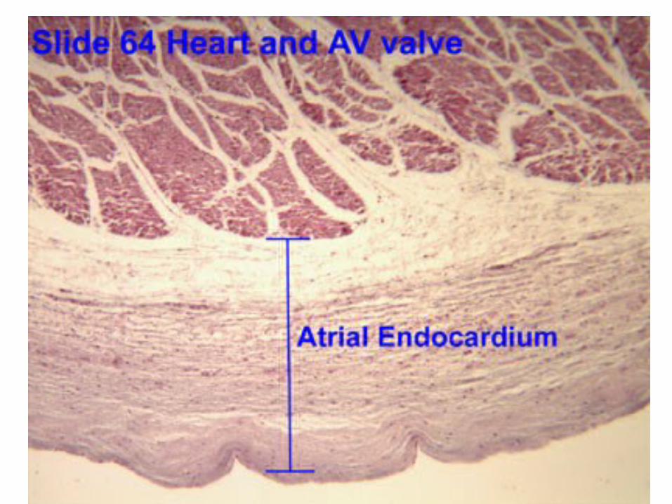
• Lining the interior surface of the heart’s chambers, valves, and blood vessels we find the endocardium or endothelium
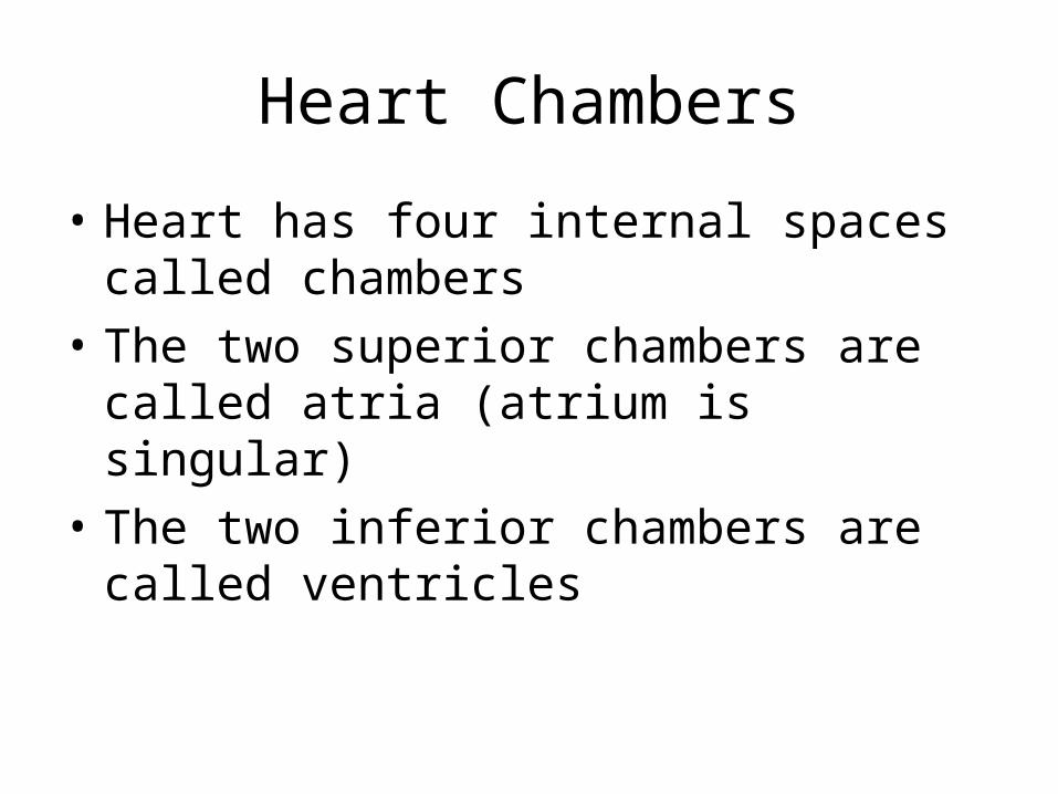
Heart Chambers
• Heart has four internal spaces called chambers
• The two superior chambers are called atria (atrium is singular)
• The two inferior chambers are called ventricles


Atria
• Function as receiving chambers for blood entering the heart
• Push blood “next door” to ventricles• Walls are thin and contain very few muscle
fibers• Ear-shaped appendages called auricles• Fossa ovalis - remains of what was
present in fetal heart (blood shunted from right atrium to left atrium, bypassing lungs)
Atria cont’d
• Right atrium receives blood from vena cava
• Left atrium receives blood from pulmonary vein

Ventricles
• Provide force necessary to push blood into the body’s circulatory network
• Thicker walls than atria (left is thickest)
• Right ventricle pumps blood into pulmonary artery
• Left ventricle pumps blood into aorta

Heart Valves
• Located between chambers of heart and openings into major blood vessels
• Allow flow of blood in only one direction
• Remain closed while chamber is filling; open to allow blood to flow when heart muscle contracts

Atrioventricular Valves
• Located between atrium and ventricle
• Valve located between right atrium and right ventricle is called the tricuspid valve, because it has three flaps, or cusps
• Valve located between left atrium and left ventricle is the bicuspid valve, also known as mitral valve

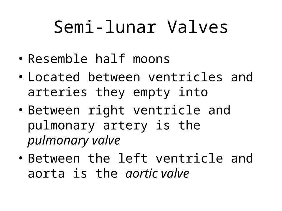
Semi-lunar Valves
• Resemble half moons
• Located between ventricles and arteries they empty into
• Between right ventricle and pulmonary artery is the pulmonary valve
• Between the left ventricle and aorta is the aortic valve


Cardiac Cycle
• Made up of events that are required to produce a single heartbeat
• Include periods of synchronized contraction (systole) and relaxation (diastole)
• P. 378
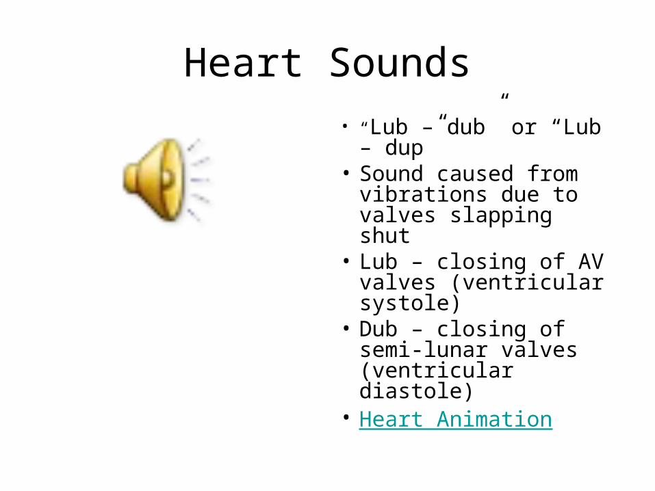
Heart Sounds
• “Lub – dub” or “Lub – dup”
• Sound caused from vibrations due to valves slapping shut
• Lub – closing of AV valves (ventricular systole)
• Dub – closing of semi-lunar valves (ventricular diastole)
• Heart Animation


Blood Vessels
• Form a closed delivery system for blood• Consists of arteries, arterioles, capillaries,
venules, and veins• All but capillaries have a three-layered
wall; from superficial to deep:– Tunica adventitia – CT, anchors vessel– Tunica media – smooth muscle and elastic
fibers– Tunica intima – CT, rich in elastic fibers


Blood Vessels cont’d
• The opening in the middle of a blood vessel through which blood flows is called the lumen
• Blood vessels are all contractile and elastic
• Contractility helps blood circulate by “squeezing” it along
• Elasticity is necessary due to changing fluid pressure (can be felt as pulse)
• Blood vessel comparison

Arteries
• All arteries carry blood away from the heart
• Carry oxygenated blood except for pulmonary arteries
• Thickest walls of any blood vessel; carries blood with the highest fluid pressure
• Very thick tunica media allows for dramatic vasoconstriction and vasodilation
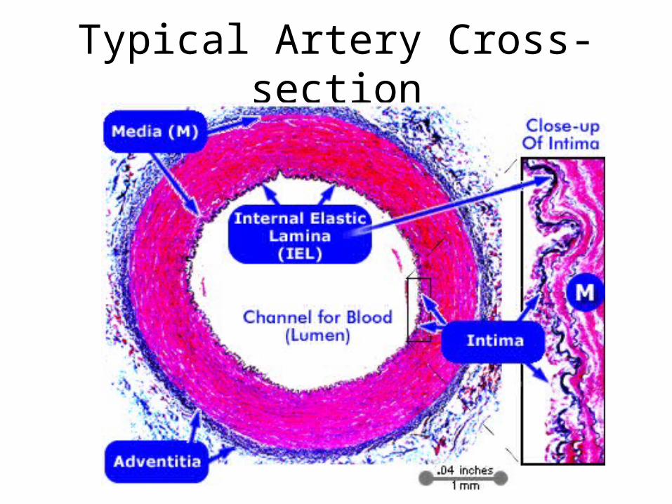
Typical Artery Cross-section

Arterioles
• Branch off of arteries
• Same structure as arteries but smaller diameter
• Usually unnamed; vary from individual to individual

Capillaries
• Capillaries are microscopic blood vessels
• Smallest diameter, single layer of epithelium
• Wall is thin enough to permit diffusion of gases and nutrients into interstitial fluid
• Organized into capillary beds which can be opened or closed to regulate flow of blood as needed
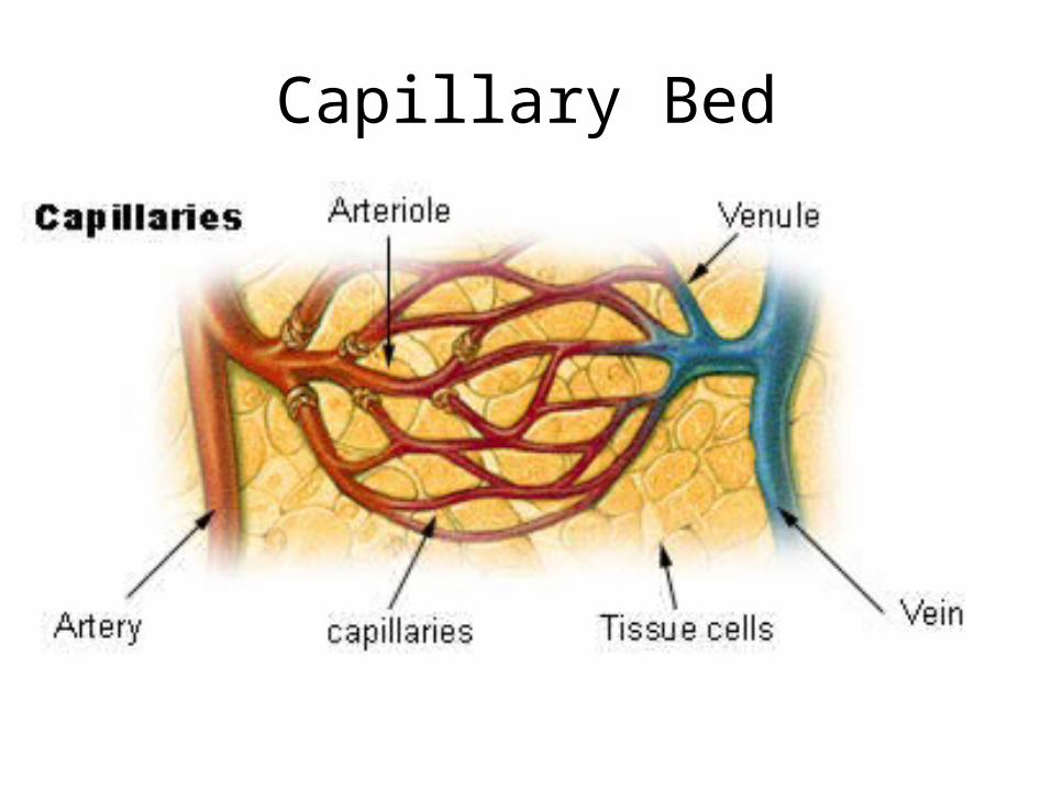
Capillary Bed

Capillaries cont’d
• A central channel called a thoroughfare channel allows blood to pass through when pre- and post-capillary sphincters are closed

Venules and Veins
• Carry blood toward heart; usually deoxygenated blood
• Thinner walls than arteries and arterioles; limited contractility and extensibility
• Carry blood at much lower pressures• Lumen contains one-way valves which prevent
back-flow of blood• Often, surrounding skeletal muscles assist with
circulation• Distensibility allows for variations in pressure
and blood volume; permanent distended state is the cause of varicose veins


![IOT BASED HEALTH MONITORING SYSTEM USING ARDUINO … · 2018. 12. 29. · [1-2], to monitor heart beats of personby using heart beats sensor [1] and to monitor the temperature of](https://static.documents.pub/doc/80x56/5fe664bf7a172b5b61112922/iot-based-health-monitoring-system-using-arduino-2018-12-29-1-2-to-monitor.jpg)


