TISSUE-SPECIFIC STEM CELLS The CD44 1 ALDH 1 Population of Human Keratinocytes Is Enriched for Epidermal Stem Cells with Long-Term Repopulating Ability AKOS Z. SZABO, a STEPHEN FONG, a LILI YUE, a KAI ZHANG, a LAUREN R. STRACHAN, a KENNETH SCALAPINO, b MARIA LAURA MANCIANTI, c RUBY GHADIALLY a a Department of Dermatology, University of California San Francisco and Veteran Affairs Medical Center, Dermatology Service, San Francisco, California, USA; b Department of Medicine, University of California San Francisco and Veterans Affairs Medical Center, Medical Service, Arthritis Division, San Francisco, California, USA; c Department of Pathology, Alta Bates Medical Center, Berkeley, California, USA Key Words. Keratinocyte • Stem cell • Epidermis • Human • Aldehyde dehydrogenase • CD44 ABSTRACT Like for other somatic tissues, isolation of a pure popula- tion of stem cells has been a primary goal in epidermal biol- ogy. We isolated discrete populations of freshly obtained human neonatal keratinocytes (HNKs) using previously untested candidate stem cell markers aldehyde dehydrogen- ase (ALDH) and CD44 as well as the previously studied combination of integrin a6 and CD71. An in vivo transplan- tation assay combined with limiting dilution analysis was used to quantify enrichment for long-term repopulating cells in the isolated populations. The ALDH 1 CD44 1 popu- lation was enriched 12.6-fold for long-term repopulating epidermal stem cells (EpiSCs) and the integrin a6 hi CD71 lo population was enriched 5.6-fold, over unfractionated cells. In addition to long-term repopulation, CD44 1 ALDH 1 ke- ratinocytes exhibited other stem cell properties. CD44 1 ALDH 1 keratinocytes had self-renewal ability, dem- onstrated by increased numbers of cells expressing nuclear Bmi-1, serial transplantation of CD44 1 ALDH 1 cells, and holoclone formation in vitro. CD44 1 ALDH 1 cells were multipotent, producing greater numbers of hair follicle-like structures than CD44 2 ALDH 2 cells. Furthermore, 58% 6 7% of CD44 1 ALDH 1 cells exhibited label-retention. In vitro, CD44 1 ALDH 1 cells showed enhanced colony forma- tion, in both keratinocyte and embryonic stem cell growth media. In summary, the CD44 1 ALDH 1 population exhibits stem cell properties including long-term epidermal regener- ation, multipotency, label retention, and holoclone forma- tion. This study shows that it is possible to quantify the relative number of EpiSCs in human keratinocyte popula- tions using long-term repopulation as a functional test of stem cell nature. Future studies will combine isolation strat- egies as dictated by the results of quantitative transplanta- tion assays, in order to achieve a nearly pure population of EpiSCs. STEM CELLS 2013;31:786–799 Disclosure of potential conflicts of interest is found at the end of this article. INTRODUCTION Human epidermis comprises a multilayered squamous epithe- lium. Epidermal stem cells (EpiSCs) are thought to reside in the basal layer and enable the epidermis to undergo massive expansion during development and wound healing. Given the critical role of EpiSCs in maintaining the epidermis and their potential involvement in the development of squamous and basal cell carcinomas, methods for the isolation and quantita- tive analysis of human EpiSCs are needed. Human stem cells and their progeny have previously been studied ex vivo using multiple approaches. For example, human foreskin was transplanted onto nude mice and infected with a lentivirus encoding a fluorescent marker protein, in order to trace the progeny of labeled basal cells. After 28 weeks, labeled columns composed of basal cells and their progeny were observed [1]. Also, organotypic culture systems have been established, using human keratinocytes cultured on dermal equivalents [2, 3]. In this manner, human epidermis could be regenerated for months (>10 weeks) [2, 3]. Another group established a transplantation assay based on repopula- tion of epidermis inside a rat trachea implanted subcutane- ously in a severe combined immunodeficient (SCID) mouse [4, 5]. Keratinocytes implanted into the human dermis or subcu- tis as a result of surgery or trauma spontaneously form epidermal cysts [6, 7] characterized by a stratified squamous epithelium reminiscent of that on the skin surface and the uppermost part of the hair follicle [8–10]. The experimental Author contributions: A.S.: conception and design, data collection and/or assembly, data analysis and interpretation, and manuscript writing; S.F. data collection and/or assembly and data analysis and interpretation; K.Z. and K.S.: conception and design and data collection and/or assembly; L.R.S.: conception and design and data analysis and interpretation; L.Y.: data analysis and interpretation; M.L.M.: manuscript writing and data analysis and interpretation; R.G.: conception and design, data analysis and interpretation, and manuscript writing. A.S. and S.F. contributed equally to this article. Correspondence: Ruby Ghadially, MBChB, FRCP(C)Derm, Department of Dermatology, University of California, San Francisco, Epithelial Section, Eli and Edythe Broad Center of Regeneration Medicine and Stem Cell Research, 4150 Clement Street MC190, San Francisco, California 94121, USA. Telephone: 415-221-4810x3373; Fax: 415-750-6959; e-mail: [email protected]Received January 18, 2011; accepted for publication December 15, 2012; first published online in STEM CELLS EXPRESS January 17, 2013. V C AlphaMed Press 1066-5099/2013/$30.00/0 doi: 10.1002/stem.1329 STEM CELLS 2013;31:786–799 www.StemCells.com
Transcript
TISSUE-SPECIFIC STEM CELLS
The CD441ALDH1 Population of Human Keratinocytes Is Enriched
for Epidermal Stem Cells with Long-Term Repopulating Ability
AKOS Z. SZABO,a
STEPHEN FONG,a
LILI YUE,a
KAI ZHANG,a
LAUREN R. STRACHAN,a
KENNETH SCALAPINO,b
MARIA
LAURA MANCIANTI,c RUBY GHADIALLYa
aDepartment of Dermatology, University of California San Francisco and Veteran Affairs Medical Center,
Dermatology Service, San Francisco, California, USA; bDepartment of Medicine, University of California San
Francisco and Veterans Affairs Medical Center, Medical Service, Arthritis Division, San Francisco, California,
USA; cDepartment of Pathology, Alta Bates Medical Center, Berkeley, California, USA
Like for other somatic tissues, isolation of a pure popula-tion of stem cells has been a primary goal in epidermal biol-ogy. We isolated discrete populations of freshly obtainedhuman neonatal keratinocytes (HNKs) using previouslyuntested candidate stem cell markers aldehyde dehydrogen-ase (ALDH) and CD44 as well as the previously studiedcombination of integrin a6 and CD71. An in vivo transplan-tation assay combined with limiting dilution analysis wasused to quantify enrichment for long-term repopulatingcells in the isolated populations. The ALDH
1CD44
1popu-
lation was enriched 12.6-fold for long-term repopulatingepidermal stem cells (EpiSCs) and the integrin a6hiCD71lo
population was enriched 5.6-fold, over unfractionated cells.In addition to long-term repopulation, CD441ALDH1 ke-ratinocytes exhibited other stem cell properties.CD44
1ALDH
1keratinocytes had self-renewal ability, dem-
onstrated by increased numbers of cells expressing nuclear
Bmi-1, serial transplantation of CD441
ALDH1
cells, andholoclone formation in vitro. CD441ALDH1 cells weremultipotent, producing greater numbers of hair follicle-likestructures than CD442ALDH2 cells. Furthermore, 58% 67% of CD44
1ALDH
1cells exhibited label-retention. In
vitro, CD441
ALDH1
cells showed enhanced colony forma-tion, in both keratinocyte and embryonic stem cell growthmedia. In summary, the CD44
1ALDH
1population exhibits
stem cell properties including long-term epidermal regener-ation, multipotency, label retention, and holoclone forma-tion. This study shows that it is possible to quantify therelative number of EpiSCs in human keratinocyte popula-tions using long-term repopulation as a functional test ofstem cell nature. Future studies will combine isolation strat-egies as dictated by the results of quantitative transplanta-tion assays, in order to achieve a nearly pure population ofEpiSCs. STEM CELLS 2013;31:786–799
Disclosure of potential conflicts of interest is found at the end of this article.
INTRODUCTION
Human epidermis comprises a multilayered squamous epithe-lium. Epidermal stem cells (EpiSCs) are thought to reside inthe basal layer and enable the epidermis to undergo massiveexpansion during development and wound healing. Given thecritical role of EpiSCs in maintaining the epidermis and theirpotential involvement in the development of squamous andbasal cell carcinomas, methods for the isolation and quantita-tive analysis of human EpiSCs are needed.
Human stem cells and their progeny have previously beenstudied ex vivo using multiple approaches. For example,human foreskin was transplanted onto nude mice and infectedwith a lentivirus encoding a fluorescent marker protein, in
order to trace the progeny of labeled basal cells. After 28weeks, labeled columns composed of basal cells and theirprogeny were observed [1]. Also, organotypic culture systemshave been established, using human keratinocytes cultured ondermal equivalents [2, 3]. In this manner, human epidermiscould be regenerated for months (>10 weeks) [2, 3]. Anothergroup established a transplantation assay based on repopula-tion of epidermis inside a rat trachea implanted subcutane-ously in a severe combined immunodeficient (SCID) mouse[4, 5].
Keratinocytes implanted into the human dermis or subcu-tis as a result of surgery or trauma spontaneously formepidermal cysts [6, 7] characterized by a stratified squamousepithelium reminiscent of that on the skin surface and theuppermost part of the hair follicle [8–10]. The experimental
Author contributions: A.S.: conception and design, data collection and/or assembly, data analysis and interpretation, and manuscriptwriting; S.F. data collection and/or assembly and data analysis and interpretation; K.Z. and K.S.: conception and design and datacollection and/or assembly; L.R.S.: conception and design and data analysis and interpretation; L.Y.: data analysis and interpretation;M.L.M.: manuscript writing and data analysis and interpretation; R.G.: conception and design, data analysis and interpretation, andmanuscript writing. A.S. and S.F. contributed equally to this article.
Correspondence: Ruby Ghadially, MBChB, FRCP(C)Derm, Department of Dermatology, University of California, San Francisco,Epithelial Section, Eli and Edythe Broad Center of Regeneration Medicine and Stem Cell Research, 4150 Clement Street MC190, SanFrancisco, California 94121, USA. Telephone: 415-221-4810x3373; Fax: 415-750-6959; e-mail: [email protected] ReceivedJanuary 18, 2011; accepted for publication December 15, 2012; first published online in STEM CELLS EXPRESS January 17, 2013.VC AlphaMed Press 1066-5099/2013/$30.00/0 doi: 10.1002/stem.1329
STEM CELLS 2013;31:786–799 www.StemCells.com
production of this type of epithelial structure has been used tostudy multiple epithelia including epidermis [9, 10], lung[11], and mammary epithelium [12], as well as epidermal dif-ferentiation of induced pluripotent stem cells [13].
Putative EpiSC markers in human skin include integrin b1[14, 15], integrin a6 in combination with CD71 [16, 17],AC133 [18], p63 [19], keratin 15 [20], ABCG2 [21, 22], anddelta 1 [23]. Integrin a6hiCD71lo is one of the most usedEpiSC-enriched populations in the field of epidermal biology[16, 24].
One strategy to find candidate tissue stem cell markers isto use conserved stem and progenitor cell functions that maybe applicable to multiple tissues. Aldehyde dehydrogenase 1(ALDH1) is a detoxifying enzyme that oxidizes intracellularaldehydes [25]. ALDH oxidizes retinol to retinoic acid andmay have a role in early stem cell differentiation [26]. It hasbeen shown that hematopoietic [27, 28] [29], neural [30, 31],and mammary [32] stem cells have high ALDH1 activity.CD44þ is a marker of normal prostate epithelial stem cells[33] and of cancer stem cells from multiple epithelia [34–36].
In addition to long-term repopulation, self-renewal abilityis another key defining stem cell property. In previous studies,evidence for self-renewal has included serial transplantationsin vivo [37–39], holoclone formation in vitro [40, 41], andexpression of nuclear Bmi-1 (B-lymphoma Moloney murineleukemia virus insertion region-1, an epigenetic regulatorknown to be involved in the self-renewal of stem cells frommultiple tissues) [42–45]. Finally, multipotency [46] andlabel-retaining ability are features of stem cells [47].
In the study presented here, we quantified the frequencyof long-term repopulating cells in the ALDHþCD44þ popula-tion of keratinocytes, previously untested in epidermis. Injec-tion of keratinocytes into murine subcutis was combined withlimiting dilution analysis to quantitatively assess the fre-quency of long-term repopulating EpiSCs in fluorescenceactivated cell sorting (FACS)-selected populations of humankeratinocytes. The ALDHþCD44þ population showed long-term repopulating ability, self-renewal ability, multipotency,and label retention. This study is a step toward the goal ofcombining EpiSC markers in a strategic fashion in order toselect a nearly pure population of EpiSCs.
MATERIALS AND METHODS
Animals. Non-obese diabetic SCID (NOD/SCID) mice (Jack-son Laboratories, Bar Harbor, ME, http://www.jax.org) weremaintained and cared for in accordance with an approved Institu-tional Animal Care and Use Committee Protocol.
Human Skin Samples. A fresh human adult scalp sample andneonatal foreskins were obtained with the appropriate approvalsfrom the UCSF Committee on Human Research and all studiesabided by the rules of the Internal Review Board and the tenetsof the Declaration of Helsinki.
Generation of Human Epidermis In Vivo. Human neonatalforeskins were incubated in Dispase (25 U/ml, BD Biosciences,San Jose, CA, http://www.bdbiosciences.com) for 24 hours at4�C. Epidermis was then peeled from dermis and incubated in0.05% trypsin-EDTA (Invitrogen, Carlsbad, CA, http://www.invitrogen.com) at 37�C for 12 minutes. Keratinocytes were collectedby centrifugation at 1,000 rpm for 5 minutes. Human neonatalkeratinocyte (HNK) suspensions in keratinocyte growth medium
(KGM) (Invitrogen) were mixed with Matrigel (BD Biosciences)1:1(vol/vol) and injected into the subcutis of NOD/SCID mice.
Analysis of Epidermal Regeneration. Xenografts were har-vested by punch biopsy (Acuderm, Fort Lauderdale, FL, http://www.acuderm.com). Frozen sections were stained with hematox-ylin and eosin and examined for presence (positive sample) orabsence (negative sample) of human epidermal repopulation units(ERUs).
Flow Cytometry
Keratinocytes were sorted using a FACSAria (BD) and analyzedwith CellQuest software. ALDHþ cells were selected usingALDEFLUOR (Stem Cell Technologies, Vancouver, BC, http://www.stemcell.com) according to the manufacturer’s protocol.allophycocyanin (APC) conjugated CD44 (IM7, #17-0441, 0.6lg/ml) was from eBioscience, San Diego, CA, http://ebioscience.-com. FITC-conjugated integrin a6 (GoH3, #555735, 1:5), APC-conjugated CD71 (M-A712, #551374, 1:5) and isotype controlswere from BD Pharmingen (San Diego, http://www.bdbioscien-ces.com/index[lowen]us.shtml).
Assessment of Human Versus Murine Tissue Origin
Hoechst 33258 dye (Invitrogen) was used to confirm the humanderivation of the xenograft ERUs. Human nuclei can be distin-guished from murine nuclei, as the latter have multiple hyper-chromatic chromocenters, resulting in a characteristic punctatepattern of fluorescence in contrast to the homogeneous staining ofhuman nuclei [48, 49]. In addition, fluorescent in situ hybridiza-tion (FISH) was performed using a CY3-day-UTP-labeledhuman lymphocyte-derived genomic DNA probe [46] or a Spec-trum-Orange-labeled Human Y chromosome-specific DNA probe(provided by H.U. Weier, Berkeley, CA) [50, 51].
Assessing the Single Cell Origin of ERUs with CellTracker Nontoxic Carbocyanine Dyes, Vybrant DiIand DiO
Freshly isolated, unsorted HNKs were labeled with either VybrantDiI (565 nm) or Vybrant DiO (501 nm) (Invitrogen) (followingthe manufacturer’s protocol) and then mixed 1:1. Either 6,250 or1,562 cells from the mixture were transplanted subcutaneously,harvested after 1 week (five experiments, 767 ERUs studied), andfrozen sections were examined under an Axio Plan Z fluorescencemicroscope (Carl Zeiss Inc., Thornwood, NY, http://www.zeiss.de/micro.com).
Immunostaining
For immunofluorescence staining, frozen sections were fixed withacetone and incubated with primary antibodies including cytoker-atin 14 (rabbit polyclonal, ab15461, 1:1000), involucrin (SY5,mouse monoclonal, ab68, 1:1000), filaggrin (SPM181, mousemonoclonal, ab17808, 1:300), laminin (LT3, Rat monoclonal,ab3003, 1:10), Bmi-1 (rabbit polyclonal, ab38295, 1:100), CD44(F10-44-2, mouse monoclonal, ab6124, 1:100) (all from Abcam,Cambridge, MA, http://www.abcam.com), and ALDH1A1 (rabbitpolyclonal, HPA002123, 1:1,000, Sigma), then secondary anti-bodies including Alexa Fluor 488 IgG and Alexa Fluor 594 IgG(Invitrogen). Propidium iodide or 40,6-diamidino-2-phenylindolewas used to counterstain nuclei.
For immunoperoxidase staining, formalin-fixed, paraffin-em-bedded sections were deparaffinized and rehydrated. Slides wereincubated with primary antibodies including cytokeratin 14, invo-lucrin, and filaggrin (see above), then secondary antibodiesincluding biotinylated anti-rabbit IgG and anti-mouse IgG (allfrom Vector Laboratories, Burlingame, CA, http://www.vectorlabs.com). Immunolabeled structures were visualized with ABCreagent and ImmPACT NovaRED Peroxidase Substrate (VectorLaboratories). Hematoxylin was used as a counterstain.
Szabo, Fong, Yue et al. 787
www.StemCells.com
For alkaline phosphatase staining, formalin-fixed, paraffin-embedded sections were incubated with primary antibodiesincluding cytokeratin 17 (rabbit polyclonal, ab53707, 1:100,Abcam) and cytokeratin 6 (EPR1603Y, rabbit monoclonal,ab52620, 1:500, Abcam). The UltraVision Quanto detectionsystem and Permanent Fast Red Quanto substrate system (Ther-moFisher Scientific, Fremont, CA, http://www.thermoscientific.comG) were used to detect signal. Hematoxylin was used as acounterstain.
Serial Transplantation
CD44þALDHþ or CD44�ALDH� keratinocytes were each mixedwith 5 � 106 cultured human neonatal fibroblasts and trans-planted into recipient NOD/SCID mice. Experiments 1–3 used700/10,000, 1,000/10,000, and 350/4,000 CD44þALDHþ/CD44�ALDH� keratinocytes, respectively. Grafts were harvested9 weeks later. A cell suspension was again generated using 300collagen digestion U/ml of Collagenase, 370 U/ml of Hyaluroni-dase, and 100 Kunitz U/ml DNase I (Sigma, St. Louis, MO) inHank’s solution (Cambrex, Walkersville, MD, http://www.cam-brex.com) with 10% fetal bovine serum (Invitrogen) for 30minutes at 37�C. Keratinocytes were then transplanted into a sec-ond NOD/SCID recipient for 6–9 weeks and presence or absenceof human ERUs was determined by histology.
Holoclone Assay. Single FACS-sorted CD44þALDHþ,CD44�ALDH�, or unfractionated (UNF) human keratinocyteswere inoculated onto 96-well plates containing Mitomycin C-treated 3T3 cells maintained in Dulbecco’s modified Eagle’s me-dium with 10% fetal bovine serum. At 10–12 days, primary colo-nies were photographed. Each colony was then removed by trypsini-zation, and cells were transferred to 100 mm dishes with MitomycinC treated 3T3 cells at 80%–90% confluence. After 12–14 days, sec-ondary colonies were fixed and stained with toluidine blue (Sigma).Each plate was photographed and the identity of the cell thatfounded each primary colony was determined [holoclone, mero-clone, or paraclone based on previously described methods [40]].
(25,000), or UNF (25,000) HNKs were mixed with freshly sortedgreen fluorescent protein (GFPþ) mouse dermal papilla cells(400 K-1.1 M) and Matrigel (50 ll) and injected subcutaneouslyinto NOD/SCID mice. Biopsies were harvested at 17–21 daysand sections were stained with H&E.
Iododeoxyuridine (IdU) Incorporation Studies Using Organo-typic Cultures. AlloDerm regenerative tissue matrix (LifeCellCorp., Branchburg, NJ) was placed in 0.4 cm diameter cell cultureinserts (Millipore Corp, Billerica, MA, http://www.millipore.com).Human fibroblasts were first inoculated onto AlloDerm (60,000cells per m2) for 24 hours [3]. Human keratinocytes were then ino-culated at 500,000 cells per cm2 [3]. Cultures were maintainedsubmerged in 0.07 mM calcium CnT-57 growth medium (ZenBio,Inc., Research Triangle Park, NC) for 8 days and then raised tothe air–liquid interface in 1.2 mM calcium CnT-02-3DP1 (ZenBio,Inc.) with 200 Units/ml aprotinin [3]. Cultures were pulse labeledwith IdU (1 lM) every 12 hours for 2 weeks. At the end of a 4-week chase, cultures were harvested and incubated in 25 Units/mlDispase at 37�C for 2 hours. Live CD44þALDHþ cells were thenFACS-sorted and inoculated onto microscope slides. IdU wasdetected with 5-bromo-2’-deoxyuridine (BrdU)/IdU antibody (B44,#347580, 1:10, BD Biosciences).
Colony Formation In Vitro. FACS-sorted cells were plated at1,000 cells per well in uncoated six-well plates (clonal density)and cultured for 3–4 weeks in either embryonic stem cell me-dium, ESC-Sure (Applied StemCell, Sunnyvale, CA, http://www.appliedstemcell.com), or KGM (Invitrogen). Plates werestained with toluidine blue.
Statistical Analysis. L-Calc (Stemsoft, Vancouver, BC, http://stemsoft.com) was used to determine the frequency of ERUs inthe selected cell populations. Limiting dilution analysis was sub-jected to a v2 test. Lack of a significant p value in the v2 test wasused to demonstrate internal consistency in the distribution ofresults. Stem cell frequencies from limiting dilution experiments atdifferent weeks (1, 2, 4, 6, 9, and 12) were compared using stand-ard ‘‘single-hit’’ Poisson models for limiting dilution experiments[52]. Results for these analyses were obtained using R statisticalsoftware, version 2.9.0 (R Development Core Team, 2009). Forcomparison of the number of CD44þALDHþ versus integrina6hiCD71lo versus UNF HNKs with nuclear Bmi-1 expression, aone-way ANOVA was used. For comparison of the percent holo-clones in CD44þALDHþ versus UNF populations and the numberof hair follicle-like structures produced by CD44þALDHþ versusCD44�ALDH� populations, a paired Student’s t test was used. Invitro colony forming ability of the five different subpopulationswas analyzed using the Kruksal Wallis test, with post hoc pair-wise comparisons using the Bonferroni Dunn test.
RESULTS
Generation of Epidermis/ERUs in a Xenograftmodel, by Injection of Human Keratinocytes
Freshly obtained HNKs transplanted into NOD/SCID subcutisformed human keratinizing epidermis (Fig. 1A; supporting in-formation Fig. 1), as seen in previous studies [9, 10]. Epider-mal cysts generated in this fashion were termed ERUs pursu-ant to longstanding terminology in hematopoiesis [53]. As innormal human epidermis, keratin 14 antibody immunostainedthe basal layers of ERUs [54, 55] (Fig. 1B), involucrin anti-body stained all suprabasal epidermal layers [56, 57] (Fig.1C), and filaggrin antibody stained the uppermost epidermallayers [56] (Fig. 1D). Immunofluorescence with FITC-conju-gated laminin antibody produced a linear pattern at the base-ment membrane [58] (Fig. 1E). These findings confirm theproduction of a differentiated keratinizing epidermis, as seenpreviously by others [8–10].
To determine whether ERUs originate from single cells,freshly isolated HNKs were labeled with Vybrant DiI (red) orVybrant DiO (green). DiI and DiO labeled HNKs were mixedin equal numbers (1:1) and then transplanted into NOD/SCIDmice. Using doses of 1,562 or 6,250 cells (five separate experi-ments), 766 out of a total of 767 ERUs were found to be eitherred or green but not mixed (Fig. 1F), indicating that at thesedoses ERUs almost always derive from a single cell, ratherthan resulting from cell aggregation. The lipid staining dyesare diluted as cells multiply and because of this ERU forma-tion was assessed at 2 weeks, when label was still visible.Because of this, we cannot be sure that on occasion some cystsdo not merge at later time points, due to their close proximity.
The human origin of ERU keratinocytes was confirmedusing Hoechst 33258 staining [48, 49]. Nuclei in the humanERUs (Fig. 1G, left panel) showed the expected homogenousnuclear staining with Hoechst 33258 (Fig. 1G, middle panel),while murine nuclei displayed the characteristic punctatestaining of hyperchromatic chromocenters (Fig. 1G, rightpanel). Additionally, FISH with a CY3-day-UTP-labeledhuman lymphocyte derived genomic DNA probe also stainedthe ERUs (Fig. 1H, middle panel) but not murine epidermis(Fig. 1H, right panel), confirming their human derivation.
In summary, H&E, keratin 14, involucrin, filaggrin, andlaminin staining confirmed epidermal differentiation reminis-cent of that in vivo, while Hoechst 33258 and FISHconfirmed the human derivation of ERUs in this model.
788 CD44þALDHþ Enriches for Keratinocyte Stem Cells
Figure 1. ERUs in the xenograft model show epidermal differentiation and human derivation. (A): H&E staining of a human ERU, producedby injecting human neonatal keratinocyte (HNKs) into murine subcutis (9 weeks) and showing keratinizing epidermis. (B): Keratin 14 is presentin the basal layers of human ERUs. (C): Involucrin is present in the suprabasal layers. (D): Filaggrin is present in the uppermost layers of the ep-idermis. Appropriate positive and negative controls were performed (supporting information Fig. 1). (E): Linear pattern of fluorescence of lamininexpression along the basement membrane of the ERU (Inset, lower power view of ERU). (F): To determine whether ERUs from implantedHNKs were derived from single cells, HNKs were labeled with Vybrant DiI or Vybrant DiO and mixed in a 1:1 ratio before injection into non-obese diabetic severe combined immunodeficient mice. Resultant ERUs were either green or red. DAPI was used to counter stain nuclei. (G):Hoechst 33258 staining confirmed that characteristic homogeneous staining was seen in human ERU nuclei (middle panel). In contrast the charac-teristic bright punctate pattern of hyperchromatic chromocenters was seen in the murine epidermal nuclei (right panel). (H): FISH against humangenomic DNA labels the human ERU (middle panel) but not murine epidermis (right panel). (I): The frequency of cells capable of formingERUs at weeks 1, 2, 4, 6, 9, and 12 was determined by limiting dilution analysis. At 9 weeks the frequency of ERUs stops decreasing signifi-cantly and thereafter remains constant. (A, B, C, D, G) scale bars ¼ 10 lm. (E, F, H) scale bars ¼ 50 lm. Abbreviations: DAPI, 40,6-diamidino-2-phenylindole; ERU, epidermal repopulating units; FISH, fluorescent in situ hybridization.
Szabo, Fong, Yue et al. 789
www.StemCells.com
Additionally, DiI and DiO staining of keratinocytes revealed asingle cell origin of most ERUs at the doses examined.
Selection of the Endpoint at Which Only EpiSCsOriginally Injected (and Their Progeny) Persist
In order to study stem cell frequency quantitatively usinglong-term repopulation assays it is necessary to determine thetime point at which ERUs can be identified as arising fromlong-term repopulating stem cells versus short-term repopulat-ing transit amplifying cells (TACs) [37, 53, 59, 60]. At thetime point at which short-term proliferating TAC-derivedERUs have exhausted their replicative ability and no longerexist, the frequency of ERUs stops decreasing and thereafterremains constant [37, 53, 59-61], allowing the assessment ofEpiSCs originally injected and their progeny. For HNKs, theERU frequency at weeks 1, 2, 4, 6, 9, and 12 was determinedby limiting dilution analysis (Fig. 1I). It can be seen that theERU frequency decreases from 1 in 96 keratinocytes at 1week to 1 in 792 keratinocytes at 2 weeks and then to 1 in1,939 keratinocytes by 6 weeks, following which the fre-quency appears to remain stable. The overall likelihood ratiotest for differences in stem cell frequencies between weeks 1and 12 was significant (p < .001). Likelihood ratio tests foradequacy of the single-hit Poisson model were nonsignificantfor all analyses, indicating an acceptable fit to the data.Inspection of the week-specific results indicated that observeddifferences in frequencies were due primarily to changesoccurring in the first 4 weeks, with relatively stable results inweeks 6–12. This observation was confirmed by a separatelikelihood ratio test for differences between results in weeks6–12, which was not significant (p ¼ .95). A similar analysislimited to weeks 9–12 also yielded nonsignificant results (p ¼.99). The relatively large p-values obtained in analyses limitedto later time points, coupled with the relatively small differen-ces in observed frequencies and large overlap in 95% confi-dence intervals for these time points supported the use of 9weeks as the point after which ERU frequency remains stable.We therefore used 9 weeks in subsequent experiments as theendpoint at which to study long-term repopulating EpiSCs.
The CD441ALDH1 and Integrin a6hiCD71lo Popula-tions of Human Keratinocytes Are Enriched forLong-Term Repopulating EpiSCs
To quantify long-term repopulating EpiSCs, putative humanEpiSCs were selected by FACS, transplanted into the subcutisof NOD/SCID mice, and limiting dilution analysis of ERUfrequency was performed at week 9. ALDHþ cells comprised1.6%–13.5% of total HNKs and CD44þALDHþ cells com-prised 0.03%–0.2% of total HNKs (Fig. 2A). Integrin a6hiC-D71lo cells (7%–10% of total, Fig. 2B) were selected usingpublished methods [16, 24].
In neonatal human foreskin epidermis, immunohistochem-istry for ALDH1 and CD44 each showed staining in the basallayers and occasional costained cells were detected (Fig. 2C).In adult epidermis, the coexpression of CD44 and ALDH wasfound in the depths of the rete ridges (deepest part of retepeg) (Fig. 2D). In the adult hair follicle, the bulge region andthe opening of the sebaceous gland showed coexpression ofCD44 and ALDH (Fig. 2E). 82% 6 2% of CD44þALDHþ
cells were keratin 14þ, evidence that CD44þALDHþ cellsare mostly basal cells (supporting information Fig. 2).
The frequency of long-term repopulating cells was 1 in586 cells (SE 783-438) in the ALDHþ population versus 1 in69,918 (SE 283,641-17,235) in the ALDH� population (notshown), 1 in 170 cells (SE 240-120) in the CD44þALDHþ
population versus 1 in 218,510 (SE 591,130-80,770) in the
CD44�ALDH� population, and 1 in 390 cells (SE 520-290)in the integrin a6hiCD71lo population versus 1 in 24,030 (SE36,860-15,660) in the integrin a6loCD71hi population, whilethe frequency of long-term repopulating cells in the UNF pop-ulation was 2,160 (SE 2,610-1,790) (Fig. 3A). When com-pared to UNF keratinocytes, ERUs were enriched 3.7-fold inthe ALDHþ population (not shown), 12.6-fold in theCD44þALDHþ population, and 5.6-fold in the integrin a6hiC-D71lo population (Fig. 3A, 3B). Thus there was significantenrichment for EpiSCs in both the CD44þALDHþ (1 in 170)and integrin a6hiCD71lo populations (1 in 390) over UNF.However, the enrichment for CD44þALDHþ versus integrina6hiCD71lo was not significantly different (p ¼ .06).
CD44þALDHþ cells produced ERUs with a normal patternof epidermal differentiation (Fig. 3C) and CD44 and ALDHwere both present in the basal layer of ERUs (Fig. 3D).
To further investigate the longevity of these cells using analternative methodology [60, 61], 5,000 CD44þALDHþ or50,000 UNF HNKs, mixed with one million human neonatalfibroblasts were implanted in chambers on the backs of NOD/SCID mice. Transplants from four out of four CD44þALDHþ
samples and three out of four UNF samples showed a fullydifferentiated epidermis at 9 weeks (Fig. 3E). Staining forlaminin, keratin 14, and filaggrin showed a fully differentiatedepidermis. Epidermis from CD44þALDHþ cells was indistin-guishable from that from UNF cells by H&E.
The CD441ALDH1 Population of KeratinocytesExhibits Self-Renewal Ability In Vivo and In Vitro
Bmi-1 is involved in the self-renewal of stem cells from multipletissues [42–45]. CD44þALDHþ HNKs and integrin a6hiCD71lo
HNKs contained significantly greater numbers of cells express-ing nuclear Bmi-1 (36.1% and 35.8%, respectively) versus UNFHNKs (12.7%, p ¼ .004 and 0.024, respectively, Fig. 4A).
To study self-renewal in vivo, CD44þALDHþ keratino-cytes mixed with 5 � 106 cultured human neonatal fibroblastsor CD44�ALDH� keratinocytes mixed with 5 � 106 culturedhuman neonatal fibroblasts were injected into recipient NOD/SCID mice. Experiments 1–3 used 700/10,000, 1,000/10,000,and 350/4,000 CD44þALDHþ/CD44�ALDH� keratinocytes(dependent on the quantity of tissue available). Grafts wereharvested 9 weeks later. A cell suspension was again gener-ated from the harvested graft, transplanted into a secondNOD/SCID recipient, and the presence or absence of humanepidermal ERUs was determined by histology. Secondarytransplants derived from human CD44þALDHþ keratinocytesonce again regenerated ERUs, providing evidence for self-renewal capacity of the CD44þALDHþ keratinocytes (Fig.4B). ERUs were not seen in any of the CD44�ALDH�trans-plants. It was noted that, unlike in the primary transplants(Fig. 3C), ERUs in secondary transplants were clusters ofbasaloid cells lacking stratification (Fig. 4B), suggestive ofundifferentiated keratinocytes. Finally, because more than onecell was transplanted in the experiments above, we cannotrule out the possibility that the primary and secondary ERUswere derived from distinct CD44þALDHþ cells.
Holoclone formation in vitro was studied as further evi-dence for self-renewal ability. For each sample, 108–288 sin-gle CD44þALDHþ, CD44�ALDH�, or UNF cells wereplated, one cell per well, in 96-well plates. After 10–13 days,colonies were counted and secondarily plated. No colonieswere produced from over 160 CD44�ALDH� individuallyplated cells (three independent experiments). After 13–16days, secondary colonies were stained and assessed for largesmooth colonies with central differentiation (holoclones) usingthe classification of Barrandon and Green [40] (Fig. 4C). The
790 CD44þALDHþ Enriches for Keratinocyte Stem Cells
Figure 2. Fluorescence-activated cell sorting (FACS) isolation and immunohistochemistry of CD44þALDHþ keratinocytes. (A): FACS isolationof CD44þALDHþ keratinocytes. DEAB, an ALDH inhibitor, was used as a negative control. CD44 isotype control performed but not shown.(B): FACS isolation of 7%–10% of integrin a6hiCD71lo keratinocytes. Isolation performed as previously described [20]. (isotype controls per-formed but not shown). (C): In neonatal human foreskin epidermis, immunohistochemistry for ALDH1 showed staining in the basal layers. CD44staining was also located in the basal layers. There were occasional double positive cells in the basal layer. (D): In adult epidermis, cells coex-pressing CD44 and ALDH were found in the depths of the rete ridges (tips of the rete pegs) (arrowheads). (E): In the adult hair follicle (facialskin) cells that coexpressed CD44 and ALDH were located in the bulge region (arrowheads) and at the opening of the sebaceous gland (asterisk).Negative controls were performed with no primary antibody. Scale bars ¼ 10 lm. Abbreviation: ALDH, aldehyde dehydrogenase.
Szabo, Fong, Yue et al. 791
www.StemCells.com
Figure 3. The CD44þALDHþ and integrin a6hiCD71lo populations of keratinocytes are enriched in cells with long-term repopulating ability.(A): Limiting dilution analysis of EpiSC frequency in selected keratinocyte populations at 9 weeks. There is a significant increase in EpiSC fre-quency in the CD44þALDHþ and integrin a6hiCD71lo populations versus the UNF population. For each cell dose the number of positiveresponses (injection sites with at least one ERU) over the total number of injection sites is the response ratio. (B): Histogram showing the foldenrichment of EpiSCs in selected populations over the UNF HNK population. The CD44þALDHþ and integrin a6hiCD71lo populations areenriched for long-term repopulating EpiSCs over UNF cells. (C): Epidermal repopulating units (ERUs) produced by injection of CD44þALDHþ
keratinocytes exhibited a fully differentiated epidermis. Keratin 14 was present in the basal layer, involucrin in the suprabasal layers and filaggrinin the uppermost layers. (D): ERUs produced by injection of CD44þALDHþ keratinocytes showed expression of CD44 (fluorescein isothiocya-nate/green) and ALDH (phycoerythrin/red) in the basal layer (there was also nonspecific staining of keratin in the center of the ERU). (E):CD44þALDHþ keratinocytes, in chambers on the dorsum of mice, produced a well-differentiated epidermis that persisted for at least 9 weeks.Expression of laminin was present along the basement membrane, keratin 14 in the basal layer and filaggrin in the uppermost layer. Scale bars ¼10 lm. Abbreviations: ALDH, aldehyde dehydrogenase; EpiSC, epidermal stem cell; UNF, unfractionated population.
792 CD44þALDHþ Enriches for Keratinocyte Stem Cells
number of holoclones in the CD44þALDHþ population of ke-ratinocytes was twice that in the UNF population; 1.4% 60.46% vs. 0.7% 6 0.35%, n ¼ 3, p ¼ .01, (Fig. 4D).
The CD441
ALDH1
Population of Keratinocytes IsMultipotent
To determine whether CD44þALDHþ HNKs are multipotent,HNKs were implanted with trichogenic murine dermal cells,
using previously described methods to generate chimeric hairfollicles from human keratinocytes [46, 62]. Doses of 2,500–25,000 CD44þALDHþ or CD44�ALDH� HNKs wereimplanted with 7 � 105 GFPþ dermal papilla cells and graftswere studied for the formation of chimeric hair follicles at18–21 days (four independent experiments). 100,000-700,000dermal papilla cells alone did not produce hair follicle-likestructures (n ¼ 3). CD44þALDHþ cells produced significantlygreater numbers of hair follicle-like structures than did
Figure 4. CD44þALDHþ keratinocytes display the stem cell property of self-renewal. (A): Bmi-1 expression in CD44þALDHþ keratinocytes.The CD44þALDHþ and integrin a6hiCD71lo populations of keratinocytes have greater numbers of cells expressing nuclear Bmi-1 than the UNFpopulation. (B): CD44þALDHþ keratinocytes display the stem cell property of self-renewal. Injection of 700 CD44þALDHþ and 10,000CD44�ALDH� cells, followed by secondary transplantation of the resultant grafts (harvested at 9 weeks). Secondary ERUs were seen inCD44þALDHþ derived secondary transplants. No ERUs were seen in CD44�ALDH� secondary transplants (n ¼ 3). Scale bar ¼ 20 lm. (C): Holo-clone formation in CD44þALDHþ versus UNF HNKs. Single cells were plated in 96-well plates. Colonies formed were each secondarily plated intoa single dish. In secondary plating, holoclones formed large smooth colonies with central differentiation (left panels, low and high power) and para-clones formed small terminal colonies (right panels, low and high power) (meroclones were intermediate between the two). (D): The number of hol-oclones in CD44þALDHþ keratinocytes was twice that in UNF keratinocytes; 1.4% 6 0.46% versus 0.7% 6 0.35%, n ¼ 3, p ¼ .01. (B) scale bars¼ 10 lm. (C) scale bar ¼ 1 mm. Abbreviations: ALDH, aldehyde dehydrogenase; ERUs, Epidermal repopulating units; UNF, unfractionated.
Szabo, Fong, Yue et al. 793
www.StemCells.com
CD44�ALDH� cells (9.0 6 2.1 vs.1.9 þ 0.9 per 25,000HNKs injected, p ¼ .007; Fig. 5A). CD44 and ALDH stainingwas present in a location consistent with regeneration of a
bulge region (Fig. 5B). H&E, keratin 6, and keratin 17 stain-ing (Fig. 5C–5E) confirmed the hair follicle-like nature of thestructures observed, and FISH using a human Y chromosome-
Figure 5. The CD44þALDHþ population of keratinocytes is multipotent. (A): The histogram illustrates how CD44þALDHþ keratinocytesshowed significantly greater production of hair follicle-like structures than CD44�ALDH� keratinocytes. H&E staining of CD44þALDHþ derivedhair follicle-like structures (arrows) is shown in the panels on the right. (B): The hair follicle-like structures produced from CD44þALDHþ kera-tinocytes contain CD44þALDHþ keratinocytes in a location consistent with regeneration of a bulge region. (C): Higher power view of hair fol-licle-like structure (H&E). (D): Immunohistochemistry of hair-follicle-like structures for hair-associated proteins keratin 6 (alkaline phosphatase-red) and (E) keratin 17 (alkaline phosphatase-red) (for (D) and (E) hematoxylin alone was used as counterstain). Controls are adult human hairfollicles showing follicular staining (red). (F): FISH using a human specific y-chromosome DNA probe showed the presence of human cells(bright pink staining in human nuclei) in the hair follicle-like structures. (B, D-right panel) (E-right panel) scale bars ¼ 50 lm. (D-left panel) (E-left panel) (F) scale bars ¼ 10 lm. (C) scale bar ¼ 20 lm. Abbreviations: ALDH, aldehyde dehydrogenase; DAPI, 40,6-diamidino-2-phenylin-dole; FISH, fluorescence in situ hybridization.
794 CD44þALDHþ Enriches for Keratinocyte Stem Cells
specific Spectrum Orange-labeled DNA-probe confirmed thepresence of human cells in the follicle-like structures (Fig.5F).
The CD441ALDH1 Population of KeratinocytesContains Label-Retaining Cells
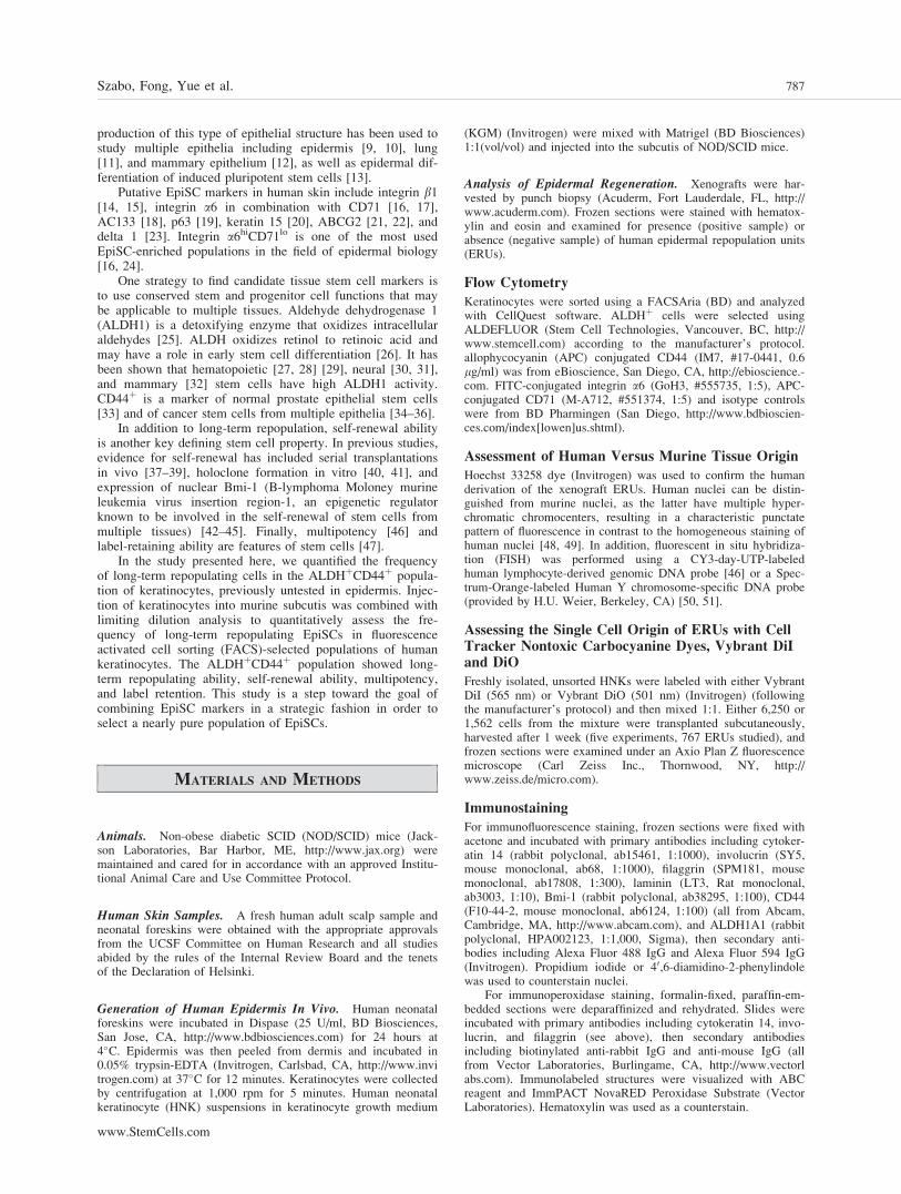
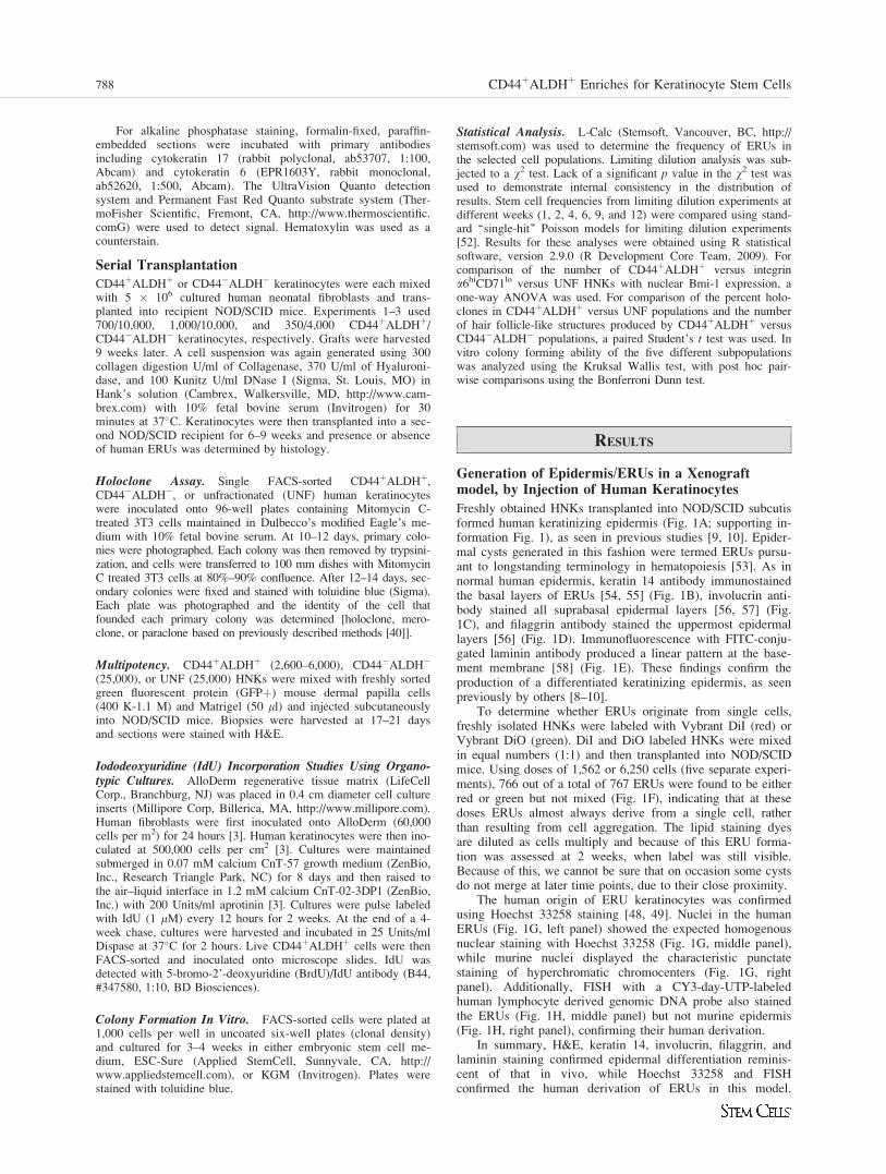
Stem cells are expected to cycle slowly and therefore retainlabel. Organotypic cultures were prepared using 500,000CD44þALDHþ HNKs per cm2, as previously described [3].After 2 weeks, cultures were pulse-labeled with IdU every 12hours for 2 further weeks. After the 2-week pulse, IdU-treatedcultures showed almost 100% IdUþ basal cells (Fig. 6A).After a 4-week chase, the cultures were harvested, a cell sus-pension was isolated, and CD44þALDHþ keratinocytes wereFACS-sorted onto microscope slides for immunofluorescencestudies. 58% 6 7% of the CD44þALDHþ keratinocytes wereIdU-expressing label-retaining cells at 4 weeks of chase (n ¼3, Fig. 6B).
The CD441
ALDH1
Population of KeratinocytesPossesses Enhanced In Vitro Colony FormingAbility
To test CD44þALDHþ keratinocytes for their growth proper-ties in vitro, HNK subpopulations were plated at 1,000 cellsper well (six-well plates) and cultivated for 21–28 days ineither KGM (Invitrogen) or embryonic stem cell media(ESC-Sure, Applied StemCell).
In KGM, there was a significant difference in the number
of colonies produced in the five groups (CD44þALDHþ,
and UNF, p ¼ .002). CD44þALDHþ HNKs formed colonies
whereas CD44�ALDH� HNKs did not and colony forming
ability of CD44þALDHþ HNKs was significantly greater
than CD44�ALDH� HNKs (3.4 6 0.7 vs. 0 6 0, p < .05),
using a post hoc pairwise comparison (Bonferroni-Dunn)
(Fig. 7A left panel; supporting information Fig. 3). Colony
forming ability of integrin a6hiCD71lo HNKs was also signif-
icantly greater than integrin a6loCD71hi HNKs (5.8 6 2.8 vs.
0.01 6 0.01, p < .05 Fig. 7B left panel). No significant dif-
ference was detected in the colony forming ability of
CD44þALDHþ versus integrin a6hiCD71lo HNKs (n ¼ 4,
Fig. 7).We then tested whether these cells would grow in a
medium designed to support the growth of primitive stemcells. In embryonic stem cell medium, there was a significantdifference in the five groups (CD44þALDHþ, CD44�ALDH�,integrin a6hiCD71lo, integrin a6loCD71hi, and UNF, p ¼ .003)and colony forming ability of CD44þALDHþ HNKs was sig-nificantly greater than CD44�ALDH� HNKs (3.5 6 1.2 vs.0.01 6 0.01, p ¼ <.05) and a6hiCD71lo HNKs (3.5 6 1.2 vs.0.0 6 0, p ¼ <.01) using a post hoc pairwise comparison(Bonferroni-Dunn) Fig. 7A). While neither integrin a6hiC-D71lo nor a6hiCD71hi HNKs (putative EpiSCs and TACs,respectively [16, 63] grew in embryonic stem cell medium,integrin a6loCD71hi HNKs did, although a statistically
Figure 6. The CD44þALDHþ population of keratinocytes contains label-retaining cells. (A): After iododeoxyuridine administration twice dailyfor 2 weeks, almost 100% of keratinocytes in the basal layer of an organotypic epidermis (from unfractionated keratinocytes) were labeled. Nega-tive controls included untreated keratinocytes (no iododeoxyuridine) (shown) as well as iododeoxyuridine-treated keratinocytes (2.5 lM twice aday for 2 days) with no primary antibody (not shown). Iododeoxyuridine-treated keratinocytes with primary antibody added were used as a posi-tive control (not shown). (B): After a 4-week chase, keratinocytes from organotypic epidermis were fluorescence-activated cell-sorted to collectCD44þALDHþ keratinocytes and stained for iododeoxyuridine. 58% 6 7% of CD44þALDHþ keratinocytes were label retaining cells. Scale bar¼ 10 lm. Abbreviations: ALDH, aldehyde dehydrogenase; DAPI, 40,6-diamidino-2-phenylindole; IdU, iododeoxyuridine.
Szabo, Fong, Yue et al. 795
www.StemCells.com
significant difference was not detected between a6hiCD71lo
and integrin a6loCD71hi HNKs (0 6 0 vs. 0.39 6 0.18)(Fig. 7B).
DISCUSSION
The assay presented here provides a quantitative assessmentof the ability of putative stem cell markers to enrich for cellscapable of long-term regeneration of the epidermis. This studyquantitatively demonstrates a 12.6-fold enrichment for EpiSCsin CD44þALDHþ HNKs, previously untested in epidermis.While the CD44þALDHþ population is enriched so that 1 in170 cells is an EpiSC, the ultimate goal is to obtain a nearlypure population of EpiSCs, as has been accomplished in hem-atopoiesis. Isolation of a nearly pure population of EpiSCs isof high importance for regenerative medicine applications,targeting EpiSCs for genetic manipulations and for more basicstudies of EpiSC regulation and differentiation.
In these studies, CD44þALDHþ and integrin a6hiCD71lo
human keratinocytes regenerated epidermis for the long-termwhen injected subcutaneously into NOD/SCID mice. It hasbeen demonstrated that skin, corneal, and esophageal epithe-lial each maintained their distinctive in vivo phenotype wheninjected subcutaneously and reacquire their distinctive in vivokeratin patterns [9]. In cell culture, using fibroblast support,this was not the case indicating that these differences do notdepend on specific mesenchymal instruction but on permissivefactors present in vivo but not in vitro [9]. Injected keratino-cytes reacquired a normal ultrastructural appearance with ker-atohyalin granules, lamellar bodies, and enucleated hornycells [10] and expressed bullous pemphigoid antigen and lam-inin at the epithelial–stromal interface [64]. Our studiesshowed normal expression of keratin 14, involucrin, fillagrin,and laminin.
The absolute frequency of repopulating cells detected inthis assay is almost certainly affected by the methods. FACSplaces significant stress on cells and engagement of cell
Figure 7. Colony forming efficiency of CD44þALDHþ and integrin a6hiCD71lo keratinocytes. (A): The CD44þALDHþ population hasenhanced colony-forming efficiency versus the CD44�ALDH� population in both keratinocyte growth medium and embryonic stem cell medium.To determine the in vitro colony forming efficiency of the selected cells, CD44þALDHþ, CD44�ALDH�, and UNF keratinocytes were plated insix-well plates, 1,000 cells per well, in either keratinocyte growth medium (left) or embryonic stem cell medium (right). One representative platefrom each experiment is shown (see also supporting information Fig. 3). Bar graphs show mean 6 SE (four experiments). (B): The integrina6hiCD71lo population of cells has enhanced colony forming efficiency in keratinocyte growth medium but not in embryonic stem cell medium.The difference in colony forming efficiency between CD44þALDHþ and integrin a6hiCD71lo HNKs in KGM was not significant, but in embry-onic stem cell medium CD44þALDHþ human neonatal keratinocytes had significantly greater colony forming efficiency (p < .01). One represen-tative plate for each experiment is shown (see also supporting information Fig. 3). Bar graphs show mean 6 SE (1,000 cells per well, fourexperiments). Abbreviations: ALDH, aldehyde dehydrogenase; CFE, colony forming efficiency; UNF, unfractionated.
796 CD44þALDHþ Enriches for Keratinocyte Stem Cells
surface receptors may modulate cell function and behavior.However, the assay provides a quantitative comparison of therelative frequency of cells capable of long-term repopulationpresent in different keratinocyte populations. Consistent withthe studies done using the subcutaneous transplantation assay,longevity studies using keratinocytes transplanted with neona-tal human fibroblasts onto the backs of NOD/SCID mice alsoshowed that CD44þALDHþ keratinocytes produced epidermisfor the long-term.
We performed FACS analysis to determine whether theCD44þALDHþ and integrin a6hiCD71lo populations of kerati-nocytes were the same population. We found that only 1.1%–2.3% of integrin a6hiCD71lo keratinocytes were ALDHþ, whilethere was no overlap with the CD44þALDHþ keratinocytes.This is interesting in view of the in vitro findings whereCD44þALDHþ but not integrin a6hiCD71lo keratinocytes wereable to grow in embryonic stem cell medium. Thus, both theFACS analysis and the growth in embryonic stem cell mediumstudies suggest two distinct populations of progenitors.
While in neonatal foreskin CD44þALDHþ cells werefound along the entire basal layer, in adult epidermisCD44þALDHþ cells were found in known stem cell niches,including both the bulge [65] and sebaceous gland [66].CD44þALDHþ interfollicular cells were predominantly in thedepths of the rete ridges (tips of rete pegs). This location issimilar to that of K15þ integrin a6hi cells [17] and desmo-glein-3dim cells [67] but different from the location of integrinb1hi [15] and delta 1þ cells [23] that are located in the shal-lowest parts of the rete ridges (suprapapillary plate). The rea-son for these different findings is not clear but may possiblyinvolve previously described methodological issues [17].
CD44þALDHþ HNKs produced ERUs when seriallytransplanted. ERUs in secondary transplants were clusters ofbasaloid cells lacking stratification. This may reflect the prim-itive nature of the cell that has the capability for serialtransplantation.
CD44þALDHþ cells displayed enhanced multipotencyversus CD44�ALDH� cells. However, while hairs did notgrow from dermal papilla cells injected alone, some hair-likestructures did grow from CD44�ALDH� cells injected incombination with dermal papilla cells. This is may be due tothe fact that there are groups of progenitor cells distinct fromthe CD44þALDHþ cells (e.g., integrin a6hiCD71lo) that arealso capable of hair production. Also, it is possible that inthis assay, progenitor cells that are not as primitive as stemcells may produce hair-like structures, as is the case in hema-topoesis where multipotent progenitors can be either long-term repopulating cells or less primitive progenitors [68, 69].
Holoclone formation was increased 2-fold inCD44þALDHþ over UNF HNKs, whereas, in vivo, long-termrepopulating cells were enriched 12-fold. This apparent dis-crepancy may be explained by evidence that primitive stemcells do not have clonal capacity in vitro. Hematopoietic stemcells with long-term bone marrow repopulating capacity havebeen shown to have minimal to no clonal capacity in vitro
[70, 71], and there is prior evidence that stem cell-rich frac-tions exhibit equal or substantially lower colony forming effi-ciency than the source total population in human neonatalforeskin [16], human limbal epithelium, [72] and rat epider-mis [73].
A previous study found that integrin a6lo keratinocytes(not stem cells) had poor regenerative capacity in vitro (over14 days) but could produce an in vivo epidermis in rat trachea(over 6 weeks) [2]. In the studies presented here, likewise,integrin a6lo (CD71hi) keratinocytes did not grow in KGM buthad a small number of long-term repopulating cells and didgrow in embryonic stem cell medium. Altogether, the abovestudies suggest that integrin a6lo keratinocytes contain a smallpopulation of primitive cells. In addition, in the studies pre-sented here, integrin a6hiCD71lo keratinocytes grew only inKGM, not embryonic stem cell medium, but contained long-term repopulating cells. Thus some long-term repopulatingcells appear to grow preferentially in KGM and some in em-bryonic stem cell medium, suggesting that not all long-termrepopulating cells behave similarly in vitro.
While classically the epidermis has been thought to besustained by a EpiSC/TAC hierarchy of progenitors, someobservations contradict the EpiSC/TAC model [for review see[74, 75]]. Using inducible genetic labeling, clone-size distri-butions were consistent with a model of homeostasis involv-ing only one type of progenitor cell [76]. Thus in the studypresented here, the loss of repopulating units over time maybe wrongly attributed to the loss of TAC-derived units andcould conceivably represent either the disappearance of multi-ple tiers of progenitors (as in other tissues) or the chance lossof ERUs from a single type of epithelial progenitor.
While the properties of the integrin a6hiCD71lo keratino-cyte population have been well documented, here we eval-uated stem cell properties of the CD44þALDHþ keratino-cytes, previously untested as EpiSC markers. In future studiesthere is a need to systematically quantitate the ability of indi-vidual potential stem cell markers to select for cells with thefunctional property of long-term tissue regeneration, with thegoal of combining markers to select a nearly pure populationof EpiSCs. Different progenitor populations can then be corre-lated with specific phenotypes and in vitro growth behaviors.
ACKNOWLEDGMENTS
We thank Corey Largman and Alex Charruyer for useful discus-sions and Ruby Gribi for her help with the FACS.
DISCLOSURE OF POTENTIAL CONFLICTS OF
INTEREST
The authors indicate no potential conflicts of interest
REFERENCES
1 Ghazizadeh S, Taichman LB. Organization of stem cells and theirprogeny in human epidermis. J Invest Dermatol 2005;124:367–72.
2 Li A, Pouliot N, Redvers R et al. Extensive tissue-regenerativecapacity of neonatal human keratinocyte stem cells and their progeny.J Clin Invest 2004;113:390–400.
3 Muffler S, Stark H-J, Amoros M et al. A stable niche supports long-term maintenance of human epidermal stem cells in organotypic cul-tures. Stem Cells 2008;26:2506–2515.
4 Pouliot N, Redvers RP, Ellis S et al. Optimization of a transplantmodel to assess skin reconstitution from stem cell-enriched primaryhuman keratinocyte populations. Exp Dermatol 2005;14:60–69.
5 Paquet-Fifield S, Redvers RP, Pouliot N, Kaur P. A transplant modelfor human epidermal skin regeneration. Methods Mol Biol 2010;585:369–382.
6 Silver SG, Ho VCY. Benign epithelial tumors. In: Freedberg I, EisenA, Wolff K et al. Fitzpatrick’s Dermatology in General Medicine.Columbus, OH: McGraw-Hill, 2003:778–780.
7 Hall JP, Sheffey RJ, Chagares WE et al. Epidermal inclusion cyst inthe foot of a Vietnam Veteran. J Am Pediatr Med Assoc 2006;96:445–447.
Szabo, Fong, Yue et al. 797
www.StemCells.com
8 Ohnishi T, Watanabe S. Immunohistochemical observation of cytoker-atins in keratinous cysts including plantar epidermoid cyst. J CutanPathol 1999;26:424–429.
9 Doran TI, Vidrich A, Sun TT. Intrinsic and extrinsic regulation of thedifferentiation of skin, corneal and esophageal epithelial cells. Cell1980;22(1 pt 1):17–25.
10 Lavker RM, Sun TT. Rapid modulation of keratinocyte differentiationby the external environment. J Invest Dermatol 1983;80:228–237.
11 Yu W, Fang X, Ewald A et al. Formation of cysts by alveolar type IIcells in three-dimensional culture reveals a novel mechanism for epi-thelial morphogenesis. Mol Biol Cell 2007;18:1693–1700.
12 Debnath J, Brugge JS. Modelling glandular epithelial cancers in three-dimensional cultures. Nat Rev Cancer 2005;5:675–688.
13 Takahashi K, Tanabe K, Ohnuki M et al. Induction of pluripotentstem cells from adult human fibroblasts by defined factors. Cell 2007;131:861–872.
14 Jones PH, Watt FM. Separation of human epidermal stem cells fromtransit amplifying cells on the basis of differences in integrin functionand expression. Cell 1993;73:713–724.
15 Jones PH, Harper S, Watt FM. Stem cell patterning and fate in humanepidermis. Cell 1995;80:83–93.
16 Li A, Simmons PJ, Kaur P. Identification and isolation of candidatehuman keratinocyte stem cells based on cell surface phenotype. ProcNatl Acad Sci USA 1998;95:3902–3907.
17 Webb A, Li A, Kaur P. Location and phenotype of human adult kera-tinocyte stem cells of the skin. Differentiation 2004;72:387–395.
18 Belicchi M, Pisati F, Lopa R et al. Human skin-derived stem cellsmigrate throughout forebrain and differentiate into astrocytes afterinjection into adult mouse brain. J Neurosci Res 2004;77:475–486.
19 Pellegrini G, Dellambra E, Golisano O et al. p63 identifies keratino-cyte stem cells. Proc Natl Acad Sci USA 2001;98:3156–3161.
20 Lyle S, Christofidou-Solomidou M, Liu Y et al. The C8/144B mono-clonal antibody recognizes cytokeratin 15 and defines the location ofhuman hair follicle stem cells. J Cell Sci 1998;111 (pt 21):3179–3188.
21 Terunuma A, Jackson KL, Kapoor V et al. Side population keratino-cytes resembling bone marrow side population stem cells are distinctfrom label-retaining keratinocyte stem cells. J Invest Dermatol 2003;121:1095–1103.
22 Triel C, Vestergaard ME, Bolund L et al. Side population cells inhuman and mouse epidermis lack stem cell characteristics. Exp CellRes 2004;295:79–90.
23 Lowell S, Jones P, Le Roux I et al. Stimulation of human epidermaldifferentiation by delta-notch signalling at the boundaries of stem-cellclusters. Curr Biol 2000;10:491–500.
24 Li A, Kaur P. Epidermal Cells: FACS enrichment of human keratino-cyte stem cells. Methods Mol Biol. 2005;289:87–96.
25 Marchitti SA, Brocker C, Stagos D et al. Non-P450 aldehyde oxidiz-ing enzymes: The aldehyde dehydrogenase superfamily. Expert OpinDrug Metab Toxicol 2008;4:697–720.
26 Chute JP, Muramoto GG, Whitesides J et al. Inhibition of aldehydedehydrogenase and retinoid signaling induces the expansion of humanhematopoietic stem cells. Proc Natl Acad Sci USA 2006;103:11707–11712.
27 Armstrong L, Stojkovic M, Dimmick I et al. Phenotypic characteriza-tion of murine primitive hematopoietic progenitor cells isolated on ba-sis of aldehyde dehydrogenase activity. Stem Cells 2004;22:1142–1151.
28 Christ O, Lucke K, Imren S et al. Improved purification of hematopoi-etic stem cells based on their elevated aldehyde dehydrogenase activ-ity. Haematologica 2007;92:1165–1172.
29 Hess DA, Meyerrose TE, Wirthlin L et al. Functional characterizationof highly purified human hematopoietic repopulating cells isolatedaccording to aldehyde dehydrogenase activity. Blood 2004;104:1648–1655.
30 Corti S, Locatelli F, Papadimitriou D et al. Identification of a primi-tive brain-derived neural stem cell population based on aldehydedehydrogenase activity. Stem Cells 2006;24:975–985.
31 Corti S, Locatelli F, Papadimitriou D et al.. Transplanted ALDHhiS-SClo neural stem cells generate motor neurons and delay diseaseprogression of nmd mice, an animal model of SMARD1. Hum MolGenet 2006;15:167–187.
32 Ginestier C, Hur MH, Charafe-Jauffret E et al. ALDH1 is a marker ofnormal and malignant human mammary stem cells and a predictor ofpoor clinical outcome. Cell Stem Cell 2007;1:555–567.
33 Liu AY, True LD, LaTray L et al. Cell–cell interaction in prostategene regulation and cytodifferentiation. Proc Natl Acad Sci USA1997;94:10705–10710.
34 Al-Hajj M, Wicha MS, Benito-Hernandez A et al. Prospective identifi-cation of tumorigenic breast cancer cells. Proc Natl Acad Sci 2003;100:3983–3988.
35 Prince ME, Sivanandan R, Kaczorowski A et al. Identification of asubpopulation of cells with cancer stem cell properties in head and
neck squamous cell carcinoma. Proc Natl Acad Sci USA 2007;104:973–978.
36 Patrawala L, Calhoun T, Schneider-Broussard R et al. Highly purifiedCD44þ prostate cancer cells from xenograft human tumors areenriched in tumorigenic and metastatic progenitor cells. Oncogene2006;25:1696–1708.
37 Charruyer A, Strachan LR, Yue L et al. CD133 is a marker for long-term repopulating murine epidermal stem cells. J Invest Dermatol2012;132:2522–2533.
38 Yahata T, Takanashi T, Muguruma Y et al. Accumulation of oxidativeDNA damage restricts the self-renewal capacity of human hematopoi-etic stem cells. Blood 2011;118:2941–2950.
39 Kunimoto H, Fukuchi Y, Sakurai M et al. Tet2 disruption leads toenhanced self-renewal and altered differentiation of fetal liver hemato-poietic stem cells. Sci Rep 2012;2:273.
40 Barrandon Y, Green H. Three clonal types of keratinocyte with differ-ent capacities for multiplication. Proc Natl Acad Sci USA 1987;84:2302–2306.
41 Pellegrini G, Ranno R, Stracuzzi G et al. The control of epidermalstem cells (holoclones) in the treatment of massive full-thicknessburns with autologous keratinocytes cultured on fibrin. Transplantation1999;6:868–879.
42 Sangiorgi E, Capecchi MR. Bmi1 is expressed in vivo in intestinalstem cells. Nat Genet 2008;40:915–20.
43 Lessard J, Sauvageau G. Bmi-1 determines the proliferative capacityof normal and leukaemic stem cells. Nature 2003;423:255–60.
44 Leung C, Lingbeek M, Shakhova O et al. Bmi1 is essential for cere-bellar development and is overexpressed in human medulloblastomas.Nature 2004;428:337–341.
45 Molofsky AV, Pardal R, Iwashita T et al. Bmi-1 dependence distin-guishes neural stem cell self-renewal from progenitor proliferation.Nature 2003;425:962–967.
46 Ehama R, Ishimatsu-Tsuji Y, Iriyama S et al. Hair follicle regenera-tion using grafted rodent and human cells. J Invest Dermatol 2007;127:2106–2115.
47 Bickenbach JR, Holbrook KA. Label-retaining cells in human embry-onic and fetal epidermis. J Invest Dermatol 1987;88:42–46.
48 Moser FG, Dorman BP, Ruddle FH. Mouse-human heterokaryon anal-ysis with a 33258 Hoechst-Giemsa technique. J Cell Biol 1975;66:676–680.
49 Cunha GR, Vanderslice KD. Identification in histological sections ofspecies origin of cells from mouse, rat and human. Stain Technol1984;59:7–12.
50 Kuperwasser C, Chavarria T, Wu M et al. Reconstruction of function-ally normal and malignant human breast tissues in mice. Proc NatlAcad Sci USA 2004;101:4966–4971.
51 Parrott JA, Nilsson E, Mosher R et al. Stromal–epithelial interactionsin the progression of ovarian cancer: Influence and source of tumorstromal cells. Mol Cell Endocrinol 2001;175:29–39.
52 Bonnefoix T, Bonnefoix P, Verdiel P et al. Fitting limiting dilutionexperiments with generalized linear models results in a test of the sin-gle-hit Poisson assumption. J Immunol Methods 1996;194:113–119.
53 Szilvassy SJ, Humphries RK, Lansdorp PM et al. Quantitative assayfor totipotent reconstituting hematopoietic stem cells by a competitiverepopulation strategy. Proc Natl Acad Sci USA 1990;87:8736–8740.
54 Moll R, Franke WW, Schiller DL et al. The catalog of human cyto-keratins: Patterns of expression in normal epithelia, tumors and cul-tured cells. Cell 1982;31:11–24.
55 van der Velden LA, Schaafsma HE, Manni JJ et al. Cytokeratin andvimentin expression in normal epithelium and benign lesions of thevocal cords. Acta Otolaryngol 1996;116:325–331.
56 Gerritsen MJ, Elbers ME, de Jong EM et al. Recruitment of cyclingepidermal cells and expression of filaggrin, involucrin and tenascin inthe margin of the active psoriatic plaque, in the uninvolved skin ofpsoriatic patients and in the normal healthy skin. J Dermatol Sci1997;14:179–188.
57 Murphy GF, Flynn TC, Rice RH et al. Involucrin expression in nor-mal and neoplastic human skin: A marker for keratinocyte differentia-tion. J Invest Dermatol 1984;82:453–457.
58 Kanitakis J. Anatomy, histology and immunohistochemistry of normalhuman skin. Eur J Dermatol 2002;12:390–399, quiz 400–401.
59 Strachan LR, Scalapino KJ, Lawrence HJ et al. Rapid adhesion to col-lagen isolates murine keratinocytes with limited long-term repopulat-ing ability in vivo despite high clonogenicity in vitro. Stem Cells2008;26:235–243.
60 Charruyer A, Barland CO, Yue L et al. Transit-amplifying cell fre-quency and cell cycle kinetics are altered in aged epidermis. J InvestDermatol 2009;129:2574–2583.
61 Schneider TE, Barland C, Alex AM et al. Measuring stem cell fre-quency in epidermis: A quantitative in vivo functional assay for long-term repopulating cells. Proc Natl Acad Sci USA 2003;100:11412–11417.
798 CD44þALDHþ Enriches for Keratinocyte Stem Cells
62 Ferraris C, Bernard BA, Dhouailly D. Adult epidermal keratinocytesare endowed with pilosebaceous forming abilities. Int J Dev Biol1997;41:491–498.
63 Tani H, Morris RJ, Kaur P. Enrichment for murine keratinocyte stemcells based on cell surface phenotype. Proc Natl Acad Sci USA 2000;97:10960–10965.
64 Grimwood RE, Baskin JB, Nielsen LD et al. Fibronectin extracellularmatrix assembly by human epidermal cells implanted into athymicmice. J Invest Dermatol 1988;90:434–440.
65 Cotsarelis G. Epithelial stem cells: A folliculocentric view. J InvestDermatol 2006;126:1459–1468.
66 Horsley V, O’Carroll D, Tooze R et al. Blimp1 defines a progenitorpopulation that governs cellular input to the sebaceous gland. Cell2006;126:597–609.
67 Wan H, Stone MG, Simpson C et al. Desmosomal proteins, includingdesmoglein 3, serve as novel negative markers for epidermal stemcell-containing population of keratinocytes. J Cell Sci 2003;116 (pt20):4239–4248.
68 Kondo M, Weissman IL, Akashi K. Identification of clonogenic com-mon lymphoid progenitors in mouse bone marrow. Cell 1997;91:661–672.
69 Akashi K, Traver D, Miyamoto T et al. A clonogenic common mye-loid progenitor that gives rise to all myeloid lineages. Nature 2000;404:193–197.
70 Sutherland HJ, Lansdorp PM, Henkelman DH et al. Functional charac-terization of individual human hematopoietic stem cells cultured atlimiting dilution on supportive marrow stromal layers. Proc Natl AcadSci USA 1990;87:3584–3588.
71 Haylock DN, To LB, Dowse TL et al. Ex vivo expansion and matura-tion of peripheral blood CD34þ cells into the myeloid lineage. Blood1992;80:1405–1412.
72 Budak MT, Alpdogan OS, Zhou M et al. Ocular surface epitheliacontain ABCG2-dependent side population cells exhibiting featuresassociated with stem cells. J Cell Sci 2005;118 (pt 8):1715–1724.
73 Pavlovitch JH, Rizk-Rabin M, Jaffray P et al. Characteristics ofhomogeneously small keratinocytes from newborn rat skin: Possibleepidermal stem cells. Am J Physiol 1991;261(6 pt 1):C964–C972.
74 Jones PH, Simons BD, Watt FM. Sic transit gloria: Farewell to theepidermal transit amplifying cell? Cell Stem Cell 2007;1:371–381.
75 Jones P, Simons BD. Epidermal homeostasis: Do committed progeni-tors work while stem cells sleep? Nat. Rev Mol Cell Biol 2008;9:82–88.
76 Clayton E, Doup�e DP, Klein AM et al. A single type of progenitorcell maintains normal epidermis. Nature 2007;446:185–189.
See www.StemCells.com for supporting information available online.