THE .JOURNAL OF BIOLOGICAL CHEMISTRY Vol. 266, No. 21, Issue of July 25, pp. 13821-13827, 1991 Printed in U.S.A. The Chicken Tropomyosin 1 Gene Generates Nine mRNAs by Alternative Splicing* (Received for publication, February 19, 1991) Suzanne Forry-Schaudies and Stephen H. Hughes$ From the ABL-Basic Research Program. National Cancer Institute-Frederick Cancer Research and Development Center, - . Frederick, Maryland 21 702-1201 Skeletal muscle /3-tropomyosin,smooth muscle a-tro- pomyosin, and a low molecular weight fibroblast tro- pomyosin are generated by alternatively splicing RNA transcripts of the chicken tropomyosin 1 (TM 1) gene (Forry-Schaudies, S,, Maihle, N. J., and Hughes, S. H. (1990) J. Mol. Biol. 211; 321-330). Two novel tropo- myosin cDNAs that derive from mRNAs of the TM 1 gene have been isolated from a chicken embryo brain cDNA library. Brain cDNA BRT-1 is 2.2 kilobases in length and encodes 283 amino acids. It is identical to skeletal muscle &tropomyosin from amino acids 1 to 268. The sequence 3’ of this point is unique to BRT-1; a comparison to genomic sequence indicates that a new carboxyl-terminal exon is used to generate thisse- quence. 1.4-kilobase brain cDNA BRT-2 contains se- quences found in both fibroblast cDNA FT-/3 (5”end) and skeletal muscle cDNA SKT-8 (3”end). RNase and S1 nuclease assays using RNA samples from leg mus- cle, gizzard, fibroblasts, and brain indicate that the TM 1 gene expresses four additional tropomyosin RNAs by alternately splicing previously characterized exons. These results demonstrate that the chicken TM 1 gene encodes nine tropomyosin RNAs through the use of two promoters, two internal exons that are mutually exclu- sive, and three 3”exons. Implications for the regula- tion of alternative splicing are discussed. The tropomyosins are a large family of cytoskeletal proteins that have regions of close homology. They have been found in all cell types studied, withsome nonmuscle cells having as many as nine distinct protein isoforms (2-5). The function of tropomyosins in skeletal muscle is relatively well understood; however, the significance of the numerous similar proteins in nonmuscle cells is less well understood. Both skeletal and nonmuscle tropomyosins form dimeric a-helical coiled coils (6). In skeletal muscle, the tropomyosin helices align head- to-tail and lie in the groove of thedouble-strandedactin filament (7). In cooperation with the troponin complex, tro- pomyosins regulate Ca”-activated muscle contraction (6, 8). The tropomyosins associate with microfilaments in nonmus- * This work was supported by the National Cancer Institute, De- partment of Health and Human Services, under contract NO1-CO- 74101 with ABL. The costs of publication of this article were defrayed in part by the payment of page charges. This article must therefore be hereby marked “aduertisement” in accordance with 18 U.S.C. Section 1734 solely to indicate this fact. The nucleotide sequence(s) reported in thispaper has been submitted to the GenBank‘rM/EMBL Data Bank with accession number(s) M64287 and M64288. $ To whom correspondence should be addressed ABL-Basic Re- search Program, NCI-Frederick Cancer Research and Developmental Center, P.O. Box B, Frederick, MD 21702-1201. Tel.: 301-846-1619. cle cells (9) and appear to stabilize filamentous actin and prevent it from being depolymerized (10, 11). The large number of the tropomyosin proteins arises both from the existence of several genomic loci in all eukaryotes and from the alternative splicing of the RNA transcripts that derive from these loci. Four distinct classes of tropomyosin genes havebeen found in vertebrates. These include the genes encoding fast skeletal muscle a-tropomyosin (12-16), slow skeletal muscle a-tropomyosin (17, 18), and skeletal muscle 0-tropomyosin (1, 19-22). A fourth gene thatgenerates a platelet-like low molecular weight tropomyosin andprobably the cardiac tropomyosin in chickens has also been described (23-26). The known mRNAs from the four tropomyosin loci do not account for all of the tropomyosin proteins that are known tobepresentineukaryotic cells. Ithasnot been determined whether more tropomyosin genes exist or whether additional mRNAs are generated by thecurrently known genes by alternative splicing. These possibilities are not mu- tually exclusive; both may be correct. Our knowledge of tropomyosin gene structure suggests that all vertebrate tropomyosin genes have evolved from an ances- tral gene that includes two alternate promoters, one set of mutually exclusive internal exons, and two alternatively spliced 3”exons (25). The chicken gene that gives rise to skeletal muscle @-tropomyosin (the chicken tropomyosin 1 gene (1)) is structurally similar to the proposed ancestral gene (1, 21, 22) and is known to generate three mRNAs by alter- natively splicing RNA transcripts in a tissue-specific fashion. The tissue-specific expression of the internal exons of the rat and chicken @-tropomyosin loci has led to studies of how tissue-specific alternative splicing is regulated (27-30). The most structurally complex tropomyosin gene now known is the rat a-tropomyosin gene (16). The gene generatesnine mRNAs through the use of two alternate promoters, two sets of mutually exclusive internal exons, and four carboxyl-ter- minal alternate exons. One of the carboxyl-terminal exons appears to be expressed exclusively in the brain: a correspond- ing exon has not been found in any other tropomyosingene. However, the presence of additional carboxyl-terminal exons in the rat a-tropomyosin gene suggests the possibility that other tropomyosin genes may have a similar structure. In this study, we report that the chicken tropomyosin 1 gene (the @-tropomyosin locus) encodesnine distinct mRNAs, one of which defines a new carboxyl-terminal exon. The differential expression of these mRNAs in chicken embryo skeletal muscle, smooth muscle, fibroblasts, and brain sug- gests that the alternate exons are not used in a strictly tissue- specific manner and has implications for the regulation of alternative splicing. 13821
Transcript
THE .JOURNAL OF BIOLOGICAL CHEMISTRY Vol. 266, No. 21, Issue of July 25, pp. 13821-13827, 1991 Printed in U.S.A.
The Chicken Tropomyosin 1 Gene Generates Nine mRNAs by Alternative Splicing*
(Received for publication, February 19, 1991)
Suzanne Forry-Schaudies and Stephen H. Hughes$ From the ABL-Basic Research Program. National Cancer Institute-Frederick Cancer Research and Development Center, - . Frederick, Maryland 21 702-1201
Skeletal muscle /3-tropomyosin, smooth muscle a-tro- pomyosin, and a low molecular weight fibroblast tro- pomyosin are generated by alternatively splicing RNA transcripts of the chicken tropomyosin 1 (TM 1) gene (Forry-Schaudies, S,, Maihle, N. J., and Hughes, S. H. (1990) J. Mol. Biol. 211; 321-330). Two novel tropo- myosin cDNAs that derive from mRNAs of the TM 1 gene have been isolated from a chicken embryo brain cDNA library. Brain cDNA BRT-1 is 2.2 kilobases in length and encodes 283 amino acids. It is identical to skeletal muscle &tropomyosin from amino acids 1 to 268. The sequence 3’ of this point is unique to BRT-1; a comparison to genomic sequence indicates that a new carboxyl-terminal exon is used to generate this se- quence. 1.4-kilobase brain cDNA BRT-2 contains se- quences found in both fibroblast cDNA FT-/3 (5”end) and skeletal muscle cDNA SKT-8 (3”end). RNase and S1 nuclease assays using RNA samples from leg mus- cle, gizzard, fibroblasts, and brain indicate that the TM 1 gene expresses four additional tropomyosin RNAs by alternately splicing previously characterized exons. These results demonstrate that the chicken TM 1 gene encodes nine tropomyosin RNAs through the use of two promoters, two internal exons that are mutually exclu- sive, and three 3”exons. Implications for the regula- tion of alternative splicing are discussed.
The tropomyosins are a large family of cytoskeletal proteins that have regions of close homology. They have been found in all cell types studied, with some nonmuscle cells having as many as nine distinct protein isoforms (2-5). The function of tropomyosins in skeletal muscle is relatively well understood; however, the significance of the numerous similar proteins in nonmuscle cells is less well understood. Both skeletal and nonmuscle tropomyosins form dimeric a-helical coiled coils (6). In skeletal muscle, the tropomyosin helices align head- to-tail and lie in the groove of the double-stranded actin filament (7). In cooperation with the troponin complex, tro- pomyosins regulate Ca”-activated muscle contraction (6, 8). The tropomyosins associate with microfilaments in nonmus-
* This work was supported by the National Cancer Institute, De- partment of Health and Human Services, under contract NO1-CO- 74101 with ABL. The costs of publication of this article were defrayed in part by the payment of page charges. This article must therefore be hereby marked “aduertisement” in accordance with 18 U.S.C. Section 1734 solely to indicate this fact.
The nucleotide sequence(s) reported in thispaper has been submitted to the GenBank‘rM/EMBL Data Bank with accession number(s) M64287 and M64288.
$ To whom correspondence should be addressed ABL-Basic Re- search Program, NCI-Frederick Cancer Research and Developmental Center, P.O. Box B, Frederick, MD 21702-1201. Tel.: 301-846-1619.
cle cells (9) and appear to stabilize filamentous actin and prevent it from being depolymerized (10, 11).
The large number of the tropomyosin proteins arises both from the existence of several genomic loci in all eukaryotes and from the alternative splicing of the RNA transcripts that derive from these loci. Four distinct classes of tropomyosin genes have been found in vertebrates. These include the genes encoding fast skeletal muscle a-tropomyosin (12-16), slow skeletal muscle a-tropomyosin (17, 18), and skeletal muscle 0-tropomyosin (1, 19-22). A fourth gene that generates a platelet-like low molecular weight tropomyosin and probably the cardiac tropomyosin in chickens has also been described (23-26). The known mRNAs from the four tropomyosin loci do not account for all of the tropomyosin proteins that are known to be present in eukaryotic cells. It has not been determined whether more tropomyosin genes exist or whether additional mRNAs are generated by the currently known genes by alternative splicing. These possibilities are not mu- tually exclusive; both may be correct.
Our knowledge of tropomyosin gene structure suggests that all vertebrate tropomyosin genes have evolved from an ances- tral gene that includes two alternate promoters, one set of mutually exclusive internal exons, and two alternatively spliced 3”exons (25). The chicken gene that gives rise to skeletal muscle @-tropomyosin (the chicken tropomyosin 1 gene (1)) is structurally similar to the proposed ancestral gene (1, 21, 22) and is known to generate three mRNAs by alter- natively splicing RNA transcripts in a tissue-specific fashion. The tissue-specific expression of the internal exons of the rat and chicken @-tropomyosin loci has led to studies of how tissue-specific alternative splicing is regulated (27-30). The most structurally complex tropomyosin gene now known is the rat a-tropomyosin gene (16). The gene generates nine mRNAs through the use of two alternate promoters, two sets of mutually exclusive internal exons, and four carboxyl-ter- minal alternate exons. One of the carboxyl-terminal exons appears to be expressed exclusively in the brain: a correspond- ing exon has not been found in any other tropomyosin gene. However, the presence of additional carboxyl-terminal exons in the rat a-tropomyosin gene suggests the possibility that other tropomyosin genes may have a similar structure.
In this study, we report that the chicken tropomyosin 1 gene (the @-tropomyosin locus) encodes nine distinct mRNAs, one of which defines a new carboxyl-terminal exon. The differential expression of these mRNAs in chicken embryo skeletal muscle, smooth muscle, fibroblasts, and brain sug- gests that the alternate exons are not used in a strictly tissue- specific manner and has implications for the regulation of alternative splicing.
13821
13822 The Chicken Tropomyosin 1 Gene
MATERIALS AND METHODS'
RESULTS AND DISCUSSION
cDNA Isolation and Characterization-The chicken tropo- myosin 1 gene is known to include two alternate promoters, two internal alternate exons, and two carboxyl-terminal al- ternate exons (1,21,22). Through alternative splicing of these exons, the chicken tropomyosin 1 gene could generate eight distinct mRNAs. The gene is known to produce three major mRNAs: a 1.2 kb2 smooth muscle a-tropomyosin mRNA (pSMT-10 (31)), a 1.6-kb skeletal muscle @-tropomyosin mRNA (SKT-@ (32)), and a 1.1-kb fibroblast mRNA (FT-@ (32)). The proteins encoded by the smooth and skeletal muscle mRNAs are 284 amino acids in length and are generated from the upstream promoter and transcription initiation site. The fibroblast mRNA is transcribed from the downstream pro- moter and encodes a low molecular weight 248-amino acid protein (1). The fibroblast and smooth muscle mRNAs con- tain the same internal and carboxyl-terminal exons, whereas the skeletal muscle mRNA contains the alternate form of the internal and 3"exons.
RNA isolated from various chicken embryo tissues was analyzed by Northern transfer using a smooth muscle tropo- myosin (SMT-10) probe to determine whether tropomyosin RNAs of different sizes are generated by the tropomyosin 1 gene (Fig. 1). Skeletal muscles (lanes 1 and 2) express abun- dant 1.6-kb tropomyosin 1 mRNAs as well as smaller amounts of 1.1- and 1.2-kb mRNAs. The predominant mRNA in giz- zard (lane 3 ) is 1.2 kb, although 1.1-kb mRNA is also present. Similarly, the predominant RNA in fibroblasts (lane 4 ) is 1.1 kb, although 1.2-kb mRNA also appears to be expressed. Both gizzard and fibroblasts express minor quantities of 1.6-kb mRNA. Chicken embryo brain appears to express very small quantities of tropomyosin RNA on 1-day exposures (lane 5 ) . Seven-day exposures (lane 6) indicate that 17-day chicken embryo brain expresses 2.2-, 1.6-, 1.2-, and 1.1-kb tropomyosin 1 mRNAs.
To determine which tropomyosin mRNAs are expressed by the tropomyosin 1 gene in brain tissue, we constructed and screened a 17-day chicken embryo brain cDNA library. Four classes of tropomyosin cDNAs were isolated. The first class contained cDNAs identical to the smooth muscle SMT-10 cDNA (31,32). These cDNAs correspond to the 1.2-kb mRNA seen on Northern analysis (Fig. 1, lane 6).
The second class of cDNAs had not been previously isolated from libraries made from RNA isolated from other tissues; the sequence of the longest clone in this class, which we term BRT-1, is shown in Fig. 2. BRT-1 is 2173 base pairs (bp) long. It contains 77 bp of 5"untranslated sequence, 849 bp of coding sequence, 1247 bp of 3"untranslated region, and a poly(A) tail. BRT-1 has 5 more base pairs of sequence at its 5'-end than does SKT-P, the cDNA encoding skeletal muscle @-tropomyosin (32). I t is identical to SKT-@ through the second nucleotide of the codon for amino acid 258 (Fig. 3). 3' of this position, BRT-1 differs from all other tropomyosin c3NAs derived from the tropomyosin 1 gene. The nucleotide sequence in this region encodes 25 amino acids, and the protein encoded by the corresponding mRNA is 283 amino acids long. The 3"untranslated region of BRT-1 contains 943 nucleotides 3' of the T of the TGA stop codon that are
I Portions of this paper (including "Materials and Methods" and Figs. 1, 4, 5, and 8) are presented in miniprint at the end of this paper. Miniprint is easily read with the aid of a standard magnifying glass. Full size photocopies are included in the microfilm edition of the Journal that is available from Waverly Press. ' The abbreviations used are: kb, kilobase(s); bp, base pair(s).
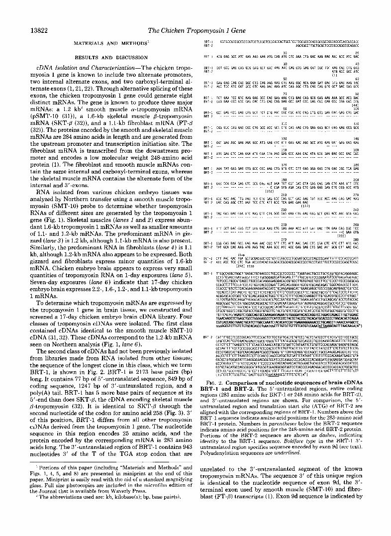
FIG. 2. Comparison of nucleotide sequences of brain cDNAs BRT-1 and BRT-2. The 5"untranslated regions, entire coding regions (283 amino acids for BRT-1 or 248 amino acids for BRT-2),
untranslated region and translation start site (ATG) of BRT-2 are and 3'-untranslated regions are shown. For comparison, the 5'-
aligned with the corresponding regions of BRT-1. Numbers above the BRT-1 sequence indicate amino acid positions for the 283-amino acid BRT-1 protein. Numbers in parentheses below the BRT-2 sequence indicate amino acid positions for the 248-amino acid BRT-2 protein. Portions of the BRT-2 sequence are shown as dashes, indicating identity to the BRT-1 sequence. Boldface type in the BRT-1 3'- untranslated region specifies sequence encoded by exon 9d (see text). Polyadenylation sequences are underlined.
unrelated to the 3"untranslanted segment of the known tropomyosin mRNAs. The sequence 3' of this unique region is identical to the nucleotide sequence of exon 9d, the 3'- terminal exon used by smooth muscle (SMT-10) and fibro- blast (FT-@) transcripts (1). Exon 9d sequence is indicated by
The Chicken Tropomyosin 1 Gene la 2 l b 3 4 5 6a 6b 7 8 9a 9c
TM 1 Gene 9d
U E II I A
o o a m n r t m
(SMT- 10)
o r t m e l g $ s Skeletal Muscle (SKT-P )
m N P m N m a Fibroblast (FT- P )
- z 2 2 2 5 ( " M N
l n a m 2 2 % (1
Brain (BRT- I )
Brain - z 2 2 2 2 2 : s (BRT-2)
D O N - m N m
m N C m N m
Brain - e m 2 2 2 ' Z N " - m
(BRT-3) N N
m N P m N m N 3" 2 2 2 2 : r t m
Brain (BRT-4)
o * m (1" a
Smooth Muscle (SMT-2)
5;; m o m r t m - f
Fibroblast (FT-2)
SK SM
- ++
++ -
m +
- m5
rn? rn
rn-
m r n
m +
rn? +
13823
BR __ +
-
m
+
m
rn
m
-
m?
FIG. 3. Diagram of chicken tropomyosin 1 (TM I ) gene (top) and its mRNA products. Black boxes indicate constitutive exons or regions common to all nine RNAs. The remaining boxes indicate alternate exons. Lines between the boxes denote introns. To facilitate comparison to other tropomyosin genes, the exons are numbered according to Lees-Miller et al. (16, 25). Numbers above mRNAs indicate positions of amino acids. Right, table summarizing tissue-specific expression of mRNAs shown (left). The data from numerous S1 nuclease analyses and RNase protection assays were used to establish expression patterns in chicken embryo skeletal muscle ( S K ) , gizzard ( S M ) , fibroblasts (FB) , and brain ( B R ) . Symbols indicate relative quantities of each RNA: ++, present in abundance; +, present in moderate quantity; m , present in minor amount; m?, definitive presence or absence cannot be determined from data; -, absent.
the boldface type in Fig. 2. BRT-1 is polyadenylated at the terminus of exon 9d, which is the site used by both the smooth muscle SMT-10 and fibroblast FT-P mRNAs (1, 32).
Fig. 2 also shows the sequence of BRT-2, the longest clone (1.45 kb) of the third class of tropomyosin cDNAs isolated from the chicken embryo brain cDNA library. BRT-2 is diagramed in Fig. 3. I t is identical to fibroblast cDNA FT-P (32) from its 5'-end through the second nucleotide of the codon for amino acid 222. BRT-2 contains the same 3'- terminal exon as skeletal muscle cDNA SKT-/3 (exon 9a); therefore, the region encoding amino acids 223-248 and the 3"untranslated region in BRT-2 are identical to those encod- ing amino acids 258-284 and the 3"untranslated region in
A fourth class of cDNA was isolated from the chicken embryo brain library. The clone is identical to cDNA SKT-P from the nucleotide position encoding SKT-p amino acid 126 through the poly(A) site, but lacks sequence 5' of this position. Because sequence corresponding to a first coding exon is missing in this cDNA, these clones do not allow us to deter- mine whether brain expresses mRNAs identical to skeletal muscle SKT-P or whether a new mRNA that uses the down- stream promoter is expressed in brain.
Comparison to Genomic Sequence-We have completely sequenced the chicken tropomyosin 1 gene (1). The sequence of cDNA BRT-1 was compared with genomic sequence to determine where the sequence found in the BRT-1 cDNA lies in relation to the previously described exons. These compar-
SKT-8 (32).
isons show that the BRT-1 sequence is colinear with genomic sequence from the position 1324 nucleotides upstream of the polyadenylation site of exon 9d. Based on this comparison, the structure of the tropomyosin 1 gene has been modified to include an extended exon 9d, as shown in Fig. 3. This new exon can be termed exon 9c using a nomenclature similar to that used by Lees-Miller et al. (16, 25).
The structure of the modified chicken T M 1 gene is most like that of the rat a-tropomyosin gene (16). The rat a- tropomyosin gene is the only other gene whose expression has been studied in brain. Both genes contain two alternate pro- moters and mutually exclusive internal exons 6a and 6b, and both include carboxyl-terminal exons 9c/9d. Since the chicken tropomyosin 1 gene and the rat a-tropomyosin gene are not homologous, these results suggest that exon 9c sequences may be a common feature of the ancestral vertebrate tropomyosin gene.
Although the chicken tropomyosin 1 gene is structurally similar to the rat a-tropomyosin gene, it appears to be more complex than the corresponding gene in rats (the rat P- tropomyosin gene (20)). Like the chicken T M 1 gene, the rat P-tropomyosin gene gives rise to skeletal muscle P-tropomy- osin and a smooth muscle tropomyosin. However, the rat gene does not appear to contain an exon Ib and therefore does not appear to encode low molecular weight isoforms. In addition, the exon 9c sequences have not been found in the rat gene (25). A similar cross-species difference in complexity exists between the chicken and rat genes that generate the platelet-
13824 The Chicken Tropomyosin 1 Gene
like low molecular weight isoforms (25,26). These differences in cross-species gene complexity and the resulting lack of certain tropomyosin isoforms presumably have implications for the organism’s precise requirements for tropomyosin.
The intron between exons 9a and 9c is -590 bp long. 100 bp of the intron sequence immediately upstream of exon 9c are shown in Fig. 4. A potential lariat branch point has been located 50-60 bp 5‘ of the intronlexon 9c border (underlined in Fig. 4). All 7 bases of this putative branch point (TTCTAAC) conform to the loosely defined consensus branch point sequence (YNYURAY (33, 34)). A 22-base pair poly- pyrimidine tract is present between this branch point and the exon 9c splice acceptor. The splice acceptor sequence con- forms to consensus sequence rules for higher eukaryotes (35, 36).
Amino Acid Sequence Comparisons-BRT-1 has a derived amino acid sequence identical to that of skeletal muscle cDNA SKT-P from amino acids 1 to 257 (32). The amino acid sequence of the carboxyl terminus of BRT-1 differs from those of the other cDNAs derived from the tropomyosin 1 gene and is shown in Fig. 5. Fig. 5 compares the derived amino acid sequence of BRT-1 amino acids 258-283 with the se- quences of chicken smooth muscle cDNA SMT-10 (31), chicken skeletal muscle cDNA SKT-P (32), and rat brain tropomyosin cDNA TMBr-1 (16). The rat brain TMBr-1 sequence was chosen for these comparisons because it is the only other tropomyosin cDNA whose carboxyl terminus is encoded by an exon 9c. A comparison to cDNA FT-P is not shown because FT-P and SMT-10 are identical in this region. BRT-1 shows 31% identity (8 of 26 amino acids) to SMT-10 and 15% identity (4 of 26 amino acids) to SKT-P in this region. It should be noted that SMT-10 and SKT-P show only 22% identity (6 of 27 amino acids) to each other in this region. BRT-1 is 42% identical to rat brain cDNA TMBr-1 (10 of 24 amino acids). In particular, the region from amino acids 265 to 275 shows 80% identity to the rat cDNA. The conservation of this sequence in nonhomologous genes of rats and chickens suggests that this region may be important in the maintenance of protein structure or in the interaction with regulatory proteins.
The BRT-1 protein is predicted to bind actin strongly since the first 90% of the protein is identical to skeletal muscle P- tropomyosin. The sequence of the carboxyl terminus indicates that this portion would bind actin relatively weakly, as is the case for the rat TMBr-1 protein.
The derived amino acid sequences of cDNAs SMT-10, SKT-P, and FT-P have been previously published (31, 32). The derived amino acid sequence of BRT-2 is a combination of FT-P amino acids 1-222 and SKT-P amino acids 258-284 (amino acids 222-248 for BRT-2) and is not shown in this report. Because the first 90% of the BRT-2 protein is identical to the low molecular weight fibroblast tropomyosin FT-/3 (32), we can anticipate that it would have an a-helical coiled-coil structure, but would bind actin weakly.
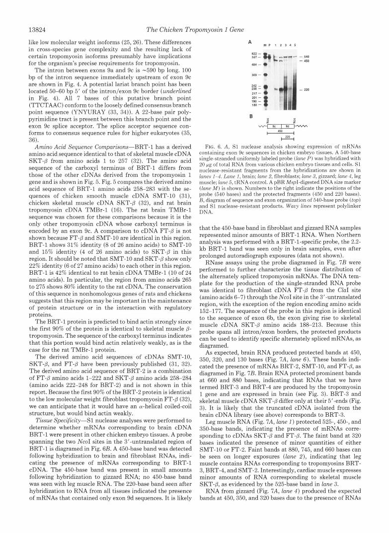
Tissue Specificity-S1 nuclease analyses were performed to determine whether mRNAs corresponding to brain cDNA BRT-1 were present in other chicken embryo tissues. A probe spanning the two NcoI sites in the 3’-untranslated region of BRT-1 is diagramed in Fig. 6B. A 450-base band was detected following hybridization to brain and fibroblast RNAs, indi- cating the presence of mRNAs corresponding to BRT-1 cDNA. The 450-base band was present in small amounts following hybridization to gizzard RNA; no 450-base band was seen with leg muscle RNA. The 220-base band seen after hybridization to RNA from all tissues indicated the presence of mRNAs that contained only exon 9d sequences. I t is likely
A M P 1 2 3 4 5
450
e% FIG. 6. A, S1 nuclease analysis showing expression of mRNAs
containing exon 9c sequences in chicken embryo tissues. A 540-base single-stranded uniformly labeled probe (lane P ) was hybridized with 20 pg of total RNA from various chicken embryo tissues and cells. S1 nuclease-resistant fragments from the hybridizations are shown in lanes 1-4. Lane 1, brain; lane 2, fibroblasts; lane 3, gizzard; lane 4, leg muscle; lane 5 , tRNA control. A pBR MspI-digested DNA size marker (lane M ) is shown. Numbers to the right indicate the positions of the probe (540 bases) and the protected fragments (450 and 220 bases). R, diagram of sequence and exon organization of 540-base probe ( top) and S1 nuclease-resistant products. W a y lines represent polylinker DNA.
that the 450-base band in fibroblast and gizzard RNA samples represented minor amounts of BRT-1 RNA. When Northern analysis was performed with a BRT-1-specific probe, the 2.2- kb BRT-1 band was seen only in brain samples, even after prolonged autoradiograph exposures (data not shown).
RNase assays using the probe diagramed in Fig. 7B were performed to further characterize the tissue distribution of the alternately spliced tropomyosin mRNAs. The DNA tem- plate for the production of the single-stranded RNA probe was identical to fibroblast cDNA FT-P from the ClaI site (amino acids 6-7) through the NcoI site in the 3”untranslated region, with the exception of the region encoding amino acids 152-177. The sequence of the probe in this region is identical to the sequence of exon 6b, the exon giving rise to skeletal muscle cDNA SKT-P amino acids 188-213. Because this probe spans all intron/exon borders, the protected products can be used to identify specific alternately spliced mRNAs, as diagramed.
As expected, brain RNA produced protected bands a t 450, 350, 320, and 130 bases (Fig. 7A, lane 6). These bands indi- cated the presence of mRNAs BRT-2, SMT-10, and FT-P, as diagramed in Fig. 7B. Brain RNA protected prominent bands a t 660 and 880 bases, indicating that RNAs that we have termed BRT-3 and BRT-4 are produced by the tropomyosin 1 gene and are expressed in brain (see Fig. 3). BRT-3 and skeletal muscle cDNA SKT-P differ only a t their 5’-ends (Fig. 3). I t is likely that the truncated cDNA isolated from the brain cDNA library (see above) corresponds to BRT-3.
Leg muscle RNA (Fig. 7A, lane 1 ) protected 525-, 450-, and 350-base bands, indicating the presence of mRNAs corre- sponding to cDNAs SKT-8 and FT-0. The faint band at 320 bases indicated the presence of minor quantities of either SMT-10 or FT-2. Faint bands a t 880, 745, and 660 bases can be seen on longer exposures (lune 2), indicating that leg muscle contains RNAs corresponding to tropomyosins BRT- 3, BRT-4, and SMT-2. Interestingly, cardiac muscle expresses minor amounts of RNA corresponding to skeletal muscle SKT-P, as evidenced by the 525-base band in lane 3.
RNA from gizzard (Fig. 7A, lane 4 ) produced the expected bands at 450,350, and 320 bases due to the presence of RNAs
A 1 2 3 4 5 6 7 M P
1630 - I ." .. - I4 - 622
The Chicken Trol
* -527
0 - 404
0 - 309
-217 0 - 201 t = l g o 160 * -180 * -180
6 -123
-116
120 . , 5 2 5
350
450 . . 3% 525 I 220
& I
450
I I
660
680 I I I 745
FIG. 7. A, RNase protection assay demonstrating expression of alternatively spliced tropomyosin 1 mRNAs in various chicken em- bryo tissues and cells. A 1630-base uniformly labeled antisense RNA probe (/ane P ) was hybridized to 20 pg of total RNA. Ten pg of poly(A)' brain RNA was used. RNase-resistant fragments from hy- bridizations with RNA from various chicken embryo tissues and cells are shown in lanes 1-6. Lane 1, leg muscle; lane 2, leg muscle; lane 3, heart; lane 4, gizzard; lane 5 , fibroblasts; lane 6, brain; lane 7, tRNA control. The RNase-resistant products were exposed to autoradi- ographic film for either 1 day (lanes 1 and 6) or 7 days (lanes 2-5). Numbers to the right are sizes in bases as indicated by a pBR MspI- digested DNA marker (lane M). Numbers to the left indicate positions of the probe and the RNase-resistant fragments. R, diagram of sequence and exon composition of probe (top) and resistant frag- ments. Black boxes represent RNA sequences common to all mRNAs. White boxes represent RNA sequences derived from alternate exons Ib, 6b, and 9d. The numbers above the probe diagram indicate amino acid positions. Size in bases is shown above each resistant fragment. The wavy line represents polylinker and unrelated RNAs.
corresponding to smooth muscle cDNA SMT-10 and fibro- blast cDNA FT-P. Fibroblasts (lane 5 ) showed the same set of bands. Neither gizzard nor fibroblast RNAs protected 525- base bands. However, a prominent band a t 745 bases in gizzard samples indicated the expression of an RNA termed SMT-2 (Fig. 3). SMT-2 differs from SMT-10 only in the choice of internal exon 6b instead of 6a. The band at 130 bases in both gizzard and fibroblasts indicated that they contain RNAs corresponding to either BRT-2 or FT-2.
To confirm the assignments shown in Figs. 6 and 7, addi- tional S1 nuclease analyses were performed both with uni- formly labeled and end-labeled DNA probes (data not shown). Analyses were done with probes derived from the SKT-@ cDNA clone, the BRT-2 cDNA clone, the SMT-10 cDNA clone, and a DNA construction that was built to confirm the structures we have proposed for BRT-4. Since many of the analyses can yield small fragments of similar size, we have used the protection of large fragments as the most definitive diagnostic technique. The data were compiled in tabular form so that the results of the various RNase and S1 nuclease protection assays could be compared. Based upon these anal- yses, a decision was made on the presence (or absence) of a
oomyosin 1 Gene 13825
particular form of tropomyosin RNA in a particular tissue. The details of this analysis are available upon request. The combined results of the analyses are shown diagrammatically in Fig. 3.
Implications for Alternative Splicing-The results summa- rized in Fig. 3 have implications for the regulation of alter- native splicing. The differential splicing of structurally iden- tical primary transcripts in a tissue-specific manner indicates that tissue-specific trans-acting factors are involved in alter- native splicing (for review, see Ref. 37). For example, mRNAs BRT-2 and BRT-3 are the differentially spliced products of a primary RNA that initiates transcription with exon Ib and is polyadenylated at the terminus of exon 9a. BRT-2 appears to be expressed in all tissues; BRT-3 is expressed in leg muscle and brain, but not in gizzard and fibroblasts.
The patterns of expression shown in Fig. 3 indicate that all tissues studied are capable of using both promoters. Likewise, both carboxyl-terminal exons 9a and 9d are expressed in all tissues. The expression of carboxyl-terminal exon 9c is more limited, suggesting that the exon 9c splice acceptor is not recognized in all tissues. RNA secondary structure in this region may prevent the use of the splice acceptor, and/or the binding of tissue-specific positive or negative regulatory fac- tors in this region may also regulate splicing.
Our data suggest that exon 6a is preferred over exon 6b in some tissues, whereas in other tissues, the opposite choice is made. This is most obvious in leg muscle and fibroblasts, both of which contain relatively homogeneous populations of cells. Leg muscle appears to preferentially incorporate exon 6b; the preference for exon 6a in fibroblasts is evidenced by the production of four mRNAs that include exon 6a (i.e. SMT- 10, FT-P, BRT-2, and FT-2). The only fibroblast mRNA that includes exon 6b is BRT-1. Exon preference is less clear in gizzard and brain. In our experiments, the presence of both exons 6a and 6b in RNAs from gizzard and brain could be due to the presence of various cell types. Each cell type could use only one of the two exons due to the presence or absence of specific factors. Alternatively, the factors that control exon use may be expressed in varying amounts in the different cell types, allowing the use of both exons. Our data suggest that the second possibility is more likely. We know that gizzard is composed largely of smooth muscle cells and contains only minor amounts of connective tissue. Gizzard uses exon 6a in mRNA SMT-10 and exon 6b in mRNA SMT-2. The possi- bility that SMT-2 is made only in the connective tissue subpopulation is unlikely since fibroblasts make no detectable SMT-2. It is therefore probable that smooth muscle cells themselves express exon 6b.
I n vitro splicing studies using a minigene that includes exons 5-8 of the rat @-tropomyosin gene has indicated that the critical event in alternative splicing was the joining of either exon 6a or 6b to the downstream exon 7 (27). Through analysis of this and other alternatively spliced genes, it has been proposed that alternative splicing factors influence the outcome of a single pivotal splicing event and that all other splicing events follow (38). In the process of screening a 10- day chicken embryo body wall cDNA library, we have isolated a cDNA for a tropomyosin RNA that has not made an internal exon splicing choice (Fig. 8). The existence of this cDNA suggests that the splicing of mutually exclusive exons 6a and 6b can follow all of the other processing and splicing events. This cDNA does not by itself rule out the possibility that the splicing of either exon 6a or 6b to exon 7 normally precedes the splicing of these exons to exon 5, as suggested by in vitro experiments (27); however, it does suggest the possibility that splicing of the internal alternate exons is not the pivotal event
13826 The Chicken Tropomyosin 1 Gene
in the further processing and splicing of primary tropomyosin transcripts.
Acknowledgments-We are grateful to Hilda Marusiodis for typing the manuscript and to Marilyn Powers for supplying the oligonucle- otides.
REFERENCES 1. Forry-Schaudies, S., Maihle, N. J., and Hughes, S. H. (1990) J.
2. Matsumura, F., Lin, J . J.-C., Yamashiro-Matsumura, S., Thomas, G. P., andTopp, W. C . (1983) J. Biol. Chem. 258,13954-13964
3. Giometti, C . S., and Anderson, N. L. (1984) J. Bid. Chem. 259,
4. Hendricks, M., and Weintraub, H. (1984) Mol. Cell. Biol. 4,1823-
5. Lin, J. J.-C., Helfman, D. M., Hughes, S. H., and Chou, C.3.
6. Srnillie, L. B. (1979) Trends Biochem. Sci. 4, 151-154 7. Ebashi, S., Endo, M., and Ohtsuki, I. (1969) Q. Rev. Biophys. 2,
8. Taylor, E. W. (1979) Crit. Rev. Biochem. 6, 103-164 9. Lazarides, E. (1975) J. Cell Bid. 65, 549-561
Mol. Bid . 211, 321-330
14113-14120
1833
(1985) J. Cell Biol. 100, 692-703
351-384
10. Fattoum, A., Hartwig, J. H., and Stossel, T. P. (1983) Biochem-
11. Payne, M. R., and Rudnick, S. E. (1984) Trends Biochem. Sci. 9,
12. Pearson-White, S. H., and Emerson, C. P., Jr. (1987) J. Biol.
13. MacLeod, A. R., and Gooding, C. (1988) Mol. Cell. B id . 8, 433-
14. Wieczorek, D. F., Smith, C. W. J., and Nadal-Ginard, B. (1988)
15. Lindquester, G. J., Flach, J. E., Fleenor, D. E., Hickman, K. H.,
16. Lees-Miller, J. P., Goodwin, L. O., and Helfman, D. M. (1990)
17. MacLeod, A. R., Houlker, C., Reinach, F. C., and Talbot, K.
18. Clayton, L., Reinach, F. C., Chumbley, G. M., and MacLeod, A.
19. MacLeod, A. R., Houlker, C., Reinach, F. C., Smillie, L. B., Talbot, K., Modi, G., and Walsh, F. S. (1985) Proc. Natl. Acud. Sci. U. S. A. 82, 7835-7839
istry 22,1187-1193
361-363
Chem. 262, 15998-16010.
440
Mol. Cell. Bid. 8, 679-694
and Devlin, R. B. (1989) Nucleic Acids Res. 17, 2099-2118
Mol. Cell. Biol. 10, 1729-1742
(1986) Nucleic Acids Res. 14, 8413-8426
R. (1988) J. Mol. Bid. 201, 507-515
20.
21.
22.
23.
24.
25.
26.
27.
28.
29.
30.
31.
32.
33.
34. 35. 36.
37.
38. 39.
40.
41.
42.
Helfman, D. M., Cheley, S., Kuismanen, E., Finn, L. A., and Yamawaki-Kataoka, Y. (1986) Mol. Cell. Biol. 6, 3582-3595
Libri, D., Lemonnier, M., Meinnel, T., and Fiszman, M. Y. (1989) J. Bid. Chem. 264,2935-2944
Libri, D., Mouly, V., Lemonnier, M., and Fiszman, M. Y. (1990) J. Biol. Chem. 265,3471-3473
MacLeod, A. R., Talbot, K., Smillie, L. B., and Houlker, C . (1987) J. Mol. B id . 194, 1-10
Yamawaki-Kataoka, Y., and Helfman, D. M. (1987) J. Biol. Chem.
Lees-Miller, J. P., Yan, A., and Helfman, D. M. (1990) J. Mol.
Forry-Schaudies, S., Gruber, C. G., and Hughes, S. H. (1990) Cell
Helfman, D. M., Ricci, W. M., and Finn, L. A. (1988) Genes &
Goux-Pelletan, M., Libri, D., Aubenton-Carafa, Y., Fiszman, M.,
Helfman, D. M., Roscigno, R. F., Mulligan, G. J., Finn, L. A.,
Libri, D., Goux-Pelletan, M., Brody, E., and Fiszman, M. Y.
Helfman, D. M., Feramisco, J . R., Ricci, W. M., and Hughes, S.
Bradac, J. A,, Gruber, C. E., Forry-Schaudies, S., and Hughes, S.
Keller, E. B., and Noon, W. A. (1984) Proc. Natl. Acad. Sci.
Reed, R., and Maniatis, T. (1985) Cell 41, 95-105 Mount, S. (1982) Nucleic Acids Res. 10, 459-472 Shapiro, M. B., and Senapathy, P. (1987) Nucleic Acids Res. 15,
Smith, C. W. J., Patton, J. G., and Nadal-Ginard, B. (1989) Annu.
Latchman, D. S. (1990) The New Biologist 2,297-303 Maniatis, T., Fritsch, E. F., and Sambrook, J. (1982) Molecular
Brody, E., and Marie, J. (1990) EMBO J. 9, 241-249
and Weber, K. S. (1990) Genes & Dev. 4,98-110.
(1990) Mol. Cell. Biol. 10, 5036-5046
H. (1984) J. Bid. Chem. 259, 14136-14143
H. (1989) Mol. Cell. Biol. 9, 185-192
U. 5'. A . 81, 7417-7420
7155-7174
Rev. Genet. 23, 527-577
Cold Siring Harbor,"NY ~- - .
Saneer. F.. Nicklen. S.. and Coulson. A. R. (1977) Proc. Natl. A&d: Sci. U. S. A,' 74, 5463-5467 '
Gilman, M. (1987) in Current Protocols in Molecular Biology (Ausubel, F. F., Brent, R., Kingston, R. E., Moore, D. D., Smith, J. A,, Seidman, J. G., and Struhl, K., eds) John Wiley & Sons, Inc., New York
Sap, J., Munoz, A., Damm, K., Goldberg, Y., Ghysdael, J., Leutz, A., Beug, H., and Vennstrom, B. (1986) Nature 324,635-640
The Chicken Tropomyosin 1 Gene 13827
RNA I s o l a t i o n a d northern Transfer - 17- or 18-day chicken embryos were u n u x m ~ N D MODI
dissected and the tissues immediately frozen in liquid nitrogen. Total RNA was isolated from the tissues according to the RNA201 procedure (cinnal Biotecx Laboratories). Northern transfer was p r f o m e d as in (26). A "P- labeled, nick-translated prob. prepared from the inmert in smooth muscle tropomyosin =DNA SWT-10 (32) was Used. Kodak M - 5 film was e w s e d either overnight or for 7 days at -70 C with intensifying scrams.
cDNl Library PrOducfion - Total RNA was prepared from 17-day chicken embryo
directed in the l R N A mrification Kit protocol (Phamscia, Inc). brain tissue as above. Polysdenylated RNA was isolated from total RNA a8
First-strand and second-strand synthesis were perfomad as directed in the SUprScript Plasmid Cloning System Uaers Manual (BRL Life Technologies.
primed by a 42-bp primer-adaptor that contains fifteen dT residues and four Inc) , starting with 3.4 pg of poly(A)' brain RNA. First-strand BynthesiS Was
Sizes of first-strand products ranged from 0.5 kb to 6.6 kb, as determined by rare restriction endonuclease sites including H o t I , HruI , X b a I , and Sper .
alkaline agarose gel electrophoresis. 300 Pmole Of ECORI adaptors (Promega Biotac) were ligated to the =DNA products of the second-strand reaction for 18 h at 16.c. After DNA precipitation and resuspnsion in TEN buffer (lo mn Tris-HCl (pH 7.5), 1 mJ4 EDTA. 25 IUI NaClI, the EDNA Was Site fractionated On i) Sephscryl 500 HR chromatography column as described in the Superscript Plasmid cloninq system protocol. Fractions containing the majority of the Cerenkov radiation were combined and S'-tsrminal phosphate was added using 2 mn ATP and polynucleotide kinase (Phsmacia. Inc). Approximately 50 ng of =DNA were ligated to 1 pg of ECORI digested, phosphatassd lambda gtll arms
packaged using Gigspask XI Plus packaging extract (Stratagens Cloning (Stratagene Cloning systems) for 16 h at 4'C. 500 nq of this ligation was
Systems). The library was titered in the preaence of X-gal (5-bromo-l- fhlo~0-3-i~d~yl-8-D-q~l~ctopyrsnoPlde~ and IPTC (isopropyl-8-thio-gslacto- pyranoside) and found to contain 3.6 x lo5 plaque coming units (pfu) per microgram of ligated DNA. 36\ of the plaque forming units were white. indicating the presence Of inserts. The library was amplified according to the Gigapack 11 Plus packaging extract instruction manual, using the E. c o l i host strain Y1088. Supernatants containing the amplified library were stored at 4'C.
the chicken embryo brain library were screened using a "P-labeled, nick- translated probe prepared from the insert Of smooth muscle tropomyosin cDNA SUT-10 (32). Thirty-one plaques were isolated. All 31 plaques were seem- darily screened with one or several of the following nick-translated probes: EDNA SWT-10 (above): the fragment extending from the Xb.1 Site through the
c ~ A Library Screening - 6.3 x 10s pfu (aquivalent to 2.1 x 10% white pfu) of
SIC1 site 600 bases 3' of it in the tropomyosin 1 gene A6 clone (1); a 500-bp fragment containing the 3' untranslatsd region of skeletal muscle cDNA SKT-p ( 3 2 ) . from the H i n c l l site to the EcoRl site in the polylinksr; a 310-bp
C t J I site [equivalent to the position encoding amino acid 7 in fibroblast fragment extending from the Sphl site upstream of exon lb through the exon lb
CDNA FT-8 (32)). All hybridization and filter washing was performed as described (32).
standard procedures (39). EcoRI fragments were isolated and ligated into the EcoRI site of Bluescript E(-) (stratagens Cloning Systems).
Phage from the secondary plaques were amplified and DNA vas isolated by
restriction endonucleasn digestions of aubclonsd brain CDNAS, the tropomyosin By combining data from secondary screening, sequence analysis, and
Clone8 isolated from the brain cDNA library could be classified into several groups. -0 Clones Yere identical to smooth muscle cDNA SWT-10. Seven Clones Of varying lengths represented a new brain tropomyosin that we have termed BRT-1. Thrne clones were a new brain tropomyosin that we have termed BRT-2. One clone is identical to EDNA SKT-8 from the position encoding amino acid 126 through the poly(A) tail. Five clones were derived that arise from a different tropomyosin genomic locus. The identity of the remaininq thirteen clones was not dstsmined.
Oh1 Squencc Analysis - E c o m inserts were isolated from plasmid DNA prepara-
dideoxy chain termination sequencing (40) was prformed using sequenase tions and subcloned into M13mpl8 or M13mp19. muble- and single-stranded
(United States Biochemical corp.). me sequence of both strands of the BRT-~
nucleotide primers. CDNA and one strand of the BRT-2 =DNA was determined using specific oligo-
as described (1). An IIl3mpl8 clone containing the 450-b~ HcoI - NcoI fragment from the 3' Untranslated region of CDNA BRT-I was used as a tamplate. extended Produst Was linearized with ECORI to generate a 540-base probe.
$1 HUClClSe h r l Y S i S - The Single-stranded, UnifOrm1y labeled probe was made
P 4 Of pOly(A)' Chicken embryo brain RNA or 20 pq of total RNA from chicken S1 nuclease analysis was perfomad using standard procedures (39). 3.4
embryo fibroblasts. leg muscle, or gizzard was hybridized with 6 X 10' cpm of probe for 16 h at 48'C. Hybrids were digested with 120 U of S1 nuclease and fractionated on 5\ polyacrylamide-7.6 II urea gels.
replacing the 380-bp Apr l - Hind111 fragment of fibroblast =DNA PP-8 (32) with the corresponding ApaI - Hind111 fragment O f skeletal muscle CDNA SKT-8 (32). These two Ap.1 - Hind111 fragments differ only in the region encoding amino acids 188-213 (152-177 in FT-8). The resulting clone Was digested with NCOI
RHaSe Protec t ion Assays - Construction O f the DNA template entailed first
to qenerate an 880-bp fragment that extends from the HcoI site corresponding to the AUG translation Start site of FT-# through the NCOI site in the 3' UDtranSlated region. The H C O I sites Yere filled in using DNA polymerase I Klanov fragment, and the fragment was cloned in the reverse orientation into
BmtI site. the ECORV site of a KS(+) plasmid containing 750 bp Of unrelated DNA in the
essentially as described (41). The DNA template described above Was linear-
transcribed using T7 RNA polymerase in the presence of [dP)UTP. 5 x lo' ized at the CIaI site corresponding to amino acids 6-7. Antisense RNA vas
cpm of probe RNA was incubated with either 10 pq of poly(A)' chicken embryo brain RNA or 20 pg of total RNA from all other chicken embryo tissues, overnight at 45'C. Hybrids were exposed to a mixture Of RNase TI and RNase A
for 60 mi" at 30'C. Products were fractionated on St polyacrylamide - 7.6 I4 "rea gels.
Production of RNA probes and psrfonsncs of RNase protection assays was
I 2 3 4 5 6
r -2.2
-1.2 -1.6 - 1.1
Pig. 1. Northern transfer showing sxprasSiOn Of chicken embryo RNAs that hybridize to a nick-translated SWT-10 (smooth muscle tropomyosin) probe. 20 Lq of total RNA vas used in each sample. Lane I, 18-day leg muscle: lane 2.'18-day breast muscle: l a m 3. 18-day gizzard;
day brain. Lanes I t o 5 are a 1-day autoradiographic exposure: l a m lane 4. chicken embryo fibroblasts: lane 5. 17-day brain: lane 6. 17-
6 is a 7-day exposure.
Fig. 4. Nucleotide sequence of the intron upstream of exon 9E. The intron is shown in m a 1 1 Capi ta ls and exon 9c is boxed and shown in large capitals. Amino acids 258 to 262 are shown. Dots are placed at 10 nucleotide intervals in the intron sequence. A potential branch point is underlined.
BUT-1 GLU ARC SER ARC GLN GLU ALA GLU L r s ASH ARC VAL LEU 260 270
SMT-10 --- SER LEU ALA SER ALA LYS --- GLU ASH VAL GLV ILE SKT-s ASP GLU VAL TYR ALA GLN LIS MET --- TYR LYS ALA ILE THBR-1 ASP GLN LEU TYR HIS GLN LEU --- GLN --- --- ARC ---
280 BUT-1 TWR Asn GLU LEU ARC VaL ILE LEU THR GLU LEU ASH Asn SMT-10 HIS GLN VAL --- ASP GLN THR --- LEU GLU --- --- --- LEU SKT-s SER GLU --- --- ASP ASH ALA --- ASH ASP ILE THR SER LEU TMBR-1 --- --- --- --- LIS LEU ALA --- ASN --- ASP
Fig. 5. Comparison of the derived amino acid sequence Of the Carboxy terminus of chicken brain EDNA BRT-1 to the corresponding regions of chicken smooth muscle EDNA SUT-10 132). chicken skeletal muscle =DNA SKT-B (32). and rat brain EDNA TIiBrli (16). N u d e n indicate amino acid position. Some amino acids are Shown as dasher, indicating identity to BRT-1.
Fig. 8 . Diagram Of the Sequence of an incompletely spliced tropomyosin 1 =DNA isolated from a lo-day chicken embryo body wall library (32 42). The black boxes represent =DNA sequences derived from consti:
alternatively spliced exons la, 2, 6a, 6b. and 9a. The horizontal tutive exons. The whi te boxes represent sequences derived from
l ines indicate intron sequencer. Humbcrr above the =DNA specify amino acid positions.


![Alternative splicing of [ -tropomyosin pre-mRNA: cis-actinggenesdev.cshlp.org/content/5/11/2096.full.pdf · [Key Words: [3-Tropomyosin gene; alternative splicing; RNA processing]](https://static.documents.pub/doc/80x56/5f0253b47e708231d403b8ce/alternative-splicing-of-tropomyosin-pre-mrna-cis-key-words-3-tropomyosin.jpg)


