74
Biology 30S WAEC 1 Biology 30S THE CIRCULATORY SYSTEM Name: ___________________ This module adapted from bblearn.merlin.mb.ca
Biology 30S WAEC
1
Biology 30S
THE
CIRCULATORY
SYSTEM
Name: ___________________
This module adapted from bblearn.merlin.mb.ca

Biology 30S WAEC
2
Introduction to Circulation
The first organ to form, and the last organ to die. The heart is the pump of life.
The main functions of the circulatory system:
______________________________________________________________________________________________________________________________________________________________________________________________________________________________________________________________________________________________________________________________________________________________
Types of circulatory system in animals:
_______________________________________________________________________________________________________________________________________________________________________________________________________________________________________________________________________________________
Cardiovascular disease accounts for the death of more Canadians than any other disease. In 1999, cardiovascular disease accounted for 78,942 Canadian deaths. 35% of all male deaths in Canada in 1999 were due to heart diseases, diseases of the blood vessels and stroke. For women, the toll was even higher – 37% of all female deaths in 1999 were due to cardiovascular disease.
Why do you think Cardiovascular related deaths are so high in Canada?
The circulatory or cardiovascular system plays a vital role of maintaining homeostasis in the human body. This homeostasis depends on the continuous and controlled movement of blood through the thousands of kilometres of blood vessels that ultimately reach every cell in the body. It is in the microscopic blood vessels that blood performs its ultimate transport function. Nutrients and other essential materials pass from blood into fluids surrounding the cells and waste products are removed. In this module, you will study the structure and function of the human circulatory system including the heart and the lymphatic system. You will also study and research some of the disorders and diseases that affect this system.
Biology 30S WAEC
3
Introduction to Lesson 1
All living things require nutrients to grow and reproduce. Single celled organisms and very simple multicellular organisms are able to diffuse these nutrients from the environment into their cells. As organisms become more sophisticated (more than two cell layers thick), a transport mechanism is required to transport nutrients to the cells and transport wastes away from cells. In humans, the circulatory system performs this transport function.
By the end of this lesson, you should be able to:
-List six ways in which the circulatory system maintains homeostasis in the human body.
-Explain how the structure of the five different types of blood vessels (arteries, arterioles, veins, venules, capillaries) is related to their function.
-Describe how the structure of the heart is related to its function, i.e. double pump.
-Identify and trace blood flow through the following structures of the heart from a specimen, model, or diagram:
left and right atria left and right ventricle left and right pulmonary arteries left and right pulmonary veins superior/inferior venae cavae septum aorta left and right semilunar valves left and right atrioventricular valves
Lesson 1 Outcomes

Biology 30S WAEC
4
-Describe the difference between the systemic and pulmonary circulatory system.
-Identify the following systemic blood vessels from a specimen, model, or diagram:
carotid arteries jugular veins subclavian artery and vein superior/inferior venae cavae coronary artery and vein renal artery and vein iliac artery and vein hepatic portal vein
Lesson 1 Overview
Following is a list of topics covered in this lesson.
The Circulatory System and Homeostasis Blood Vessels The Heart Major Systemic Blood Vessels Fetal Circulation
The Circulatory System and Homeostasis
The human circulatory system (also known as the cardiovascular system) consists of the heart, which is a muscular pumping device, and a closed system of vessels that are known as arteries, arterioles, veins, venules and capillaries. Blood contained in the circulatory system is pumped by the heart around a closed circle or circuit of vessels as it passes again and again through the various "circulations" of the body.
The circulatory system is vital to the maintenance of homeostasis in the body. Maintaining homeostasis depends on the continuous and controlled movement of blood through the thousands of kilometres of capillaries that permeate every tissue and reach every cell in the body. It is in the microscopic capillaries that blood performs its transport function.

Biology 30S WAEC
5
The circulatory system performs a number of important homeostatic functions in the human body. These include:
__________________________________________________________________________________________________________________________________________________________________________________________________________________________________________________________________________________________________________________________________________________________________________________________________________________________________________________________________________________________________________
Blood Vessels
Blood vessels are the channels through which blood is distributed to body tissues. The vessels make up two closed systems of tubes that begin and end at the heart. These two systems are:
________________________________________________________________________________________________________________________________________________________________________________________________________________________________________________________________________________________
Arteries
Characteristics:
____________________________________________________________________________________________________________________________________________________________________________________________________________________________________________________________________________________________________________________________________________________________________________________________________________________________________
The aorta, the largest artery in the human body, is about 25 mm in diameter.
Arterioles on the other hand are only about 0.2 mm in diameter.
Blood Vessels - Arteries

Biology 30S WAEC
6
Artery walls are thick, strong and muscular and made of three tissue layers.
In large arteries the middle layer is made mostly of elastic fibers. These fibers allow the large arteries to accommodate surges of blood pumped by the heart. The thick elastic walls stretch with the changing blood flow. This property of elasticity means that they can expand to accept a volume of blood, and then contract and squeeze back to their original size after the pressure is released. A good way to think of them is like a balloon. When you blow into the balloon, it inflates to hold the air. When you release the opening, the balloon squeezes the air back out.
As the arteries get smaller, the blood pressure gets less and the need for elastic fibers in the middle section diminishes. This layer becomes mostly muscle fibers that contract, changing the size of the arterial channel, regulating the pressure and amount of blood that enters the capillaries. The artery's outer layer is made of fibrous, connective tissue, nerves, and tiny blood vessels that nourish the artery's walls.
The diagram below illustrates the structure of arteries.
Figure 5.1.1 – Structure of Arteries
(http://training.seer.cancer.gov/module_anatomy/unit7_3_cardvasc_blood1_classification.html

Biology 30S WAEC
7
Capillaries and Veins
Capillaries
Characteristics:
__________________________________________________________________________________________________________________________________________________________________________________________________________________
The average diameter of a capillary is 7/1000 mm (7 µm), just wide enough to let red blood cells pass through single file.
Capillaries are really more like a web than a branched tube. It is in the capillaries that the exchange between the blood and the cells of the body takes place. Here the blood gives up its oxygen and takes on carbon dioxide. In the special capillaries of the kidneys, the blood gives up many waste products in the formation of urine. Capillary beds are also the sites where white blood cells are able to leave the blood and defend the body against harmful invaders. The diagram below illustrates a capillary bed.
Figure 5.1.2 – Capillary Bed
(http://training.seer.cancer.gov/module_anatomy/unit7_3_cardvasc_blood1_classification.html)
There are approximately 100 000 km of capillaries in an adult. Because these minute capillaries are so numerous, (about 10 billion) they present a huge surface area to the tissue. More than 800 square meters of surface area allows for a great deal of exchange between the blood and the tissues. Tissues are so permeated with capillaries that rarely are any cells more than one cell layer away from a capillary.

Biology 30S WAEC
8
This is necessary to allow for the diffusion of materials between the body cells and the walls of the capillaries.
The walls of capillaries are only one cell thick.
Substances in the blood and substances in body tissue are exchanged only across the capillary endothelium.
Capillary networks serve nearly all of the living tissue of the body, their concentration in the tissue depending on the local need for exchange of materials. Muscles, which are called upon frequently to move the body, and the kidneys, which must remove waste products constantly, require great quantities of food and oxygen and are well supplied with capillaries. On the other hand, the cornea of the eye, a very inactive tissue, has none.
Veins
Characteristics:
______________________________________________________________________________________________________________________________________________________________________________________________________________________________________________________________________________________________________________________________________________________________
Because the walls of the veins are thinner and less rigid than arteries, veins can hold more blood. Almost 70 percent of the total blood volume is in the veins at any given time.
Although the blood is forced into the arteries under pressure, by the time it reaches the veins, this pressure is very low. Blood pressure in the veins is less than 1/10 of the pressure in the aorta. Therefore, another mechanism must be present for getting blood back to the heart.
How do veins function in moving the blood despite the low pressure?
____________________________________________________________________________________________________________________________________________________________________________________________________________________________________________________________________________________________________________________________________________________________________________________________________________________________________

Biology 30S WAEC
9
The diagram below illustrates the structure of veins.
Veins, like other blood vessels are subject to problems.
________________________________________________________________________________________________________________________________________________________________________________________________________________________________________________________________________________________
What is varicose veins?
____________________________________________________________________________________________________________________________________________
One of the first important events of your life took place about three and one-half weeks into your embryonic development. Your heart began to beat. You may be excused for not remembering as you were only about 2.5 mm in size at the time.
Your heart now is about the size of a large fist and has a mass of approximately 300 grams. It is a tough muscular organ which beats about 70 times and pumps 5 liters of blood every minute. Pumping over 7000 liters of blood each day, it has pumped about 35 million liters in your life time as a grade eleven student.
While most of the hollow organs of the body do have muscular layers, the heart is almost entirely muscle. Unlike most of the other hollow organs, whose muscle layers are composed of smooth muscle, the heart is composed of cardiac muscle called the myocardium. The heart is surrounded by a fluid-filled membrane called the pericardium.The pericardial fluid bathes the heart, preventing friction between its outer wall and the membrane.
The Heart

Biology 30S WAEC
10
The human heart is really two pumps working side by side. A thick wall of muscle, called the septum, separates the heart's right and left sides. Each side is divided into two chambers: the atrium and the ventricle. The upper chambers, the left atrium and right atrium, collect blood returning to the heart through veins. The thin muscles of their walls push blood a short distance into the lower chambers, the left ventricle and right ventricle. The thick, muscular walls of the ventricles contract forcefully, pushing blood out of the heart to the lungs and body through arteries.
The heart is responsible for pumping the blood to every cell in the body. It is also responsible for pumping blood to the lungs, where the blood gives up carbon dioxide and takes on oxygen. The heart is able to pump blood to both regions efficiently because there are really two separate circulatory circuits with the heart as the common link. Some even refer to the heart as two separate hearts, a right heart (pulmonary system) and left heart (systemic system).
In the pulmonary system, blood leaves the heart through the pulmonary trunk which branches into the left and right pulmonary arteries, goes to the lungs, and returns to the heart through the left and right pulmonary veins. In the systemic system, blood leaves the heart through the aorta, goes to all the organs of the body through the systemic arteries, and then returns to the heart through the systemic veins.
Arteries carry blood away from the heart and veins carry blood toward the heart. Most of the time, arteries carry oxygenated blood and veins carry deoxygenated blood. However, there is an exception. The pulmonary arteries leaving the right ventricle for the lungs carry deoxygenated blood and the pulmonary veins carry oxygenated blood. The diagram below illustrates this relationship.
Figure 5.1.4 – Pulmonary Circulation(http://training.seer.cancer.gov/module_anatomy/unit7_3_cardvasc_blood3_
pathways.html)

Biology 30S WAEC
11
Blood from any body tissue other than the lungs returns to the heart through either of two veins: superior vena cava and inferior vena cava.
__________________________________________________________________________________________________________________________________________________________________________________________________________________________________________________________________________________________________________________________________________________________________________________________________________________________________________________________________________________________________________
Refer to the diagram below.
Figure 5.1.5 – Blood Flow though the Heart
(http://www.tmc.edu/thi/anatomy.html)

Biology 30S WAEC
12
A simplified diagram of the blood flow:
Two valves regulate the flow of blood between the atria and ventricles.
These valves, commonly called the atrioventricular (AV) valves, consist of three flaps of tissue that together form a more or less funnel-shaped arrangement, the narrow end extending into the ventricle. The pressure of the blood in the atrium forces the valve open, but when pressure develops in the ventricle, the pressure pushes the flaps
against each other, effectively closing the opening.
These two valves that regulate blood flow between the atria and ventricles are -
Tricuspid valve:
__________________________________________________________________________________________________________________________________________________________________________________________________________________
Mitral valve:
__________________________________________________________________________________________________________________________________________________________________________________________________________________
Two valves regulate the flow of blood between the ventricles and the major vessels leaving those ventricles. These valves are commonly known as semilunar valves.
The two semilunar valves are –
Pulmonary valve:
________________________________________________________________________________________________________________________________________________________________________________________________________________________________________________________________________________________

Biology 30S WAEC
13
Aortic valve:
________________________________________________________________________________________________________________________________________________________________________________________________________________________________________________________________________________________
The branches of the aorta carry oxygenated blood to all parts of the body except the lungs. In the brain, a muscle, a gland, or some other organ, the oxygenated blood becomes deoxygenated blood as it releases its oxygen and accepts carbon dioxide from the tissues.
Major Systemic Arteries and Veins
All systemic arteries are branches, either directly or indirectly, from the aorta. The aorta ascends from the left ventricle, curves to the left, and descends through the thorax and abdomen. This geography divides the aorta into three portions:
__________________________________________________________________________________________________________________________________________________________________________________________________________________
After blood delivers oxygen to the tissues and picks up carbon dioxide, it returns to the heart through a system of veins. The capillaries, where gas exchange occurs, merge into venules and these converge to form larger and larger veins until the blood reaches either the superior vena cava or inferior vena cava, which drain into the right atrium.
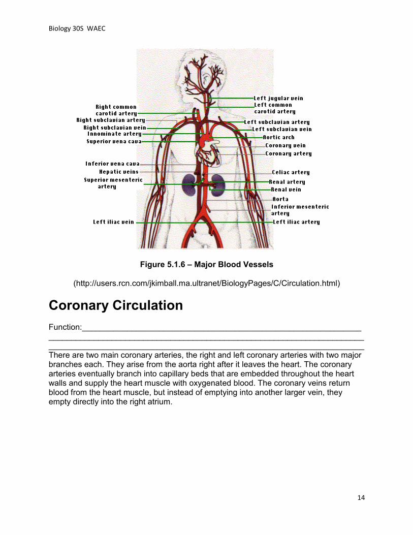
The diagram below illustrates the major blood vessels in the human body.
Systemic and Coronary
Circulation
Biology 30S WAEC
14
Figure 5.1.6 – Major Blood Vessels
(http://users.rcn.com/jkimball.ma.ultranet/BiologyPages/C/Circulation.html)
Coronary Circulation
Function:__________________________________________________________________________________________________________________________________________________________________________________________________________There are two main coronary arteries, the right and left coronary arteries with two major branches each. They arise from the aorta right after it leaves the heart. The coronary arteries eventually branch into capillary beds that are embedded throughout the heart walls and supply the heart muscle with oxygenated blood. The coronary veins return blood from the heart muscle, but instead of emptying into another larger vein, they empty directly into the right atrium.

Biology 30S WAEC
15
Figure 5.1.7 Coronary Arteries
(http://www.tmc.edu/thi/coroanat.html)
Disease in coronary arteries prevents the heart from receiving enough oxygen. These diseases will be discussed in lesson 5.
Fetal Circulation
In the human fetus, the lungs are not functional; the placenta substitutes for the lungs as the organ of gas exchange. How does the fetus’ gas exchange work? ______________________________________________________________________________________________________________________________________________________________________________________________________________________________________________________________________________________________________________________________________________________________ Oxygenated blood is delivered to the fetus from the placenta by the umbilical vein. This highly oxygenated blood flows into the inferior vena cava, which enters the right atrium. However, deoxygenated blood being returned from the internal organs contaminates the pure placental blood flowing in the inferior vena cava. Fortunately, the volume of placental blood is large, so that the mixture entering the right atrium is relatively well oxygenated. Ordinarily, blood would flow directly from the right atrium into the right ventricle, and, in turn, would leave the heart through the pulmonary trunk to the lungs. This would be a useless course in the fetus since the lungs are inactive.
Biology 30S WAEC
16
The main volume of the relatively pure blood in the right atrium crosses through a special opening, known as the foramen ovale, into the left atrium. From the left atrium the blood reaches the left ventricle, which pumps the blood into the aorta to be delivered through the systemic system.
Thus, the foramen ovale is an important device to ensure that a considerable portion of the oxygenated blood passes directly from the right atrium into the left atrium.
Blood that passes from the right atrium to the right ventricle will be directed through a second shunt, called the ductus arteriosus that leads to the aorta. Some of the blood will reach the lungs through the pulmonary trunk, but a greater part arising from the right ventricle will continue through the ductus arteriosus to the aorta.
Fetal circulation ceases at birth. When the lungs of the newborn expand with air, pulmonary circulation begins so that there will be an adequate supply of oxygen to the body. Constriction of the ductus arteriosus occurs shortly after birth with the result that the blood leaving the right ventricle no longer bypasses the lungs. Also, the foramen ovale is gradually sealed.
What is a “Blue Baby?”
____________________________________________________________________________________________________________________________________________________________________________________________________________________________________________________________________________________________________________________________________________________________________________________________________________________________________

Biology 30S WAEC
17
1. List 5 ways that the human circulatory system contributes to homeostasis.
2. Compare and contrast the structure and function of arteries, capillaries and veins.
3. Describe main differences between arteries and veins in terms of structures, functions and locations.
4. Why is it necessary to have 100 000 km of capillaries in your body?
5. Differentiate between the pulmonary and systemic circulatory systems.
6. Name and give the function of the four valves found in the heart.
7. Using the following diagram of the heart on the next page, label the major structures and trace the flow of blood using arrows. Use the colors blue and red to distinguish between deoxygenated and oxygenated blood.
Lesson 1 Exercise

Biology 30S WAEC
18
8. How does the heart receive its supply of blood?
9. What two modifications to the circulatory system are found in developing embryos?
Biology 30S WAEC
19
Lesson 1 Summary
In this lesson, I learned: ________________________________________________________________________________________________________________________________________________________________________________________________________________________________________________________________________________________________________________________________________________________________________________________________________________________________________________________________________________________________________________________________________________________________________________
Introduction to Lesson 2 - Heartbeat Activity: Locate your pulse at rest. Count how many times it beats in 15 seconds (look at a clock), then multiply this number by 4. This is your pulse rate_________________ Approximately how many heartbeats have you had since you were born? Since your heart has been beating since you were 2 weeks old “in utero” (inside the uterus), how many heartbeats have you had since your heart was formed?
The human heart beats an average of 70 times per minute, 24 hours a day, and 365 days a year. For those who have never had heart problems, the regular rhythmic beating of the heart is usually taken for granted. Did you know that the average heart beats or contracts over 3 billion times during a normal lifetime?

Biology 30S WAEC
20
By the end of this lesson, you should be able to:
1. Differentiate between systole and diastole and relate these to heart sounds.
2. Describe the intrinsic control of heartbeat, i.e. nervous (SA Node, AV Node, Perkinje Fibers, Bundle of HIS), and chemical (adrenaline, noradrenaline).
3. Explain the role of pacemakers in regulating heartbeat.
4. Describe the effects of adrenaline and noradrenaline on heart rate.
5. Measure your own heart rate.
6. Explain the effect of physical activity on heart rate.
7. Calculate cardiac output given heart rate and stroke volume.
8. Relate cardiac output to fitness levels.
Lesson 2 Overview
Following is a list of topics covered in this lesson.
Heartbeat Control of Heartbeat Artificial Pacemakers Cardiac Output Heart Rate, Stroke Volume and Fitness
Lesson 2 Outcomes

Biology 30S WAEC
21
A heartbeat is a two-part pumping action that takes approximately one second. The contraction of the heart and its anatomy cause the distinctive sounds heard when listening to the heart with a stethoscope. Systole and Diastole and The “Lubb – Dubb” sound: __________________________________________________________________________________________________________________________________________________________________________________________________________________ See the diagram below.
Figure 5.2.1 – Systole and Diastole
(http://www.tmc.edu/thi/systole.html)
After blood moves into the pulmonary artery and the aorta, the ventricles relax, and the pulmonary and aortic valves close. The lower pressure in the ventricles causes the tricuspid and mitral valves to open, and the cycle begins again. This series of contractions is repeated over and over again, increasing during times of exertion and decreasing while you are at rest.
Your heart does not work alone, though. Your brain tracks the conditions around you—climate, stress, and your level of physical activity—and adjusts your cardiovascular system to meet those needs.
Heartbeat

Biology 30S WAEC
22
The sinoatrial Node (SA Node):
______________________________________________________________________________________________________________________________________________________________________________________________________________________________________________________________________________________________________________________________________________________________
Why is the SA Node called the “natural pacemaker”?
______________________________________________________________________
The atrioventricular Node (AV Node):
______________________________________________________________________________________________________________________________________________________________________________________________________________________________________________________________________________________________________________________________________________________________
Bundle of His:
____________________________________________________________________________________________________________________________________________
The impulse started in the SA node and picked up by the AV node reaches the muscles of the ventricles and causes them to contract.
Purkinje fibers :
________________________________________________________________________________________________________________________________________________________________________________________________________________________________________________________________________________________
Control of Heartbeat

Biology 30S WAEC
23
Series of events in one heartbeat:
____________________________________________________________________________________________________________________________________________________________________________________________________________________________________________________________________________________________________________________________________________________________________________________________________________________________________
The following diagrams illustrate the sequence of events involved in a heart contraction.

Biology 30S WAEC
24
Figure 5.2.2 â The Contraction of the Heart
(Images from Purves et al., Life: The Science of Biology, 4th Edition, by Sinauer Associates,
www.sinauer.com and www.whfreeman.com
A physician listening carefully to the heart with a stethoscope can detect if the valves are closing completely or not. Instead of a distinctive valve sound, the physician may hear a swishing sound if they are letting blood flow backward. When the swishing is heard tells the physician where the leaky valve is located. This condition is known as a heart murmur.
Electrocardiograph:
__________________________________________________________________________________________________________________________________________________________________________________________________________________
The EKG shows three slow, negative changes, known as P, R, and T.
Positive deflections are the Q and S waves. The P wave represents the contraction impulse of the atria, the T wave the ventricular contraction.
EKGs are useful in diagnosing heart abnormalities.

Biology 30S WAEC
25
Figure 5.2.3 - Electrocardiogram
(Image from Purves et al., Life: The Science of Biology, 4th Edition, by Sinauer Associates,
www.sinauer.com and www.whfreeman.com
The SA node sends electrical impulses at a certain rate, but your heart rate may still change depending on physical demands, stress, or hormonal factors.
For example, when you run to catch a bus, the increased activity in your muscles produces a faster rate of cellular respiration. This leads to an increase in the amount of carbon dioxide in your blood. The medulla oblongata detects this increase and sends impulses along the nervous system causing the release of a hormone called noradrenaline. When noradrenaline reaches the SA node, it makes the node fire more rapidly. Once you have boarded the bus, your heart gradually slows to a resting rate due to an increase in blood pressure. This response is detected by special blood pressure receptors located in the walls of the aorta and carotid arteries that send messages to the medulla oblongata.
Physical activity is not the only trigger for an increased heart rate. Your nervous system releases another hormone called adrenaline when you are nervous, angry, excited or after a sudden shock or sharp pain. All of these conditions produce what is called the "fight or flight" response “ a physiological change that prepares the body for anticipated activity. This response increases heart rate, increasing blood flow to the muscles.

Biology 30S WAEC
26
Artificial Pacemakers and
Cardiac Output
Artificial Pacemakers
Arrythmia:
____________________________________________________________________________________________________________________________________________
When the natural pacemaker fails to work properly, doctors can implant a small, battery-operated device called an artificial pacemaker to help the heart beat in a regular rhythm.
Artificial pacemakers can be permanently implanted into a person's chest or they may be temporary and located outside of the body. Both types use batteries to send electrical impulses to the heart. A wire or electrode is placed next to the heart and transmits small electrical charges to the heart.
Most current pacemakers are demand pacemakers which have sensing devices to turn the pacemaker on when the heartbeat falls below a certain level.
Cardiac Output
The amount of blood pumped by the heart is called cardiac output. This is a measure of the volume of blood pumped from each ventricle per unit of time. It is also a measure of the level of oxygen delivery to the body.
Two factors affect cardiac output:
____________________________________________________________________________________________________________________________________________

Biology 30S WAEC
27
Cardiac output can be calculated by multiplying stroke volume and heart rate. The average person has a stroke volume of about 70 mL and a resting heart rate of about 70 beats/minute.
Therefore, the average person has a cardiac output of 70 x 70 = 4 900 mL/min. Since the average person has about 5 L of blood, your total blood volume circulates through your body approximately once every minute.
Heart Rate, Stroke Volume
and Fitness
Maximum heart rate (also known as Target heart rate) is the highest heart rate you can attain during strenuous physical activity. This rate will diminish as you get older although maximum heart rate does not appear to be related to fitness. The more important indicator of fitness is the length of time it takes for your heart to return to its resting level following physical activity. This is called recovery time , and this amount of time will diminish as you become more fit.
Two factors affect stroke volume –
___________________________________________________________________________________________________________________________________________Regular cardiovascular exercise will enlarge the ventricular chambers and increase their distensibility (stretchiness).
Athletes who are very fit have high stroke volumes. This means they can maintain high oxygen delivery to tissues at low heart rates. Some elite endurance trained athletes like Olympic cross country skiers have resting heart rates of as low as 30 beats/minute.
Biology 30S WAEC
28
Lesson 2
Exercise
1. What causes the characteristic "lubb-dubb" heart sounds?
2. Describe the structures involved and the sequence of events involved in a heart contraction.
3. What is an EKG or ECG? Why are they useful?
4. Why do some people require an artificial pacemaker?
5. Describe the conditions that would cause the release of noradrenaline and adrenaline and their resulting effects of on heart rate.
6. How are heart rate and stroke volume related to fitness?
7. Explain the changes in pulse rate during exercise.
8. Extension: How does an AED (Automated External Defibrillator) device work?

Biology 30S WAEC
29
Heart Rate Activity
Effects of Exercise on Heart Rate
Complete the following activity. Once you have performed the lab, prepare a report and submit it in the Assignments Tool — M4 L2 Heart Rate. Your report should have the following categories:
1. Purpose 2. Observations/Data 3. Analysis/Conclusions
Lesson 2 Lab Activity
Effects of Exercise on Heart Rate
Purpose:
To measure your heart rate, recovery time and cardiac output before, during and after vigorous exercise. If you have a physical condition that makes it unadvisable for you to exercise vigorously, do not participate in the exercise portion of this activity.
Procedure:
1. Find your resting heart rate by measuring your pulse. A pulse is a change in the diameter of arteries following a heart contraction. The easiest locations for measuring your pulse are directly under the back of your jawbone toward the neck or on the underside of your wrist directly beneath the thumb area.
2. While in a sitting position, record your pulse rate for 15 seconds. Multiply this number by four to determine your resting heart rate per minute. Record this value in the data table below. Repeat three times and determine the average.
3. Perform jumping jacks or running on the spot for one minute. Immediately after completing the exercise, measure your pulse rate for 15 seconds and calculate your heart rate after vigorous exercise.

Biology 30S WAEC
30
4. Continue to measure your pulse rate in one minute intervals until your heart rate reaches its resting rate. The time it takes for your heart to reach its resting rate is your recovery time. Record this time in the data table.
5. Repeat steps 3 and 4 two more times and calculate the average of the three trials.
Data Table
Trial 1 2 3 Average
Resting
Vigorous Exercise
Recovery Time
Analysis
1. What do you notice about the change in your heart rate from resting to vigorous exercise?
2. Compare your recovery time with other students. How does it compare?
3. Calculate your cardiac output for resting and vigorous exercise by using your averages and a stroke volume of 70 mL.
4. Why did you perform three trials for each heart rate measurement?
Lesson 2 Summary
In this lesson, you studied the physiology of heartbeat and the factors that influence heart rate. You also had an opportunity to measure your own heart rate and recovery rate. In the next lesson, you will study blood pressure and fluid exchange.
Biology 30S WAEC
31
Introduction to Blood Pressure and
Fluid Exchange
Uncontrolled high blood pressure can lead to stroke, heart attack, heart failure or kidney failure. However, because there are no symptoms, nearly one-third of people with high blood pressure don't even know they have it. This is why high blood pressure is often called the "silent killer". The only way to tell if you have high blood pressure is to have your blood pressure checked. Do you know your blood pressure?
Lesson 3 Outcomes
By the end of this lesson, you should be able to:
5.3.1 Identify systolic and diastolic blood pressure using a sphygmomanometer.
5.3.2 List and describe extrinsic factors (e.g., exercise, caffeine, nicotine) which affect transient blood pressure.
5.3.3 Differentiate between vasodilation and vasoconstriction.
5.3.4 Describe the control of blood pressure by the autonomic nervous system.
5.3.5 List and describe factors which affect arteriolar resistance.
5.3.6 Explain how changes in blood pressure help to maintain homeostasis in the body.
5.3.7 Explain how blood pressure and osmotic pressure contribute to fluid exchange at the capillary level.
5.3.8 Describe the term hypertension and discuss its causes, effects and treatment.
Biology 30S WAEC
32
Lesson 3 Overview
Following is a list of topics covered in this lesson.
Blood pressure Measuring Blood pressure Hypertension Exchanges between Blood and Cells Regulation of Blood Pressure
Blood Pressure
Blood pressure is defined as ________________________________________________________________________________________________________________________________________________________________________________________________________________________________________________________________________________________ The highest pressure occurs in the aorta, the large vessel that carries oxygenated blood away from the heart. As the blood passes into smaller vessels and the distance from the heart becomes greater, the pressure becomes reduced.
The pressure in any artery varies as a result of two major factors.
1. Cardiac Output
o Volume of blood.
____________________________________________________________________________________________________________________________________________________________________________________________________________________________________________
o Heart rate.
____________________________________________________________________________________________________________________________________________________________________________________________________________________________________________

Biology 30S WAEC
33
2. Arteriolar Resistance
o Size.
____________________________________________________________________________________________________________________________________________________________________________________________________________________________________________
o Elasticity.
____________________________________________________________________________________________________________________________________________________________________________________________________________________________________________
o Measuring Blood Pressure
____________________________________________________________________________________________________________________________________________________________________________________________________________________________________________
Two different pressures are measured and compared in a blood pressure reading.
Systolic pressure
____________________________________________________________________________________________________________________________________________
Diastolic pressure
____________________________________________________________________________________________________________________________________________
The pressure of the blood pressing against the arterial walls can be measured using a device called a sphygmomanometer. To measure blood pressure, an inflatable rubber cuff is wrapped around the upper arm. As air is pumped into the cuff, the cuff presses on the arteries of the arm. When the pressure in the cuff is high enough, the blood flow through the arteries ceases.
A stethoscope is placed over one of the arteries in the elbow, and the air in the cuff is gradually released. At first, the person listening through the stethoscope hears no sound. Then, a sharp tapping sound is heard. This sound is made by the blood spurting

Biology 30S WAEC
34
through a narrow opening in the compressed artery. The pressure reading just as this sound is heard is the systolic pressure. As more air is released from the cuff, the sound becomes muffled and then stops as the cuff ceases to press on the artery. The pressure reading just as the sound stops is the diastolic pressure.
Blood pressure is measured in millimeters of mercury (mm Hg). It is expressed as a ratio of systolic pressure to diastolic pressure. A reading of 120/70 means that the person's systolic pressure is 120 mm Hg and the diastolic pressure is 70 mm Hg. It is expressed verbally as "120 over 70."
Normal blood pressure is less than 130 mm Hg systolic and less than 85 mm Hg diastolic. Optimal blood pressure is less than 120 mm Hg systolic and less than 80 mm Hg diastolic. A typical reading for a healthy adult is 120/70. Readings for children and adolescents may be slightly higher.
A physician can infer much about a person's health by taking a blood pressure reading.
Hypertension
High blood pressure or hypertension is defined in an adult as a blood pressure greater than or equal to 140 mm Hg systolic pressure or greater than or equal to 90 mm Hg diastolic pressure. High blood pressure directly increases the risk of coronary heart disease (which can lead to heart attack) and stroke, especially along with other risk factors.
High blood pressure can occur in children or adults, but it's more common among people over age 35. It's particularly prevalent in middle-aged and elderly people, obese people, heavy drinkers and women who are taking birth control pills. It may run in families, but many people with a strong family history of high blood pressure never have it. People with diabetes mellitus, gout or kidney disease are more likely to have hypertension.
Recall:
Systolic Pressure:
_____________________________________________________________________
Diastolic Pressure:
_____________________________________________________________________
Biology 30S WAEC
35
Which is more dangerous to be higher than normal, systolic or diastolic pressure? ______________Why?_________________________________________________________________________________________________________________________
Blood pressure is normally controlled by nerves that have their center in the brain. If the blood pressure in certain vessels increases, the brain sends nerve impulses to the heart and to the blood vessels, causing the heart rate to slow and the blood vessels to widen. As a result the blood pressure decreases. If the blood pressure becomes too low, the brain sends impulses that cause the heart rate to increase and the blood vessels to narrow, increasing the blood pressure. This is another case of homeostasis—maintaining a constant internal environment. If this regulatory mechanism cannot bring the blood pressure to normal levels, a condition known as hypertension is evident and medical assistance is required.
The factors causing hypertension are not well understood. In 90% of the cases of hypertension, the cause is unknown.
What do you think can cause hypertension?
________________________________________________________________________________________________________________________________________________________________________________________________________________________________________________________________________________________
Risk Factors You Can Control
Smoking Physical Inactivity Obesity Diet (Salt Intake) Diabetes Stress
Risk Factors You Can’t Control
Age Ethnicity (South Asians, First Nations/Aboriginal Peoples or Inuit and Blacks are
at increased risk) Family history
The goal of treatment is to reduce the diastolic blood pressure to less than 90 mm Hg. Treatment consists of a combination of no-added-salt diet, weight loss if the person is over-weight, and drug medication. The excess fluid that sodium (salt) holds in the body may also put an added burden on the heart and "waterlog" the blood vessels, causing them to contract or narrow more easily. The blood vessels then take less diluted blood to the organs of the body than the quantity of normal blood that is required, depriving
Biology 30S WAEC
36
the cells of some oxygen and nutrients that they need. For this reason, low-sodium diets are used in treating mild to moderately severe hypertension. However, in individuals with severe hypertension, salt restriction must be severe.
When the demand for blood in various parts of the body is high (e.g. during exercise), the heart must pump faster, increasing the blood pressure in the vessels. Fatty tissue requires a lot of blood to feed it. Therefore, another way to reduce blood pressure and the stress on the heart is to lose weight. In addition to possibly lowering blood pressure and reducing weight, a low-fat, low-cholesterol diet may also help delay the beginning of arteriosclerosis. Medications called diuretics, which help rid the body of excess salt and therefore, of excess water, are often prescribed by the doctor in the treatment of hypertension.
Blood Pressure in Capillaries and in Veins
Blood pressure in the Capillaries
The pressure of arterial blood is significantly reduced when the blood enters the capillaries. Capillaries are tiny vessels with a diameter just about that of a red blood cell (7 µm). Although the diameter of a single capillary is quite small, the number of capillaries supplied by a single arteriole is so great that the total area available for the flow of blood is increased. Therefore, the pressure of the blood as it enters the capillaries decreases.
Blood pressure in the veins
When blood leaves the capillaries and enters the venules and veins, little pressure remains to force it along. Blood in the veins below the heart is helped back up to the heart by the muscle pump. This is simply the squeezing effect of contracting muscles on the veins running through them. One-way flow to the heart is achieved by valves within the veins.
Biology 30S WAEC
37
Exchanges between Blood and
Cells
Our blood does not come into direct contact with the cells it nourishes. However, the distribution of capillaries is so extensive that cells are never farther away than 1 cell layer from a capillary.
When blood enters the arteriole end of a capillary, it is still under pressure produced by the contraction of the ventricle. As a result of this pressure, a substantial amount of water and some plasma proteins filter through the walls of the capillaries into the tissue space.
This fluid, called interstitial fluid, is blood plasma minus most of the proteins. Interstitial fluid bathes the cells in the tissue space. Substances in the fluid can enter the cells by diffusion or active transport. Substances, like carbon dioxide, can diffuse out of cells and into the interstitial fluid.
Near the venous end of a capillary, the blood pressure is greatly reduced. Here, another force comes into play. Although the composition of interstitial fluid is similar to that of blood plasma, it contains a smaller concentration of proteins than plasma and a greater concentration of water. This difference sets up an osmotic pressure. Although the osmotic pressure is small, it is greater than the blood pressure at the venous end of the capillary. Consequently, the fluid re-enters the capillary at the venous end.
Regulation of Blood Pressure
An adult human has been estimated to have some 100 000 km of capillaries with a total surface area of some 800 -1000 m2(an area greater than three tennis courts). The total volume of this system is roughly 5 liters, the same as the total volume of blood. However, if the heart and major vessels are to be kept filled, all the capillaries cannot be filled at once. Therefore, a continual redirection of blood from organ to organ takes place in response to the changing needs of the body. For example, during vigorous exercise, capillary beds in the skeletal muscles open at the expense of those in the abdomen. The reverse occurs after a heavy meal.
Biology 30S WAEC
38
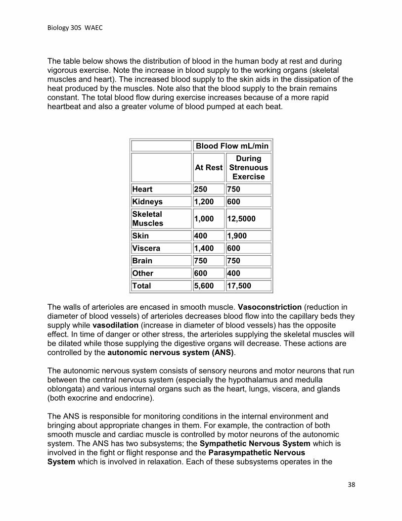
The table below shows the distribution of blood in the human body at rest and during vigorous exercise. Note the increase in blood supply to the working organs (skeletal muscles and heart). The increased blood supply to the skin aids in the dissipation of the heat produced by the muscles. Note also that the blood supply to the brain remains constant. The total blood flow during exercise increases because of a more rapid heartbeat and also a greater volume of blood pumped at each beat.
Blood Flow mL/min
At Rest During
Strenuous Exercise
Heart 250 750
Kidneys 1,200 600
Skeletal Muscles
1,000 12,5000
Skin 400 1,900
Viscera 1,400 600
Brain 750 750
Other 600 400
Total 5,600 17,500
The walls of arterioles are encased in smooth muscle. Vasoconstriction (reduction in diameter of blood vessels) of arterioles decreases blood flow into the capillary beds they supply while vasodilation (increase in diameter of blood vessels) has the opposite effect. In time of danger or other stress, the arterioles supplying the skeletal muscles will be dilated while those supplying the digestive organs will decrease. These actions are controlled by the autonomic nervous system (ANS).
The autonomic nervous system consists of sensory neurons and motor neurons that run between the central nervous system (especially the hypothalamus and medulla oblongata) and various internal organs such as the heart, lungs, viscera, and glands (both exocrine and endocrine).
The ANS is responsible for monitoring conditions in the internal environment and bringing about appropriate changes in them. For example, the contraction of both smooth muscle and cardiac muscle is controlled by motor neurons of the autonomic system. The ANS has two subsystems; the Sympathetic Nervous System which is involved in the fight or flight response and the Parasympathetic Nervous System which is involved in relaxation. Each of these subsystems operates in the

Biology 30S WAEC
39
reverse of the other (antagonism). Both systems affect the same organs and act in opposition to maintain homeostasis. For example: when you are scared, the sympathetic system causes your heart to beat faster; the parasympathetic system reverses this effect.
Baroreceptors
____________________________________________________________________________________________________________________________________________ When blood pressure exceeds acceptable levels, the receptors send nerve impulse messages to the medulla oblongata which causes the sympathetic (flight or fight) nerve impulses to decrease. This results in arteriole dilation and increased outflow of blood from the artery. The parasympathetic (relaxation) nerve impulses are increased, causing heart rate and stroke volume to decrease. The decreased cardiac output slows the movement of blood into the arteries, lowering blood pressure.
Low blood pressure is also adjusted by the sympathetic nerve. Without nerve information from the pressure receptors of the carotid artery and aorta, the sympathetic nerve will continue to be stimulated, causing cardiac output to increase and arterioles to constrict. The increased flow of blood into the artery, accompanied by a decreased outflow raises blood pressure to acceptable levels.
Control of Blood Pressure
Regulation of Blood Pressure by Hormones
The role of the kidney is __________________________________________________________________________________________________________________________________________________________________________________________________________________
Local Control of Blood Pressure in the Capillary Beds
Cells where infection or other damage is occurring release substances like histamine that dilate the arterioles and increase blood flow in the area.
Nitric oxide (NO) is also a potent dilator of blood vessels. When the endothelial cells that line blood vessels are stimulated, they synthesize nitric oxide. It quickly diffuses into the muscular walls of the vessels causing them to relax.
Biology 30S WAEC
40
Nitroglycerine is often prescribed to reduce the pain of angina (heart pain). It does so by generating nitric oxide, which relaxes the walls of the arteries and arterioles. The prescription drug sildenafil citrate ("Viagra ") does the same for vessels supplying blood to the penis. The effects of these two drugs are additive and using them together could precipitate a dangerous drop in blood pressure.
Shock
Trauma to the body or severe bleeding may cause shock which may result in capillary beds opening without others closing in compensation. Although the volume of blood remains unchanged, blood pressure declines abruptly as blood pools in the capillary beds. The heart can only pump as much blood as it receives. If insufficient blood gets back to the heart, its output - and hence blood pressure - drops. The tissues fail to receive enough oxygen. This is especially critical for the brain and the heart itself. If untreated, shock is usually fatal.
To cope with the problem, arterioles constrict and shut down the capillary beds - except those in the brain and heart. This reduces the volume of the system and helps maintain normal blood pressure. Air-breathing vertebrates that spend long periods under water (e.g., seals, penguins, turtles, and alligators) employ a similar mechanism to ensure that the oxygen supply of the heart and brain is not seriously diminished. When the animal dives, the blood supply to the rest of the body is sharply reduced so that what oxygen remains will be available for those organs needing it most: the brain and heart.

Biology 30S WAEC
41
Lesson 3 Exercise
1. a) What is blood pressure?
b) List and describe two major factors that affect blood pressure.
2. a) Differentiate between systolic and diastolic blood pressure.
b) How is blood pressure measured?
c) What are "normal" blood pressure measurements?
d) What effect does exercise have on blood pressure?
3. a) Define hypertension.
b) What are some of the possible side effects of hypertension?
c) Discuss the causes, risk factors and treatment of hypertension.
Biology 30S WAEC
42
4. a) Explain how blood pressure contributes to fluid exchange in the tissues
b) What other factor contributes to fluid exchange at the tissue level.
5. Differentiate between vasoconstriction and vasodilation.
6. How is blood flow to the muscles, kidneys and brain affected by vigorous exercise?
7. Describe how the Autonomic Nervous System contributes to homeostasis by controlling blood pressure. Include the role of blood pressure receptors in your answer.
8. Describe the role of the kidney in controlling blood pressure.
9. Why is nitroglycerin used as a treatment for angina?
10. Why is shock sometimes fatal if untreated?

Biology 30S WAEC
43
Lesson 3 Summary
In this lesson, you have learned about blood pressure, how it is measured, and some problems associated with high blood pressure. You have also studied how blood pressure helps to maintain homeostasis and fluid exchange at the cell level. In the next lesson, you will study the Lymphatic System.
Introduction to Lesson 4 - The Lymphatic System
Your circulatory system is not your body’s only vascular transport system. Closely associated with the blood vessels of the circulatory system is the lymphatic system. The lymphatic system is a network of glands and vessels that extend throughout your body.
Lesson 4 Outcomes
By the end of this lesson, you should be able to:
Describe the function of the lymphatic system in the human body.
List the components of lymph in the human body, i.e., fat, protein, water, white blood cells.
Identify the following structures of the lymphatic system from a specimen, model, or diagram:
adenoids tonsil lymph nodes spleen thoracic duct
Differentiate between lymph vessels and blood vessels.

Biology 30S WAEC
44
Lesson 4 Overview
Following is a list of topics covered in this lesson.
Function of the Lymphatic System Lymph Lymphatic Organs
The lymphatic system consists of fluid called lymph, vessels that transport the lymph and organs that contain lymphoid tissue.
The lymphatic system has three primary functions.
First of all, it returns excess interstitial fluid (the fluid that surrounds the cells) to the blood. Interstitial fluid is very abundant, making up about 15% of the body mass. It is the clear, colourless liquid that appears when the skin is grazed or a blister is broken.
All of the cells and tissues of the body must be continuously bathed by fluids. These fluids enable nutrients to pass from the capillaries to the cell membranes and waste products to return to the capillaries. Some of the fluid in blood is constantly passing through the capillary walls and entering the spaces between the cells. The walls of the capillaries prevent most of the blood cells and some of the proteins of the plasma from leaving the blood stream. However, some white blood cells leave the capillaries by forcing their way out between the cells that make up the capillary walls. The composition of intercellular fluid is much the same as the plasma of the blood.
Interstitial fluid bridges the gap between capillaries and the isolated cells which are not in direct contact with a capillary. It is laden with substances needed by the cells and also takes away waste substances from the cells.
Of the fluid that leaves the capillaries, about 90 percent is returned. The 10 percent that does not return becomes part of the interstitial fluid that surrounds the tissue cells. Lymph capillaries pick up the excess interstitial fluid and proteins and return them to the venous blood.
The Lymphatic
System

Biology 30S WAEC
45
The second and probably most well known function of the lymphatic system is defense against invading microorganisms and disease. Lymph nodes and other lymphatic organs filter the lymph to remove microorganisms and other foreign particles. Lymphatic organs also contain lymphocytes that destroy invading organisms. Lymphatic organs will be discussed later in the lesson.
The third function of the lymphatic system is the absorption of fats and fat-soluble vitamins from the digestive system and the subsequent transport of these substances to the venous circulation. The lining of the small intestine is covered with fingerlike projections called villi. There are blood capillaries and special lymph capillaries, called lacteals, in the center of each villus. The blood capillaries absorb most nutrients, but the fats and fat-soluble vitamins are absorbed by the lacteals. The absorbed fat droplets are transported to adipose (fat) tissue where they are stored.
Figure 5.4.3 Lacteal
(http://users.rcn.com/jkimball.ma.ultranet/BiologyPages/G/GITract.html#intestine)

Biology 30S WAEC
46
Lymph
Lymph is a fluid similar in composition to blood plasma (90% water, salts, proteins, hormones, nutrients, waste products, gases). As discussed above, it is derived from blood plasma as fluids pass through capillary walls at the arterial end. As the interstitial fluid begins to accumulate, it is picked up and removed by tiny lymphatic vessels and returned to the blood. As soon as the interstitial fluid enters the lymph capillaries, it is called lymph. Returning the fluid to the blood prevents a condition known as edema (swelling of the tissue) and helps to maintain normal blood volume and pressure.
The lymphatic system consists of capillaries and lymph vessels that correspond to the capillaries and veins of the blood circulatory system. Lymphatic vessels, unlike blood vessels, only carry fluid away from the tissues. The smallest lymphatic vessels are the lymph capillaries, which begin in the tissue spaces as blind-ended sacs. Lymph capillaries are found in all regions of the body except the bone marrow, central nervous system, and tissues, such as the epidermis, that lack blood vessels. The wall of the lymph capillary is composed of endothelium in which the simple squamous cells overlap to form a simple one-way valve. This arrangement permits fluid to enter the capillary but prevents lymph from leaving the vessel.
The lymph capillaries do not form a net like the blood capillaries but they resemble microscopic fingers that connect with the lymph vessels. The smaller lymph vessels unite, forming larger ones. See the diagram below.

Biology 30S WAEC
47
Figure 5.4.1 – Lymph Capillaries
(http://training.seer.cancer.gov/module_anatomy/unit8_2_lymph_compo.html)
Small lymphatic vessels join to form larger tributaries, called lymphatic trunks, which drain large regions. Lymphatic trunks merge until the lymph enters the two lymphatic ducts. The right lymphatic duct drains lymph from the upper right quadrant of the body. The thoracic duct drains all the rest. The diagram below illustrates the major lymphatic vessels and organs.
Figure 5.4.2 – Lymphatic Vessels and Organs
(http://www.acm.uiuc.edu/sigbio/project/lymphatic/index.html)
Like veins, the lymphatic tributaries have thin walls and have valves to prevent backflow of blood. There is no pump in the lymphatic system like the heart in the cardiovascular system. The pressure gradients to move lymph through the vessels come from the skeletal muscle action, respiratory movement, and contraction of smooth muscle in vessel walls.

Biology 30S WAEC
48
Lymphatic organs are characterized by clusters of lymphocytes and other white blood cells, such as macrophages in a framework of short, branching connective tissue fibers. The lymphocytes originate in the red bone marrow with other types of blood cells and are carried in the blood from the bone marrow to the lymphatic organs. When the body is exposed to microorganisms and other foreign substances, the lymphocytes multiply within the lymphatic organs and are sent in the blood to the site of the invasion. This is part of the immune response that attempts to destroy the invading agent.
Lymph nodes are small bean-shaped structures that are usually less than 2.5 cm in length. They are widely distributed throughout the body along the lymphatic pathways where they filter the lymph before it is returned to the blood. There are three regions on each side of the body where lymph nodes tend to cluster. These areas are the groin, the armpit, and the neck.
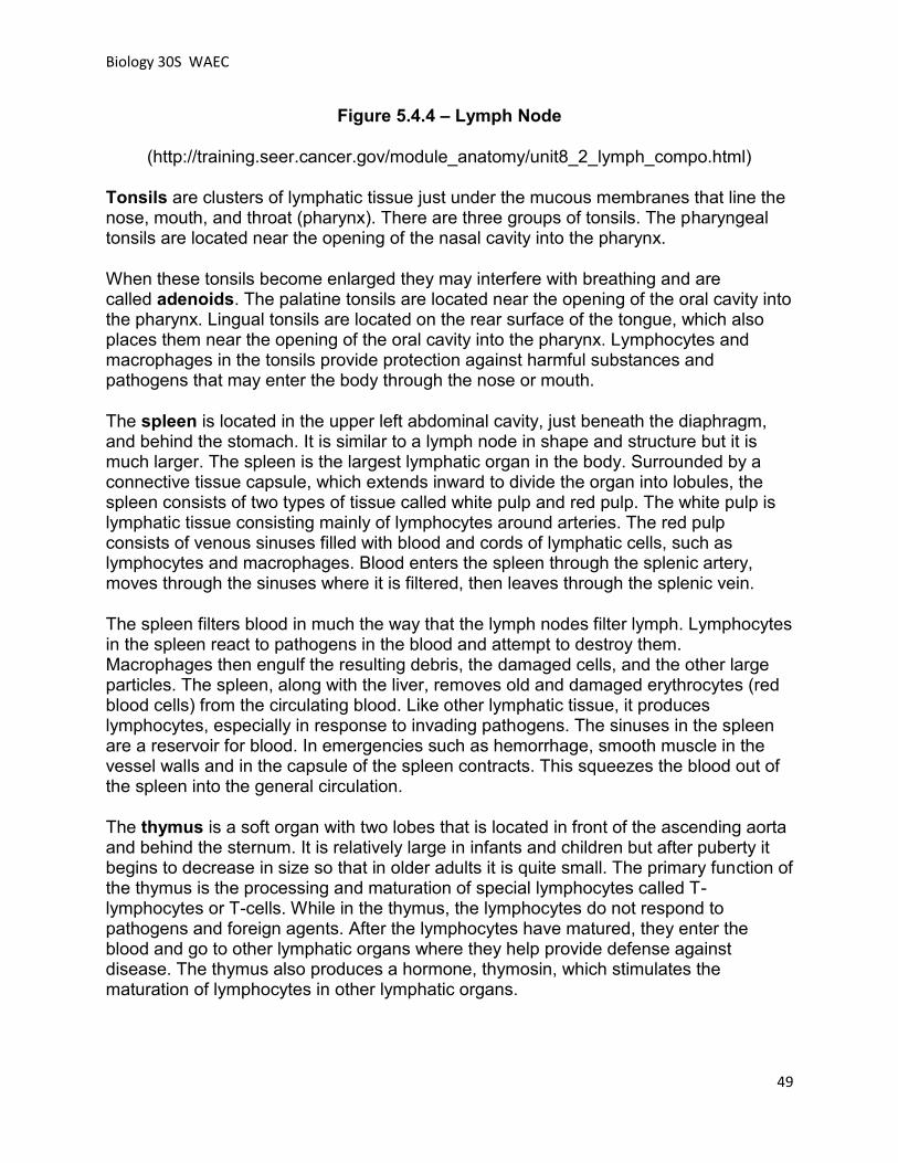
The typical lymph node is surrounded by a connective tissue capsule and divided into compartments called lymph nodules. The lymph nodules are dense masses of lymphocytes and macrophages and are separated by spaces called lymph sinuses. Several afferent lymphatic vessels, which carry lymph into the node, enter the node on the convex side. The lymph moves through the lymph sinuses and enters an efferent lymphatic vessel, which carries the lymph away from the node. Because there are more afferent vessels than efferent vessels, the passage of lymph through the sinuses is slowed down, which allow time for the cleansing process. See the diagram below.
Lymphatic
Organs
Biology 30S WAEC
49
Figure 5.4.4 – Lymph Node
(http://training.seer.cancer.gov/module_anatomy/unit8_2_lymph_compo.html)
Tonsils are clusters of lymphatic tissue just under the mucous membranes that line the nose, mouth, and throat (pharynx). There are three groups of tonsils. The pharyngeal tonsils are located near the opening of the nasal cavity into the pharynx.
When these tonsils become enlarged they may interfere with breathing and are called adenoids. The palatine tonsils are located near the opening of the oral cavity into the pharynx. Lingual tonsils are located on the rear surface of the tongue, which also places them near the opening of the oral cavity into the pharynx. Lymphocytes and macrophages in the tonsils provide protection against harmful substances and pathogens that may enter the body through the nose or mouth.
The spleen is located in the upper left abdominal cavity, just beneath the diaphragm, and behind the stomach. It is similar to a lymph node in shape and structure but it is much larger. The spleen is the largest lymphatic organ in the body. Surrounded by a connective tissue capsule, which extends inward to divide the organ into lobules, the spleen consists of two types of tissue called white pulp and red pulp. The white pulp is lymphatic tissue consisting mainly of lymphocytes around arteries. The red pulp consists of venous sinuses filled with blood and cords of lymphatic cells, such as lymphocytes and macrophages. Blood enters the spleen through the splenic artery, moves through the sinuses where it is filtered, then leaves through the splenic vein.
The spleen filters blood in much the way that the lymph nodes filter lymph. Lymphocytes in the spleen react to pathogens in the blood and attempt to destroy them. Macrophages then engulf the resulting debris, the damaged cells, and the other large particles. The spleen, along with the liver, removes old and damaged erythrocytes (red blood cells) from the circulating blood. Like other lymphatic tissue, it produces lymphocytes, especially in response to invading pathogens. The sinuses in the spleen are a reservoir for blood. In emergencies such as hemorrhage, smooth muscle in the vessel walls and in the capsule of the spleen contracts. This squeezes the blood out of the spleen into the general circulation.
The thymus is a soft organ with two lobes that is located in front of the ascending aorta and behind the sternum. It is relatively large in infants and children but after puberty it begins to decrease in size so that in older adults it is quite small. The primary function of the thymus is the processing and maturation of special lymphocytes called T-lymphocytes or T-cells. While in the thymus, the lymphocytes do not respond to pathogens and foreign agents. After the lymphocytes have matured, they enter the blood and go to other lymphatic organs where they help provide defense against disease. The thymus also produces a hormone, thymosin, which stimulates the maturation of lymphocytes in other lymphatic organs.

Biology 30S WAEC
50
1. What are the three main functions of the lymphatic system?
2. Explain the importance of interstitial fluid.
3. a) What is the composition of lymph?
b) How is lymph different from interstitial fluid?
4. a) What are lymph vessels and what is their relationship to the circulatory system?
b) What are the two main lymph ducts in the body?
c) How is the structure and function of lymph vessels similar to that of veins?
Lesson 4 Exercise
Biology 30S WAEC
51
5. How does the lymphatic system contribute to the immune response?
6. a) Where are the main clusters of lymph nodes located?
b) What is the main function of lymph nodes?
7. What are adenoids?
8. Why is the spleen an important organ?
9. What role does the thymus play in immunity?

Biology 30S WAEC
52
Lesson 4 Summary
The lymphatic system plays a vital role in the maintenance of homeostasis in the human body. This lesson has introduced you to the role of the lymphatic system in the maintenance of homeostasis in the human body, the relationship between the circulatory and lymphatic systems and the function of the lymphatic system in other body systems. The next lesson will study diseases and disorders of the circulatory system.
Introduction to Lesson 5 -
Cardiovascular Disease
Cardiovascular disease (heart disease and stroke) is the leading cause of death of over one-third of Canadians. It not only affects the elderly but is also the third leading cause of premature death under age 75. Mortality (death) rates for heart disease and acute myocardial infarction (heart attack) continue to decrease, but mortality rates for stroke have not changed significantly during the past ten years.
The number of elderly in the Canadian population has been increasing in recent years. As a result of this trend, there has been an increase in the number of deaths due to stroke and heart disease. This trend is expected to continue for the next fifteen years.
Lesson 5 Outcomes
By the end of this lesson, you should be able to: Describe the effects an aneurysm may have on the body. Explain the dangers of atherosclerosis and the risk factors that accelerate its
development. Describe angina and the factors that can cause this condition. Explain 3 possible medical procedures used to rectify atherosclerosis (i.e.,
coronary bypass, angioplasty, drug therapy). Distinguish between congenital heart defects and those related to lifestyle. Discuss lifestyle factors which contribute to heart disease, i.e., smoking,
obesity, diabetes, diet, kidney problems.

Biology 30S WAEC
53
Lesson 5 Overview
Following is a list of topics covered in this lesson.
Cardiovascular Disease Atherosclerosis Thrombus Aneurysm Congenital Heart Disease Congestive Heart Failure Artificial Heart Valves Cardiovascular Disease Risk Factors
Cardiovascular diseases are defined as diseases and injuries of the cardiovascular system: the heart, the blood vessels of the heart, and the system of blood vessels (veins and arteries) throughout the body and within the brain. Stroke is the result of a blood flow problem in the brain. It is considered a form of cardiovascular disease. The exact number of Canadians who have cardiovascular disease is unknown. It is estimated that one in four Canadians has some form of heart disease, disease of the blood vessels or is at risk for stroke. If this estimate is accurate, approximately eight million Canadians have some sort of cardiovascular disease.
Cardiovascular disease deaths
Cardiovascular disease accounts for the death of more Canadians than any other disease. In 1999 (the latest year for which Statistics Canada has data), cardiovascular disease accounted for 78,942 Canadian deaths. 35% of all male deaths in Canada in 1999 were due to heart diseases, diseases of the blood vessels and stroke. For women, the toll was even higher – 37% of all female deaths in 1999 were due to cardiovascular disease.
Cardiovascular Diseases and
Deaths

Biology 30S WAEC
54
54% of all cardiovascular deaths are due to coronary artery disease; 20% to stroke; 16% to other forms of heart disease such as problems with the electrical system of the heart, viral heart infections, and heart muscle disease, and the remaining 10% to vascular problems such as high blood pressure and hardening of the arteries.
Atherosclerosis
Atherosclerosis is a form of arteriosclerosis, a general term for the thickening and hardening of the arteries. Atherosclerosis comes from two Greek words: athero (meaning gruel or paste) and sclerosis (hardness). In atherosclerosis, the walls of the arteries have a build-up of plaque, a combination of cholesterol, cellular waste products, calcium and fibrin (a clotting material in the blood). Plaque rupture can trigger the formation of a blood clot.
Atherosclerosis affects large and medium-sized arteries. The type of artery involved and the location of the plaque varies with each person. Researchers are still trying to determine why plaque is "patchy" (i.e., why it doesn't occur consistently throughout the artery but is found only in certain locations). Atherosclerosis is a slow, progressive disease that may start as early as childhood. People's susceptibility to atherosclerosis varies with their genetic make-up and their lifestyles.
The causes of atherosclerosis are complex and still not entirely understood. Blood vessels have a thin lining composed of endothelial cells. Many scientists think atherosclerosis begins when this inner lining becomes damaged. The blood vessel wall reacts to this injury by stimulating various types of cells to grow and reproduce. The result is a progressive thickening of the blood vessel wall.
Risk factors for atherosclerosis include:
High levels of LDL cholesterol and triglycerides in the blood; Lipoprotein oxidation, the process whereby cholesterol is modified by
elements called "free radicals" and becomes more damaging to the blood vessels;
High blood pressure; Smoking. Cigarette smoke greatly aggravates and speeds up the growth of
atherosclerosis in the coronary arteries; Genetics. There appears to be a strong genetic component to
atherosclerosis.

Biology 30S WAEC
55
A person with atherosclerosis may remain symptom-free until the disease is far enough advanced to block a significant portion of some important blood vessel. If the blockage occurs in a coronary artery (one which supplies the heart muscle), the result is angina. Angina (angina pectoris is the full medical term) is chest pain. It is sometimes described as "pressure" or "discomfort" rather than pain; it may also radiate to the throat, jaw, back, or arms. Angina usually follows a predictable pattern. Pain generally occurs at about the same point when exercising and/or under emotional stress. The pain usually comes on with activity and/or emotional stress and goes away with rest and/or nitroglycerin within three to five minutes. Angina is a warning signal. It is the heart muscle’s way of telling the body that it is being forced to work too hard and needs to slow down.
Atherosclerosis can cause a heart attack or myocardial infarction in one of two ways. First, it can block coronary arteries to such an extent that little or no blood can get through to the heart. Second, rupture of plaque can trigger the formation of blood clots, which may then block a coronary artery.
Heart Attack Warning Signs
Pain
sudden discomfort or pain that does not go away with rest pain that may be in the chest, neck, jaw, shoulder, arms or back pain that may feel like burning, squeezing, heaviness, tightness or pressure in women, pain may be more vague
Shortness of Breath
difficulty breathing
Nausea
indigestion vomiting
Sweating
cool, clammy skin

Biology 30S WAEC
56
Fear
anxiety denial
Atherosclerosis can also cause a stroke by blocking cerebral blood vessels (those within the brain) or by triggering a clot which then blocks cerebral blood vessels.
Atherosclerosis can be diagnosed using angiography, arteriography or Doppler ultrasound testing. The progress of atherosclerosis can be significantly slowed by avoiding the risk factors for the disease. Keeping blood pressure within healthy limits, adopting a non-smoking lifestyle, exercising regularly and eating a balanced, low-fat diet will all help control atherosclerosis.
If atherosclerosis progresses to the point where it is seriously obstructing blood flow in one or more coronary arteries, angioplasty may be recommended. This catheter-based procedure unblocks arteries without major surgery. Traditionally, angioplasty has been used to widen narrowed blood vessels in the heart. The catheter is positioned where a blood vessel has been narrowed by the buildup of plaque (atherosclerosis). A balloon tip is inflated, which presses against the atherosclerotic plaque and widens the blood vessel.
Stenting is similar in many ways to angioplasty. In addition to using a balloon, a wire mesh tube (called a stent) is inserted into the narrowed artery. When the balloon tip is inflated, the stent pops open. The balloon is then removed, leaving the stent in place. The stent helps to prevent the blood vessel from collapsing and re-narrowing.
Another option is bypass surgery, which replaces "clogged" arteries with unblocked arteries taken from the patient’s own leg or chest.

Biology 30S WAEC
57
A thrombus (plural - thrombi) is a blood clot that forms inside a blood vessel or cavity of the heart. The term "thrombosis" refers to the process by which blood clots form.
A thrombus is dangerous because it can block a blood vessel partially or entirely, cutting off blood flow to the area supplied by that vessel. A thrombus may form for a variety of reasons, including trauma (injury) or rupture of an atherosclerotic plaque. Risk factors for thrombus formation include atherosclerosis or other cardiovascular disorders, blood disorders, obesity and heredity.
A thrombus will produce different symptoms depending on where it forms. A coronary thrombus will produce chest pain (angina) and may result in a heart attack. A thrombus in the brain will result in a temporary ischemic attack (TIA) or stroke. A thrombus in the limbs may produce a sharp pain in the affected area and a bluish tinge (associated with lack of circulation) below the clot.
A thrombus causing stroke or heart attack can be identified on the basis of a physical examination plus an electrocardiogram (ECG) or electroencephalogram (EEG). Doppler ultrasound tests might be used to detect a thrombus in the limbs.
Aneurysm
An aneurysm is a bulging out of part of the wall of a blood vessel. It forms where the wall has weakened, often due to the build-up of plaque. It may also be an inherited condition or a complication of high blood pressure (hypertension). Left untreated, aneurysms may tear or burst (a ruptured aneurysm). Ruptures are very painful events that cause massive internal bleeding. The patient must be treated within minutes in order to have a chance of survival. If an aneurysm bursts in the brain, it could cause a hemorrhagic stroke. If an aneurysm bursts in the chest, there is only a 20 percent chance of survival. Therefore, early diagnosis and treatment are critical. Because aneurysms often produce no symptoms or mild symptoms (e.g., back pain), routine physical examinations are strongly encouraged so that a physician can regularly test for warning signs.
Aneurysms and
Thrombi

Biology 30S WAEC
58
There are a number of different types of aneurysms. The two most common are:
Aortic aneurysm. A general condition characterized by the distention, or ballooning out, of part of the wall of the aorta. The aorta is the main artery carrying oxygen-rich blood from the heart to the rest of the body. Typically, the widened part of the aorta is considered to be an aneurysm when it is more than 1.5 times its normal size.
Cerebral aneurysm. Also known as a berry aneurysm, this is a bulge in the wall of a blood vessel in the brain (one of the cerebral arteries). A cerebral aneurysm is typically found where the arteries branch at the base of the brain. Cerebral aneurysms occur more commonly in adults than in children and are slightly more common in women than in men, however they may occur at any age. Before an aneurysm ruptures, the individual may experience such symptoms as a sudden and usually severe headache, nausea, vision impairment, vomiting, and loss of consciousness or the individual may be asymptomatic, experiencing no symptoms at all. Onset is usually sudden and without warning. Rupture of a cerebral aneurysm is dangerous and usually results in bleeding in the brain or in the area surrounding the brain, leading to an intracranial hematoma (a mass of blood—usually clotted—within the skull)
Congenital heart problems are those present at birth. They include defects in the valves and chambers and also circulatory problems. About eight of every 1,000 infants are born with one or more heart or circulatory problems, and about half these cases are serious enough to require treatment.
The good news is that congenital defects are being detected earlier than ever - sometimes in the womb - and are being treated with refined medical and surgical methods, including less invasive methods than those used in the past
Congenital Heart Diseases

Biology 30S WAEC
59
.
Causes of Congenital Heart Disease
The exact cause of a congenital heart defect is unknown. Although genetic factors seem to play a part, families should be aware that medical researchers cannot predict most cases. Therefore, there's no point in trying to assess genetic "blame" or determine which side of the family "caused" the problem.
In addition to genetic factors, certain environmental and behavioral factors have been identified as interfering with the development of the fetus's heart during the first 10 weeks of development. Some conditions that alert a physician to the possibility of congenital heart disease in an infant include:
Congenital heart disease in the mother or father. Congenital heart disease in a previous child or other relative. Diabetes in the mother. Rubella (German measles), toxoplasmosis (a protozoa infection transmitted via
cat feces), or HIV infection in the mother. The mother's excessive use of alcohol. The mother's use of cocaine or other drugs. The mother's exposure during pregnancy to certain anticonvulsant and
dermatologic medications.
Types of Congenital Heart Disease
The most common congenital heart defects are:
Abnormalities that impede the flow of blood through the vessels. Heart valves that are malformed, missing, or blocking blood flow. Problems with the structure of the heart that allow blood to flow from one side to
the other outside the normal circulatory path. Problems with the connections between the main arteries or veins and the heart.
Even though there seem to be both genetic and environmental links to congenital heart disease, a pregnant woman's exposure to one or more of these environmental threats doesn't necessarily mean that her baby will be born with a heart defect. For example, not every mother who contracts rubella during pregnancy delivers a baby with a defective heart. Likewise, unless a specific chromosomal defect has been identified, the fact that an earlier child or close family member had a congenital heart defect does not guarantee that a baby will have a similar problem.

Biology 30S WAEC
60
Other Heart Conditions
Congestive Heart Failure (CHF) usually develops gradually. It is a condition in which the heart does not pump as strongly as it should. The body does not get the right amount of blood and oxygen it needs to work properly. The weakened pumping action can cause a backup of fluid (congestion) in the lungs and other parts of the body. An abnormal buildup of fluid in the lungs is known as pulmonary edema. Without a proper oxygen supply and with congestion, you may feel tired and short of breath at times.
Causes Congestive Heart Failure (CHF) has many causes:
long-standing impaired blood flow to the heart for some time (this may or may not produce chest pain or angina);
heart muscle damage from a previous heart attack; long-standing high blood pressure; a heart valve that is not working properly (heart valve disease); an infection causing inflammation of the heart muscle; excessive use of alcohol or drugs; a disease of the heart muscle itself from an unknown cause.
Valvular stenosis is the result of diseases such as rheumatic fever, which causes the opening through a valve in the heart to become so narrow that blood can flow through only with difficulty. The result can be blood damming up behind the valve. Valvular regurgitation occurs when the valves become so worn that they cannot close completely, and blood flows back into the atria or the ventricles. If the blood can flow backward, the efficiency of the cardiac stroke is drastically reduced.
A heart murmur is an abnormal heart sound. ("Normal" heart sounds, such as the familiar "LUB-DUB" noise made by the beating heart, are produced by the heart valves opening and shutting.
Heart murmurs may be caused by turbulent blood flow in the heart resulting from congenital (present at birth) heart defects. Some heart murmurs are caused by defective heart valves. Others may be the result of a heart attack.
Usually, there are no symptoms associated with a simple heart murmur. If a heart murmur is caused by a heart valve disorder, the patient may experience shortness of breath.

Biology 30S WAEC
61
Heart murmurs are easily heard by a doctor, listening to heart sounds through a stethoscope. If a heart valve problem is suspected, the patient will be referred for more testing. Diagnostic tests for valve disorders can include a chest x-ray, electrocardiogram (ECG) testing, echocardiograms or cardiac catheterization.
Most heart murmurs are quite harmless. They tend to be common in children and disappear when the child grows up. If a heart murmur is the result of a problem with a heart valve, treatment may be necessary. Heart valve problems are often treated with medication. Sometimes, surgical repair or replacement of a damaged valve is required.
Artificial Heart Valves
Artificial heart valves are man-made or animal-derived valves which can be surgically implanted in a human heart to replace a damaged or diseased valve. Valve replacement surgery dramatically lowers the death rate from valvular disease. Mortality from the surgery is quite low, around 5%, depending upon the age of the patient and his/her overall health and functioning. Each year, there are approximately 4,000 heart valve surgeries in Canada. Human heart valves can be replaced with mechanical valves, or with specially prepared heart valves from human or animal donors (known as bioprosthetic or tissue valves).
Mechanical valves (made of metal alloys, carbon and various plastics) are very durable, but can promote the formation of blood clots, which can lead to a heart attack or stroke. To prevent this, patients with mechanical valves must take blood-thinning medication every day for the rest of their lives.
Bioprosthetic valves come from two sources: human donors and animals. Valves from animal sources (usually cows or pigs) are very similar to those found in the human heart. They are well tolerated by the body, and do not promote clot formation to the same degree as mechanical valves. On the other hand, bioprosthetic valves from pigs or cows are usually not as durable as the mechanical kind. Human heart valves are well tolerated and tend to last longer than animal valves.
Cardiovascular Disease Risk Factors

Biology 30S WAEC
62
Blood Pressure
Your blood pressure is the measure of the pressure or force of circulating blood against the walls of your blood vessels. In adults, high blood pressure or hypertension is defined as a blood pressure that is consistently greater than or equal to 140 mm Hg systolic pressure, or greater than or equal to 90 mm Hg diastolic pressure.
Cholesterol and Triglycerides Cholesterol is a soft waxy substance manufactured by human and animal bodies. LDL cholesterol is often called the "bad" cholesterol. It doesn’t really deserve this name - our bodies need normal amounts of LDL cholesterol for cell growth and repair. However, if blood levels of LDL cholesterol are too high, they can cause the gradual build up of plaque on the walls of our blood vessels. This leads to a condition called atherosclerosis which is the main cause of heart disease and stroke.
The body contains another type of fat called triglyceride. While not a cholesterol, triglyceride is the most common form of fat found within our bodies. Research has shown that quite a large number of people who have heart disease also have high triglyceride levels. On the other hand, some people with very high triglyceride levels show no sign of plaque buildup. For this reason, experts can’t be sure that triglycerides are a direct cause of atherosclerosis. On the other hand, high triglyceride levels are often associated with low HDL ("good") cholesterol.
Unlike LDL cholesterol, triglycerides do not adhere to the walls of the blood vessels. Triglycerides are more like a "thick cream" in the blood and increase the tendency of the blood to clot. The greater the tendency to clot, the greater the risk of a heart attack or stroke. High triglyceride levels are often associated with excess alcohol consumption, excess weight or poorly controlled diabetes. Their presence may therefore be a signal that additional heart disease risk factors are present or that lifestyle changes are needed.
Diabetes Diabetes is when the body can't process sugar properly. "Juvenile diabetes" (develops in childhood) must be treated with insulin. "Adult onset diabetes" often develops in overweight adults. Both forms of diabetes can increase the risk of high blood pressure, atherosclerosis, coronary heart disease and stroke, particularly if blood sugar levels are poorly controlled. Insulin resistant people also have an increased risk of developing cardiovascular problems. More than 80% of people with diabetes die from some form of heart or blood vessel disease.
Obesity People who are overweight or obese are at risk of developing high blood pressure, high blood lipids and diabetes - all of which put them at high risk of cardiovascular or heart disease.

Biology 30S WAEC
63
Smoking Smoking is a dangerous health hazard for teenagers. It is the single most important cause of preventable illness and premature death for Canadians.
Coronary Heart Disease Risk Factors Coronary heart disease risk factors are behaviours or medical conditions which make people more likely to develop coronary heart disease. Some risk factors are well known. Others are less well recognized.
Risk factors you CAN influence
High blood cholesterol High blood pressure Lifestyle factors (lack of exercise, being overweight, smoking, drinking too much
alcohol, stress) Diabetes
Risk factors you CAN'T change
Age and gender (55+ for women, 45+ for men) Ethnic descent (African, South Asian, and First Nation populations are at higher
risk) Family medical history - heart attack or stroke before age 65, angina, tendency to
develop high blood cholesterol or blood pressure

Biology 30S WAEC
64
Lesson 5 Exercise
1. Why is it important to learn about cardiovascular disease?
2. a) What is congenital heart disease?
b) Can congenital heart disease be prevented?
3. a) What is atherosclerosis?
b) Why is it potentially dangerous?
c) Describe two common procedures used to relieve the symptoms of atherosclerosis of the coronary arteries.
4. a) What is the most common symptom of atherosclerosis?
b) What are some common symptoms of a heart attack?
5. a) What are some common risk factors of cardiovascular disease?
b) Which of these risk factors are considered congenital and which are considered lifestyle related?

Biology 30S WAEC
65
6. a) What is an aneurysm and why are they potentially fatal?
b) Describe the two most common types of aneurysms.
7. Why is a thrombus a dangerous condition?
8. a) What is congestive heart failure?
9.
b) How does CHF lead to pulmonary edema?
10. Explain the statement "Most heart murmurs are quite harmless although some may be an indication of a serious heart disorder".
11. Differentiate between a mechanical and bioprosthetic valves.

Biology 30S WAEC
66
Module 4 Project Assignment
Case Study - Wake-Up Call The Module 4 Project Assignment is a Case Study related to cardiovascular disease. Please read the case study on the following page and answer the questions after each section. You may need to do research to answer some of the questions. Submit your work for assessment in the Assignments Tool — M4 L5 Case Study.
Read the following case study and answer the questions related to each part. Makes sure that you keep the structure of the case study and label the answers accordingly. You may need to research to find some of the answers. You could try the Heart and Stroke Foundation web site. Click on Web Links and select Heart and Stroke.
The case study questions are repeated in the Assignment M4 L5 section.
Wake-Up Call
Part 1 - "Panic!"
It was 4:36 a.m. She was in a cold sweat and having difficulty breathing. She felt as though she had run a marathon. Fear swept through her—something terrible was going to happen. Panic-stricken, she woke her husband, Jeremy.
"Denise, what is it? Is it a nightmare?"
"No, it's like I'm having an asthma attack. I feel lightheaded and I can't catch my breath. My heart feels like it's beating a thousand times a minute."
Afraid to upset her husband further, Denise didn't tell him that an immense feeling of apprehension suddenly overcame her. She got up to drink some water and waited for the anxiety to subside. Her mind was racing. Jeremy had a family history of heart disease. This couldn't be happening to her. It was his problem. A few months earlier Jeremy was diagnosed with coronary artery disease. He was only 48 years old, the same age as Denise. The scare had encouraged him to gradually end years of chain
Case
Study

Biology 30S WAEC
67
smoking and adopt a healthier lifestyle. He was currently working on giving up the occasional cigarette for good.
"No," Denise thought to herself. "There's no way this was a sign of heart troubles. I didn't have a pain in my chest, I'm physically fit, and I have no family history. There's just no way."
After assuring herself of this, Denise was somehow able to fall back asleep.
Questions:
1. How likely is this to be a heart problem? Asthma? Panic attack? Or...? 2. Why do you say this? What are the symptoms that are consistent with your
preliminary diagnosis? Is there anything unusual?
Part 2 - "A Voice from Within"
The next day at work, Denise was having a hard time focusing. Maybe the stress of her job was finally catching up with her. Managing a catering business was no easy task. On top of that, her only daughter, Emily, had left for college this fall and, being the overprotective parent that she was, Denise found herself constantly worrying about how her daughter was faring in a different city, away from the comforts of home. Also, Denise was starting to go through the early stages of menopause. The hormonal changes, combined with fatigue, stress, and her general worrisome nature, were catching up to her. Not only that, she couldn't get last night's scary episode out of her thoughts. Was it just part of the whole perimenopause thing or was it more? Her body was trying to tell her something, but Denise wasn't sure she was ready to hear.
"I wonder if Denise realizes how all those years of second-hand smoke have taken a toll on her lungs and on ME, her heart! All that tobacco inhalation has constricted her coronary arteries. Sure, Denise tries to stay physically active but genetics and her food choices have brought her blood cholesterol up pretty high to 245 mg/dl. She could be headed for heart disease. A person's total cholesterol level shouldn't get above 200 mg/dl. That's right. I ought to know! Denise has hypercholesterolemia, a major contributor to heart disease. Geesh. Get with it, Denise.
That was a major warning last night. I'm oxygen-starved! Luckily, only a small area of my left ventricle had a big decrease in blood flow and oxygen supply (cardiac ischemia). Thank goodness. If nothing else happens, my body will start growing some new collateral vessels (bypass channels) and I can get some repair work done. Denise didn't experience chest pain (angina pectoris). But her rapid heart beat and shortness of breath sure got her attention. She had better shape up because I don't know if I can handle much more oxygen deprivation. And, hey, all this unstable plaque lurking around is not a good sign either. No indeed. Who knows when it may rupture? I don't like the looks of this at all."

Biology 30S WAEC
68
Questions:
1. Draw a sketch of the heart and show where the coronary blood vessels lie. 2. What are the characteristics of Denise's lifestyle that might lead to a heart
problem? 3. Has Denise suffered a heart attack?
Wake-Up Call
Part 3- "Heart Attack Basics"
It appears that Denise has suffered mild heart trauma, which may lead to a more severe heart attack if not treated. But wait ... isn't a heart attack when the heart stops beating? Not exactly.
Cardiac arrest is the term used when the heart muscle literally stops pumping blood. A heart attack, also known as a myocardial infarction, may lead to cardiac arrest, but it's defined as a sudden event where at least one of the three major coronary arteries (right coronary artery, left anterior descending coronary artery, and left circumflex artery) becomes partially or totally blocked, usually by a blood clot (thrombus). A more rare cause of coronary artery blockage is an artery spasm that shuts down blood flow to the heart. This can occur with cocaine use and severe emotional stress. Other rare causes of heart attack include allergic reactions, carbon monoxide poisoning, extreme hypoxia (lack of oxygen), and an unmet increased need for blood flow to the heart such as may occur during extreme physical exertion, shock, or hemorrhage.
Heart cells can live for about 20 minutes without oxygen. The loss of oxygen-rich blood to the heart cells during a heart attack leads to cell damage, which may be permanent and lead to cell necrosis (death), depending on the severity of the attack and the amount of heart tissue that the blocked artery supplies. The area of infarction is where cell necrosis occurs, if it does. Surrounding it is the area of injury, which may or may not suffer permanent damage. The outermost affected area is the zone of cardiac ischemia, which is weakened but regains function within two to three weeks.
Besides the possibility of cardiac arrest, other possible complications include the following: cardiogenic shock (where the heart is too weak to adequately pump blood), pulmonary edema (where a weakened heart causes blood backup and leakage of plasma into the lungs), irregular heart rhythm (arrhythmia), rupture of a heart wall or valve, or death.
It is a misconception that having a heart attack leads to chronic coronary artery disease (CAD). In reality, CAD and accompanying atherosclerosis (hardened, narrowed arteries) is the number one cause of heart attacks. What causes CAD? The main culprit is arteriosclerosis, or plaque buildup in the coronary arteries. Plaque is a material composed mainly of lipids, cholesterol (lipoproteins), and calcium. Cholesterol (a type of lipid necessary for synthesis of hormones, vitamin D, and bile) is carried

Biology 30S WAEC
69
through the bloodstream by two main types of lipoproteins: high-density lipoproteins (HDL’s) or "good" cholesterol, and low-density lipoproteins (LDL’s) or "bad" cholesterol. HDL’s help prevent heart disease by transporting lipids and cholesterol from the arteries to the liver. LDL’s, which contain more fat and less protein, are unstable and stick to artery walls to help contribute to plaque formation.
LDL’s (cholesterol-handling system) produce toxins that form tiny lesions on the inner walls of arteries. These lesions attract triglycerides and other substances in the bloodstream. White blood cells (inflammatory system) rush to the injury site, but cause the inner wall to become stickier and thus attract more LDL’s. Platelets (blood-clotting system) collect at the lesion site, only to trap more lipids and white blood cells. Plaque build-up slowly occurs. (Note that cholesterol is not the sole cause of plaque formation.) Over time, some of the plaque can develop a thick, hard, calcified fibrous cap and is called stable plaque, yet causes the arteries to become narrower and harder (atherosclerosis). Other plaque can develop a large lipid and macrophage core, decreased smooth muscle cell content, and a thinner, softer, more unpredictable fibrous cap (due to increased enzyme activity). This can rupture, producing a thrombosis (artery blockage), cardiac ischemia, and a heart attack can ensue.
Questions:
1. Define these terms: cardiac arrest, myocardial infarction, thrombus, necrosis, cardiac ischemia, cardiogenic shock, pulmonary edema, arrhythmia, plaque.
2. How long can heart cells live without oxygen? 3. What is the difference between HDL’s and LDL’s? 4. What does LDL have to do with heart attacks?
Wake-Up Call
Part 4 - "Call 911!"
It was March. Emily was home for spring break and Denise was enjoying having her 19-year-old daughter around. Unfortunately, it was going to be hard to spend much time with her because it was that time of the year when weddings and other catered events were picking up again after the post-New Year's lull. Denise was feeling the pressure pile up again. She constantly felt fatigued and out of breath, but she attributed these to perimenopause.
Emily could sense that her mother was tense and out of sorts, so she planned a relaxing evening for her parents and offered to cook mushroom lasagna, her mother's favorite dish. All was going well until dessert, when Emily noticed her mother's face growing paler by the minute. Suddenly, just like that night back in October, Denise

Biology 30S WAEC
70
began to have severe trouble breathing and her heart began racing. The room began to spin and, without warning, she fainted on the dining room floor.
"Oh my God! Dad, call 911!"
"Uh oh. Oh! Oh no! Denise. Denise! Do you read me? I'm in the middle of a heart attack!! I know it. I can feel it! That plaque in your left anterior descending coronary artery just ruptured. Now everything is going crazy. Everyone in the whole body seems to be swimming by. BAD things are happening, Denise. Really, really BAD!
Plaque ruptures. Platelets stick to the exposed lipid core at the site of rupture. The blood clot grows...too big. Oh too big. Is it going to break? Say it isn't going to break. Not thrombosis, please....
.... It's been 10 minutes since my heart cells supplied by the blocked artery have been without oxygen. If something isn't done soon, my cells are going to die. Necrosis! I never thought I could say that word. They say a heart attack can take over four to six hours. This first hour is horrible—the most critical period. Parts of the blood clot may break loose, travel in the blood, and stick in some tiny little blood vessel. My God, it could get in a coronary artery or the brain! An embolism. I need help! Now... NOW. HELP!!
I've got to get my self in hand. It's the only way in a crisis. Right? Right! Why didn't Denise go to her doctor to complain about her chronic breathlessness, fatigue, and nausea? All this stress elevated her blood pressure and further increased her risk for a heart attack. Alright, so she didn't know that she had a mutation in her LDL receptor gene. How could she know that LDL was not being efficiently removed from her blood? Whatever. At least she should have known her LDL blood levels were very high. So were her levels of lipoprotein (LP a). This stuff increases heart disease risk. Why didn't anyone warn her?
Sure, I know I'm involved. I'm taking it personally. Wouldn't you? But maybe, just maybe, if Denise had been more aware of the symptoms of heart disease she would have sought help. I happen to know that heart attacks are the number one cause of death in Canada. More people die from cardiovascular disease (including heart attacks, atherosclerosis, and hypertension) each year than the next six leading causes of death combined, including cancer and automobile accidents. It's an epidemic that people need to be educated about. So get it. I'm here to tell you. Denise. If you won't listen to me, who will you listen to?
Questions:
1. Why is the first hour of a heart attack the most critical? 2. What is the cause of Denise's breathlessness, fatigue, and nausea? 3. What are platelets and what do they have to do with Denise's heart problem? 4. What is an embolism and what is its connection to thrombosis?

Biology 30S WAEC
71
5. How does hypertension develop and what does it have to do with a high risk of heart attacks?
Wake-Up Call
Part 5 - "Emergency Room"
The doctor spoke calmly to Jeremy in the waiting room. "Mr. Belmore, your wife is in no immediate danger but she has suffered a heart attack to her left ventricle. She's in the emergency room right now, with the aid of an oxygen mask. We noticed some scar tissue, meaning that some prior heart trauma occurred as well. Is this your wife's first attack?"
"Yeah. I'm actually the one who has been diagnosed with heart disease in the house, and I'm the one with a family history. I don't understand. Where did this come from? Denise is conscious of her weight, and she's healthier than I am. She's the one who usually looks out for me and my daughter."
"Well, from her records, your wife hasn't had her blood pressure and cholesterol tested in a few years. Unfortunately, they were highly elevated, which greatly increased her risk of heart disease. Although she looked fit on the outside, blood work would have revealed hidden dangers. Tell me, had your wife been feeling out of sorts these past few months?"
"She has always been an on-the-go person and tends to worry a lot. Her job is pretty stressful. I did notice that these past few months she seemed more tired than usual and acted almost asthmatic. But, don't heart attack victims experience chest pain? Denise has never complained of that."
"That's a good question. The simple answer is that women's heart disease symptoms can be subtler than men's and are often overlooked. Take a look at the charts on the wall over there and you'll see what I mean. Patients may experience all, some, or none of those symptoms. It is even possible to have a silent heart attack."
Women's Symptoms
Angina (chest pain may radiate into jaw and down left shoulder and arm) Breathlessness (especially at night) Chronic fatigue (usually overwhelming) Dizziness or even blackouts Edema or swelling, especially in the
Men's Symptoms
1. Sudden immense pressure or pain in the chest center (may persist or occur on and off) 2. Pain that radiates from chest center to neck, shoulders, and arms 3. Dizziness, nausea, sweating 4. Sudden onset of rapid heartbeat

Biology 30S WAEC
72
ankles Fluttering (rapid heartbeat) and pallor Gastric upset (nausea) and sweating
The doctor continued, "This is a pamphlet that gives you some background on cardiovascular disease and the factors that go into them. You'll notice that some of these are things you can't change. We call them "non-modifiable." They include your gender, age and your hereditary background; we’re all stuck with these. Then there are the "modifiable" factors, things like smoking, stress, and a high fat diet. When more than one factor is present, risk further increases. Once Denise is better I think you both need some time together to consider how you might change your lifestyle."
"Well, it's been four hours since the chaos began here in Denise's heart. I'm pooped! Here's the way I see it. A bunch of my cells are dead. So now there's an inflammatory response of neutrophils and monocytes and an elevated body temperature. Enzyme levels in the bloodstream are up. I don't know one enzyme from the other. They're all just proteins to me. But here's what I heard the doctors say—I mean it, they really use these big words: Creatine phosphokinase (CPK) has become elevated and will peak within 12 to 24 hours since the attack and with luck it'll return to normal within 48 to 72 hours. Its isoenzyme, CK-MB, is also elevated. CK-MB2 undergoes a change to CK-MB when released into the bloodstream. The ratio of CK-MB2 to CK-MB1 is more than 1.5 for heart attack patients, which is a benchmark doctors use to diagnose myocardial infarction within 6 hours of symptom onset. The blood level of aspartate aminotransferase (AST or GOT) has become elevated due to cell injury, will peak in 24 to 48 hours, and will return to normal in five days. In contrast to the rapid rise and decline of these enzymes, lactate dehydrogenase (LDH) will begin to elevate within a day of the attack onset and will persist at high levels for 10 to 20 days.
Cardiac troponins T and I (which help me contract) will remain elevated in the blood for 10 to 15 days after myocardial injury. This means that if the doctors find that the troponins levels are up, they can really be sure the heart has been injured. Well, that's sure to be what happened to me. So now what have I got to look forward to? Some rest and healing time. With luck, four to six weeks from now, Denise's body will have deposited collagen fibers and scar tissue at the plaque rupture site. Some more collateral vessels will have been built. But for me, things will never be the same. Any of my heart tissue that died from oxygen starvation will be lost and replaced with scar tissue ... unless doctors can find a way to regenerate it. Geesh, I never thought this would happen to me. Denise is so young...."

Biology 30S WAEC
73
Assignment:
Denise is back home and on cholesterol-lowering medication and is learning how to better handle stress. Your assignment is to help Denise and her family research the key measures in preventing heart disease, or in Denise's case, another heart attack. Try the Heart and Stroke Foundation at http://ww.heartandstroke.ca . Answer the following questions briefly and directly. You may include a table if desired.
1. Heart-Healthy Diet
a. What foods/nutrients should be limited and specifically what foods/nutrients are beneficial and why? (Example: what are the benefits of folic acid, monounsaturated fats, omega 3 fats, etc? Why are saturated fats bad?)
1. Lifestyle Changes a. What activities are hazardous to heart health and what are some
solutions? (Example: handle stress with stress management, not overeating.)
b. What are the benefits of exercise concerning heart health?
2. Aspirin a. How can aspirin help in preventing heart disease?
3. Create a pamphlet that the doctor could give to Denise about altering her life style. It should include information on smoking, cholesterol, blood pressure, obesity, diabetes, physical activity, diet, and stress.
4.
Lesson 5 Summary
In this lesson, you have studied some of the most common cardiovascular diseases.

Biology 30S WAEC
74


