CONSENSUS PAPER The Classification of Autosomal Recessive Cerebellar Ataxias: a Consensus Statement from the Society for Research on the Cerebellum and Ataxias Task Force Marie Beaudin 1,2 & Antoni Matilla-Dueñas 3 & Bing-Weng Soong 4,5 & Jose Luiz Pedroso 6 & Orlando G. Barsottini 6 & Hiroshi Mitoma 7 & Shoji Tsuji 8,9 & Jeremy D. Schmahmann 10 & Mario Manto 11,12 & Guy A Rouleau 13 & Christopher Klein 14 & Nicolas Dupre 1,2 # The Author(s) 2019 Abstract There is currently no accepted classification of autosomal recessive cerebellar ataxias, a group of disorders characterized by important genetic heterogeneity and complex phenotypes. The objective of this task force was to build a consensus on the classification of autosomal recessive ataxias in order to develop a general approach to a patient presenting with ataxia, organize disorders according to clinical presentation, and define this field of research by identifying common pathogenic molecular mechanisms in these disorders. The work of this task force was based on a previously published systematic scoping review of the literature that identified autosomal recessive disorders characterized primarily by cerebellar motor dysfunction and cerebellar degeneration. The task force regrouped 12 international ataxia experts who decided on general orientation and specific issues. We identified 59 disorders that are classified as primary autosomal recessive cerebellar ataxias. For each of these disorders, we present geographical and ethnical specificities along with distinctive clinical and imagery features. These primary recessive ataxias were organized in a clinical and a pathophysiological classification, and we present a general clinical approach to the patient presenting with ataxia. We also identified a list of 48 complex multisystem disorders that are associated with ataxia and should be included in the differential diagnosis of autosomal recessive ataxias. This classification is the result of a consensus among a panel of international experts, and it promotes a unified understanding of autosomal recessive cerebellar disorders for clinicians and researchers. Keywords Spinocerebellar degenerations . Cerebellar ataxia . Friedreich ataxia . Ataxia telangiectasia . Genetics . Classification * Nicolas Dupre [email protected]1 Axe Neurosciences, CHU de Québec–Université Laval, Québec, QC, Canada 2 Department of Medicine, Faculty of Medicine, Université Laval, Quebec City, QC, Canada 3 Department of Neuroscience, Health Sciences Research Institute Germans Trias i Pujol (IGTP), Universitat Autònoma de Barcelona, Badalona, Barcelona, Spain 4 Department of Neurology, Shuang Ho Hospital and Taipei Neuroscience Institute, Taipei Medical University, Taipei, Taiwan, Republic of China 5 National Yang-Ming University School of Medicine, Taipei Veterans General Hospital, Taipei, Taiwan, Republic of China 6 Ataxia Unit, Department of Neurology, Universidade Federal de São Paulo, São Paulo, SP, Brazil 7 Medical Education Promotion Center, Tokyo Medical University, Tokyo, Japan 8 The University of Tokyo, Tokyo, Japan 9 International University of Health and Welfare, Chiba, Japan 10 Department of Neurology, Massachusetts General Hospital and Harvard Medical School, Boston, MA, USA 11 Service de Neurologie, Médiathèque Jean Jacquy, CHU-Charleroi, 6000 Charleroi, Belgium 12 Service des Neurosciences, UMons, Mons, Belgium 13 McGill University, Montreal, QC, Canada 14 Mayo Clinic, Rochester, MN, USA The Cerebellum https://doi.org/10.1007/s12311-019-01052-2 (2019) 18:1098–1125 Published online: 2 2019 July
Transcript
CONSENSUS PAPER
The Classification of Autosomal Recessive Cerebellar Ataxias:a Consensus Statement from the Society for Researchon the Cerebellum and Ataxias Task Force
Marie Beaudin1,2& Antoni Matilla-Dueñas3 & Bing-Weng Soong4,5
& Jose Luiz Pedroso6& Orlando G. Barsottini6 &
Hiroshi Mitoma7 & Shoji Tsuji8,9 & Jeremy D. Schmahmann10& Mario Manto11,12
& Guy A Rouleau13&
Christopher Klein14& Nicolas Dupre1,2
# The Author(s) 2019
AbstractThere is currently no accepted classification of autosomal recessive cerebellar ataxias, a group of disorders characterizedby important genetic heterogeneity and complex phenotypes. The objective of this task force was to build a consensus onthe classification of autosomal recessive ataxias in order to develop a general approach to a patient presenting with ataxia,organize disorders according to clinical presentation, and define this field of research by identifying common pathogenicmolecular mechanisms in these disorders. The work of this task force was based on a previously published systematicscoping review of the literature that identified autosomal recessive disorders characterized primarily by cerebellar motordysfunction and cerebellar degeneration. The task force regrouped 12 international ataxia experts who decided on generalorientation and specific issues. We identified 59 disorders that are classified as primary autosomal recessive cerebellarataxias. For each of these disorders, we present geographical and ethnical specificities along with distinctive clinical andimagery features. These primary recessive ataxias were organized in a clinical and a pathophysiological classification, andwe present a general clinical approach to the patient presenting with ataxia. We also identified a list of 48 complexmultisystem disorders that are associated with ataxia and should be included in the differential diagnosis of autosomalrecessive ataxias. This classification is the result of a consensus among a panel of international experts, and it promotes aunified understanding of autosomal recessive cerebellar disorders for clinicians and researchers.
1 Axe Neurosciences, CHU de Québec–Université Laval,Québec, QC, Canada
2 Department of Medicine, Faculty of Medicine, Université Laval,Quebec City, QC, Canada
3 Department of Neuroscience, Health Sciences Research InstituteGermans Trias i Pujol (IGTP), Universitat Autònoma de Barcelona,Badalona, Barcelona, Spain
4 Department of Neurology, Shuang Ho Hospital and TaipeiNeuroscience Institute, Taipei Medical University, Taipei, Taiwan,Republic of China
5 National Yang-Ming University School of Medicine, Taipei VeteransGeneral Hospital, Taipei, Taiwan, Republic of China
6 Ataxia Unit, Department of Neurology, Universidade Federal de SãoPaulo, São Paulo, SP, Brazil
7 Medical Education Promotion Center, Tokyo Medical University,Tokyo, Japan
8 The University of Tokyo, Tokyo, Japan9 International University of Health and Welfare, Chiba, Japan
10 Department of Neurology, Massachusetts General Hospital andHarvard Medical School, Boston, MA, USA
11 Service de Neurologie, Médiathèque Jean Jacquy, CHU-Charleroi,6000 Charleroi, Belgium
12 Service des Neurosciences, UMons, Mons, Belgium13 McGill University, Montreal, QC, Canada14 Mayo Clinic, Rochester, MN, USA
The Cerebellumhttps://doi.org/10.1007/s12311-019-01052-2
The classification of hereditary ataxias represents a significantchallenge due to the large number of neurological and meta-bolic diseases that present with cerebellar dysfunction and thephenotypic heterogeneity in known genetically defined disor-ders. Indeed, ataxia is a presenting feature in degenerativedisorders that target mainly the cerebellum, but it may bepresent in hereditary spastic paraplegias, inborn errors of me-tabolism, and various encephalopathies. Proper classificationand phenotypic understanding is of primary importance in thisfield where the high prevalence of repeat expansion disorders,which are not adequately covered by the next-generation se-quencing (NGS) techniques [1, 2], precludes NGS as a firstdiagnostic step and requires phenotypic evaluation to performcustom gene testing when applicable. Nevertheless, autoso-mal recessive cerebellar ataxias have remained an ill-definedand disorganized group of disorders for two main reasons.First, unlike the dominant ataxias that have been organizedwith a numerical naming system, recessive disorders present-ing with ataxia have been named in a highly heterogeneousmanner according to clinical features, physicians’ surname, orregions of high prevalence. Second, several recessivemultisystemic or complex metabolic disorders present withataxia, such that it is difficult to properly circumscribe thisgroup of disorders and classify it in a meaningful way for bothclinicians and researchers. Hence, the Society for Research onthe Cerebellum and Ataxias (SRCA) Task Force on theClassification of Recessive Cerebellar Ataxias was created in2016 to regroup a panel of international ataxia experts in orderto propose a classification relevant to clinical practice andresearchers. As a first step, we undertook a systematic scopingreview of the literature to identify all recessive disorders pre-senting with ataxia, select those in which cerebellar degener-ation was a core feature, and propose a first classification. Thissystematic scoping review has been previously published [3]and served as the basis for the current work.
Recently, the Movement Disorder Society Task Force onClassification and Nomenclature of Genetic MovementDisorders proposed a revised naming system based on thegene name associated with a phenotypical prefix. They pre-sented a list of 92 gene-defined recessive disorders associatedwith ataxia for which this naming system would be appliedand an exhaustive list of disorders that may occasionally pres-ent with ataxia [4]. This represents a useful reference for in-terpretation of NGS results. However, in a significant numberof listed disorders, the cerebellum is only one of the manyaffected organs in multisystemic and metabolic disorders.For example, maple syrup urine disease, caused byBCKDHB mutations, and congenital disorders of glycosyla-tion 1a, 1c, and 1q have been included. These disorders areinborn errors of metabolism characterized by developmentaldelay, hypotonia, and metabolic defects, and ataxia is only
mild, found in a minority of patients, or present solely duringepisodes of metabolic decompensation. Hence, there remainsa need for a classification system that focuses on disordersaffecting primarily the cerebellum and organizes clinical andparaclinical information to promote an understanding of cer-ebellar disorders useful not only to ataxia experts but also togeneral neurologists, learners, patients, and researchers.
The objective of this task force was to build a consensus onthe classification of autosomal recessive ataxias in order todevelop a general approach to a patient presenting with ataxia,organize disorders according to clinical presentation, and de-fine this field of research by identifying common pathophys-iological mechanisms in recessive disorders presenting withataxia. This aims at bringing together clinicians and re-searchers to promote a common understanding of recessivecerebellar disorders in order to advance research and improvepatient care.
Materials and Methods
The first step was to identify all recessive disorders presentingwith ataxia. Recessive cerebellar ataxias were defined as dis-orders with autosomal recessive inheritance characterized by acerebellar motor syndrome of gait ataxia, dysmetria,adiadochokinesia, nystagmus, and dysarthria associated withcerebellar degeneration as demonstrated by imagery or pathol-ogy. A pathogenic mutation had to be identified in at least twoindependent families for a specific gene to be included. Purelymalformative disorders were excluded, and disorders withcomplex phenotypes where ataxia is a secondary or late fea-ture were also excluded. We conducted a systematic scopingreview of the literature to identify relevant reports. The meth-odology and results of this systematic review have been pub-lished previously [3]. In the first publication, this review pro-cess had allowed the identification of 2354 records and wascurrent as of September 2016. The literature search was up-dated and is current as of October 2018.
The second step was to regroup a panel of 12 internationalataxia experts to create a logical classification system andbuild a consensus. Ataxia experts were identified from variousgeographical regions and areas of expertise within the field ofataxias, ensuring proper representation of regional differencesin prevalence and clinical approach to ataxias. Discussionsspanned over 2 years, included meetings at two SRCA inter-national conferences, and concerned general orientation, clin-ical approach, specific disorders, classification issues, and re-gional specificities. The first author (MB) reviewed identifiedrecords for inclusion, extracted clinical, epidemiological, andmolecular data to build the classifications and wrote the textintegrating all authors’ input and comments. All authors ap-proved the final manuscript and list of included disorders.
Cerebellum (2019) 18:1098 1125–1099
Results
The final list of included autosomal recessive cerebellarataxias is presented in Table 1 and includes 59 primary reces-sive ataxias, which regroup 15 disorders that are more preva-lent and widely distributed and 44 disorders that are less fre-quent and reported only in certain populations or few families.Because ethnic and regional specificities are an essential ele-ment to consider in the appraisal of a patient with a recessiveataxia, areas where the disorder has been reported to date arelisted. Metabolic or mitochondrial disorders where ataxia isonly a secondary nonspecific finding in a multisystemic phe-notype were excluded, as cerebellar pathology is not central inthese disorders. However, clinicians must bear in mind thatsome of these disorders may present with a milder juvenile oradult onset phenotype where cerebellar ataxia may predomi-nate, for example, in Niemann-Pick disease type C, Tay-Sachsdisease, sialic acid storage disorders, congenital disorders ofglycosylation, and Zellweger spectrum disorders. As some ofthese metabolic disorders may benefit from early treatment,clinicians must keep a high index of suspicion to test for thesedisorders, and they should be included in large NGS genepanels for ataxia. These and other complex disorders thatmay occasionally present with ataxia are presented inTable 2. This second list is not exhaustive and presents onlythe main or most frequent disorders occasionally associatedwith ataxia. Disorders in which the cerebellar phenotype is notclearly established have been excluded.
Clinical Approach to a Patient Presenting with Ataxia
1. The first step in evaluating a patient with ataxia is toperform a detailed clinical evaluation that includes a clin-ical history, a family history, a targeted neurological andsystemic physical evaluation, and relevant paraclinicaltests. The temporal course is a central element in deter-mining the underlying etiology. Indeed, a chronic pro-gressive evolution over months to years, without traumaor toxin exposure, is suggestive of a hereditary disorder,whereas acute or subacute onset points towards an ac-quired etiology. A clinical history and physical examina-tion are essential to assess the severity of the cerebellarsyndrome and the presence of associated neurologicalfeatures or systemic involvement. Headache, fever, or anassociated autoimmune disorder should prompt the con-sideration of acquired etiologies. A detailed family historyshould be obtained to search for relatives with similarsymptomatology. Laboratory tests may be useful to ruleout acquired causes or as biomarkers for certain disorders.Neuroimaging, preferably with magnetic resonance imag-ing, is an essential tool to evaluate the presence of cere-bellar atrophy or signal anomalies, to search for associat-ed pontine atrophy, and to rule out space-occupying
lesions. Electromyography and nerve conduction studiescan prove the presence of clinically suspected or subclin-ical neuropathy and provide evidence of associatedmyopathy.
2. Following the clinical assessment, one should verify thatacquired and treatable causes for ataxia have been exclud-ed. These include vascular disease, trauma, infection, pri-mary or metastatic tumor, excess alcohol consumption,vitamin deficiency, Creutzfeldt-Jakob disease, andimmune-mediated cerebellar ataxias such asmultiple scle-rosis, gluten ataxia, anti-GAD (glutamic acid decarboxyl-ase) ataxia, and paraneoplastic cerebellar degenerations.Clinical evaluation should reveal previous exposure totoxins or traumatic injuries, along with specific signsand symptoms suggestive of infectious, vascular, or met-astatic disease. Laboratory tests are useful to identify vi-tamin deficiencies or autoimmune conditions.Specifically, testing for antibodies involved inparaneoplastic or autoimmune cerebellar degenerationmay be particularly useful for patients with a subacuteprogression, older age at onset, and absence of familyhistory. The paraneoplastic antibodies most associatedwith cerebellar degeneration are anti-Yo, anti-Hu, anti-Tr, and anti-mGluR1 antibodies; the tumors most ofteninvolved are breast and gynecological tumors, Hodgkinlymphoma, and small-cell lung carcinoma [218]. Largeparaneoplastic autoantibody panels are now availableand may reduce the delay associated with serial testing.
3. Once acquired causes have been ruled out, a genetic eti-ology may be considered, especially in the presence of apositive family history, early onset, chronic progressivecourse, or with a set of clinical signs and symptoms thatis reminiscent of a well-described genetic disorder. Oneshould bear in mind that a negative family history doesnot rule out a genetic cause, and sporadic cases may bedue to recessive or mitochondrial inheritance, de novomutations, genetic anticipation, incomplete penetrance,variability in disease expression, paternity error, gonadicmosaicism, or incomplete phenotyping of family mem-bers. Indeed, recessive disorders may appear as sporadicin small kindred or with incomplete family history. Inother cases, a complete family history should allow iden-tification of the mode of transmission.
4. If autosomal recessive inheritance is suspected, the nextstep in clinical evaluation is to consider age at onset andclinical signs and symptoms to evaluate if the clinicalpicture is reminiscent of a well-described disorder.Presentation in infancy suggests ataxia telangiectasia orautosomal recessive ataxia of Charlevoix-Saguenay.Childhood or teenage onset should raise the suspicion
Cerebellar and sensory ataxia, dysarthria,progressive external ophthalmoplegia,myoclonus, epilepsy, myopathy,migraine, variable age at onset, signalabnormalities in the cerebellum andthalamus
[18–20]
ATX-SYNE1 ARCA1 610743 Worldwide Pure cerebellar ataxia with occasionalupper and/or lower motor neuroninvolvement, cognitive impairment,late onset, cerebellar atrophy
[21–23]
HSP/ATX-SPG7 SPG7 607259 Described worldwide, frequentin Europe
Spastic paraparesis with pyramidal signs,tremor, childhood or teenage onset, T2hyperintensity in internal capsules,parietal and occipital white matter,cerebellar peduncles, and pyramidaltracts
ATX-PEX10 PBD 6B or ZSD 614871 Caucasians, Japan Axonal motor or sensorimotorneuropathy, variable cognitiveimpairment, nystagmus, hypo orhyperreflexia, childhood to adolescentonset, cerebellar atrophy
[92, 93]
ATX-PMPCA SCAR2 213200 Lebanon, France, FrenchCanadians
Intellectual disability, hypotonia, shortstature, severe phenotype with lacticacademia and ophthalmoplegia,congenital or infantile onset, cerebellaratrophy
for Friedreich ataxia, ataxia with oculomotor apraxia 1and 2, and POLG-related disorders. Finally, recessiveataxia with onset in adulthood is evocative of autosomalrecessive cerebellar ataxia 1 and 3 and spastic paraplegia7. However, there are large variations in the age at onset ofmost of the presented disorders, and Friedreich ataxia isone of the best examples with some patients presentingwith late-onset (> 25 years of age) or very-late-onsetFriedreich ataxia (> 40 years of age). Clinical signs andsymptoms may provide clues to identify the mutatedgene. Indeed, certain discriminating clinical features orcombinations of neurological symptoms may be helpfulto guide the clinician towards specific genes (Fig. 1 andTable 1). As one may observe in Fig. 1, none of the auto-somal recessive ataxias reported up to now presents with apure cerebellar phenotype. Even SYNE1-related autoso-mal recessive cerebellar ataxia 1, which used to be theprototype of a pure cerebellar phenotype [21], has recent-ly been reported to be associated with upper and/or lowermotor neuron involvement in 58% of cases, with somerare patients presenting with a very severe early-onsetneuromuscular phenotype [22]. The presence of motorneuron involvement, polyneuropathy, extrapyramidalmovement disorders, eye movement abnormalities suchas oculomotor apraxia, intellectual impairment, and asso-ciated multisystemic involvement may guide the clinician
towards a particular diagnosis. Some clinical syndromesare particularly evocative of specific disorders.Multisystemic involvement with sensory loss, muscleweakness, cardiomyopathy, diabetes, optic atrophy, andsensorineuronal hearing loss is characteristic ofFriedreich ataxia, which is the prototype of a disorderassociated with mitochondrial dysfunction. Other associ-ated disorders present with similar features and occasion-ally epilepsy, retinal involvement, or ophthalmoplegia,such as POLG-related disorders, autosomal recessive cer-ebellar ataxia 2, and Marinesco-Sjogren syndrome.Extrapyramidal involvement with oculomotor apraxia, el-evated α-fetoprotein, and occasional polyneuropathy aretypical findings of ataxia telangiectasia, ataxiatelangiectasia-like disorder, spinocerebellar ataxia reces-sive 26, and ataxia with oculomotor apraxia types 1, 2,and 4. Nevertheless, autosomal recessive ataxias are char-acterized by important phenotypic variability and signifi-cant clinical overlap between different pathologies, suchthat predicting the mutated gene according to the clinicalphenotype is prone to errors even for ataxia experts [219].Some laboratory tests may serve as useful biomarkers forrecessive ataxias. Altered levels of vitamin E, α-fetopro-tein, albumin, coenzyme Q10, cholesterol, cholestanol,lactate, sex hormones, and gonadotropins have been asso-ciated with specific disorders (see Table 1). Dosing of
Table 1 (continued)
MDS nomenclature1
or gene nameAlternate nomenclature2 OMIM Geographic specificities Additional clinical clues and
neuroimaging findingsReferences
ATX-WDR81 CAMRQ2/DES2 610185 Turkey, Yemen Occasional quadrupedal gait, intellectualdisability, congenital onset, cerebellarhypoplasia; allelic with Congenitalhydrocephalus type 3 with brainanomalies
[128, 129]
XRCC1 SCAR26 617633 India, Pakistan Oculomotor apraxia with nystagmus,peripheral sensorimotor axonalneuropathy, cognitive impairment,childhood to adult onset, progressivecerebellar atrophy
[130, 131]
In part inspired from [3]1MDS nomenclature: nomenclature proposed by the Movement Disorder Society Task Force on Classification and Nomenclature of Genetic MovementDisorders [4] with a phenotypical prefix followed by the gene name. ATX ataxia, HSP hereditary spastic paraplegia,MYC myoclonus2AOA ataxia with oculomotor apraxia, ARCA autosomal recessive cerebellar ataxia, ARSACS autosomal recessive spastic ataxia of Charlevoix-Saguenay, AT ataxia telangiectasia, ATLD ataxia telangiectasia-like disorder, AVED ataxia with vitamin E deficiency, BNS Boucher-Neuhäuser syn-drome, CA Cayman ataxia, CAMRQ cerebellar ataxia mental retardation with or without quadrupedal locomotion, DCMA dilated cardiomyopathy withataxia, DES disequilibrium syndrome, EAOH early-onset ataxia with oculomotor apraxia and hypoalbuminemia, FRDA Friedreich ataxia,GHS GordonHolmes syndrome,HLD hypomyelinating leukodystrophy, IOSCA infantile onset spinocerebellar ataxia, LIKNS Lichtenstein-Knorr syndrome,MGCA53-methyglutaconic aciduria type 5, MIRAS mitochondrial recessive ataxia syndrome, MC4D mitochondrial complex 4 deficiency, MSS Marinesco-Sjogren syndrome, MTDPS7 mitochondrial DNA depletion syndrome 7, NBIA neurodegeneration with brain iron accumulation, OMCS OliverMcFarlane syndrome, PBD peroxisome biogenesis disorder, PEOA3 progressive external ophthalmoplegia with mitochondrial DNA deletions, auto-somal dominant 3, PHARC polyneuropathy hearing loss ataxia retinitis pigmentosa and cataract, SANDO sensory ataxic neuropathy with dysarthria andophthalmoparesis, SCAE spinocerebellar ataxia with epilepsy, SCAN1 spinocerebellar ataxia with axonal neuropathy 1, SCAR spinocerebellar ataxiaautosomal recessive, SeSAME seizures sensorineural deafness ataxia mental retardation and electrolyte imbalance, SPAX spastic ataxia, SPG spasticparaplegia, UMN upper motor neuron, ZSD Zellweger spectrum disorder
Cerebellum (2019) 18:1098 1125– 1106
Table 2 Other metabolic or complex autosomal recessive disorders that have ataxia as an associated feature
Developmental delay, ataxia, hypotonia, neonatal breathingabnormalities, intellectual disability, nephronophthisis,congenital onset, agenesis of the cerebellar vermis with molartooth sign; in COACH syndrome, associated with ocularcolobomas and hepatic fibrosis
603147 Developmental delay, intellectual disability, language dysfunction,hypotonia, hyporeflexia, autistic behavior and hallucinations,infantile to childhood onset, T2 hypersignal in globi pallidi
DYT/ATX-ATP7B Wilson disease 277900 Tremor, dystonia, parkinsonism, choreoathetosis, liver disease,psychiatric involvement, Kayser-Fleischer rings, childhood toadult onset, T2 hypersignal in basal ganglia or brainstem
[138]
ATP8A2 CAMRQ4 615268 Global development delay, cognitive impairment, microcephaly,ataxia or quadrupedal gait, choreoathetoid movement, congenitalonset, cerebellar and cerebral atrophy or delay in myelination
[139, 140]
HSP/ATX-B4GALNT1 SPG26 609195 Pyramidal signs, amyotrophy, progressive hyporeflexia, cognitiveimpairment, axonal peripheral neuropathy, occasional cerebellarataxia and extrapyramidal signs, scoliosis, childhood to teenageonset, cerebral cortical atrophy, T2/F white matter hyperintensity
254800 Stimulus-sensitive and action-sensitive myoclonus, tonic-clonicgeneralized seizures, mild cerebellar ataxia, cognitiveimpairment, emotional lability, childhood to adolescent onset,normal brain MRI
[146]
EIF2B1, EIF2B2,EIF2B3, EIF2B4,EIF2B5
Vanishing white matterdisease
603896 Cerebellar ataxia with spasticity, clinical deterioration followinghead trauma, febrile illness or surgery, infantile to adult onset,symmetric and diffusely abnormal cerebral white matter thatappears isointense to CSF
HSP/ATX-GAN1 Giant axonal neuropathy 1 256850 Peripheral sensorimotor neuropathy, weakness, amyotrophy,areflexia, pes cavus, typical frizzly hair, ataxia, nystagmus,pyramidal signs, seizures, cognitive impairment, childhood onset,cerebellar or cerebral white matter T2 hypersignal
[156, 157]
DYT/PARK-GLB1 GM1 gangliosidosis type II 230600 Developmental regression in childhood with gait disorder andcognitive impairment, dystonia, hepatosplenomegaly, ataxia,skeletal dysplasia, cardiomyopathy, infantile to childhood onset,progressive diffuse brain atrophy
[158, 159]
ATX/HSP-HEXA Tay-Sachs disease 272800 Infantile form with weakness, motor regression, startle reaction,myoclonic jerks, decreased attentiveness, cherry red spots,dementia, blindness. Juvenile form with ataxia, dysarthria,incoordination; adult form with ALS-like symptomatology
[160, 161]
ATX/HSP-HEXB Sandhoff disease 268800 Similar to Tay-Sachs with organomegaly [162]
MYC/ATX-NEU1 Neuraminidase deficiencyor sialidosis type I and II
256550 Myoclonic epilepsy, visual impairment, cherry red spots, ataxia,hyperreflexia, severe phenotype with dysmorphic features,dysostosis multiplex, hepatomegaly, developmental delay,increased urinary bound sialic acid, variable age at onset, diffusecerebellar and cerebral atrophy
[174, 175]
NKX6-2 SPAX8 withhypomyelinatingleukodystrophy
617560 Nystagmus, developmental delay, hypotonia followed by rapidlyprogressive spasticity, weakness, dystonia, dysphagia, ataxia,visual impairment, infantile to childhood onset, brainhypomyelination, occasional cerebellar atrophy
Severe neonatal phenotype with ascites, failure to thrive and earlydeath. Milder infantile phenotype with hypotonia, cerebellarataxia and intellectual disability, infantile to adult onset,hypomyelination, cerebellar atrophy
Sensorimotor neuropathy, optic atrophy, blindness, sensorineuralhearing loss, respiratory insufficiency, bulbar involvement,childhood onset, absence of cerebellar atrophy; ataxia is on aspectrum between Brown-Vialetto-Van Laere syndrome type 2and SCAR3
[202–204]
SLC6A19 Hartnup disorder 234500 Transient manifestations of pellagra, cerebellar ataxia, psychosis,nystagmus and ophthalmoparesis, cognitive impairment, aminoaciduria, early onset
[205]
SLC25A46 CMT6B 616505 Optic atrophy, blindness, severe sensorimotor neuropathy,hyporeflexia, amyotrophy, pes cavus, sensory loss in lower limbs,sensitive and cerebellar ataxia, nystagmus, divergent strabismus,neonatal to childhood onset, cerebellar and brain atrophy, T2hyperintensity in cerebellar white matter
ATX-TTC19 MC3DN2 615157 Muscular hypotonia progressing to spasticity, developmental delay,neurological regression with loss of language and ambulation,cognitive regression, rapid evolution, axonal motor neuropathy,psychiatric features, infantile to adult onset, cerebral andcerebellar atrophy, T2 hypersignal in basal ganglia, bilateralinferior olive involvement
immunoglobulins, very long chain fatty acids, and hexos-aminidasemay be relevant according to clinical suspicion.
5. Once the clinical assessment is complete, genetic test-ing is indicated to confirm the mutated gene or allow amore specific diagnosis if the clinical picture is non-specific. Initial testing should include searching for theFriedreich ataxia-associated trinucleotide repeat ex-pansion in the FXN gene considering the high preva-lence of this mutation, its incomplete coverage throughthe next-generation sequencing methods [1], and theheterogeneous clinical phenotype. Searching for aFXN repeat expansion can be done with frataxin pro-tein analysis or gene analysis with Southern blot orPCR. Moreover, clinicians may consider testing foranother specific gene through Sanger sequencing ormultiplex ligation-dependent probe amplification(MLPA) if the clinical and paraclinical data are highlyevocative of a particular disorder, if there is a con-firmed mutation in a relative or in isolated populationswhere selected disorders are highly prevalent. Finally,a panel for the dominantly inherited CAG-repeat ex-pansion spinocerebellar ataxias may also be consideredas part of the initial assessment if family history isinconclusive regarding the mode of inheritance andconsidering the high prevalence of these mutationsand their incomplete coverage through the next-generation sequencing methods [1].
6. If single gene testing does not provide a molecular diag-nosis, one should consider the high-throughput NGSmethods either with a multigene panel, whole exome
sequencing, or whole genome sequencing. Several studieshave demonstrated the efficacy and cost efficiency ofmultigene panels [220], targeted exome sequencing[219, 221], or whole exome sequencing [222, 223], witha diagnostic yield varying between 18 and 80%. Thehighest yield is obtained for patients with early-onset atax-ia and positive family history and consanguinity amongparents. NGS panels allow for better coverage of includedgenes and reduce the volume of genetic variants that areunrelated to the clinical phenotype, while exome sequenc-ing may reveal mutations in genes that were not previous-ly known to be associated with ataxia [1]. Whole genomesequencing may be considered in selected cases with ap-propriate genetic counseling, but its diagnostic yield isuncertain [224]. Once genetic testing is completed and apathogenic mutation has been identified, it is of primaryimportance to provide specialized genetic counseling forthe patient and his or her relatives along with symptommanagement and disease treatment when available.Figure 2 presents a graphical summary of the proposedclinical approach.
The importance of a proper recessive ataxia classification goesbeyond the clinical diagnosis perspective. Autosomal reces-sive ataxias can be regrouped according to the deficient cellu-lar and metabolic pathways involved, which provide a betterunderstanding of cerebellar physiology and of its selectivevulnerability to certain metabolic defects. This is also essential
Diabetes mellitus, optic atrophy, diabetes insipidus, deafness, renalabnormalities, ataxia, intellectual disability, psychiatric features,childhood to adolescent onset, generalized brain and cerebellaratrophy
WWOX SCAR12 614322 Tonic-clonic epilepsy, intellectual disability, spasticity, neonatal tochildhood onset, variable cerebellar or cerebral atrophy,phenotypic spectrum with infantile epileptic encephalopathyassociated with psychomotor retardation and growth retardation
[216, 217]
1MDS nomenclature: nomenclature proposed by the Movement Disorder Society Task Force on Classification and Nomenclature of Genetic MovementDisorders [4] with a phenotypical prefix followed by the gene name. ATX ataxia, DYT dystonia, HSP hereditary spastic paraplegia, MYC myoclonus,NBIA neurodegeneration with brain iron accumulation, PARK Parkinsonism2ALS amyotrophic lateral sclerosis, BVVLS2 Brown-Vialetto-Van Laere syndrome type 2, CAMRQ cerebellar ataxia mental retardation with or withoutquadrupedal locomotion, CDG congenital disorder of glycosylation, CLN neuronal ceroid lipofuscinosis, CMT Charcot-Marie-Tooth, COACH cerebel-lar vermis hypoplasia, oligophrenia, congenital ataxia, ocular coloboma, and hepatic fibrosis, EPM progressive myoclonic epilepsy, FAHN fatty acidhydroxylase-associated neurodegeneration, GMS Galloway-Mowat syndrome, IMNEPD infantile-onset multisystem neurologic, endocrine, and pan-creatic disease,MC3DN2 mitochondrial complex III deficiency, nuclear type 2,MMYAT mitochondrial myopathy and ataxia, NBIA neurodegenerationwith brain iron accumulation, PBD peroxisome biogenesis disorder, PCH pontocerebellar hypoplasia, SCAR spinocerebellar ataxia autosomal recessive,SPAX spastic ataxia, SPG spastic paraplegia
Cerebellum (2019) 18:1098 1125– 1110
from a therapeutic perspective, as disorders that belong to thesame metabolic pathway may respond to the same treatmentoptions, indicating potential for drug repurposing. Figure 3presents a pathophysiological classification of autosomal re-cessive ataxias. Certain genes are presented more than oncesince some proteins are involved in several metabolic path-ways or may interfere with other cellular processes as theyaccumulate in neurons or glial cells. Table 3 presents a moredetailed listing of the pathogenic pathways involved alongwith relevant references.
Certain pathways are predominantly involved, notably mi-tochondrial dysfunction, which may result from abnormal mi-tochondrial DNA maintenance with progressive mutagenesis,defective mitochondrial protein synthesis and quality control,increased levels of reactive oxygen species and oxidativestress, deficient coenzymeQ10metabolism, alteredmitochon-drial dynamics, defective mitochondrial chain assembly, orabnormal mitochondrial RNA maturation and processing
(Table 3). Interestingly, many of the disorders caused by mi-tochondrial dysfunction also present with a mitochondrialclinical syndrome as shown in Fig. 1. Disorders of DNA repairmechanisms are also common, with double-strand break re-pair pathway or single-strand break repair complexes predom-inantly involved. Pathogenic mutations in these genes are alsoassociated with a susceptibility to ionizing radiations and pre-disposition for cancers, but the neurological syndrome is char-acterized by cerebellar involvement and extrapyramidalmovement disorders. It remains debated whether defectiveDNA repair is the main pathogenic mechanism causing theneurological phenotype [230], but the fact that severalinteracting genes in this pathway are involved in degenerativecerebellar ataxias suggests that the cerebellum has a peculiarsusceptibility to DNAdamage for which the underlyingmech-anism is not understood. Finally, altered synaptic morphologyor synaptic dysfunction of Purkinje cells (PC) is frequentlyinvolved in recessive ataxias and is associated with aberrant
Fig. 1 Clinical classification of autosomal recessive ataxias. The geneassociated with each primary recessive ataxia is classified according tothe most frequent clinical syndrome described for this disorder. Note that
some disorders havemore complex or variable phenotypes and are placedin the overlapping areas between two categories. Genes presented inlarger font represent the most prevalent ataxias
Cerebellum (2019) 18:1098 1125–1111
morphology at the PC/parallel fiber synapse, impaired den-dritic architecture, or dysregulation of glutamate transmission.Other disorders have been implicated in synaptic dysfunctionthrough indirect evidence, for example, SLC9A1, which lo-calizes in presynaptic terminals and is involved in the modu-lation of synaptic activity [254, 275]. Of interest, many ofthese disorders are characterized by significant cognitive im-pairment that goes beyond what is expected in the cerebellarcognitive-affective syndrome and cause intellectual disability,developmental delay, or dementia, highlighting the impor-tance of synaptogenesis in cognitive development.
Discussion
We present a new clinical classification of autosomal recessiveataxias in parallel with a pathophysiological classification.The objective of this classification is to provide a tool forclinicians and researchers that facilitates the understandingof this complex group of disorders and defines this field ofresearch. This work is based on the results of our systematicscoping review of the literature [3]. We updated this literaturereview and regrouped a panel of 12 international ataxia ex-perts to build a consensus on the definition and classificationof cerebellar ataxias. The task force vision is that a
classification goes beyond the listing of disorders and mustorganize diseases in a way that allows better understandingand clinical mastery of this group of disorders. Hence, weproposed a clinical classification along with a pathophysiolog-ical classification, which enabled us to observe that there issignificant overlap between these two classifications,highlighting how clinical presentation is in some cases a goodprojection of the underlying biochemical defect. This has po-tential applications from bench to bedside since treatmentsthat address a specific pathogenic pathway may have thera-peutic potential in all disorders in which this pathway is af-fected. The clinical classification is presented along with astructured clinical approach to a patient presenting with ataxia,which is intended as a clinical tool for expert and nonexpertclinicians. Despite the increasing accessibility of the NGStechniques, there remains a critical place for clinical judgmentin the prescription of genetic tests and interpretation of results,taking into account the technical limitations and risk of findingvariants of unknown significance. Recently, Renaud and col-leagues published the results of a diagnostic algorithm forrecessive ataxias that integrates 124 clinical features to pro-pose three potential diagnoses among a list of 67 recessivedisorders that may present with ataxia [285]. This is a verypromising tool, but its pragmatic impact on molecular testingstrategy, final diagnostic rate, patient management, or timeefficiency remains to be validated. In the meantime, it is es-sential for clinicians to be at ease with a general approach torecessive ataxias with the NGS techniques often permittingmolecular diagnosis when the clinical picture is nonspecific.
One of the major strengths of this classification proposal isthat it is based on a consensus from a panel of internationalataxia experts, thereby ensuring a proper representation ofregional differences in the prevalence and clinical approachto ataxias. Moreover, the literature search was based on asystematic scoping review of the literature whose methodolo-gy has been published before and which permitted an unbi-ased appraisal of all potentially relevant articles. Nevertheless,there are some limitations to this classification proposal thatare inherent to classifying a group of diseases that evolvesvery rapidly and that is highly heterogeneous. First, as newevidence emerges regarding the identification of novel ataxia-associated genes and as new phenotypes are described forpreviously described disorders, this classification will needto be updated. This was highlighted by the significant addi-tions to the list of primary recessive ataxias since the originalsystematic review was conducted in 2016. Indeed, many newgenes and new phenotypes of previously described genes havebeen reported in only 2 years, which suggests that there is aneed for periodic updates to the present classification or anonline resource. Moreover, several decisions were made in theelaboration of this classification regarding general orientation,purpose of a classification, inclusion of specific disorders, andclassification categories. The lists presented here offer in our
Fig. 2 Graphical summary of the clinical approach to a patient presentingwith ataxia
Cerebellum (2019) 18:1098 1125– 1112
opinion the best compromise between synthesis and exhaus-tiveness for the expert and nonexpert clinician.
Compared with a previously published report by theMovement Disorders Society Task Force [4], we decided to
exclude disorders in which cerebellar involvement is a minoror late finding in a complex multisystem phenotype or disor-ders that are already classified on their own, such as genesassociated with Joubert syndrome. The objective was to
Fig. 3 Pathophysiological classification of autosomal recessive ataxias.A Purkinje cell is depicted along with a granule cell and parallel fibers.Subcellular organelles and structures are represented graphically. Each
gene is classified at one or more subcellular localizations according tothe different metabolic pathways involved
Cerebellum (2019) 18:1098 1125–1113
identify the core disorders that are involved in autosomal re-cessive ataxias in order to define this field of research andbuild a classification that would be accessible for all clini-cians. Indeed, with the progressive advent of affordableNGS diagnostic testing, we believe that it is most importantfor clinicians to be at ease with one classification and familiarwith the most frequent disorders in their unique ethnical andclinical context. Disorders in which ataxia has been reportedas a rare or late finding should be included in large NGStesting strategies, but in our opinion should not be categorized
as primary ataxias per se. From this perspective, our classifi-cation complements the proposal by the Movement DisordersSociety Task Force.
There remain some important challenges to be addressed inthe field of autosomal recessive ataxias. First, the issue of aproper nomenclature system has been much debated.Recently, the Movement Disorders Society Task Force pro-posed a revised naming system based on an ataxia prefix as-sociated with the gene name [4]; this was part of a larger effortto revise the nomenclature of all genetic movement disorders.
CGN cerebellar granule neuron, MF mossy fiber, PC Purkinje cell
Cerebellum (2019) 18:1098 1125– 1114
This system overcomes the limitations of the numbered no-menclature, notably unconfirmed genes, and erroneously at-tributed phenotypes, but its ease of use by nonexperts andpatients remains uncertain. Moreover, some disorders wereassigned as many as three phenotypic prefixes while someother disorders that are among the most prevalent causes ofrecessive ataxia, such as POLG, were not assigned an ataxiaprefix. Hence, there remains a debate concerning the attribu-tion of prefixes and the integration of this naming system withother fields in neurology and other specialties as many genesinvolved in ataxia have very complex multisystem pheno-types. Finally, one of the most important challenges in thisfield of orphan diseases is to develop targeted treatment strat-egies that address the pathogenic mechanism underlyingsymptom progression. To this end, we believe that identifyingcommon pathophysiological pathways may provide an oppor-tunity for drug repurposing or enlarge the number of patientsthat are admissible for drug trials in order to find treatments forthese rare but debilitating diseases.
Conclusion or Summary
We present a clinical and a pathophysiological classificationof autosomal recessive cerebellar ataxias along with a clinicalapproach to a patient presenting with ataxia. This classifica-tion is the result of a consensus among a panel of internationalexperts, and it promotes a unified understanding of autosomalrecessive cerebellar disorders for clinicians and researchers.
Acknowledgments We thank Miruna Anohim for her contribution to thedata collection on geographical specificities. Marie Beaudin is supportedby the Canadian Institutes of Health Research.
Compliance with Ethical Standards
Conflict of Interest The authors declare that they have no conflict ofinterest.
Open Access This article is distributed under the terms of the CreativeCommons At t r ibut ion 4 .0 In te rna t ional License (h t tp : / /creativecommons.org/licenses/by/4.0/), which permits unrestricted use,distribution, and reproduction in any medium, provided you give appro-priate credit to the original author(s) and the source, provide a link to theCreative Commons license, and indicate if changes were made.
References
1. Klein CJ, Foroud TM. Neurology individualized medicine: whento use next-generation sequencing panels. Mayo Clin Proc.2017;92(2):292–305. https://doi.org/10.1016/j.mayocp.2016.09.008.
2. Bahlo M, Bennett MF, Degorski P, Tankard RM, Delatycki MB,Lockhart PJ. Recent advances in the detection of repeat
expansions with short-read next-generation sequencing.F1000Res. 2018;7. https://doi.org/10.12688/f1000research.13980.1.
3. Beaudin M, Klein CJ, Rouleau GA, Dupre N. Systematic reviewof autosomal recessive ataxias and proposal for a classification.Cerebellum Ataxias. 2017;4:3. https://doi.org/10.1186/s40673-017-0061-y.
4. Rossi M, Anheim M, Durr A, Klein C, Koenig M, Synofzik M,et al. The genetic nomenclature of recessive cerebellar ataxias.Mov Disord. 2018;33(7):1056–76. https://doi.org/10.1002/mds.27415.
5. Campuzano V, Montermini L, Molto MD, Pianese L, Cossee M,Cavalcanti F, et al. Friedreich’s ataxia: autosomal recessive diseasecaused by an intronic GAA triplet repeat expansion. Science.1996;271(5254):1423–7.
6. Durr A, Cossee M, Agid Y, Campuzano V, Mignard C, Penet C,et al. Clinical and genetic abnormalities in patients withFriedreich’s ataxia. N Engl J Med. 1996;335(16):1169–75.https://doi.org/10.1056/nejm199610173351601.
7. Savitsky K, Bar-Shira A, Gilad S, Rotman G, Ziv Y, Vanagaite L,et al. A single ataxia telangiectasia gene with a product similar toPI-3 kinase. Science. 1995;268(5218):1749–53.
8. Wright J, Teraoka S, Onengut S, Tolun A, Gatti RA, Ochs HD,et al. A high frequency of distinct ATM gene mutations in ataxia-telangiectasia. Am J Hum Genet. 1996;59(4):839–46.
9. Levy A, Lang AE. Ataxia-telangiectasia: a review of movementdisorders, clinical features, and genotype correlations. MovDisord. 2018. https://doi.org/10.1002/mds.27319.
10. Date H, Onodera O, Tanaka H, Iwabuchi K, Uekawa K, IgarashiS, et al. Early-onset ataxia with ocular motor apraxia and hypoal-buminemia is caused bymutations in a newHITsuperfamily gene.Nat Genet. 2001;29(2):184–8. https://doi.org/10.1038/ng1001-184.
11. Moreira MC, Barbot C, Tachi N, Kozuka N, Uchida E, Gibson T,et al. The gene mutated in ataxia-ocular apraxia 1 encodes the newHIT/Zn-finger protein aprataxin. Nat Genet. 2001;29(2):189–93.https://doi.org/10.1038/ng1001-189.
12. Renaud M, Moreira MC, Ben Monga B, Rodriguez D, Debs R,Charles P, et al. Clinical, biomarker, and molecular delineationsand genotype-phenotype correlations of ataxia with oculomotorapraxia type 1. JAMA Neurol. 2018;75(4):495–502. https://doi.org/10.1001/jamaneurol.2017.4373.
13. Moreira MC, Klur S, Watanabe M, Nemeth AH, Le Ber I, MonizJC, et al. Senataxin, the ortholog of a yeast RNA helicase, ismutant in ataxia-ocular apraxia 2. Nat Genet. 2004;36(3):225–7.https://doi.org/10.1038/ng1303.
14. Le Ber I, Bouslam N, Rivaud-Pechoux S, Guimaraes J, BenomarA, Chamayou C, et al. Frequency and phenotypic spectrum ofataxia with oculomotor apraxia 2: a clinical and genetic study in18 patients. Brain. 2004;127(Pt 4):759–67. https://doi.org/10.1093/brain/awh080.
15. Anheim M, Fleury M, Monga B, Laugel V, Chaigne D, Rodier G,et al. Epidemiological, clinical, paraclinical and molecular studyof a cohort of 102 patients affected with autosomal recessive pro-gressive cerebellar ataxia from Alsace, Eastern France: implica-tions for clinical management. Neurogenetics. 2010;11(1):1–12.https://doi.org/10.1007/s10048-009-0196-y.
16. Engert JC, Berube P, Mercier J, Dore C, Lepage P, Ge B, et al.ARSACS, a spastic ataxia common in northeastern Quebec, iscaused by mutations in a new gene encoding an 11.5-kb ORF.Nat Genet. 2000;24(2):120–5. https://doi.org/10.1038/72769.
17. Criscuolo C, Banfi S, Orio M, Gasparini P, Monticelli A, ScaranoV, et al. A novel mutation in SACS gene in a family from southernItaly. Neurology. 2004;62(1):100–2.
18. Van Goethem G, Martin JJ, Dermaut B, Lofgren A, Wibail A,Ververken D, et al. Recessive POLG mutations presenting with
sensory and ataxic neuropathy in compound heterozygote patientswith progressive external ophthalmoplegia. Neuromuscul Disord.2003;13(2):133–42.
19. Winterthun S, Ferrari G, He L, Taylor RW, Zeviani M, TurnbullDM, et al. Autosomal recessive mitochondrial ataxic syndromedue to mitochondrial polymerase gamma mutations. Neurology.2005;64(7):1204–8. https://doi.org/10.1212/01.wnl.0000156516.77696.5a.
20. Cohen BH, Chinnery PF, Copeland WC. POLG-related disorders.In: Adam MP, Ardinger HH, Pagon RA, Wallace SE, Bean LJH,Stephens K et al., editors. GeneReviews((R)). Seattle (WA) 1993.
21. Gros-Louis F, Dupre N, Dion P, Fox MA, Laurent S, Verreault S,et al. Mutations in SYNE1 lead to a newly discovered form ofautosomal recessive cerebellar ataxia. Nat Genet. 2007;39(1):80–5. https://doi.org/10.1038/ng1927.
22. Synofzik M, Smets K, Mallaret M, Di Bella D, Gallenmuller C,Baets J, et al. SYNE1 ataxia is a common recessive ataxia withmajor non-cerebellar features: a large multi-centre study. Brain.2016;139(Pt 5):1378–93. https://doi.org/10.1093/brain/aww079.
23. Izumi Y, Miyamoto R, Morino H, Yoshizawa A, Nishinaka K,Udaka F et al. Cerebellar ataxia with SYNE1 mutation accompa-nying motor neuron disease. Neurology. 2013;80(1).
24. Casari G, De Fusco M, Ciarmatori S, Zeviani M, Mora M,Fernandez P, et al. Spastic paraplegia and OXPHOS impairmentcaused by mutations in paraplegin, a nuclear-encoded mitochon-drial metalloprotease. Cell. 1998;93(6):973–83.
25. Pfeffer G, Pyle A, Griffin H, Miller J, Wilson V, Turnbull L, et al.SPG7 mutations are a common cause of undiagnosed ataxia.Neurology. 2015;84(11):1174–6. https://doi.org/10.1212/WNL.0000000000001369.
26. Lagier-Tourenne C, Tazir M, Lopez LC, Quinzii CM, AssoumM,Drouot N, et al. ADCK3, an ancestral kinase, is mutated in a formof recessive ataxia associated with coenzyme Q10 deficiency. AmJ Hum Genet. 2008;82(3):661–72. https://doi.org/10.1016/j.ajhg.2007.12.024.
27. Mollet J, Delahodde A, Serre V, Chretien D, Schlemmer D,Lombes A, et al. CABC1 gene mutations cause ubiquinone defi-ciency with cerebellar ataxia and seizures. Am J Hum Genet.2008;82(3):623–30. https://doi.org/10.1016/j.ajhg.2007.12.022.
28. Vermeer S, Hoischen A, Meijer RP, Gilissen C, Neveling K,Wieskamp N, et al. Targeted next-generation sequencing of a12.5 Mb homozygous region reveals ANO10 mutations in pa-tients with autosomal-recessive cerebellar ataxia. Am J HumGenet. 2010;87(6):813–9. https://doi.org/10.1016/j.ajhg.2010.10.015.
29. Chamova T, Florez L, Guergueltcheva V, RaychevaM, Kaneva R,Lochmuller H, et al. ANO10 c.1150_1151del is a founder muta-tion causing autosomal recessive cerebellar ataxia inRoma/Gypsies. J Neurol. 2012;259(5):906–11. https://doi.org/10.1007/s00415-011-6276-6.
30. Renaud M, Anheim M, Kamsteeg EJ, Mallaret M, Mochel F,Vermeer S, et al. Autosomal recessive cerebellar ataxia type 3due to ANO10 mutations: delineation and genotype-phenotypecorrelation study. JAMA Neurol. 2014;71(10):1305–10. https://doi.org/10.1001/jamaneurol.2014.193.
31. Ouahchi K, Arita M, Kayden H, Hentati F, Ben Hamida M, SokolR, et al. Ataxia with isolated vitamin E deficiency is caused bymutations in the alpha-tocopherol transfer protein. Nat Genet.1995;9(2):141–5. https://doi.org/10.1038/ng0295-141.
32. Yokota T, Shiojiri T, Gotoda T, Arita M, Arai H, Ohga T, et al.Friedreich-like ataxia with retinitis pigmentosa caused by theHis101Gln mutation of the alpha-tocopherol transfer protein gene.Ann Neurol. 1997;41(6):826–32. https://doi.org/10.1002/ana.410410621.
33. El Euch-Fayache G, Bouhlal Y, Amouri R, Feki M, Hentati F.Molecular, clinical and peripheral neuropathy study of Tunisian
patients with ataxia with vitamin E deficiency. Brain. 2014;137(Pt2):402–10. https://doi.org/10.1093/brain/awt339.
34. Cali JJ, Hsieh CL, Francke U, Russell DW. Mutations in the bileacid biosynthetic enzyme sterol 27-hydroxylase underliecerebrotendinous xanthomatosis. J Biol Chem. 1991;266(12):7779–83.
35. Leitersdorf E, Reshef A,Meiner V, Levitzki R, Schwartz SP, DannEJ, et al. Frameshift and splice-junction mutations in the sterol 27-hydroxylase gene cause cerebrotendinous xanthomatosis in Jewsor Moroccan origin. J Clin Invest. 1993;91(6):2488–96. https://doi.org/10.1172/JCI116484.
36. Wong JC, Walsh K, Hayden D, Eichler FS. Natural history ofneurological abnormalities in cerebrotendinous xanthomatosis. JInherit Metab Dis. 2018;41(4):647–56. https://doi.org/10.1007/s10545-018-0152-9.
37. Anttonen AK, Mahjneh I, Hamalainen RH, Lagier-Tourenne C,Kopra O, Waris L, et al. The gene disrupted in Marinesco-Sjogrensyndrome encodes SIL1, an HSPA5 cochaperone. Nat Genet.2005;37(12):1309–11. https://doi.org/10.1038/ng1677.
38. Senderek J, Krieger M, Stendel C, Bergmann C, Moser M,Breitbach-Faller N, et al. Mutations in SIL1 cause Marinesco-Sjogren syndrome, a cerebellar ataxia with cataract and myopathy.Nat Genet. 2005;37(12):1312–4. https://doi.org/10.1038/ng1678.
39. Nikali K, Suomalainen A, Saharinen J, Kuokkanen M, SpelbrinkJN, Lonnqvist T, et al. Infantile onset spinocerebellar ataxia iscaused by recessive mutations in mitochondrial proteins Twinkleand Twinky. Hum Mol Genet. 2005;14(20):2981–90. https://doi.org/10.1093/hmg/ddi328.
40. ParkMH,Woo HM, Hong YB, Park JH, Yoon BR, Park JM, et al.Recessive C10orf2 mutations in a family with infantile-onsetspinocerebellar ataxia, sensorimotor polyneuropathy, and myopa-thy. Neurogenetics. 2014;15(3):171–82. https://doi.org/10.1007/s10048-014-0405-1.
41. Fiskerstrand T, H’Mida-Ben Brahim D, Johansson S, M’ZahemA, Haukanes BI, Drouot N, et al. Mutations in ABHD12 cause theneurodegenerative disease PHARC: an inborn error ofendocannabinoid metabolism. Am J Hum Genet. 2010;87(3):410–7. https://doi.org/10.1016/j.ajhg.2010.08.002.
42. Eisenberger T, Slim R, Mansour A, Nauck M, Nurnberg G,Nurnberg P, et al. Targeted next-generation sequencing identifiesa homozygous nonsense mutation in ABHD12, the gene underly-ing PHARC, in a family clinically diagnosed with Usher syn-drome type 3. Orphanet J Rare Dis. 2012;7:59. https://doi.org/10.1186/1750-1172-7-59.
43. Pierson TM, Adams D, Bonn F, Cherikuri PF, Teer JK, HansonNF, et al. Whole exome sequencing identifies AFG3L2 mutationin a novel recessive progressive myoclonic epilepsy-ataxia-neuropathy syndrome. Ann Neurol. 2010;68:S68–S9.
44. Eskandrani A, AlHashem A, Ali ES, AlShahwan S, Tlili K,Hundallah K, et al. Recessive AFG3L2 mutation causes progres-sive microcephaly, early onset seizures, spasticity, and basal gan-glia involvement. Pediatr Neurol. 2017;71:24–8. https://doi.org/10.1016/j.pediatrneurol.2017.03.019.
45. Bomar JM, Benke PJ, Slattery EL, Puttagunta R, Taylor LP, SeongE, et al. Mutations in a novel gene encoding a CRAL-TRIO do-main cause human Cayman ataxia and ataxia/dystonia in the jitterymouse. Nat Genet. 2003;35(3):264–9. https://doi.org/10.1038/ng1255.
46. Manzoor H, Bruggemann N, Hussain HMJ, Baumer T, Hinrichs F,Wajid M, et al. Novel homozygous variants in ATCAY,MCOLN1, and SACS in complex neurological disorders.Parkinsonism Relat Disord. 2018. https://doi.org/10.1016/j.parkreldis.2018.02.005.
47. Paternoster L, Soblet J, Aeby A, Vilain C, Smits G, Deconinck N.A new mutation of carbonic anhydrase 8 gene expanding the cer-ebellar ataxia, mental retardation and disequilibrium syndrome
48. Kaya N, Aldhalaan H, Al-Younes B, Colak D, Shuaib T, Al-Mohaileb F, et al. Phenotypical spectrum of cerebellar ataxia as-sociated with a novel mutation in the CA8 gene, encoding carbon-ic anhydrase (CA) VIII. Am J Med Genet B NeuropsychiatrGenet. 2011;156b(7):826–34. https://doi.org/10.1002/ajmg.b.31227.
49. Gan-Or Z, Bouslam N, Birouk N, Lissouba A, Chambers DB,Veriepe J, et al. Mutations in CAPN1 cause autosomal-recessivehereditary spastic paraplegia. Am J HumGenet. 2016;98(6):1271.https://doi.org/10.1016/j.ajhg.2016.05.009.
50. Wang Y, Hersheson J, Lopez D, Hammer M, Liu Y, Lee KH, et al.Defects in the CAPN1 gene result in alterations in cerebellar de-velopment and cerebellar ataxia in mice and humans. Cell Rep.2016;16(1):79–91. https://doi.org/10.1016/j.celrep.2016.05.044.
51. Depienne C, Bugiani M, Dupuits C, Galanaud D, Touitou V,Postma N, et al. Brain white matter oedema due to ClC-2 chloridechannel deficiency: an observational analytical study. LancetNeurol. 2013;12(7):659–68. https://doi.org/10.1016/s1474-4422(13)70053-x.
52. Zeydan B, Uygunoglu U, Altintas A, Saip S, Siva A, AbbinkTEM, et al. Identification of 3 novel patients with CLCN2-related leukoencephalopathy due to CLCN2 mutations. EurNeurol. 2017;78(3–4):125–7. https://doi.org/10.1159/000478089.
53. Martinez Lyons A, Ardissone A, Reyes A, Robinson AJ, MoroniI, Ghezzi D, et al. COA7 (C1orf163/RESA1) mutations associatedwith mitochondrial leukoencephalopathy and cytochrome c oxi-dase deficiency. J Med Genet. 2016;53(12):846–9. https://doi.org/10.1136/jmedgenet-2016-104194.
54. Higuchi Y, Okunushi R, Hara T, Hashiguchi A, Yuan J, YoshimuraA, et al. Mutations in COA7 cause spinocerebellar ataxia withaxonal neuropathy. Brain. 2018. https://doi.org/10.1093/brain/awy104.
55. Szklarczyk R, Wanschers BF, Nijtmans LG, Rodenburg RJ,Zschocke J, Dikow N, et al. A mutation in the FAM36A gene,the human ortholog of COX20, impairs cytochrome c oxidaseassembly and is associated with ataxia and muscle hypotonia.Hum Mol Genet. 2013;22(4):656–67. https://doi.org/10.1093/hmg/dds473.
56. Doss S, Lohmann K, Seibler P, Arns B, Klopstock T, Zuhlke C,et al. Recessive dystonia-ataxia syndrome in a Turkish familycaused by a COX20 (FAM36A) mutation. J Neurol.2014;261(1):207–12. https://doi.org/10.1007/s00415-013-7177-7.
57. Burns R, Majczenko K, Xu J, PengW, Yapici Z, Dowling JJ, et al.Homozygous splice mutation in CWF19L1 in a Turkish familywith recessive ataxia syndrome. Neurology. 2014;83(23):2175–82. https://doi.org/10.1212/wnl.0000000000001053.
58. Nguyen M, Boesten I, Hellebrekers DM, Vanoevelen J, Kamps R,de Koning B, et al. Pathogenic CWF19L1 variants as a novelcause of autosomal recessive cerebellar ataxia and atrophy. Eur JHum Genet. 2016;24(4):619–22. https://doi.org/10.1038/ejhg.2015.158.
59. Tsaousidou MK, Ouahchi K, Warner TT, Yang Y, Simpson MA,Laing NG, et al. Sequence alterations within CYP7B1 implicatedefective cholesterol homeostasis in motor-neuron degeneration.Am J Hum Genet. 2008;82(2):510–5. https://doi.org/10.1016/j.ajhg.2007.10.001.
60. Schols L, Rattay TW,Martus P,Meisner C, Baets J, Fischer I, et al.Hereditary spastic paraplegia type 5: natural history, biomarkersand a randomized controlled trial. Brain. 2017;140(12):3112–27.https://doi.org/10.1093/brain/awx273.
61. Scheper GC, van der Klok T, van Andel RJ, van Berkel CG,Sissler M, Smet J, et al. Mitochondrial aspartyl-tRNA synthetasedeficiency causes leukoencephalopathywith brain stem and spinal
cord involvement and lactate elevation. Nat Genet. 2007;39(4):534–9. https://doi.org/10.1038/ng2013.
62. van Berge L, Hamilton EM, Linnankivi T, Uziel G, SteenwegME,Isohanni P, et al. Leukoencephalopathy with brainstem and spinalcord involvement and lactate elevation: clinical and genetic char-acterization and target for therapy. Brain. 2014;137(Pt 4):1019–29. https://doi.org/10.1093/brain/awu026.
63. Al Teneiji A, Siriwardena K, George K, Mital S, Mercimek-Mahmutoglu S. Progressive cerebellar atrophy and a novel homo-zygous pathogenic DNAJC19 variant as a cause of dilated cardio-myopathy ataxia syndrome. Pediatr Neurol. 2016;62:58–61.https://doi.org/10.1016/j.pediatrneurol.2016.03.020.
64. Davey KM, Parboosingh JS, McLeod DR, Chan A, Casey R,Ferreira P, et al. Mutation of DNAJC19, a human homologue ofyeast inner mitochondrial membrane co-chaperones, causesDCMA syndrome, a novel autosomal recessive Barth syndrome-like condition. J Med Genet. 2006;43(5):385–93. https://doi.org/10.1136/jmg.2005.036657.
65. Ucar SK, Mayr JA, Feichtinger RG, Canda E, Coker M,Wortmann SB. Previously unreported biallelic mutation inDNAJC19: are sensorineural hearing loss and basal ganglia le-sions additional features of dilated cardiomyopathy and ataxia(DCMA) syndrome? JIMD Rep. 2017;35:39–45. https://doi.org/10.1007/8904_2016_23.
66. Hammer MB, Eleuch-Fayache G, Schottlaender LV, Nehdi H,Gibbs JR, Arepalli SK, et al. Mutations in GBA2 causeautosomal-recessive cerebellar ataxia with spasticity. Am J HumGenet. 2013;92(2):245–51. https://doi.org/10.1016/j.ajhg.2012.12.012.
67. Votsi C, Zamba-Papanicolaou E, Middleton LT, Pantzaris M,Christodoulou K. A novel GBA2 gene missense mutation in spas-tic ataxia. Ann Hum Genet. 2014;78(1):13–22. https://doi.org/10.1111/ahg.12045.
68. Eidhof I, Baets J, Kamsteeg EJ, Deconinck T, van Ninhuijs L,Martin JJ, et al. GDAP2 mutations implicate susceptibility to cel-lular stress in a new form of cerebellar ataxia. Brain. 2018;141(9):2592–604. https://doi.org/10.1093/brain/awy198.
69. Uhlenberg B, Schuelke M, Ruschendorf F, Ruf N, Kaindl AM,Henneke M, et al. Mutations in the gene encoding gap junctionprotein alpha 12 (connexin 46.6) cause Pelizaeus-Merzbacher-likedisease. Am J Hum Genet. 2004;75(2):251–60. https://doi.org/10.1086/422763.
70. Henneke M, Combes P, Diekmann S, Bertini E, Brockmann K,Burlina AP, et al. GJA12 mutations are a rare cause of Pelizaeus-Merzbacher-like disease. Neurology. 2008;70(10):748–54. https://doi.org/10.1212/01.wnl.0000284828.84464.35.
71. CorbettMA, SchwakeM, BahloM, Dibbens LM, LinM, GandolfoLC, et al. Amutation in the Golgi Qb-SNARE gene GOSR2 causesprogressive myoclonus epilepsy with early ataxia. Am J HumGenet. 2011;88(5):657–63. https://doi.org/10.1016/j.ajhg.2011.04.011.
72. van Egmond ME, Verschuuren-Bemelmans CC, Nibbeling EA,Elting JW, Sival DA, Brouwer OF, et al. Ramsay Hunt syndrome:clinical characterization of progressive myoclonus ataxia causedby GOSR2 mutation. Mov Disord. 2014;29(1):139–43. https://doi.org/10.1002/mds.25704.
73. Utine GE, Haliloglu G, Salanci B, Cetinkaya A, Kiper PO, AlanayY, et al. A homozygous deletion in GRID2 causes a human phe-notype with cerebellar ataxia and atrophy. J Child Neurol.2013;28(7):926–32. https://doi.org/10.1177/0883073813484967.
74. Hills LB, Masri A, Konno K, Kakegawa W, Lam AT, Lim-MeliaE, et al. Deletions in GRID2 lead to a recessive syndrome ofcerebellar ataxia and tonic upgaze in humans. Neurology.2013;81(16):1378–86. https:/ /doi.org/10.1212/WNL.0b013e3182a841a3.
75. Guergueltcheva V, Azmanov DN, Angelicheva D, Smith KR,Chamova T, Florez L, et al. Autosomal-recessive congenital cere-bellar ataxia is caused by mutations in metabotropic glutamatereceptor 1. Am J Hum Genet. 2012;91(3):553–64. https://doi.org/10.1016/j.ajhg.2012.07.019.
76. Rossi PI, Musante I, Summa M, Pittaluga A, Emionite L, IkehataM, et al. Compensatory molecular and functional mechanisms innervous system of the Grm1(crv4) mouse lacking the mGlu1 re-ceptor: a model for motor coordination deficits. Cereb Cortex.2013;23(9):2179–89. https://doi.org/10.1093/cercor/bhs200.
77. Smith KR, Damiano J, Franceschetti S, Carpenter S, Canafoglia L,Morbin M, et al. Strikingly different clinicopathological pheno-types determined by progranulin-mutation dosage. Am J HumGenet. 2012;90(6):1102–7. https://doi.org/10.1016/j.ajhg.2012.04.021.
78. Almeida MR, Macario MC, Ramos L, Baldeiras I, Ribeiro MH,Santana I. Portuguese family with the co-occurrence offrontotemporal lobar degeneration and neuronal ceroidlipofuscinosis phenotypes due to progranulin gene mutation.Neurobiol Aging. 2016;41:200.e1–5. https://doi.org/10.1016/j.neurobiolaging.2016.02.019.
79. Gerber S, Alzayady KJ, Burglen L, Bremond-Gignac D,Marchesin V, Roche O, et al. Recessive and dominant de novoITPR1 mutations cause Gillespie syndrome. Am J Hum Genet.2016;98(5):971–80. https://doi.org/10.1016/j.ajhg.2016.03.004.
80. Paganini L, Pesenti C, Milani D, Fontana L, Motta S, Sirchia SM,et al. A novel splice site variant in ITPR1 gene underlying reces-sive Gillespie syndrome. Am J Med Genet A. 2018. https://doi.org/10.1002/ajmg.a.38704.
81. Dor T, Cinnamon Y, Raymond L, Shaag A, Bouslam N,Bouhouche A, et al. KIF1C mutations in two families with hered-itary spastic paraparesis and cerebellar dysfunction. J Med Genet.2014;51(2):137–42. https://doi.org/10.1136/jmedgenet-2013-102012.
82. Yucel-Yilmaz D, Yucesan E, Yalnizoglu D, Oguz KK, SagirogluMS, Ozbek U, et al. Clinical phenotype of hereditary spastic para-plegia due to KIF1C gene mutations across life span. Brain Dev.2018;40(6):458–64. https://doi.org/10.1016/j.braindev.2018.02.013.
83. Bockenhauer D, Feather S, Stanescu HC, Bandulik S, Zdebik AA,Reichold M, et al. Epilepsy, ataxia, sensorineural deafness,tubulopathy, and KCNJ10 mutations. N Engl J Med.2009 ; 3 60 ( 19 ) : 1960–70 . h t t p s : / / d o i . o rg / 10 . 1056 /NEJMoa0810276.
84. Scholl UI, Choi M, Liu T, Ramaekers VT, Hausler MG, GrimmerJ, et al. Seizures, sensorineural deafness, ataxia, mental retarda-tion, and electrolyte imbalance (SeSAME syndrome) caused bymutations in KCNJ10. Proc Natl Acad Sci U S A. 2009;106(14):5842–7. https://doi.org/10.1073/pnas.0901749106.
85. Celmina M, Micule I, Inashkina I, Audere M, Kuske S, Pereca J,et al. EAST/SeSAME syndrome: review of the literature and in-troduction of four new Latvian patients. Clin Genet. 2018. https://doi.org/10.1111/cge.13374.
86. Topcu M, Jobard F, Halliez S, Coskun T, Yalcinkayal C, GercekerFO, et al. L-2-Hydroxyglutaric aciduria: identification of a mutantgene C14orf160, localized on chromosome 14q22.1. Hum MolGenet. 2004;13(22):2803–11. https://doi.org/10.1093/hmg/ddh300.
87. Steenweg ME, Jakobs C, Errami A, van Dooren SJ, AdevaBartolome MT, Aerssens P, et al. An overview of L-2-hydroxyglutarate dehydrogenase gene (L2HGDH) variants: agenotype-phenotype study. Hum Mutat. 2010;31(4):380–90.https://doi.org/10.1002/humu.21197.
88. Stewart GS, Maser RS, Stankovic T, Bressan DA, Kaplan MI,Jaspers NG, et al. The DNA double-strand break repair gene
hMRE11 is mutated in individuals with an ataxia-telangiectasia-like disorder. Cell. 1999;99(6):577–87.
89. Pitts SA, Kullar HS, Stankovic T, Stewart GS, Last JI, BedenhamT, et al. hMRE11: genomic structure and a null mutation identifiedin a transcript protected from nonsense-mediated mRNA decay.Hum Mol Genet. 2001;10(11):1155–62.
90. Crosby AH, Patel H, Chioza BA, Proukakis C, Gurtz K, PattonMA, et al. Defective mitochondrial mRNA maturation is associ-ated with spastic ataxia. Am J Hum Genet. 2010;87(5):655–60.https://doi.org/10.1016/j.ajhg.2010.09.013.
91. Martin NT, Nakamura K, Paila U, Woo J, Brown C, Wright JA,et al. Homozygous mutation of MTPAP causes cellular radiosen-sitivity and persistent DNA double-strand breaks. Cell Death Dis.2014;5:e1130. https://doi.org/10.1038/cddis.2014.99.
92. Regal L, Ebberink MS, Goemans N, Wanders RJ, De Meirleir L,Jaeken J, et al. Mutations in PEX10 are a cause of autosomalrecessive ataxia. Ann Neurol. 2010;68(2):259–63. https://doi.org/10.1002/ana.22035.
93. Yamashita T, Mitsui J, Shimozawa N, Takashima S, Umemura H,Sato K, et al. Ataxic form of autosomal recessive PEX10-relatedperoxisome biogenesis disorders with a novel compound hetero-zygous gene mutation and characteristic clinical phenotype. JNeurol Sci. 2017;375:424–9. https://doi.org/10.1016/j.jns.2017.02.058.
94. Jobling RK, AssoumM, Gakh O, Blaser S, Raiman JA,Mignot C,et al. PMPCA mutations cause abnormal mitochondrial proteinprocessing in patients with non-progressive cerebellar ataxia.Brain. 2015;138(Pt 6):1505–17. https://doi.org/10.1093/brain/awv057.
95. Choquet K, Zurita-Rendon O, La Piana R, Yang S, Dicaire MJ,Boycott KM, et al. Autosomal recessive cerebellar ataxia causedby a homozygous mutation in PMPCA. Brain. 2016;139(Pt 3):e19. https://doi.org/10.1093/brain/awv362.
96. Joshi M, Anselm I, Shi J, Bale TA, Towne M, Schmitz-Abe K,et al. Mutations in the substrate binding glycine-rich loop of themitochondrial processing peptidase-alpha protein (PMPCA) causea severe mitochondrial disease. Cold Spring Harb Mol Case Stud.2016;2(3):a000786. https://doi.org/10.1101/mcs.a000786.
97. Bras J, Alonso I, Barbot C, Costa MM, Darwent L, Orme T, et al.Mutations in PNKP cause recessive ataxia with oculomotor aprax-ia type 4. Am J Hum Genet. 2015;96(3):474–9. https://doi.org/10.1016/j.ajhg.2015.01.005.
98. Paucar M, Malmgren H, Taylor M, Reynolds JJ, Svenningsson P,Press R, et al. Expanding the ataxia with oculomotor apraxia type4 phenotype. Neurol Genet. 2016;2(1):e49. https://doi.org/10.1212/nxg.0000000000000049.
99. Schiess N, Zee DS, Siddiqui KA, SzolicsM, El-Hattab AW. NovelPNKPmutation in siblings with ataxia-oculomotor apraxia type 4.J Neurogenet. 2017;31(1–2):23–5. https://doi.org/10.1080/01677063.2017.1322079.
100. Synofzik M, Gonzalez MA, Lourenco CM, Coutelier M, HaackTB, Rebelo A, et al. PNPLA6 mutations cause Boucher-Neuhauser and Gordon Holmes syndromes as part of a broadneurodegenerative spectrum. Brain. 2014;137(Pt 1):69–77.https://doi.org/10.1093/brain/awt326.
101. Wiethoff S, Bettencourt C, Paudel R,Madon P, Liu YT, HershesonJ, et al. Pure cerebellar ataxia with homozygous mutations in thePNPLA6 gene. Cerebellum. 2016. https://doi.org/10.1007/s12311-016-0769-x.
102. Bernard G, Chouery E, Putorti ML, Tetreault M, Takanohashi A,Carosso G, et al. Mutations of POLR3A encoding a catalytic sub-unit of RNA polymerase Pol III cause a recessive hypomyelinatingleukodystrophy. Am J Hum Genet. 2011;89(3):415–23. https://doi.org/10.1016/j.ajhg.2011.07.014.
103. Wolf NI, Vanderver A, van Spaendonk RM, Schiffmann R, BraisB, Bugiani M, et al. Clinical spectrum of 4H leukodystrophy
caused by POLR3A and POLR3B mutations. Neurology.2014;83(21):1898–905. https://doi.org/10.1212/WNL.0000000000001002.
104. Saitsu H, Osaka H, Sasaki M, Takanashi J, Hamada K, YamashitaA, et al. Mutations in POLR3A and POLR3B encoding RNApolymerase III subunits cause an autosomal-recessivehypomyelinating leukoencephalopathy. Am J Hum Genet.2011;89(5):644–51. https://doi.org/10.1016/j.ajhg.2011.10.003.
105. Tetreault M, Choquet K, Orcesi S, Tonduti D, Balottin U,Teichmann M, et al. Recessive mutations in POLR3B, encodingthe second largest subunit of Pol III, cause a rare hypomyelinatingleukodystrophy. Am J Hum Genet. 2011;89(5):652–5. https://doi.org/10.1016/j.ajhg.2011.10.006.
106. Margolin DH, Kousi M, Chan YM, Lim ET, Schmahmann JD,HadjivassiliouM, et al. Ataxia, dementia, and hypogonadotropismcaused by disordered ubiquitination. N Engl JMed. 2013;368(21):1992–2003. https://doi.org/10.1056/NEJMoa1215993.
107. Alqwaifly M, Bohlega S. Ataxia and hypogonadotropichypogonadism with intrafamilial variability caused by RNF216mutation. Neurol Int. 2016;8(2):6444. https://doi.org/10.4081/ni.2016.6444.
108. Schmidt WM, Rutledge SL, Schule R, Mayerhofer B, Zuchner S,Boltshauser E, et al. Disruptive SCYL1 mutations underlie a syn-drome characterized by recurrent episodes of liver failure, periph-eral neuropathy, cerebellar atrophy, and ataxia. Am J Hum Genet.2015;97(6):855–61. https://doi.org/10.1016/j.ajhg.2015.10.011.
109. Shohet A, Cohen L, Haguel D,Mozer Y, ShomronN, Tzur S, et al.Variant in SCYL1 gene causes aberrant splicing in a family withcerebellar ataxia, recurrent episodes of liver failure, and growthretardation. Eur J Hum Genet. 2018. https://doi.org/10.1038/s41431-018-0268-2.
110. Thomas AC, Williams H, Seto-Salvia N, Bacchelli C, Jenkins D,O’Sullivan M, et al. Mutations in SNX14 cause a distinctiveautosomal-recessive cerebellar ataxia and intellectual disabilitysyndrome. Am J Hum Genet. 2014;95(5):611–21. https://doi.org/10.1016/j.ajhg.2014.10.007.
111. Akizu N, Cantagrel V, Zaki MS, Al-Gazali L, Wang X, Rosti RO,et al. Biallelic mutations in SNX14 cause a syndromic form ofcerebellar atrophy and lysosome-autophagosome dysfunction.Nat Genet. 2015;47(5):528–34. https://doi.org/10.1038/ng.3256.
112. Guissart C, Li X, Leheup B, Drouot N, Montaut-Verient B, RaffoE, et al. Mutation of SLC9A1, encoding the major Na(+)/H(+)exchanger, causes ataxia-deafness Lichtenstein-Knorr syndrome.Hum Mol Genet. 2015;24(2):463–70. https://doi.org/10.1093/hmg/ddu461.
114. Lise S, Clarkson Y, Perkins E, Kwasniewska A, Sadighi Akha E,Schnekenberg RP, et al. Recessive mutations in SPTBN2 impli-cate beta-III spectrin in both cognitive and motor development.PLoS Genet. 2012;8(12):e1003074. https://doi.org/10.1371/journal.pgen.1003074.
115. Yildiz Bolukbasi E, Afzal M, Mumtaz S, Ahmad N, Malik S,Tolun A. Progressive SCAR14 with unclear speech, developmen-tal delay, tremor, and behavioral problems caused by a homozy-gous deletion of the SPTBN2 pleckstrin homology domain. Am JMed Genet A. 2017;173(9):2494–9. https://doi.org/10.1002/ajmg.a.38332.
116. Shi Y, Wang J, Li JD, Ren H, Guan W, He M, et al. Identificationof CHIP as a novel causative gene for autosomal recessive cere-bellar ataxia. PLoS One. 2013;8(12):e81884. https://doi.org/10.1371/journal.pone.0081884.
117. Synofzik M, Schule R, Schulze M, Gburek-Augustat J, SchweizerR, Schirmacher A, et al. Phenotype and frequency of STUB1
120. Sun Y, Almomani R, Breedveld GJ, Santen GW, Aten E, LefeberDJ, et al. Autosomal recessive spinocerebellar ataxia 7 (SCAR7) iscaused by variants in TPP1, the gene involved in classic late-infantile neuronal ceroid lipofuscinosis 2 disease (CLN2 disease).Hum Mutat. 2013;34(5):706–13. https://doi.org/10.1002/humu.22292.
121. Dy ME, Sims KB, Friedman J. TPP1 deficiency: rare cause ofisolated childhood-onset progressive ataxia. Neurology.2015;85(14) :1259–61. h t tps : / /doi .org/10.1212/wnl .0000000000001876.
122. Bilguvar K, Tyagi NK, Ozkara C, Tuysuz B, Bakircioglu M, ChoiM, et al. Recessive loss of function of the neuronal ubiquitin hy-drolase UCHL1 leads to early-onset progressive neurodegenera-tion. Proc Natl Acad Sci U S A. 2013;110(9):3489–94. https://doi.org/10.1073/pnas.1222732110.
124. Boycott KM, Flavelle S, Bureau A, Glass HC, Fujiwara TM,Wirrell E, et al. Homozygous deletion of the very low densitylipoprotein receptor gene causes autosomal recessive cerebellarhypoplasia with cerebral gyral simplification. Am J Hum Genet.2005;77(3):477–83. https://doi.org/10.1086/444400.
125. Ali BR, Silhavy JL, Gleeson MJ, Gleeson JG, Al-Gazali L. Amissense founder mutation in VLDLR is associated withdysequilibrium syndrome without quadrupedal locomotion.BMC Med Genet. 2012;13:80. https://doi.org/10.1186/1471-2350-13-80.
126. Gauthier J, Meijer IA, Lessel D, Mencacci NE, Krainc D, HempelM, et al. Recessive mutations in >VPS13D cause childhood onsetmovement disorders. Ann Neurol. 2018;83(6):1089–95. https://doi.org/10.1002/ana.25204.
127. Seong E, Insolera R, Dulovic M, Kamsteeg EJ, Trinh J,Bruggemann N, et al. Mutations in VPS13D lead to a new reces-sive ataxia with spasticity and mitochondrial defects. Ann Neurol.2018;83(6):1075–88. https://doi.org/10.1002/ana.25220.
128. Gulsuner S, Tekinay AB, Doerschner K, Boyaci H, Bilguvar K,Unal H, et al. Homozygosity mapping and targeted genomic se-quencing reveal the gene responsible for cerebellar hypoplasia andquadrupedal locomotion in a consanguineous kindred. GenomeRes. 2011;21(12):1995–2003. https://doi.org/10.1101/gr.126110.111.
129. Komara M, John A, Suleiman J, Ali BR, Al-Gazali L. Clinical andmolecular delineation of dysequilibrium syndrome type 2 and pro-found sensorineural hearing loss in an inbred Arab family. Am JMed Genet A. 2016;170a(2):540–3. https://doi.org/10.1002/ajmg.a.37421.
130. Hoch NC, Hanzlikova H, Rulten SL, Tetreault M, Komulainen E,Ju L, et al. XRCC1 mutation is associated with PARP1 hyperac-tivation and cerebellar ataxia. Nature. 2017;541(7635):87–91.https://doi.org/10.1038/nature20790.
131. O’Connor E, Vandrovcova J, Bugiardini E, Chelban V, Manole A,Davagnanam I, et al. Mutations in XRCC1 cause cerebellar ataxiaand peripheral neuropathy. J Neurol Neurosurg Psychiatry. 2018.https://doi.org/10.1136/jnnp-2017-317581.
132. Ferland RJ, Eyaid W, Collura RV, Tully LD, Hill RS, Al-Nouri D,et al. Abnormal cerebellar development and axonal decussationdue to mutations in AHI1 in Joubert syndrome. Nat Genet.2004;36(9):1008–13. https://doi.org/10.1038/ng1419.
133. Parisi M, Glass I. Joubert syndrome and related disorders. In:Pagon RA, Adam MP, Ardinger HH, Wallace SE, Amemiya A,Bean LJH, et al., editors. GeneReviews(R). Seattle: University ofWashington, Seattle University of Washington, Seattle. All rightsreserved; 1993.
134. Pearl PL, Gibson KM, Acosta MT, Vezina LG, Theodore WH,Rogawski MA, et al. Clinical spectrum of succinic semialdehydedehydrogenase deficiency. Neurology. 2003;60(9):1413–7.
135. Trettel F, Malaspina P, Jodice C, Novelletto A, Slaughter CA,Caudle DL, et al. Human succinic semialdehyde dehydrogenase.Molecular cloning and chromosomal localization. Adv Exp MedBiol. 1997;414:253–60.
136. Imbach T, Burda P, Kuhnert P, Wevers RA, Aebi M, Berger EG,et al. A mutation in the human ortholog of the Saccharomycescerevisiae ALG6 gene causes carbohydrate-deficient glycoproteinsyndrome type-Ic. Proc Natl Acad Sci U S A. 1999;96(12):6982–7.
137. Morava E, Tiemes V, Thiel C, Seta N, de Lonlay P, de Klerk H,et al. ALG6-CDG: a recognizable phenotype with epilepsy, prox-imal muscle weakness, ataxia and behavioral and limb anomalies.J Inherit Metab Dis. 2016;39(5):713–23. https://doi.org/10.1007/s10545-016-9945-x.
138. Bull PC, Thomas GR, Rommens JM, Forbes JR, Cox DW. TheWilson disease gene is a putative copper transporting P-typeATPase similar to the Menkes gene. Nat Genet. 1993;5(4):327–37. https://doi.org/10.1038/ng1293-327.
139. Onat OE, Gulsuner S, Bilguvar K, Nazli Basak A, Topaloglu H,Tan M, e t a l . Mi s s en se mu ta t i on in the ATPase ,aminophospholipid transporter protein ATP8A2 is associated withcerebellar atrophy and quadrupedal locomotion. Eur J HumGenet.2013;21(3):281–5. https://doi.org/10.1038/ejhg.2012.170.
140. Alsahli S, Alrifai MT, Al Tala S, Mutairi FA, Alfadhel M. Furtherdelineation of the clinical phenotype of cerebellar ataxia, mentalretardation, and disequilibrium syndrome type 4. J Cent Nerv SystDis. 2018;10:1179573518759682. https://doi.org/10.1177/1179573518759682.
141. Boukhris A, Schule R, Loureiro JL, Lourenco CM,Mundwiller E,GonzalezMA, et al. Alteration of ganglioside biosynthesis respon-sible for complex hereditary spastic paraplegia. Am J Hum Genet.2013;93(1):118–23. https://doi.org/10.1016/j.ajhg.2013.05.006.
142. Pomponio RJ, Reynolds TR, Cole H, Buck GA, Wolf B.Mutational hotspot in the human biotinidase gene causes pro-found biotinidase deficiency. Nat Genet. 1995;11(1):96–8.https://doi.org/10.1038/ng0995-96.
143. Wolf B. Biotinidase deficiency. In: Adam MP, Ardinger HH,Pagon RA, Wallace SE, Bean, LJH, Stephens K et al., editors.GeneReviews((R)). Seattle (WA)1993.
144. Klockars T, Savukoski M, Isosomppi J, Peltonen L. Positionalcloning of the CLN5 gene defective in the Finnish variant of theLINCL. Mol Genet Metab. 1999;66(4):324–8. https://doi.org/10.1006/mgme.1999.2832.
145. Yoshida K, Furihata K, Takeda S, Nakamura A, Yamamoto K,Morita H, et al. A mutation in the ceruloplasmin gene is associatedwith systemic hemosiderosis in humans. Nat Genet. 1995;9(3):267–72. https://doi.org/10.1038/ng0395-267.
146. Pennacchio LA, Lehesjoki AE, Stone NE, Willour VL, VirtanevaK, Miao J, et al. Mutations in the gene encoding cystatin B in
147. Leegwater PA, Vermeulen G, Konst AA, Naidu S, Mulders J,Visser A, et al. Subunits of the translation initiation factor eIF2Bare mutant in leukoencephalopathy with vanishing white matter.Nat Genet. 2001;29(4):383–8. https://doi.org/10.1038/ng764.
148. Fogli A, Schiffmann R, Bertini E, Ughetto S, Combes P, Eymard-Pierre E, et al. The effect of genotype on the natural history ofeIF2B-related leukodystrophies. Neurology. 2004;62(9):1509–17.
149. Minassian BA, Lee JR, Herbrick JA, Huizenga J, Soder S,Mungall AJ, et al. Mutations in a gene encoding a novel proteintyrosine phosphatase cause progressive myoclonus epilepsy. NatGenet. 1998;20(2):171–4. https://doi.org/10.1038/2470.
150. Chan EM, Young EJ, Ianzano L, Munteanu I, Zhao X,Christopoulos CC, et al. Mutations in NHLRC1 cause progres-sive myoclonus epilepsy. Nat Genet. 2003;35(2):125–7. https://doi.org/10.1038/ng1238.
151. Sijbers AM, de Laat WL, Ariza RR, Biggerstaff M, Wei YF,Moggs JG, et al. Xeroderma pigmentosum group F caused by adefect in a structure-specific DNA repair endonuclease. Cell.1996;86(5):811–22.
152. Doi H, Koyano S, Miyatake S, Nakajima S, Nakazawa Y, KuniiM, et al. Cerebellar ataxia-dominant phenotype in patients withERCC4 mutations. J Hum Genet. 2018;63(4):417–23. https://doi.org/10.1038/s10038-017-0408-5.
153. Edvardson S, Hama H, Shaag A, Gomori JM, Berger I, Soffer D,et al. Mutations in the fatty acid 2-hydroxylase gene are associatedwith leukodystrophy with spastic paraparesis and dystonia. Am JHum Genet. 2008;83(5):643–8. https://doi.org/10.1016/j.ajhg.2008.10.010.
154. Steinfeld R, Grapp M, Kraetzner R, Dreha-Kulaczewski S, HelmsG, Dechent P, et al. Folate receptor alpha defect causes cerebralfolate transport deficiency: a treatable neurodegenerative disorderassociated with disturbed myelin metabolism. Am J Hum Genet.2009;85(3):354–63. https://doi.org/10.1016/j.ajhg.2009.08.005.
155. GrappM, Just IA, Linnankivi T,Wolf P, Lucke T, Hausler M, et al.Molecular characterization of folate receptor 1 mutations delin-eates cerebral folate transport deficiency. Brain. 2012;135(Pt 7):2022–31. https://doi.org/10.1093/brain/aws122.
156. Bomont P, Cavalier L, Blondeau F, Ben Hamida C, Belal S, TazirM, et al. The gene encoding gigaxonin, a new member of thecytoskeletal BTB/kelch repeat family, is mutated in giant axonalneuropathy. Nat Genet. 2000;26(3):370–4. https://doi.org/10.1038/81701.
157. Kuhlenbaumer G, Timmerman V, Bomont P. Giant axonal neu-ropathy. In: Adam MP, Ardinger HH, Pagon RA, Wallace SE,Bean LJH, Stephens K et al., editors. GeneReviews((R)). Seattle(WA) 1993.
158. Nishimoto J, Nanba E, Inui K, Okada S, Suzuki K. GM1-gangliosidosis (genetic beta-galactosidase deficiency): identifica-tion of four mutations in different clinical phenotypes amongJapanese patients. Am J Hum Genet. 1991;49(3):566–74.
159. Karimzadeh P, Naderi S, Modarresi F, Dastsooz H, Nemati H,Farokhashtiani T, et al. Case reports of juvenile GM1gangliosidosisis type II caused by mutation in GLB1 gene. BMCMed Genet. 2017;18(1):73. https://doi.org/10.1186/s12881-017-0417-4.
160. Myerowitz R, Costigan FC. The major defect in Ashkenazi Jewswith Tay-Sachs disease is an insertion in the gene for the alpha-chain of beta-hexosaminidase. J Biol Chem. 1988;263(35):18587–9.
161. Gort L, de Olano N, Macias-Vidal J, Coll MA, Spanish GMWG.GM2 gangliosidoses in Spain: analysis of the HEXA and HEXBgenes in 34 Tay-Sachs and 14 Sandhoff patients. Gene.2012;506(1):25–30. https://doi.org/10.1016/j.gene.2012.06.080.
162. O’Dowd BF, Klavins MH, Willard HF, Gravel R, Lowden JA,Mahuran DJ. Molecular heterogeneity in the infantile and juvenileforms of Sandhoff disease (O-variant GM2 gangliosidosis). J BiolChem. 1986;261(27):12680–5.
163. Pierce SB, Walsh T, Chisholm KM, Lee MK, Thornton AM,Fiumara A, et al. Mutations in the DBP-deficiency proteinHSD17B4 cause ovarian dysgenesis, hearing loss, and ataxia ofPerrault syndrome. Am J Hum Genet. 2010;87(2):282–8. https://doi.org/10.1016/j.ajhg.2010.07.007.
164. Matsukawa T, Koshi KM, Mitsui J, Bannai T, Kawabe M, IshiuraH, et al. Slowly progressive d-bifunctional protein deficiency withsurvival to adulthood diagnosed by whole-exome sequencing. JNeurol Sci. 2017;372:6–10. https://doi.org/10.1016/j.jns.2016.11.009.
165. Stevanin G, Santorelli FM, Azzedine H, Coutinho P, Chomilier J,Denora PS, et al. Mutations in SPG11, encoding spatacsin, are amajor cause of spastic paraplegia with thin corpus callosum. NatGenet. 2007;39(3):366–72. https://doi.org/10.1038/ng1980.
166. Schule R, Schlipf N, Synofzik M, Klebe S, Klimpe S, Hehr U,et al. Frequency and phenotype of SPG11 and SPG15 in compli-cated hereditary spastic paraplegia. J Neurol NeurosurgPsychiatry. 2009;80(12):1402–4. https://doi.org/10.1136/jnnp.2008.167528.
167. VanBogaert P, Azizieh R, Desir J, AebyA,DeMeirleir L, Laes JF,et al. Mutation of a potassium channel-related gene in progressivemyoclonic epilepsy. Ann Neurol. 2007;61(6):579–86. https://doi.org/10.1002/ana.21121.
168. Kousi M, Anttila V, Schulz A, Calafato S, Jakkula E, Riesch E,et al. Novel mutations consolidate KCTD7 as a progressive my-oclonus epilepsy gene. J Med Genet. 2012;49(6):391–9. https://doi.org/10.1136/jmedgenet-2012-100859.
170. Leegwater PA, Yuan BQ, van der Steen J, Mulders J, Konst AA,Boor PK, et al. Mutations of MLC1 (KIAA0027), encoding aputat ive membrane protein, cause megalencephal icleukoencephalopathy with subcortical cysts. Am J Hum Genet.2001;68(4):831–8. https://doi.org/10.1086/319519.
171. Nasca A, Scotton C, Zaharieva I, Neri M, Selvatici R, MagnussonOT, et al. Recessive mutations in MSTO1 cause mitochondrialdynamics impairment, leading to myopathy and ataxia. HumMutat. 2017;38(8):970–7. https://doi.org/10.1002/humu.23262.
172. Gal A, Balicza P, Weaver D, Naghdi S, Joseph SK, Varnai P, et al.MSTO1 is a cytoplasmic pro-mitochondrial fusion protein, whosemutation induces myopathy and ataxia in humans. EMBO MolMed. 2017;9(7):967–84. https://doi.org/10.15252/emmm.201607058.
173. Sharp D, Blinderman L, Combs KA, Kienzle B, Ricci B, Wager-Smith K, et al. Cloning and gene defects in microsomal triglycer-ide transfer protein associated with abetalipoproteinaemia. Nature.1993;365(6441):65–9. https://doi.org/10.1038/365065a0.
174. Bonten E, van der Spoel A, Fornerod M, Grosveld G, d’Azzo A.Characterization of human lysosomal neuraminidase defines themolecular basis of the metabolic storage disorder sialidosis. GenesDev. 1996;10(24):3156–69.
175. Lai SC, Chen RS, Wu Chou YH, Chang HC, Kao LY, Huang YZ,et al. A longitudinal study of Taiwanese sialidosis type 1: an in-sight into the concept of cherry-red spotmyoclonus syndrome. EurJ Neurol. 2009;16(8):912–9. https://doi.org/10.1111/j.1468-1331.2009.02622.x.
176. Dorboz I, Aiello C, Simons C, Stone RT, Niceta M, Elmaleh M,et al. Biallelic mutations in the homeodomain of NKX6-2 underliea severe hypomyelinating leukodystrophy. Brain. 2017;140(10):2550–6. https://doi.org/10.1093/brain/awx207.
177. ChelbanV, Patel N, Vandrovcova J, Zanetti MN, Lynch DS, RytenM, et al. Mutations in NKX6-2 cause progressive spastic ataxia
and hypomyelination. Am J Hum Genet. 2017;100(6):969–77.https://doi.org/10.1016/j.ajhg.2017.05.009.
178. Carstea ED, Morris JA, Coleman KG, Loftus SK, Zhang D,Cummings C, et al. Niemann-Pick C1 disease gene: homologyto media tors of choles terol homeostas i s . Sc ience .1997;277(5323):228–31.
179. Nadjar Y, Hutter-Moncada AL, Latour P, Ayrignac X, Kaphan E,Tranchant C, et al. Adult Niemann-Pick disease type C in France:clinical phenotypes and long-term miglustat treatment effect.Orphanet J Rare Dis. 2018;13(1):175. https://doi.org/10.1186/s13023-018-0913-4.
180. Naureckiene S, Sleat DE, Lackland H, Fensom A, Vanier MT,Wattiaux R, et al. Identification of HE1 as the second gene ofNiemann-Pick C disease. Science. 2000;290(5500):2298–301.https://doi.org/10.1126/science.290.5500.2298.
181. Schaaf CP, BlazoM, Lewis RA, Tonini RE, Takei H,Wang J, et al.Early-onset severe neuromuscular phenotype associated withcompound heterozygosity for OPA1 mutations. Mol GenetMetab. 2011;103(4):383–7. https://doi.org/10.1016/j.ymgme.2011.04.018.
182. Bonneau D, Colin E, Oca F, Ferre M, Chevrollier A, Gueguen N,et al. Early-onset Behr syndrome due to compound heterozygousmutations in OPA1. Brain. 2014;137(Pt 10):e301. https://doi.org/10.1093/brain/awu184.
183. Shimozawa N, Suzuki Y, Orii T, Moser A, Moser HW, WandersRJ. Standardization of complementation grouping of peroxisome-deficient disorders and the second Zellweger patient with peroxi-somal assembly factor-1 (PAF-1) defect. Am J Hum Genet.1993;52(4):843–4.
185. van den Brink DM, Brites P, Haasjes J, Wierzbicki AS, Mitchell J,Lambert-Hamill M, et al. Identification of PEX7 as the secondgene involved in Refsum disease. Am J Hum Genet. 2003;72(2):471–7.
186. Mihalik SJ, Morrell JC, Kim D, Sacksteder KA, Watkins PA,Gould SJ. Identification of PAHX, a Refsum disease gene. NatGenet. 1997;17(2):185–9. https://doi.org/10.1038/ng1097-185.
187. Morgan NV, Westaway SK, Morton JE, Gregory A, Gissen P,Sonek S, et al. PLA2G6, encoding a phospholipase A2, is mutatedin neurodegenerative disorders with high brain iron. Nat Genet.2006;38(7):752–4. https://doi.org/10.1038/ng1826.
188. Salih M, Mundwiller E, Khan A, Al Drees A, Elmalik S, HassanH, et al. PLA2G6 gene mutations cause evolving spinocerebellarataxia influenced by the genotype. J Neurol. 2013;260:S10–S1.
189. Matthijs G, Schollen E, Pardon E, Veiga-Da-Cunha M, Jaeken J,Cassiman JJ, et al. Mutations in PMM2, a phosphomannomutasegene on chromosome 16p13, in carbohydrate-deficient glycopro-tein type I syndrome (Jaeken syndrome). Nat Genet. 1997;16(1):88–92. https://doi.org/10.1038/ng0597-88.
190. Schiff M, Roda C, Monin ML, Arion A, Barth M, Bednarek N,et al. Clinical, laboratory and molecular findings and long-termfollow-up data in 96 French patients with PMM2-CDG(phosphomannomutase 2-congenital disorder of glycosylation)and review of the literature. J Med Genet. 2017;54(12):843–51.https://doi.org/10.1136/jmedgenet-2017-104903.
191. Meneret A, Gaudebout C, Riant F, Vidailhet M, Depienne C, RozeE. PRRT2 mutations and paroxysmal disorders. Eur J Neurol.2013;20(6):872–8. https://doi.org/10.1111/ene.12104.
192. Delcourt M, Riant F, Mancini J, Milh M, Navarro V, Roze E, et al.Severe phenotypic spectrum of biallelic mutations in PRRT2 gene.J Neurol Neurosurg Psychiatry. 2015;86(7):782–5. https://doi.org/10.1136/jnnp-2014-309025.
193. Hu H,Matter ML, Issa-Jahns L, JijiwaM, Kraemer N, Musante L,et al. Mutations in PTRH2 cause novel infantile-onset multisystemdisease with intellectual disability, microcephaly, progressive atax-ia, and muscle weakness. Ann Clin Transl Neurol. 2014;1(12):1024–35. https://doi.org/10.1002/acn3.149.
194. Picker-Minh S, Mignot C, Doummar D, Hashem M, Faqeih E,Josset P, et al. Phenotype variability of infantile-onset multisystemneurologic, endocrine, and pancreatic disease IMNEPD. OrphanetJ Rare Dis. 2016;11(1):52. https://doi.org/10.1186/s13023-016-0433-z.
195. Agamy O, Ben Zeev B, Lev D, Marcus B, Fine D, Su D, et al.Mutations disrupting selenocysteine formation cause progressivecerebello-cerebral atrophy. Am J HumGenet. 2010;87(4):538–44.https://doi.org/10.1016/j.ajhg.2010.09.007.
196. van Dijk T, Vermeij JD, van Koningsbruggen S, Lakeman P, BaasF, Poll-The BT. A SEPSECS mutation in a 23-year-old womanwith microcephaly and progressive cerebellar ataxia. J InheritMetab Dis. 2018;41(5):897–8. https://doi.org/10.1007/s10545-018-0151-x.
197. Verheijen FW, Verbeek E, Aula N, Beerens CE, Havelaar AC,Joosse M, et al. A new gene, encoding an anion transporter, ismutated in sialic acid storage diseases. Nat Genet. 1999;23(4):462–5. https://doi.org/10.1038/70585.
198. Aula N, Salomaki P, Timonen R, Verheijen F, Mancini G,Mansson JE, et al. The spectrum of SLC17A5-gene mutationsresulting in free sialic acid-storage diseases indicates somegenotype-phenotype correlation. Am J Hum Genet. 2000;67(4):832–40. https://doi.org/10.1086/303077.
199. Barmherzig R, Bullivant G, Cordeiro D, Sinasac DS, Blaser S,Mercimek-Mahmutoglu S. A new patient with intermediate severeSalla disease with hypomyelination: a literature review for Salladisease. Pediatr Neurol. 2017;74:87–91 e2. https://doi.org/10.1016/j.pediatrneurol.2017.05.022.
200. Seidner G, Alvarez MG, Yeh JI, O’Driscoll KR, Klepper J, StumpTS, et al . GLUT-1 defic iency syndrome caused byhaploinsufficiency of the blood-brain barrier hexose carrier. NatGenet. 1998;18(2):188–91. https://doi.org/10.1038/ng0298-188.
201. Hully M, Vuillaumier-Barrot S, Le Bizec C, Boddaert N,Kaminska A, Lascelles K, et al. From splitting GLUT1 deficiencysyndromes to overlapping phenotypes. Eur J Med Genet.2015;58(9):443–54. https://doi.org/10.1016/j.ejmg.2015.06.007.
202. Johnson JO, Gibbs JR, Megarbane A, Urtizberea JA, HernandezDG, Foley AR, et al. Exome sequencing reveals riboflavin trans-porter mutations as a cause of motor neuron disease. Brain.2012;135(Pt 9):2875–82. https://doi.org/10.1093/brain/aws161.
203. Foley AR,Menezes MP, Pandraud A, Gonzalez MA, Al-Odaib A,Abrams AJ, et al. Treatable childhood neuronopathy caused bymutations in riboflavin transporter RFVT2. Brain. 2014;137(Pt1):44–56. https://doi.org/10.1093/brain/awt315.
204. Guissart C, Drouot N, Oncel I, Leheup B, Gershoni-Barush R,Muller J, et al. Genes for spinocerebellar ataxia with blindnessand deafness (SCABD/SCAR3, MIM# 271250 and SCABD2).Eur J Hum Genet. 2016;24(8):1154–9. https://doi.org/10.1038/ejhg.2015.259.
205. Kleta R, Romeo E, Ristic Z, Ohura T, Stuart C, Arcos-Burgos M,et al. Mutations in SLC6A19, encoding B0AT1, cause Hartnupdisorder. Nat Genet. 2004;36(9):999–1002. https://doi.org/10.1038/ng1405.
206. Abrams AJ, Hufnagel RB, Rebelo A, Zanna C, Patel N, GonzalezMA, et al. Mutations in SLC25A46, encoding a UGO1-like pro-tein, cause an optic atrophy spectrum disorder. Nat Genet.2015;47(8):926–32. https://doi.org/10.1038/ng.3354.
207. Hammer MB, Ding J, Mochel F, Eleuch-Fayache G, Charles P,Coutelier M, et al. SLC25A46 mutations associated with autoso-mal recessive cerebellar ataxia in North African families.
208. Cantagrel V, Lefeber DJ, Ng BG, Guan Z, Silhavy JL, Bielas SL,et al. SRD5A3 is required for converting polyprenol to dolicholand is mutated in a congenital glycosylation disorder. Cell.2010;142(2):203–17. https://doi.org/10.1016/j.cell.2010.06.001.
209. Wheeler PG, Ng BG, Sanford L, Sutton VR, Bartholomew DW,Pastore MT, et al. SRD5A3-CDG: expanding the phenotype of acongenital disorder of glycosylation with emphasis on adult onsetfeatures. Am J Med Genet A. 2016;170(12):3165–71. https://doi.org/10.1002/ajmg.a.37875.
210. Ghezzi D, Arzuffi P, Zordan M, Da Re C, Lamperti C, Benna C,et al. Mutations in TTC19 cause mitochondrial complex III defi-ciency and neurological impairment in humans and flies. NatGenet. 2011;43(3):259–63. https://doi.org/10.1038/ng.761.
211. Morino H, Miyamoto R, Ohnishi S, Maruyama H, Kawakami H.Exome sequencing reveals a novel TTC19 mutation in an autoso-mal recessive spinocerebellar ataxia patient. BMC Neurol.2014;14:5. https://doi.org/10.1186/1471-2377-14-5.
212. Ardissone A, Granata T, Legati A, Diodato D, Melchionda L,Lamantea E, et al. Mitochondrial complex III deficiency causedby TTC19 defects: report of a novel mutation and review of liter-ature. JIMD Rep. 2015;22:115–20. https://doi.org/10.1007/8904_2015_419.
213. Colin E, Huynh Cong E, Mollet G, Guichet A, Gribouval O,Arrondel C, et al. Loss-of-function mutations in WDR73 are re-sponsible for microcephaly and steroid-resistant nephrotic syn-drome: Galloway-Mowat syndrome. Am J Hum Genet.2014;95(6):637–48. https://doi.org/10.1016/j.ajhg.2014.10.011.
215. Strom TM, Hortnagel K, Hofmann S, Gekeler F, Scharfe C, RablW, et al. Diabetes insipidus, diabetes mellitus, optic atrophy anddeafness (DIDMOAD) caused by mutations in a novel gene(wolframin) coding for a predicted transmembrane protein. HumMol Genet. 1998;7(13):2021–8.
216. Mallaret M, Lagha Boukbiza O, Drouot N, RenaudM,Klein FAC,Anheim M, et al. The tumor suppressor gene WWOX is mutatedin autosomal recessive cerebellar ataxia with epilepsy and mentalretardation. Mov Disord. 2013;28:S398.
217. Mignot C, Lambert L, Pasquier L, Bienvenu T, Delahaye-DuriezA, Keren B, et al. WWOX-related encephalopathies: delineationof the phenotypical spectrum and emerging genotype-phenotypecorrelation. J Med Genet. 2015;52(1):61–70. https://doi.org/10.1136/jmedgenet-2014-102748.
218. Dalmau J, Rosenfeld MR. Paraneoplastic syndromes of the CNS.Lancet Neurol. 2008;7(4):327–40. https://doi.org/10.1016/S1474-4422(08)70060-7.
219. Coutelier M, Hammer MB, Stevanin G, Monin ML, Davoine CS,Mochel F, et al. Efficacy of exome-targeted capture sequencing todetect mutations in known cerebellar ataxia genes. JAMANeurol.2018. https://doi.org/10.1001/jamaneurol.2017.5121.
220. Nemeth AH, Kwasniewska AC, Lise S, Parolin Schnekenberg R,Becker EB, Bera KD, et al. Next generation sequencing for mo-lecular diagnosis of neurological disorders using ataxias as a mod-el. Brain. 2013;136(Pt 10):3106–18. https://doi.org/10.1093/brain/awt236.
221. Sun M, Johnson AK, Nelakuditi V, Guidugli L, Fischer D, ArndtK, et al. Targeted exome analysis identifies the genetic basis ofdisease in over 50% of patients with a wide range of ataxia-relatedphenotypes. Genet Med. 2018. https://doi.org/10.1038/s41436-018-0007-7.
222. Valence S, Cochet E, Rougeot C, Garel C, Chantot-Bastaraud S,Lainey E, et al. Exome sequencing in congenital ataxia identifies
two new candidate genes and highlights a pathophysiological linkbetween some congenital ataxias and early infantile epileptic en-cephalopathies. Genet Med. 2018. https://doi.org/10.1038/s41436-018-0089-2.
223. Fogel BL, Lee H, Deignan JL, Strom SP, Kantarci S, Wang X,et al. Exome sequencing in the clinical diagnosis of sporadic orfamilial cerebellar ataxia. JAMA Neurol. 2014;71(10):1237–46.https://doi.org/10.1001/jamaneurol.2014.1944.
224. Kang C, Liang C, Ahmad KE, Gu Y, Siow SF, Colebatch JG, et al.High degree of genetic heterogeneity for hereditary cerebellarataxias in Australia. Cerebellum. 2018. https://doi.org/10.1007/s12311-018-0969-7.
225. Jiang B, Glover JN, Weinfeld M. Neurological disorders associat-ed with DNA strand-break processing enzymes. Mech AgeingDev. 2017;161(Pt A):130–40. https://doi.org/10.1016/j.mad.2016.07.009.
226. Tahbaz N, Subedi S, Weinfeld M. Role of polynucleotide kinase/phosphatase in mitochondrial DNA repair. Nucleic Acids Res.2012;40(8):3484–95. https://doi.org/10.1093/nar/gkr1245.
227. El-Hattab AW, Craigen WJ, Scaglia F. Mitochondrial DNA mainte-nance defects. Biochim Biophys Acta Mol basis Dis. 2017;1863(6):1539–55. https://doi.org/10.1016/j.bbadis.2017.02.017.
228. Almajan ER, Richter R, Paeger L, Martinelli P, Barth E, Decker T,et al. AFG3L2 supports mitochondrial protein synthesis andPurkinje cell survival. J Clin Invest. 2012;122(11):4048–58.https://doi.org/10.1172/JCI64604.
229. Levytskyy RM, Germany EM, Khalimonchuk O. Mitochondrialquality control proteases in neuronal welfare. J NeuroImmunePharmacol. 2016;11(4):629–44. https://doi.org/10.1007/s11481-016-9683-8.
230. ChoyKR,Watters DJ. Neurodegeneration in ataxia-telangiectasia:multiple roles of ATM kinase in cellular homeostasis. Dev Dyn.2018;247(1):33–46. https://doi.org/10.1002/dvdy.24522.
231. Wong A, Yang J, Cavadini P, Gellera C, Lonnerdal B, Taroni F,et al. The Friedreich’s ataxia mutation confers cellular sensitivityto oxidant stress which is rescued by chelators of iron and calciumand inhibitors of apoptosis. Hum Mol Genet. 1999;8(3):425–30.
232. Abeti R, Parkinson MH, Hargreaves IP, Angelova PR, Sandi C,Pook MA, et al. Mitochondrial energy imbalance and lipid perox-idation cause cell death in Friedreich’s ataxia’. Cell Death Dis.2016;7:e2237. https://doi.org/10.1038/cddis.2016.111.
233. Cuddihy SL, Ali SS, Musiek ES, Lucero J, Kopp SJ, Morrow JD,et al. Prolonged alpha-tocopherol deficiency decreases oxidativestress and unmasks alpha-tocopherol-dependent regulation of mi-tochondrial function in the brain. J Biol Chem. 2008;283(11):6915–24. https://doi.org/10.1074/jbc.M702572200.
234. Garcia-Diaz B, Barca E, Balreira A, Lopez LC, Tadesse S, KrishnaS, et al. Lack of aprataxin impairs mitochondrial functions viadownregulation of the APE1/NRF1/NRF2 pathway. Hum MolGenet. 2015;24(16):4516–29. https://doi.org/10.1093/hmg/ddv183.
235. Stefely JA, Licitra F, Laredj L, Reidenbach AG, Kemmerer ZA,Grangeray A, et al. Cerebellar ataxia and coenzyme Q deficiencythrough loss of unorthodox kinase activity. Mol Cell. 2016;63(4):608–20. https://doi.org/10.1016/j.molcel.2016.06.030.
237. Bradshaw TY, Romano LE, Duncan EJ, Nethisinghe S, Abeti R,Michael GJ, et al. A reduction in Drp1-mediated fission compro-mises mitochondrial health in autosomal recessive spastic ataxiaof Charlevoix Saguenay. Hum Mol Genet. 2016;25(15):3232–44.https://doi.org/10.1093/hmg/ddw173.
238. Anding AL, Wang C, Chang TK, Sliter DA, Powers CM,Hofmann K, et al. Vps13D encodes a ubiquitin-binding proteinthat is required for the regulation of mitochondrial size and clear-ance. Curr Biol. 2018;28(2):287–95 e6. https://doi.org/10.1016/j.cub.2017.11.064.
239. Aradjanski M, Dogan SA, Lotter S, Wang S, Hermans S, WibomR, et al. DARS2 protects against neuroinflammation and apoptoticneuronal loss, but is dispensable for myelin producing cells. HumMol Genet. 2017;26(21):4181–9. https://doi.org/10.1093/hmg/ddx307.
240. Bratic A, Clemente P, Calvo-Garrido J, Maffezzini C, Felser A,Wibom R, et al. Mitochondrial polyadenylation is a one-step pro-cess required for mRNA integrity and tRNA maturation. PLoSGenet. 2016;12(5):e1006028. https://doi.org/10.1371/journal.pgen.1006028.
241. Lu YW, Claypool SM. Disorders of phospholipid metabolism: anemerging class of mitochondrial disease due to defects in nucleargenes. Front Genet. 2015;6:3. https://doi.org/10.3389/fgene.2015.00003.
242. Rzem R, Achouri Y, Marbaix E, Schakman O,Wiame E, Marie S,et al. A mouse model of L-2-hydroxyglutaric aciduria, a disorderof metabolite repair. PLoS One. 2015;10(3):e0119540. https://doi.org/10.1371/journal.pone.0119540.
243. Rao L, Sha Y, Eissa NT. The E3 ubiquitin ligase STUB1 regulatesautophagy and mitochondrial biogenesis by modulating TFEBactivity. Mol Cell Oncol. 2017;4(6):e1372867. https://doi.org/10.1080/23723556.2017.1372867.
244. Regal JA, Festerling TA, Buis JM, Ferguson DO. Disease-associated MRE11 mutants impact ATM/ATR DNA damage sig-naling by distinct mechanisms. Hum Mol Genet. 2013;22(25):5146–59. https://doi.org/10.1093/hmg/ddt368.
245. Cohen S, Puget N, Lin YL, Clouaire T, AguirrebengoaM, RocherV, et al. Senataxin resolves RNA:DNA hybrids forming at DNAdouble-strand breaks to prevent translocations. Nat Commun.2018;9(1):533. https://doi.org/10.1038/s41467-018-02894-w.
246. Fogel BL, Cho E, Wahnich A, Gao F, Becherel OJ, Wang X, et al.Mutation of senataxin alters disease-specific transcriptional net-works in patients with ataxia with oculomotor apraxia type 2.Hum Mol Genet. 2014;23(18):4758–69. https://doi.org/10.1093/hmg/ddu190.
247. HirasawaM, Xu X, Trask RB, Maddatu TP, Johnson BA, NaggertJK, et al. Carbonic anhydrase related protein 8 mutation results inaberrant synaptic morphology and excitatory synaptic function inthe cerebellum. Mol Cell Neurosci. 2007;35(1):161–70. https://doi.org/10.1016/j.mcn.2007.02.013.
248. Kakegawa W, Miyoshi Y, Hamase K, Matsuda S, Matsuda K,Kohda K, et al. D-serine regulates cerebellar LTD and motor co-ordination through the delta2 glutamate receptor. Nat Neurosci.2011;14(5):603–11. https://doi.org/10.1038/nn.2791.
249. Sugawara T, Hisatsune C, Le TD,Hashikawa T, HironoM, HattoriM, et al. Type 1 inositol trisphosphate receptor regulates cerebellarcircuits by maintaining the spine morphology of Purkinje cells inadult mice. J Neurosci. 2013;33(30):12186–96. https://doi.org/10.1523/JNEUROSCI.0545-13.2013.
250. Perkins E, Suminaite D, Jackson M. Cerebellar ataxias: beta-IIIspectrin’s interactions suggest common pathogenic pathways. JPhysiol. 2016;594(16):4661–76. https://doi.org/10.1113/JP271195.
251. Razafsky D, Hodzic D. A variant of Nesprin1 giant devoid ofKASH domain underlies the molecular etiology of autosomal re-cessive cerebellar ataxia type I. Neurobiol Dis. 2015;78:57–67.https://doi.org/10.1016/j.nbd.2015.03.027.
252. Itoh M, Li S, Ohta K, Yamada A, Hayakawa-Yano Y, Ueda M,et al. Cayman ataxia-related protein is a presynapse-specific cas-pase-3 substrate. Neurochem Res. 2011;36(7):1304–13. https://doi.org/10.1007/s11064-011-0430-5.
253. Bossi S, Musante I, Bonfiglio T, Bonifacino T, Emionite L,Cerminara M, et al. Genetic inactivation of mGlu5 receptor im-proves motor coordination in the Grm1(crv4) mouse model ofSCAR13 ataxia. Neurobiol Dis. 2018;109(Pt A):44–53. https://doi.org/10.1016/j.nbd.2017.10.001.
254. Dietrich CJ, Morad M. Synaptic acidification enhances GABAAsignaling. J Neurosci. 2010;30(47):16044–52. https://doi.org/10.1523/JNEUROSCI.6364-09.2010.
255. Praschberger R, Lowe SA,Malintan NT, Giachello CNG, Patel N,Houlden H, et al. Mutations in Membrin/GOSR2 reveal stringentsecretory pathway demands of dendritic growth and synaptic in-tegrity. Cell Rep. 2017;21(1):97–109. https://doi.org/10.1016/j.celrep.2017.09.004.
256. Husain N, Yuan Q, Yen YC, Pletnikova O, Sally DQ, Worley P,et al. TRIAD3/RNF216 mutations associated with GordonHolmes syndrome lead to synaptic and cognitive impairmentsvia arc misregulation. Aging Cell. 2017;16(2):281–92. https://doi.org/10.1111/acel.12551.
257. Sakurai M, Sekiguchi M, Zushida K, Yamada K, Nagamine S,Kabuta T, et al. Reduction in memory in passive avoidance learn-ing, exploratory behaviour and synaptic plasticity in mice with aspontaneous deletion in the ubiquitin C-terminal hydrolase L1gene. Eur J Neurosci. 2008;27(3):691–701. https://doi.org/10.1111/j.1460-9568.2008.06047.x.
258. Duncan EJ, Lariviere R, Bradshaw TY, Longo F, Sgarioto N,Hayes MJ, et al. Altered organization of the intermediate filamentcytoskeleton and relocalization of proteostasis modulators in cellslacking the ataxia protein sacsin. Hum Mol Genet. 2017;26(16):3130–43. https://doi.org/10.1093/hmg/ddx197.
259. Menade M, Kozlov G, Trempe JF, Pande H, Shenker S,Wickremasinghe S, et al. Structures of ubiquitin-like (Ubl) andHsp90-like domains of sacsin provide insight into pathologicalmutations. J Biol Chem. 2018;293(33):12832–42. https://doi.org/10.1074/jbc.RA118.003939.
260. Ichhaporia VP, Kim J, Kavdia K, Vogel P, Horner L, Frase S, et al.SIL1, the endoplasmic-reticulum-localized BiP co-chaperone,plays a crucial role in maintaining skeletal muscle proteostasisand physiology. Dis Model Mech. 2018;11(5). https://doi.org/10.1242/dmm.033043.
261. Shi CH, Schisler JC, Rubel CE, Tan S, Song B, McDonough H,et al. Ataxia and hypogonadism caused by the loss of ubiquitinligase activity of the U box protein CHIP. Hum Mol Genet.2014;23(4):1013–24. https://doi.org/10.1093/hmg/ddt497.
262. Lenz D, McClean P, Kansu A, Bonnen PE, Ranucci G, Thiel C,et al. SCYL1 variants cause a syndrome with low gamma-glutamyl-transferase cholestasis, acute liver failure, and neurode-generation (CALFAN). Genet Med. 2018;20(10):1255–65.https://doi.org/10.1038/gim.2017.260.
263. Patron M, Sprenger HG, Langer T. m-AAA proteases, mitochon-drial calcium homeostasis and neurodegeneration. Cell Res.2018;28(3):296–306. https://doi.org/10.1038/cr.2018.17.
264. Wanitchakool P, Ousingsawat J, Sirianant L, Cabrita I, Faria D,Schreiber R, et al. Cellular defects by deletion of ANO10 are dueto deregulated local calcium signaling. Cell Signal. 2017;30:41–9.https://doi.org/10.1016/j.cellsig.2016.11.006.
265. Synofzik M, Helbig KL, Harmuth F, Deconinck T, Tanpaiboon P,Sun B, et al. De novo ITPR1 variants are a recurrent cause ofearly-onset ataxia, acting via loss of channel function. Eur JHum Genet. 2018;26(11):1623–34. https://doi.org/10.1038/s41431-018-0206-3.
266. Shanmughapriya S, Rajan S, Hoffman NE, Higgins AM, TomarD, Nemani N, et al. SPG7 is an essential and conserved compo-nent of the mitochondrial permeability transition pore. Mol Cell.2015;60(1):47–62. https://doi.org/10.1016/j.molcel.2015.08.009.
267. Gotzl JK, Colombo AV, Fellerer K, Reifschneider A, Werner G,Tahirovic S, et al. Early lysosomal maturation deficits in microglia
triggers enhanced lysosomal activity in other brain cells ofprogranulin knockout mice. Mol Neurodegener. 2018;13(1):48.https://doi.org/10.1186/s13024-018-0281-5.
268. Sleat DE, Wiseman JA, El-Banna M, Kim KH, Mao Q, Price S,et al. A mouse model of classical late-infantile neuronal ceroidlipofuscinosis based on targeted disruption of the CLN2 gene re-sults in a loss of tripeptidyl-peptidase I activity and progressiveneurodegeneration. J Neurosci. 2004;24(41):9117–26. https://doi.org/10.1523/JNEUROSCI.2729-04.2004.
269. Bryant D, Liu Y, Datta S, Hariri H, Seda M, Anderson G, et al.SNX14mutations affect endoplasmic reticulum-associated neutrallipid metabolism in autosomal recessive spinocerebellar ataxia 20.Hum Mol Genet. 2018;27(11):1927–40. https://doi.org/10.1093/hmg/ddy101.
270. Ulatowski L, Parker R, Warrier G, Sultana R, Butterfield DA,Manor D. Vitamin E is essential for Purkinje neuron integrity.Neuroscience. 2014;260:120–9. https://doi.org/10.1016/j.neuroscience.2013.12.001.
271. Joshi A, Shaikh M, Singh S, Rajendran A, Mhetre A, Kamat SS.Biochemical characterization of the PHARC-associated serine hy-drolase ABHD12 reveals its preference for very-long-chain lipids.J Biol Chem. 2018;293(44):16953–63. https://doi.org/10.1074/jbc.RA118.005640.
272. Nie S, Chen G, Cao X, Zhang Y. Cerebrotendinousxanthomatosis: a comprehensive review of pathogenesis, clinicalmanifestations, diagnosis, andmanagement. Orphanet J Rare Dis.2014;9:179. https://doi.org/10.1186/s13023-014-0179-4.
273. Woeste MA, Wachten D. The enigmatic role of GBA2 in control-ling locomotor function. Front Mol Neurosci. 2017;10:386.https://doi.org/10.3389/fnmol.2017.00386.
274. Dutta S, Rieche F, Eckl N, Duch C, Kretzschmar D. Glial expres-sion of Swiss cheese (SWS), the Drosophila orthologue of neu-ropathy target esterase (NTE), is required for neuronalensheathment and function. Dis Model Mech. 2016;9(3):283–94.https://doi.org/10.1242/dmm.022236.
275. Liu Y, Zaun HC, Orlowski J, Ackerman SL. CHP1-mediatedNHE1 biosynthetic maturation is required for Purkinje cell axonhomeostasis. J Neurosci. 2013;33(31):12656–69. https://doi.org/10.1523/JNEUROSCI.0406-13.2013.
276. Abrams CK. Diseases of connexins expressed in myelinating glia.Neurosci Lett. 2019;695:91–9. https://doi.org/10.1016/j.neulet.2017.05.037.
277. Schirmer L, Mobius W, Zhao C, Cruz-Herranz A, Ben Haim L,Cordano C, et al. Oligodendrocyte-encoded Kir4.1 function isrequired for axonal integrity. Elife. 2018;7. https://doi.org/10.7554/eLife.36428.
278. Tingaud-Sequeira A, Raldua D, Lavie J, Mathieu G, Bordier M,Knoll-Gellida A, et al. Functional validation of ABHD12 muta-tions in the neurodegenerative disease PHARC. Neurobiol Dis.2017;98:36–51. https://doi.org/10.1016/j.nbd.2016.11.008.
279. Blanz J, Schweizer M, Auberson M, Maier H, Muenscher A,Hubner CA, et al. Leukoencephalopathy upon disruption of thechloride channel ClC-2. J Neurosci. 2007;27(24):6581–9. https://doi.org/10.1523/JNEUROSCI.0338-07.2007.
280. Duchesne A, Vaiman A, Frah M, Floriot S, Legoueix-RodriguezS, Desmazieres A, et al. Progressive ataxia of Charolais cattlehighlights a role of KIF1C in sustainable myelination. PLoSGenet. 2018;14(8):e1007550. https://doi.org/10.1371/journal.pgen.1007550.
281. Takanashi J, Osaka H, Saitsu H, Sasaki M, Mori H, Shibayama H,et al. Different patterns of cerebellar abnormality andhypomyelination between POLR3A and POLR3B mutations.Brain and Development. 2014;36(3):259–63. https://doi.org/10.1016/j.braindev.2013.03.006.
282. Ady V, Toscano-Marquez B, Nath M, Chang PK, Hui J, Cook A,et al. Altered synaptic and firing properties of cerebellar Purkinje
cells in a mouse model of ARSACS. J Physiol. 2018;596(17):4253–67. https://doi.org/10.1113/JP275902.
283. Cavallin M, Rujano MA, Bednarek N, Medina-Cano D, BernabeGelot A, Drunat S, et al. WDR81 mutations cause extreme micro-cephaly and impair mitotic progression in human fibroblasts andDrosophila neural stem cells. Brain. 2017;140(10):2597–609.https://doi.org/10.1093/brain/awx218.
284. Kizhakkedath P, Loregger A, John A, Bleijlevens B, Al-BlooshiAS, Al-Hosani AH, et al. Impaired trafficking of the very lowdensity lipoprotein receptor caused by missense mutations associ-ated with dysequilibrium syndrome. Biochim Biophys Acta.
285. Renaud M, Tranchant C, Martin JVT, Mochel F, Synofzik M, vande Warrenburg B, et al. A recessive ataxia diagnosis algorithm forthe next generation sequencing era. Ann Neurol. 2017;82(6):892–9. https://doi.org/10.1002/ana.25084.
Publisher’s Note Springer Nature remains neutral with regard to jurisdic-tional claims in published maps and institutional affiliations.

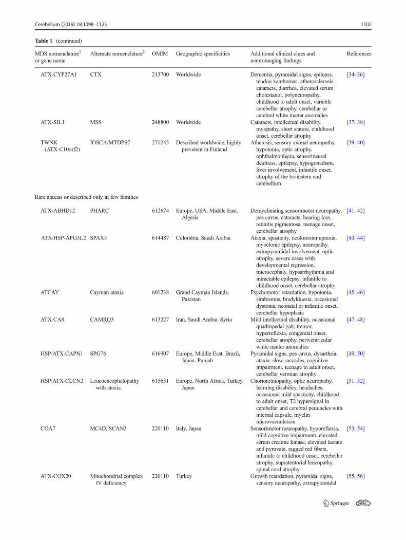
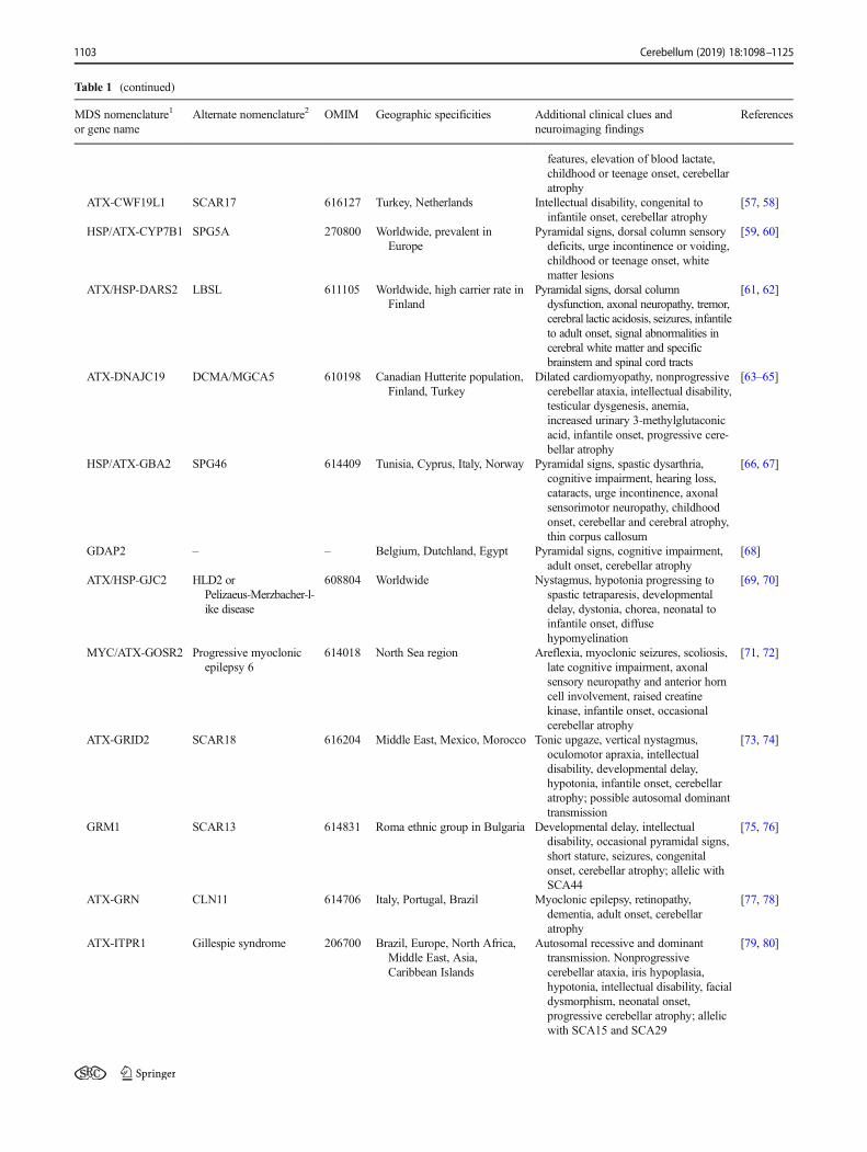

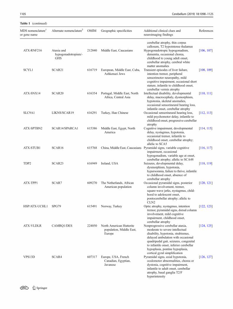
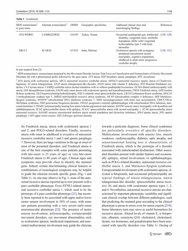
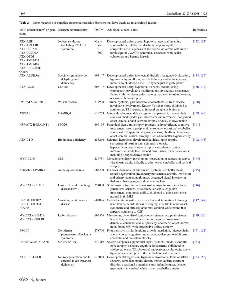
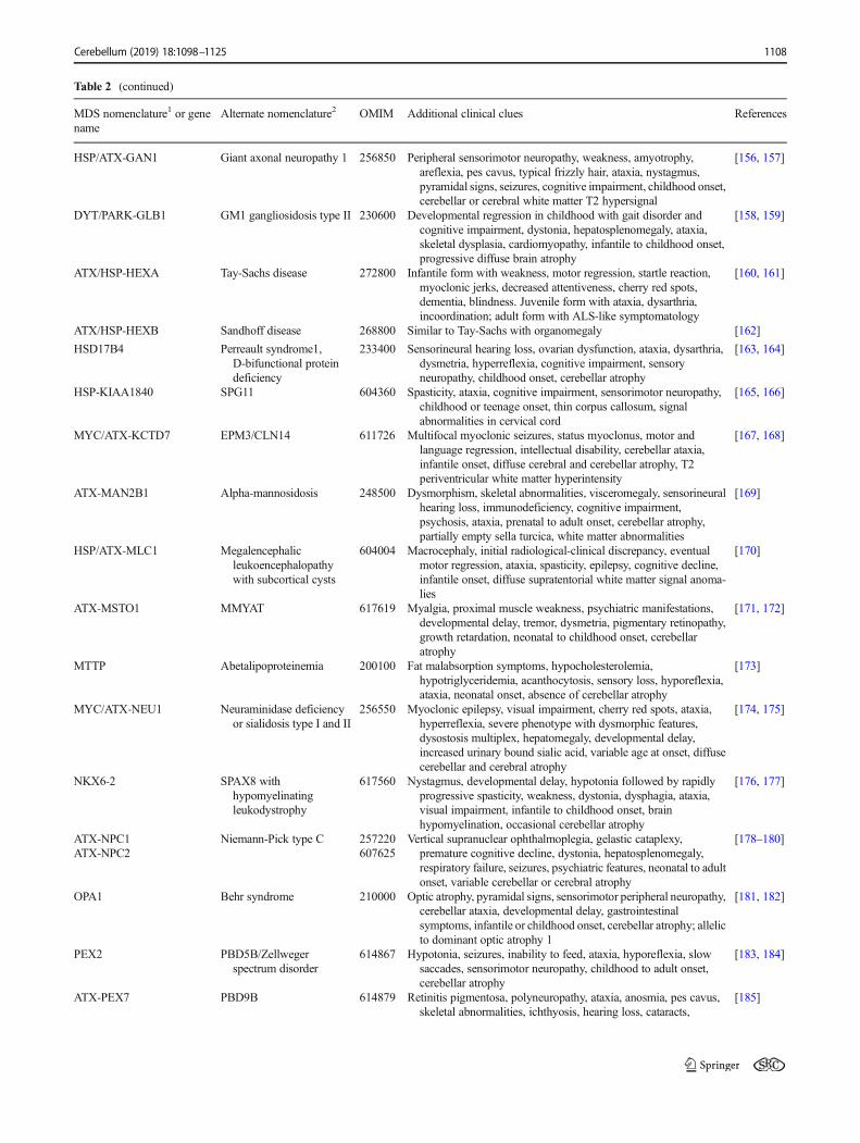
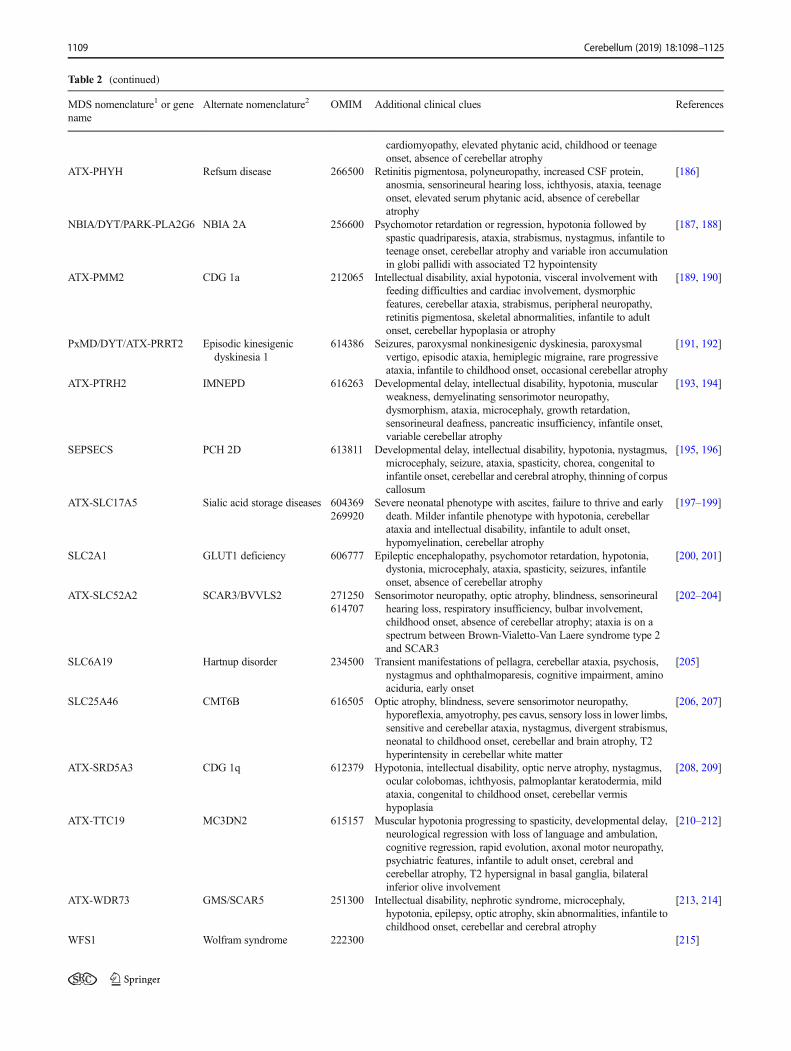
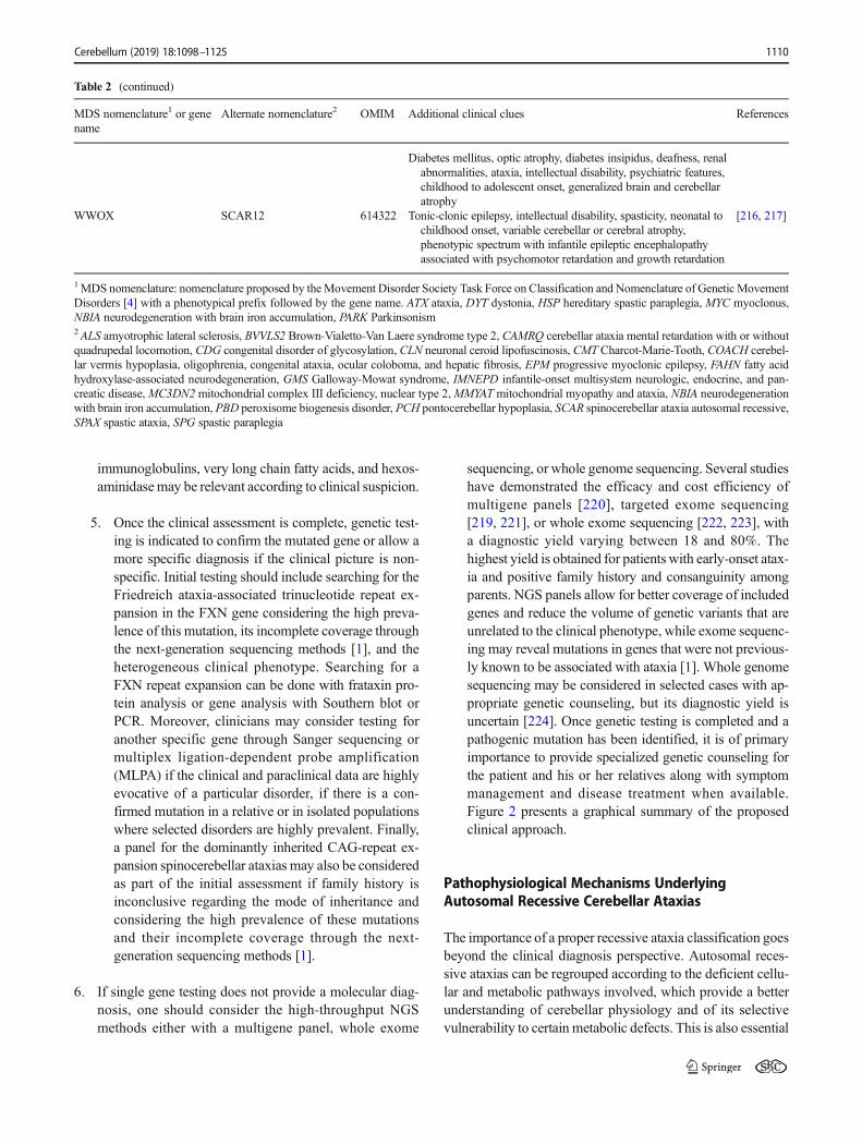
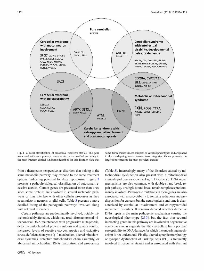

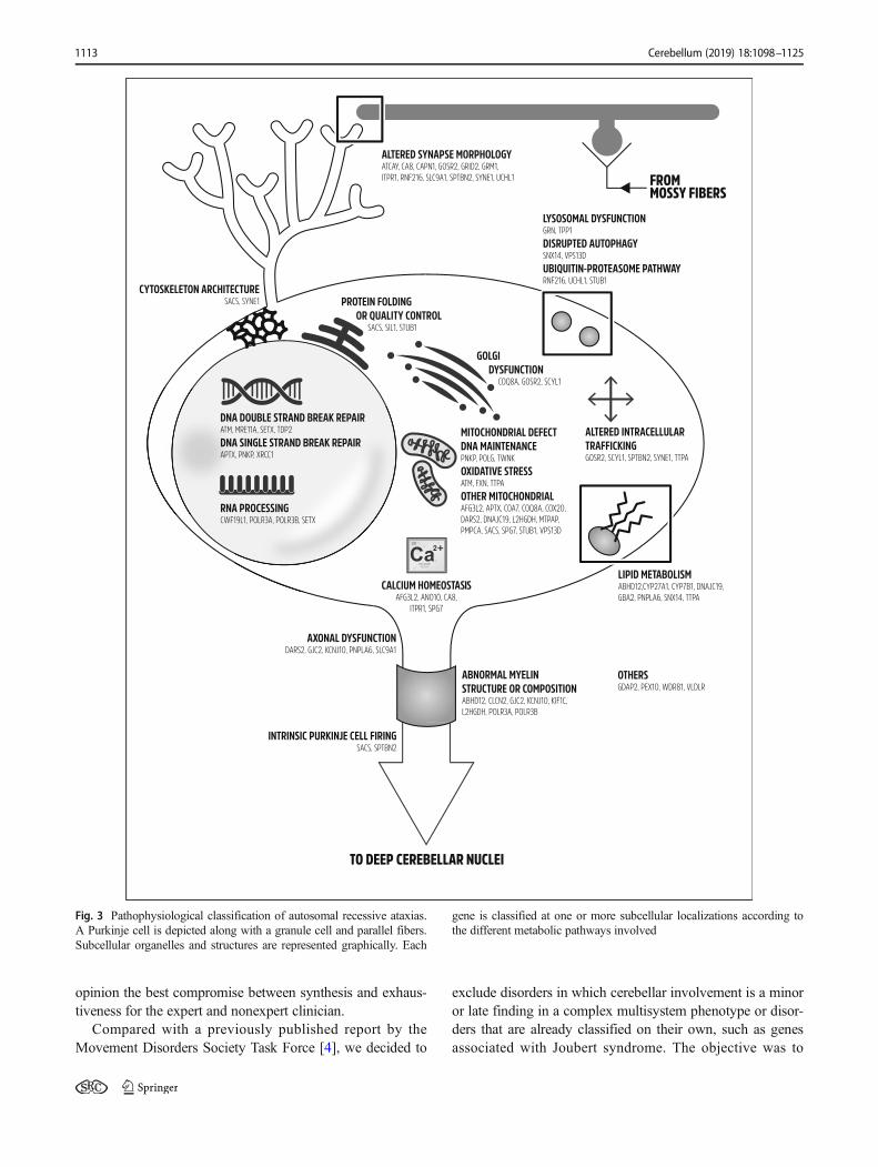
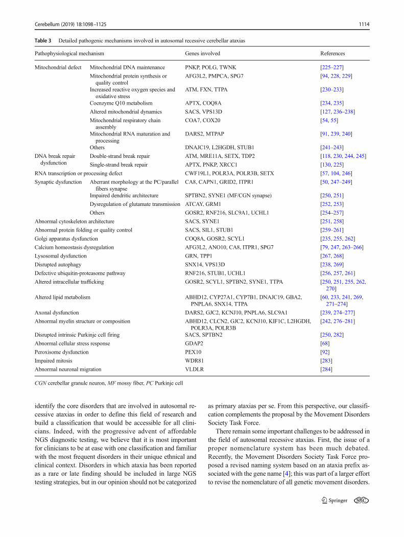


![Autosomal recessive ichthyosis with limb reduction defect ... · including autosomal dominant, autosomal recessive and X-linked inheritance [1,2]. Associated cutaneous and extracutaneous](https://static.documents.pub/doc/80x56/5ec8c9b91adfdf12ab3e663c/autosomal-recessive-ichthyosis-with-limb-reduction-defect-including-autosomal.jpg)


![Ataxia telangiectasia: a reviewataxia, oculocutaneous telangiectasia and frequent pul-monary infection [1]. Definition A-T is an autosomal recessive cerebellar ataxia [2]. It has also](https://static.documents.pub/doc/80x56/60c0274fdc425b48211dfd10/ataxia-telangiectasia-a-review-ataxia-oculocutaneous-telangiectasia-and-frequent.jpg)