The Clinical Microbiology Laboratory: a Fundamental Resource for Infection Preventionists! Janet A. Hindler, MCLS MT(ASCP) F(AAM) UCLA Health (retired) LA County DPH, Microbiology Laboratory [email protected]1
Transcript
The Clinical Microbiology Laboratory: a Fundamental Resource for
Infection Preventionists!
Janet A. Hindler, MCLS MT(ASCP) F(AAM)UCLA Health (retired)LA County DPH, Microbiology [email protected]
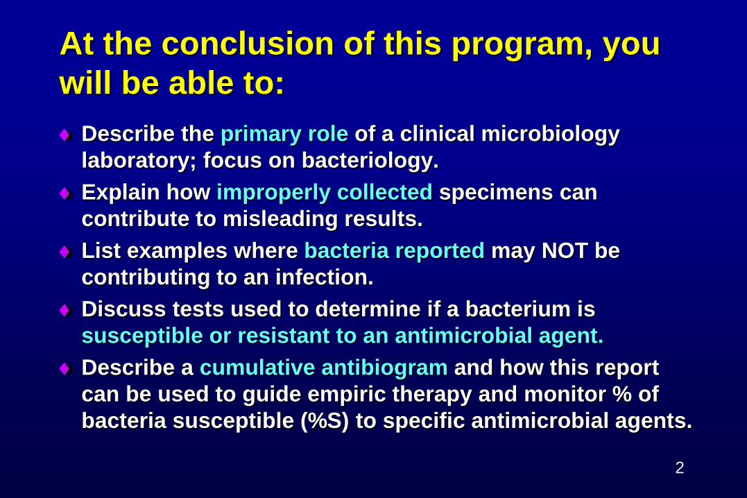
♦ Describe the primary role of a clinical microbiology laboratory; focus on bacteriology.
♦ Explain how improperly collected specimens can contribute to misleading results.
♦ List examples where bacteria reported may NOT be contributing to an infection.
♦ Discuss tests used to determine if a bacterium is susceptible or resistant to an antimicrobial agent.
♦ Describe a cumulative antibiogram and how this report can be used to guide empiric therapy and monitor % of bacteria susceptible (%S) to specific antimicrobial agents.
At the conclusion of this program, you will be able to:
2
Scenario:Physician sends a specimen to the microbiology lab.What does he/she want to know?
Does the specimen contain pathogens? What type?
How many?
What are the antimicrobial susceptibility profiles of the pathogens in the specimen?
3
Scenario:IP practitioner / epidemiologist reviews microbiology laboratory reports.What does he/she want to know?
Could the pathogens isolated have been acquired while the
patient was in the facility?
What can be done to prevent further spread of
the pathogens?
4
= some key messages!
Examining Patient Specimens for Microorganisms
5
6
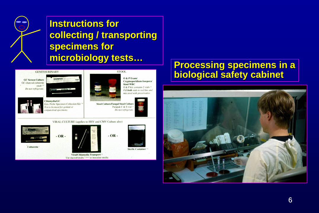
Instructions for collecting / transporting specimens for microbiology tests…
6
Processing specimens in a biological safety cabinet
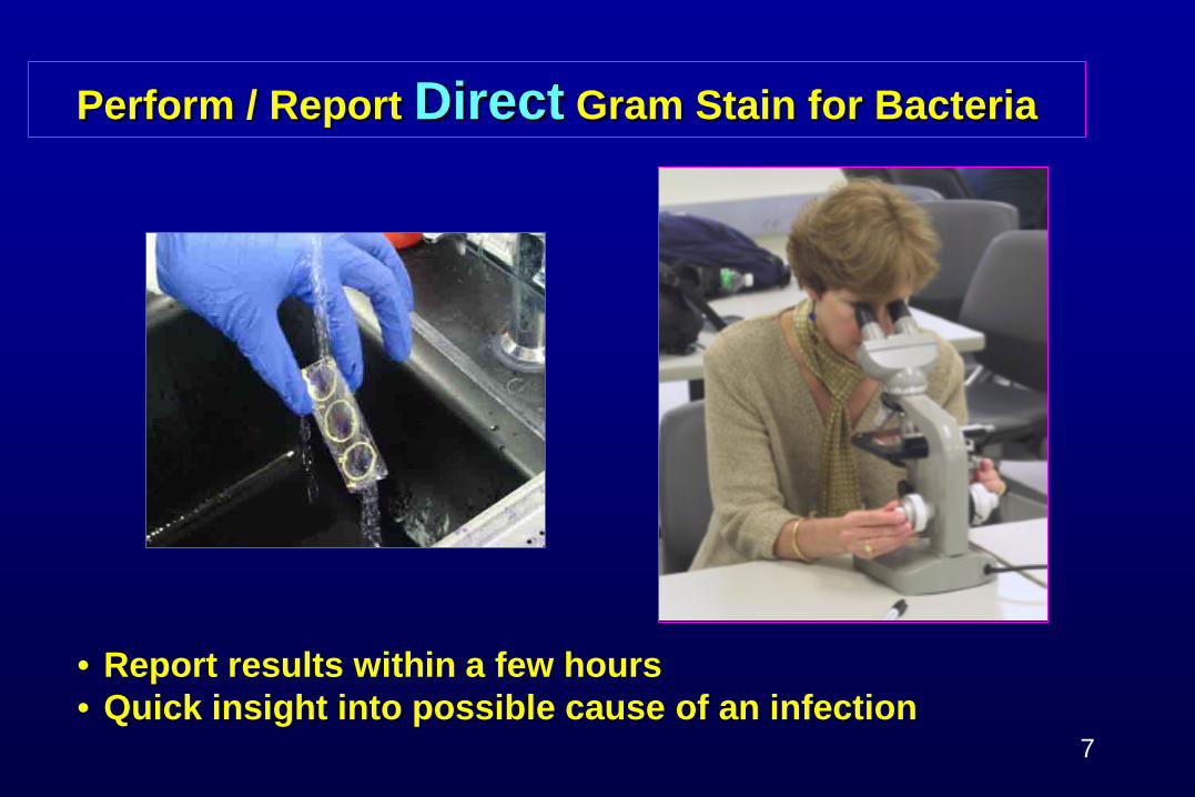
Perform / Report Direct Gram Stain for Bacteria
• Report results within a few hours• Quick insight into possible cause of an infection
7
Gram Reactions for Select Bacteria
Gram positive
KlebsiellaStaphylococcus Streptococcus E. coli
Gram negative
Pseudomonas
cocci in clusters cocci in chains
rods
cocci Neisseria
8
Direct Gram stain (pus from wound): Gram-positive cocci in clusters + white blood cells
9
Direct Gram stain (urethral discharge): Gram-negative diplococci (gonorrhoeae) within white blood cells
10
Place inoculated plates in incubator…
11
Should I identify these bacteria? Should I perform antimicrobial susceptibility tests on them?
next day
Criteria Used to Identify BacteriaTraditional methods:♦ Gram stain and microscopic
exam♦ Growth rate and colony
appearance on various types of agar media
♦ Reactivity with various chemicals / reagents
Modern (molecular) methods:♦ DNA / RNA content of
microorganisms♦ Protein profile (MALDI-TOF) of
microorganisms
12
MALDI-TOF = Matrix-assisted laser desorption ionization – time of flight mass spectrometry
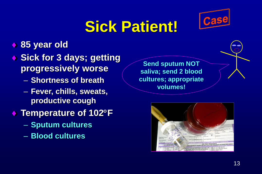
Sick Patient!♦ 85 year old♦ Sick for 3 days; getting
progressively worse– Shortness of breath– Fever, chills, sweats,
productive cough ♦ Temperature of 102°F
– Sputum cultures– Blood cultures
13
Send sputum NOT saliva; send 2 blood cultures; appropriate
volumes!
Direct Gram Stain Assess Sputum Specimen Quality
# SEC / low power field Interpretation
<10 No significant ”mouth” contamination
≥10 Indicates poorly collected specimen
♦ If saliva vs. sputum collected, may NOT recover “pathogens”
♦ Prepare direct Gram stain (put specimen on slide)
♦ Count number of squamous epithelial cells (SEC)
14
GOOD!
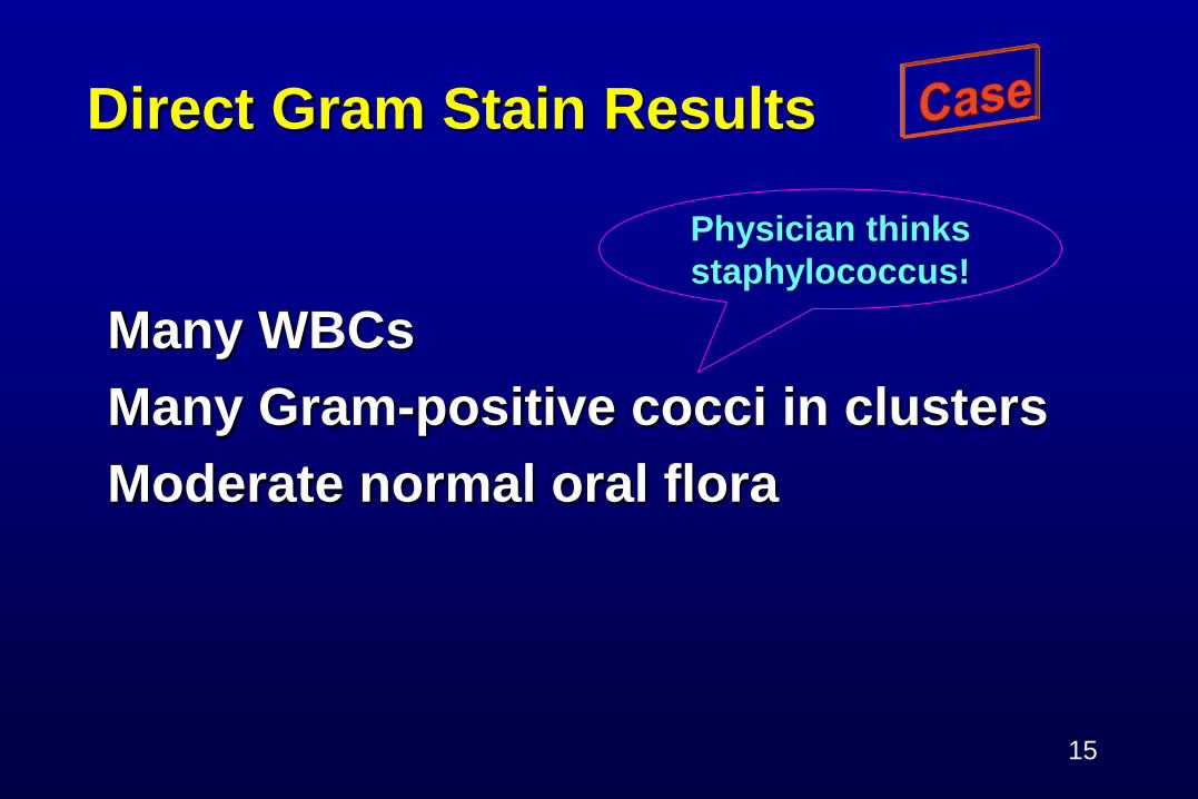
Direct Gram Stain Results
Many WBCsMany Gram-positive cocci in clustersModerate normal oral flora
15
Physician thinks staphylococcus!
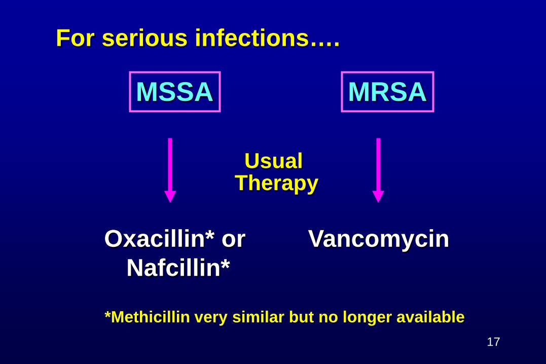
When Staphylococcus suspected…
♦Questions:– Is this Staphylococcus aureus?
• If yes, is this methicillin-resistant S. aureus (MRSA) or methicillin-susceptible S. aureus (MSSA)?
– Is this another species of Staphylococcus, typically lumped into “coagulase-negative staphylococci” (CoNS) group?
• Often contaminant; less clinically significant than MRSA or MSSA
pneumoniae, and Legionella pneumophila− Often more difficult to recover / identify
♦ Hospital-acquired pneumonia (HAP); most often ICU or ventilator-associated − Klebsiella pneumoniae− Pseudomonas aeruginosa
♦ Either CAP or HAP− Staphylococcus aureus (MRSA or MSSA)
18
Yeast uncommon cause of pneumonia or other respiratory tract infection unless present in large quantities and/or immunosuppressed.
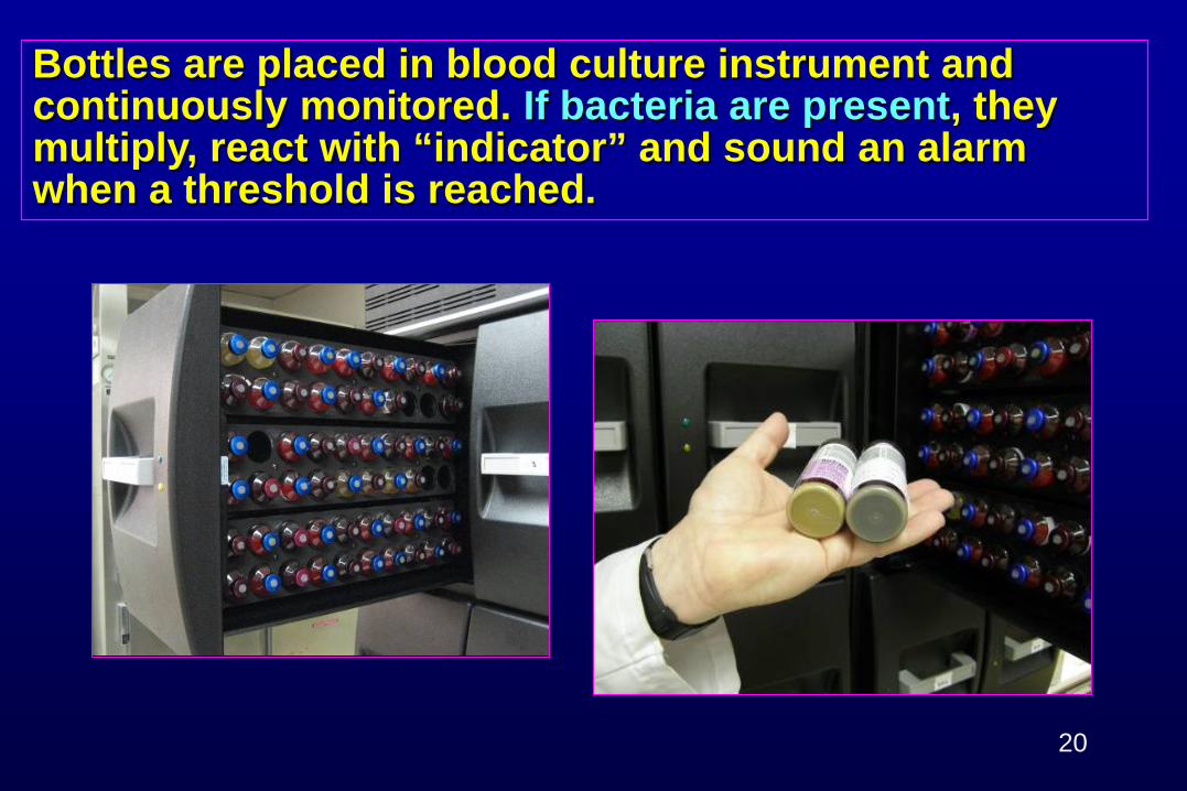
Blood specimen for bacterial culture: blood is injected directly into bottle of broth at bedside and sent to the lab.
19
Timing – collect before antibiotics given
Volume – check instructions; 2 sets!
Bottles are placed in blood culture instrument and continuously monitored. If bacteria are present, they multiply, react with “indicator” and sound an alarm when a threshold is reached.
20
“Positive” blood cultures are Gram stained, subcultured and subjected to other “tests”!
21
Gram stain: gram-positive cocci in clusters
Preliminary Report
Gram Stain
Pos Blood Culture
Sheep’s Blood Agar Medium
Colonies show:Staphylococcus spp.Perform coagulase test to determine if S. aureus
16-20 hours
Blood “Traditional” Culture Workup (1)
Pos
22
neg poscoagulase
Gram Stain
Pos Blood Culture
Molecular AssayResults:MSSA or MRSA or
CoNS
1-2 hoursPos
www.bd.com/geneohm
23
Blood “Molecular” Culture Workup (2)
Sick Patient (Blood Culture)
Gram Stain:Gram-positive cocci in clusters
Culture:Staphylococcus aureus (MRSA)
Clindamycin RDaptomycin SLinezolid SOxacillin RVancomycin S
24
Final Report with Optional Comment
“MRSA isolated. Please check infection control policies.”
Usually, for these bacteria to be considered as causing infection, two sets of blood cultures must be positive PLUS patient must show specific signs and symptoms of bloodstream infection.
25
Urine Collection / Transport
• If UTI symptoms – send for culture! • Best if culture performed ONLY on specimens with
significant pyuria (auto-reflex to culture); e.g., IF positive for leukocyte esterase and/or nitrite tests which suggest infection, THEN culture.
♦Must test within 2 hours of collection if stored at room temp♦Must test within 24 hours if
refrigerated♦Must test within 2 days if in boric
acid preservative
26
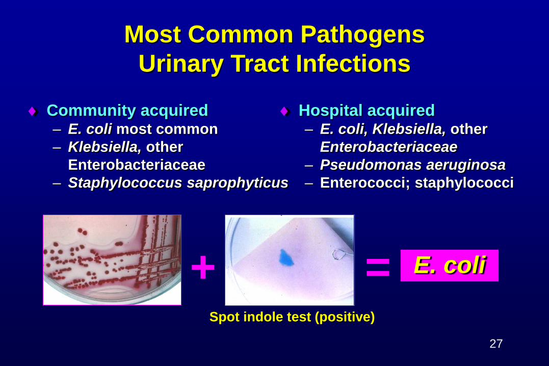
27
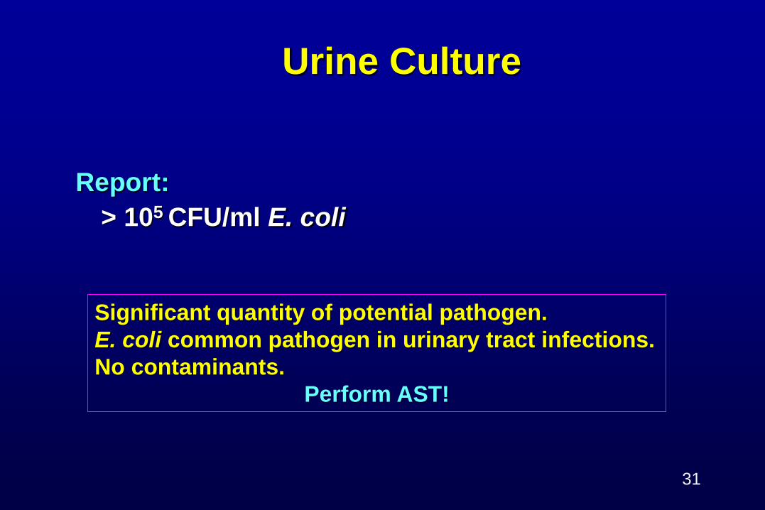
Most Common Pathogens Urinary Tract Infections
♦ Community acquired– E. coli most common– Klebsiella, other
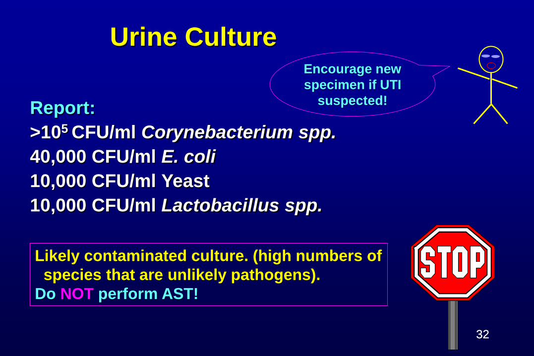
Likely contaminated culture. (high numbers of species that are unlikely pathogens).
Do NOT perform AST!
32
Encourage new specimen if UTI
suspected!
33
Sputum Culture Gram Stain:
Many oral floraMany Gram positive diplococciMany WBCs
Culture:Many Normal FloraMany Streptococcus pneumoniae
Good correlation of Gram stain with culture.Significant quantity of potential pathogen.S. pneumoniae relatively common pathogen in respiratory tract
infections. Perform AST!
33
Foot Wound Culture Gram Stain:
Many Gram positive cocci in clustersMany pleomorphic Gram positive rodsNo WBCs
Culture:Many coagulase-neg staphylococciMany diphtheroidsFew E. coli-like coloniesFew Proteus-like colonies
Poor correlation of Gram stain with culture.Small quantity of potential pathogens.“Skin flora” suggests likely contaminated culture.Do NOT perform AST! 34
Send “pus”
Throat Culture
Many Group A Streptococcus
“Group A Streptococcus is always susceptible to penicillin.”
Not necessary to perform AST on bacteria that are always (predictably) susceptible to the antimicrobial agents
typically prescribed.
35
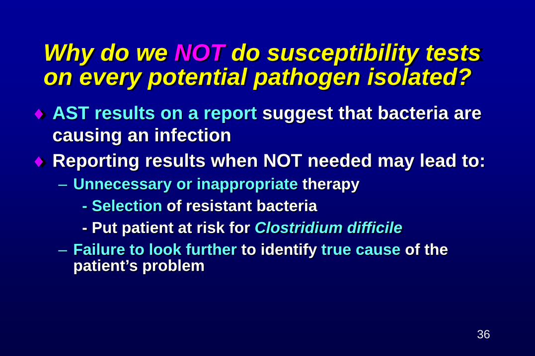
xWhy do we NOT do susceptibility tests on every potential pathogen isolated?♦ AST results on a report suggest that bacteria are
causing an infection♦ Reporting results when NOT needed may lead to:
– Unnecessary or inappropriate therapy- Selection of resistant bacteria- Put patient at risk for Clostridium difficile
– Failure to look further to identify true cause of the patient’s problem
36
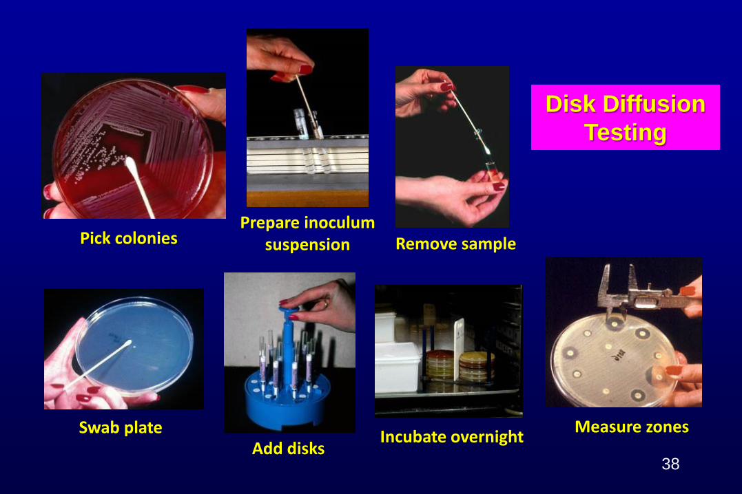
Disk diffusion (Kirby Bauer)
Broth microdilution MIC
Antimicrobial Susceptibility Tests
MIC = minimal inhibitory concentration (lowest concentration of drug that inhibits growth of the test bacteria)
Reported results:♦Susceptible (S) – drug likely
to work providing it can get to the infection site
♦Resistant (R) – drug won’t work
♦ Intermediate (I) – drug may or may not work depending on site of infection and patient’s status
37
Prepare inoculum suspension
Swab plate
Remove sample
Incubate overnightAdd disks
Disk Diffusion Testing
Pick colonies
Measure zones
38
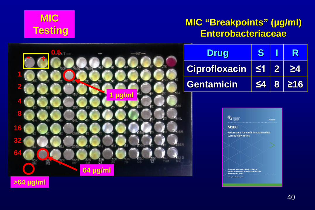
Zone Diameter “Breakpoints” (mm) Enterobacteriaceae
Drug S I RCiprofloxacin ≤1 2 ≥4Gentamicin ≤4 8 ≥16
MIC “Breakpoints” (µg/ml) Enterobacteriaceae
MICTesting
Commercial Antimicrobial Susceptibility Test Systems
Vitek 2
Sensititre
Phoenix
Etest
MicroScan
41
Review of S, I, R most important for IPFor MIC tests, must report S, I, R with or without MIC value.
Lab Report
42
Agent #1 #2Ampicillin S RCefazolin S RCefepime RCeftriaxone RCiprofloxacin S RErtapenem SGentamicin S SMeropenemNitrofurantoin S RPiper-tazo STrimeth-sulfa S R
Acquired “R” to all PO agents. Request fosfomycin – usually not tested routinely!
Broad Spectrum drug results suppressed when “S” to narrow spectrum drugs! 43
2 urine E. coli isolates
“Typical” E. coli - NO “R”!
Agent #1 #2 #3 #4 #5Ampicillin S R R R RCefazolin S R R R RCefepime R R R RCeftriaxone R R R RCiprofloxacin S R R R RErtapenem S R R RGentamicin S S S S SMeropenem R R RNitrofurantoin S R R R RPiper-tazo S R R RTrimeth-sulfa S R R R R
Potential outbreak?
44
3 more E. coli isolatesALL CRE!
CRE = carbapenem-resistant Enterobacteriaceae
CRE = R to doripenem,ertapenem, imipenem ORmeropenem
Lab Report
CREwith comments
45
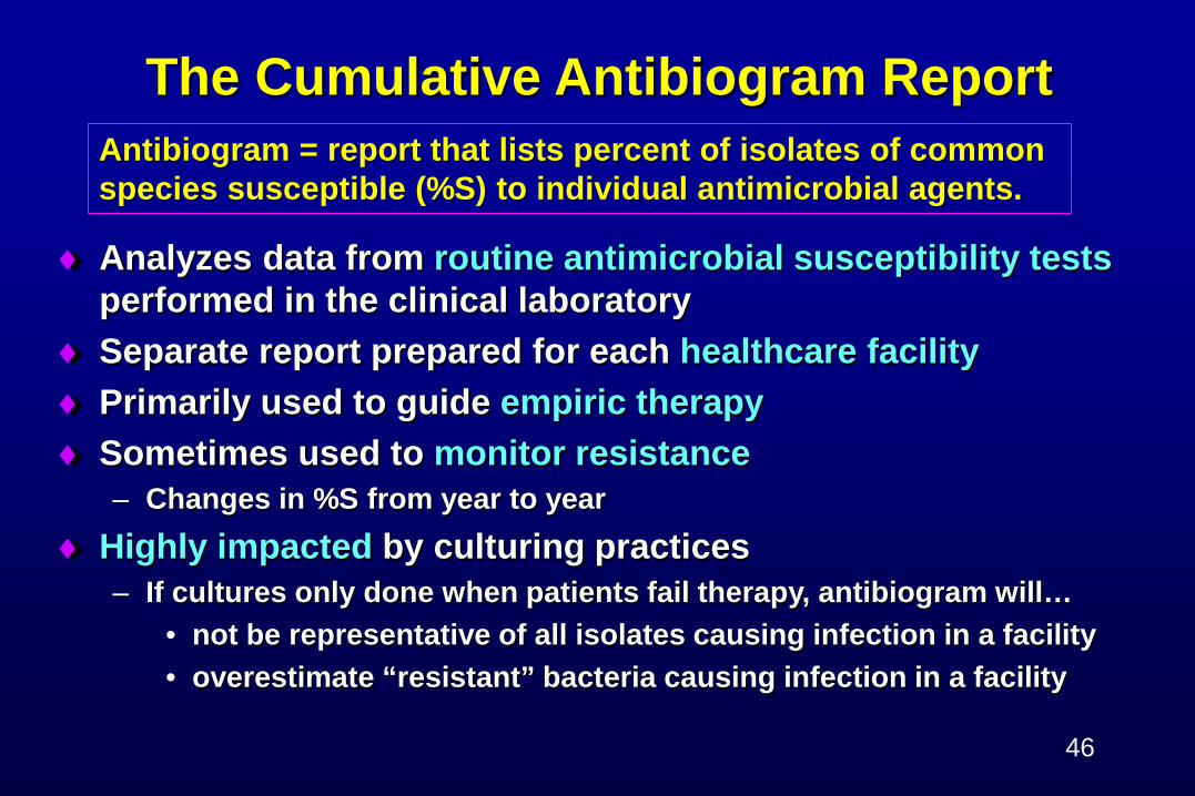
The Cumulative Antibiogram Report
♦ Analyzes data from routine antimicrobial susceptibility tests performed in the clinical laboratory
♦ Separate report prepared for each healthcare facility♦ Primarily used to guide empiric therapy♦ Sometimes used to monitor resistance
– Changes in %S from year to year♦ Highly impacted by culturing practices
– If cultures only done when patients fail therapy, antibiogram will…• not be representative of all isolates causing infection in a facility • overestimate “resistant” bacteria causing infection in a facility
Antibiogram = report that lists percent of isolates of common species susceptible (%S) to individual antimicrobial agents.
46
Recommendations Preparation of Cumulative Antibiogram
Analyze/present data at least annually Include only species with ≥ 30 isolates of each species Include diagnostic (not surveillance) isolates Include the 1st isolate/patient; no duplicate patient
isolates
Often difficult to get 30 isolates in LTCFs
47
“Routine” Cumulative antibiogramGenerally…all isolates from a facility
CLSI M39-A4. 48
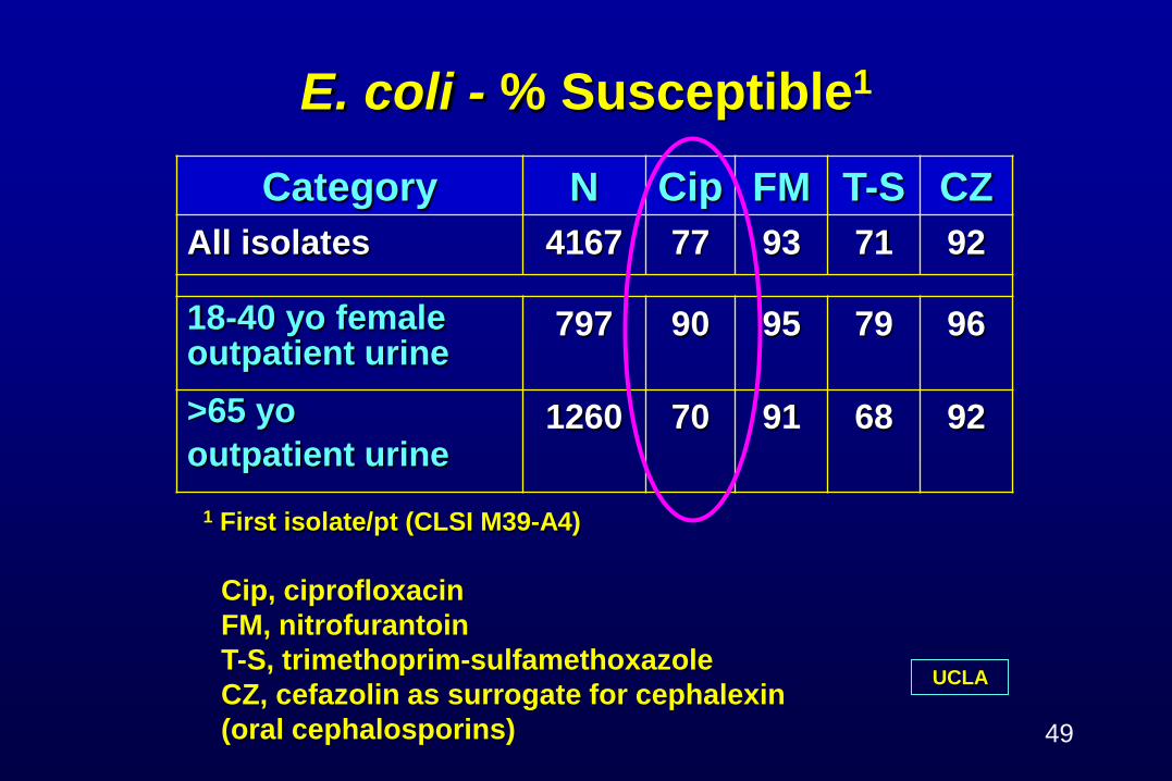
E. coli - % Susceptible1
Category N Cip FM T-S CZAll isolates 4167 77 93 71 92
18-40 yo female outpatient urine
797 90 95 79 96
>65 yooutpatient urine
1260 70 91 68 92
1 First isolate/pt (CLSI M39-A4)
UCLA
Cip, ciprofloxacinFM, nitrofurantoinT-S, trimethoprim-sulfamethoxazoleCZ, cefazolin as surrogate for cephalexin (oral cephalosporins) 49
Routine Cumulative Antibiogram% Susceptible
Organism N Amp P-T Ceftriax Erta Mero Amk Gent Cip T-S
K. pneumoniae 450 R 88 85 95 98 98 92 88 82
50
• Meropenem = carbapenem• 98% “S”• ≈ 2% CRE
CRE = carbapenem-resistant Enterobacteriaceae
Number of CRE Patients
Examine all isolates (not just first isolate/patient).Number of Enterobacteriaceae/year tested = approximately 5000 isolates.
UCLA
51CRE = carbapenem-resistant Enterobacteriaceae
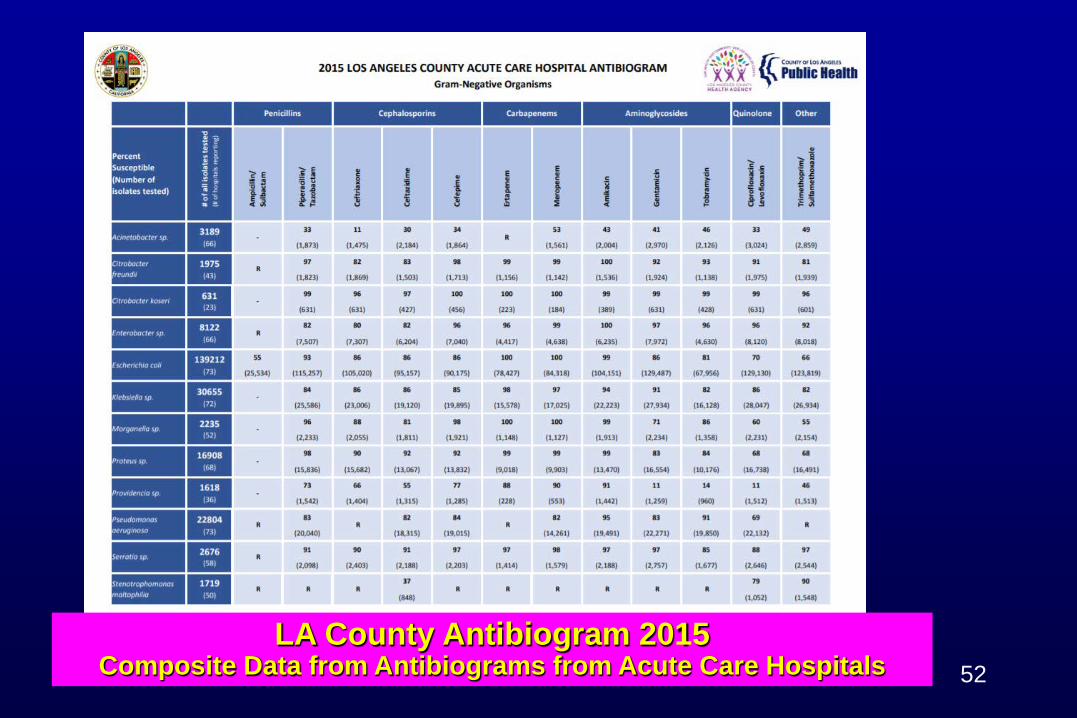
LA County Antibiogram 2015 Composite Data from Antibiograms from Acute Care Hospitals 52
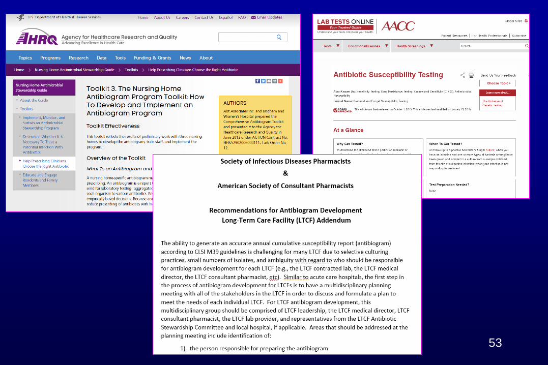
53
Summary♦ Assessment of patient’s clinical symptoms together with reliable
clinical microbiology laboratory results are essential for accurate diagnosis of infections.– Reliable clinical microbiology laboratory results are dependent on:
• appropriate collection and transport of specimens.• accurate identification and antimicrobial susceptibility testing.• good communication between healthcare providers and lab.
♦ Review of clinical microbiology laboratory results is key to identification of potential nosocomial transmission of microbes.
♦ Additional clinical microbiology laboratory tests may be needed for epidemiological investigations.
♦ A local cumulative antibiogram can help guide empiric therapy decisions and monitor “%S” for antimicrobial agents appropriate for common pathogens.