Page 1
The Cloning and Characterization of a Profilin Homolog Encoded by Orthopoxviruses
Christine Kathrine Butler-Cole B.Sc., University College of the Fraser Valley, 2002
A Thesis Submitted in Partial Fulfillment of the Requirements for the Degree of
MASTER OF SCIENCE
in the Department of Biochemistry and Microbiology
O CHRISTINE KATHRINE BUTLER-COLE, 2005 University of Victoria
All rights reserved. This thesis may not be reproduced in whole or in part, by photocopying or other means, without the permission of the author.
Page 2
Supervisor: Dr. Chris Upton
This thesis focuses on the characterization of a gene in ectromelia virus that encodes a
homolog of profilin, a cellular actin-binding protein. The profilin homolog protein
family is found exclusively within the orthopoxviruses, and orthologs share greater than
90% amino acid identity. The conservation of the gene in orthopoxviruses, in addition to
its location in the variable terminal region of the genome, suggests that it is important for
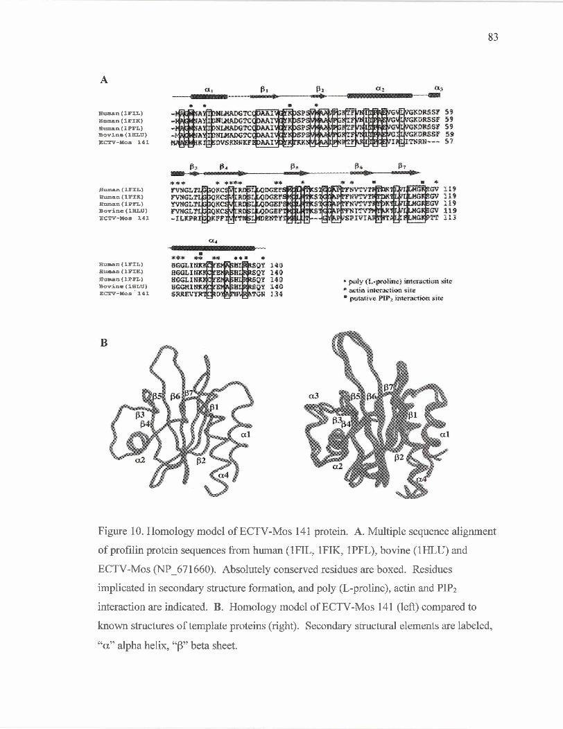
increasing viral fitness during infection. A homology model of the ECTV-Mos 141
protein, suggests that although the profilin homolog and mammalian profilin share only
30% amino acid identity, the three-dimensional structures of the proteins are similar.
There are differences at the amino acid level, however, which may have important
implications in the localization and function of the profilin homolog in vivo.
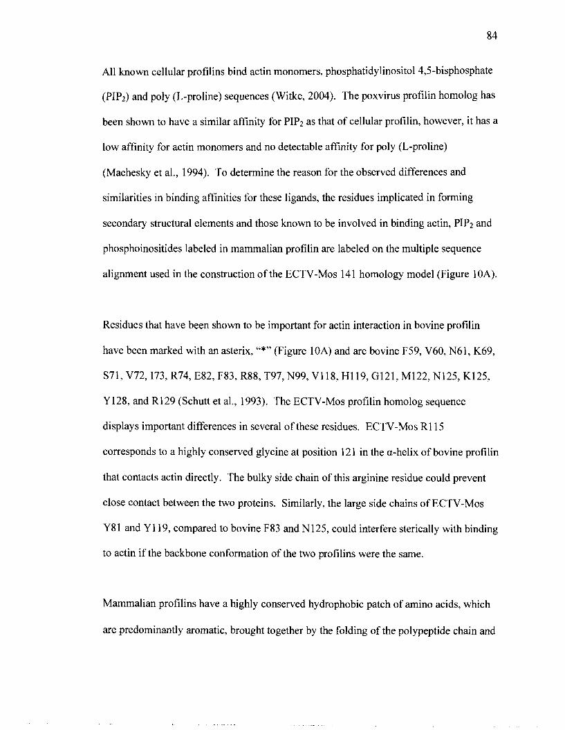
Interestingly, ECTV-Mos 14 1 associates with cellular tropomyosin and viral A-type
inclusion proteins in virus infected cells. Colocalization of ECTV-Mos 14 1, tropomyosin
and truncated A-type inclusion protein at putative actin tails and CEV-induced
protrusions from the cell surface, suggests a role for these proteins in intercellular spread
of the virus. Additionally, ECTV-Mos 141 associates with A-type inclusion bodies
formed by both truncated and full-length A-type inclusion proteins; these structures are
important in the protection and dissemination of the virus outside the host. The
formation of these bodies may be facilitated by the action of the profilin homolog and
utilization of the microtubule cytoskeleton.
Page 3
Table of Contents
Abstract
Table o f Contents
List of Tables
List of Figures
List of Abbreviations
Acknowledgments
Chapter 1 - Introduction
History of Poxviruses
Classification of Poxviruses
Biology and Life Cycle of Poxviruses
Virion Structure
Genome
Replication Cycle
Motility
Dissemination
Poxvirus Virulence Factors
Host Immune Evasion
A-type Inclusion Bodies
Profilin Homolog
Significance of Poxvirus Research
Thesis Rationale
Contributors to Work Presented in this Thesis
Chapter 2 - Materials and Methods
ii
iii
iv
V
vi
vii
Page 4
Chapter 3 - Results
Selection of Gene Targets
Cloning of Gene Targets
Location of Gene Target ortholog ORFs in the ECTV-Mos genome
Expression of Recombinant Proteins
Purification of ECTV-Mos 14 1 protein, a profilin homolog
Analysis of the Orthopoxvirus Profilin Homolog Protein Family
Homology Model of ECTV-Mos 14 1 protein
Coimmunoprecipitation of ECTV-Mos 14 1 and ECTV-Mos 14 1 - interacting proteins from poxvirus-infected cells
ECTV-Mos 14 1 and tropomyosin interact directly
Analysis of Orthopoxvirus A-type Inclusion Proteins
Localization of ECTV-Mos 141 and A-type Inclusion Proteins in vivo
Localization of ECTV-Mos 141 and cellular tropomyosin in vivo
Chapter 4 - Discussion
Concluding Remarks
Literature Cited
Page 5
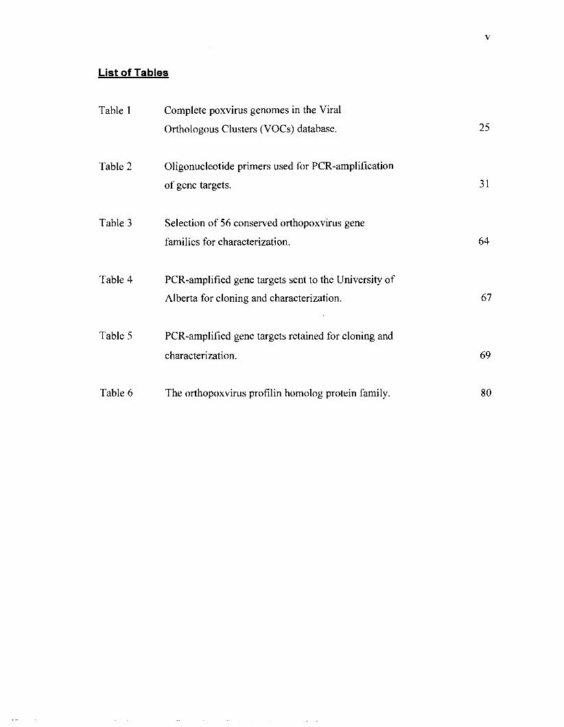
List of Tables
Table 1
Table 2
Table 3
Table 4
Table 5
Table 6
Complete poxvirus genomes in the Viral
Orthologous Clusters (VOCs) database.
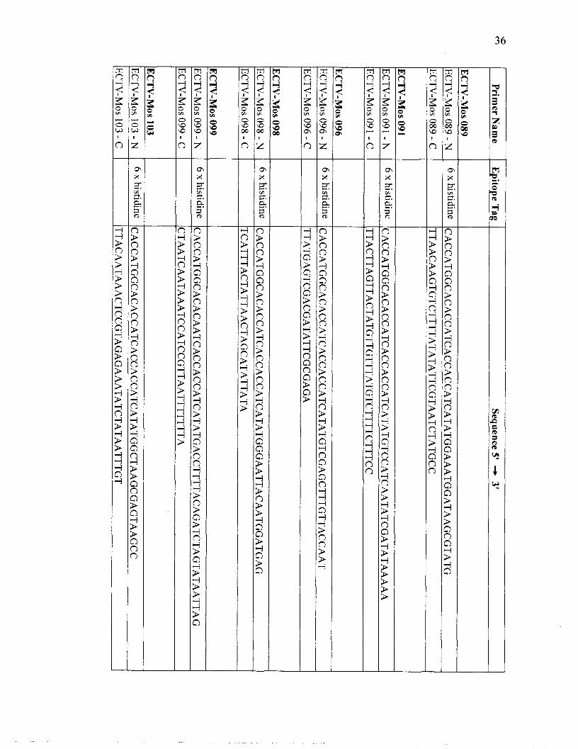
Oligonucleotide primers used for PCR-amplification
of gene targets.
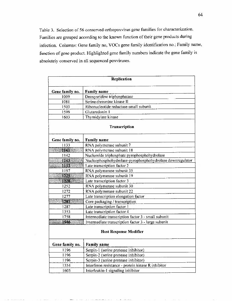
Selection of 56 conserved orthopoxvirus gene
families for characterization.
PCR-amplified gene targets sent to the University of
Alberta for cloning and characterization.
PCR-amplified gene targets retained for cloning and
characterization.
The orthopoxvirus profilin homolog protein family.
Page 6
List of Fiaures
Figure 1
Figure 2
Figure 3
Figure 4
Figure 5
Figure 6
Figure 7
Figure 8
Figure 9
Figure 10
Figure 11
Replication cycle of vaccinia virus.
Overview of intracellular and intercellular virion
movement.
Cellular profilin performs a diversity of functions.
Design of oligonucleotide primers for PCR-
amplification of gene targets.
Cloning strategy for the production of pDEST14
expression clones.
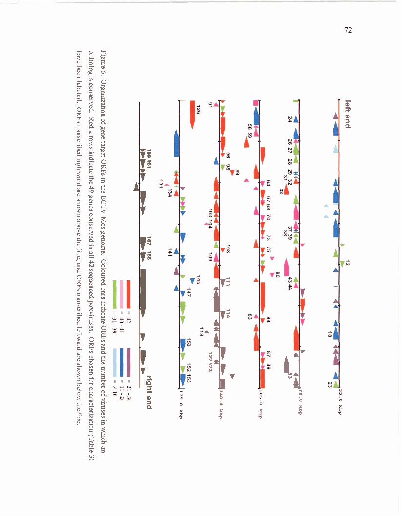
Organization of gene target ORFs in the ECTV-Mos
genome.
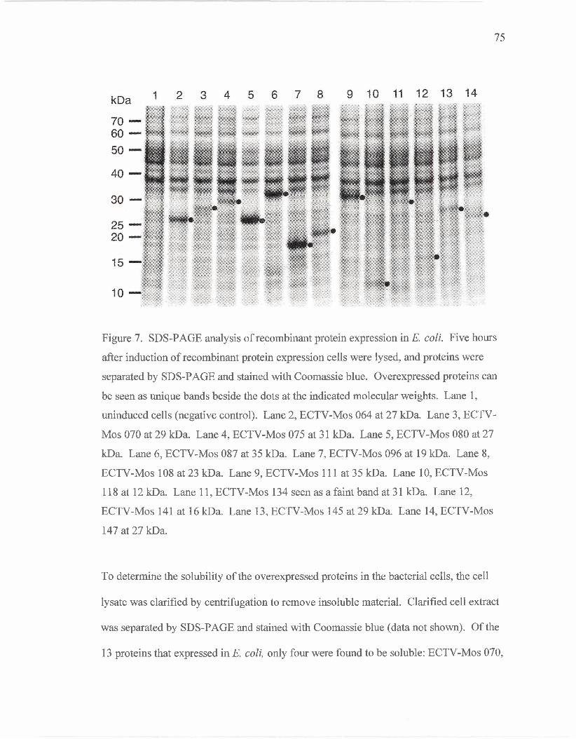
SDS-PAGE analysis of recombinant protein
expression in E. coli.
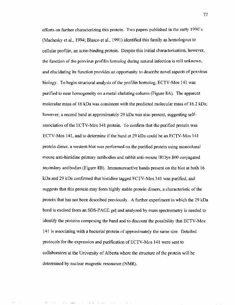
SDS-PAGE and western blot analysis of purified
ECTV-Mos 14 1 protein.
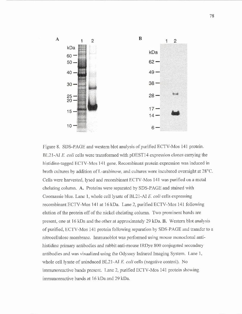
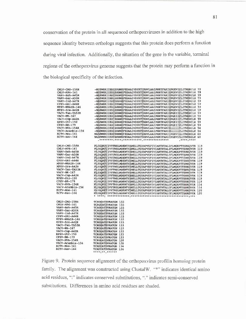
Protein sequence alignment of the orthopoxvirus
profilin homolog protein family.
Homology model of ECTV-Mos 14 1 protein.
Coimmunoprecipitation and identification of proteins
interacting with ECTV-Mos 14 1 during infection.
Page 7
Figure 12 Western blot to detect actin in coirnmunoprecipitated
proteins.
Figure 13 Far western analysis of the interaction between
ECTV-Mos 141 and tropomyosin.
Figure 14 Graphical representation of sequence alignment of
A-type inclusion proteins.
Figure 15 Colocalization of ECTV-Mos 141 and VACV-WR 148
in virus-infected cells.
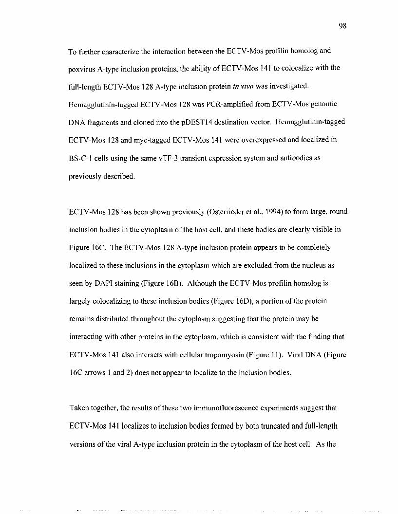
Figure 16 Colocalization of ECTV-Mos 141 and ECTV-Mos 128
in virus-infected cells.
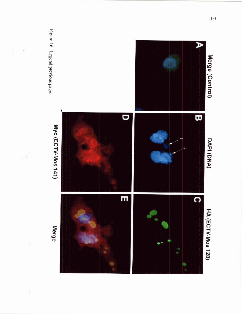
Figure 1 7 Colocalization of ECTV-Mos 14 1 and tropomyosin
in virus-infected cells.
vii
8 9
Page 8
. . . Vl l l
List of Abbreviations
a, alpha
aa, amino acid(s)
~ m ~ ~ , ampicillin resistant
araBAD, arabinose operon
ATI, A-type inclusion
ATPase, adenosine triphosphate phosphatase
p, beta
BS-C- 1 , African green monkey cells
BLASTP, Basic Local Alignment Search Tool-Protein
bp, basepair
BSA, bovine serum albumin
C-terminus, carboxy terminus
CEV, cell-associated enveloped virus
CMLV, camelpox virus
COz, carbon dioxide
CPXV, cowpox virus
DAPI, 4',6'-diamidino-2-phenylindole
DMEM, Dulbecco's minimal essential medium
DMSO, dimethyl sulphoxide
DNA, deoxyribonucleic acid
dNTP, deoxyribonucleotide triphosphate
DTT, dithiothreitol
EB, elution buffer
EDTA, ethylenediaminetetraacetic acid
EEV, extracellular enveloped virion
ECTV, ectromelia virus
FBS, fetal bovine serum
FITC, fluorescein isothiocyanate
Y, gamma
Page 9
HA, haemagglutinin
HBS, HEPES-buffered saline solution
HEPES, N-2-hydroxyethylpiperazine-N' -2' -ethanesulfonic acid
HGT, horizontal gene transfer
His, histidine
IFN, interferon
IL, interleukin
IMV, intracellular mature virion
an^, kanamycin resistant
kb, kilobasepair
kDa, kiloDalton
LB, Luria-Bertani
MALDI-TOF, Matrix Assisted Laser Desorption Ionization-Time of Flight
ml, millilitre
y g, microgram
p1, microlitre
mM, millimolar
MOI, multiplicity of infection
MPXV, monkeypox virus
mRNA, messenger ribonucleic acid
N-terminus, amino terminus
ng, nanogram
NMR, Nuclear Magnetic Resonance
NTP-PPH, nucleoside triphosphate pyrophosphohydrolase
OD, optical density
O W , open reading frame
PAGE, polyacryalamide gel electrophoresis
PBS, phosphate buffered saline
PCR, polymerase chain reaction
PEG, polyethylene glycol
pfu, plaque forming units
Page 10
PH-PPH, nucleophosphohydrolase-pyrophosphohydrolase downregulator
PIP2, phosphatidylinositol4,5-bisphosphate
PKR, RNA-dependent protein kinase
PMSF, phenyl methyl sulfonyl fluoride
RNA, ribonucleic acid
rpm, revolutions per minute
RPXV, rabbitpox virus
SDS, sodium dodecylsulfate
SSC, standard saline citrate
TAE, tris, acetic acid, EDTA
TBS, tris buffered saline
VARV, variola virus
VACV, vaccinia virus
VASP, Vasodilator Stimulated Phosphoprotein
VOCs, Viral Orthologous Clusters
N-WASP, neural Wiskott-Aldrich syndrome protein
WIP, WASP-interacting protein
Page 11
Acknowledaments
I thank my supervisor Dr. Chris Upton for his guidance on this project throughout the
previous three years and in supporting my decision to complete my Masters and pursue a
degree in law. I appreciate your understanding. I thank my committee members Dr.
Nano and Dr. Koop for their help throughout my studies. Thank you to all of the
individuals who have contributed to the work presented in this thesis, including: Dr.
Mark Buller, Arwen Hunter, Roderick Haesevoets, Guiyun Lee, Dr. David Esteban, and
Shan Sundararaj. I would also like to express my gratitude to Melisa Da Silva and
Angelika Ehlers for their friendship and computer expertise. Thanks are also extended to
Scott Scholz, Albert Labossiere and Stephen Horak for their technical support and to
Melinda Powell and Deb Penner for their help through the administrative process.
Finally I would like to thank my family, friends and my husband Chris for their love and
support.
Page 12
Chapter 1 : Introduction
I. History of Poxviruses
Smallpox was once the most serious disease faced by mankind; it has had an enormous
impact on human history. It claimed the lives of hundreds of millions of people between
its first recorded out break in Ancient Egypt and its eradication in 1979 (Mahalingam et
al., 2004). Variola virus, the causative agent of smallpox, is speculated to have emerged
in Africa or Asia some 5,000 years ago (Moss, 2001). Although its exact origins remain
obscure, an ancestral virus of variola present in a wild animal reservoir likely adapted to
the human population through an intermediate host such as cattle or small rodent. Large
high-density populations and the domestication of animals are thought to have been
necessary for the emergence and continuance of this pathogen (Ellner, 1998).
In about 1000 A.D., one of the first effective preventative measures against an infectious
disease was initiated against smallpox. Variolation, a risky procedure in which people
were inoculated with material collected from individuals infected with smallpox, offered
some protection against smallpox infection (Moss, 2001). In 1796 Edward Jenner
demonstrated that cowpox virus, which is less virulent to humans, could be used to
protect against contraction of smallpox and marked the beginning of a scientific
investigation into vaccination (Ellner, 1998). Global eradication of smallpox was
declared in 1979 following a World Health Organization (WHO)-led vaccination
program (Ellner, 1998). The vaccine against smallpox contained vaccinia virus, now
considered the prototypic poxvirus. The absence of a non-human reservoir for variola
Page 13
virus in addition to sociopolitical factors contributed to the successful eradication of this
disease (Mahalingam et al., 2004).
Due to its role in the eradication of smallpox, vaccinia virus has the longest and most
extensive history of use in humans compared to any other virus and has been studied
extensively in the laboratory. It was the first animal virus seen microscopically, grown in
tissue culture, accurately titered, physically purified, and chemically analyzed (Shen and
Nemunaitis, 2004). Poxvirus research has, and continues to be, an active area of
investigation.
II. Classification of Poxviruses
The Poxviridae, as a family, are ubiquitous, infecting mammals, birds, reptiles and
invertebrates. Two members of this family, variola virus and molluscum contagiousum
virus, are obligate human pathogens although many other poxviruses can be transmitted
to humans from other animal hosts. Poxviruses can be divided into two subfamilies
based on their ability to replicate within vertebrates (Chordopoxvirinae) and insects
(Entomopoxvirinae). The Chordopoxvirinae consists of eight genera: Orthopoxvirus
(camelpox, cowpox, ectromelia, monkeypox, raccoonpox, skunkpox, vaccinia, variola,
volepox), Parapoxvirus (ecthyma, orf, pseudocowpox, parapox of deer, sealpox),
Avipoxvirus (canarypox, fowlpox, juncopox, mynahpox, pigeonpox, psittacinepox,
quailpox, sparrowpox, starlingpox, turkeypox), Capripoxvirus (goatpox, lumpy skin
disease, sheeppox), Leporipoxvirus (myxoma, hare fibroma, rabbit fibroma, squirrel
fibroma), Suipoxvirus (swinepox), Molluscipoxvirus (molluscum contagiosum) and
Page 14
Yatapoxvirus (tanapox, Yaba monkey tumor). Viruses belonging to the same genus are
genetically and antigenically related and have a similar morphology and host range
(Moss, 2001). The Entomopoxvirinae subfamily contains three genera, which are
distinguished from one another by their insect host range and virion morphology (Moss,
2001; Arif, 1984). Genus A viruses infect coleopterans, genus B viruses infect
lepidopterans and orthopterans, and genus C viruses infect dipterans. Insects are the only
known hosts of entomopoxviruses, and their viral host range is restricted to a small
number of related species (Afonso et a]., 1999).
Ill. Biology and Life Cycle of Poxviruses
Virion Structure
Poxviruses are the largest known animal viruses, and are discernable by light microscopy
(Dubochet et al., 1994). The structure of poxvirus virions has been studied extensively
using vaccinia virus, the prototypic virus of the family, although the basic features may
largely apply to other family members as well. Vaccinia virions are oval or brick-shaped,
approximately 300 x 240 x 120 nm in size, and consist of a lipoprotein envelope
surrounding a complex core structure (Moss, 2001). The virion core contains the viral
genome associated with a number of virus-encoded enzymes required for transcription;
including the multisubunit DNA dependent RNA polymerase, early transcription factor
(VETF), enzymes for methylation and capping of mRNA, a poly (A) polymerase and
nucleoside triphosphate phosphohydrolase (NTP-PPH) (Moss, 2001 ; Smith et al., 2002).
Page 15
Vaccinia virus produces four different types of virion from each infected cell that have
different abundance, structure, location and roles in the virus life cycle (Husain and
Moss, 2005). First, intracellular mature virus (IMV) particles are formed within
cytoplasmic factors from non-infectious precursors and represent the majority of
infectious progeny. IMV are released from the cell during cell lysis and are important for
viral transmission from one host to another (Moss, 2001). The majority of IMV remain
in the cell until lysis, however, some IMV become wrapped by a double layer of
intracellular membrane to form intracellular enveloped virus (IEV) (Hiller and Weber,
1985). The composition of this membrane is different from that of the host, and contains
at least seven poxvirus-encoded polypeptides (Husain and Moss, 2005; Lorenzo et al.,
1998). IEV then move to the cell periphery where the outer membrane fuses with the
plasma membrane exposing a cell-associated enveloped virus (CEV) on the surface.
CEV are involved in actin tail formation that is instrumental in the intercellular spread of
the virus (Blasco and Moss, 1992). CEV released from the cell surface are called
extracellular enveloped virus (EEV). Although less abundant than IMV or CEV, EEV
play a role in the long range dissemination of the virus within tissue culture and the host
(Payne, 1980). CEV and EEV are physically indistinguishable and contain one fewer
membrane that IEV and one more membrane than IMV, respectively (Smith et al., 2002).
Genome
The poxvirus genome is not infectious and consists of a linear, double-stranded DNA
molecule with covalently closed ends (Baroudy et al., 1982). The size of the poxvirus
genome varies from approximately 130 kbp in parapoxviruses (Delhon et al., 2004) to
Page 16
about 380 kbp in avipoxviruses (Laidlaw and Skinner, 2004). Like many other viruses,
poxviruses have inverted terminal repeats (ITRs), which are identical but oppositely
oriented sequences at either end of the genome and are required for poxvirus DNA
replication. The ITRs are variable in length due to deletions, repetitions, and
transpositions. The general composition of ITRs is: an A+T rich hairpin loop at each end
of the genome that links the two DNA strands together; a sequence of approximately 100
bp important for the disassociation of concatemers during viral replication; variable-
length sets of short, tandemly repeated sequences; and up to several open reading frames
(ORFs). Examination of poxvirus genomic maps reveals a high degree of utilization of
the genomic DNA, with few, if any, non-coding sequences. ORFs are present on both
strands and are organized in clusters that are predominantly transcribed toward the closest
end of the genome (Moss, 200 1).
To date, 49 absolutely conserved poxvirus genes have been identified in 42 sequenced
poxvirus genomes, while the vertebrate-infecting chordopoxviruses share 90 conserved
genes (Upton et al., 2003). Analysis of complete poxvirus genomes have demonstrated
that the poxvirus genome consists of a highly conserved central region that contains
genes essential for replication, and more variable terminal regions containing genes
involved in virulence and host interaction. Additionally, the localization of individual
ORFs in this central region is largely preserved in chordopoxviruses (Upton et al., 2003;
Moss, 2001).
Page 17
The genomes of poxviruses are not static, but are subject to frequent events of gene
duplication, deletion, and horizontal gene transfer (HGT) from their hosts (Hughes and
Friedman, 2005). These gene loss and gene gain events have been consistent
characteristics of poxvirus genome evolution. Genes that are acquired and lost during
poxvirus evolution are likely to have host specific effects such as host range or evasion of
host antiviral defense mechanisms (McLysaght et al., 2003). These fluctuations in the
content of the genome, therefore, are likely opportunities for virus adaptation.
Interestingly, the rate of gene acquisition is not constant over time, and it has increased in
the orthopoxviruses. Although it is not yet clear what has changed the rate of gene
acquisition and retention in orthopoxviruses it has been suggested that this is associated
with the unique features of orthopoxvirus infection, replication, and virulence (Hughes
and Friedman, 2005; McLysaght et al., 2003).
Replication Cycle
Poxvirus genomes are large and complicated by the standards of many other viruses and
their life cycle reflects this. Poxviruses are unique among the DNA viruses in that their
replication cycle occurs exclusively within the cytoplasm of the infected host cell and
therefore must encode all of the enzymes and factors necessary for genome replication
and transcription of viral mRNAs (Moss, 2001). The details of poxvirus replication have
been obtained primarily by studying vaccinia virus infections of tissue culture cells. The
time required to complete a single replication cycle varies considerably from virus to
virus and ranges from 12 to 24 hours by vaccinia and up to 75 hours by Yaba virus
(Buller and Palumbo, 1991).
Page 18
The poxvirus replication cycle begins with the entry of the virus into the host cell (Figure
1). The mechanism by which poxviruses penetrate cells is poorly understood, in part
because the complexity of the virus makes it difficult to determine which of the numerous
known or predicted membrane proteins are involved (Senkevich and Moss, 2005).
Vaccinia produced two forms of infectious virions: enveloped extracellular virus (EEV)
and intracellular mature virus (IMV) that bind to different receptors (Vanderplasschen
and Smith, 1997) and use different mechanisms for entry into the host cell (Moss, 2001;
Vanderplasschen and Smith, 1997). IMV attachment is enhanced by viral proteins in the
viral membrane that bind to proteoglycans on the cell surface (Senkevich and Moss,
2005). Entry of IMV occurs by fusion with the plasma membrane or vesicles that are
formed by surface invaginations in a pH-independent manner (Doms et al., 1 99O),
although non-fusion mechanisms have also been suggested (Locker et al., 2000). EEV
entry into cells is dependant on low pH, suggesting that an endocytic pathway is used,
although vaccinia virus may be too large for internalization through clathrin pits (Husain
and Moss, 2005). Once inside the vesicle, low pH disruption of the outer membrane
results in the release of the IMV particle which then fuses to the vesicle membrane
(Husain and Moss, 2005; Ichihashi, 1996).
Upon entry into the cell, virus particles undergo two stages of disassembly. During the
first stage, the nucleoprotein core is released from the outer coat and early mRNA is
synthesized. Viral gene expression is tightly controlled and the three classes of genes,
early, intermediate and late, are expressed in a temporal cascade with the transcription of
each gene class being dependent upon prior expression of genes of the previous class
Page 19
(Smith et al., 2004). Within 20 minutes of infection, early transcription begins,
generating capped, polyadenylated mRNAs that encode proteins required for intermediate
gene transcription, viral genome replication, nucleotide biosynthesis and the down-
regulation of a variety of host immune functions. Approximately 50% of the poxvirus
genome consists of early genes that are characterized by an AIT rich promoter region that
is bound and transcribed by a virus-encoded transcription factor and RNA polymerase
(Moss, 2001). Early gene expression is followed by a second round of uncoating that
facilitates replication of the virus genome. DNA synthesis occurs in discrete areas of the
cytoplasm called factories or virosomes, and results in thousands of genome copies per
cell only half of which are packaged into mature virus particles. Although specific origin
sequences have not been defined, synthesis appears to start near the ends of the genome
because a 200 bp sequence in the ITR is required for optimal template replication (De
Silva and Moss, 2005; Du and Traktman, 1996). The onset of replication varies
considerably for individual poxviruses, anywhere fiom 4 to 16 hours post-infection, and
is influenced by cell type and multiplicity of infection. Genome replication produces
concatameric intermediates that are not resolved until the products of late genes are
synthesized (Beaud, 1995).
Viral DNA replication is followed by sequential transcription of intermediate and late
genes, processes that are dependent upon the presence of naked DNA template (Keck et
al., 1990). Intermediate mRNAs appear approximately 100 minutes post infection and
are translated into late transcription factors that regulate late stage transcription.
Transcription and translation of late mRNAs produces early gene transcription factors,
Page 20
virion structural proteins and several enzymes that are later incorporated with viral
genomes into viral particles (Moss, 2001). The first visible structures are crescent-shaped
and are composed of virus protein and host-derived lipid. These structures grow to form
immature virus (IV) particles that are initially non-infectious but gain infectivity during a
process that involves condensation of the virus core and proteolytic processing of several
major structural proteins to produce IMV (Smith and Law, 2004).
Figure 1 (adapted from Moss, 2001). Replication of vaccinia virus.
Page 21
Motility
Poxviruses utilize the actin and microtubule cytoskeletons for the intracellular and
intercellular movement of virions and viral components (Figure 2). It has been estimated
that if poxviruses relied on diffusion through the viscous cytoplasm it would take 10
hours to move from the site of replication to the cell periphery; however by exploiting the
host cell transport machinery, it takes less than one minute (Hall, 2004). After entering
the cell, the viral core attaches to microtubules and moves to the perinuclear region of the
host cell where the virus then replicates its DNA within viral factories. The core proteins
that interact with microtubules remain to be defined, but in vitro studies have suggested
that vaccinia (strain Copenhagen) AlOL and L4R might be involved (Smith et al., 2003;
Ploubidou et al., 2000). A subset of IMVs are transported away from the viral factory on
microtubules to the site of wrapping near the microtubule organizing centre (MTO)
where they are enveloped by an extra double membrane to become IEV (Smith et al.,
2002). This process requires the A27L protein because if the A27L gene not is
expressed, IMV are not transported away from factories (Sanderson et a]., 2000). The
host protein(s) required for attachment of the IMVs to the microtubule is currently
unknown, although recent evidence suggests the involvement of both kinesin and dynein
microtubule motors (Ward, 2005). IEVs are then transported from the site of wrapping to
the cell periphery on microtubules by kinesin, a protein normally involved in transporting
cellular cargo (protein complexes or vesicles) from the Golgi network to the plasma
membrane. The interaction between the IEV and kinesin is mediated through the
vaccinia envelope protein, A36R, which binds directly to the kinesin light chain through
its amino terminus (Ward and Moss, 2004). Another protein present on the IEV
Page 22
envelope, vaccinia F 12R, has also been implicated in the movement of IEV, although a
specific role for the encoded protein has not yet been identified (Smith and Law, 2004).
Once at the cell periphery, the outermost IEV membrane fuses with the plasma
membrane depositing the cell-associated enveloped virus (CEV) on the cell surface. The
vaccinia A36R protein, in addition to its vital role in microtubule-based motility of IEVs,
is necessary for the formation of actin tails that promote intercellular spread of the virus.
After CEV are deposited on the cell surface, vaccinia A36R is situated just underneath of
the CEV with the majority of the protein on the cytosolic side of the plasma membrane
and becomes phosphorylated on certain serine, threonine, and tyrosine residues by a host
cell tyrosine kinase called Src (Gouin et a]., 2005; Frischknecht et al., 1999). A viral
protein, vaccinia B5R, which is associated with the membrane of the CEV, interacts with
an unknown host-cell protein to promote Src activation and phosphorylation of A36R
(Gouin et a]., 2005). Once phosphorylated, it has been proposed that A36R triggers
dissociation of the IEV from kinesin (Newsome et al., 2004). Phosphorylation of A36R
also results in the recruitment of a complex of cellular proteins composed of Nck, N-
WASP and WASP-interacting protein (WIP) that stimulates the actin-nucleating activity
of the cellular Arp213 complex. Actin polymerization occurs directly beneath the CEV
on the cytosolic side of the membrane resulting in thick actin structures known as actin
tails. As the viral particle sits at the tip of these finger-like membrane extensions of actin
filaments, it is propelled into neighbouring cells (Ward, 2005). The importance of actin
tail formation in the intercellular spread of vaccinia virus is highlighted by the fact that
all mutant viruses unable to form actin tails have a reduced plaque size (Smith et al.,
Page 23
2003). As such, inhibition of intracellular movements provides a potential strategy to
limit pathogenicity. The viral factors that interact with host cell motors and the
microtubule and actin filament tracks are potential therapeutic targets (Bearer and
Satpute-Krishnan, 2002).
EEV
Microtubules
Microtubules
Figure 2 (adapted from Smith et al., 2002). Intracellular and extracellular virion
movement. After entry, the viral cores move on microtubules to the perinuclear region.
IMV are made in a virus factory and move on microtubules to the wrapping membranes
derived from the trans-Golgi network or early endosomes. IMV are wrapped by a double
membrane to form IEV that move to the cell surface on microtubules. At the cell surface
the outermost IEV membrane fuses with the plasma membrane to form CEV that induce
actin tail formation to drive the virion away from the cell. CEV may also be released to
form EEV (Smith et al., 2002).
Page 24
Dissemination
Poxviruses, as a family, infect a wide range of hosts and thus use a variety of different
routes to facilitate their transmission. Myxoma and shope fibroma viruses are transmitted
between their rabbit hosts through insect vectors such as fleas and mosquitoes (Willer et
al., 1999) while swinepox is transmitted primarily through lice (Afonso et al., 2002b).
Smallpox is spread by one of two mechanisms, either by inhalation of aerosols directly
from an infected person or indirectly through fomites (Buller and Palumbo, 1991).
Several orthopoxvirus species, not including vaccinia virus, form proteinaceous bodies in
the host cell cytoplasm late in infection that may contain IMV. These 'A-type' inclusions
are thought to prolong survival and more efficient dissemination of the virions outside the
host after lysis of the cell (Meyer and Rziha, 1993).
IV. Poxvirus Virulence Factors
Host Immune Evasion
The successful propagation of poxviruses within the mammalian host requires the
evasion or manipulation of the hosts' immune defenses (Seet et al., 2003). Mechanisms
of immune evasion have been characterized for several poxviruses including the
orthopoxviruses vaccinia, cowpox, ectromelia, and rabbitpox and the leporipoxvirus
myxoma. The process of immune evasion in variola virus, the causative agent of
smallpox, is one of the least understood among the orthopoxviruses, in part because of
the difficulty in finding an appropriate animal model and because variola DNA is not
available to the general scientific community. Therefore, much of what is known about
Page 25
the mechanisms of immune evasion is inferred from studies of orthologous genes,
particularly in vaccinia and ectromelia viruses (Dunlop et al., 2003; Buller and Palumbo,
1991). Approximately 25% of the 200 open reading frames (ORFs) present in vaccinia
are 'nonessential' for virus replication in cell culture, however, some have been
demonstrated and many others proposed to express important functions which modulate
host responses during the virus life cycle. These host-response modifiers (HRMs), or
virulence factors, are located in the terminal regions of the genome and show much
variability among the poxvirus species in function and specificity (Chen et al., 2000). No
single immunomodulatory ortholog is common to every poxvirus, a property that
highlights differences in pathogenesis and host tropism among viruses (Seet et al., 2003;
Chen et al., 2000).
Vaccinia has accumulated a wide range of immune evasion strategies. Soon after entry
into the host cell, the virus arrests DNA, RNA and protein synthesis of cellular origin
(Boone and Moss, 1978; de Gouttes Olgiati et al., 1976; Esteban et al., 1973), effectively
preventing class I and class I1 major histocompatability complex (MHC) molecule
production and presentation. Interference with MHC presentation leads to poor
recognition of the virus infected cells by T cells. Vaccinia virus also blocks the function
of many immune defense molecules by secreting truncated, soluble receptors for these
molecules to prevent them from binding to their natural cell surface receptors, including:
interleukin (1L)-1 P, IL-18, interferon (1FN)-a, IFN-P, IFN-y, tumor necrosis factor
(TNF)-a, TNF-P and a chemokine-binding protein (Seet et al., 2003).
Page 26
Additionally, poxviruses manipulate a variety of intracellular signal transduction
pathways such as the apoptotic response and the complement cascade (Dunlop et al.,
2003). Many of the poxvirus genes that disrupt these pathways have been "captured"
directly from the host, while others have demonstrated no clear resemblance to any
known host genes (Seet et al., 2003). Apoptosis is a mechanism by which the host
eliminates infected cells, thereby terminating further replication and spread of the virus.
Poxviruses prevent this host response by producing viral proteins that are rapidly
expressed during the early stages of replication. These anti-apoptotic proteins have
different modes of action. They can be secreted and neutralize signals emanating from
the extracellular environment, such as the TNF decoy, or they can act to manipulate
transduction of cell death pathways within the cell, such as the virus-encoded serpins and
PRK inhibitors. Complement is another means by which the host organisms inactivate
and clear viruses, and vaccinia encodes a secreted complement control protein (VCP) that
inhibits pathways of complement activation (Seet et al., 2003). Furthermore, vaccinia
incorporates host complement control proteins in the outer envelope of EEV, allowing
the virus to evade the consequences of complement activation (Shen and Nemunaitis,
2004).
A-type Inclusion Bodies
In addition to the immunomodulatory proteins encoded by poxviruses, there are a number
of other viral proteins that increase the iitness of the virus both inside and outside of host
cells and can be considered virulence factors. In certain orthopoxvirus species including
cowpox, ectromelia and raccoonpox viruses, A-type inclusion proteins form large
Page 27
cytoplasmic inclusions late in infection that may contain IMV (Funahashi et al., 1988). It
has been assumed, by analogy with the inclusions of insect viruses, that such bodies
allows the prolonged survival of the virus outside the host and results in more efficient
dissemination of the virions (Meyer and Rziha, 1993; Funahashi et al., 1988). A-type
inclusion bodies can be classified into two groups according to whether they contain
virus particles (v') or whether they contain few if any virus particles (V] and is a strain
specific phenotype (Pate1 et al., 1986). The P4c protein, present on the surface of IMV
appears to have a role in directing the insertion of the virus particles into the A-type
inclusion bodies (McKelvey et al., 2002). Interestingly, orthopoxvirus species that do not
produce large A-type inclusion bodies, such as vaccinia, variola, monkeypox and
camelpox viruses, maintain a truncated version of the full-length A-type inclusion protein
found in cowpox virus, suggesting an alternative role for the truncated A-type inclusion
protein during the virus life cycle.
Profilin Homolog
The coevolution of viruses and their hosts has had a significant impact on how each has
evolved, and the consequences of this interaction are evident in both host and viral
genomes. The presence of cellular gene homologs in the genome of poxviruses suggests
these viruses occasionally acquire genes from their host and retain those that confer a
selective advantage to the virus (Hughes and Friedrnan, 2005; McLysaght et al., 2003;
Bugert and Darai, 2000). During infection, poxviruses utilize the cellular cytoskeleton to
move virus components and virions to different locations throughout the cytoplasm, and
to enhance intercellular virus spread. An intensive area of poxvirus research has been
Page 28
delineating the mechanisms by which these viruses are able to control the actin and
microtubule cytoskeletons to facilitate their own life cycle (Newsome et al., 2004).
Orthopoxviruses encode a homolog of cellular profilin, a protein intimately involved in
the regulation of the actin cytoskeleton. Although the profilin homolog is 'nonessential'
for virus replication in tissue culture, the gene may increase the fitness of the virus during
natural infection (Blasco et al. 1991).
Profilins are small actin-regulating proteins that are essential in all organisms examined
to date. Once thought to bind only to actin, it is now recognized that they function as
hubs that control a complex network of molecular interactions, the importance of which
is just beginning to be understood (Witke, 2004). Profilins mediate these cellular
processes through interactions with ligands at three conserved domains that bind actin,
poly (L-proline) or phosphoinositides (Figure 3) (Witke, 2004).
In addition to their role in regulating actin polymerization (Carlsson et al., 1997) and
modulating the activity of actin regulatory proteins (Yamamoto et al., 2001), profilins
have been implicated in a wide variety of other cellular processes. In mammalian cells,
profilins are involved in membrane trafficking, as indicated by the presence of profilin 1
at budding Golgi vesicles and the profilin- 1 -dependent recruitment of dynamin 2 to the
Golgi that is required for vesicle budding (Dong et al., 2000). Further, a role for
profilins in endocytosis is suggested by experiments showing that profilin 1 forms
complexes with clathrin, a protein that assembles at membrane sites of endocytosis to
form coated pits, and valosine-containing protein (VCP), a protein involved in vesicle
Page 29
transport (Witke et al., 1998). The interaction of profilin with scaffolding proteins in
neurons suggests that profilins may be involved in formation of receptor scaffolds in both
the presynaptic and the postsynaptic compartments (Miyagi et al., 2002; Wang et al.,
1999). Interestingly, profilin 1 has been shown to distribute to the nucleus and associate
with subnuclear structures (ribonuclear particles and Cajal bodies) and has been
implicated in pre-mRNA processing; although the significance of these findings is not yet
known (Skare et al., 2003). In recent years, the number of known profilin-binding
proteins from different organisms has increased to more than fifty characterized ligands,
although this is probably only a fraction of the number of actual profilin-binding partners.
The binding of profilin to such a variety of ligands might provide a means of linking
different pathways to cytoskeletal dynamics. Alternatively, the profilin -1igand
interaction might work in an actin-independent manner to regulate the ligands directly
(Witke, 2004). Whichever is the case, given the activities of profilins and their
involvement in such a variety of cellular processes and the requirement for utilization of
the host cytoskeleton during the virus life cycle, one can envision how the acquisition of
a profilin protein could contribute to the evolutionary success of orthopoxviruses.
Page 30
Actin-hinding domain Phosphoinositidc-binding domain
Regulation of actin polymerbation Modulation of actin through direct interaction with actin regulator) proteins
Profilin 1
4
Poly (L-proline)-binding donlain
Formation and R ~ ~ n l a t i o n of Formation of focal regulation of the membrane trafficking contacts synaptic scaffold
Figure 3 (adapted from Witke, 2004). Cellular profilin performs a diversity of functions.
Profilin 1 contains actin, phosphoinositide and poly (L-proline)-binding domains.
Interaction with ligands at these domains influences a variety of cellular processes
(Witke, 2004).
V. Significance of Poxvirus Research
The study of poxviruses has been and continues to be a highly worthwhile endeavor.
Biotechnological and medical applications resulting from the study of poxvirus virulence
factors are apparent. Recombinant poxvirus expression vectors have been used
successfully as tools to study protein processing and intracellular trafficking, antigen
Page 31
presentation, cell fusion, protein-protein interactions, structure/function relationships and
determinants of humoral and cellular immunity (Carroll, 1997; Miner and Hruby, 1990).
There has also been considerable interest in the development of recombinant poxvirus
vaccines to prevent infectious diseases and in cancer therapy. Several aspects of poxvirus
vectors make this a promising prospect from a safety perspective; they are non-oncogenic
and can be engineered to reduce disseminated infections after immunization, and block
spread to non-vaccinated contacts. Concerns regarding the safety of poxviruses for
human and veterinary applications have been largely addressed by creating attenuated
vaccina virus strains, such as vaccinia Ankara (MVA) and NYVAC (Sutter and Moss,
1992; Tartaglia et al., 1992). Both these viruses have proven to be immunogenic and
effective, despite a highly attenuated phenotype in immunocompetent and
immunocompromised animal models (Perkus, 1995). Recently, vaccinia virus has
become the platform of many exploratory approaches to treat cancer, and has been used
as a delivery vehicle for anti-cancer transgenes, as vaccine carrier for tumor-associated
antigens and immunoregulatory molecules in cancer therapy, and as an oncolytic agent
that selectively replicates in and lyses cancer cells (Shen and Nemunaitis, 2004).
Additionally, there is still an abundance of proteins encoded in the poxvirus genome for
which there is no known function. The analysis of these poxviral proteins may provide
insight into aspects of the poxvirus life cycle that are still not well understood, and may
not only result in a deeper understanding of the poxvirus life cycle and virus-host
interactions, but may also aid in identification of drug, antibody, vaccine and detection
targets (Upton et a]., 2003).
Page 32
VI. Thesis Rationale
The object of this project was to identify and begin preliminary characterization of genes
conserved within the orthopoxviruses. Although the genomes of 42 poxviruses have
been completely sequenced, there are many genes for which no function has yet been
determined, or merely a prediction of function made based on sequence similarity. In
order to gain a clearer picture of poxvirus biology and virus-host interactions, it is critical
that the function of these genes be determined. The initial high-throughput cloning and
expression of 56 conserved orthopoxvirus genes was undertaken in this project. The
focus of this thesis, however, is the characterization of the ECTV-Mos 141 gene from
ectromelia virus, a virulent orthopoxvirus. This gene encodes a protein that is
homologous to cellular profilin 1, a protein involved in the regulation of the cellular
cytoskeleton. Vaccinia virus utilizes both the actin and microtubule cytoskeletons to
facilitate its own life cycle. It is therefore predicted that the viral profilin homolog has a
function in viral manipulation of the host cytoskeleton during infection.
Thus, the research objectives are as follows:
Identify conserved orthopoxvirus genes with little or no previous characterization
Determine location of gene target ORFs in the genome of ECTV-Mos to predict the
'essential' or 'non-essential' function of the encoded proteins in the virus life cycle
Amplify gene targets from ectromelia virus DNA, clone gene targets into Gateway
Technology and express the recombinant proteins
Purify the profilin homolog for structural analysis by NMR
Build a homology model of the ECTV-Mos 14 1 protein to begin structural analysis
Page 33
6) Determine what protein(s) the profilin homolog associates with in vivo and where
this co-localization occurs in :he cell
Page 34
VII. Contributors to work presented in this thesis
I would like to specially thank the following people for their contribution to the work
presented in my thesis.
a) Dr. R. Mark Buller (Department of Molecular Microbiology and Immunology, Saint
Louis University of Health Sciences Center, St. Louis, USA) for preparing the
ECTV-Mos genomic DNA fragments.
b) Arwen Hunter (present address: Department of Pathology and Laboratory Medicine,
University of British Columbia, Vancouver, Canada) assisting in choosing gene
targets.
c) Roderick Haesevoets (Department of Biology, University of Victoria, Victoria,
Canada) performed the automated sequencing of plasmid constructs.
d) Guiyun Lee (Department of Biochemistry and Microbiology, University of Victoria,
Victoria, Canada) for assisting with the cloning of ECTV-Mos 1 18 and ECTV-Mos
123.
e) Dr. David Esteban (Department of Biochemistry and Microbiology, University of
Victoria, Victoria, Canada) for assisting with the immunofluorescence microscopy
experiments.
f) Shan Sundararaj (current address: Department of Computing Science and Biological
Sciences, University of Alberta, Edmonton, Alberta, Canada) for constructing the
homology model of ECTV-Mos 14 1.
Page 35
Chapter 2: Materials and Methods
Oligonucleotide primers were obtained from Invitrogen Life Technologies (Carlsbad,
CA, USA). Unless otherwise indicated, chemicals were obtained from Sigma-Aldrich
Canada Ltd. (Oakville, ON, Canada).
Dutabase search for conserved gene families in orthopoxviruses'
The Viral Orthologs Clusters (VOCs) database version 2.0 (Ehlers et al., 2002), which is
available from the Poxvirus Bioinformatics Resource Center (http://www.poxvirus.org),
was used to search for gene families conserved between 40 complete chordopoxvirus
genomes (including 18 complete orthopoxvirus genomes) and 2 entomopoxvirus
genomes (Table 1). This database groups all poxvirus protein orthologs into separate
families that are then assessed by a human database curator.
' This work was performed in part by Arwen Hunter (current address: Department of
Pathology and Laboratory Medicine, University of British Columbia, Vancouver,
Canada)
Page 36
Table 1: C
omplete poxvirus genom
es in the Viral O
rthologs Clusters D
atabase (VO
Cs).
Genom
e A
bbreviation G
enBank no.
Reference
Chordopoxviruses
Bovine papular stom
atitis virus (AR
02) C
amelpox virus (C
MS)
Cam
elpox virus (Kazakhstan M
-96) C
anarypox virus (AT
CC
VR
- 1 1 1) C
owpox virus (B
righton Red)
Cow
pox virus (GR
I-90) D
eerpox virus (W-848-83)
Deerpox virus (W
- 1 170-84) E
ctromelia virus (M
oscow)
Ectrom
elia virus (Naval)
Fowlpox virus (V
irulent-Iowa)
Fowlpox virus (H
P 1-43 8 Munich)
Goatpox virus (G
20-LK
V)
Goatpox virus (Pellor)
Lum
py skin disease virus (Neethling vaccine L
W 1959)
Lum
py skin disease virus (Neethling 2490)
Lum
py skin disease virus (Neethling W
armbaths L
W)
Molluscum
contagiosum virus subtype 1
Monkeypox V
irus (Walter R
eed 267) M
onkeypox virus strain (Zaire)
Myxom
a virus (Lausanne)
Orf virus (O
V-IA
82)
BP
SV-A
R02
CM
LV
-CM
S C
ML
V-M
96 C
NP
V
CP
XV
-BR
C
PXV
-GR
I D
PV
- W83
DPV
- W 84
EC
TV
-Mos
EC
TV
-Nav
FWPV
-Vir-Iow
a FW
PV-M
unich G
TPV
-G20L
KV
G
TPV
-Pellor L
SDV
- 1959 L
SDV
-Neeth
LSD
V-W
arm
MO
CV
- 1 M
PXV
-WR
AIR
M
PXV
-Zre
MY
XV
-Laus
OR
FV-IA
82
(Delhon et al., 2004)
(Gubser and S
mith 2002)
(Afonso et al., 2002a)
(Tulm
an et al., 2004) U
npublished U
npublished (A
lfonso et al., 2005) (A
lfonso et al., 2005) U
npublished U
npublished (A
fonso et al., 2000) (L
aidlaw and S
kinner, 2004) (T
ulman et al., 2002)
(Tulm
an et al., 2002) (K
ara et al., 2003) (T
ulman et al., 2001)
(Kara et al., 2003)
(Senkevich et al., 1997) U
npublished (Shchelkunov et al, 2001) (C
ameron et al., 1999)
(Delhon et al., 2004)
Page 38
Ectrornelia virus DNA purijkation and amplification1
A plaque-purified isolate of the ECTV-Mos (ATCC VR- 1374) was propagated in an
African green monkey kidney cell line, BS-C-1 (ATCC CCL 26) (Chen et al., 1992). The
viral DNA was extracted from virions by an SDS and proteinase K treatment followed by
phenol-chloroform purification (Moss and Earl, 1998). The ECTV-Mos genome, except
the hairpin loops, the 32 kbp of the right-hand end (Chen et al., 2000) and the 1.5 kbp
right hand terminal repeat, was split into 16 overlapping fragments of approximately 1 1
kb. Each fragment was amplified from purified genomic DNA using Expand Long
Template PCR System (Roche Diagnostics Corp., Indianapolis, IN, USA) following the
manufacturer's instructions. In order to ensure sequence accuracy, each base position was
sequenced at least once on both forward and reverse strands. Sequencing reactions were
carried out using CEQ 2000 Dye Terminator Cycle Sequencing with Quick Start Kit
(Beckman Coulter Inc., Fullerton, CA, USA), and run on CEQ 2000XL DNA Analysis
System (Beckman Coulter Inc.).
2 This work was performed by Dr. R. Mark. L. Buller (Department of Molecular
Microbiology and Immunology, Saint Louis University Health Sciences Center St. Louis,
Missouri, USA).
Page 39
Vaccinia virus DNA isolation
VACV genomic DNA was used as template in PCR reactions to amplify the gene
encoding the VACV-WR 148 A-type inclusion protein and was prepared by a method
adapted from Roper, 2004. Approximately 2.48 x lo7 pfu (-100 pl), of the recombinant
VACV-WR strain vTF7-3 (passage 3, ATCC VR-2153) were aliquoted into a 1.5 ml
Micro Tube (Cat # 72690, SARSTEDT AG & Co., Niimbrecht, Germany). Viral
aggregates and cellular debris were broken up or removed by sonication for 60 pulses at
output level 8 followed by centrifugation for 10 seconds at 14,000 x g in a microfuge
(Eppendorf centrifuge 541 5C; Brinkmann Instruments). Supernatant was removed and
dispensed into a new 1.5 ml Micro Tube to which 100 p12 x PCR detergent (1 00 mM
KC1, 20 mM Tris pH 8.3, 3 mM MgC12, 0.01% gelatin, 0.9% TWEEN-20 and 0.9%
IGEPAL) and 12 pl proteinase K (2 pglml in: 10 mM Tris-HC1 pH 7.5, 20 mM CaC12,
50% glycerol) were added. The tube was incubated at 37OC for 1 hour, followed by heat
inactivation of the proteinase K by incubation at 95•‹C for 5 minutes. 1 p1 of the resulting
crude viral genomic DNA preparation was used directly in PCR reactions.
Polymerase chain reaction (PCR)
ORFs corresponding to orthologs of the 56 conserved orthopoxvirus genes in addition to
the gene encoding the ECTV-Mos 128 A-type inclusion protein were amplified from
ECTV-Mos genomic DNA fragments provided by Dr. Mark Buller. The gene encoding
the VACV-WR 148 A-type inclusion protein was amplified from a crude VACV-WR
genomic DNA preparation. Oligonucleotide primers were designed from genomic DNA
Page 40
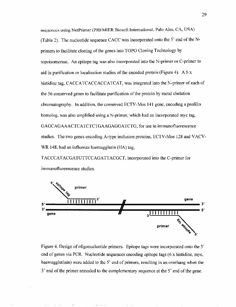
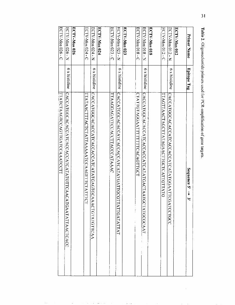
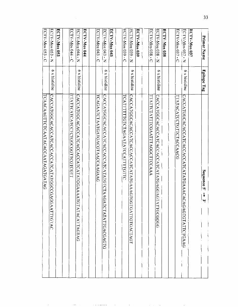
sequences using Netprimer (PREMIER Biosoft International, Palo Alto, CA, USA)
(Table 2). The nucleotide sequence CACC was incorporated onto the 5' end of the N-
primers to facilitate cloning of the genes into TOP0 Cloning Technology by
topoisomerase. An epitope tag was also incorporated into the N-primer or C-primer to
aid in purification or localization studies of the encoded protein (Figure 4). A 6 x
histidine tag, CACCATCACCACCATCAT, was integrated into the N-primer of each of
the 56 conserved genes to facilitate purification of the protein by metal chelation
chromatography. In addition, the conserved ECTV-Mos 14 1 gene, encoding a profilin
homolog, was also amplified using a N-primer, which had an incorporated myc tag,
GAGCAGAAACTCATCTCTGAAGAGGATCTG, for use in immunofluorescence
studies. The two genes encoding A-type inclusion proteins, ECTV-Mos 128 and VACV-
WR 148, had an influenza haemagglutin (HA) tag,
TACCCATACGATGTTCCAGATTACGCT, incorporated into the C-primer for
immunofluorescence studies.
5' a, 9% primer
l l l l l l l l l l l I gene 5' 3' 3' H
/I 1 1 1 1 1 1 1 1 1 1 1 5' gene
primer
5'
Figure 4. Design of oligonucleotide primers. Epitope tags were incorporated onto the 5'
end of genes via PCR. Nucleotide sequences encoding epitope tags (6 x histidine, myc,
haemagglutinin) were added to the 5' end of primers, resulting in an overhang when the
3' end of the primer annealed to the complementary sequence at the 5' end of the gene.
Page 41
PCR reactions (50 p1 total volume) were performed in 200 p1 thin-walled PCR tubes (Cat
# TW6200, Gordon Technologies, Mississauga, ON, Canada) in Minicycler PTC-150-25
(MJ Research, Watertown, MA, USA). Reaction mixes consisted of lxPCR buffer (50
mM KC1, 10 mM Tris pH 8.3, 1.5 mM MgC12, 0.01% gelatin), 0.1 mM dNTP mix
(Invitrogen Life Technologies, Carlsbad, CA, USA), 0.1 yM each of the forward and
reverse primer, 1 ng template DNA and 1 unit Pfu polymerase (Cat # 6001 35, Stratagene,
San Diego, CA, USA). Reactions were overlaid with two drops of mineral oil to prevent
evaporation of the reaction mixture. The following thermocycler conditions were used to
amplify the 56 conserved genes: initial denaturation at 94OC for 30 seconds; 26 cycles at
94•‹C for 30 seconds, 50•‹C for 30 seconds, 72•‹C for 2 minutes; a final extension at 72OC
was performed for 10 minutes and then samples were held at 4OC.
The genes encoding the A-type inclusion proteins, ECTV-Mos 128 and VACV-WR 148,
were amplified using similar PCR conditions as for the 56 conserved genes except the
extension time was 5 minutes.
Page 42
*
cc, T
able 2. Oligonucleotide prim
ers used for PCR
-amplification of gene targets.
Prim
er Nam
e
t--- E
pitope Tag
Sequence 5' -,
3'
histidine
I CACCATGGCACACCATCACCACCATCATATGGAATTCGATCCTGCC
TTAGTTAACTAGCTTATAGAACTTGCTCATTGTTATG
histidine CACCATGGCACACCATCACCACCATCATATGACTAATGCTATGCGCAAT
CTATTGTAGGAATTTTTTTTCACAGTTGCT
histidine CACCATGGCACACCATCACCACCATCATATGATTGCGTTATTGATATTAT
TTAAGGAGATTCCACCTTACCCATAAAC
histidine CACCATGGCACACCATCACCACCATCATATGAGTGCAAACTGTATGTTCAA
TTATAACTTTACTCTATTAAAAATCCAAGTTTCTATTTCT
histidine CACCATGGCACACCATCACCACCATCATATGTTCAACATGAATATTAACTCACC
TTATCTAAGTCCAGTTGATCCAAATCCTT
Page 43
ic;
Prim
er Nam
e
EC
TV
-Mos 027
EC
TV
-Mos 027 - N
E
CT
V-M
os 027 - C
EC
TV
-Mos 028
EC
TV
-Mos 028 - N
EC
TV
-Mos 028 - C
Epitope Tag
histidine
EC
TV
-Mos 029
EC
TV
-Mos 029 - N
EC
TV
-Mos 029 - C
Sequence 5'
-+ 3'
CA
CC
AT
GG
CA
CA
CC
AT
CA
CC
AC
CA
TC
AT
AT
GA
AC
AT
GG
AT
CA
MT
TA
TA
GA
TA
T
TTACCATCTTATCCCATTCCATATATTCC
histidine
EC
TV
-MO
S 031
EC
TV
-Mos 03 1 - N
CACCATGGCACACCATCACCACCATCATATGGAACCGATCCTTGCA
TTAAAAGTCAACATCTAAAGAAAAAATGATTGTC
histidine
EC
TV
-Mos 03 1 - C
CACCATGGCACACCATCACCACCATCATATGAGTAAAATACTCACATTTGTTAAA
TCAATTTATTGTAAAAAAAGAATCGGTTTTATAC
histidine
CTATTTTGGTGGAGGATTATATGATATAATTCG
EC
TV
-Mos 032 - N
EC
TV
-Mos 032 - C
CACCATGGCACACCATCACCACCATCATATGGAGGGATCTAAACGCA
EC
TV
-MO
S 033
EC
TV
-Mos 033 - N
EC
TV
-Mos 033 - C
histidine CACCATGGCACACCATCACCACCATCATATGGCGGAAACTAAAGAGTTT
TTAAACGTATAAAAACGTTCCGTATCTGTATTT
ti histidine
CACCATGGCACACCATCACCACCATCATATGGGTGTTGCCAATGATT
TT
AG
TT
TC
CG
CC
AT
TT
AT
CC
AG
TC
TG
Page 44
Prim
er Nam
e
EC
TV
-Mos 037
ECTV-Mos 037 - N
ECTV-MOS 037 - C
Epitope T
ag
ECTV-Mos 038 - N
ECTV-Mos 038 - C
Sequence 5' -, 3'
histidine
EC
TV
-Mos 039
ECTV-Mos 039 - N
ECTV-Mos 039 - C
CACCATGGCACACCATCACCACCATCATATGAAACACAGAGTGTATTCTGAAG
TTATACATCCTGTTCTACCAACG
histidine
EC
TV
-Mos 043
ECTV-Mos 043 - N
ECTV-MOS 043 - C
CACCATGGCACACCATCACCACCATCATATGAGGAGTATTGCGGGG
TTATTCTATTTCGAATTTAGGCTTCCAAA
histidine
ECTV-MOS 044 - C
CACCATGGCACACCATCACCACCATCATATGAAAGTGGTGATTGTGACTAGT
TCATTTTTTGTCTAGAATATCCATTTTGTTC
x
TT
AT
TC
AT
CA
TC
CT
CT
GG
CG
GT
TC
GT
CG
TT
CACCATGGCACACCATCACCACCATCATATGTCTAAGATCTATATTGACGAGTG
TCAGAATCTAATGATGACGTAACCAAGAAG
Page 45
Prim
er Nam
e
EC
TV
-Mos 058
EC
TV
-Mos 058 - N
E
CT
V-M
OS 058 - C
Epitope T
ag
EC
TV
-Mos 059 - N
EC
TV
-MO
S 059 - C
Sequence 5'
-, 3'
histidine
EC
TV
-Mos 064
EC
TV
-Mos 064 - N
EC
TV
-MO
S 064 - C
CACCATGGCACACCATCACCACCATCATATGGCGGATGCTATAACC
TTAACTTTTCATTAATAGGGACTTGACGTAC
listidine
EC
TV
-Mos 068 - N
E
CT
V-M
OS 068 - C
CACCATGGCACACCATCACCACCATCATATGAATAACTTTGTTAAACAAGTAGC
TCAAAGAATATGTGACAAAGTCCTAGTTGTATAC
histidine
EC
TV
-MO
S 070
EC
TV
-Mos 070 - N
EC
TV
-Mos 070 - C
CACCATGGCACACCATCACCACCATCATATGCCATTTAGAGATCTAATTTT
CTATGGAGTTTGGCCACCTGTTACCGAATA
histidine CACCATGGCACACCATCACCACCATCATATGGATCCGGTTGATTTTAT
TCACCCTTTAAGGTAATCAATTTGCC
histidine CACCATGGCACACCATCACCACCATCATATGAGCATCCGTATAAAAATCG
TTAGTCTAAAAACGCCATAAAGATGTTAATCTT
Page 46
-. m
I
Prim
er Nam
e I E
pitope Tag I
Sequence 5' -* 3'
EC
TV
-MO
S 073
ECTV-Mos 073 - N
ti
histidine CAC
CA
TG
GC
AC
AC
CA
TC
AC
CA
CC
AT
CA
TA
TG
GA
AG
TT
AT
CG
CT
GA
TC
G
ECTV-MOS 073 - C
T
TA
TA
GT
AT
AA
AG
TA
AT
AA
AA
AA
TA
GT
TA
AT
GT
GA
TG
AC
TT
G
ECTV-Mos 075 - N
ti
histidine CA
CC
AT
GG
CA
CA
CC
AT
CA
CC
AC
CA
TC
AT
AT
GA
GT
CT
AC
TG
CT
AG
AA
AA
CC
TC
ECTV-Mos 075 - C
T
CA
AT
CC
TT
TG
TT
GG
AA
TA
TC
TG
TT
AG
AG
G
EC
TV
-Mos 080
ECTV-Mos 080 - N
lktidine
CA
CC
AT
GG
CA
CA
CC
AT
CA
CC
AC
CA
TC
AT
AT
GA
AC
CA
AT
AC
AA
CG
TA
AA
AT
AT
C
ECTV-MOS 080 - C
T
TA
AT
CA
GC
GA
CT
GA
AA
TA
AC
AG
AT
CT
AT
CG
EC
TV
-Mos 083
ECTV-Mos 083 - N
ti
histidine CAC
CA
TG
GC
AC
AC
CA
TC
AC
CA
CC
AT
CA
TA
TG
GA
TA
AG
AA
AA
GT
TT
GT
AT
AA
AT
AC
T
ECTV-MOS 083 - C
T
TA
AT
TC
TT
AT
CA
AT
CA
CA
TA
TT
TT
TC
TA
TG
AT
GT
CT
ECTV-Mos 084 - N
ti
lGstidine
CA
CC
AT
GG
CA
CA
CC
AT
CA
CC
AC
CA
TC
AT
AT
GG
AT
AA
AA
CT
AC
TT
TA
TC
AG
TA
AA
C
ECTV-MOS 084 - C
C
TA
TT
CC
AT
AT
TA
CT
AA
GA
TC
GG
AA
CA
CC
A
EC
TV
-Mos 087
ECTV-Mos 087 - N
ti
histidine CAC
CA
TG
GC
AC
AC
CA
TC
AC
CA
CC
AT
CA
TA
TG
GC
GT
GG
TC
AA
TT
AC
G
ECTV-Mos 087 - C
T
TA
CT
TC
TT
AC
AA
GT
TT
TA
AC
TT
TT
TT
AC
GA
AC
AA
Page 47
w
m
EC
TV
-Mos 089
EC
TV
-Mos 089 - N
Prim
er Nam
e
EC
TV
-MO
S 089 - C
histidine
TTAACAAGTGTCTTTTATATATTCGTAATCTATGCC
EC
TV
-Mos 091
EC
TV
-Mos 09 1 - N
EC
TV
-Mos 09 1 - C
Epitope T
ag
CACCATGGCACACCATCACCACCATCATATGGAAATGGATAAGCGTATG
CACCATGGCACACCATCACCACCATCATATGTCCATCAATATCGATATAAAAA
TTACTTAGTTACTATGTTGTTTATGTCTTTTCTTTCC
EC
TV
-MO
S 096
EC
TV
-Mos 096 - N
EC
TV
-MO
S 096 - C
Sequence 5'
-, 3'
CACCATGGCACACCATCACCACCATCATATGTCGAGCTTTGTTACCAAT
TTATGAGTCGACGATATTCGCGAGA
EC
TV
-Mos 098
EC
TV
-Mos 098 - N
E
CT
V-M
OS 098 - C
lGstidine
TCATTTACTATTAACTAGCATATTATA
EC
TV
-Mos 099
EC
TV
-Mos 099 - N
EC
TV
-MO
S 099 - C
CACCATGGCACACCATCACCACCATCATATGGGAATTACAATGGATGAG
lGstidine
CACCATGGCACACAATCACCACCATCATATGACCTTTTACAGATCTAGTATAATTAG
CTAATCAATAAATCCATCCGTTAATTTTTTTA
Page 48
" I
Prim
erNam
e E
pitope Tag I
Sequence 5' 4
3'
histidine C
AC
CA
TG
GC
AC
AC
CA
TC
AC
CA
CC
AT
CA
TA
TG
AA
TC
TA
CG
AT
TA
TG
TA
GC
GG
TTATACGTCTAATGAGCAAGTAGAAAACCTCT
histidine C
AC
CA
TG
GC
AC
AC
CA
TC
AC
CA
CC
AT
CA
TA
TG
GC
AG
AC
AC
AG
AC
GA
TA
TT
A
TTAGAATTTATACGAATATCGTTCTCTAAATGTAACA
TTAGAATTTATACGAATATCGTTCTCTAAATGTAACA
histidine C
AC
CA
TG
GC
AC
AC
CA
TC
AC
CA
CC
AT
CA
TA
TG
TT
CG
AA
CC
AG
TA
CC
AG
AT
C
CTAAGTGAAGTATTTTAGTAACGTATCCTTATCCC
TTAAATAATTTTAATTCGTTTAACGAATATCTTGAG
histidine C
AC
CA
TG
GC
AC
AC
CA
TC
AC
CA
CC
AT
CA
TA
TG
TT
CG
TA
GA
CG
AT
AA
TT
CG
TT
TTACTTATCATTTACTAGACGAAAAGGTGGTG
Page 49
Epitope T
ag Sequence 5'
-+
3'
histidine C
AC
CA
TG
GC
AC
AC
CA
TC
AC
CA
CC
AT
CA
TA
TG
GA
TA
GT
AC
CA
AC
GC
GC
TTAACTCGCAAAATCGTTAAGAAGTTTAAGC
TTAGGTAGTAAAAAATAAGTCAGAATATGCCCTAT
CACCATGGCACACCATCACCACCATCATATGGATAATCTATTTACCTTTCTACA
TCATTTTAGAAGCAATTCTTTTAGACGATC
CACCATGGAGGTCACGAACCTTATTGAAAA
hemagluttinin C
TA
AG
CG
TA
AT
CT
GG
AA
CA
TC
GT
AT
GG
GT
AA
GT
AG
AT
AT
TG
GT
AG
MG
AT
AT
GC
histidine C
AC
CA
TG
GC
AC
AC
CA
TC
AC
CA
CC
AT
CA
TA
TG
CA
GT
AT
CC
GC
GG
G
TTATAATATATTAGAAGCTGACAAAATTTTTTTACAC
histidine C
AC
CA
TG
GC
AC
AC
CA
TC
AC
CA
CC
AT
CA
TA
TG
AA
TT
GT
TT
TC
AA
GA
AA
AA
CA
G
TTATGATACATTTTTTGACGACGATGATT
Page 50
Prim
er Nam
e
* t----- E
pitope Tag
Sequence 5' -,
3'
CACCATGGCACACCATCACCACCATCATATGGCGGCCGAATGG
TTAATTACCAGTTGCTCGCACATTAGT
I
mY
c C
AC
CA
TG
GA
GC
AG
AA
AC
TC
AT
CT
CT
GA
AG
AG
GA
TC
TG
AT
GG
CG
GC
CG
MT
G
TT
AA
TT
AC
CA
GT
TG
CT
CG
CA
CA
TT
AG
T
histidine C
AC
CA
TG
GC
AC
AC
CA
TC
AC
CA
CC
AT
CA
TA
TG
GC
GT
TT
GA
TA
TA
TC
AG
TT
AA
TT
AT
AC
AT
CC
GT
TT
CC
CT
GT
CG
GT
T
histidine C
AC
CA
TG
GC
AC
AC
CA
TC
AC
CA
CC
AT
CA
TA
TG
AA
CT
TT
CA
AG
GA
CT
TG
TG
TT
TTACATAACTCCATTCATTAATACGCGC
Page 51
Primer Name
Epitope Tag Sequence 5'
-+ 3'
ECTV-MOS 153
ECTV-Mos 153 - N
histidine CA
CC
AT
GG
CA
CA
CC
AT
CA
CC
AC
CA
TC
AT
AT
GG
CG
AT
GT
TT
TA
CA
CA
CA
ECTV-MOS 153 - C
T
TA
AA
CT
TT
TA
TA
TA
TG
AC
AC
CC
AT
TC
AT
CT
GG
ECTV-Mos 160
ECTV-Mos 160 - N
histidine CA
CC
AT
GG
CA
CA
CC
AT
CA
CC
AC
CA
TC
AT
AT
GG
AA
TC
CT
TC
AA
GT
AT
TG
TT
T
ECTV-MOS 160 - C
T
CA
AT
CT
TG
TA
TA
AA
CA
GT
CT
AC
GT
AG
TC
TG
TC
A
ECTV-Mos 16 1 - N
histidine C
AC
CA
TG
GC
AC
AC
CA
TC
AC
CA
CC
AT
CA
TA
TG
GA
TA
TC
TT
CA
GG
GA
GA
TC
ECTV-MOS 16 1 - C
TTAATTAGTTGTTGGAGAGCAATATCTACCA
ECTV-MOS 167 - C
T
TA
GT
AG
AT
GG
GT
AG
TG
TA
TC
GT
GT
AC
TA
TA
TA
AC
TA
TT
C
ECTV-Mos 168
ECTV-Mos 168 - N
histidine CA
CC
AT
GG
CA
CA
CC
AT
CA
CC
AC
CA
TC
AT
AT
GT
CT
AC
TT
GG
CA
TG
TT
GT
CA
ECTV-MOS 168 - C
T
TA
TT
GT
GG
AT
AG
CA
GT
AT
TT
CC
CT
AT
AA
AA
A
VACV-WR 148 - N
C
AC
CA
TG
GA
GG
TC
AC
GA
AC
CT
TA
TT
GA
AA
A
VACV-WR 148 - C
hemagglutinin T
TA
AG
CG
TA
AT
CT
GG
AA
CA
TC
GT
AT
GG
GT
AA
GA
CG
TC
GC
AT
CT
CT
CT
CT
GT
TT
C
Page 52
Agurose gel electrophoresis
PCR products and plasmid DNA were resolved by agarose gel electrophoresis. Gels
were prepared by dissolving OmniPur Agarose (EMD Chemicals Inc., Gibbstown, NJ,
USA) in Tris-acetate buffer (TAE; 40 mM Tris-acetate, 1 mM EDTA) for a final agarose
concentration of 1%. DNA samples were mixed with 6 x DNA loading buffer (0.25%
bromophenol blue, 0.25% xylene cyan01 FF, 40% sucrose, 50 mM EDTA) prior to
loading on the gel. Mini gels were loaded with 2 - 1Oyl of each sample and 0.4 yg of 1
Kb Plus DNA Ladder (Cat # 1078701 8, Invitrogen Life Technologies). Electrophoresis
was performed at 100 volts (Bio-Rad Power Pac 300; Bio-Rad, Richmond, CA, USA) for
30 minutes in TAE buffer. Following electrophoresis, the DNA was stained for
approximately 15 minutes in buffer containing 0.5 yglml ethidium bromide, visualized
using a MultiImage Light Cabinet (Alpha Innotech, San Leandro, CA, USA) and
photographed.
Purzjcution of PCR products
PCR products were purified using the QIAquick PCR Purification kit (Cat # 28 104
QIAGEN, Chatsworth, CA, USA) according to the manufacturer's instructions. After
purification, DNA was eluted from the QIAquick column by application of 50 yl Buffer
EB (10 mM Tris-HC1, pH 8.5) and stored at -20•‹C.
Page 53
Cloning o f recombinant genes into the p E N T W D - Topo Entry Vector
Purified PCR products were introduced into the cloning site of pENTR/SD/D-Topo entry
vector according to the instructions in the pENTR/SD/D-Topo Cloning Kit (Cat #
0219, Invitrogen Life Technologies) (Figure 5). Briefly, 2.5 ng fresh PCR product, 1 y1
salt solution (250 mM NaCl, 10 mM MgC12), and 250 ng pENTR/SD/D-Topo entry
vector were mixed together and brought up to a final volume of 5 yl with water. A
negative control, in which the PCR product was omitted from the reaction mixture, was
included the first time the reaction was performed. The reactions were mixed gently and
allowed to incubate at room temperature for 5 minutes. Directionality of cloning was
achieved through the unique design of the entry vector and use of topoisomerase I, which
is covalently bound to the 3' phosphate of the linearized entry vector. The entry vector
contains a single-strand GTGG overhang on the 5' end and a blunt end on the 3' end.
The four-nucleotide overhang invades the double-stranded DNA of the PCR product and
anneals to the CACC sequence incorporated in the 5' primer. Topoisomerase then ligates
the PCR product in the correct orientation. After incubation, the reaction mixture was
frozen at -20•‹C or used to transform One Shot TOP1 0 E. coli cells (Invitrogen Life
Technologies).
Page 54
+ PCR product
Destination Vcctor
Entry Vector
Expression Clone
Figure 5. Cloning strategy for the production of pDEST14 expression clones.
1 Entry Clone
LR Reaction
Page 55
Transformation of E. coli cells
2 p1 of pENTR/SD/D-Topo entry vector or pDEST14 expression clone DNA was added
to 1.5 ml screw-top microfuge tubes containing 50 pl of thawed, chemically competent
One Shot TOP1 0 E. coli (Cat # 440301, Invitrogen Life Technologies) or One Shot
BL2 1 -A1 Chemically Competent E. coli (Cat # 440 184, Invitrogen Life Technologies),
respectively, and swirled gently to mix. The cells were incubated on ice for 30 minutes
then heat-shocked at 42•‹C for 30 seconds without agitation. The microfuge tubes were
returned to ice, and 250 p1 room temperature SOC medium (0.5% yeast extract, 2%
tryptone, 10 mM NaC1,2.5 mM KC1, 10 mM MgC12,lO mM MgS04, 20 mM glucose)
was added. The vials were shaken horizontally (200 rpm) at 37•‹C for 30 or 60 minutes in
an Innova 4000 Shaking Incubator (New Brunswick Scientific Co. Inc., Edison, NJ,
USA) to permit expression of the antibiotic resistance gene prior to plating 150 p1 of the
transformation reaction onto pre-warmed Luria Bertani (LB; log tryptone, 5g yeast
extract, 10 g NaC1, in a total of 1 L of distilled water, adjusted to pH 7.0 with 5 M NaOH)
2% agar plates containing the appropriate antibiotic.
Isolation and purzjkation of plasmid DNA
Single colonies were used to inoculate Luria Bertani broth containing the appropriate
antibiotic and grown overnight at 200 rpm, 37•‹C in a shaking incubator. Plasmid DNA
was isolated using the QIAprep Spin Miniprep Kit (Cat # 271 04, QIAGEN) as outlined in
the QIAprep Spin Miniprep Kit manual. This DNA isolation procedure is based on rapid
alkaline lysis, as described by Birnboim and Doly (1979). After isolation and
Page 56
purification, DNA was eluted from the QIAprep spin column by application of 50 yl
sterile water.
Quantitation o f DNA
A solid-state fluorimeter (SSF-600, Tyler Research Instruments Corporation, Edmonton,
AB, Canada) was zeroed with ethidium bromide solution (5 mM Tris pH 8.0, 0.5 mM
EDTA, 0.5 ng/ml EtBr), and calibrated with a standard containing 50 pg herring sperm
DNA in 2 ml ethidium bromide solution. DNA sample concentration was determined by
adding 1 p1 of the sample DNA to 2 ml of ethidium bromide solution and comparing to
the standard.
IdentiJication ofpositive clones
Entry clones containing insert in the correct orientation in the pENTR/SD/D-Topo entry
vector were identified by PCR analysis. A small amount of colony was removed from
the transformation plate with a sterile toothpick and resuspended in 100 p1 sterile water in
a 1.5 ml Micro Tube. Typically, 1 p1 of this cell suspension was used as template in the
following PCR reaction: 1 x PCR buffer, 1 unit Tag polymerase (cloned in our
laboratory), 0.1 mM dNTP mix, 0.1 pM gene-specific N-primer and 0.1 pM vector-
specific M13 reverse primer (Cat # 460691, Invitrogen Life Technologies). Thermocyler
conditions were as previously described. PCR products were analyzed by agarose gel
electrophoresis.
Page 57
DNA sequencing
Plasmid DNA from positive clones were checked by DNA sequencing to verify that
errors had not been introduced by Pfu polymerase during PCR. High quality DNA
suitable for sequencing was prepared using a QIAprep Spin Miniprep Kit (QIAGEN)
according to the manufacturer's instructions. Approximately 1 pg of plasmid DNA was
provided to the University of Victoria Centre for Environmental Health Sequencing
Group. Sequences were obtained using a LI-COR fluorescent DNA sequencer. Raw data
was analyzed with the EditView program (version 4.1 1, Applied Biosystems, Foster City,
CA, USA).
Subcloning o f recombinant genes into pDEST14 Destination Vector
For expression of the recombinant proteins, expression clones were constructed by
transferring recombinant genes from the pENTR/SD/D-Topo entry clone to the pDEST14
Destination Vector ( ~ m ~ ~ ) via the LR recombination reaction. The reaction strategy to
produce a pDEST14 expression clone is given in Figure 5. LR reaction conditions were
followed as per manufacturer instructions outlined in the Gateway Technology Cloning
Manual (Invitrogen Life Technologies); however, reaction volumes were halved to
minimize cost. To perform the LR reaction, typically 100 ng pENTRlSD/D-Topo entry
clone, 1 50 ng pDEST 14 destination vector (Invitrogen Life Technologies), 2 p1 LR
Reaction Buffer (Invitrogen Life Technologies), 5 pl TE buffer (TE; 10 mM Tris-C1, pH
7.5, 1 mM EDTA pH KO), and 2 p1 LR Clonase Enzyme Mix (Invitrogen Life
Technologies) were mixed together in a 1.5 ml Micro Tube and allowed to incubate at
Page 58
room temperature for 1 hour. To stop the reaction, 2 yl proteinase K solution (2 yglml
in: 10 mM Tris-HC1 pH 7.5, 20 mM CaC12, 50% glycerol) was added and the reaction
was incubated at 37•‹C for 10 minutes. BL2 1-A1 E. coli cells (Cat # C607003, Invitrogen
Life Technologies) were transformed with 1 yl LR reaction for expression of the
recombinant proteins.
Expression andpuriJication of recombinant proteins
4 x 2 L Erlenmeyer flasks containing 500 ml LB broth plus 100 yglml ampicillin were
inoculated with 12 ml of overnight culture and grown at 200 rpm, 37"C, in the shaking
incubator until the OD600 was 0.4 (approximately 2 hours) as measured using a Beckman
DU-65 Spectrophotometer (Beckrnan Coulter Inc.). The flask was rapidly cooled on ice
to room temperature prior to the addition of 20 % L-arabinose to a final concentration of
0.2%. Expression of the recombinant protein from the expression vector is regulated by
the araBAD promoter (PBAD), which controls expression of the T7 RNA polymerase in
BL21 -A1 cells. In the presence of arabinose, the PBAD promoter permits expression of the
T7 RNA polymerase, and therefore transcription of the gene of interest. After induction
with arabinose, the cells were grown for an additional 4 hours at 2528•‹C to permit
recombinant protein expression after which the cells were harvested by centrifugation at
5000 x g in a Beckman J2-HC centrifuge (JA-14 rotor) for 10 minutes at 4•‹C. The
supernatants were discarded and the pellet resuspended in 40 ml lysis / purification buffer
(50 mM NaP04, 0.5 M NaCl pH 7.0, 1 mM PMSF). The cells were lysed by passing the
cell suspension through a French Pressure Cell Press (American Instrument Company,
Silver Spring, Maryland, USA) at 1260 psi a total of 3 times. Insoluble material was
Page 59
removed by centrifugation at 12,000 x g for 45 minutes at 4OC. The clarified extract was
loaded onto 2 ml nitrilotetraacetic agarose columns (Cat # R90101, Ni-NTA resin,
Invitrogen Life Technologies) at a flow rate of 0.5 mllmin for 80 minutes on a Bio-Rad
Biologic low pressure chromatography system (Bio-Rad). Contaminating proteins were
removed by washing the column with low pH lysis I purification buffer (pH 5.0) for 45
min at 0.5 mllmin. The histidine-tagged proteins were eluted with an elution buffer (50
mM NaP04, 0.5 M NaCl pH 7.0, lmM PMSF) gradient from 0 mM to 300 mM imidazole
over 2 hours at 0.5 mllmin, collecting 2 ml fractions. EDTA and glycerol were added to
the fractions (final concentrations of I mM and lo%, respectively) to prevent degradation
and aggregation of the proteins, which were then stored at -20•‹C. Fractions containing
significant amounts of purified protein were identified by SDS-PAGE.
Polyacrylamide gel electrophoresis
Sodium dodecyl sulfate polyacrylamide gel electrophoresis (SDS-PAGE) was performed
using an Xcell SureLock Mini-cell apparatus (Invitrogen Life Technologies). Protein
samples were prepared by adding NuPAGE LDS sample buffer (to a final concentration
of 1 x); Cat # NP0007, Invitrogen Life Technologies), DTT (dithiothreitol; to a final
concentration of 10 mM) and heating to 70•‹C for 10 minutes. If cell lysate was run on
the gel, DNA was sheared with an 18-gauge syringe needle prior to loading. 5 pL
BenchMark Protein Marker (Cat # 107470 12, Invitrogen Life Technologies) or 10 pL
SeeBlue Plus2 (Cat # LC5925, Invitrogen Life Technologies) protein molecular weight
standard was used if the gel was to be stained with Coomassie blue or used in western
blot analysis, respectively. Protein samples and protein standards were loaded onto pre-
Page 60
cast NuPAGE Novex 4 - 12% Bis-Tris 12 well gels (Cat # NP0322BOX, Invitrogen Life
Technologies) and run at 200 volts for 35 minutes in MES running buffer (0.05 M 2-(N-
morpholino) ethane sulfonic acid, 0.05M tris base, 3.47 mM SDS, 1.025 mM EDTA, pH
7.3). Gels to be stained with Coomassie blue were placed in fixing solution (50%
methanol, 10% acetic acid, 40% water) for 30 minutes, stained in Coomassie blue G-250
(0.1% Brilliant Blue G, 25% methanol, and 5% acetic acid) for 1 hour and then placed in
destaining buffer (1 0% methanol, 10% acetic acid, 80% water) until background was
sufficiently reduced (4 hours to overnight). Gels were digitally photographed using the
Odyssey Infrared Imaging System (model 9120, Li-COR Biosciences, Lincoln, NB,
USA), and dried in BioDesignGelWrap membrane (Cat# G101, BioDesign Inc., Carmel,
NY, USA).
Bradford assays
Protein concentrations were determined using a Bradford assay in which 1.5 ml Bradford
Reagent (Cat # B6916, Sigma) was added to 50 p1 of bovine serum albumin (BSA)
standards (serially diluted from 2 mglml to 0.25 mglml) or protein samples in disposable
cuvettes. The samples were gently mixed and incubated for 20 minutes at room
temperature to allow colour development. The absorbance of the samples was read at
595 nrn on a Beckman DU-65 Spectrophotometer (Beckman Coulter Inc.). The
concentrations of the protein samples were determined by construction of a standard
curve using the absorbance readings for the BSA standards.
Page 61
Western blots
Purified protein or cellular lysates were separated by SDS-PAGE on 4 - 12% acrylamide
gels. Proteins were transferred to a Trans-Blot nitrocellulose membrane (Cat # 120 1 1,
Bio-Rad) in transfer buffer (20% methanol, 25 mM Tris base, 192 mM glycine, 0.035
mM SDS) at 80 volts for 90 minutes using a Bio-Rad mini trans-blot cell apparatus (Bio-
Rad). Membranes were blocked with Odyssey Blocking Buffer (Cat # 9270000, Li-COR
Biosciences) for 1 hour at room temperature and then incubated with primary antibody
diluted in Odyssey Blocking Buffer 1 : 1500 for 1 hour at 4•‹C. Membranes were washed
4 x 5 minutes with PBS (phosphate buffered saline; 137 mM NaC1,2.7 mM KCl, 4.3 mM
Na2HP04.7H20, 1.4 mM KH2P04 pH 7.3) + 0.1% TWEEN-20 and then incubated in
secondary antibody 1 :2500 dilution in Odyssey Blocking Buffer for 1 hour at 4•‹C.
Histidine-tagged proteins were detected and visualized using mouse IgGl anti-penta
histidine primary antibody (Cat # 34660, QIAGEN) and rabbit anti-mouse IgG (H&L)
IRDye 800 conjugate secondary antibody (Cat # 610432020, Rockland
Immunochemicals Inc., Gilbertsville, PA, USA). Myc-tagged ECTV-Mos 141 protein
was detected and visualized using rabbit polyclonal anti-myc primary antibody (Cat #
2272, Cell Signaling Technology, Beverly, MA, USA) and goat anti-rabbit IgG (H&L)
IRDye 800 conjugate secondary antibody (Cat # 61 1132122, Rockland
Immunochemicals Inc.). Hemagglutinin-tagged proteins, ECTV-Mos 128 and VACV-
WR 148, were detected and visualized using mouse IgGl anti - hemagglutinin Alexa
Fluor 488 conjugate antibody (Cat # A21287, Molecular Probes Inc., Eugene, OR, USA)
and rabbit anti-mouse IgG (H&L) IRDye 800 conjugate secondary antibody (Cat #
Page 62
610432020, Rockland Irnmunochemicals Inc.). Actin was detected and visualized using
rabbit IgG anti-actin primary antibody (Cat # A5060, Sigma-Aldrich) and goat anti-rabbit
IgG (H&L) IRDye 800 conjugate secondary antibody (Cat # 61 1132122, Rockland
Immunochemicals Inc.). After incubation in secondary antibody, membranes were
washed 4 x 5 minutes each in PBS + 0.1% TWEEN-20 and then once in PBS alone for 5
minutes at room temperature. Blots were allowed to dry and then visualized and digitally
photographed using the Odyssey Infiared Imaging System (Li-COR Biosciences).
Ectromelia virus (Moscow) genome map
The Viral Orthologs Clusters Database (VOCs) was used to create the genomic map of
ECTV-Mos. Genes were selected for display and coloured based on their degree of
conservation in 42 sequenced poxvirus genomes (Table 1). The resulting JPEG file was
modified in OmniGraffle Professional software.
Homology modeling of ECTV-Mos 141
The ECTV-Mos 141 primary protein sequence (NP - 671 660.1) was submitted to the
SWISS-MODEL (Schwede et al., 2003) server using the "First Approach" mode with
default settings. The server identified 4 profilin proteins as having a high degree of
sequence identity with ECTV-Mos 141 based on BLASTp results (Altschul et al., 1990):
human platelet profilin 1 (1 FIL), human profilin (IFIK), human profilin NMR structure
(IPFL), and bovine profilin bound with actin (lHLU). SWISS-MODEL created a
multiple sequence alignment of ECTV-Mos 141 and the 4 template proteins using the
Page 63
SIM sequence alignment program (Huang, X and M. Webb, 1 Wl), and used the
alignment in the ProModII (Peitsch, 1996) modeling program to build the ECTV-Mos
141 homology model. WHAT-CHECK (Hooft et al., 1996) and VADAR (Willard et al.,
2003) protein verification tools were used to analyze the protein structure quality.
Images of the homology model were generated using the Protein Explorer
macromolecular visualization tool (Martz, 2002).
This work was performed by Shan Sundararaj (current address: Department of
Computing Science and Biological Sciences, University of Alberta, Edmonton, Alberta,
Canada)
Page 64
Mammalian cell culture
Tissue culture reagents were obtained from Gibco BRL Inc. (Gaithersburg, MD, USA)
unless otherwise stated. The African green monkey kidney cell line BS-C-1 (ATCC CCL
26), was grown in complete Dulbecco's modified Eagle medium (D-MEM; 5.56 mM D-
glucose, 4 mM L-glutamine, 0.01 96 mM pyroxine hydrochloride, 1 mM sodium pyruvate
and 44.05 mM NaHC03) supplemented with 10% newborn bovine serum, 50 U/ml
penicillin, 50 pglml streptomycin and 5 ml/L (1 00X) GlutaMAX-11, in a humidified
37"C, 7% C02 incubator (Sanyo C02 Incubator, Caltec Scientific Ltd., Vancouver, BC,
Canada). Cells were passaged by harvesting confluent monolayers from T75 tissue
culture flasks (Falcon, Beckton Dickinson Labware, Franklin Lakes, NJ, USA) followed
by seeding approximately 20% of the cells back to the flasks. Briefly, the medium was
decanted into neutral buffer formalin (NBF; 4% formaldehyde pH 7.0, 0.145 M NaCl,
0.03 M Na2HPOd,0.03 M NaH2P04*H20) and the cells washed with pre-warmed, 37•‹C
PBS. The cells were removed from the monolayer by the addition of 5 ml of 1 x PBS
containing approximately 0.75 pglml trypsin and incubated at 37OC until the cells began
to round up and detach. 5 ml of complete D-MEM + serum was added to inhibit trypsin
activity and cell clumping. The cell suspension was transferred to 15 ml conical tubes,
pelleted by centrifugation at 400 x g for 5 minutes (Beckman GS- 15 Centrifuge,
Beckrnan Instruments) and resuspended in 5 ml of complete D-MEM. 20% of the cells
were added to a T75 flask containing 14 ml complete D-MEM then returned to the COz
incubator.
Page 65
Virus infection and transfection of mammalian cells
This protocol was adapted from Fuerst et al., 1986. When BS-C-1 cells had reached
approximately 90% confluency, the growth medium was removed and an appropriate
volume of complete D-MEM containing virus at the desired multiplicity of infection
(MOI) was added. For 100 mm and 6 well tissue culture dishes and chamber slides, the
infection volumes were 3 ml, 500 y1 and 200 yl respectively. Virus absorption was
allowed to proceed at 37OC for 1 hour with gentle rocking every 10 minutes, the virus
inoculum was removed and replaced with complete D-MEM + serum (10 ml, 2 ml and
200 y1, respectively). The infected cells were incubated at 37OC for an additional 2 hours
before transfection with plasmid DNA.
During the last hour of infection, plasmid DNA was prepared for transfection of the BS-
C- 1 cells. For 100 mm and 6 well tissue culture dishes and chamber slides, the cells were
transfected with a total of 25 yg, 2.5 yg or 200 ng pDEST14 expression clone DNA,
respectively. The appropriate amount of DNA was brought up to a final volume of 700
1-11, 230 y1, or 93 yl, respectively, with sterile water and mixed with 75 yl, 25 y1, or 17 y1
of 2.5 M CaC12, respectively. The DNA / CaC12 solution was added dropwise to 750 yl,
250 y1, or 25 yl, respectively of 2 x HBS (HEPES-buffered saline solution; 0.28 M NaC1,
0.05 M HEPES, 1.5 mM Na2HP04, pH 7.5, filter sterilized through a 0.45 ym filter),
vortexed, and incubated at room temperature for 1 hour. After the 2 hour infection
incubation, the medium was removed from the cells and the DNA precipitate was added
dropwise over cells. The precipitate was left on cells for 15 minutes with occasional
rocking, and then 10 ml, 1.5 ml, and 200 p1, respectively, of pre-warmed 37•‹C complete
Page 66
D-MEM was added to the dishes or chamber slides and cells were incubated an additional
4 hours in the C02 incubator. The medium was removed, cells were washed 2 x with
PBS, and an appropriate amount of pre-warmed complete D-MEM + serum was added to
the dishes or chambers, and the cells were incubated overnight in the C02 incubator.
Immunoprecipitation
Protocol was adapted from Husain and Moss, 2003. BS-C-1 (passage 7) cells were
seeded in 9 x 100 mm tissue culture dishes and grown to 90% confluency (approximately
6.3 x lo7 cells / dish). Cells were infected with a recombinant VACV strain WR vTF7-3
(passage 3, ATCC VR-2153) expressing a T7 polymerase, at a multiplicity of infection
(MOI) of 10 and transfected with 25 pg ECTV-Mos 141 (histidine-tagged) pDEST14
Expression Clone plasmid DNA per 100 mm dish. Cells were incubated overnight in a
humidified 37"C, 7% C02 incubator. The next day (-16 hours later), the tissue culture
dishes were removed from the incubator and placed on ice, the growth medium was
removed, and the cells were washed 4 x with 10 ml ice-cold PBS. 350 p1 ice-cold non-
denaturing lysis buffer (50 mM Tris-HC1 pH 7.5,300 mM NaC1, 1% Triton X-100, 10
mM imidazole) containing protease inhibitor cocktail (Cat # 1836153, Roche Applied
Science) was added to each 100 rnrn tissue culture dish. After a 10 minute incubation in
lysis buffer on ice, cells were removed from the monolayer using a cell scraper, and
transferred to a 30 ml glass centrifuge tube. The cell suspension (-3.15 ml) was mixed
gently incubated an additional 30 minutes on ice. The lysate was cleared by centrifuging
at 20,000 x g for 10 min at 4OC. The supernatant was transferred to a new tube and 15 yl
Protein G-Plus Agarose (Cat # sc2002, Santa Cruz Biotechnology, Santa Cruz, CA, USA)
Page 67
was added to pre-clear the extract. After rotating the tube at 4OC for 1 hour, the Protein
G-Plus Agarose was pelleted by centrifuging at 1000 x g for 1 minute. The supernatant
was removed and dispensed into a fresh centrifuge tube and Penta-His Antibody (Cat #
34660, mouse Penta-His Antibody IgG,, QIAGEN) was added to a concentration of 5
pglml. The tube was rotated for 3 hours at 4OC before 60 p1 Protein G-Plus Agarose was
added and the incubation continued overnight. The next day (- 16 hours later), the
Protein G-Plus Agarose was pelleted by centrifuging at 1000 x g for 1 minute and the
supernatant was removed. The agarose was resuspended in 3 ml of wash buffer (0.1%
Triton X-100, 50 mM Tris-C1, pH 7.5,300 mM NaCl), mixed gently, and centrifuged at
1000 x g to pellet the agarose. The agarose was washed as above a total of 4 times before
a final wash with PBS and resuspension in 80 p1 of 1 x NuPAGE LDS sample buffer (Cat
# NP0007, Invitrogen Life Technologies), 10 mM DTT (dithiothreitol) and heated to
70•‹C for 10 minutes. A control was performed which followed the identical protocol
with the exception that cells were transformed with herring sperm DNA instead of
pDEST 14 expression clone DNA.
Mass spectrometry
Coomassie blue-stained bands were excised from the gel using a new scalpel and were
prepared and analyzed by the Genome BC Proteomics Centre. The gel slices were
subjected to an automated in-gel trypsin digestion, and were analyzed by MALDI-TOF
using a Voyager-DE STR mass spectrometer (Applied Biosystems, Foster City, CA,
USA). The Mascot search engine (Perkins et al., 1999) was used to identify the primary
Page 68
protein sequences of the samples from the mass spectrometry data by searching primary
sequence databases.
Far Western analysis
Protocol was adapted from Einarson and Orlinick, 2002. 2 pg of each of porcine muscle
tropomyosin (Cat # T2400, Sigma-Aldrich), ECTV-Mos 14 1 (histidine-tagged profilin
homolog, metal chelation chromatography purified), rabbit muscle actin (Cat # A2522,
Sigma-Aldrich), RelA (histidine-tagged bacterial protein, a gift from Dr. Edward
Ishiguro, Dept. Biochemistry and Microbiology, University of Victoria), and Bovine
Serum Albumin (BSA; Cat # A9647, Sigma-Aldrich) were separated by SDS-PAGE.
After transfer to a nitrocellulose membrane, proteins were refolded by a denaturation and
renaturation cycle in guanidine hydrochloride as described by Rea et al., 2004. The
~nembrane was washed twice in 50 ml of denaturation buffer (6 M guanidine
hydrochloride, 20 mM HEPES pH 7.5,50 mM KC1, 10 mM MgC12, 1 mM DTT, 0.1%
Nonidet P-40) for 10 minutes at 4OC with gentle agitation. The denaturation buffer was
diluted 1 : 1 with basic buffer (20 mM HEPES pH 7.5, 50 mM KCl, 10 mM MgCls, 1 mM
DTT, 0.1% Nonidet P-40) and the membrane was washed as before. This dilution and
wash cycle was repeated four more times until the final wash contained 175 mM
guanidinium hydrochloride. Porcine muscle tropomyosin was used as the probe protein,
and was diluted to a final concentration of 20 pglml in interaction buffer (1% nonfat dry
milk in basic buffer, 5% glycerol, 1 mM PMSF) and was incubated with the membrane
for 5 hours at 4•‹C with gentle agitation. The tropomyosin solution was removed and the
membrane was washed 4 x 10 minutes in buffer #1 (0.2% Triton X-100 in PBS) at 4OC
Page 69
with gentle agitation, followed by 2 x 10 minutes in buffer #2 (0.2% Triton X- 100, 100
mM KC1 in PBS) at 4•‹C with gentle agitation. The membrane was exposed to mouse
monoclonal anti-tropomyosin IgGl primary antibody (Cat # T2780, Sigma-Aldrich)
diluted 1 : 1000 in 1 : 1 Odyssey Blocking Buffer and PBS + 0.2% TWEEN-20, overnight
at 4•‹C with gentle agitation. The next day the primary antibody was removed and the
membrane was washed 4 x 5 minutes with PBS + 0.1% TWEEN-20. The membrane was
then exposed to rabbit anti-mouse IgG (H&L) IRDye 800 conjugate secondary antibody
(Cat # 61 0432020, Rockland Immunochemicals Inc.) at 1 :2500 dilution for 1 hour at 4OC
with gentle agitation. The secondary antibody was removed and the membrane was
washed 4 x 5 minutes with PBS + 0.1% TWEEN-20 and once with PBS alone. The blot
was visualized and digitally photographed using the Odyssey Infrared Imaging System
(Li-COR Biosciences).
Protocol was adapted from Ye et al., 2004. BS-C-I cells (passage 12) were grown on
cover slips to 80% confluency, infected at a MOI of 10 with recombinant VACV western
reserve strain vTF7-3 (passage 3; ATCC VR-2153) expressing T7 RNA polymerase, and
then transfected with a total of 200 ng pDEST14 expression clone plasmid DNA per
chamber. After infection and transfection, cells were incubated overnight (approximately
16 hours) in the 37"C, COs incubator. The next day, the growth medium was removed,
cells were washed once with room temperature Tris-buffered saline (TBS; 150 mM
Tris.HC1 pH 7.4, 150 mM NaC1) and then fixed for 10 minutes in 4% paraformaldehyde
(in PBS). The paraformaldehyde was removed, cells were washed 5 minutes with TBS,
Page 70
permeabilized in 0.2% Triton X-100 (in PBS) for 5 minutes at room temperature and then
washed 3 x 5 minutes each with TBS. Cells were quenched in fresh 0.1% sodium
borohydride (in PBS) for 5 minutes, washed 3 x 5 minutes with TBS, blocked (in 10%
fetal bovine serum, 1% BSA, 0.02% NaN3, in PBS) for 1 hour at room temperature with
gentle agitation and then washed once for 5 minutes with TBS.
Prior to staining of the cells, all antibodies were centrifuged for 20 minutes at 12,000 x g
in a microfuge refrigerated at 4OC to remove any aggregated material. Cells were
incubated in primary antibody diluted in 1% BSA (in TBS) overnight at 4OC with gentle
agitation. The next day (-16 hours) cells were washed 3 x 5 minutes with TBS and then
incubated with secondary antibody diluted in 1% BSA (in TBS) at room temperature in
the dark for 45 minutes. Proteins containing a myc tag were visualized using rabbit
polyclonal anti-myc primary antibody (Cat # 2272, Cell Signaling Technology, Beverly,
MA, USA) 1 : 100 dilution, and Alexa Fluor 568 conjugate goat anti-rabbit IgG (H+L)
secondary antibody (Cat # A1 10 1 1, Molecular Probes) 1 :200 dilution. Proteins
containing a hemagglutinin tag were visualized using Alexa Fluor 488 conjugate mouse
monoclonal IgGl anti-hemagglutinin antibody (Cat # A21287, Molecular Probes) 1 :200
dilution. Endogenous cellular tropomyosin was visualized using mouse monoclonal anti-
tropomyosin IgGl primary antibody (Cat # T2780, Sigma-Aldrich) 1 :200 dilution, and
goat anti-mouse IgG (whole molecule) FITC conjugate secondary antibody (Cat # F2012,
Sigma-Aldrich) 1 :40 dilution. After incubation with secondary antibody, cells were
washed 3 x 5 minutes each with TBS in low lighting, and DNA was visualized by
incubation of cells with DAPI (4',6-Diamidino-2-phenyindole; Cat # D5964, Sigma-
Page 71
Aldrich) at 1 ng/ml in TBS for 5 minutes in the dark. Controls cells for the co-
localization of ECTV-Mos 141 with AT1 proteins VACV-WR 148 and ECTV-Mos 128,
were infected with vTF7-3, transformed with herring sperm DNA, and stained with
primary and secondary antibodies and DAPI. Control cells for the co-localization of
ECTV-Mos 141 with endogenous cellular tropomyosin were not infected with vTF7-3
but were transformed with herring sperm DNA and stained with primary and secondary
antibodies and DAPI. After staining with DAPI, cells were washed 3 x 5 minutes each
with TBS in low lighting and coverslips were mounted on slides using the Prolong
Antifade Kit (Cat # P748 1, Molecular Probes). Pictures of cells at 1000x magnification
were taken using the Leica DM6000 B microscope (Leica Microsystems, Richmond Hill,
ON, Canada) using the autoexposure option.
Protein sequence alignments
Primary protein sequences for: human profilin-1 (P07737), bovine profilin (P02584),
murine profilin- 1 (P62962), rat profilin- 1 (AAH62405), ectromelia virus profilin
homolog (NP-67 1660.1) and vaccinia virus profilin homolog (NP-063836.1) were
retrieved from the National Center for Biotechnology Information (NCBI) website at
http://www.ncbi.nlm.nih.gov/. Alignments of amino acid sequences were created with
the Clustal W Multiple Sequence Alignment Program (Thompson et al., 1994) using
default settings as described by Higgins et al., 1994.
Page 72
A multiple sequence alignment from primary protein sequences was also created for three
A-type inclusion (ATI) protein orthologs ECTV-Mos 128 (CAA49 168), VACV-WR 148
(P2 1 1 17) and CPXV-BR 158 (NP - 6 19942) using the same method as above.
Page 73
Chapter 3: Results
Selection of 56 conserved orthopoxvirus gene.fumilies.for structural and functional characterization
Despite the large body of poxvirus genome sequence information now available, many
poxvirus proteins, including a significant number that are present in all sequenced
poxviruses, are either totally uncharacterized or merely have a predicted function based
on sequence similarity (Upton et al., 2003). The goal of this thesis was to contribute to
the understanding of poxvirus biology by beginning preliminary characterization of genes
conserved between the orthopoxviruses, a genus of poxviruses containing viruses of
human, veterinary and zoonotic importance (Bugert et al., 2000). The Viral Orthologs
Clusters (VOCs) database (Upton et al., 2003), a repository for poxvirus genomic
information, was used to identify 133 orthologous gene families conserved between the
orthopoxviruses (see Materials and Methods for genomes searched). Many of these 133
gene families, however, have been well characterized in previous research, and further
characterization would provide little or no additional insight into the poxvirus life cycle.
As such, 56 gene families were chosen for characterization (Table 3) based both on a
paucity of information available about the gene families in addition to their conservation
in orthopoxviruses. Nearly half of these conserved orthopoxvirus gene families are
involved in essential viral functions, such as DNA replication, mRNA transcription and
virion maturation (Upton et al., 2003). Five of the 56 gene families encode proteins that
are orthopoxvirus-specific and modulate host immune responses during the virus life
cycle. These viral immunoregulatory proteins are involved in blocking the host
Page 74
inflammatory and immune response to infection, and probably contribute significantly to
the virulence of the virus in a natural infection (Seet et al., 2003). Interestingly, 15 of the
56 gene families in addition to being absolutely conserved within the orthopoxviruses are
also found in all other sequenced poxviruses, from the insect-infecting entomopoxviruses
to the vertebrate-infecting chordopoxviruses. The absolute conservation of these genes
suggests that they are essential in the poxvirus life cycle, and further characterization may
eventually lead to the identification of drug, antibody, vaccine and detection targets
(Upton et al., 2003). Furthermore, 27 of the gene families chosen for characterization
have a yet unidentified function, and further characterization of these genes may not only
provide an excellent opportunity for significant contributions to the understanding of the
poxvirus life cycle, but might also illuminate novel aspects of the virus-host relationship.
Page 75
Table 3. Selection of 56 conserved orthopoxvirus gene families for characterization.
Families are grouped according to the known function of their gene products during
infection. Columns: Gene family no, VOCs gene family identification no.; Family name,
function of gene product. Highlighted gene family numbers indicate the gene family is
absolutely conserved in all sequenced poxviruses.
Replication
I Transcription I
Gene family no. 1009 108 1 1503 1599 1603
I Gene family no. I Family name
Family name Deoxyuridine triphosphatase Serine-threonine kinase I1 Ribonucleotide reductase-small subunit Glutaredoxin 1 Thymidylate kinase
1252 1272
Host Response Modifier
RNA polymerase subunit 30 RNA polymerase subunit 22
1287 1353
Late transcription factor 1 Late transcription factor 4
Gene family no. 1196 1196 1196 1554 1605
Family name Serpin- 1 (serine protease inhibitor) Serpin-2 (serine protease inhibitor) Serpin-3 (serine protease inhibitor) Interferon resistance - protein kinase R inhibitor Interleukin-l signaling inhibitor
Page 76
Table 3. Continued.
Virion Maturation
1 Gene family no. I Family name
1351 I Virion core protein
1270 -$&& -w- ' >' $$'$
yet *, ,T+cn, ,;
Limited Characterization
Tyrosine-serine phosphatase Core packaging / transcription
I 1224 I Unknown 1238 1260 1268 1309
Unknown - ubiquitin ligase Unknown Unknown - putative telomere binding protein Unknown
1595 1609 1613 1614
Unknown - Schlafen-like Unknown - profilin homolog Unknown Unknown - cytoplasmic protein
- -
-
-
-
-
-
-
-
- -
- -
- -
1617 1673 1745 1746
Unknown - Kelch-like Unknown - putative monoglyceride lipase Unknown - Kelch-like Unknown - Kelch-like
Page 77
Cloning ofgene targets
Following selection of the 56 gene targets for characterization, orthologous genes were
identified in ectromelia virus strain Moscow (ECTV-Mos) (Tables 4 and 5 ) , a virulent
orthopoxvirus that is the causative agent of mousepox. Ectromelia virus has been used
previously as a model for smallpox infection (Buller and Palumbo, 199 1) as many
proteins shared between ECTV and variola virus share greater than 90% amino acid
identity (Chen et al., 2000). The polymerase chain reaction (PCR) was utilized to
amplify the target genes from ECTV-Mos DNA and incorporate a 6 x histidine tag onto
the amino terminus of each gene, via design of the primers (Figure 4), to aid in
purification of the proteins. The only gene that could not be amplified was ECTV-Mos
098 (gene family no. 1142, nucleoside triphosphate pyrophosphohydrolase (NTP-PPH)),
possibly because primers could not be fully optimized due to sequence-dependant design
restrictions e.g. formation of hairpins. As this was a collaborative project, 23 of the
amplified genes were sent to collaborators at the University of Alberta for cloning and
characterization (Table 4).
Page 78
Table 4. Twenty-three of the 56 conserved orthopoxviruses genes identified in ECTV-
Mos were PCR-amplified and sent to collaborators at the University of Alberta for
cloning and characterization. Columns: ECTV-Mos gene no., gene identification no. in
ECTV-Mos; Start, first nucleotide of start codon; Stop, last nucleotide of stop codon;
Gene family no., VOCs gene family identification no.; Family name, function of gene
product.
ECTV-Mos gene no.
012 018 023 024 026 027 028
Gene family no.
029 03 1 032 033
168 1 193640 1 194572 1 1 196 1 Serpin-1 (serine protease inhibitor)
Family name Start
19658 29158 35291 37765 40232 41708 42678
038 039 043 044 053 067 13 1 150 160 161
Our laboratory retained the remaining 32 amplified genes for cloning and
characterization (Table 5). Invitrogen's Gateway Technology was chosen for this high-
throughput cloning project, since it is an efficient means of cloning genes into a variety of
vectors for expression of protein in a wide range of expression systems. Additionally, the
Stop
43927 44528 45224 46530
20383 27620 34170 36932 39789 40260 41719
I 167 11915221 193201 1 1617 IUnknown-Kelch-like I
51705 52407 57098 57934 68528 82204 150134 168929 181212 182164
43703 44331 44586 4521 1
1238 1745 1 196 1673 1009 1504 1503
51229 51712 56526 57155 68202 82395 149217 1 70620 182072 183198
Unknown - ubiquitin ligase Unknown - Kelch-like Serpin-3 (serine protease inhibitor) Unknown - putative monoglyceride lipase Deoxyuridine triphosphatase Unknown - Kelch-like Ribonucleotide reductase - small subunit
1564 1614 100 1 1065
Unknown Unknown - cytoplasmic protein Unknown - putative maturation protein Serine-threonine kinase
1148 1309 1554 1252 1599 1 133 1 197 1746 1569 1196
Unknown Unknown Interferon resistance RNA polymerase subunit 30 Glutaredoxin 1 RNA polymerase subunit 7 RNA polymerase subunit 35 Unknown - Kelch-like Unknown - serine-threonine kinase Serpin-2 (serine protease inhibitor)
Page 79
technology employs the action of topoisomerase, an enzyme that facilitates the
directional insertion of the gene into the entry vector, thereby reducing the number of
genes inserted into the vector incorrectly. Gene targets were cloned into the
pENTWSD/D-Topo entry vector, creating entry clones that were subsequently sequenced
to verify the absence of errors introduced by polymerase during PCR. Initially, cloning
of the genes into the entry vector was problematic due to incompatible components
within the Gateway system itself; however, after resolving these issues and optimizing
the cloning reactions, cloning proceeded more efficiently. Of the 32 genes retained by
our laboratory, 28 were successfully cloned into the pENTRlSD/D-Topo entry vector.
Genes that could not be cloned into the entry vector include: ECTV-Mos 037 (function
unknown), ECTV-Mos 089 (function unknown), ECTV-Mos 099
(nucleophosphohydrolase - pyrophosphohydrolase (NPH-PPH) downregulator) and
ECTV-Mos 126 (intermediate transcription factor-large subunit). These genes
consistently produced entry clones containing sequence errors introduced by Pfu
polymerase during PCR amplification. These errors were not due to the length of the
gene, since gene sizes ranged from 2 13bp (ECTV-Mos 037) to 1 146bp (ECTV-Mos 126).
Additionally, errors did not consistently occur at a specific area on the genes, which
would be expected if the polymerase was stuttering and introducing errors at
homonucleotide stretches (Sarisky and Weber, 1994). Although cloning of these genes
was unsuccessful, viable entry clones could undoubtedly be created if more attempts
were made. After gene targets were cloned into the entry vector, they were then
subcloned, via a recombination reaction, into the pDEST14 destination vector to generate
expression clones.
Page 80
Table 5. Thirty-three of the 56 conserved orthopoxviruses genes identified in ECTV-Mos
were retained by our laboratory for PCR-amplification and cloning. Columns: ECTV-
Mos gene no., gene identification no. in ECTV-Mos; Start, first nucleotide of start codon;
Stop, last nucleotide of stop codon; Gene family no., VOCs gene family identification
no.; Family name, function of gene product.
098 1 112831 1 113472 1 1142 I NTP-PPH
Family name Gene family no.
ECTV-Mos gene no.
099 103 104 108 109 1 1 1
Start
113469 119172 119867 122979
114 118 122 123 126
Stop
124588 126798
114221 118720 119193 123473
130684 133274 1367 12 137069 13886 1
123470 127664
1143 1153 1228 1225
13 1640 132990 136479 1367 13 140009
PH-PPH downregulator Late transcription factor 2 Late transcription factor 3 RNA polymerase subunit 19
1224 1758
Unknown Intermediate transcription factor 3-small
1217 1546 1545 1202 946
subunit Unknown Unknown Unknown Unknown Intermediate transcription factor 3-large
Page 81
Location of gene target ortholog ORFs in the ECTV-Mos genome
It has long been observed that poxvirus genes centrally located in the genome tend to be
relatively conserved within members of the Chordopoxvirinae family and encode
proteins that perform common essential molecular functions such as DNA replication,
mRNA transcription, and virion maturation. Terminally located genes tend to be more
variable and express an array of proteins that mediate the biological specificity of
infections through mechanisms such as host range restriction and modulation of host
antiviral responses (Johnston and McFadden, 2003; Upton et al., 2003). A genome map
of ECTV-Mos (Figure 6) generated in the VOCs database (Upton et al., 2003) shows the
location of open reading frames (ORFs) in the genome, and the number of sequenced
poxviral genomes in which the ortholog is conserved. Genes chosen for characterization
in this thesis have been labeled. In agreement with previous analyses (Upton et al.,
2003), 49 gene families absolutely conserved in all 42 sequenced poxvirus genomes are
located centrally in the poxvirus genome (Figure 6 red arrows). It is interesting to note
the location of the 27 gene families with unknown functions chosen for characterization
in the present study. Six of these gene families, ECTV-Mos 32, 68, 75, 84, 114,123 are
located centrally in the genome and are absolutely conserved in all sequenced poxviruses
(Figure 6 labeled red arrows), suggesting that these genes encode proteins with essential
functions in the virus life cycle. Similarly, 6 of the gene families located within this
central core, ECTV-Mos 58,59,73,89, 109, and 122 (Figure 6 labeled pink arrows) are
conserved within all sequenced chordopoxviruses, indicating that these genes encode
proteins highly advantageous to the viral life cycle within vertebrate cells. There are,
however, exceptions to this trend because although ECTV-Mos 37,38,39 and 118 and
Page 82
centrally located in the genome, these genes are conserved only within certain
chordopoxviruses. This anomaly is likely not specific to the genome of ECTV because
previous restriction endonuclease analyses of poxvirus genomes have shown that the
central region of orthopoxviruses is highly conserved (Macket and Archard, 1975; Muller
et al., 1977). Additionally, 11 of the gene families with unknown functions, ECTV-Mos
12, 18, 24, 27, 29, 3 1, 14 1, 150, 153, 160, 167, are located near the terminal regions of
the genome (Figure 6 labeled green and blue arrows). Their location in the genome and
conservation in a subset of sequenced chordopoxvirus genomes, principally the
orthopoxviruses, suggests that these genes encode proteins that may have a role in
virulence or host range restriction.
Page 83
left en
d
b 70.0 kbp
4
44
d3
111
114
b
b d
ql
40
.0
b
kbp
4 1
22
123
16a 1
61
167
168 rig
ht e
nd
b
I
r,
. -m
. F- -
Db
ill
Figure 6. Organization of gene target O
RFs in the E
CT
V-M
os genome. C
oloured bars indicate OR
Fs and the number of viruses in w
hich an
ortholog is conserved. Red arrow
s indicate the 49 genes conserved in all 42 sequenced poxviruses. OR
Fs chosen for characterization (Table 3)
have been labeled. OR
Fs transcribed rightward are show
n above the line, and OR
Fs transcribed leftward are show
n below the line.
Page 84
Expression of recombinant proteins in E. coli
A prokaryotic expression system was chosen for expressing the recombinant proteins
because the large number of proteins to be processed necessitated a relatively fast and
inexpensive method to generate the large quantity of protein needed for structural
analysis. The BL21-A1 E. coli strain was utilized for these initial expression experiments
because protein expression is tightly regulated from the arabinose-inducible araBAD
promoter, keeping basal protein expression low, which is advantageous when expressing
proteins that are potentially toxic in E. coli. In addition, this strain lacks the lon and
Ompt T proteases, thereby reducing degradation of overexpressed heterologous proteins
(Newman and Fuqua, 1999). The 28 pDEST14 expression clones generated in the
Gateway cloning recombination reaction were introduced into BL2 1 -A1 E. coli, and the
transformants treated with arabinose to induce expression of the recombinant proteins.
After induction, cells were incubated for 5 hours before being disrupted using a French
Pressure Cell Press. To visualize recombinant protein expression, the cell lysate was
separated by SDS-PAGE and stained with Coomassie blue (Figure 7). Thirteen
recombinant proteins expressed in E. coli including:
ECTV-Mos 064 (late transcription elongation factor)
ECTV-Mos 070 (late transcription factor 1)
ECTV-Mos 075 (involved in core packaging and transcription)
ECTV-Mos 080 (RNA polymerase subunit 22)
ECTV-Mos 087 (late transcription factor 4)
ECTV-Mos 096 (RNA polymerase subunit 18)
ECTV-Mos 108 (RNA polymerase subunit 1 9)
ECTV-Mos 1 09 (unknown)
ECTV-Mos 1 1 1 (intermediate transcription factor 3-small subunit)
Page 85
ECTV-Mos 1 1 8 (unknown)
ECTV-Mos 134 (ATPase)
ECTV-Mos 14 1 (profilin homolog)
ECTV-Mos 145 (interleukin- 1 signaling inhibitor)
ECTV-Mos 147 (thymidylate kinase)
Interestingly, E .coli transformed with the pDEST14 expression clone containing the
ECTV-Mos 147 gene encoding thymidylate kinase exhibited slower growth compared to
other cultures after induction of recombinant protein expression (data not shown) and is
consistent with previous observations (Hughes et al., 1991; Su and Sclafani, 1991).
Although bacteria also encode a thymidylate kinase, overexpression of the poxviral
kinase or overexpression of the thymidylate kinase in general, appears to be toxic to
bacteria. Observed sizes of overexpressed proteins were consistent with predicted protein
sizes, except in the case of ECTV-Mos 087, which had a predicted protein size of 24 kDa
and an observed protein size of 35 kDa. The ability of ECTV-Mos 087, virion late
transcription factor 4, to self-interact has just recently been described by Dellis et al.,
2004 and is suggested to be integral to the function of the protein during transcription.
Therefore, the 35 kDa band is most likely an ECTV-Mos 087 protein dimer, although this
conjecture must be confirmed using mass spectrometry in future experiments. Notably,
the transcription factors and the RNA polymerase subunits expressed exceptionally well.
All overexpressed proteins were confirmed to contain a histidine tag by performing a
western blot on cell lysate using mouse monoclonal anti-histidine primary antibodies and
goat anti-mouse IRDye 800 conjugated secondary antibodies (data not shown).
Page 86
Figure 7. SDS-PAGE analysis of recombinant protein expression in E. coli. Five hours
after induction of recombinant protein expression cells were lysed, and proteins were
separated by SDS-PAGE and stained with Coomassie blue. Overexpressed proteins can
be seen as unique bands beside the dots at the indicated molecular weights. Lane 1,
uninduced cells (negative control). Lane 2, ECTV-Mos 064 at 27 kDa. Lane 3, ECTV-
Mos 070 at 29 kDa. Lane 4, ECTV-Mos 075 at 3 1 kDa. Lane 5, ECTV-Mos 080 at 27
kDa. Lane 6, ECTV-Mos 087 at 35 kDa. Lane 7, ECTV-Mos 096 at 19 kDa. Lane 8,
ECTV-Mos 108 at 23 kDa. Lane 9, ECTV-Mos 1 1 1 at 35 kDa. Lane 10, ECTV-Mos
1 18 at 12 kDa. Lane 1 1, ECTV-Mos 134 seen as a faint band at 3 1 kDa. Lane 12,
ECTV-Mos 141 at 16 kDa. Lane 13, ECTV-Mos 145 at 29 kDa. Lane 14, ECTV-Mos
147 at 27 kDa.
To determine the solubility of the overexpressed proteins in the bacterial cells, the cell
lysate was clarified by centrifugation to remove insoluble material. Clarified cell extract
was separated by SDS-PAGE and stained with Coomassie blue (data not shown). Of the
13 proteins that expressed in E. coli, only four were found to be soluble: ECTV-Mos 070,
Page 87
087, 108 and 141. The insoluble proteins were most likely contained within inclusion
bodies, which are aggregates of unfolded protein in the cellular cytoplasm. Inclusion
body formation is a common problem when overexpressing proteins in bacteria (Stevens,
2000), and is documented as particularly problematic when overexpressing poxvirus
proteins (McDonald and Traktman, 1994; Slabaugh et al., 1993; Stuart et al., 1993).
Several strategies were attempted to improve protein solubility (Stevens, 2000); including
cooling cultures on ice to 25OC prior to induction of protein expression, inducing protein
expression with half of the recommended amount of arabinose, and growing bacteria at
25•‹C-28•‹C overnight after induction. These strategies were aimed at decreasing the rate
of bacterial growth and recombinant protein production, thereby decreasing inclusion
body formation. These attempts were met with limited success, however, improving
solubility to at most 10% (data not shown). Further experiments are required to optimize
protein expression conditions to decrease or eliminate inclusion body formation.
Alternatively, other expression systems, more amenable to the expression of poxvirus
proteins, such as a baculovirus expression system or in vitro transcriptionltranslation,
could be sought and tested. The E. coli expression system, although not ideal for the
overexpression of poxvirus proteins, was successfully employed to quickly screen a large
number of proteins that could be expressed in a soluble form in an inexpensive
expression system.
PuriJication of ECTV-Mos 141 protein, a profilin homolog
Of the 4 soluble recombinant proteins expressed in E. coli (Figure 7), ECTV-Mos 14 1,
encoding a profilin homolog, has been the least studied and we therefore decided to focus
Page 88
efforts on further characterizing this protein. Two papers published in the early 1990's
(Machesky et a]., 1994; Blasco et a]., 1991) identified this family as homologous to
cellular profilin, an actin-binding protein. Despite this initial characterization, however,
the function of the poxvirus profilin homolog during natural infection is still unknown,
and elucidating its function provides an opportunity to describe novel aspects of poxvirus
biology. To begin structural analysis of the profilin homolog, ECTV-Mos 141 was
purified to near homogeneity on a metal chelating column (Figure 8A). The apparent
molecular mass of 16 kDa was consistent with the predicted molecular mass of 16.2 kDa;
however, a second band at approximately 29 kDa was also present, suggesting self-
association of the ECTV-Mos 14 1 protein. To confirm that the purified protein was
ECTV-Mos 141, and to determine if the band at 29 kDa could be an ECTV-Mos 14 1
protein dimer, a western blot was performed on the purified protein using monoclonal
mouse anti-histidine primary antibodies and rabbit anti-mouse IRDye 800 conjugated
secondary antibodies (Figure 8B). Immunoreactive bands present on the blot at both 16
kDa and 29 kDa confirmed that histidine tagged ECTV-Mos 14 1 was purified, and
suggests that this protein may form highly stable protein dimers, a characteristic of the
protein that has not been described previously. A further experiment in which the 29 kDa
band is excised from an SDS-PAGE gel and analyzed by mass spectrometry is needed to
identify the proteins composing the band and to discount the possibility that ECTV-Mos
141 is associating with a bacterial protein of approximately the same size. Detailed
protocols for the expression and purification of ECTV-Mos 14 1 were sent to
collaborators at the University of Alberta where the structure of the protein will be
determined by nuclear magnetic resonance (NMR).
Page 89
Figure 8. SDS-PAGE and western blot analysis of purified ECTV-Mos 141 protein.
BL21-A1 E, coli cells were transformed with pDEST14 expression clones carrying the
histidine-tagged ECTV-Mos 14 1 gene. Recombinant protein expression was induced in
broth cultures by addition of L-arabinose, and cultures were incubated overnight at 28•‹C.
Cells were harvested, lysed and recombinant ECTV-Mos 14 1 was purified on a metal
chelating column. A. Proteins were separated by SDS-PAGE and stained with
Coomassie blue. Lane 1, whole cell lysate of BL21-A1 E. coli cells expressing
recombinant ECTV-Mos 141 at 16 kDa. Lane 2, purified ECTV-Mos 14 1 following
elution of the protein off of the nickel chelating column. Two prominent bands are
present, one at 16 kDa and the other at approximately 29 kDa. B. Western blot analysis
of purified, ECTV-Mos 14 1 protein following separation by SDS-PAGE and transfer to a
nitrocellulose membrane. Immunoblot was performed using mouse monoclonal anti-
histidine primary antibodies and rabbit anti-mouse IRDye 800 conjugated secondary
antibodies and was visualized using the Odyssey Infrared Imaging System. Lane 1,
whole cell lysate of uninduced BL21-A1 E. coli cells (negative control). No
immunoreactive bands present. Lane 2, purified ECTV-Mos 141 protein showing
immunoreactive bands at 16 kDa and 29 kDa.
Page 90
Analysis of the orthopoxvirus projXn homolog protein family
Although the central region of orthopoxvirus genomes is highly conserved and displays a
high degree of sequence identity at the amino acid level, the terminal regions of the
genome are less well conserved (Macket and Archard, 1979; Muller et al., 1977). These
variable regions in orthopoxvirus genomes contain deletions and insertions in one
genome relative to the other, determining the species specific gene complement that is
responsible for the virus host adaptation, virulence and ability to interfere with the host's
defense machinery (Seet et al., 2003). ECTV-Mos 141 is located near the right hand
terminal region in the ECTV-Mos genome (Figure 6). The extent of sequence
conservation of the ECTV-Mos profilin homolog with orthologs in other orthopoxviruses
was investigated by constructing an alignment of the protein family with sequences
(Table 6) retrieved from the VOCs database (Upton et al., 2003).
Page 91
Table 6. The poxvirus profilin homolog protein family. Columns: Virus name,
abbreviation of virus name and strain; Gene name, name of gene in virus strain; GenBank
ID, gene identification no. in GenBank; Protein ID, protein identification in NCBI.
VARV (variola virus), VACV (vaccinia virus), MPXV (monkeypox virus), CMLV
(camelpox virus), ECTV (ectromelia virus), RPXV (rabbitpox virus) and CPXV (cowpox
virus).
Protein ID
EXV-WRAIR CMLV-M96
CMLV-CMS ECTV-Mos
ECTV-Nav
The protein sequences were aligned using the ClustalW (Thompson et al., 1994)
GenBank ID Virus name
sequence alignment program (Figure 9). The amino acid sequences encoding the profilin
Gene name
146 161 158R 141
164
homolog are 92% identical, excluding a five amino acid deletion in VACV-MVA and
VACV-Acambis. The protein sequence is remarkably well conserved considering that in
Not in GenBank 18640395
19718132 22 164747
Not in GenBank
the conserved central region of the genome, ECTV-Mos shares 96-97% amino acid
identity with VACV-Cop, VARV-Bsh and CPXV-BR (Chen et al., 2003). The
------ NP-57055 1 . I AAG37657.1 NP-671660.1
------
Page 92
conservation of the protein in all sequenced orthopoxviruses in addition to the high
sequence identity between orthologs suggests that this protein does perform a function
during viral infection. Additionally, the situation of the gene in the variable, terminal
regions of the orthopoxvirus genome suggests that the protein may perform a function in
the biological specificity of the infection.
ILK 59
ILK 59 .. M ILK 59 ILK 59 ILK 59 ILX 53 ILK 59 ILK 59
Figure 9. Protein sequence alignment of the orthopoxvirus profilin homolog protein
family. The alignment was constructed using ClustalW. "*" indicates identical amino
acid residues, ":" indicates conserved substitutions, "." indicates semi-conserved
substitutions. Differences in amino acid residues are shaded.
Page 93
Homology model of ECTV-Mos 141 protein
A 3-dimensional structure for the poxvirus profilin homolog has not yet been determined,
however, the structures for a number of profilins, including both bovine (Cedergren-
Zeppezauer et al., 1994) and human (Metzler et al., 1995) profilin have been solved. A
homology model of the ECTV-Mos 141 protein (Figure lOB), was constructed in
SWISS-MODEL (Schwede et a]., 2003) using four human and bovine profilin template
sequences: human profilin crystallized in high salt and low salt (1FIL and IFIK,
respectively), human profilin NMR (lPFL), and bovine profilin bound with actin
(1HLU). Although the viral profilin homolog shares only 30% sequence identity with
human and bovine profilin, the resulting model suggests that the three-dimensional
structure of the protein is conserved and displays many of the features of a characteristic
profilin fold, consisting of 4 alpha helices and 7 beta sheets (Figure 1 OB). This is
consistent with previous reports that demonstrate profilins maintain very similar three-
dimensional structures even if the amino acid sequence is poorly conserved between
organisms (Mahoney et al., 1997). Two obvious differences between the structure of the
template proteins and the model are a lack of alpha helix 3 and a shortened loop between
the fifth and sixth beta sheets in ECTV-Mos 141. Absence of amino acid residues in
ECTV-Mos 14 1 at positions that correspond to residues 57-60 and 91 -93 in the
mammalian profilins are the cause of these two structural differences.
Page 94
s u m a n ( l a r L ) Human ( 1 F I K ) Humon ( I P F L ) B o v i n e ( IRLU) p c r v - M O S 1 4 1
s u m a n ( l l I L ) Xuman (1FZK) Human ( 1 P F L ) B o v i n e ( 1HLU) ECTV-Moa 1 4 1
Human ( l F I L ) Ruman (1PXK) Human ( 1 P P L ) B o v i n e (1HLU) ECTV-Mom ' 1 4 1
Figure 1 0. Homology model of ECTV-Mos 14 1 protein. A. Multiple sequence alignment
of profilin protein sequences from human (IFIL, lFIK, IPFL), bovine (1HLU) and
ECTV-Mos (NP-67 1 660). Absolutely conserved residues are boxed. Residues
implicated in secondary structure formation, and poly (L-proline), actin and PIP2
interaction are indicated. B. Homology model of ECTV-Mos 141 (left) compared to
known structures of template proteins (right). Secondary structural elements are labeled,
"a" alpha helix, "(3" beta sheet.
Page 95
All known cellular profilins bind actin monomers, phosphatidylinositol4,5-bisphosphate
(PIP2) and poly (L-proline) sequences (Witke, 2004). The poxvirus profilin homolog has
been shown to have a similar affinity for PIP2 as that of cellular profilin, however, it has a
low affinity for actin monomers and no detectable affinity for poly (L-proline)
(Machesky et al., 1994). To determine the reason for the observed differences and
similarities in binding affinities for these ligands, the residues implicated in forming
secondary structural elements and those known to be involved in binding actin, PIP2 and
phosphoinositides labeled in mammalian profilin are labeled on the multiple sequence
alignment used in the construction of the ECTV-Mos 141 homology model (Figure 10A).
Residues that have been shown to be important for actin interaction in bovine profilin
have been marked with an asterix, "*" (Figure 10A) and are bovine F59, V60, N61, K69,
S71, V72,173, R74, E82, F83, R88, T97, N99, V118, H119, G121, M122, N125, K125,
Y128, and R129 (Schutt et al., 1993). The ECTV-Mos profilin homolog sequence
displays important differences in several of these residues. ECTV-Mos R115
corresponds to a highly conserved glycine at position 121 in the a-helix of bovine profilin
that contacts actin directly. The bulky side chain of this arginine residue could prevent
close contact between the two proteins. Similarly, the large side chains of ECTV-Mos
Y81 and Y 1 19, compared to bovine F83 and N125, could interfere sterically with binding
to actin if the backbone conformation of the two profilins were the same.
Mammalian profilins have a highly conserved hydrophobic patch of amino acids, which
are predominantly aromatic, brought together by the folding of the polypeptide chain and
Page 96
are identified in Figure 10A by a dot, "a ". These aromatic residues have been directly
implicated in the binding of poly (L-proline) and correspond to bovine profilin W4, Y7,
W32, H134, L135, and Y140 (Bjorkegren et al., 1993). The ECTV-Mos profilin
homolog lacks four of these aromatic residues. Bovine profilin has a tyrosine instead of
an isoleucine at ECTV-Mos position 8, a tryptophan instead of leucine at ECTV-Mos 141
position 33, a histidine instead of an asparagine at ECTV-Mos 14 1 position 128, and a
tyrosine instead of an asparagine at ECTV-Mos 141 position 134.
Although the PIP2 binding site on profilin is not as well defined as that for actin or poly
(L-proline), five residues in human profilin that form a highly positive electrostatic
surface potential have been implicated in PIP2 binding (Skare and Karlsson, 2002) and
are identified in Figure 10A with squares, " w ". These residues are K26, K105, K116,
Kl26 and Rl36 in human profilin, which correspond to residues K27, R99, K110, R120
and R130 in ECTV-Mos 141. If the ECTV-Mos profilin homolog has the same three-
dimensional arrangement as that for human profilin, these amino acid residues will form
a surface with a highly positive electrostatic surface potential, suitable for binding of
PIP2.
Coimmunoprecipitation of ECTV-Mos 141 and ECTV-Mos 1 4 l -interacting proteins from
poxvirus-infected cells
To determine what, if any, proteins interact with ECTV-Mos 141 in vivo during viral
infection, an immunoprecipitation was performed to isolate ECTV-Mos 141 and ECTV-
Mos 14 1 -interacting proteins from poxvirus infected cells. A vaccinia virus (vTF7-3)
Page 97
transient expression system was used to overexpress histidine-tagged ECTV-Mos 14 1 in
BS-C-1 cells, an African green monkey kidney cell line. BS-C-1 cells were transfected
with pDEST 14 expression plasmids containing the histidine-tagged ECTV-Mos 14 1 gene
under the control of the T7 promoter and were then infected with recombinant VACV-
WR expressing the T7 RNA polymerase (vTF7-3). At a late time during infection (-1 6
hours) cells were lysed under non-denaturing conditions and histidine-tagged ECTV-Mos
14 1 and ECTV-Mos 14 1 -interacting proteins were isolated by incubating Protein G-Plus
agarose and mouse monoclonal anti-histidine antibodies with the cell lysate. A control,
in which BS-C-1 cells were transfected with herring sperm DNA instead of pDEST14
expression plasmid, was run in parallel to identity proteins interacting non-specifically
with the antibodies or the Protein G-Plus agarose. In future experiments, however, the
pDEST 14 plasmid containing no insert should be used as control DNA.
Immunoprecipitated proteins were separated by SDS-PAGE and stained with Coomassie
blue (Figure 1 1). Proteins interacting specifically with ECTV-Mos 141 were identified
on the gel, excised, and sent for identification by mass spectrometry analysis.
Coimmunoprecipitated proteins were identified as VACV-WR 148, an 84 kDa viral A-
type inclusion protein (protein accession no. AAO89427. I), and a tropomyosin, a 38 kDa
cellular actin-binding protein (protein accession no. AAA61226). In addition, the capture
of ECTV-Mos 14 1 was confirmed. One band present at approximately 29 kDa in Lane 2
labeled as "Unknown" was not excised from the gel for identification by mass
spectrometry, but may be an ECTV-Mos 14 1 dimer, as self-association of the protein was
Page 98
also observed in Figure 8 . The interaction of ECTV-Mos 141 with cellular tropomyosin
and the viral A-type inclusion protein observed in this experiment is novel.
VACV-WR 148
Tropomyosin
Unknown
25 20
Figure 1 1. Coimmunoprecipitation and identification of proteins interacting with ECTV-
Mos 141 during viral infection. A vTF7-3 transient expression system was used to
overexpress histidine-tagged ECTV-Mos 14 1 in BS-C- 1 cells. Mouse monoclonal anti-
histidine antibodies and Protein G-Plus agarose were used to immunoprecipitate ECTV-
Mos 14 1 and ECTV-Mos 141 -interacting proteins from cell lysate. Proteins were
separated by SDS-PAGE and stained with Coomassie blue. Lane 1, proteins interacting
non-specifically with antibodies and agarose were identified by performing the
immunoprecipitation on cell lysate containing no recombinant ECTV-Mos 14 1 (negative
control). Lane 2, proteins isolated from immunoprecipitation on cells expressing
recombinant ECTV-Mos 141. Three bands at 16 kDa, 38 kDa and 84 kDa were excised
from the gel and identified by mass spectrometry as indicated.
Page 99
As the poxvirus profilin homolog is known to bind actin weakly (Machesky et al., 1994),
there was a possibility that actin also coimmunoprecipitated with ECTV-Mos 14 1, and
that tropomyosin, which is an actin-binding protein, may have been pulled-down with
actin and not through a direct interaction with ECTV-Mos 141. To determine if actin did
coimmunoprecipitate, a western blot was performed using polyclonal anti-actin primary
antibodies and goat anti-rabbit IRDye 800 conjugated secondary antibodies and was
visualized using the Odyssey Infrared Imaging System (Figure 12). Actin was not
detected in the coimmunoprecipitated proteins, suggesting that tropomyosin co-
immunoprecipitated through a direct interaction with ECTV-Mos 14 1. An
immunoreactive band in Lane 3 at approximately 17 kDa, corresponds to the molecular
mass of Protein G. It appears as though even after separation on an SDS-PAGE gel and
transfer to a membrane under denaturing conditions, this protein retains its ability to bind
to antibodies.
Page 100
Figure 12. Western blot for detection of actin in coimmunoprecipitated proteins. Lane 1,
purified rabbit muscle actin showing an immunoreactive band at 42 kDa (positive
control). Lane 2, cell lysate from which ECTV-Mos 14 1 and ECTV-Mos 14 1 -interacting
proteins were isolated. An immunoreactive band is present at approximately 42 kDa
confirming the presence of actin in the cell lysate (positive control). Lane 3, proteins that
coimmunoprecipitated with ECTV-Mos 141. Absence of an immunoreactive band at 42
kDa indicating no actin is present.
ECTV-Mos 141 and tropomyosin interact directly
To eliminate the possibility that the interaction between tropomyosin and the ECTV-Mos
profilin homolog detected in the irnmunoprecipitation experiment was mediated through
the histidine tag on the ECTV-Mos 141 protein, a far western analysis was performed to
show specific interaction of the proteins. Equal amounts of purified porcine muscle
Page 101
tropomyosin, ECTV-Mos 141, rabbit muscle actin, relA (a histidine-tagged bacterial
enzyme), and bovine serum albumin (BSA) protein were separated by SDS-PAGE and
transferred to a nitrocellulose membrane. Proteins were then refolded on the membrane
though a guanidine hydrochloride denaturation and renaturation process. The membrane
was incubated with purified porcine muscle tropomyosin diluted in interaction buffer and
protein-protein interactions were allowed to form overnight at 4•‹C. A western blot using
mouse monoclonal anti-tropomyosin primary antibodies and rabbit anti-mouse IRDye
800 conjugated secondary antibodies was performed to detect tropomyosin localization
on the membrane. The blot was visualized using the Odyssey Infrared Imaging System
as shown (Figure 13A). Tropomyosin interacts with ECTV-Mos 141, as shown by the
presence of an immunoreactive band at 15 kDa in Lane 2, but does not interact with
histidine-tagged RelA in Lane 5 or BSA in Lane 4. These results suggest that the
interaction between ECTV-Mos 141 and tropomyosin is not mediated through the
histidine tag on ECTV-Mos 14 1 and must represent interaction between functional
domains of the proteins. An interaction between tropomyosin and the ECTV-Mos 14 1
dimer at 29 kDa was not detected, however, this may be due to the relatively small
amount of the dimerized protein present on the gel. To control for the amount of protein
loaded onto the SDS-PAGE gel for the far western analysis, a second gel was loaded with
equal amounts of the same proteins and was stained with Coomassie blue (Figure 13B).
This confirms that any protein interactions, or lack thereof, were not the result of over- or
under-loading of the gel.
Page 102
Figure 13. Far western analysis of the interaction between ECTV-Mos 141 and
tropomyosin. A. Far western analysis showing specific interaction between ECTV-Mos
141 and tropomyosin. Proteins were separated by SDS-PAGE, transferred to a
nitrocellulose membrane and refolded. The membrane was incubated with purified
porcine muscle tropomyosin overnight, and tropomyosin localization on the membrane
was detected with mouse anti-tropomyosin primary antibodies and rabbit anti-mouse
IRDye conjugated secondary antibodies. The blot was visualized with the Odyssey
Infrared Imaging System. Lane 1, purified porcine muscle tropomyosin showing an
immunoreactive band at 37 kDa (positive control). Lane 2, purified ECTV-Mos 141
showing an immunoreactive band at 15 kDa, representing tropomyosin - ECTV-Mos 14 1
interaction. Lane 3, purified rabbit muscle actin showing immunoreactive bands at 42
kDa and 43 kDa, representing tropomyosin interaction with different actin isoforrns,
respectively (positive control). Lane 4, and Lane 5 contain histidine-tagged relA and
bovine serum albumin, respectively. No immunoreactive bands are present (negative
control). B. SDS-PAGE separated proteins stained with Coomassie blue. Lane
assignments are as described in A. Equal amounts of protein were loaded in each lane.
Page 103
Analysis of Orthopoxvirus A-type inclusion proteins
Results from the coimmunoprecipitation experiment (Figure 11) suggest that the ECTV-
Mos profilin homolog interacts in vivo with VACV-WR 148 (A-type inclusion protein)
during viral infection. All known orthopoxviruses encode an A-type inclusion protein
that is expressed late in infection, at roughly the same time as the profilin homolog
(Meyer and Rziha, 1993). A-type inclusion proteins are present in all orthopoxviruses in
one of two forms; a full-length protein found in cowpox virus (CPXV) and ECTV, and a
truncated form of the protein found in most other orthopoxviruses. Full-length A-type
inclusion proteins form large bodies in the cytoplasm that may contain IMV, and are
thought to be important in survival and dissemination of the virions (Ichihashi et al.,
1971). The truncated A-type inclusion proteins, however, have no known function,
although the conservation of these truncated genes suggests the protein does confer an
advantage to the virus during its life cycle. A protein sequence alignment of three A-type
inclusion protein orthologs, from CPXV-BR, ECTV-Mos, and VACV-WR was
performed using the ClustalW multiple sequence alignment program (data not shown). A
graphical representation of the sequence alignment is shown in Figure 14.
CPXV encodes the full-length, complete A-type inclusion protein, and consists of a large
amino terminus, 10 tandem repeats, and a smaller carboxy terminus (Funahashi et al.,
1988). ECTV encodes the only other known full-length A-type inclusion protein,
however, six of the tandem repeats have been deleted from the centre of the protein and
the carboxy terminus of the protein shares only 60% sequence identity with CPXV
(Osterrieder et al., 1993). All poxvirus A-type inclusion proteins have a highly conserved
Page 104
amino terminus, sharing >70% sequence identity (data not shown). The A-type inclusion
proteins encoded by VACV (de Carlos and Paez, 1991), camelpox virus (CMPXV)
(Meter and Rziha, 1993), variola virus (VARV) (Fenner et al., 1989) and monkeypox
virus (MPXV) (Fenner et al., 1989) are truncated within the first four tandem repeats
(Osterrieder, 1994).
Figure 14. Graphical representation of a protein sequence alignment between A-type
inclusion proteins: CPXV-BR 158 (protein accession no. NP-671647. I), ECTV-Mos 128
(protein accession no. NP-671647.1) and VACV-WR 148 (protein accession no.
AA089427.1). The amino termini of the proteins (striped bar) are well conserved,
however, proteins differ in conservation of 10 tandem repeats (black bars) and presence
of a carboxy terminus (solid grey bar). Numbers indicate amino acid residues at the
beginning and end of the amino and carboxy termini. Tandem repeats are 27 - 3 1 amino
acid residues in length.
Page 105
Localization of the ECTV-Mos profilin homolog and viral A-type inclusion proteins in
vivo
The coimmunoprecipitation of the ECTV-Mos profilin homolog and VACV-WR 148
AT1 protein (Figure 1 I), suggested that the proteins interact in vivo during viral infection.
To determine if ECTV-Mos 141 and VACV-WR 148 colocalize in poxvirus-infected
cells, epitope-tagged versions of the proteins were overexpressed in vivo using a vTF7-3
transient expression system and localization of the proteins was investigated by indirect
immunofluorescence. Hemagglutinin-tagged VACV-WR 148 and myc-tagged ECTV-
Mos 141 were PCR-amplified from crude viral DNA and from viral genomic DNA
fragments, respectively, incorporating epitope tags via the design of the primers (Figure
4). Histidine-tagged proteins were not used in localization experiments because this
would cause a higher background staining with antibodies due to the fact that eukaryotic
cells express a number of proteins containing poly-histidine (Jones et al., 2004). The
epitope-tagged genes were then cloned into the pDEST 14 destination vector under the
control of the T7 promoter. BS-C-1 cells were transfected with equal amounts of both
expression clones and then infected with vTF7-3, a recombinant vaccinia virus that
expresses the T7 RNA polymerase. Cells were incubated approximately 16 hours and
then fixed. Localization of myc-tagged ECTV-Mos 141 was determined by incubation of
the cells with rabbit polyclonal anti-myc primary antibodies and Alexa Fluor 568
conjugated goat anti-rabbit secondary antibodies, while localization of hemagglutinin-
tagged VACV-WR 148 was determined using Alexa Fluor 488 conjugated mouse
monoclonal anti-hemagglutinin antibodies. Cellular and viral DNA in the infected cells
was visualized by DAPI staining.
Page 106
Both VACV-WR 148 (Figure 15C) and ECTV-Mos 14 1 (Figure 15D) are cytoplasmic
proteins that are excluded from the nucleus as shown by DAPI staining (Figure 15B).
The proteins appear to colocalize in higher concentrations in a least two areas in the
cytoplasm (Figure 15E arrows 1 and 2). These areas could be inclusion bodies formed by
the aggregation of truncated VACV-WR 148 A-type inclusion protein. The aggregation
of truncated A-type inclusion proteins to form inclusion bodies has been documented
previously in the literature (Pate1 et al., 1986) however these bodies are thought to be
unstable and formed rarely. Although no photographs of such inclusion bodies have been
published, they are described as being small and irregularly shaped, matching the
morphology of the putative inclusion bodies in Figure 15E (arrows 1 and 2). Two areas
of concentrated viral DNA (Figure 15B arrows 1 and 2) also show colocalization to the
putative inclusion bodies, suggesting that although inclusion bodies formed by truncated
A-type inclusion proteins are unstable, intracellular mature virions (IMV) can still be
sequestered in these bodies. A viral factory, which is a discrete area in the cytoplasm that
contains actively replicating viral DNA (Figure 15B arrow 4), does not colocalize to the
putative inclusion bodies. Interestingly, there is a structure near the cell periphery (Figure
15E arrow 3) to which ECTV-Mos 14 1 and VACV-WR 148 colocalize. This structure
resembles protrusions from the cell surface induced by cell associated virions (CEV)
during infection that are important for the intercellular spread of the virus (Goudin et al.,
2005).
Page 107
Figure 15 (following page). ECTV-Mos 141 colocalizes with VACV-WR 148, a
truncated A-type inclusion protein, i n vivo. Hemagglutinin-tagged VACV-WR 148 and
myc-tagged ECTV-Mos 141 were overexpressed in BS-C-1 cells using a vTF7-3
transient expression system. Cells were fixed and then stained with antibodies, as
described in the text, to localize recombinant proteins. Indirect fluorescence microscopy
was used to visualize and photograph the cells. A, control cells, infected with virus and
transfected with calf thymus DNA, show little background staining with antibodies
(negative control). B, DAPI staining of cellular nuclei and viral DNA. A viral factory is
indicated by arrow 4. Discrete areas of concentrated viral DNA in the cytoplasm are
indicated with arrows 1 and 2. C and D, VACV-WR 148 and ECTV-Mos 141,
respectively, are cytoplasmic proteins. Arrows 1,2 and 3 indicate where proteins
colocalize. E, merged view of panels B, C and D. Arrows 1 and 2 indicate areas where
VACV-WR 148, ECTV-Mos 141 and viral DNA colocalize to putative inclusion bodies.
Arrow 3 indicates where VACV-WR 148 and ECTV-Mos 141 colocalize to putative
protrusions from the cell surface.
Page 109
To further characterize the interaction between the ECTV-Mos profilin homolog and
poxvirus A-type inclusion proteins, the ability of ECTV-Mos 141 to colocalize with the
full-length ECTV-Mos 128 A-type inclusion protein in vivo was investigated.
Hemagglutinin-tagged ECTV-Mos 128 was PCR-amplified from ECTV-Mos genomic
DNA fragments and cloned into the pDEST14 destination vector. Hemagglutinin-tagged
ECTV-Mos 128 and myc-tagged ECTV-Mos 141 were overexpressed and localized in
BS-C-1 cells using the same vTF-3 transient expression system and antibodies as
previously described.
ECTV-Mos 128 has been shown previously (Osterrieder et al., 1994) to form large, round
inclusion bodies in the cytoplasm of the host cell, and these bodies are clearly visible in
Figure 16C. The ECTV-Mos 128 A-type inclusion protein appears to be completely
localized to these inclusions in the cytoplasm which are excluded from the nucleus as
seen by DAPI staining (Figure 16B). Although the ECTV-Mos profilin homolog is
largely colocalizing to these inclusion bodies (Figure 16D), a portion of the protein
remains distributed throughout the cytoplasm suggesting that the protein may be
interacting with other proteins in the cytoplasm, which is consistent with the finding that
ECTV-Mos 141 also interacts with cellular tropomyosin (Figure 1 1). Viral DNA (Figure
16C arrows 1 and 2) does not appear to localize to the inclusion bodies.
Taken together, the results of these two imrnunofluorescence experiments suggest that
ECTV-Mos 141 localizes to inclusion bodies formed by both truncated and full-length
versions of the viral A-type inclusion protein in the cytoplasm of the host cell. As the
Page 110
amino terminus and first 2 tandem repeats are the only domains shared between these two
A-type inclusion proteins, it is reasonable to conclude that either, or both, of these
regions on the protein contain the site of interaction with the profilin homolog. These
results also confirm previous findings (Pate1 et al., 1986) suggesting that truncated A-type
inclusion proteins have the ability to form A-type inclusion bodies. The association of
ECTV-Mos 141 and IMV with these bodies, however, is a novel finding. In addition, the
colocalization of the viral profilin homolog and truncated A-type inclusion protein to
protrusions from the cell surface suggests that these proteins may be involved in
intercellular transport of the virus.
Figure 16 (following page). ECTV-Mos 141 colocalizes with ECTV-128, a full length
A-type inclusion protein, in vivo. Hemagglutinin-tagged ECTV-Mos 128 and myc-tagged
ECTV-Mos 14 1 were overexpressed in virus-infected BS-C- 1 cells using a vTF7-3
transient expression system. Cells were fixed and stained with antibodies, as described in
the text, to localize recombinant proteins. Indirect fluorescence microscopy was used to
visualize and photograph the cells. A, control cells, infected with virus and transfected
with calf thymus DNA, show little background staining with antibodies (negative
control). B, DAPI staining of cellular nuclei, and viral DNA. Areas containing viral
DNA are indicated by arrows 1 and 2. C, ECTV-Mos 128 forms large inclusion bodies
throughout the cytoplasm. D, ECTV-Mos 141 localizes to inclusion bodies but remains,
in part, distributed throughout the cytoplasm. E, merged view of panels B, C and D
showing localization of ECTV-Mos 141, but not viral DNA, to inclusion bodies.
Page 112
Localization ofthe ECTV-Mos projilin homolog and cellular tropomyosin in vivo
The results of the immunoprecipitation experiment (Figure 11) and far western analysis
(Figure 13) suggest that the ECTV-Mos profilin homolog and cellular tropomyosin
interact in vivo during viral infection. To investigate this interaction further, localization
of the proteins in infected BS-C-1 cells was determined using indirect
immunofluorescence. ECTV-Mos 141 was overexpressed in BS-C-1 cells using a vTF7-
3 transient expression system and was localized using antibodies as previously described,
while endogenous cellular tropomyosin was visualized using mouse monoclonal anti-
tropomyosin primary antibodies and goat anti-mouse FITC conjugated secondary
antibodies.
Tropornyosin is a cytoplasmic protein (Figure 17C) that is excluded from the nucleus as
shown by DAPI staining (Figure 17B). The ECTV-Mos profilin homolog and
endogenous cellular tropomyosin colocalize to structures resembling actin tails (Figure
17E arrow 1) and to protrusions from the cell surface (Figure 17E arrow 2). Actin tails
are formed just after budding of the virus from the cell and support protrusions from the
cell surface that are important for intercellular spread of the virus (Gouin et al, 2005).
Interestingly, it appears as though small inclusion bodies are forming throughout the
cytoplasm (Figure 17E arrow 3) by aggregation of the truncated A-type inclusion protein
encoded by vTF7-3, the recombinant vaccinia virus used in the transient expression
system. These inclusion bodies are more spherical than the irregularly shaped inclusion
bodies formed during overexpression of the truncated A-type inclusion protein VACV-
WR 148, suggesting that overexpression of the protein may affect the morphology of the
Page 113
inclusion body. It would be expected that viral DNA would localize to these putative
inclusion bodies, as viral DNA was shown to localize to the inclusion bodies formed by
VACV-WR 148. The large amount of viral DNA present in the cytoplasm in the vicinity
of the putative inclusion bodies, however, makes localization of the DNA difficult to
determine with certainty.
Together, these results suggest that ECTV-Mos 141 and cellular tropomyosin interact
during viral infection and that these proteins may have a role in intercellular transport of
the virus.
Figure 17 (following page). ECTV-Mos 141 and cellular tropomyosin colocalize in vivo.
Myc-tagged ECTV-Mos 14 1 was overexpressed in virus-infected BS-C- 1 cells using a
vTF7-3 transient expression system. Cells were fixed and stained with antibodies, as
described in the text, to localize recombinant proteins. Indirect fluorescence microscopy
was used to visualize and photograph the cells. A, uninfected control cells stained with
anti-tropomyosin antibodies, show a relatively uniform distribution of endogenous
tropomyosin throughout the cytoplasm. B, DAPI staining of the cellular nucleus and
viral DNA. C and D, endogenous tropomyosin and ECTV-Mos 141 are distributed
throughout the cytoplasm but localize to structures resembling actin tails (arrow 1) and to
protrusions from the cell surface (arrow 2). ECTV-Mos 141 is also localizing to areas in
the cytoplasm that resemble inclusion bodies formed by truncated A-type inclusion
protein, VACV-WR-148 (arrow 3). E, merged view of panels B, C and D showing
colocalization of tropomyosin and ECTV-Mos 141 to structures at the cell periphery,
indicated by arrows 1,2. Arrow 3 indicates location of a putative inclusion body.
Page 115
Chapter 4: Discussion
The goal of this thesis was to identify and begin preliminary characterization of gene
families conserved between the orthopoxviruses, a genus of poxviruses containing
viruses of medical and veterinary importance. The identification of fifty-six highly
conserved orthopoxvirus gene families (Table 3) was achieved by coupling the large
body of poxvirus genome sequence information now available with software designed
specifically for poxvirus genome analysis (Upton et al., 2003). All of these families have
had little or no previous characterization and approximately half have a yet unknown
function. The genes encoding proteins with an unidentified function were particularly
interesting because characterization of these proteins provides a unique opportunity to
describe novel aspects of the virus life cycle and virus-host interactions. The localization
of gene target ORFs (Figure 6) in the genome of ECTV-Mos 141 revealed that six of
these families with an unknown function are absolutely conserved in all poxviruses and
are located centrally within the genome, indicating an essential function for the encoded
proteins. An additional six gene families with unknown function are similarly centrally
located in the genome and are highly conserved within the chordopoxviruses, suggesting
that the encoded protein is advantageous for viral replication in vertebrate cells.
Furthermore, approximately eleven of these completely uncharacterized families are
located in the terminal regions of the genome and are primarily conserved within the
orthopoxviruses, indicating that these proteins may be involved in host range restriction
or virulence (Johnston and McFadden, 2003; Upton et al., 2003). These results, coupled
with the cloning of 28 of the gene targets (Table 5) into expression vectors, provide a
Page 116
basis for further characterization of the proteins. Additionally, gene families identified in
this study that are unique to the orthopoxviruses may prove to be potential drug, vaccine
and detection targets.
This study focused on the preliminary characterization of ECTV-Mos 14 1, a gene
encoding a protein homologous to mammalian profilin 1, an actin binding protein (Blasco
et al., 1991). During infection, poxviruses utilize the cellular cytoskeleton to move virus
components and virions to different locations throughout the cytoplasm (Ward, 2005).
An intensive area of poxvirus research has been delineating the mechanisms by which
these viruses are able to control the actin and microtubule cytoskeletons to facilitate their
own life cycle (Newsome et al., 2004). An excellent candidate for the involvement of a
viral protein in these processes would be the profilin homolog, as its cellular counterpart
is known to be involved in the regulation of the actin cytoskeleton (Witke, 2004).
The profilin homolog is found exclusively within the orthopoxviruses (Table 6) , and an
alignment of the profilin homolog protein family demonstrates that the protein sequence
is highly conserved between the viruses comprising this genus, sharing over 92%
sequence identity (Figure 9). The absolute conservation of the profilin homolog in
orthopoxviruses, in addition to the high degree of amino acid identity shared between the
protein orthologs, suggests the protein does have a function during viral infection.
It has been known for some time that certain dsDNA viruses, including poxviruses,
express proteins homologous to immune signaling molecules or receptors found in their
Page 117
vertebrate hosts (Hughes and Friedman, 2005). The genes encoding these homologous
proteins were most likely acquired by the virus through horizontal gene transfer from the
host, and maintained as they increase the fitness of the virus (Shackelton and Holmes,
2004; Hughes and Friedman, 2004; McLysaght et al., 2003). Although many proteins
essential to the viral life cycle are also shared between poxviruses and vertebrate cells,
such as RNA polymerase subunits and ribonucleotide reductase, they do not show
evidence of recent horizontal gene transfer and likely form part of the ancient viral
genome. Interestingly, a recent evolutionary analysis of the poxvirus genome provides
evidence suggesting that the gene encoding the profilin homolog was acquired by an
ancestral orthopoxvirus through a horizontal gene transfer event (McLysaght et al.,
2003). A BLASTp (Altschul et al., 1990) search of the NCBI protein sequence databases
revealed that the ECTV-Mos profilin homolog shares a higher amino acid identity (30%)
with human and bovine profilin 1 than with profilin from any other organism (data not
shown). These observations together suggest that the gene encoding the profilin homolog
was likely acquired by an ancestral orthopoxvirus from a mammalian host in a horizontal
gene transfer event.
The observation that the ECTV-Mos 141 gene is located in the terminal regions of the
ECTV-Mos genome (Figure 6), in addition to evidence indicating it has been acquired by
horizontal gene transfer (McLysaght et al., 2003), suggests that this protein does not have
an essential function during the viral life cycle. This hypothesis is consistent with
previous reports that the profilin homolog is not essential for virus replication in cell
culture (Blasco et al., 1991), although it is suspected that the profilin homolog confers an
Page 118
advantage to the virus at some point during its life cycle. If the profilin homolog has a
nonessential function that increases the fitness of the virus in its host, such as
involvement in modulation of the host immune responses, this function may only become
apparent during natural infection.
It has been demonstrated that profilins maintain very similar three-dimensional structures
even if the amino acid sequence is poorly conserved between organisms. The
conservation of the characteristic profilin fold is important in binding of actin, poly (L-
proline and phosphoinosities (Mahoney et al., 1997). In agreement with these previous
observations, a homology model of ECTV-Mos 141 (Figure 10B) suggests that although
mammalian profilin and the viral profilin homolog share only 30% sequence identity
(Figure lOA), ECTV-Mos 141 likely shares a structure similar to that of its mammalian
counterpart. There are, however, differences between the proteins in both the structure
(Figure 10B) and in the composition of residues comprising ligand binding domains
(Figure 1 OA), which might have important implications in the function of the profilin
homolog. The low affinity of ECTV-Mos 141 for actin in comparison to all other known
profilins (Machesky et al., 1994) can be explained in part by the acquisition of residues in
the profilin homolog with bulkier side chains at R116, Y8 1 and Y 1 19 compared with the
corresponding residues, G 12 1, F83 and N 125 in bovine profilin, which may prevent close
contact between the profilin homolog and actin (Schutt et al. 1993). Similarly, absence
of alpha helix 3 in the viral profilin homolog, shown to be important for actin interaction
in bovine profilin may also account for decreased affinity for actin (Schutt et al., 1993).
Additionally, the viral profilin homolog does not bind poly (L-proline) sequences, which
Page 119
may be due to the loss of several aromatic residues at 18, L33, N129, N134,
corresponding to bovine profilin Y7, W32, H134 and Y 140, shown to be involved in
forming this interaction (Bjorkegren et al., 1993). Interestingly, the VACV profilin
homolog maintains an ability to bind phosphoinosities with an affinity comparable to that
of mammalian profilin (Machesky et al., 1994), possibly through the conservation of
positively charged basic residues at K27, R99, K110, R120 and R130. These positively
charged basic residues have been shown to form a positive electrostatic surface potential
on human profilin that is suitable for the binding of PIP2. It is not yet clear what
advantage the loss of affinity for actin and poly (L-proline) coupled with the retention of
affinity for phosphoinositides confers upon the viral profilin homolog during viral
infection. The loss of affinity for poly (L-proline) may affect localization of the profilin
homolog in vivo. Cellular profilins bind to the proline-rich sequences of several proteins,
such as WASP and VASP, which are intimately involved in actin polymerization, and
therefore may act to localize profilin to the sites of rapid actin assembly (Paavilainen et
al., 2004). The loss of affinity for poly (L-proline) and may allow the profilin homolog
to disperse into other areas of the cell where actin is not being rapidly assembled to
perform functions advantageous to the virus life cycle, while still maintaining an ability
to modulate actin regulatory proteins through interaction with phosphoinositides.
Further evidence supporting the hypothesis that the profilin homolog may not interact
with the actin cytoskeleton directly comes from observations made during purification of
ECTV-Mos 14 1 from E. coli overexpressing the protein. The profilin homolog
demonstrates ability to self-associate, as putative dimers of the purified protein were
Page 120
visible on a Coomassie stained SDS-PAGE gel (Figure 7A), and on a western blot using
antibodies directed against the epitope tag on the protein (Figure 7B). Although there
have been no previous reports of the poxvirus profilin homolog having the ability to self-
interact, multimerization has been described for human platelet profilin (Babich et al.,
1996). Human profilin 1 forms both dimers and tetramers after purification that are
readily visible on an SDS-PAGE gel, and it has been demonstrated that the tetramers are
the high affinity actin binding form (Babich et al., 1996). The inability to detect viral
profilin homolog tetramers during SDS-PAGE analysis of the purified protein (Figure
7A) suggests another reason why the protein has a lower affinity for actin compared to
cellular profilin, and supports the suggestion that the profilin homolog does not interact
with actin directly during infection. The interpretation of this experiment depends on the
assumption that the properties of the recombinant ECTV-Mos profilin homolog are
representative of the native protein. Although it has not yet been possible to purify native
protein from virus infected cells, other reports which have extensively characterized
recombinant human profilins produced in E. coli have not found substantial functional or
structural differences compared to native proteins (Federov et al., 1994; Vinson et al.,
1993). Future experiments are required to confirm the formation of ECTV-Mos 14 1
dimers on the SDS-PAGE gel, and in addition, more sensitive detection methods should
be employed to confirm the absence of profilin homolog tetramers.
Surprisingly, the profilin homolog coimmunoprecipitated with a-tropomyosin 1 and
VACV-WR 148, a truncated viral A-type inclusion protein from VACV-infected cells
(Figure 1 1). The interactions between ECTV-Mos 141 and tropomyosin was shown to be
Page 121
specific and not mediated through actin or the eptiope tag on ECTV-Mos 141 in
subsequent control experiments (Figures 12 and 13). Although the function of a-
tropomyosin 1 is poorly understood in non-muscle cells, it has been linked to intracellular
transport of cellular vesicles via actin microfilaments (Liu and Bretscher, 1992). The
coimmunoprecipitation of the ECTV-Mos profilin homolog with a cellular protein
implicated in intracellular trafficking of cellular cargo lends further evidence to the
hypothesis that the profilin homolog may be involved in the manipulation of the
cytoskeleton by orthopoxviruses for the transportation of viral cargo.
The colocalization of the profilin homolog with tropomyosin during viral infection in
vivo was determined using indirect immunofluorescence (Figure 17). Proteins were
shown to colocalize to structures at the periphery of the cell resembling actin tails (Figure
17 arrow I), and at virus-induced protrusions from the cell surface (Figure 17 arrow 2),
which are important for intercellular spread of the virus (Smith et al., 2002). Although
the profilin homolog has been shown to be nonessential for the formation of these
protrusions in cell culture (Blasco et al., 1991), the possibility exists that the profilin
homolog may affect these processes during natural infection of a host, and the
localization of ECTV-Mos 141 to these structures in vivo supports this prospect.
The interaction between the poxvirus profilin homolog and viral A-type inclusion
proteins has been detected in a yeast 2-hybrid analysis (McCraith et al., 2000), however
the immunoprecipitation data presented in this thesis is the only known work
demonstrating that these proteins interact during viral infection in vivo. Most
Page 122
orthopoxvimses, including vaccinia, variola and monkeypox, contain a truncated A-type
inclusion protein that has a highly conserved N-terminal domain, sharing greater than
70% amino acid identity (Figure 14) (Osterrieder, 1994). Although no function has yet
been ascribed to the truncated A-type inclusion proteins, the high accumulation of this
protein in infected cells (about 4% of total cellular protein) and the conservation of this
gene in all known orthopoxviruses, suggests that the protein is maintained because its
confers some advantage to the virus (de Carlos and Paez, 199 1).
Colocalization of the ECTV-Mos profilin homolog and both the truncated and full-length
A-type inclusion proteins, was investigated in vivo using indirect immunofluoresence.
The profilin homolog was shown to colocalize with both the truncated and full-length A-
type inclusion proteins. Association of the profilin homolog and the truncated A-type
inclusion protein occurs at higher concentrations in several areas throughout the
cytoplasm (Figure 15E arrows 1 and 2), and may be colocalizing to inclusion bodies
formed by the aggregation of the truncated A-type inclusion protein. Although inclusion
bodies formed by truncated A-type inclusion proteins are rarely formed and are
purportedly unstable, they have been described previously in the literature (de Carlos and
Paez, 199 1). Interestingly, IMV also appear to be localizing to these putative inclusion
bodies (Figure 15E arrows 1 and 2), an observation that has not been previously
documented in the literature. It is possible that truncated A-type inclusion proteins are
able to form inclusion bodies into which IMV can be sequestered, although the inherent
instability of these bodies makes this event difficult to visualize. The profilin homolog
and the truncated A-type inclusion protein also colocalize at the cell periphery to
Page 123
structures resembling virus-induced protrusions (Figure 15 arrow 3), although the
significance of protein colocalization at these locations remains unclear.
Similarly, the profilin homolog also associated with large inclusion bodies formed by a
full-length A-type inclusion protein (Figure 16E), although some of ECTV-Mos 14 1
remained distributed throughout the cytoplasm suggesting that the profilin homolog may
be interacting with other proteins, possibly tropomyosin, in the cytoplasm.
Although the vTF7-3 transient expression system is commonly employed in poxvirology
to investigate localization of proteins in vivo (Fuerst et al., 1996), further experiments
with additional controls are needed to confirm the results of these immunofluorescence
experiments and to discount the possibility that colocalization of ECTV-Mos 141, with
tropomyosin, and the A-type inclusion proteins in vivo is an artifact of overexpression of
the viral proteins. Similar localization experiments performed using recombinant viruses
encoding epitope-tagged versions of the viral proteins would be more representative of
the levels of these proteins produced during natural infection and would provide a clearer
picture of where protein colocalization occurs. Additionally, localization of actin within
the cell is needed to confirm the identity of the structures putatively described as actin
tails and virus-induced protrusions from the cell surface.
The function of the viral profilin homolog during natural viral infection of a host remains
unclear. The association of the viral profilin homolog with A-type inclusion proteins and
tropomyosin, offers few insights into the function of the viral profilin homolog, as the
functions of these proteins are also poorly understood. However, a clue to this puzzle
Page 124
may be slowly emerging. It is now understood that intracellular enveloped virions (IEVs)
are transported to the cell periphery along the microtubule cytoskeleton powered by
kinesin motor proteins (Rietdorf et al., 2001), as simple diffusion through the viscous
cytoplasm would be a slow and inefficient process (Sodeik, 2000). For the same reasons
that active transport mechanisms are needed to move IEVs to the plasma membrane,
similar mechanisms may be necessary for the retrograde transport of both IMVs and the
viral A-type inclusion proteins to the sites where these proteins colocalize (McKelvey et
al., 2002). It has been suggested that certain high molecular weight, non-muscle
tropomyosins, may be involved in organelle transport that utilizes both microtubules and
actin filaments (Pelham et a]., 1996; Liu et al., 1992). By overexpressing tropomyosin 3
in mammalian cells, dramatic retrograde translocation and accumulation of organelles
into the perinuclear area is observed, although the mechanism through which
tropomyosin brings about this effect is far from clear (Pelham et al., 1996). It is possible
that the viral profilin homolog and tropomyosin may be involved in the retrograde
transport of the A-type inclusion proteins to the sites where they will colocalize with
IMV. The interaction with tropomyosin mediated through the profilin homolog may also
act to tether the A-type inclusion bodies to the cytoskeleton. The localization of the viral
profilin homolog, the truncated A-type inclusion protein, and tropomyosin to structures
resembling actin tails and virus-induced protrusions from the cell surface may indicate
that these proteins are involved in intercellular transport of virions. Until further
experiments are conducted that characterize the interactions between these proteins in
greater depth, one can only speculate on the functional significance of this complex.
Page 125
Although a specific function cannot yet be attributed to the profilin homolog, these
results lay the foundation for further characterization of the protein and suggest that the
protein may be involved in utilizing the cellular cytoskeleton for transport of intracellular
transport of viral proteins and for the intercellular transport of virions.
Page 126
Conclusion
This thesis focused on the further characterization of a highly conserved orthopoxvirus
gene encoding a profilin homolog. The variety of cellular processes influenced by
cellular profilin includes regulation of actin polymerization and modulation of actin
binding proteins. The ability of poxviruses to utilize the cellular cytoskeleton for the
transport of virions and viral components during viral infection suggests that the profilin
homolog may play a role in these processes.
Although it is still not possible to assign a role to the profilin homolog in poxvirus
infection, this work has contributed to the overall picture by demonstrating that the
ECTV-Mos 14 1 interacts directly with cellular tropomyosin and viral A-type inclusion
proteins in vivo. The significance of this interaction remains unclear, but suggests that
the profilin homolog may be important for intracellular transport of viral proteins in the
cytoplasm on the cellular microtubule cytoskeleton. The profilin homolog also co-
localizes with tropomyosin and the truncated A-type inclusion protein to structures at the
cell periphery important for intercellular transport of the virus, implicating the proteins in
this process. A homology model of the ECTV-Mos profilin homolog demonstrates that
the protein maintains many features of the characteristic profilin fold found in all
profilins to date (Nodelman et al., 1999). In contrast to all other known profilins,
however, the viral profilin homolog displays a decreased affinity for actin and loss of
affinity for poly (L-proline) (Machesky et al., 1994), a result of the mutation or deletion
of key residues involved in forming these interactions. The maintenance of the
Page 127
phosphoinositide binding domain in the profilin homolog suggests that the protein could
maintain an ability to modulate actin regulatory proteins.
I hope that this thesis lays the foundation for further characterization of the profilin
homolog, as many aspects of this preliminary characterization suggest an important role
for the protein during the viral life cycle. Illuminating the mechanisms by which
poxviruses manipulate the cytoskeleton will not only result in a deeper understanding of
the virus-host relationship, but may also give a fresh insight into mechanisms by which
cells organize and control the actin and microtubule cytoskeletons. In addition, the
cloning of 28 of the 56 conserved orthopoxvirus genes identified at the onset of this
project is the first step in further characterization of these genes, which will undoubtedly
resulting in an enhanced understanding of poxvirus biology.
Page 128
Literature Cited
Afonso, C.L., G. Delhon, E. R. Tulman, Z. Lu, A. Zsak, V. M. Becerra, L. Zsak, G. F.
Kutish, and D. L. Rock. 2005. Genome of Deerpox Virus. Journal of Virology.
79:966-977.
Afonso, C.L., E.R. Tulman, Z. Lu, L. Zsak, N.T. Sandybaev, U.Z. Kerembekova, V.L.
Zaitsev, G.F. Kutish, D.L. Rock. 2002a. The genome of camelpox virus.
Virology. 295: 1-9.
Afonso, C.L., E.R. Tulman, Z. Lu, L. Zsak, F.A. Osorio C. Balinsky, G.F. Kutish and
D.L. Rock. 2002b. The genome of swinepox virus. Journal of Virology. 76:783-
790.
Afonso, C.L., E.R. Tulman, Z. Lu, L. Zsak, G.F. Kutish and K.L. Rock. 2000. The
genome of fowlpox virus. Journal of Virology. 74:3815-383 1 .
Afonso, C.L., E.R. Tulman, Z. Lu, E. Oma, G.F. Kutish and D.L. Rock. 1999. The
genome of Melanoplus sanguinipes entomopoxvirus. Journal of Virology.
73:533-552.
Altschul, S.F., T.L. Madden, A.A. Schaffer, J. Zhang, Z. Zhang, W. Miller and D.J.
Lipman. 1997. Gapped BLAST and PSI-BLAST: a new generation of protein
database search programs. Nucleic Acids Research. 25:3389-3402.
Altschul, S.F., W. Gish, W. Miller, E.W. Myers and D.J. Lipman. 1990. "Basic local
alignment search tool." Journal of Molecular Biology. 2 l5:403-4 10.
Antoine, G., F. Scheiflinger, F. Dorner, and F.G. Falkner. 1998. The complete genomic
sequence of the modified vaccinia Ankara strain: comparison with other
orthopoxviruses. Virology. 244:365396.
Page 129
Arif, B.M. 1984. The entomopoxviruses. Advances in Virus Research. 29: 195-2 13.
Babich, M., L. Foti, L. Sykaluk and C. Clark. 1996. Profilin forms tetramers that bind to
G-actin. Biochemical and Biophysical Research Communications. 2 18: 125- 13 1.
Baroudy, B.M., S. Venkatesan and B. Moss. 1982. Incompletely base-paired flip-flop
terminal loops link the two DNA strands of the vaccinia virus genome into one
uninterrupted polynucleotide chain. Cell. 28:3 15-324.
Barquet, N., and P. Domingo. 1997. Smallpox: the triumph over the most terrible of the
ministers of death. Annals of Internal Medicine. 127:635-642.
Bawden, A. L., K. J. Glassberg, J. Diggans, R. Shaw, W. Farmerie and R. W. Moyer.
2000. Complete genomic sequence of the Amscata moorei entomopoxvirus:
analysis and comparison with other poxviruses. Virology. 274: 120-139.
Bearer, E. L. and P. Satpute-Krishnan. 2002. The role of the cytoskeleton in the life
cycle of viruses and intracellular bacteria: tracks, motors, and polymerization
machines. Current Drug Targets for Infectious Disorders. 2:247-264.
Beaud, G. 1995. Vaccinia virus DNA replication: A short review. Biochimie. 77:774-
779.
Bharadwaj, S., S. Hitchcock-DeGregori, A. Thorburn and G.L. Prasad. 2004. N
terminus is essential for tropomyosin functions. The Journal of Biological
Chemistry. 279: 14039-14048.
Birnboim, H.C., and J. Doly. 1979. A rapid alkaline extraction procedure for screening
recombinant plasmid DNA. Nucleic Acids Research. 7: 15 13-23.
Bjorkegren C., M. Rozycki, C.E. Schutt, U. Lindberg and R. Karlsson. 1993.
Mutagenesis of human profilin locates its poly(L-proline)-binding site to a
hydrophobic patch of aromatic amino acids. FEBS Journal. 333 : 123- 126.
Page 130
Blasco, R. and B. Moss. 1992. Role of cell-associated enveloped vaccinia virus in cell-
to-cell spread. Journal of Virology. 66:4170-4179.
Boone, R.F. and B. Moss. 1978. Sequence complexity and relative abundance of
vaccinia virus mRNA's synthesized in vivo and in vitro. Journal of Virology.
26:554-569.
Bray, M., and C.J. Roy. 2004. Antiviral prophylaxis of smallpox. Journal of
Antimicrobial Chemotherapy. 54: 1-5.
Broyles, S. Vaccinia virus transcription. 2003. Journal of General Virology. 84:2293-
2303.
Brunetti,C.R., H. Amano, Y. Ueda, J. Qin, T. Miyamura, T. Suzuki, X. Li, J.W. Barrett
and G. McFadden. 2003. Complete Genomic Sequence and Comparative
Analysis of the Tumorigenic Poxvirus Yaba Monkey Tumor Virus. Journal of
Virology. 77:13335-13347.
Bugert, J.J. and G. Darai. 2000. Poxvirus homologues of cellular genes. Virus Genes.
21:lll-133.
Buller, R.M. and G.J. Palumbo. 1991. Poxvirus pathogenesis. FEMS Microbiology
Reviews. 55:80-122.
Cameron, C., S. Hota-Mitchell, L. Chen, J. Barrett, J.X. Cao, C. Macaulay, D. Willer, D.
Evans, and G. McFadden. 1999. The complete DNA sequence of myxoma virus.
Virology. 264:298-3 18.
Carlsson, L., L.E. Nystrom, I. Sundkvist, F. Markey and U. Lindberg. 1977. Actin
polymierization is influenced by profilin, a low molecular weight protein in non-
muscle cells. Journal of Molecular Biology. 1 15:465-483.
Carroll, M.W., B. Moss. 1997. Poxviruses as expression vectors. Current Opinion in
Biotechnology. 8573-577.
Page 131
Chen, N., R.M. Buller, E.M. Wall and C. Upton. 2000. Analysis of host response
modifier ORFs of ectromelia virus, the causative agent of mousepox. Virus
Research. 66: 155-1 73.
Chen, W., R. Drillien, D. Spehner and R.M. Buller. 1992. Restricted replication of
ectromelia virus in cell culture correlates with mutations in virus-encoded host
range gene. Virology. 1 87:433-442.
de Carlos, A. and E. Paez, 1991. Isolation and characterization of mutants of vaccinia
virus with a modified 94-kDa inclusion protein. Virology. 185:768-778.
Delhon,G., E.R. Tulman, C.L. Afonso, Z. Lu, A. de la Concha-Bermejillo, H.D.
Lehrnkuhl, M.E. Piccone, G.F. Kutish and D.L. Rock. 2004. Genomes of the
Parapoxviruses Orf Virus and Bovine Papular Stomtatitis Virus. Journal of
Virology. 78: 168- 1 77.
des Gouttes Olgiati, B.G. Pogo and S. Dales. 1976. Biogenesis of vaccinia: specific
inhibition of rapidly labeled host DNA in vaccinia inoculated cells. Virology.
71:325-335.
De Silva, F. S. and B. Moss. 2005. Origin-independent plasmid replication occurs in
vaccinia virus cytoplasmic factories and requires all five known poxvirus
replication factors. Virology Journal. 2:23.
Doms, R. W., R. Blumenthal and B. Moss. 1990. Fusion of intra- and extracellular
forms of vaccinia virus with the cell membrane. Journal of Virology. 64:4884-
4892.
Dong, J., B. Radau, A. Otto, E. Muller, C. Lindschau and P. Westermann. 2000. Profilin
I attached to the Golgi is required fro the formation of constitutive transport
vesicles at the trans-Golgi network. Biochimica et Biophysica Acta. 1497:253-
260.
Page 132
Du, S. and P. Traktman. 1996. Vaccinia virus DNA replication: two hundred base pairs
of telomeric sequence confer optimal replication efficiency on minichromosome
templates. Proceedings of the National Academy of Sciences USA. 93: 9693-
9698.
Dubochet, J., M. Adrian, K. Richter, J. Garces and R. Wittek. 1994. Structure of
intracellular mature vaccinia virus observed by cryoelectron microscopy. Journal
of Virology. 68:1935-1941.
Dunlop, L.R., K.A. Oehlberg, J.J. Reid, D. Avci and A.M. Rosengard. 2003. Variola
virus immune evasion proteins. Microbes and Infection. 5: 1049- 1056.
Einarson, M.B. and J.R. Orlinick. 2002. Identification of protein-protein interactions
with glutathione-S-transferase fusion proteins. In " Erica Golemis (ed), Protein-
protein interactions." Cold Spring Harbor Laboratory Press, Cold Spring Harbor,
NY, USA, pp. 37-57.
Ellner, P.D. 1998. Smallpox: gone but not forgotten. Infection. 26:263-269.
Esteban, M. and D.H. Metz. 1973. Early virus protein synthesis in vaccinia virus
infected cells. Journal of General Virology. l9:2O 1-206.
Fedorov, A.A. K.A. Magnus, H. Graupe, E.E. Lattman, T.D. Pollard and S.C. Almo.
1994.
Fenner, F., R. Wittek and K.R. Dumbell. 1989. In " The Orthopoxviruses. New
York&London: Academic Press, San Diego, CA, USA. pp. 198-239.
Frischknecht, F., V. Moreau, S. Rottger, S. Gonfloni, I. Rechrnan, G. Superti-Furga and
M. Way. 1999. Actin-based motility of vaccinia virus mimics receptor kinase
signaling. Nature. 401:926-929.
Page 133
Fuerst, T.R, P.L. Earl and B. Moss. 1986. Eukaryotic transient-expression system based
on recombinant vaccinia virus that synthesizes bacteriophage T7 polymerase.
Proceedings of the National Academy of Sciences USA. 83 :8 122-8 126.
Funahashi, S., T. Sato and H. Shida. 1988. Cloning and characterization of the gene
encoding the major protein of the A-type inclusion body of cowpox virus.
Journal of General Virology. 69:35-47.
Goebel, S.J., G.P. Johnson, M.E. Perkus, S.W. Davis, J.P. Winslow and E. Paoletti.
1990. The complete DNA sequence of vaccinia virus. Virology. 179:247-266.
Gouin, E. M.D. Welch and P. Cossart. 2005. Actin-based motility of intracellular
pathogens. Current Opinion in Microbiology. 8:35-45.
Gubser, C. and G.L. Smith. 2002. The sequence of camelpox virus shows it is most
closely related to variola virus, the cause of smallpox. Journal of General
Virology. 83:855-872.
Haarer, B. and S. Brown. 1990. Structure and function of profilin. Cell Motility and the
Cytoskeleton. 17:7 1-74.
Hall. A. 2004. Src launches vaccinia. Science. 306:65-67.
Harrison, S.C., B. Alberts, E. Ehrenfeld, L. Enquist, H. Fineberg, S.L. McKnight, B.
Moss, M. O'Donnell, H. Ploegh, S.L. Schmid, K.P. Walter, J. Theriot. 2004.
Discovery of antivirals against smallpox. Proceedings of the National Academy
ofsciences. 101:11178-11192.
Higgins, D., J. Thompson, J.D Thompson, D.G. Higgins, T.J. Gibson. 1994. CLUSTAL
W: improving the sensitivity of progressive multiple sequence alignment through
sequence weighting, position-specific gap penalties and weight matrix choice.
Nucleic Acids Research. 22:4673-4680.
Page 134
Hiller, G. and K. Weber. 1985. Golgi-derived membranes that contain an acylated viral
polypeptide are used for vaccinia virus envelopment. Journal of Virology.
55:65 1-659.
Hooft, R.W.W., G. Vriend, C. Sander and E.E. Abola. 1996. Errors in protein structures.
Nature. 38 1 :272-272.
Huang, X., and M. Webb. 1991. A time-efficient, linear-space local similarity
algorithm. Advances in Applied Mathematics. 12:337-3 57.
Hughes, A. and R. Friedman. 2005. Poxvirus genome evolution by gene gain and loss.
Molecular Phylogenetics and Evolution. 3 5: 186- 195.
Hughes, S.J., L.H. Johnston, A. de Carlos and G.L. Smith. 1991. Vaccinia virus encodes
an active thymidylate kinase that complements a cdc8 mutant of saccharomyces
cerevisiae. The Journal o f Biological Chemistry. 30:20103-20 109.
Husain, M. and B. Moss. 2005. Role of receptor-mediated endocytosis in the formation
of vaccinia virus extracellular enveloped particles. Journal of Virology. 79:4080-
4089.
Husain, M. and B. Moss. 2003. Intracellular trafficking of a palmitoylated membrane-
associated protein component of enveloped vaccinia virus. 2003. Journal of
Virology. 77:9OO8-90 19.
Ichihashi, Y. 1996. Extracellular enveloped vaccinia virus escapes neutralization.
Virology. 21 7:478-485.
Ichihashi, Y., S. Matsumoto, and S. Dales. 1971. Biogenesis of poxviruses: role of A-
type inclusions and host cell membranes in virus dissemination. Virology.
46:507-532.
Page 135
Jones, A.L., M.D. Hulett and C.R. Parish. Histidine-rich glycoprotein binds to cell-
surface heparan sulfate via its N-terminal domain following Zn2+ chelation.
Journal of Biological Chemistry. 279:30114-3022.
Kamal, A. and L. Goldstein. 2002. Principles of cargo attachment to cytoplasmic motor
proteins. Current Opinion in Cell Biology. 14:63-68.
Kara, P.D., C.L. Afonso, D.B. Wallace, G.F. Kutish, C. Abolnik, Z. Lu, F.T. Vreede,
L.C.F. Taljaard, A. Zsak, G.J. Viljoen and D.L. Rock. 2003. Comparative
sequence analysis of the South African vaccine strain and two virulent field
isolates of Lumpy skin disease virus. Archives of Virology. 148: 1335-1 356.
Keck, J.G., C.J. Baldick Jr. and B. Moss. 1990. Role of DNA replication in vaccinia
virus gene expression: a naked template is required for transcription of three late
trans-activator genes. Cell. 6 1 :801-809.
Kyte, J., and R.F. Doolittle. 1982. A simple method for displaying the hydopathic
character of a protein. Journal of Molecular Biology. 157: 105- 132.
Laidlaw, S.M.and M.A. Skinner. 2004. Comparison of the genome sequence of FP9, an
attenuated, tissue culture-adapted European strain of Fowlpox virus, with those of
virulent American ad European viruses. Journal of General Virology. 85:305-
322.
Landy, A. 1989. Dynamic, structural, and regulatory aspects of lambda site-specific
recombination. Annual Review of Biochemistry. 58:913-945.
Langford, G.M. 1995. Actin-and microtubule-dependent organelle motors:
interrelationship between the two motility systems. Current Opinions in Cell
Biology. 7:82-88.
Lee, H.J., K. Essani and G.L. Smith. 2001. The genome sequence of Yaba-like disease
virus, a yatapoxvirus. Virology. 28 1 : 170-1 92.
Page 136
Locker, J K., A. Kuehn, S. Schleich, G. Rutter, H. Hohenberg, R. Wepf and G. Griffiths.
2000. Entry of the two infectious forms of vaccinia virus at the plasma membrane
is signaling-dependent for the IMV but not the EEV. Molecular Biology of the
Cell. 1 1 :2497-25 1 1.
Lorenzo, M., E. Herrera, R. Blasco and S. Isaacs. 1998. Functional analysis of vaccinia
virus B5R protein: role of the cytoplasmic tail. Virology. 252:450-457.
Machesky, L. N.B. Cole, B. Moss and D. Pollard. 1994. Vaccinia virus expresses a
novel profilin with a higher affinity for polyphosphoinosities than actin.
Biochemistry. 33: 1081 5-10824.
MacLeod, A.R., and C. Gooding. 1988. Human hTMa gene: expression in muscle and
nonmuscle tissue. Molecular and Cellular Biology. 8:433-440.
Mahalingam, S., I. Damon and B. Lidbury. 2004. 25 years since the eradication of
smallpox: why poxvirus research is still relevant. TRENDS in Immunology.
25:636-639.
Mahoney, N.M., P.A. Janrney and S.C. Almo. 1997. Structure of the profilin-poly-l-
proline complex involved in morphogenesis and cytoskeletal regulation. Nature
Structural Biology. 4:953-960.
Makett, M. and L.C. Archard. 1979. Conservation and variation in Orthopoxvirus
genome structure. Journal of General Virology. 45:683-701.
Martz, E. 2002. Protein Explorer: easy yet powerful macromolecular visualization.
Trends in Biochemical Sciences. 27: 107- 109.
Massung, R.F., L.I. Liu, J. Qi, J.C. Knight, T.E. Yuran, A.R. Kerlavage, J.M. Parsons,
J.C. Venter and J.J. Esposito. 1994. Analysis of the complete genome of
smallpox variola major virus strain Bangladesh - 1975. Virology. 201 :2 15-240.
Page 137
McCraith, S., T. Holtzman, B. Moss and S. Fields. 2000. Genome-wide analysis of
vaccinia virus protein-protein interactions. Proceedings ofthe National Academy
of Sciences USA. 97:4879-4884.
McDonald, W.F. and P. Traktman. 1994. Overexpression and purification of the
vaccinia virus DNA polymerase. Protein Expression and Purzjcation. 5:409-
421.
McFadden, F. 2005. Poxvirus tropism. Nature Reviews in Microbiology. 3:2O 1-2 13.
McKelvey, T., S. Andrews, S. Miller, C. Ray, and D. Pickup. 2002. Identification of the
orthopoxvirus p4c gene, which encodes a structural protein that directs
intracellular mature virus particles into A-type inclusions. Journal of Virology.
76:11216-11225.
McLysaght, A., P. Baldi and B. Gaut. 2003. Extensive gene gain associated with
adaptive evolution f poxviruses. Proceedings of the National Academy of
Sciences USA. 100: 156%- 15660.
Meyer, H. and H.J. Rziha. 1993. Characterization of the gene encoding the A-type
inclusion protein of camelpox virus and sequence comparison with other
orthopoxviruses. Journal of General Virology. 74: 1679- 1684.
Miner, J.N. and D.E. Hruby. 1990. Vaccinia virus: A versatile tool for molecular
biologists. Trends in Biotechnology. 8:20-25.
Miyagi, Y., T. Yamashita, M. Fukaya, T. Sonoda, T. Okuno, K. Yamada, M. Watanabe,
Y. Nagashima, I. Aoki, K. Okuda, M. Mishina and S. Kawamoto. 2002.
Delphilin: a novel PDZ and formin homology domain-containing protein that
synaptically colocalizes and interacts with glutamate receptor 62 subunit. Journal
of Neuroscience. 22:803-8 14.
Page 138
Moritz, R., J. Eddes, J. Hong, G. Reid and R. Simpson. 1995. Rapid separation of
proteins and peptides using conventional silica-based supports: identification of
20 gel proteins following in-gel proteolysis. In " J.W. Crabb (ed.), Techniques in
protein chemistry." Academic Press, San Diego, CA, USA, vol. 6. pp. 3 1 1-3 19.
Moss, B. 2001. Poxviridae: The viruses and their replication. In Fundamental
Virology. B.N. Fields, D.M. Knipe, and P.M. Howley, editors. Lippincott-Raven
Publishers, Philadelphia. 1 163- 1 197.
Moss, B. and P.L. Earl. 1998. Expression of proteins in mammalian cells using vaccinia
viral vectors. Overview of the vaccinia virus expression system. In "Current
Protocols in Molecular Biology." John Wiley & Sons, Inc. Publishers, Hoboken,
NJ, USA, Vol. 3,pp. 16.15.1-16.15.5.
Muller, H.K., R. Wittek, W. Schaffner, D. Schumperli, A. Menna, R. Wyler. 1978.
Comparison of five poxvirus genomes by analysis with restriction endonucleases
HindIII, BamI and EcoRI. Journal of General Virology. 38: 135- 147.
Newrnan, J.R and C. Fuqua. 1999. Broad-host-range expression vectors that carry the L-
arabinose-inducible Escherichia coli araBAD promoter and the araC regulator.
Gene. 227: 197-203.
Newsome, T.P., N. Scaplehorn and M. Way. 2004. SRC mediates a switch from
microtubule-to actin-based motility of vaccinia virus. Science. 306:2264-2267.
Niemialtowski M., I. Spohr de Faundez, M. Gierynska, E. Malicka, F. Toka, A.
Schollenberger and A. Popis. 1994. The inflammatory and immune response to
mousepox (infectious ectromelia) virus. Acta virologica. 38:299-307.
Osterrieder, N., H. Meyer and M. Pfeffer. 1994. Characterization of the gene encoding
the A-type inclusion body protein of Mousepox virus. Virus Genes. 8: 125-1 35.
Page 139
Paavilainen, V., E. Bertling, S. Falck and P. Lappalainen. 2004. Regulation of
cytoskeletal dynamics by actin-monomer-binding proteins. TRENDS in Cell
Biology. 14:386-394.
Payne, L.G. 1980. Significance of extracellular virus in the in vitro and in vivo
dissemination of vaccinia virus. Journal of General Virology. 50:89-100.
Peitsch, M.C. 1996. ProMod and Swiss-Model: Internet-based tools for automated
comparative protein modeling. Biochemical Society Transactions. 24:274-279.
Pelham, R., J. Jung-Ching Lin, and Y. Wang. 1996. A high molecular mass non-muscle
tropomyosin isoform stimulates retrograde organelle transport. Journal of Cell
Science. 1 09% 1-989.
Perkins, D.N., D.J. Pappin, D.M. Creasy and J.S. Cottrell. 1999. Probability-based
protein identification by searching sequence databases using mass spectrometry
data. Electrophoresis. 20:355 1-3567.
Perkus, M.A., J. Tartaglia and E. Paoletti. 1995. Poxvirus-based vaccine candidates for
cancer, AIDS, and other infectious diseases. Journal of Leukocyte Biology. 58: 1-
13.
Perry, S.V. 2001. Vertebrate tropomyosin: distribution, properties and function.
Journal of Muscle Research and Cell Motility. 225-49.
Ploubidou, A., V. Moreau, K. Ashman, I. Reckmann, C. Gonalez and M. Way. 2000.
Vaccinia virus infection disrupts microtubule organization and centrosome
function. The EMBO Journal. 19:3932-3944.
Rea, G., P. Iacovacci, P. Ferrante, M. Zelli, B. Brunetto, D. Lamba, A. Boffi, C. Pini, R.
Federico. 200. Refolding of the Cupressus arizonica major pollen allergen
Cupal .O overexpressed in Escherichia coli. Protein Expression and Purification.
37: 419-425.
Page 140
Rietdorf, J. A. Ploubidou, I. Reckmann. A. Holmstrom, F. Frischknecht, M. Zetti, T.
Zimmermann and M. Way. 2001. Kinesin-dependent movement on microtubules
precedes actin-based motility of vaccinia virus. Nature Cell Biology. 3:992-
1000.
Roper, R.L. 2004. Rapid preparation of vaccinia virus DNA template for analysis and
cloning by PCR. Methods in Molecular Biology. 269:2 13-2 18.
Sanchez, G., A. Bosch and R.M. Pinto. 2003. Genome variability and capsid structural
constraints of hepatitis a virus. Journal of Virology. 77:452-459.
Sanderson , C. M., M. Hollinshead and G. L. Smith. 2000. The vaccinia virus A27L
protein is needed for the microtubule-dependent transport of intracellular mature
virus particles. Journal of General Virology. 83:2915-293 1 .
Sarisky, R.T. and P.C. Weber. 1994. Role of anisomorphic DNA conformations in the
negative regulation of herpes simplex virus type 1 promoter. Virology. 204;569-
579.
Schutt, C., J.C. Myslik, M.D. Rozychi, N.C. W. Goonesekere and U. Lindberg. 1993.
The structure of crystalline profilin-beta-actin. Nature. 365: 8 10-8 16.
Schwede T., J. Kopp, N. Guex and M.C. Peitsch. 2003. SWISS-MODEL: an automated
protein homology-modeling server. Nucleic Acids Research. 3 1 :3 3 8 1-33 85.
Seet B.T., J.B. Johnston, C.R. Brunetti, J.W. Barrett, H. Everett, C. Cameron, J. Sypula,
S.H. Nazarian, A. Lucas and G. McFadden. 2003. Poxviruses and immune
evasion. Annual Reviews of Immunology. 2 1 :377-423.
Senkevich, T.G. and B. Moss. 2005. Vaccinia virus H2 protein is an essential component
of a complex involved in virus entry and cell-cell fusion. Journal of Virology.
79:4744-4754.
Page 141
Senkevich, T.G., E.V. Koonin, J.J. Bugert, G. Darai, and B. Moss. 1997. The genome of
molluscum contagiosum virus: analysis and comparison with other poxviruses.
Virology. 233 : 19-42.
Shackelton, L. and E. Holmes. 2004. The evolution of large DNA viruses: combining
genomic information of viruses and their hosts. TRENDS in Microbiology.
12:458-465.
Shchelkunov, S. N., A.V. Totmenin, I.V. Babkin, P.F. Safronov, 0.1. Ryazankina, N.A.
Petrov, V.V. Gutorov, E.A. Uvarova, M.V. Mikheev, J.R. Sisler, J.J. Esposito,
P.B. Jahrling, B. Moss and L.S. Sandakhchiev. 2001. Human monkeypox and
smallpox viruses: genomic comparison. FEBS Letters. 509:66-70.
Shchelkunov, S. N., A.V. Totmenin, I.V. Babkin, P.F. Safronov, 0.1. Ryazankina, N.A.
Petrov, V.V. Gutorov, E.A. Uvarova, M.V. Mikheev, J.R. Sisler, J.J. Esposito,
P.B. Jahrling, B. Moss and L.S. Sandakhchiev. 1996a. Study of the structure-
activity organization of the smallpox viral genome. V. Sequencing and analysis
of the nucleotide sequence of the left terminus of the India-1 967 strain genome.
Molecular Biology (Moscow). 30:595-6 12.
Shchelkunov, S. N., A.V. Totmenin, I.V. Babkin, P.F. Safronov, 0.1. Ryazankina, N.A.
Petrov, V.V. Gutorov, E.A. Uvarova, M.V. Mikheev, J.R. Sisler, J.J. Esposito,
P.B. Jahrling, B. Moss and L.S. Sandakhchiev. 1996b. Analysis of the nucleotide
sequence of 23.8 kbp from the left terminus of the genome of variola major virus
strain India- 1967. Virus Research. 40: 169-1 83.
Shen, Y. and J. Nemunaitis. 2004. Fighting cancer with vaccinia virus: teaching new
tricks to an old dog. Molecular Therapy. 1 1 : 180- 195.
Shine, J., and Dalgarno, L. 1975. Terminal-sequence analysis of bacterial ribosomal
RNA. Correlation between the 3'-terminal polypyrimidine sequence of 16-S
RNA and translational specificity of the ribosome. European journal of
Biochemistry. 57:22 1-230.
Page 142
Skare, P., J.P. Kreivi, A. Bergstrom and R. Karlsson. 2003. Profilin I colocalizes with
speckles and Cajal bodies: a possible role in pre-mRNA splicing. Experimental
Cell Research. 286: 12-2 1.
Skare, P. and R. Karlsson. 2002. Evidence for two interaction regions for
phosphatidylinositol(4,5)-bisphosphate on mammalian profilin I. FEBS Letters.
522:199-124.
Slabaugh, M.B., R.E. Davis, N.A. Roseman and C.K. Mathews. 1993. Vaccinia virus
ribonucleotide reductase expression and isolation of the recombinant large
subunit. Journal of Biological Chemistry. 268: 17803- 178 10.
Smith, L. and M. Law. 2004. The exit of vaccinia virus from infected cells. Virus
Research. 106: 189- 197.
Smith G.L., B.J. Murphy and M. Law. 2003. Vaccinia virus motility. Annual Review of
Microbiology. 57:323-342.
Smith. G.L., A. Vanderplasschen and M. Law. 2002. The formation and function of
extracellular enveloped vaccinia virus. Journal oj'General Virology. 83: 2915-
293 1.
Sodeik, B. 2000. Mechanisms of viral transport in the cytoplasm. TRENDS in
Microbiology. 8:465-472.
Stevens, R.C. 2000. Design of high-throughput methods of protein production for
structural biology. Structure. 8:R177-R185.
Stuart, D.T., C. Upton, M.A. Higman, E.G. Niles and G. McFadden. 1993. A poxvirus-
encoded uracil DNA glycosylase is essential for virus viability. Journal of
Virology. 67:2503-25 12.
Page 143
Su, J.Y. and R.A. Sclafani. 1991. Molecular cloning and expression of the human
deoxythymidylate kinase gene in yeast. Nucleic Acids Research. 19:823-827.
Sutter, G. and B. Moss. 1992. Nonreplicating vaccinia vector efficiently expresses
recombinant genes. Proceedings of the National Academy of Sciences, USA.
89:10847-10851.
Szajner,P., A.S. Weisberg and B. Moss. 2001. Unique temperature-sensitive defect I
vaccinia virus morphogenesis maps to a single nucleotide substitution in the
A30L gene. Journal of Virology. 75:11222-11226.
Thompson, J.D., D.G. Higgins and T.J. Gibson. 1994. CLUSTAL W: improving the
sensitivity of progressive multiple sequence alignment through sequence
weighting, position-specific gap penalties and weight matrix choice. Nucleic
Acids Research. 22:4673-4680.
Tulman,E.R., C.L. Afonso, Z. Lu, L. Zsak, G.F. Kutish and D.L. Rock. 2004. The
genome of canarypox virus. Journal of Virology. 78:353-366.
Tulman, E.R., C.L. Afonso, Z. Lu, L. Zsak, J.H. Sur, N.T. Sandybaev, U.Z.
Kerembekova, V.L. Zaitsev, G.F. Kutish and D.L. Rock. 2002. The genomes of
sheeppox and goatpox viruses. Journal of Virology. 76:6054-606 1.
Tulman, E.R., C.L. Afonso, Z. Lu, L. Zsak, G.F. Kutish and D.L. Rock. 2001. Genome
of lumpy skin disease virus. Journal of Virology. 75 :7 122-7 130.
Upton, C., S. Slack, A.L. Hunter, A. Ehlers, and R.L. Roper. 2003. Poxvirus
Orthologous Clusters: toward defining the minimum essential poxvirus genome.
Journal of Virology. 77:7590-7600.
Vanderplasschen, A. and G. L. Smith. 1997. A novel virus binding assay using confocal
microscopy demonstrates that the intracellular and extracellular vaccinia virions
bind to different cellular receptors. Journal of Virology. 7 1 :4032-404 1.
Page 144
Wang, X., M. Kibschull, M.M. Laue, B. Lichte, E. Petrasch-Parwez and M.W. Kilimann.
1999. Aczonin, a 550-kD putative scaffolding protein of presynaptic active
zones, shares homology regions with Rim and Bassoon and binds profilin.
Journal of Cell Biology. 147: 15 1 - 162.
Ward, B.M. 2005. Visualization and characterization of the intracellular movement of
vaccinia virus intracellular mature virions. Journal of Virology. 79:4755-4763.
Willard, L., A. Ranjan, H. Zhang, H. Monzavi, R. Boyko, B. Sykes and D. Wishart.
2003. VADAR: a web server for quantitative evaluation of protein structure
quality. Nucleic Acids Research. 3 1 :33 l6:33 19.
Willer, D.O., G. McFadden and D.H. Evans. 1999. The complete genome sequence of
shope (rabbit) fibroma virus. Virology. 264:3 19-343.
Witke, W. 2004. The role of profilin complexes in cell motility and other cellular
processes. TRENDS in Cell Biology. l4:46 1-469.
Witke, W. 1998. In mouse brain profilin I and profilin I1 associate with regulators of the
endocytic pathway and actin assembly. European Journal of Molecular Biology.
17:967-976.
Yamamoto, M.,'D.H. Hilgemann, S. Feng, H. Bito, H. Ishihara, Y. Shibasaki, H.L. Yin.
2001. Phosphatidylinositol4,5-bisphosphate induces actin stress-fiber formation
and inhibits membrane ruffling in CVl cells. Journal of Cell Biology. 152:867-
876.
Ye, Y., Y. Shibata, C. Yun, D. Ron and T.A. Rapoport. 2004. A membrane protein
complex mediates retro-translocation from the ER lumen into the cytosol.
Nature. 429:84 1-847.