The Conserved nhaAR Operon Is Drastically Divergent between B2 and Non-B2 Escherichia coli and Is Involved in Extra-Intestinal Virulence Mathilde Lescat, Florence Reibel, Coralie Pintard, Sara Dion, J´ er´ emy Glodt, Cecile Gateau, Adrien Launay, Alice Ledda, Stephane Cruvellier, J´ erˆome Tourret, et al. To cite this version: Mathilde Lescat, Florence Reibel, Coralie Pintard, Sara Dion, J´ er´ emy Glodt, et al.. The Conserved nhaAR Operon Is Drastically Divergent between B2 and Non-B2 Escherichia coli and Is Involved in Extra-Intestinal Virulence. PLoS ONE, Public Library of Science, 2014, 9 (9), pp.e108738. <10.1371/journal.pone.0108738>. <hal-01366321> HAL Id: hal-01366321 http://hal.upmc.fr/hal-01366321 Submitted on 14 Sep 2016
Transcript
The Conserved nhaAR Operon Is Drastically Divergent
between B2 and Non-B2 Escherichia coli and Is Involved
in Extra-Intestinal Virulence
Mathilde Lescat, Florence Reibel, Coralie Pintard, Sara Dion, Jeremy Glodt,
Cecile Gateau, Adrien Launay, Alice Ledda, Stephane Cruvellier, Jerome
Tourret, et al.
To cite this version:
Mathilde Lescat, Florence Reibel, Coralie Pintard, Sara Dion, Jeremy Glodt, et al.. TheConserved nhaAR Operon Is Drastically Divergent between B2 and Non-B2 Escherichia coliand Is Involved in Extra-Intestinal Virulence. PLoS ONE, Public Library of Science, 2014, 9(9), pp.e108738. <10.1371/journal.pone.0108738>. <hal-01366321>
HAL is a multi-disciplinary open accessarchive for the deposit and dissemination of sci-entific research documents, whether they are pub-lished or not. The documents may come fromteaching and research institutions in France orabroad, or from public or private research centers.
L’archive ouverte pluridisciplinaire HAL, estdestinee au depot et a la diffusion de documentsscientifiques de niveau recherche, publies ou non,emanant des etablissements d’enseignement et derecherche francais ou etrangers, des laboratoirespublics ou prives.
Distributed under a Creative Commons Attribution 4.0 International License
The Conserved nhaAR Operon Is Drastically Divergentbetween B2 and Non-B2 Escherichia coli and Is Involvedin Extra-Intestinal VirulenceMathilde Lescat1,2*, Florence Reibel1, Coralie Pintard1, Sara Dion1,3, Jeremy Glodt1,3, Cecile Gateau1,
Adrien Launay1, Alice Ledda1, Stephane Cruvellier4, Jerome Tourret1,5, Olivier Tenaillon1
1 Institut National de la Sante et de la Recherche Medicale (INSERM), Unite Mixte de Recherche (UMR) 1137, Paris, France, 2 Laboratoire de Microbiologie, Hopital Jean
Verdier, Assistance Publique-Hopitaux de Paris, Bondy, France et Universite Paris Nord, Sorbonne Paris Cite, Paris, France, 3 UMR 1137, Universite Paris Diderot, Sorbonne
Paris Cite, Paris, France, 4 Laboratoire de Genomique Comparative, Centre national de la Recherche Scientifique (CNRS) UMR 8030, Institut de Genomique, Commissariat a
l’energie atomique et aux energies alternatives (CEA), Genoscope, Evry, France, 5 Departement d’Urologie, Nephrologie et Transplantation, Hopital Pitie-Salpetriere,
Assistance Publique-Hopitaux de Paris et Universite Pierre et Marie Curie, Paris, France
Abstract
The Escherichia coli species is divided in phylogenetic groups that differ in their virulence and commensal distribution.Strains belonging to the B2 group are involved in extra-intestinal pathologies but also appear to be more prevalent ascommensals among human occidental populations. To investigate the genetic specificities of B2 sub-group, we used 128sequenced genomes and identified genes of the core genome that showed marked difference between B2 and non-B2genomes. We focused on the gene and its surrounding region with the strongest divergence between B2 and non-B2, theantiporter gene nhaA. This gene is part of the nhaAR operon, which is in the core genome but flanked by mobile regions,and is involved in growth at high pH and high sodium concentrations. Consistently, we found that a panel of non-B2 strainsgrew faster than B2 at high pH and high sodium concentrations. However, we could not identify differences in expression ofthe nhaAR operon using fluorescence reporter plasmids. Furthermore, the operon deletion had no differential impactbetween B2 and non-B2 strains, and did not result in a fitness modification in a murine model of gut colonization.Nevertheless, sequence analysis and experiments in a murine model of septicemia revealed that recombination in nhaAamong B2 strains was observed in strains with low virulence. Finally, nhaA and nhaAR operon deletions drastically decreasedvirulence in one B2 strain. This effect of nhaAR deletion appeared to be stronger than deletion of all pathogenicity islands.Thus, a population genetic approach allowed us to identify an operon in the core genome without strong effect incommensalism but with an important role in extra-intestinal virulence, a landmark of the B2 strains.
Citation: Lescat M, Reibel F, Pintard C, Dion S, Glodt J, et al. (2014) The Conserved nhaAR Operon Is Drastically Divergent between B2 and Non-B2 Escherichia coliand Is Involved in Extra-Intestinal Virulence. PLoS ONE 9(9): e108738. doi:10.1371/journal.pone.0108738
Editor: Christophe Beloin, Institut Pasteur, France
Received January 3, 2014; Accepted September 4, 2014; Published September 30, 2014
Copyright: � 2014 Lescat et al. This is an open-access article distributed under the terms of the Creative Commons Attribution License, which permitsunrestricted use, distribution, and reproduction in any medium, provided the original author and source are credited.
Funding: This work was supported by the European Research Council under the European Union’s Seventh Framework Program (FP7/2007-2013)/ERC Grant310944. The funder had no role in study design, data collection and analysis, decision to publish, or preparation of the manuscript.
Competing Interests: The authors have declared that no competing interests exist.
20 mmol/L), each media was adjusted at pH 7 and pH 8.5 with
MOPS and TAPS, respectively (Sigma), DM was also adjusted at
pH 8 and at NaCl: 170 mmol/L and 300 mmol/L (Table S3). LB
is a complex medium, whereas DM is a minimal medium with
only one source of carbon. All the studied strains were grown
overnight (O/N) in LB medium in deep-weel plates at 37uC with
constant shaking at 280 rpm. O/N cultures were pre-diluted at 1/
100 in saline buffer and strains were inoculated in four different
wells each at 1/100 in a Costar 96 flat-bottomed well plate.
Growth was recorded by an Infinite 200 Tecan, which measured
the OD600 in each well every 5 minutes at 37uC, while shaking for
24 hours. Growth assays were repeated 3 times. The maximum
growth rate (MGR in s21) was computed from growth curves
obtained by Tecan. Briefly, OD600 were collected, log-trans-
formed, and smoothed with a spline function. The MGR was
defined as the maximum value of the derivative of the smoothed
growth curve. The doubling times (DT) (in mn) have then been
computed as followed. DT = Log2/(MGR*60). All DTs were
compared by strain and by medium using the Welch test.
Murine Septicemia modelA mouse model of systemic infection [8] was used to assess the
intrinsic virulence of strains SE15, H001, TA103 and TA435
which showed traces of recombination. To compare intrinsic
virulence of B2 strains with a recombinant nhaAR operon and B2
strains without trace of recombination at the operon locus we used
previous results of intrinsic virulence of strains CFT073, 536, F11,
S88, APEC01, UTI89, LF82 and B2S [14,25]. In order to avoid a
day-of-experiment bias, K-12 and 536 were included in all
experiments as negative and positive controls of intrinsic virulence,
respectively. To test the effect of the deletion of the nhaAR operon
on intrinsic virulence of E. coli B2 strains in different genomic
backgrounds, we tested CFT073, CFT073DnhaAR:Cm and
CFT073DnhaAR strains, a mixture of equal quantities of
CFT073 and CFT073DnhaA:Cm and a mixture of equal
quantities of CFT073DnhaAR and CFT073DnhaAR:Cm to test
for the cost of the antibiotic resistance. We also tested 536 and
536DnhaAR strains, a mixture of equal quantities of 536 and
536DnhaAR:Cm. To decipher which gene was responsible for
virulence attenuation in the operon, we tested the deleted mutant
strains 536DnhaA and 536DnhaR. Finally, we tested the
complemented strains 536DnhaAR pGCnhaAR, 536DnhaARpGCnhaA, 536DnhaA pGCnhaAR 536DnhaA pGCnhaA. The
complemented strains 536DnhaAR pGC and 536DnhaA pGC in
which the deleted mutant strains have been complemented with an
empty vector were used as control of empty vector cost in the
murine model of septicemia. The experiments were conducted as
described elsewhere [8]. Briefly, the ability of bacterial strains to
cause sepsis was determined using 5-wk old female OF1 mice
(Charles River, L’Arbresle, France). 10 mice per strain or mixture
of strains tested were used. A total of 200 ml of a suspension of 109
bacteria/ml in saline buffer was inoculated by subcutaneous
injection in the neck, and mortality was recorded during the
following 7 days. For competition assays, spleens were aseptically
collected after death, homogenized in 1 ml of saline buffer, and
plated in serial dilutions on LB agar with or without appropriate
antibiotic. For assays where strains were tested alone, spleens were
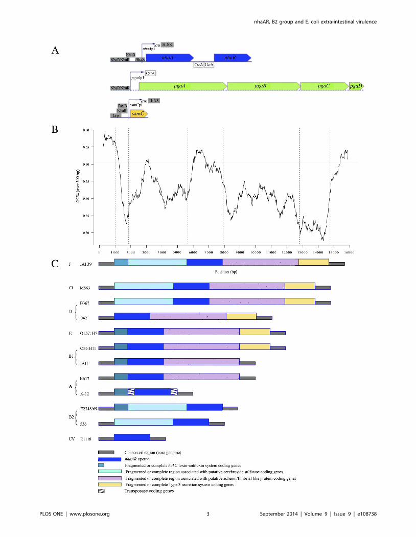
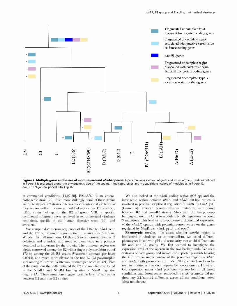
Figure 1. Genomic organization of nhaAR region. (A) Genomic representation of nhaAR and other operons under NhaR regulation in K-12 E.colistrain from http://www.ecocyc.org. All transcription or translation regulators are indicated. (B) GC percent along IAI39 nhaAR region (black line) andmean core genome GC percent (gray line) (C) Organization in various modules of nhaAR region. The modular organization of the region was definedusing synteny breaks between 10 pathogenic and commensal E. coli and 2 clades strains from various phylogenetic groups including K-12 and H617(two group A commensal strains), IAI1 and O26:H11 (two group B1 commensal strains), B367 (a group D commensal strain), 042 (anenteroaggregative group D, E2348/69 (an enteropathogenic group B2 strain), 536 (an extra-intestinal pathogen group B2 strain), O157:H7 Sakaı (anenterohemorrhagic group E strain). The two Escherichia clades (C) used were M863 (CI) and E1118 (CV). Five homologous modules have been defined,nhaAR operon being the third. Dark green: Fragmented or complete hokC toxin-antitoxin system coding genes; turquoise: Fragmented or conservedregion associated with putative cerebroside sulfatase coding genes; blue: nhaAR region; pink: Fragmented or conserved region associated withputative adhesin/fimbrial like protein coding genes; yellow: Fragmented or conserved Type 3 secretion system coding genes. IS are also indicated.doi:10.1371/journal.pone.0108738.g001
nhaAR, B2 group and E. coli extra-intestinal virulence
PLOS ONE | www.plosone.org 4 September 2014 | Volume 9 | Issue 9 | e108738
otherwise. All statistics were computed using STATA (v10.0,
College Station, TX, USA) or R (R Development Core Team,
2009, Vienna, Austria) and statistical significance was determined
at a p-value of less than 0.05.
RNA isolationTotal RNA extraction was performed on 536, 536DnhaAR,
536DnhaR and 536DnhaA after O/N culture during 18 h at 37uCin LB medium. Each culture for each bacteria was repeated three
time. Total RNA was extracted using the hot phenol method.
Residual chromosomal DNA was removed by treating samples
with a Ambion TURBO DNA-free Kit DNase-treated RNA
samples were quantified using a NanoDrop 1000 spectrophotom-
eter (Thermo Scientific).
Quantitative RT-PCR (qRT-PCR)qRT-PCR experiments were performed using a KAPA SYBR
One-Step qRT-PCR Kit (Kapa Biosystems) and a Lightcycler 480
(Roche) instrument with the program recommended by Kapa
Biosystems. We applied the comparative CT quantification (DDCt
method) of qRT-PCR for comparing changes in gene expression
of nhaR in the 536 deleted mutant strains. Relative quantification
was performed using 16SrRNA as endogenous control gene. Each
experiment was performed in duplicate.
Results, Discussion and Conclusion
ResultsGenomic analyses. To identify genetic markers in the core
genome that would differentiate the B2 phylogenetic group from
the other groups, we scanned all genes in the core genome of 128
E. coli/Escherichia clade genomes. For each gene, we computed
the number of fixed mutations between B2 and non-B2 and
compared it with a Fisher test to the pooled core genome number.
We studied the proportion of fixed sites compared to the total gene
length or to the total number of polymorphism found in that gene.
In both cases, the gene with the lowest p-value was nhaA, (p, 1e-
62), the next gene being ygbE (p, 1e-33) a conserved gene of
unknown function (Table S5). In this paper, we focused on the
nhaAR operon, as nhaA is the first gene of the list, but also because
it can be functionally assessed as it is a sodium proton antiporter
involved in pH and sodium homeostasis [16].
We first compared the genomic environment of the nhaARoperon in 10 E. coli strains and two Escherichia clades (clade I and
clade V) (Figure 1BC). We defined 5 homologous fragments
composing this region excluding fragments of transposases. Apart
from the fragment including exclusively nhaAR operon, none of
the other fragments were found in all strains, yet they were all
present in strain IAI39. The GC content in the region was on
average 42.67%, and differed significantly from the average
genome GC content of 50.63% (p,0.05) (Figure 1B). This
suggests that the region might have been acquired through
horizontal gene transfer. Nevertheless, nhaAR had a GC content
compatible with the genomic one. The pattern of gain/loss of the
fragments surrounding nhaAR appeared to be quite dynamic
(Figure 2). All the fragments seemed to have been lost and or
gained multiple times along the phylogeny. nhaAR operon has
therefore been maintained in the core genome despite highly
dynamic surrounding regions, an observation that suggests an
important contribution of this operon to E. coli niche adaptation.
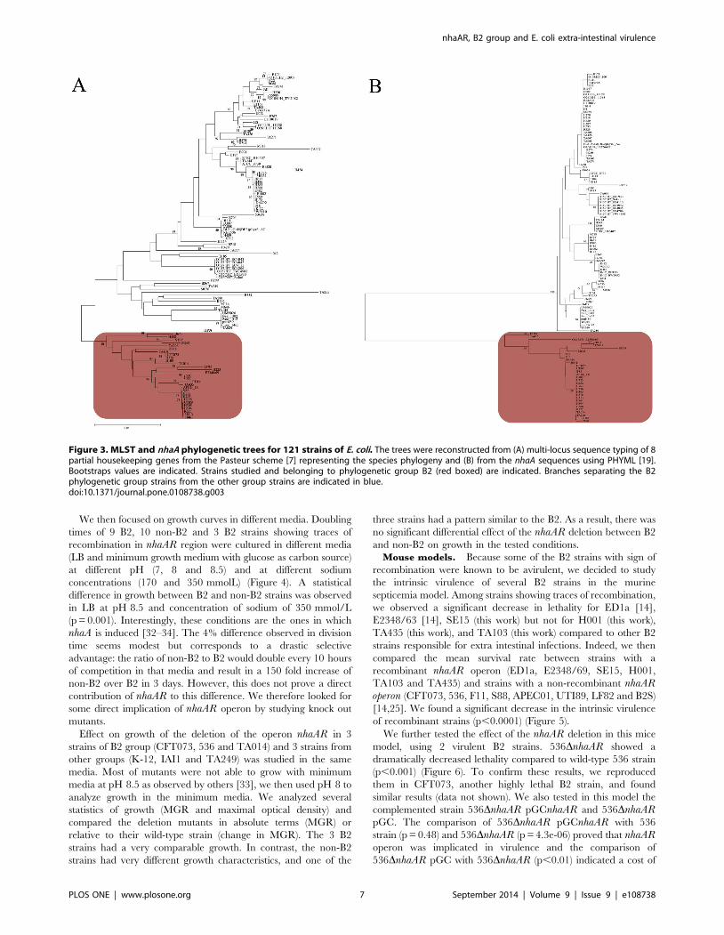
We reconstructed the phylogenetic tree of the nhaA gene from
the 121 genomes of E. coli available in data banks (Table S4).
Consistent with the screen used to identify nhaA region, we found
that in the phylogenetic tree based on nhaA the branch leading to
the B2 group of strains was much longer than what was found
using the MLST genes of the Pasteur scheme [7] (Figure 3). There
were 56 mutations that were fixed between the B2 and the non-B2
strains on a 1167 bp gene, or 4.8% of sites which contrast quite
drastically with the whole genome average of 0.14%. This could
be due to an accelerated evolution at this locus or to horizontal
gene transfer or both. Yet, when we measured Ka/Ks (corre-
sponding to the ratio of non synonymous mutation rate on the
synonymous mutation rate) between B2 and non-B2 strains, a
value between 0.01 and 0.02 was found. This means that
synonymous mutations were in large excess compared to non-
synonymous mutations. As we can exclude that selection of a
succession of non-synonymous mutations was responsible for the
long-branch, we favor horizontal gene transfer as the most likely
explanation. The 5 to 10% divergence observed between B2 and
non-B2 nhaA genes suggests that the transfer originated from a
close species like an Escherichia clade and that this transfer may
have been quite recent such that little recombination might have
occurred subsequently between the B2 and the other strains.
Accordingly, visual inspection of the nhaA sequences revealed that
a few B2 strains (ED1a, SE15, E2348/69, H001, TA103, M605,
and TA435) had three or more consecutive mutations that differed
from the other B2, which can be considered as a trace of
recombination. Similarly in the nhaR region, a long recombinant
segment in strain M605 was responsible for most of the diversity
within B2. When recombining strains were excluded from the
analysis, nhaR appeared with an even stronger B2/non-B2
differentiation than nhaA with 13.0% of fixed differences
compared to 5.8% for nhaA. Therefore the whole nhaAR operon
and not just nhaA harbors a strong divergence between B2 and
non-B2. Interestingly, while B2 strains are commonly isolated
from extra-intestinal infections, none of the strains with sign of
recombination have been isolated in extra-intestinal conditions.
ED1a, SE15, H001, TA103, M605, TA103, TA435 were sampled
nhaAR, B2 group and E. coli extra-intestinal virulence
PLOS ONE | www.plosone.org 5 September 2014 | Volume 9 | Issue 9 | e108738
in commensal conditions [14,27,28]. E2348/69 is an entero-
pathogenic strain [29]. Even more strikingly, some of these strains
are quite atypical B2 strains in terms of extra-intestinal virulence as
they are non-killer in a mouse model of septicemia. For instance,
ED1a strain belongs to the B2 subgroup VIII, a specific
commensal subgroup never retrieved in extra-intestinal virulence
conditions, specific to the human digestive track [30], and
avirulent.
We compared consensus sequences of the 1167 bp nhaA gene
and the 172 bp promoter region between B2 and non-B2 strains.
We identified 98 mutations. Of these, 3 were non-synonymous, 2
deletions and 3 indels, and none of them were in a position
described as important for the protein. The promoter region was
highly conserved among the B2 with a single polymorphism out of
172 bp among the 28 B2 strains (Watterson estimate per base:
0.0015), and much more diverse in the non-B2 (28 polymorphic
sites among 90 strains; Watterson estimate per base: 0.0321). Five
of the mutations that differentiated the B2 and non-B2 were found
in the NhaR1 and NhaR4 binding sites of NhaR regulator
(Figure 1A). These mutations suggest variable level of expression
between B2 and non-B2 strains.
We also looked at the nhaR coding region (905 bp) and the
inter-genic region between nhaA and nhaR (60 bp), which is
involved in post-transcriptional regulation of nhaR by CsrA [31]
(Figure 1A). Thirteen non-synonymous mutations were found
between B2 and non-B2 strains. Moreover, the hairpin-loop
binding site used by CsrA to modulate NhaR regulation harbored
3 mutations. This lead us to hypothesize a differential expression
of the nhaAR operon with potential consequences on the genes
regulated by NhaR, i.e. nhaA, pgaA and osmC.
Phenotypic results. To assess whether nhaAR region is
implicated in virulence or commensalism, we tested different
phenotypes linked with pH and osmolarity that could differentiate
B2 and non-B2 strains. We first wanted to investigate the
expression level of the operon in the two backgrounds. We used
3 strains of each group and introduced reporter plasmids bearing
the Gfp protein under control of the promoter regions of nhaAand osmC. Both promoters are under NhaR control and can be
used to monitor repression it imposes by flow cytometry. However,
Gfp expression under nhaA promoter was too low in all tested
conditions, and fluorescence controlled by osmC promoter did not
show any B2/non-B2 difference across all the conditions tested
(data not shown).
Figure 2. Multiple gains and losses of modules around nhaAR operon. A parsimonious scenario of gains and losses of the 5 modules definedin figure 1 is presented along the phylogenetic tree of the strains. – indicates losses and + acquisitions (colors of modules as in figure 1).doi:10.1371/journal.pone.0108738.g002
nhaAR, B2 group and E. coli extra-intestinal virulence
PLOS ONE | www.plosone.org 6 September 2014 | Volume 9 | Issue 9 | e108738
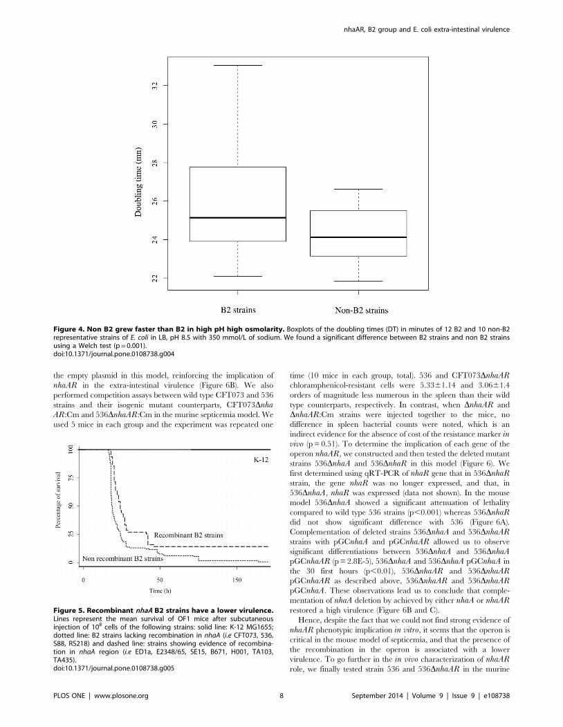
We then focused on growth curves in different media. Doubling
times of 9 B2, 10 non-B2 and 3 B2 strains showing traces of
recombination in nhaAR region were cultured in different media
(LB and minimum growth medium with glucose as carbon source)
at different pH (7, 8 and 8.5) and at different sodium
concentrations (170 and 350 mmolL) (Figure 4). A statistical
difference in growth between B2 and non-B2 strains was observed
in LB at pH 8.5 and concentration of sodium of 350 mmol/L
(p = 0.001). Interestingly, these conditions are the ones in which
nhaA is induced [32–34]. The 4% difference observed in division
time seems modest but corresponds to a drastic selective
advantage: the ratio of non-B2 to B2 would double every 10 hours
of competition in that media and result in a 150 fold increase of
non-B2 over B2 in 3 days. However, this does not prove a direct
contribution of nhaAR to this difference. We therefore looked for
some direct implication of nhaAR operon by studying knock out
mutants.
Effect on growth of the deletion of the operon nhaAR in 3
strains of B2 group (CFT073, 536 and TA014) and 3 strains from
other groups (K-12, IAI1 and TA249) was studied in the same
media. Most of mutants were not able to grow with minimum
media at pH 8.5 as observed by others [33], we then used pH 8 to
analyze growth in the minimum media. We analyzed several
statistics of growth (MGR and maximal optical density) and
compared the deletion mutants in absolute terms (MGR) or
relative to their wild-type strain (change in MGR). The 3 B2
strains had a very comparable growth. In contrast, the non-B2
strains had very different growth characteristics, and one of the
three strains had a pattern similar to the B2. As a result, there was
no significant differential effect of the nhaAR deletion between B2
and non-B2 on growth in the tested conditions.
Mouse models. Because some of the B2 strains with sign of
recombination were known to be avirulent, we decided to study
the intrinsic virulence of several B2 strains in the murine
septicemia model. Among strains showing traces of recombination,
we observed a significant decrease in lethality for ED1a [14],
E2348/63 [14], SE15 (this work) but not for H001 (this work),
TA435 (this work), and TA103 (this work) compared to other B2
strains responsible for extra intestinal infections. Indeed, we then
compared the mean survival rate between strains with a
TA103 and TA435) and strains with a non-recombinant nhaARoperon (CFT073, 536, F11, S88, APEC01, UTI89, LF82 and B2S)
[14,25]. We found a significant decrease in the intrinsic virulence
of recombinant strains (p,0.0001) (Figure 5).
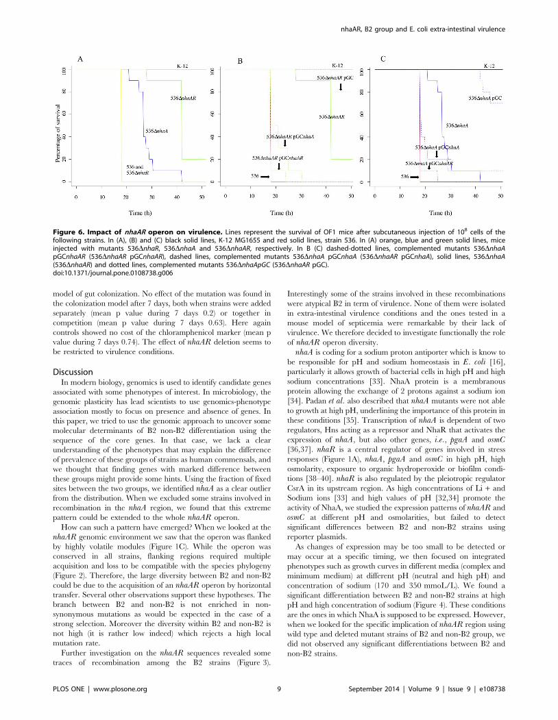
We further tested the effect of the nhaAR deletion in this mice
model, using 2 virulent B2 strains. 536DnhaAR showed a
dramatically decreased lethality compared to wild-type 536 strain
(p,0.001) (Figure 6). To confirm these results, we reproduced
them in CFT073, another highly lethal B2 strain, and found
similar results (data not shown). We also tested in this model the
complemented strain 536DnhaAR pGCnhaAR and 536DnhaARpGC. The comparison of 536DnhaAR pGCnhaAR with 536
strain (p = 0.48) and 536DnhaAR (p = 4.3e-06) proved that nhaARoperon was implicated in virulence and the comparison of
536DnhaAR pGC with 536DnhaAR (p,0.01) indicated a cost of
Figure 3. MLST and nhaA phylogenetic trees for 121 strains of E. coli. The trees were reconstructed from (A) multi-locus sequence typing of 8partial housekeeping genes from the Pasteur scheme [7] representing the species phylogeny and (B) from the nhaA sequences using PHYML [19].Bootstraps values are indicated. Strains studied and belonging to phylogenetic group B2 (red boxed) are indicated. Branches separating the B2phylogenetic group strains from the other group strains are indicated in blue.doi:10.1371/journal.pone.0108738.g003
nhaAR, B2 group and E. coli extra-intestinal virulence
PLOS ONE | www.plosone.org 7 September 2014 | Volume 9 | Issue 9 | e108738
the empty plasmid in this model, reinforcing the implication of
nhaAR in the extra-intestinal virulence (Figure 6B). We also
performed competition assays between wild type CFT073 and 536
strains and their isogenic mutant counterparts, CFT073DnhaAR:Cm and 536DnhaAR:Cm in the murine septicemia model. We
used 5 mice in each group and the experiment was repeated one
time (10 mice in each group, total). 536 and CFT073DnhaARchloramphenicol-resistant cells were 5.3361.14 and 3.0661.4
orders of magnitude less numerous in the spleen than their wild
type counterparts, respectively. In contrast, when DnhaAR and
DnhaAR:Cm strains were injected together to the mice, no
difference in spleen bacterial counts were noted, which is an
indirect evidence for the absence of cost of the resistance marker invivo (p = 0.51). To determine the implication of each gene of the
operon nhaAR, we constructed and then tested the deleted mutant
strains 536DnhaA and 536DnhaR in this model (Figure 6). We
first determined using qRT-PCR of nhaR gene that in 536DnhaRstrain, the gene nhaR was no longer expressed, and that, in
536DnhaA, nhaR was expressed (data not shown). In the mouse
model 536DnhaA showed a significant attenuation of lethality
compared to wild type 536 strains (p,0.001) whereas 536DnhaRdid not show significant difference with 536 (Figure 6A).
Complementation of deleted strains 536DnhaA and 536DnhaARstrains with pGCnhaA and pGCnhaAR allowed us to observe
significant differentiations between 536DnhaA and 536DnhaApGCnhaAR (p = 2.8E-5), 536DnhaA and 536DnhaA pGCnhaA in
the 30 first hours (p,0.01), 536DnhaAR and 536DnhaARpGCnhaAR as described above, 536DnhaAR and 536DnhaARpGCnhaA. These observations lead us to conclude that comple-
mentation of nhaA deletion by achieved by either nhaA or nhaARrestored a high virulence (Figure 6B and C).
Hence, despite the fact that we could not find strong evidence of
nhaAR phenotypic implication in vitro, it seems that the operon is
critical in the mouse model of septicemia, and that the presence of
the recombination in the operon is associated with a lower
virulence. To go further in the in vivo characterization of nhaARrole, we finally tested strain 536 and 536DnhaAR in the murine
Figure 4. Non B2 grew faster than B2 in high pH high osmolarity. Boxplots of the doubling times (DT) in minutes of 12 B2 and 10 non-B2representative strains of E. coli in LB, pH 8.5 with 350 mmol/L of sodium. We found a significant difference between B2 strains and non B2 strainsusing a Welch test (p = 0.001).doi:10.1371/journal.pone.0108738.g004
Figure 5. Recombinant nhaA B2 strains have a lower virulence.Lines represent the mean survival of OF1 mice after subcutaneousinjection of 108 cells of the following strains: solid line: K-12 MG1655;dotted line: B2 strains lacking recombination in nhaA (i.e CFT073, 536,S88, RS218) and dashed line: strains showing evidence of recombina-tion in nhaA region (i.e ED1a, E2348/65, SE15, B671, H001, TA103,TA435).doi:10.1371/journal.pone.0108738.g005
nhaAR, B2 group and E. coli extra-intestinal virulence
PLOS ONE | www.plosone.org 8 September 2014 | Volume 9 | Issue 9 | e108738
model of gut colonization. No effect of the mutation was found in
the colonization model after 7 days, both when strains were added
separately (mean p value during 7 days 0.2) or together in
competition (mean p value during 7 days 0.63). Here again
controls showed no cost of the chloramphenicol marker (mean p
value during 7 days 0.74). The effect of nhaAR deletion seems to
be restricted to virulence conditions.
DiscussionIn modern biology, genomics is used to identify candidate genes
associated with some phenotypes of interest. In microbiology, the
genomic plasticity has lead scientists to use genomics-phenotype
association mostly to focus on presence and absence of genes. In
this paper, we tried to use the genomic approach to uncover some
molecular determinants of B2 non-B2 differentiation using the
sequence of the core genes. In that case, we lack a clear
understanding of the phenotypes that may explain the difference
of prevalence of these groups of strains as human commensals, and
we thought that finding genes with marked difference between
these groups might provide some hints. Using the fraction of fixed
sites between the two groups, we identified nhaA as a clear outlier
from the distribution. When we excluded some strains involved in
recombination in the nhaA region, we found that this extreme
pattern could be extended to the whole nhaAR operon.
How can such a pattern have emerged? When we looked at the
nhaAR genomic environment we saw that the operon was flanked
by highly volatile modules (Figure 1C). While the operon was
conserved in all strains, flanking regions required multiple
acquisition and loss to be compatible with the species phylogeny
(Figure 2). Therefore, the large diversity between B2 and non-B2
could be due to the acquisition of an nhaAR operon by horizontal
transfer. Several other observations support these hypotheses. The
branch between B2 and non-B2 is not enriched in non-
synonymous mutations as would be expected in the case of a
strong selection. Moreover the diversity within B2 and non-B2 is
not high (it is rather low indeed) which rejects a high local
mutation rate.
Further investigation on the nhaAR sequences revealed some
traces of recombination among the B2 strains (Figure 3).
Interestingly some of the strains involved in these recombinations
were atypical B2 in term of virulence. None of them were isolated
in extra-intestinal virulence conditions and the ones tested in a
mouse model of septicemia were remarkable by their lack of
virulence. We therefore decided to investigate functionally the role
of nhaAR operon diversity.
nhaA is coding for a sodium proton antiporter which is know to
be responsible for pH and sodium homeostasis in E. coli [16],
particularly it allows growth of bacterial cells in high pH and high
sodium concentrations [33]. NhaA protein is a membranous
protein allowing the exchange of 2 protons against a sodium ion
[34]. Padan et al. also described that nhaA mutants were not able
to growth at high pH, underlining the importance of this protein in
these conditions [35]. Transcription of nhaA is dependent of two
regulators, Hns acting as a repressor and NhaR that activates the
expression of nhaA, but also other genes, i.e., pgaA and osmC[36,37]. nhaR is a central regulator of genes involved in stress
responses (Figure 1A), nhaA, pgaA and osmC in high pH, high
osmolarity, exposure to organic hydroperoxide or biofilm condi-
tions [38–40]. nhaR is also regulated by the pleiotropic regulator
CsrA in its upstream region. As high concentrations of Li + and
Sodium ions [33] and high values of pH [32,34] promote the
activity of NhaA, we studied the expression patterns of nhaAR and
osmC at different pH and osmolarities, but failed to detect
significant differences between B2 and non-B2 strains using
reporter plasmids.
As changes of expression may be too small to be detected or
may occur at a specific timing, we then focused on integrated
phenotypes such as growth curves in different media (complex and
minimum medium) at different pH (neutral and high pH) and
concentration of sodium (170 and 350 mmoL/L). We found a
significant differentiation between B2 and non-B2 strains at high
pH and high concentration of sodium (Figure 4). These conditions
are the ones in which NhaA is supposed to be expressed. However,
when we looked for the specific implication of nhaAR region using
wild type and deleted mutant strains of B2 and non-B2 group, we
did not observed any significant differentiations between B2 and
non-B2 strains.
Figure 6. Impact of nhaAR operon on virulence. Lines represent the survival of OF1 mice after subcutaneous injection of 108 cells of thefollowing strains. In (A), (B) and (C) black solid lines, K-12 MG1655 and red solid lines, strain 536. In (A) orange, blue and green solid lines, miceinjected with mutants 536DnhaR, 536DnhaA and 536DnhaAR, respectively. In B (C) dashed-dotted lines, complemented mutants 536DnhaApGCnhaAR (536DnhaAR pGCnhaAR), dashed lines, complemented mutants 536DnhaA pGCnhaA (536DnhaAR pGCnhaA), solid lines, 536DnhaA(536DnhaAR) and dotted lines, complemented mutants 536DnhaApGC (536DnhaAR pGC).doi:10.1371/journal.pone.0108738.g006
nhaAR, B2 group and E. coli extra-intestinal virulence
PLOS ONE | www.plosone.org 9 September 2014 | Volume 9 | Issue 9 | e108738
Because our laboratory conditions may not be the ones in which
a differentiation is strongly expressed, we tried some in vivo assays.
As a significant number of recombinant strains for nhaAR region
(ED1a and E2348/69) are known to be avirulent in a murine
model of septicemia, we tested this model for some other
recombinants identified (SE15, TA103 and TA435). We found
that among the recombinant strains half of the strains tested
showed decreased or absence of virulence in the murine model of
septicemia which is significantly different from the other B2 strains
(Figure 5) which are known to be virulent in this model [14,25].
This observation lead us to hypothesize that nhaAR could have
implication in virulence or colonization process as it is now known
that extra-intestinal and commensalism are linked [14]. We then
tested two DnhaAR mutant strains belonging to the B2 phyloge-
netic group (CFT073 and 536) and observed an important
decrease of the intrinsic virulence of the strains in this model
completely complemented by a vector bearing nhaAR operon
(Figure 6). Though, when we tested 536DnhaAR mutant in
competition in a mouse gut colonization, we did not find any
impact of the deletion. Hence, the effect of the mutation is only
marked in the virulence model. Interestingly, the deletion of
nhaAR operon seems to have a stronger impact on virulence than
the deletion of the pathogenicity island (PAI) of strain 536 in
isolations or in combination [13,41]. While the single PAI
deletions had no effect with similar inoculum as the one used
here, the mutant with all 7 PAI deleted killed 50% of mice in
28 hours compared to 42 hours in DnhaAR and 18 h in 536.
How could nhaAR contributes to virulence? NhaR is a central
regulator of expression of the genes nhaA, osmC and the operon
pgaABCD involved in stress responses such as high salinity, high
pH or biofilm formation [38–40]. Implication of these genes in the
virulence process is not clear, except for pgaABCD which has been
proved to be implicated in urinary tract ascending infections [42].
However we were not able to prove specific nhaR implication in
this model. But we clearly showed implication of nhaA gene in this
attenuation of virulence using deleted and complemented strains
with this gene. NhaA is known to be responsible for growth of
bacterial cells in high pH and high sodium concentrations [33], yet
such conditions are not the ones that seem to prevail during sepsis
where low pH seem to be dominant [43]. Further investigation will
therefore be needed to fully understand the contribution of nhaARto virulence.
ConclusionsThrough a bioinformatics approach we identified a candidate
core gene involved in B2, non-B2 genetic differentiation. Many
assays were performed to test some phenotypic expression of this
diversity in vitro without a clear success. However, when we used
in vivo experiments, though we only focused on the analysis of
knock-outs, we found a strong and so far unnoticed implication of
nhaA gene in virulence, despite a lack of effect in commensalism.
This whole process illustrates that bioinformatics approaches may
identify genes of interest whose effect is mostly if not only visible in
complex in vivo environments.
Supporting Information
Table S1 Strains and plasmids used in the in vitro and in vivoassays in this study.
(DOCX)
Table S2 List of primers used in this study.
(DOCX)
Table S3 List of conditions used in the growth curves
experiments.
(DOCX)
Table S4 List of 128 genomes used in the study to identify
markers of differentiation of the B2 phylogenetic group from other
group.
(DOC)
Table S5 List of genes classified by the proportions of fixed
differences between B2 and non-B2 between each gene of the core
and the whole set of genes pooled together using libsequence [26].
(XLS)
Acknowledgments
We thank Olivier Clermont for technical assistance, Damien Roux for
discussion and Erick Denamur for critical reading of the manuscript.
Author Contributions
Conceived and designed the experiments: ML OT. Performed the
experiments: ML FR CP JG SD A. Launay A. Ledda SC CG JT.
Analyzed the data: ML JT OT. Contributed reagents/materials/analysis
tools: ML A. Ledda JT OT. Wrote the paper: ML JT OT.
References
1. Touchon M, Hoede C, Tenaillon O, Barbe V, Baeriswyl S, et al. (2009)
Organised genome dynamics in the Escherichia coli species results in highly
diverse adaptive paths. PLoS Genet 5: e1000344. doi:10.1371/journal.p-
gen.1000344
2. Hacker J, Kaper JB (2000) Pathogenicity islands and the evolution of microbes.
Annu Rev Microbiol 54: 641–679. doi:10.1146/annurev.micro.54.1.641
3. Tenaillon O, Rodrıguez-Verdugo A, Gaut RL, McDonald P, Bennett AF, et al.
(2012) The molecular diversity of adaptive convergence. Science 335: 457–461.
doi:10.1126/science.1212986
4. Hommais F, Gouriou S, Amorin C, Bui H, Rahimy MC, et al. (2003) The FimH
A27V mutation is pathoadaptive for urovirulence in Escherichia coli B2
phylogenetic group isolates. Infect Immun 71: 3619–3622.
5. Sokurenko EV, Chesnokova V, Dykhuizen DE, Ofek I, Wu XR, et al. (1998)
Pathogenic adaptation of Escherichia coli by natural variation of the FimH
adhesin. Proc Natl Acad Sci USA 95: 8922–8926.
6. Tenaillon O, Skurnik D, Picard B, Denamur E (2010) The population genetics
of commensal Escherichia coli. Nat Rev Microbiol 8: 207–217. doi:10.1038/
nrmicro2298
7. Jaureguy F, Landraud L, Passet V, Diancourt L, Frapy E, et al. (2008)
Phylogenetic and genomic diversity of human bacteremic Escherichia colistrains. BMC Genomics 9: 560. doi:10.1186/1471-2164-9-560
8. Picard B, Garcia JS, Gouriou S, Duriez P, Brahimi N, et al. (1999) The link
between phylogeny and virulence in Escherichia coli extraintestinal infection.
Infect Immun 67: 546–553.
9. Bingen E, Picard B, Brahimi N, Mathy S, Desjardins P, et al. (1998)
Phylogenetic analysis of Escherichia coli strains causing neonatal meningitis
suggests horizontal gene transfer from a predominant pool of highly virulent B2
group strains. J Infect Dis 177: 642–650.
10. Lescat M, Clermont O, Woerther PL, Glodt J, Dion S, et al. (2013) Commensal
Escherichia coli strains in Guiana reveal a high genetic diversity with host-
dependant population structure. Environ Microbiol Rep 5: 49–57. doi:10.1111/
j.1758-2229.2012.00374.x
11. Nowrouzian FL, Oswald E (2012) Escherichia coli strains with the capacity for
long-term persistence in the bowel microbiota carry the potentially genotoxic pks
12. Secher T, Samba-Louaka A, Oswald E, Nougayrede J-P (2013) Escherichia coliProducing Colibactin Triggers Premature and Transmissible Senescence in
Mammalian Cells. PLoS ONE 8: e77157. doi:10.1371/journal.pone.0077157
13. Diard M, Garry L, Selva M, Mosser T, Denamur E, et al. (2010) Pathogenicity-
associated islands in extraintestinal pathogenic Escherichia coli are fitness
elements involved in intestinal colonization. J Bacteriol 192: 4885–4893.
doi:10.1128/JB.00804-10
14. Le Gall T, Clermont O, Gouriou S, Picard B, Nassif X, et al. (2007)
Extraintestinal virulence is a coincidental by-product of commensalism in B2
phylogenetic group Escherichia coli strains. Mol Biol Evol 24: 2373–2384.
doi:10.1093/molbev/msm172
15. Escobar-Paramo P, Clermont O, Blanc-Potard A-B, Bui H, Le Bouguenec C,
et al. (2004) A specific genetic background is required for acquisition and
nhaAR, B2 group and E. coli extra-intestinal virulence
PLOS ONE | www.plosone.org 10 September 2014 | Volume 9 | Issue 9 | e108738
expression of virulence factors in Escherichia coli. Mol Biol Evol 21: 1085–1094.
doi:10.1093/molbev/msh11816. Padan E, Bibi E, Ito M, Krulwich TA (2005) Alkaline pH homeostasis in
bacteria: new insights. Biochim Biophys Acta 1717: 67–88. doi:10.1016/
j.bbamem.2005.09.01017. Datsenko KA, Wanner BL (2000) One-step inactivation of chromosomal genes
in Escherichia coli K-12 using PCR products. Proc Natl Acad Sci USA 97:6640–6645. doi:10.1073/pnas.120163297
18. Vallenet D, Belda E, Calteau A, Cruveiller S, Engelen S, et al. (2013)
MicroScope–an integrated microbial resource for the curation and comparativeanalysis of genomic and metabolic data. Nucleic Acids Res 41: D636–647.
doi:10.1093/nar/gks119419. Guindon S, Lethiec F, Duroux P, Gascuel O (2005) PHYML Online–a web
server for fast maximum likelihood-based phylogenetic inference. Nucleic AcidsRes 33: W557–559. doi:10.1093/nar/gki352
20. Walk ST, Alm EW, Gordon DM, Ram JL, Toranzos GA, et al. (2009) Cryptic
lineages of the genus Escherichia. Appl Environ Microbiol 75: 6534–6544.doi:10.1128/AEM.01262-09
21. Chenna R, Sugawara H, Koike T, Lopez R, Gibson TJ, et al. (2003) Multiplesequence alignment with the Clustal series of programs. Nucleic Acids Res 31:
23. Zaslaver A, Bren A, Ronen M, Itzkovitz S, Kikoin I, et al. (2006) Acomprehensive library of fluorescent transcriptional reporters for Escherichiacoli. Nat Methods 3: 623–628. doi:10.1038/nmeth895
24. Bleibtreu A, Gros P-A, Laouenan C, Clermont O, Le Nagard H, et al. (2013)
Fitness, Stress Resistance, and Extraintestinal Virulence in Escherichia coli.Infect Immun 81: 2733–2742. doi:10.1128/IAI.01329-12
25. Tourret J, Aloulou M, Garry L, Tenaillon O, Dion S, et al. (2011) The
interaction between a non-pathogenic and a pathogenic strain synergisticallyenhances extra-intestinal virulence in Escherichia coli. Microbiology (Reading,
Engl) 157: 774–785. doi:10.1099/mic.0.037416-0
26. Thornton K (2003) Libsequence: a C++ class library for evolutionary geneticanalysis. Bioinformatics 19: 2325–2327.
27. Toh H, Oshima K, Toyoda A, Ogura Y, Ooka T, et al. (2010) Completegenome sequence of the wild-type commensal Escherichia coli strain SE15,
belonging to phylogenetic group B2. J Bacteriol 192: 1165–1166. doi:10.1128/JB.01543-09
28. Broad institute website. Available: http://www.broadinstitute.org. Accessed
Parallel evolution of virulence in pathogenic Escherichia coli. Nature 406: 64–67.doi:10.1038/35017546
30. Clermont O, Lescat M, O’Brien CL, Gordon DM, Tenaillon O, et al. (2008)
Evidence for a human-specific Escherichia coli clone. Environ Microbiol 10:1000–1006. doi:10.1111/j.1462-2920.2007.01520.x
31. Pannuri A, Yakhnin H, Vakulskas CA, Edwards AN, Babitzke P, et al. (2012)
Translational repression of NhaR, a novel pathway for multi-tier regulation ofbiofilm circuitry by CsrA. J Bacteriol 194: 79–89. doi:10.1128/JB.06209-11
32. Maes M, Rimon A, Kozachkov-Magrisso L, Friedler A, Padan E (2012)Revealing the ligand binding site of NhaA Na+/H+ antiporter and its pH
33. Padan E, Maisler N, Taglicht D, Karpel R, Schuldiner S (1989) Deletion of antin Escherichia coli reveals its function in adaptation to high salinity and an
alternative Na+/H+ antiporter system(s). J Biol Chem 264: 20297–20302.34. Taglicht D, Padan E, Schuldiner S (1991) Overproduction and purification of a
functional Na+/H+ antiporter coded by nhaA (ant) from Escherichia coli. J BiolChem 266: 11289–11294.
35. Padan E (2008) The enlightening encounter between structure and function in
the NhaA Na+-H+ antiporter. Trends Biochem Sci 33: 435–443. doi:10.1016/j.tibs.2008.06.007
36. Sturny R, Cam K, Gutierrez C, Conter A (2003) NhaR and RcsB independentlyregulate the osmCp1 promoter of Escherichia coli at overlapping regulatory sites.
J Bacteriol 185: 4298–4304.
37. Goller C, Wang X, Itoh Y, Romeo T (2006) The cation-responsive proteinNhaR of Escherichia coli activates pgaABCD transcription, required for
production of the biofilm adhesin poly-beta-1,6-N-acetyl-D-glucosamine.J Bacteriol 188: 8022–8032. doi:10.1128/JB.01106-06
38. Dover N, Higgins CF, Carmel O, Rimon A, Pinner E, et al. (1996) Na+-inducedtranscription of nhaA, which encodes an Na+/H+ antiporter in Escherichia coli,is positively regulated by nhaR and affected by hns. J Bacteriol 178: 6508–6517.
39. Wang X, Preston JF 3rd, Romeo T (2004) The pgaABCD locus of Escherichiacoli promotes the synthesis of a polysaccharide adhesin required for biofilm
formation. J Bacteriol 186: 2724–2734.40. Lesniak J, Barton WA, Nikolov DB (2003) Structural and functional features of
the Escherichia coli hydroperoxide resistance protein OsmC. Protein Sci 12:
2838–2843. doi:10.1110/ps.0337560341. Tourret J, Diard M, Garry L, Matic I, Denamur E (2010) Effects of single and
multiple pathogenicity island deletions on uropathogenic Escherichia coli strain536 intrinsic extra-intestinal virulence. Int J Med Microbiol 300: 435–439.
doi:10.1016/j.ijmm.2010.04.01342. Subashchandrabose S, Smith SN, Spurbeck RR, Kole MM, Mobley HLT
(2013) Genome-wide detection of fitness genes in uropathogenic Escherichia coliduring systemic infection. PLoS Pathog 9: e1003788. doi:10.1371/journal.ppat.1003788
43. MacKenzie IM (2001) The haemodynamics of human septic shock. Anaesthesia56: 130–144.
nhaAR, B2 group and E. coli extra-intestinal virulence
PLOS ONE | www.plosone.org 11 September 2014 | Volume 9 | Issue 9 | e108738


![[Adrian bondy u.s.r. murty] Grap Theory](https://static.documents.pub/doc/80x56/55c3a8c8bb61eb210b8b46cd/adrian-bondy-usr-murty-grap-theory.jpg)


