504 BIOCHIMICA ET BIOPHYSICA ACTA BBA 75 653 THE CORRELATION BETWEEN THE SATURATION OF MEMBRANE FATTY ACIDS AND THE PRESENCE OF MEMBRANE FRACTURE FACES AFTER OSMIUM FIXATION ROBERT JAMES AND DANIEL BRANTON Department of Botany, University of California, Berkeley, Calif. 94 720 (U.S.A .) {Received January 29th, I971 ) SUMMARY In osmium-fixed membranes, there is a decrease in the membrane fracture faces concomitant with an increase in unsaturation of the membrane fatty acids. The data suggest that both tim number of double bonds and their position in the fatty- acid chain are critical to the disappearance of membrane fracture faces. INTRODUCTION Membranes can be split and their inner hydrophobie face can be exposed by the fracture process of freeze-etching l-a. These fracture faces have been tile object of many detailed electron microscopic examinations (for reviews see refs. 3-5). Because the fracture process depends upon the presence of membrane lipidsG, 7, it may also depend upon the organization of these lipids within the membrane. One approach relating the appearance of the fracture faces to the organization of lipids has utilized model systems containing lipids in known phasesS, ". The results showed that lamellar phase lipids consistently produce fracture faces resembling extended, smooth sheets". Another way of relating the fractures to the lipids involves purposeful perturbations of lipid components in well characterized artificial and biological membranes. In this paper we examine the effects of OsO~ on the fracture properties of liposomes and 3(~,coplasma laidlawii cell membranes containing lipids of various degrees of saturation. We chose M. laidlawii because X-ray diffraction 1°, 11, calorimetryl2, la, and elec- tron paramagnetic resonancel~, 15 show that most of the lipids are organized as a lamellar bilayer. Furthermore, the polar lipids of M. laidlawii reside almost exclusively in the cell membrane 16, and the fatty-acid composition of these polar Iipids can be altered by supplementing the growth medium 17,~s Previous studies of the effects of OsO 4 fixation on the freeze-etch fracture process have led to apparently contradictory observations. In Bacilhts subtilis, OsO~ had little effectS", 2°, whereas in chloroplasts (R. B. PARK AND D. BRANTON, un- published observation), mitochondria2L and yeast cells (M. MOOR, personal communi- cation), OsO 4 fixation resulted in a striking loss of membrane face fractures; only cross fractures occurred. It appeared to us that the fatty-acid composition of these Biockim. Biopkys..4cta, ~33 (~97I) .504 5 I '
Transcript
504 BIOCHIMICA ET BIOPHYSICA ACTA
BBA 75 653
T H E C O R R E L A T I O N B E T W E E N T H E SATURATION OF M E M B R A N E
FATTY ACIDS AND T H E P R E S E N C E OF M E M B R A N E F R A C T U R E FACES
A F T E R OSMIUM F I X A T I O N
ROBERT JAMES AND DANIEL BRANTON
Department of Botany, University of California, Berkeley, Calif. 94 720 (U.S.A .)
{Received January 29th, I971 )
SUMMARY
In osmium-fixed membranes, there is a decrease in the membrane fracture faces concomitant with an increase in unsaturat ion of the membrane fa t ty acids. The data suggest tha t both tim number of double bonds and their position in the fat ty- acid chain are critical to the disappearance of membrane fracture faces.
INTRODUCTION
Membranes can be split and their inner hydrophobie face can be exposed by the fracture process of freeze-etching l-a. These fracture faces have been tile object of many detailed electron microscopic examinations (for reviews see refs. 3-5). Because the fracture process depends upon the presence of membrane lipidsG, 7, it m a y also depend upon the organization of these lipids within the membrane.
One approach relating the appearance of the fracture faces to the organization of lipids has utilized model systems containing lipids in known phasesS, ". The results showed tha t lamellar phase lipids consistently produce fracture faces resembling extended, smooth sheets". Another way of relating the fractures to the lipids involves purposeful perturbations of lipid components in well characterized artificial and biological membranes. In this paper we examine the effects of OsO~ on the fracture properties of liposomes and 3(~,coplasma laidlawii cell membranes containing lipids of various degrees of saturation.
We chose M. laidlawii because X- ray diffraction 1°, 11, calorimetryl2, la, and elec- tron paramagnet ic resonancel~, 15 show tha t most of the lipids are organized as a lamellar bilayer. Furthermore, the polar lipids of M. laidlawii reside almost exclusively in the cell membrane 16, and the fat ty-acid composition of these polar Iipids can be altered by supplementing the growth medium 17,~s
Previous studies of the effects of OsO 4 fixation on the freeze-etch fracture process have led to apparent ly contradictory observations. In Bacilhts subtilis, OsO~ had little effectS", 2°, whereas in chloroplasts (R. B. PARK AND D. BRANTON, un- published observation), mitochondria2L and yeast cells (M. MOOR, personal communi- cation), OsO 4 fixation resulted in a striking loss of membrane face fractures; only cross fractures occurred. I t appeared to us tha t the fat ty-acid composition of these
Biockim. Biopkys..4cta, ~33 (~97 I) .504 5 I '
FRACTURE FACES OF FIXED MEMBRANES 505
different membrane types could account for the differing effects of OsO 4. In B. subtilis, there are almost no unsa tura ted f a t ty acids 2z, 23, whereas in chloroplasts, for example, 95 % of the fa t ty acids are the unsa tura ted linolenic and linoleic acids 24.
M A T E R I A L S A N D M E T H O D S
Culture conditions M. laidlawii was grown statically at 37 ° in Io -5oo ml of medium in glass tubes
or Er lenmeyer flasks. The medium was modified only quant i ta t ively from tha t of RAZlN et al. 18, and I 1 contained the following: 20 g acetone-extracted Difco Tryptose, IO g glucose, 5 g NaC1, 3.7 g Tris, 4 g very fat ty-acid-poor bovine albumin (Grade B, CalBioehem), and Iooooo units potassium penicillin G. The unadjusted pH of this medium was 8.3. Fa t ty-ac id supplements were added as ethanol solutions to give a final concentrat ion of 12o #M. The ethanol in the medium did not exceed 0.5 %. Cells were harvested during late-log phase by centrifugation at Ioooo × g. Cells were washed 3 times in fl-buffer is diluted I : 4 and then were either (i) lipid-extracted, (2) dispersed in growth medium minus penicillin and fat ty-acid supplement for use in OsO~-fixation experiments, or (3) prepared for freeze-etching.
Lipid extraction After washing and pelleting, the cells for lipid extraction were weighed wet,
and IO vol. methanol (vol. per cell wet wt.) was added to the cells in glass centrifuge tubes. The cells were macerated using a glass rod and then incubated at 65 ° for 5 rain. 20 vol. chloroform (vol. per cell wet wt.) were added, and the suspension was incubated at 65 ° for 20 rain with in termit tant macerat ion 25. The suspension was then filtered through lipid-extracted W h a t m a n No. I filter paper. The filtrate was washed 3 times with chloroform-methanol (2:1, v/v).
Liposome preparation Dipalmitoyl lecithin, ~> 96 % pure, (Applied Science Laboratories, State College
O/ Pa). was used as the sa turated lipid in the liposome experiments. Asolectin, >~ 95 Jo o/ linolenic acid and 37 % linoleic acid by wt., phosphatides, containing approx. 3 ,.o
(Associated Concentrates, Woodside, N.Y.) was used as the unsa tura ted lipid. The methods for preparat ion of liposomes were adapted from those of BANGHAM,
el al. 26 and REEVES AND DOWBEN 27. For the extracted lipids, portions of the chloro- fo rm-methanol extract were placed in Er lenmeyer flasks, the solvent was evaporated by a moist s tream of N 2, and the remaining lipid was weighed. For the commercial lipids, weighed portions were dissolved in 2 vol. of chloroform (v/w) and dried under a moist s tream of N2.
To the dried preparation, distilled water was added to give a dispersion con- taining 0.I % lipid (w/v). These dispersions were incubated for I h at 5 °° under N 2
and with frequent shaking. The liposomes produced in this way were either harvested by centrifugation at 15000 × g and then frozen or resnspended and fixed with OsO 4.
Fixation, freeze-etching and electron microscopy Resuspended cells and liposomes were combined I : I (v/v) with 2 % OsO 4 in
a o.I M phosphate buffer (pH 6.8) and fixed for 2 h at 4 °. The fixed cells and liposomes
Biochirn. Biophys. Acta, 233 (I97I) 5o4-512
506 R. JAMES, D. BRANTON
were then pel le ted at IOOOO x g and 15ooo × g, respect ively. Cells were washed 2 t imes in o.o5 M phospha te buffer and pelleted. Liposomes were not rinsed.
Samples from the var ious pellets were p ipe t t ed into 3-mm ca rdboa rd discs and frozen in l iquid freon cooled b y l iquid N 2. Samples were f reeze-fractured at - - I I O ° wi th no etching 1, 28.
Repl icas were v iewed in a Siemens Ehniskop I using direct magnif icat ions of 4ooo-2oooo. Repl icas were scanned and areas to be pho tographed were selected solely on the basis of membrane concent ra t ion and repl ica qual i ty . Pr in ts of these photo- g raphs were used to de te rmine the number of cells or l iposomes showing m e m b r a n e face fractures, as opposed to those showing only cross fractures. Cells or l iposomes having less than I m m of m e m b r a n e face exposed at 16ooo final magnif icat ion were scored as cross fractures. A n y cells or l iposomes abou t which there was any doub t were not scored.
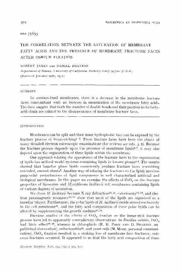
Fatty-acid analysis Lip id ex t rac t s were evapo ra t ed to I ml, IO ml chloroform added and then
e v a p o r a t e d again to I ml. Lipids in I ml chloroform were separa ted from the non- l ipid con taminan t s on a Sephadex column, according to the methods of RADIN 25. The l ipids in the "Fo lch lower phase" were then e va po ra t e d to approx. 5 ml, and the phosphol ip ids separa ted from neu t ra l l ipids in a Silicic acid column (Bio-Sil A, Bio- R a d Labora tor ies , Richmond, Calif.) b y elut ing the neu t ra l l ipids wi th chlorofor ln- me thano l (99:1, v/v) and then the phosphol ip ids wi th IOO % methanoP 8,29.
The phosphol ip ids were saponif ied and me thy l esters of the f a t t y acids p roduced according to BOTTCHER et al. a°. Analysis of the me thy l esters was per formed on a Var ian Model 6o0 gas ch roma tog raph f i t ted wi th a polar column (DEGS), flame~
Biochim. Biophys. Acta, 233 (t97~) 5o4-5 I2
FRACTURE FACES OF FIXED MEMBRANES 507
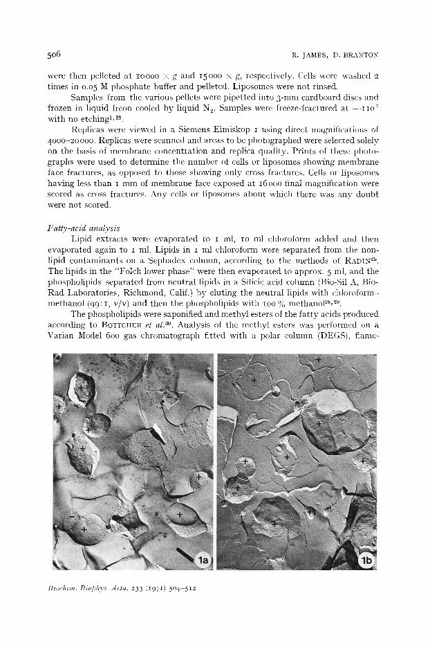
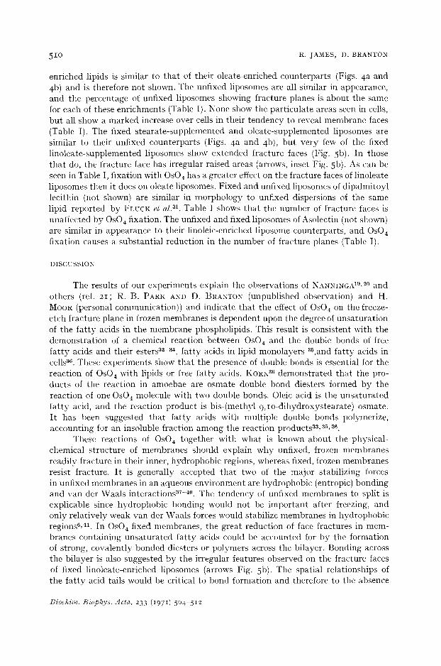
Figs. 1-3. Freeze-etch views of !l//. laidlawii cells enriched wi th s tearate (Fig. I), oleate (Fig. 2), and linoleate (Fig. 3). (a) unfixed cells; (b) fixed cells. Examples of cells scored as showing no fracture face (cross fractured) are indicated by (--) and those showing fracture faces by ( + ) . All × 20000.
t?iochim. Biophys. Acta, 233 (1971) 504-512
5 0 8 R. JAMES, I). BRANTON
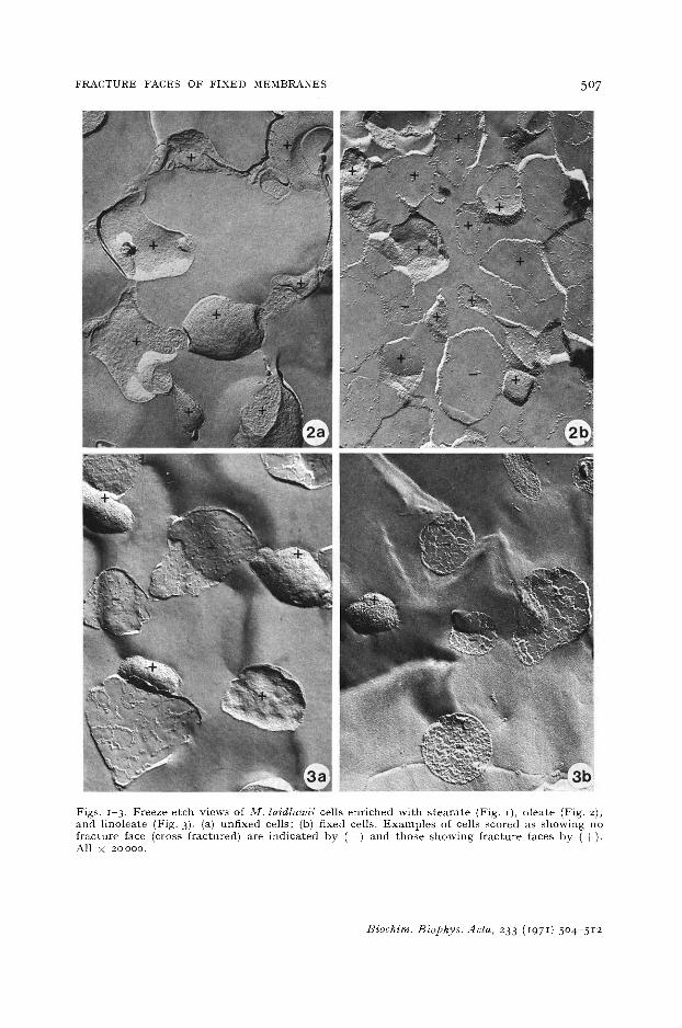
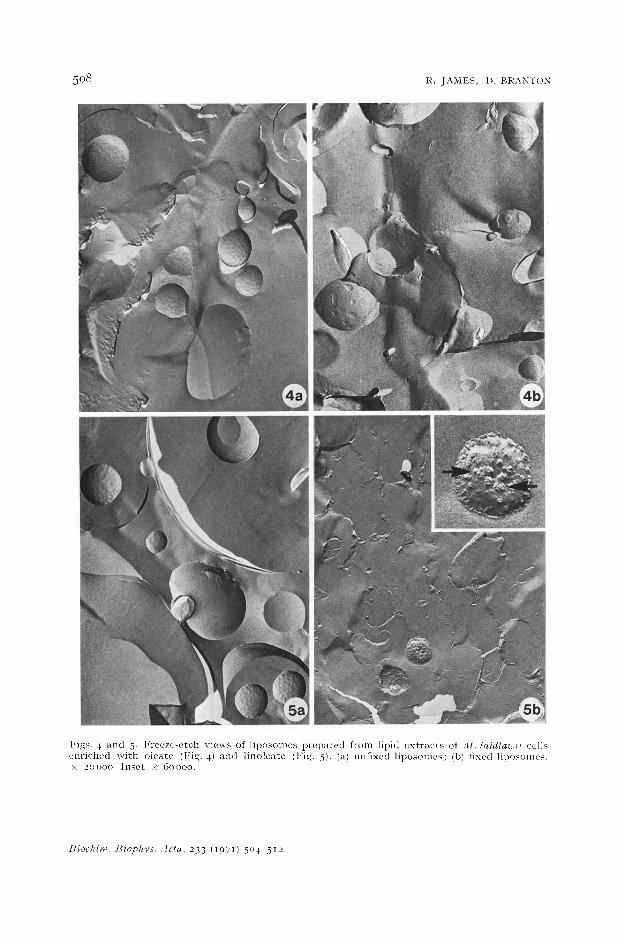
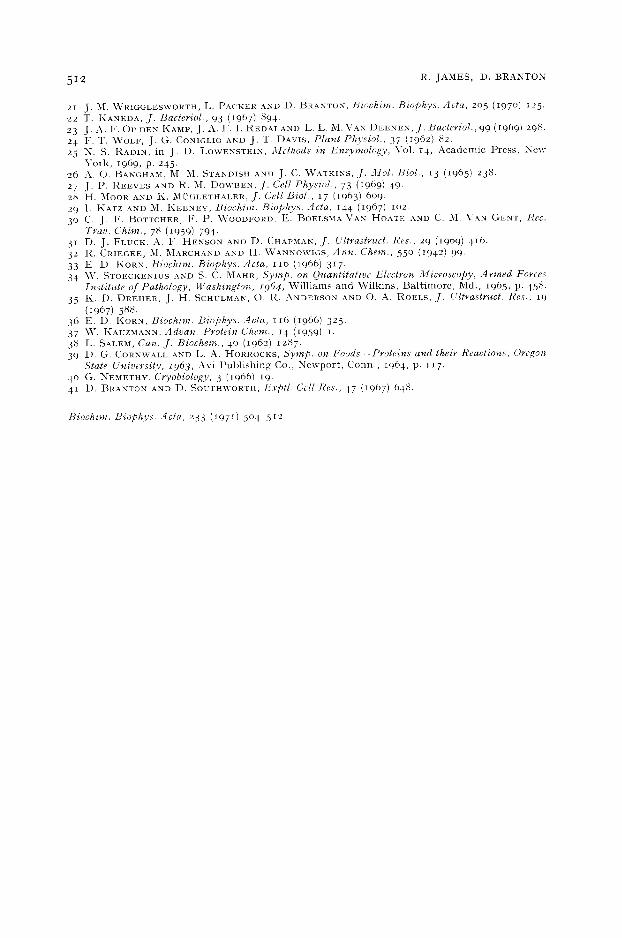
Figs . 4 a n d 5. l ' r e e z e - e t c h v i e w s of l i p o s o m c s p r e p a r e d f rom l ip id e x t r a c t s of . l l . /aidla~ii cel ls e n r i c h e d w i t h o l e a t e (Fig. 4) a n d l i n o l e a t e (l : ig. 5), (a) un f ixed l i p o s o m e s ; (b) f ixed l iposon les . x 20000. I n s e t × 60000 .
l~iochim, t?iophys. Acta, 233 (197I) 504 5 ] 2
F R A C T U R E F A C E S O F F I X E D M E M B R A N E S 5 0 9
ioniza t ion detector , and disc in tegra tor . Iden t i f ica t ion of unsa tu r a t ed me thy l esters was confirmed by hydrogena t ion of the double bonds.
R E S U L T S
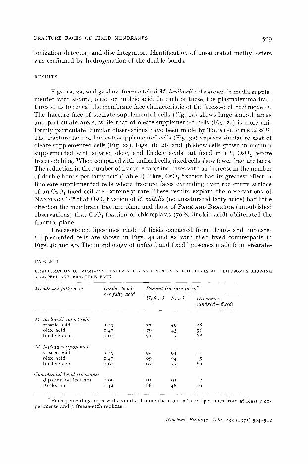
Figs. Ia , 2a, and 3a show freeze-etched M . la id law i i cells grown in media supple- men ted with stearie, oleic, or linoleic acid. In each of these, the p l a s m a l e m m a frac- tures so as to reveal the m e m b r a n e face character is t ic of the freeze-etch technique l, 2. The f rac ture face of s t ea ra t e - supp lemen ted cells (Fig. Ia) shows large smooth areas and pa r t i cu la te areas, while t ha t of o lea te - supplemented cells (Fig. 2a) is more uni- fo rmly par t icu la te . Similar observa t ions have been made by TOURTELLOTTE et al. 14.
The f rac ture face of l ino lea te -supplemented cells (Fig. 3 a) appears s imilar to t ha t of o lea te - supp lemented cells (Fig. 2a). Figs. Ib , 2b, and 3b show cells grown in med imn supp lemen ted with stearic, oleic, and linoleic acids bu t f ixed in I OJ;o OsO4 before freeze-etching. When compared with unfixed cells, f ixed cells show fewer f racture faces. The reduct ion in the number of f rac ture faces increases wi th an increase in the number of double bonds per f a t t y acid (Table I). Thus, Os04 f ixat ion had its grea tes t effect in l ino lea te - supplemented cells where f racture faces ex tend ing over the ent ire surface of an OsO4-fixed cell are ex t r eme ly rare. These results expla in the observat ions of NANNINGA19,20 t ha t OsO 4 f ixat ion of B. subl i l i s (no unsa tu r a t ed f a t t y acids) had l i t t le effect on the membrane f rac ture plane and those of PARK AND BRANTON (unpubl ished observat ions) t ha t OsO4 f ixat ion of chloroplasts (70 % linoleic acid) ob l i t e ra ted the f racture plane.
Freeze-e tched l iposomes made of l ipids ex t r ac t ed from oleate- and l inoleate- supp lemen ted cells are shown in Figs. 4 a and 5a with their fixed coun te rpar t s in Figs. 4b and 5b. The morpho logy of unfixed and fixed l iposomes made from s teara te -
TABLE I
U N S A T U R A T I O N O F M E M B R A N E F A T T Y A C I D S A N D P E R C E N T A G E O F C E L L S A N D L I P O S O M E S S H O \ V I N G
* Each percentage represents counts of more than 300 cells or liposomes from at least 2 ex- periments and 3 freeze-etch replicas.
Biochim. Biophys. Acta, 233 (1971) 5o4-512
5IO R. JAMES, D. BRANTON
enriched lipids is similar to that of their oleate-enriched counterparts (Figs. 4a and 4 b) and is therefore not shown. The unfixed liposomes are all similar in appearance, and the percentage of unfixed liposomes showing fracture planes is about the same for each of these enrichments (Table I). None show the particulate areas seen in ceils, but all show a marked increase over cells in their tendency to reveal membrane faces (Table I). The fixed stearate-supplemented and oleate-supplemented liposomes are similar to their unfixed counterparts (Figs. 4a and 4b), but very few of the fixed linoleate-supplemented liposomes show extended fracture faces (Fig. 5b). In those that do, the fracture face has irregular raised areas (arrows, inset Fig. 5b). As can be seen in Table I, fixation with OsO~ has a greater effect on the fracture faces of linoleate liposomes then it does on oleate liposomes. Fixed and unfixed liposomes of dipalmitoyl lecithin (not shown) are similar in morphology to unfixed dispersions of the same lipid reported by FLUCK et al. a~. Table I shows that the number of fracture faces is unaffected by OsO4 fixation. The unfixed and fixed liposomes of Asolectin (not shown) are similar in appearance to their linoleic-enriched liposome counterparts, and OsO~ fixation causes a substantial reduction in the number of fracture planes (Table I).
DISCUSSION
The results of our experiments explain the observations of NANNINGA 19,2° and others (ref. 21; R. B. PARK AND D. BRAXTON (unpublished observation) and H. MOOR (personal communication)) and indicate that the effect of OsO4 on the freeze- etch fracture plane in frozen membranes is dependent upon the degree of unsaturation of the fatty acids in the membrane phospholipids. This result is consistent with the demonstration of a chemical reaction between OsO~ and the double bonds of free fat ty acids and their esters a2 ~4, fatty acids in lipid monolayers ~5,and fatty acids in cells s6. These experiments show that the presence of double bonds is essential for the reaction of OsO~ with lipids or free fatty acids. KORt< sG demonstrated that the pro- ducts of the reaction in amoebae are osmate double bond diesters formed by the reaction of one OsO 4 molecule with two double bonds. Oleic acid is the unsaturated fat ty acid, and the reaction product is bis-(methyl 9,Io-dihydroxystearate) osmate. It has been suggested that fat ty acids with multiple double bonds polymerize, accounting for an insoluble fraction among the reaction products a3,'~s, 3G.
These reactions of OsO4 together with what is known about the physical- chemical structure of membranes should explain why unfixed, frozen membranes readily fracture in their inner, hydrophobic regions, whereas fixed, frozen membranes resist fracture. I t is generally accepted that two of the major stabilizing forces in unfixed membranes in an aqueous environment are hydrophobic (entropic) bonding and van der Waals interactions a7-4°. The tendency of unfixed membranes to split is explicable since hydrophobic bonding would not be important after freezing, and only relatively weak van der Waals forces would stabilize membranes in hydrophobic regions6, 41. In OsO4-fixed membranes, the great reduction of face fractures in mem- branes containing unsaturated fatty acids could be accounted for by the formation of strong, covalently bonded diesters or polymers across the bilayer. Bonding across the bilayer is also suggested by the irregular features observed on the fracture faces of fixed linoleate-enriched liposomes (arrows Fig. 5b). The spatial relationships of the fat ty acid tails would be critical to bond formation and therefore to the absence
Biochim. Biophys. Acts, 233 (1971) 504 512
FRACTURE FACES OF F I X E D MEMBRANES 5 1 1
of face fractures. The data showing that the introduction of linoleic acid causes a disproportionate decrease in the percentage of fracture faces when compared with oleic acid supports this hypothesis.
Another explanation of the decrease in face fractures is based on the observation that when monolayers of unsaturated phospholipids are reacted with 0s04, the result- ing osmate tails move from the hydrophobic to the hydrophilic region of the mono- layers 35. If osmate tails in a lipid bilayer or biological membrane also move to the hydrophilic region of the membrane, the normal fracture plane could be altered with- out any bonding across the bilayer. These two alternatives are not mutually exclusive, and the role of each may be dependent on the number and position of double bonds present.
The difference in the percentage of unfixed cells v e r s u s unfixed liposomes showing significant fracture faces is small but consistent (Table I). There are at least two pos- sible explanations: (I) the liposomes are spherical and the cells irregular, and (2) the presence of particles in the cell membranes represent bonding sites in the hydro- phobic matrix not affected by low temperature. Stronger bonds may be introduced at these sites when cells are fixed with 0 s 0 4. This is suggested by an even greater decrease in the percentage of fixed stearate and oleate cells v e r s u s fixed liposomes showing fracture faces (Table I).
A C K N O W L E D G E M E N T S
This investigation was supported by the Atomic Energy Commission Contract AT(II-I)-34. P.A. 142 (D.t3.), National Science Foundation Grant GB 13646 (D.t~), and National Institutes of Health Predoetoral Fellowship 5 F 0 I GM3997o-o2 (R.J.). The authors wish to thank Dr. Alec Keith and Mrs. E. Crump for their technical assistance, fruitful discussions, and reading of the manuscript.
REFERENCES
I D. 2 P. 3 D. 4 J . 5 D . 6 D . 7 R. 8 A. 9 D.
i o D.
BRANTON, Proc. Natl. Acad. Sci. U.S., 55 (1966) lO48. PINTO DA SILVA AND D. ]~RANTON, J. Cell Biol., 45 (197 °) 598- BRANTON, Ann. Rev. Plant Physiol., 20 (1969) 209. K. KOEHLER, Advan. Biol. Med. Phys., 12 (1968) I. SOUTHWORTH AND D. BRANTON, in p r e p a r a t i o n . BRANTON AND R. t3. PARK, J. Ultrastruct. Res., 19 (1967) 283. B. PARK AND D. t3RANTON, Brookhaven Syrup. Biol., 19 (1966) 341. STAEHELIN, jr. Ultrastruct. Res., 22 (1968) 326. DEAMER, I{. LEONARD, A. TARDIEU AND D. BRANTON, Biochim. Biophys. Acta, 219 (197 o) 47. M. ENGELMAN, J. Mol. Biol., 47 (197 °) 115"
i I 1~{. H. P. WILKINS, A. E . BLAUROCK AND D. M. ENGELMAN, Nature, in t h e press . 12 J . 3/[. STEIM, M. E. TOURTELLOTTE, J. C. I~EINERT, R. N. MCELHANEY AND R. L. RADER, Proc.
Natl. Acad. Sci. U.S., 63 (1969) lO4. 13 D. L. MELCHIOR, H. J. MOROWlTZ, J . M. STURTEVANT AND T. Y. TSONG, Biochim. Biophys.
Aeta, 219 (197 ° ) 114. 14 M. E. TOURTELLOTTE, D. BRANTON AND A. KEITH, Proc. Natl. Acad. Sci. U.S., 66 (197o) 909 . 15 R. ROTTEM, "~,7 L. HUBBELL, L. HAYFLICK AND H. M. McCoNNELL, Biochim. Biophys. Acta,
219 (197 ° ) lO4. 16 S. RAZlN, Ann. N . Y . Acad. Sci., 143 (1967) 115 . 17 R. N. I~{CELHANEY AND M. E. TOURTELLOTTE, Science, 164 (1969) 433. 18 S. RAZlN, 13. J. COSENZA AND M. E. TOURTELLOTTE, J. Bacteriol., 91 (1966) 609. 19 N. NANNINGA, J. Cell Biol., 39 (1968) 251. 20 N. NANNINGA, J. Cell Biol., 42 (1969) 733-
Biochim. Biophys. Acta, 233 (1971) 5 o 4 - 5 1 2
5 1 2 R. JAMES, D. BRANTON
21 J. ~I. ~VRIGGLESXVORTH, L. PACKER AND D. BRANTON, Bioehim. Biophys. Acta, 205 (197o) I25. 22 T. KANEDA, J. Bacteriol., 93 (I967) 894. 23 J. A. F. Op DEN KAMP, J. A. F. I . I~EDAI AND L. L. M. VAN I)EENEN, J. Bacteriol., 99 (I969) 29b. 24 F. T. WOLV, J. G. CONIGLIO AND J. T. DAVIS, Plant Physiol., 37 (1962) 82. 25 N. S. RADIN, in J. D. LOWENSTEIN, Methods in Enzymology, Vol. 14, A c a d e m i c Press , N e w
Y o r k , 1969, p. 245. 26 A. O. BANGHA-~L M. M. STAM)ISH AN1) J. C. \VATKINS, .[. 3Jol. Biol., 13 (1965) 238. 27 j . p . REEVES AND R. M. DOWBEN, .[. Cell Physiol., 73 (1969) 49. 28 H. MOOR AND K. M~L'HLETHALER, J. Cell Biol., 17 (1963) 6o9. 29 I. KATZ AND M. I~EENEY, Biochim. Biophys. Acta, 144 (1967) lO2. 3 ° C. J. F. BOTTCHER, F. P. ~VOODFORD, E. t~OELSMA VAN I-[OATE AND C. [~'I. VAN (~ENT, l~ec.
Tray. Chim., 78 (1959) 794. 31 D. J. FLUCK, A. F. HENSON AND D. CHAPMAN, ,[. Ultrastruct. Res., 29 (1969) 416. 32 R. CRIEGEE, M. MARCHAND AND H. \VANNOWlGS, Ann. Chem., 55 ° (1942) 99- 33 E. D. KORN, Biochim. Biophys. Acta, I I 6 (1966) 317 . 34 \V. STOECKENIUS AND S. (2. MAHR, Syrup. on Quantitative Electron 3licroscopy, Armed Forces
Institute of Pathology, Washington, r964, W i l l i a m s a n d \~; i lk ins , B a l t i m o r e , Md. , 1965, p. 458. 35 1,2. D. DREHER, J. H. SCHULMAN, O. R. ANDERSON AND O. A. ROELS, jr. Ultrastruct. Res., 19
(1967) 588. 36 E. D. KORN, Biochim. Biophys. Acta, 116 (1966) 325. 37 \V. KAUZMANN, Advan. Protein Chem., 14 (1959) i . 38 L. SALEM, Can..[. Biochem., 4 ° (I962) 1287. 39 D. G. CORNWALL AND L. A. HORROCKS, Syrup. on Foods--Proteins and their Reactions, Oregon
State University, z963, Avi P u b l i s h i n g Co., N e w p o r t , Conn . , 1964, p. 117. 4 ° G. NEMETH¥, Cryobiology, 3 (1966) 19. 41 D. BI~AXTON AND D. SOUTHWORTH, Exptl. Cell Res., 47 (1967) 648"