No. 3486. , JUNE 21, 1890. The Croonian Lectures ON CEREBRAL LOCALISATION. Delivered before the Royal College of Physicians of London, BY DAVID FERRIER, M.D., LL.D., F.R.S., PHYSICIAN TO KING’S COLLEGE HOSPITAL, AND TO THE NATIONAL HOSPITAL FOR THE PARALYSED AND EPILEPTIC, QUEEN SQUARE. LECTURE III. MR. PRESIDENT AND GENTLEMEN,- Schafer’s further investigations, in conjunction with Sanger-Brown, led him to agree with Munk that complete unilateral extirpation of .the occipital lobe alone caused persistent hemiopia, while bilateral extirpation caused total and enduring blindness. Admitting, however, that the lesions described by them were the cause of hemiopia or total blindness, it does not follow that these results are due to removal of the occipital ’lobe as such. Schafer himself 1 admits that the visual area I embraces not only the occipital lobe, but perhaps also part, or the whole, of the angular gyrus. The relations of the different portions of the visual centres to the retinae, founded on the phenomena of electrical irritation as well as other facts, necessitate that the angular gyrus (its posterior imb at least, according to Schaifer) should be included m the visual area, more, therefore, than the occipital lobe proper. If total blindness, therefore, is caused by removal of the occipital lobes in the line of the parieto- .occipital fissure, it is necessary to suppose that by this operation the medullary connexions of the whole of the visual area become implicated. Schäfer has himself - suggested that fibres connected with the cortex of the adjoining parts of the brain, and especially of the angular gyrus, may be cut off along with the occipital lobe, and that this is the real explanation is supported by many con- siderations.3 3 Lesions of the occipito-temporal region alone are com- petent to produce hemiopia or complete blindness, according as the lesion is unilateral or bilateral, altogether apart from affection of the angular gyrus or any other portion of the occipital lobe. I have myself recorded instance!>4 in which lesions of the occipito-temporal region were followed by temporary hemiopia, and probably similar facts have led Luciani to extend the visual area into the temporal lobe. But not only may temporary hemiopia occur, but the hemiopia so produced may be persistent. Here is an illustration 5 of the brain in one of Brown and Schafer’s experiments. The operation consisted in removal of the right temporal lobe. Posteriorly, the lesion - extended partly over the under surface of the occipital lobe. With the exception of this lesion of the occipital lobe all the rest of the visual sphere was intact, yet this animal was completely hemiopic. The inference is that the lesion severed the whole of the optic radiations, for otherwise it should, on Schafer’s hypothesis, have caused blindness only of the lower portions of the retinae. It appears, therefore, that the incision for the removal of the occipital lobe carried along the line of the external parieto-occipital fissure is calculated to sever the whole of the optic radiations of the occipito-angular region, which emerge from the primary optic nuclei about this level. ,(See Fig. 12.) In support of this I quote the following ’experiment: I exposed the lower extremity of the left .external parieto-occipital fissure, and, inserting a director at 1 Electrical Excitation of the Visual Area, Brain, April, 1888. 2 Brain, vol. x., p. 372. 3 This, however, does not fit conveniently into Schäfer’s scheme, which is as follows: 1. The whole of the visual area of one hemisphere is con- nected with the corresponding lateral half of both retinæ. 2. The upper zone of the visual area of one hemisphere is connected with the upper half of both retinæ. 3. The lower zone of the visual area is connected with the lower part of the corresponding lateral half of both retinæ. 4. The intermediate zone of the visual area is connected with the middle part of the corresponding lateral half of both retinæ (loc. cit., p. 5). The reactions would, however, be fully explained on the assump- tion that the angular gyrus has relations with the whole of the opposite eye. 4 Phil. Trans., vol. ii. 1884, Experiments 27 and 28. 5 Figs. 4a, 4b, Phil. Trans., 1888, B. 30, plate 49. No. 3486. this spot between the lower surface of the occipital lobe and the tentorium, passed a stylet bent at an oblique angle along the groove in such a manner as to make a transverse incision a few millimetres in depth across the occipito- temporal region. The result of this was right hemiopia, which, however, speedily passed off, so that it was no longer clearly perceptible on the third day. A fortnight afterwards . the right occipito-temporal region was similarly operated upon, the incision this time being made just anterior to the lower extremity of the parieto-occipital fissure. The animal died a month after the first operation. During the whole time it survived it remained absolutely hemiopic towards the left in every part of the visual field. It was found after death (see Fig. 13) that the incision on the left side was shallow and not continuous, being interrupted in the region of the lingual lobule. On the right side the incision extended across the whole occipito-temporal region, and penetrated several millimetres into the substance of the brain, dividing the medullary fibres emerging from the region of the primary optic centres. Brown and Thompson6 believe that removal of the occipi- tal lobe on one side causes hemiopia to the opposite side, altogether apart from lesion of the angular gyrus, which they entirely exclude from the visual sphere. They give particulars of a monkey, in which, after removal of the left Horizontal section of left hemisphere of monkey, on a level wit li the anterior commissure. Natural size. a. c., Anterior commissure. c. A., Cornu Ammonis. cl., Clanstrum. c. q., Corpora quadrigemina. e. c., External capsule. i. c., In- ternal capsule. i. R., Island of Reil. f., Anterior or descending (Meynert) pillar of fornix. f’., Ascending fibres or Vicq d’Azyr’s bundle. f. M., Meynert’s fasciculus. f. S., Fissure of Sylvius. n. c., Nucleus caudatus. n.l., Nucleus lenticularis. o. r., Optic radiations (Gratiolet). P., Pulvinar. p., Posterior commissure. occipital lobe, right hemiopia resulted, together with right hemiansesthesia, which was still observable on the twenty- sixth day after the operation. One hundred days after the first operation the right occipital lobe was similarly removed. This was followed by complete blindness, but they state that after three weeks the animal recovered vision to some extent towards the left. They assume-but furnish no evidence of the fact-that probably a few occipital fibres had escaped injury during the second operation. The animal died of phthisis on the 231st day. On examination it was found that the entire occipital lobe behind the angular gyrus upon each side had been removed, leaving a clean-cut surface. For some distance around this surface thepiamaterwas firmly adherent to the convolutions beneath, but it was stated not to have been thickened. It is certain, however, that their first lesion must have extended beyond the occipital lobe, owing to the hemianaesthesia, which does not occur when the lesions are restricted to the occipital lobe itself; and that the removal of the occipital lobes, as such, which they appear to have found equally complete on both sides, did not cause total loss of vision, is shown by the fact which they them- selves record, that the animal was still able to see, even if imperfectly, towards one side. In a second experiment 6 Op. cit. B B
Transcript
No. 3486.
, JUNE 21, 1890.
The Croonian LecturesON
CEREBRAL LOCALISATION.Delivered before the Royal College of Physicians of London,
BY DAVID FERRIER, M.D., LL.D., F.R.S.,PHYSICIAN TO KING’S COLLEGE HOSPITAL, AND TO THE NATIONAL
HOSPITAL FOR THE PARALYSED AND EPILEPTIC,QUEEN SQUARE.
LECTURE III.
MR. PRESIDENT AND GENTLEMEN,- Schafer’s further
investigations, in conjunction with Sanger-Brown, led himto agree with Munk that complete unilateral extirpation of.the occipital lobe alone caused persistent hemiopia, whilebilateral extirpation caused total and enduring blindness.Admitting, however, that the lesions described by themwere the cause of hemiopia or total blindness, it does notfollow that these results are due to removal of the occipital’lobe as such. Schafer himself 1 admits that the visual area
I
embraces not only the occipital lobe, but perhaps also part,or the whole, of the angular gyrus. The relations of thedifferent portions of the visual centres to the retinae, foundedon the phenomena of electrical irritation as well as otherfacts, necessitate that the angular gyrus (its posteriorimb at least, according to Schaifer) should be includedm the visual area, more, therefore, than the occipitallobe proper. If total blindness, therefore, is caused byremoval of the occipital lobes in the line of the parieto-.occipital fissure, it is necessary to suppose that by thisoperation the medullary connexions of the whole of thevisual area become implicated. Schäfer has himself
- suggested that fibres connected with the cortex of theadjoining parts of the brain, and especially of the angulargyrus, may be cut off along with the occipital lobe, andthat this is the real explanation is supported by many con-siderations.3 3
Lesions of the occipito-temporal region alone are com-petent to produce hemiopia or complete blindness, accordingas the lesion is unilateral or bilateral, altogether apart fromaffection of the angular gyrus or any other portion of theoccipital lobe. I have myself recorded instance!>4 inwhich lesions of the occipito-temporal region were followedby temporary hemiopia, and probably similar facts have ledLuciani to extend the visual area into the temporal lobe.But not only may temporary hemiopia occur, but thehemiopia so produced may be persistent.Here is an illustration 5 of the brain in one of Brown
and Schafer’s experiments. The operation consisted inremoval of the right temporal lobe. Posteriorly, the lesion- extended partly over the under surface of the occipital lobe.With the exception of this lesion of the occipital lobe allthe rest of the visual sphere was intact, yet this animalwas completely hemiopic. The inference is that the lesionsevered the whole of the optic radiations, for otherwise itshould, on Schafer’s hypothesis, have caused blindness onlyof the lower portions of the retinae.
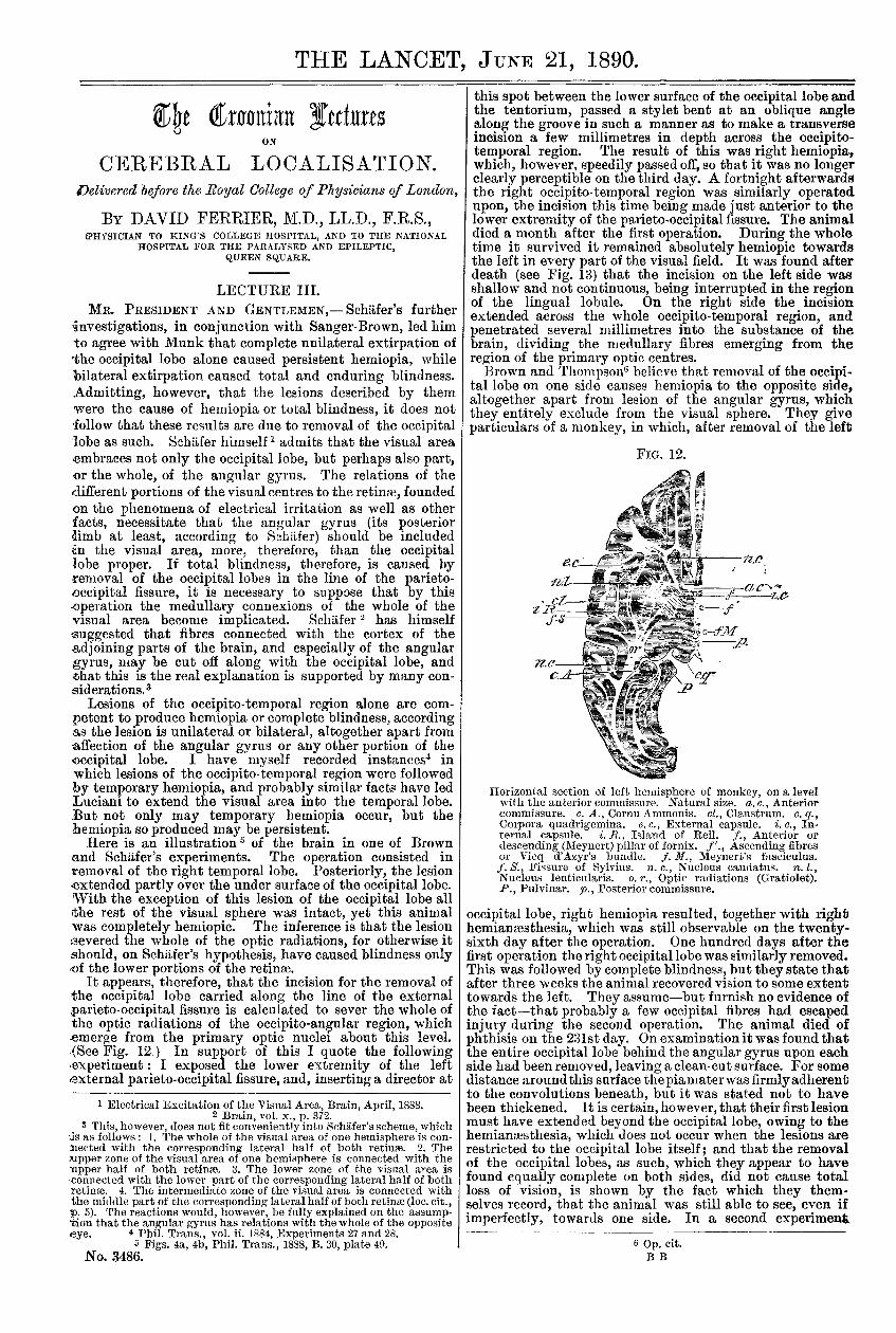
It appears, therefore, that the incision for the removal ofthe occipital lobe carried along the line of the externalparieto-occipital fissure is calculated to sever the whole ofthe optic radiations of the occipito-angular region, whichemerge from the primary optic nuclei about this level.,(See Fig. 12.) In support of this I quote the following’experiment: I exposed the lower extremity of the left.external parieto-occipital fissure, and, inserting a director at
1 Electrical Excitation of the Visual Area, Brain, April, 1888.2 Brain, vol. x., p. 372.
3 This, however, does not fit conveniently into Schäfer’s scheme, whichis as follows: 1. The whole of the visual area of one hemisphere is con-nected with the corresponding lateral half of both retinæ. 2. Theupper zone of the visual area of one hemisphere is connected with theupper half of both retinæ. 3. The lower zone of the visual area isconnected with the lower part of the corresponding lateral half of bothretinæ. 4. The intermediate zone of the visual area is connected withthe middle part of the corresponding lateral half of both retinæ (loc. cit.,p. 5). The reactions would, however, be fully explained on the assump-tion that the angular gyrus has relations with the whole of the oppositeeye. 4 Phil. Trans., vol. ii. 1884, Experiments 27 and 28.
this spot between the lower surface of the occipital lobe andthe tentorium, passed a stylet bent at an oblique anglealong the groove in such a manner as to make a transverseincision a few millimetres in depth across the occipito-temporal region. The result of this was right hemiopia,which, however, speedily passed off, so that it was no longerclearly perceptible on the third day. A fortnight afterwards
. the right occipito-temporal region was similarly operatedupon, the incision this time being made just anterior to thelower extremity of the parieto-occipital fissure. The animaldied a month after the first operation. During the wholetime it survived it remained absolutely hemiopic towardsthe left in every part of the visual field. It was found afterdeath (see Fig. 13) that the incision on the left side wasshallow and not continuous, being interrupted in the regionof the lingual lobule. On the right side the incisionextended across the whole occipito-temporal region, andpenetrated several millimetres into the substance of thebrain, dividing the medullary fibres emerging from theregion of the primary optic centres.Brown and Thompson6 believe that removal of the occipi-
tal lobe on one side causes hemiopia to the opposite side,altogether apart from lesion of the angular gyrus, whichthey entirely exclude from the visual sphere. They giveparticulars of a monkey, in which, after removal of the left
Horizontal section of left hemisphere of monkey, on a levelwit li the anterior commissure. Natural size. a. c., Anteriorcommissure. c. A., Cornu Ammonis. cl., Clanstrum. c. q.,Corpora quadrigemina. e. c., External capsule. i. c., In-ternal capsule. i. R., Island of Reil. f., Anterior or
descending (Meynert) pillar of fornix. f’., Ascending fibresor Vicq d’Azyr’s bundle. f. M., Meynert’s fasciculus.f. S., Fissure of Sylvius. n. c., Nucleus caudatus. n.l.,Nucleus lenticularis. o. r., Optic radiations (Gratiolet).P., Pulvinar. p., Posterior commissure.
occipital lobe, right hemiopia resulted, together with righthemiansesthesia, which was still observable on the twenty-sixth day after the operation. One hundred days after thefirst operation the right occipital lobe was similarly removed.This was followed by complete blindness, but they state thatafter three weeks the animal recovered vision to some extenttowards the left. They assume-but furnish no evidence ofthe fact-that probably a few occipital fibres had escapedinjury during the second operation. The animal died ofphthisis on the 231st day. On examination it was found thatthe entire occipital lobe behind the angular gyrus upon eachside had been removed, leaving a clean-cut surface. For somedistance around this surface thepiamaterwas firmly adherentto the convolutions beneath, but it was stated not to havebeen thickened. It is certain, however, that their first lesionmust have extended beyond the occipital lobe, owing to thehemianaesthesia, which does not occur when the lesions arerestricted to the occipital lobe itself; and that the removalof the occipital lobes, as such, which they appear to havefound equally complete on both sides, did not cause totalloss of vision, is shown by the fact which they them-selves record, that the animal was still able to see, even ifimperfectly, towards one side. In a second experiment
6 Op. cit.B B
1344
they found that destruction of the left angular gyruscaused no result beyond hemianmsthesia on the right side,together with some paralysis of the right arm (!). On theninetieth day a second operation was performed, consistingin excision of the whole of the right occipital lobe. Thiswas followed by left hemiopia, which still persisted whenthe paper was written, seventeen months after the opera-tion. These are the only experiments on monkeys whichthese authors have recorded. Lannegrace,7 on the otherhand, who has performed numerous experiments on theoccipital lobes and angular gyri of monkeys, states, inaccordance with my own and Yeo’s results, that destruc-tion of the occipital lobe causes no appreciable impairmentof vision, whereas destruction of the angular gyrus pro-duces crossed amblyopia of a temporary character. Hereports two cases of successive lesion of the angular gyri.In the one the first lesion induced crossed amblyopia, whichlasted four days. The second lesion, however, produced noappreciable alteration. In the other the first lesion againcaused crossed amblyopia, which disappeared in two days,while the second gave rise to lasting amblyopia. Theseresults, which are similar to those obtained by Yeo andmyself, depend no doubt on the degree of completeness ofextirpation of the gyri.
I have already mentioned that my earlier experiments,as well as those made by Professor Yeo, show that uni-lateral destructive lesion of this gyrus caused temporaryblindness of the opposite eye, and that bilateral destructioncaused for the time complete blindness in both eyes.
Though these results have been much questioned, I mustinsist on their accuracy. I have corroborated the occur-rence of apparently complete blindness of the opposite eye,on destruction of the left angular gyrus in an animal whichI lately made the subject of experiment. In this animal Ihad previously enucleated the left eye, so as to exclude allcomplications from this side. After removal of the leftangular gyrus, however, the animal, though otherwise welland in complete possession of all its senses and motor powers,was evidently absolutely blind. It responded to no test ofvision; would not move from the spot, but when urgedto move sprawled blindly and helplessly. This conditionlasted for several hours, during which it was under obser-vation. Next day there were indications of vision, butthe animal had become so prostrated, the weather beingintensely cold, that it died before any further exact
observations were possible. That le3ions of the angulargyrus may cause implication of the optic radiations ispossible, but this result is neither necessary, nor would itaccount for complete loss of vision in the opposite eye. Thebilateral relations of the angular gyrm account for thetransient nature of the amblyopia which results fromunilateral extirpation, and, as in few of my experimentswas the destruction of this gyrus complete on one side orthe other, it occasionally happened that removal of theother gyrus some time subsequently did not seem to impairvision either on one side or the other. When, however,the angular gyri are completely destroyed on both sides,
7 Op. cit.
i the animal, though for the first three or four days absolutelyblind, does not remain so permanently, but it never regains.
i normal vision. This also has been confirmed by the": researches of Lannegrace. Apart from the affection of, vision, destruction of the angular gyrus causes no othersymptoms, either motor or sensory; there is no ptosis orparalysis of the ocular muscles, and the sensibility of the.’conjunctiva is absolutely unimpaired.
, These results, confirmed by the researches of Horsley andSch1iJer, contradict the statement of Munk that the angulargyrus is the sensory sphere of the eyeball; and it will befound, on examination of Munk’s own data, that thephenomena on which he relies as indicating loss of the,sensibility of the eye are in reality due to loss of vision.Thus, he says, that after destruction of the left angulargyrus approximation of the finger to the left eye invariablycaused winking, whereas the same threat against the right.eye caused only winking when the eyelids were actuallytouched. This appears to be a proof of the sensibility ofthe eye, and the non-perception of threatened danger at a..distance. The absence of winking at threatened danger he-admits to be characteristic also of blindness; but he arguesthat inasmuch as the animal could not be blind, presumablybecause the occipital lobe was intact, therefore the non-occurrence of winking could only be due to the inability ofthe cortex to act on the sphincter palpebrarum Again, hesays that when the angular gyrus has been destroyed on oneside, and the eye on that side closed, the animal often fails..to seize things offered it or thrown down before it, especiallywhen the objects are small. This 1 consider a clear indicationof amblyopia. And he also states that after bilatera3extirpation of the angular gyrus of monkeys, "after in-complete restitution"-a phrase, however, which is not veryintelligible-are unable, like normal monkeys, to take thingsoffered them delicately with the fingers, but make grabs at,them with the whole hand. This is only a further illustra-tion of the same imperfection of vision which I have de-scribed-namely, want of precision in prehension, and a,continual tendency to place the hand over, or short of, theobject aimed at, instead of on it at once.
Schafer also8 records the symptoms in a monkey in whichhe destroyed both angular gyri. For the first few days theanimal appeared totally blind, but vision gradually returned,and before long was quite good for distant objects. The-animal could apparently see small objects, such as a raisin,at a distance, but on running up to it seemed to have somedifficulty in finding it. 5chiifer is of opinion that this,latter fact is due to absence of vision in the antero-
superior and lateral portions of the retinal. I have latelycarefully investigated the condition of vision in a monkeyin which I had completely destroyed both angular gyrLThere was no ptosis, the ocular movements were normal, theconjunctival reflexes unimpaired, sensibility wasintactevery-where, and the motor powers were perfect, but for four days atleast the animal was evidently absolutely blind. Whenurged to move, it ran against every obstacle in its path, paid noattention to threats, could not find its food except bygroping, and appeared insensible to light flashed in its eyes.On the fifth day there were evidences of returning vision.It did not now knock its head against obstacles, would not:walk over the edge of the table, showed signs of perceptionof light flashed in its eyes, and occasionally seemed to wince-when threatened. Vision gradually improved, but continuedvery imperfect, especially for minute objects, which it rarely,if ever, seized quite precisely, groping at them with the-whole hand, and reaching short, or over, or to the side. It.appeared to see objects held above, below, and to either side-much better than those held in front of its eyes. Six weeksafter the operation my colleague, Professor McHardy,examined the animal, which was very docile, with me, test--ing every portion of the visual field by pieces of apple sus-pended by a delicate thread. It was concluded that vision.was better in every part of the periphery than in the centre.Objects held directly before the eyes and at a little distance.were apparently not clearly seen, and never laid hold ofwith precision. The condition remained unchanged for-three months after the operation, similar tests being.from time to time applied, and with the same result.I noted also that the animal, when examining anyobject, always held it at full arm’s length from it&eyes. The phenomena observable in this animal were;such as would be best explained by impairment or
8 Brain, July, 1888, p. 159.
1345
loss of central vision; for it is well known that whencentral vision is lost or impaired in man, objects are bettereen at a distance than close at hand, and less distinctlywhen the eyes are immediately converged on them. This’is practically the condition seen in this animal. The loss of- central vision would thus account for the fact, noted by.;chafer in his animal, that objects were better seen at adistance than near at hand, and that Munk’s animal couldnever place its fingers precisely on any small object helddirectly in front of its eyes. There was certainly no indica-tion, but the reverse, that the upper portions of the retinaewere less sensitive than the lateral and lower portions. Itappears to me, therefore, that the symptoms resulting frombilateral destruction of the angular gyrus, described by,myself, Munk, and Scbafer, are best explained on the sup-position that the angular gyri are more particularly relatedto the area of distinct vision, and, accordingly, with themaculas lutess. The facts of disease in man render it neces-- aary to assume that the region of the yellow spot is repre-sented in each hemisphere, though more in that on theopposite than on the same side, and the probability is thatthe area of clear vision is represented mainly in the angular.gyrus of the opposite hemisphere.The retinal relations of the visual centres are not capable-of being explained by a simple division of the retinal fieldsinto correlated halves projected on the corresponding side ofach hemisphere. For unilateral lesion of the angular gyrus,produces a temporary blindness or amblyopia of the oppositeye; while bilateral destruction induces an enduring im-pairment of visual acuity in both eyes.9 The results of my.experiments seem to show that the angular gyrus has re-lations with both eyes. The crossed action, however, is the.only one which is clearly demonstrable in the lower animals,but this does not exclude the possibility of some impair-mient of vision on the same side, not perceptible by testsapplicable to them. It is certain that in man affections ofthe visual centres occasionally produce crossed amblyopia,.and not homonymous hemiopia. Not only is this characterdstic of the visual disturbances seen in hysterical hemi-.anaesthesia, the pathology of which is obscure, but it has’been noticed also in cases of organic disease. Usually, alongwith the blindness or great impairment of vision in theopposite eye, there has been some degree of contractionof the visual field of the eye on the same side. I havemyself reported several such cases and Gowersll has also:seen similar instances. A well-observed case of this naturehas been recorded by Sharkey. 12 Post-mortem examinationrevealed softening and absorption of a considerable area of’the opposite hemisphere, including the angular gyrus. Theoccipital lobe was intact, and in nowise reduced in size as,compared with the other.A distinguishing test between tract and central hemiopia
’!has been suggested by Wilbrand,13 and advocated byWernicke and Seguin, which consists in determiningwhether a pencil of light thrown on the blind side of theretinae induces contraction of the pupil or not. As the optictract is the path of the fibres which excite pupillary con-traction through the oculo-motor centres, as well as
those which excite visual sensation in the cortex, lesionof the optic tract will cause not only hemiopia, butalso paralysis of the reflex reaction of the pupils to
’light; whereas lesion of the cortical centres will cause’hemiopia, but leave intact the pupillary reaction. Thistest, however, requires great care in its application, as itia difficult to restrict the rays of light entirely to theblind side. In a case recently under my care at King’sCollege Hospital,14 in which the dividing line passed through’the fixation point, and which was carefully investigated inthis relation by Professor McHardy and myself, the pupil-lary reaction was not obtained, as a rule, when the pencil
9 This accords with Cowers’ hypothesis that " on the outer surface, infront of the occipital lobe, there is a higher visual centre in which thehalf fields are combined, and the whole opposite field is represented."Diseases of the Nervous System, vol. ii., p. 19.)
10 Cerebral Amblyopia and Hemiopia, Brain, vol. iii., p. 456.11 Diseases of the Nervous System, p. 19.
12 Medico-Chirurgical Transactions, vol. lxvii., 1884. 13 Op. cit.14 The patient was a man aged thirty-nine. Two years before he had
contracted syphilis, and at the time of his admission was suffering froma large tertiary ulcer in the soft palate. He had twitching of the rightside of the face, together with weakness and numbness of the left side.The left hand grasp was weak, and there was loss of dorsal flexion ofthe left foot. The tongue deviated to the right on protrusion. He wasfound to be absolutely hemiopic towards the left side, and a carefulperimetric examination by Professor McHardy demonstrated that thedividing line passed exactly through the fixation point, as will be seenin the chart which I show you.
of light was thrown on the right side of the retinae ;whereas it occurred readily when the light was thrown onthe left half of each retina. These facts, therefore, wouldcorroborate the hypothesis that this was a case of tracthemiopia.
I have recently verified the hemiopic pupillary reaction intwo monkeys, in which I accidentally severed the optic tractin establishing lesions of the temporal lobe. The result inboth cases was absolute hemiopia to the opposite side. Inboth cases the left optic tract was divided, and in both,along with right hemiopia, there was absence of reaction ofthe pupil when a fine pencil of electric light was thrown onthe left half of each retina ; whereas active reaction tookplace when the light was thrown on the right half. Both inthe monkey and in several cases of hemiopia in man depend-ing on lesions of the hemisphere I have found that thepupillary reaction is equally well marked whichever side ofthe retina is illuminated. There can be no question that inman and monkeys there is decussation of the optic tracts inthe chiasma. Michel, in his comparatively recent mono-graph,15 still maintains the contrary, on the basis of micro-scopical investigation ; but his results have been attributedby Singer and Münzel’16 to imperfect methods of examination.In reference to the pathology of crossed amblyopia from
lesion of the angular gyrus in the monkey, as well as ofsimilar cases of disease in man, I may here allude to thehypothesis advanced by Lannegrace. Lannegrace regardsthe eyeball as innervated by two sets of fibres-sensorial, oroptical proper, and sensory, on which the proper nutritionof the eyeball depends. The sensorial or optical decussatein the chiasma, and are distributed to the occipital lobe;while the sensory decussate in the pons, and, applying them-selves to the posterior fibres of the internal capsule, are dis-tributed mainly to the angular gyrus. Lesions of thesensory fibres produce amblyopia and sensory disturbancesin the eyeball. A similar result follows lesion of theangular gyrus, and is essentially dependent upon changeswhich are induced in the nutrition of the eye. This hypo-thesis would require that in all cases of amblyopia fromcerebral lesion there should be impairment of sensation inthe eyeball. But this is certainly not the case, for though inhysterical amblyopia there is affection of common sensationas well as of vision, it is not so in the amblyopia followingdestruction of the angular gyrus. Though affections of thefifth nerve, which cause loss or impairment of sensation ofthe eyeball, frequently also lead to trophic disorders of theeye, yet this is not necessarily so ; and even when the eye-ball is absolutely anaesthetic, provided that no trophic di s-turbances have occurred, vision is not in the least impaired.In proof of this I would refer to cases (Cases 1 and 3) re-ported by Hutchinson in the Ophthalmic Hospital Reports,vol. iv., 1863-65. Nor does complete anaesthesia of the eye-ball induced by cocaine impair visual acuteness.These appear to me to be fatal objections to the hypothesis
advanced by Lannegrace, and I contend that the only hypo-thesis which seems to harmonise with all the facts is, thatthe angular gyri are more particularly the centres for clearvision, each mainly for the eye of the opposite side.Whether the other portions of the retinae upper, lower,outer, and inner, are specially represented in correspond-ing regions of the occipital lobe, according to the hypo-theses of Munk and Scb:tfer, cannot be said to have as yetbeen established ; for, even after the most extensive de-struction of the occipital lobes, no portions of the retinaeappear to be absolutely blind ; hence, even if we admitthat the effects of irritation probably indicate a specialrelation of the different portions of the visual field tocertain portions of the occipital cortex, the relation, so faras we may judge by the facts of disease or experimentallesion, does not seem to be an exclusive one.
It is true that in man we sometimes find, besides generalhemiopic deficiency, partial, quadrant, or sector-like,defects in the upper or lower halves of the visual field.These appear, however, to be merely incomplete hemiopia ;and occasionally, as in the case which I show you, an isletof subnormal visual acuity may be seen in the defectivehalf. The pathology of these sector-like defects is a matterof conjecture. They have not been conclusively brought inrelation with lesions of any particular portion of the cortex,and the probability is that they are dependent rather upon
partial lesions of the optic radiations than of the corticalcentres themselves. This was without doubt the pathology
15 Ueber Schnerven-Degeneration und Kreuzung, 1887.16 Beitrage zur Kenntniss Schnervenkreuzung, 1889.
1346
in the case which I have alluded to, for the defect occurredin a patient who had a sudden attack ofttjMpIegia accom-panied bv hemianaesthesia and some affection of speech.
It is doubtful whether there are on record any cases ofstrictly cortical lesions of the occipital lobe proper,accompanied by hemiopia, apart from direct or indirectimplication of the optic radiations. In most of the cases ofhemiopia which have been examined after death, in which
the optic tracts, optic thalami, or corpora geniculata havenot been obviously diseased, the lesions have been found inthe medullary fibres of the posterior region, vaguely orinaccurately called the occipital lobe ; or, if the cortex hasbeen mainly affected, the lesions have been multiple anddiffuse, and not confined to the occipital region. And, inaddition to hemiopia, there have been hemiplegia, hemi-anaestbesia, aphasia, or other symptoms of implication ofthe cerebral tracts and centres beyond those of the occipitalregion.Under my direction my friend and pupil, Dr. Ewens, has
collected and analysed the majority, if not all, of therecorded cases of hemiopia (with necropsies) depending oncerebral lesions which have not obviously been of such acharacter as to cause indirect and indefinite implication ofother regions. Of 41 cases of hemiopia, 15 were fromdisease of the occipito. angular region, 2 were cases ofdisease of the angular and supra-marginal gyri only, and 15were described as being from disease of the occipital lobealone; of these 15, there were only 2 (Hun’s case 17 andDoyne’s case 18) in which there was not either a tumour,cyst, abscess, or softening of the medullary substance ofthe occipital region, or another lesion affecting the opticthalamus ; and in 1 of these cases (Doyne’s) the position ofthe lesion was not accurately described.
In the other cases the lesions were of a diffasecharacter, 6 being from lesion of the occipito-temporalregion, and 3 from lesion of the occipital, temporal, andparietal lobes simultaneously, the angular gyrus beingimplicated in all. ,
From the comparative frequency with which hemiopiahas been found associated with lesions of the cuneus and itsneighbourhood, Seguin’y and NothDagel2l’ are of opinion thatthis portion of the occipital lobe has a special relation tovisual perception ; while Wilbrand believes that the visualcentre is more especially in the apex of the occipital lobe.These hypotheses are not supported by experimental re-
search. The probability is that any apparent relationbetween lesions of the cuneus and the occurrence of hemiopiais due to the special proclivity of this region to affection bymorbid vascular conditions, and to coincident implication ofthe optic radiations of the occipito-temporal region. InSeguin’s own case,21 on which he relies in support of his
hypothesis, not only was the cuneus implicated, but alsothe fourth and fifth temporal gyri and a part of the gyrushippocampi.2 Cases have also been recorded in which notonly unilateral but bilateral lesions have been found in theoccipital lobes without any affection of vision whatever.
Irritative lesions of the angular gyrus occasionally giverise to optical illusions or flashes of light, followed by tem-porary amblyopia, as recorded by Hughes Bennett;21 whiledestructive lesions of the angular gyrus, more particularlyin the left hemisphere, are generally associated with thespecial form of sensory aphasia termed "word-blindness"
"
(Kussmaul). Word-blindness is not necessarily accompaniedby any noteworthy affection of visual sensation, though insome cases, where the lesion of the occipito-angular regionis more extensive, there may be a greater or less degree ofright hemiopia.On the other hand, right hemiopia pure and simple, is
not necessarily associated with any defects in visualideation. This would be an argument against its corticalnature. The fact that visual ideation, more particularly
19 The Journal of Nervous and Mental Diseases, vol. xiii., Jan. 1886.20 Neurolog. Centralblatt, 1887, p. 213.
21 Op. cit.22 Since this was written a case has been put on record by Delépine
(Trans. Path. Soc. Lond., May 20th, 1890) in which right hemiopia wasapparently associated with softening of the left cuneus. In this case,however, there was general arterial degeneration, and there were manycircumscribed patches of softening in various parts of the brain. Inparticular there was a small area which had destroyed the greater partof the middle occipital convolution. This case is, therefore, too complexto allow of exact conclusions as to the connexion between the hemiopiaand the lesion of the cuneus in particular.
23 Excessive Sensory Cortical Discharges and their Effects, THELANCET, March 30th and April 6th, 1889.
in reference to the association of written symbols withtheir meanings, is apt to suffer more readily than simpleperception, appears to me to illustrate the laws of the-evolution and dissolution of the nerve centres, whichhave been so ably expounded by Hughlings Jackson in hisCroonian Lectures delivered here a few years ago (1884). As.evolution is from the most simple and most stable up to themost complex and least stable, so destructive processesannihilate first of all the higher and last of all the lowerfunctional manifestations. The functions of the visualcentres in respect to mere visual sensation, or simple pre-sentation are much more stably organised than those whichimply visual ideation or re-presentation, and in particularsuch highly specialised and complex processes as are involvedin the association between visual symbols and things signi-fied. Hence, a lesion of the area of clearest vision mayparalyse visual re-presentation, while the simpler functionof visual presentation, may not be appreciably impaired.In order that this also should be entirely abolished it isnecessary that every trace of the given centre should beextirpated. In other words, re-presentative blindness willoccur sooner than presentative blindness, and the formermore readily in its most specialised modes of manifestation..
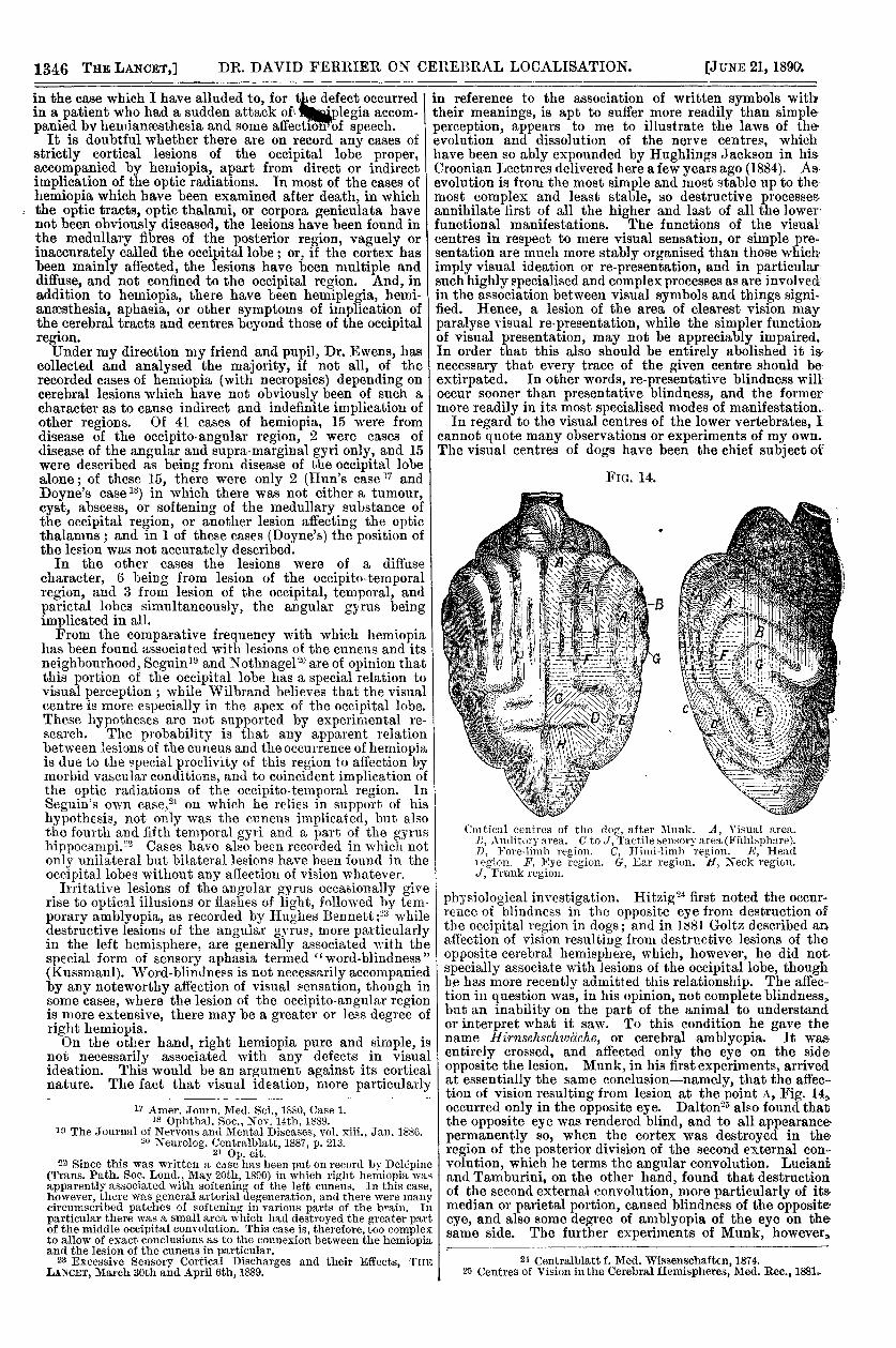
In regard to the visual centres of the lower vertebrates, Icannot quote many observations or experiments of my own.The visual centres of dogs have been the chief subject of
Ccntical centres of the dog, after Munk. A, Visual area.
A’, Auditory area. C to J, Tactile sensory area (Fuhlphare).D, Fore-limh region. C, Hil1ll-]imh region. E, Headlegion. F, Kye region. G, Ear region. tl, Neck regibn.J, Trunk region.
physiological investigation. Hitzig24 first noted the occur-rence of blindness in the opposite eye from destruction ofthe occipital region in dogs; and in 181 Goltz described anaffection of vision resulting from destructive lesions of theopposite cerebral hemisphere, which, however, he did notspecially associate with lesions of the occipital lobe, thoughbe has more recently admitted this relationship. The affec-tion in question was, in his opinion, not complete blindness,but an inability on the part of the animal to understandor interpret what it saw. To this condition he gave thename HirnselascJz2vaclae, or cerebral amblyopia. It wasentirely crossed, and affected only the eye on the sideopposite the lesion. Munk, in his first experiments, arrivedat essentially the same conclusion-namely, that the affec-tion of vision resulting from lesion at the point A, Fig. 14,occurred only in the opposite eye. Dalton25 also found thatthe opposite eye was rendered blind, and to all appearancepermanently so, when the cortex was destroyed in theregion of the posterior division of the second external con-volution, which he terms the angular convolution. Lucianhand Tamburini, on the other hand, found that destructionof the second external convolution, more particularly of its-median or parietal portion, caused blindness of the opposite-eye, and also some degree of amblyopia of the eye on thesame side. The further experiments of Munk, however:,
24 Centralblatt f. Med. Wissenschaften, 1874.25 Centres of Vision in the Cerebral Hemispheres, Med. Rec., 1881.
1347
as well as those of Loeb26 and of Goltz,27 and also the later Iexperiments of Luciani,28 appeared to show that, though in Idogs the visual area is mainly in relation with the opposite ieye, it is also in relation with the outer quadrant of theeye on the same side. Hence destruction of the visualcentre in the one hemisphere paralyses the inner three-fourths of the opposite retina, and the outer fourth ofthe retina on the same side. The condition, therefore-at least the enduring one-is that of homonymoushemiopia towards the opposite side, the defect in the eyeopposite greatly exceeding that in the eye on the same side.But thefacts recorded by Luciani and Tamburini indicate that,for a short time at least, after the destruction of the middleportion of the second external convolution there is blindnessin the opposite eye. And Goltz29 remarks that he does notthink that his former conclusions were due to defectiveobservation, but that there was probably some variationin his operative procedure. It is, however, likely thatwe have here the same relations as exist in monkeys,and that for the time at least, after complete extir-pation of the visual sphere, there is total blindnessin the opposite eye. This is confirmed by the experi-ments of Bechterew, recently published.3J Bechterewfinds that in dogs and cats there are two regions inthe cortex related to vision: the one in the occipito-parietal region, in relation with the corresponding halvesof both retinse ; the other, more especially in the parietalregion, in relation with the opposite eye alone. Lesion ofthe former causes bomonymous hemiopia ; lesion of thelatter, generally possible only in association with theformer, causes along with hemiopia amblyopia of theopposite eye by paralysing the centre of clear vision. Thiscombined affection usually after a time gives place tohomonymous hemiopia, or, on the contrary, the hemiopiadisappears and the crossed amblyopia remains. Bechterew’sconclusions may serve to explain, among others, the resultsarrived at by Gilman-Thompson, and Sanger-Brown,31which appear to be entirely at variance with those ofMunk, Goltz, and most other physiologists. For they findthat lesions of sufficient size and depth, in the posteriorpart of the occipital region in cats and dogs, invariablycause blindness of the opposite eye, with no impairment ofvision in the eye of the same side. These authors,however, seem to think that the extent of the visual sphereis a matter more of cubical capacity than accurateanatomical localisation; for they say that, in orderthat the blindness should be permanent, between 2’5 and I,3 cubic centimetres of brain tissue should be removed iin cats; and between 4’5 and 6 cubic centimetres in dogs.To render the blindness permanent, the incision mustbe at least ’5 centimetre deep, and 2 centimetres indiameter, in cats ; and 1 centimetre deep, and 3 centimetresin diameter, in dogs ; and it must involve at least two con-volutions. Smaller lesions produce complete blindness ofthe opposite eye, lasting from a day or two to six weeks.They conclude from their experiments that in cats and dogsthere is complete decussation of the optic nerves in thechiasma. This is, however, opposed by the researches ofv. Gudden,32 which show that in dogs and cats there is
partial decussation of the optic tracts ; and Nicati3J found,experimentally, that division of the chiasma in the antero-posterior or sagittal diameter did not cause complete lossof vision in either eye. The recent researches of Singer andMiinzer, previoualy alluded to, indicate that there is onlypartial decussation in the chiasma of the cat, dog, and alsoin the rabbit.The exact limits of the visual sphere in dogs are still the
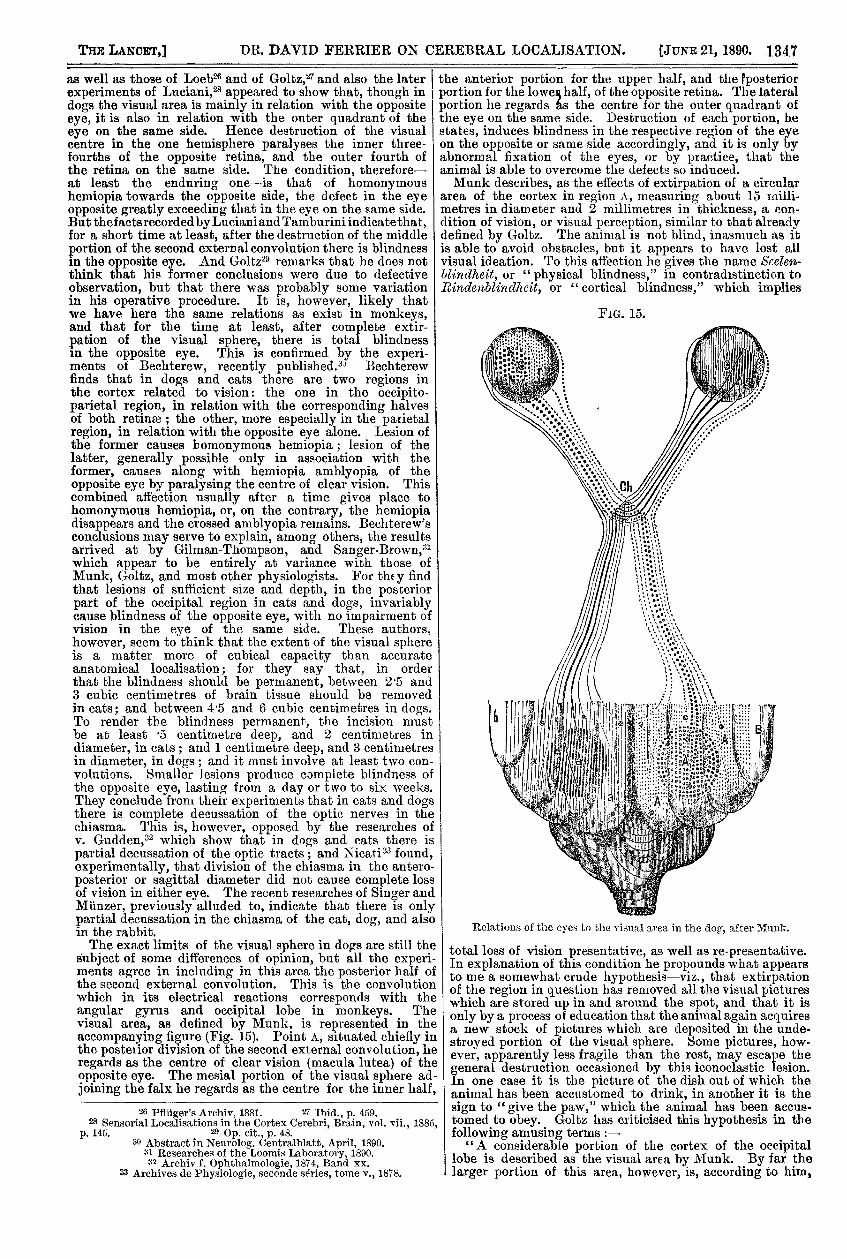
subject of some differences of opinion, but all the experi-ments agree in including in this area the posterior half ofthe second external convolution. This is the convolutionwhich in its electrical reactions corresponds with theangular gyrus and occipital lobe in monkeys. Thevisual area, as defined by Munk, is represented in theaccompanying figure (Fig. 15). Point A, situated chiefly inthe posterior division of the second external convolution, heregards as the centre of clear vision (macula lutea) of theopposite eye. The mesial portion of the visual sphere ad-joining the falx he regards as the centre for the inner half,
26 Pflüger’s Archiv, 1881. 27 Ibid., p. 459.28 Sensorial Localisations in the Cortex Cerebri, Brain, vol. vii., 1885,
p. 145. 29 Op. cit., p. 48. 30 Abstract in Neurolog. Centralblatt, April, 1890.
31 Researches of the Loomis Laboratory, 1890.32 Archiv f. Ophthalmologie, 1874, Band xx.
33 Archives de Physiologie, seconde séries, tome v., 1878.
the anterior portion for the upper half, and the Pposteriorportion for the lowel half, of the opposite retina. The lateralportion he regards as the centre for the outer quadrant ofthe eye on the same side. Destruction of each portion, hestates, induces blindness in the respective region of the eyeon the opposite or same side accordingly, and it is only byabnormal fixation of the eyes, or by practice, that theanimal is able to overcome the defects so induced.Munk describes, as the effects of extirpation of a circular
area of the cortex in region A, measuring about 15 inilli-metres in diameter and 2 millimetres in thickness, a con-dition of vision, or visual perception, similar to that alreadydefined by Goltz. The animal is not blind, inasmuch as itis able to avoid obstacles, but it appears to have lost allvisual ideation. To this affection he gives the name <S’ceblindheit, or "physical blindness," in contradistinction toRindenblinclhcit, or "cortical blindness," which implies
Relations of the eyes to the visual area in the dog, after Munl;:.
total loss of vision presentative, as well as re-presentative.In explanation of this condition he propounds what appearsto me a somewhat crude hypothesis-viz., that extirpationof the region in question has removed all the visual pictureswhich are stored up in and around the spot, and that it isonly by a process of education that the animal again acquiresa new stock of pictures which are deposited in the unde-stroyed portion of the visual sphere. Some pictures, how-ever, apparently less fragile than the rest, may escape thegeneral destruction occasioned by this iconoclastic lesion.In one case it is the picture of the dish out of which theanimal has been accustomed to drink, in another it is thesign to "give the paw," which the animal has been accus-tomed to obey. Goltz has criticised this hypothesis in thefollowing amusing terms :-"A considerable portion of the cortex of the occipital
lobe is described as the visual area by Munk. By far thelarger portion of this area, however, is, according to him,
1348
pure luxus. The memorial pictures (RrÙ’werun,q/:/bilder) ofthe visual perceptions are huddled together like sheep in astorm, in a narrow spot which occupies only about two-sevenths of the whole visual area. When this small spot,which corresponds to the macula lutea of the human retina,is destroyed on both sides, the animal is at first blind, andonly gradually learns to see, like a puppy, with the aid ofthe rest of its visual sphere. Five-sevenths of the visualsphere-a large portion of the cortex-appear retained inorder that a dog which falls into the hands of the physiologistshould again learn to see, when its accumulated visual pic-tures are cut out. All dogs which escape this fate-and these,since the creation of the dog, must be very many-carryduring their life five-sevenths of their visual sphere as an un-cultivated fallow field. A wonderful thing in hypotheses. ":;4Even if it were the case that the different portions of the
retina are represented in the indicated regions of the visualarea, it is improbable that these could be determined withany degree of certainty except by exact perimetric investi-gation. which is naturally impossible in the lower animals.The difticulties of deciding questions of this kind in the loweranimals are illustrated by Munk’s account of his experi.ences with rabbits.3a "I had at least believed that I coulddetermine with which eye the rabbit saw better and withwhich it saw worse. In this, however, I have been mis-taken, because it occasionally happened to me that wherefrom tests I had thought I had made out that the greaterimperfection of vision existed in the left eye, post-mortemexamination revealed the fact that the left tract and theright optic nerve were more atrophied than the right tractand the left optic nerve."
]oeb, after a series of carefully devised experiments, con-cludes that there is no foundation for the views of Munkthat particular segments of the retine are in special rela-tion to definite regions of the visual area. When defect ofvision occurs from lesion of the posterior lobe, it is always ofthe same hemiopic or hemiamblyopic character, whateverportion of the visual sphere be injured. The lateral portionof this sphere is not in special relation with the outer quad-rant of the eye on the same side, nor is any portion more inrela,tion with one part of the opposite retina than another.In particuhlr, central vision is precisely that which is leastaffected in all cases, whether of unilateral or bilateral lesionof the visual zone. There is never anv excentric or abnormalfixation of the eyeballs when the special regions indicatedby Munk are destroyed, such as would necessarily result ifthe corresponding portions were paralysed ; nor is the re-covery of vision, after partial lesions of the visual area, dueto practice or to the acquisition of new visual experience, in-asmuch as recovery takes place when the animal is keptabsolutely in the dark, and thus prevented from exercisingits visual faculties.
In rabbits the visual centre would, according to thehomology of the e!ectrical reactions, occupy the parieto-occipital region of the hemisphere. The exact limits of thevisual zone of these animals do not appear to have beenaccurately determined by any observer, though certain ex-periments of Moeli36 point to lesions of the region indicatedas causing at least temporary blindness of the opposite eye.It has been supposed that in this animal there is completedecussation of the optic tracts in the chiasma, inasmuch asthe experiments of Brown-Sequard have shown that sagittaldivision of the chiasma causes complete loss of vision of botheyes. The total decussation in the chiasma of the rabbitwas also supported by the earlier researches of von Gudden,who found that after enucleation of one eyeball the oppositeoptic tract only became atrophied.;)7 But, in his later re-searches, he concluded that a small fascicle of uncrossed, ordirect, fibres exist also in the optic tract of this animal, similarto that seen in higher vertebrates. Singer and Miinzer arealso of opinion that there is only partial decussation in thechiasma of the rabbit, but the uncrossed tract does notrun as a separate bundle, but as fibres more or less diffusedthroughout the optic tract. In the mouse and guinea-pig, however, the decussation is a complete one. The partialdecussation of the optic tracts in the chiasma would favourthe opinion that in the rabbit also both eyes are more orless in relation with each visual zone; and certain experi-
34 Op. cit., p. 175.35 Sitzungsberichte Akad. d. Wissensch. zu Berlin, vol. xxxi., June 20th,
1889, p. 631.36 Archives de Physiologie, 1871-72, Sur les Communications de la
Rétine avec l’Encéphale.37 Archiv f. Ophthalmologie, Bd. xx., 1874.
ments of Munk would seem to favour this notion. Thepoint, however, is one which requires further investigation.In pigeons, and in birds in general, the region which in
its electrical reactions is homologous with the visual centreof the higher animals occupies the parieto-posterior aspectof the hemisphere, where it forms a thin lamina over thecorpus striatum. McKendrick3l found that destruction ofthis region caused blindness in the opposite eye; whereasremoval of the anterior part of the hemisphere had noeffect on vision, nor removal of the posterior extremity ofthe hemisphere. Similar results have been obtained byJastrowÏtz3H and Musehold.40 Blaschko, however, foundthat vision did not seem entirely abolished in the oppositeeye by destruction of the cortex in the region indicated ;and Munk came to the conclusion that though vision atfirst seems entirely abolished in the opposite eye, yet aftera time it is regained in the extreme outer or lateral portionof the retina.
It is usually stated that in pigeons a complete decussa-tion of the optic tracts occur in the chiasma, but vonGudden expresses some doubts on this point. Singer andMiinzer, however, believe that in the pigeon a total decus-sation occurs. Muuk quotes, in favour of his conclusions,certain observations by Muller, to the effect that in theretina of the pigeon there is, besides the usual foveacentralis, another fovea, situated nearer the temporalregion of the retina. The outer fovefe would subservebinocular and the central foveae monocular vision. Thesestatements have been supported by the ophthalmoscopicinvestigation of Hirschberg.
It has seemed to me that if any bird can possess binocularvision it should be the owl, whose eyes are placed almost inthe same plane. To decide this question I recently com-pletely extirpated the right hemisphere of a,n owl. Theright eye was then securely closed. The owl reacts veryreadily to visual tests, and is keenly alive to every move-ment coming within its field of vision. This bird, how-ever, for ten days at least, remained perfectly indifferent tothe electric light flashed in its eye and to all kinds of threats,and when urged to move flew blindly against every obstaclein its course. At the end of this time there were indicationsof vision, which were found, however, to depend on the partialunclosure of the right eye. In order to ascertain if the visualcentre of the left hemisphere was undamaged, the righteye was completely freed from its closure. This had theeffect of restoring the animal’s sight, so far as the right eyewas concerned, though no indications of vision could bemade out in the left eye. The bird was able to pursue andultimately catch a mouse introduced into its cage, thoughthe mouse frequentlv escaped for the time by getting to theowl’s left. The right eye was then removed. It thenspeedily became apparent that the animal was not entirelyblind, but could see towards the right with the outer por-tion of its left eye. It noticed and winced at threats madetowards the right, came forward and pecked at pieces ofmeat held in this position, though its aim was not veryaccurate, and one day pursued about the cage and ulti-mately, after considerable difficulty, captured a mouse,which it devoured whole. No doubt can therefore be enter-tained as to the binocular relations of each cerebral hemi-sphere in the owl. It is stated, however, by Michel, as wellas by Singer and Miinzer, that there is total decussation ofthe optic tracts in this bird. If this be correct, it followsthat total decussation of the optic tracts is not inconsistentwith binocular representation in each cerebral hemisphere.My own experiments, as well as those of Munk, Horly,
and Sohtifer, show that when the lesions are strictlylimited to the visual sphere, vision alone is affected orabolished, without any implication of the other forms of sensi-bility, general or special, and absolutely without any motorparalysis. The contrary results obtained by some authorsare without doubt dependent on primary or secondaryinjury to other sensory or motor tracts or centres. InGoltz’s experiments the affections of vision from injury ofthe occipital regions appear to have been almost invariablyassociated with other forms of sensory disturbance; but themanner in which he established his lesions has not beensufficiently definite as to exclude implication of the sensorytracts of the internal capsule, or other sensory regionsof the cortex. Whether after destruction of the wholevisual sphere in the higher animals, man and monkey,,
38 Trans. Roy. Soc. of Edinburgh, January, 1873.39 Archiv. f. Psychiatrie, Bd. vi., 1876.
40 Das Selicentrum bei Tanben, August, 1878.
1349
any form of reaction to retinal impressions, beyond thatof the pupil, may continue-as has been contended forby Goltz in the case of dogs, and by Luciani andTamburini, and Lannegrace even in the case of monkeys-is not supported by clinical investigation, or by my ownexperiments, or those of Munk on monkeys. Thoughthe monkey rendered blind by total extirpation of itsvisual centres acquires the power of avoiding obstacleswhen left amidst its usual surroundings, yet this appearsto be due rather to a sharpening of its other faculties, ormore attentive appreciation of the impressions made onthese by the objects with which it is surrounded, than tovisual sensation. The question is, however, one whichmay well bear further investigation; for if retinal im-pressions are coordinated with apparently purposive actionsin the subordinate centres of the lower vertebrates,such as fishes, reptiles, and birds, there is at least thepossibility that similar reactions may be discoverable in thehigher animals, even though in a much less degree. It iscertain, however, that the visual area of the cortex is not amerely functionally differentiated region capable of re-
placing, or of being replaced by, other cortical regions,inasmuch as destruction of the visual centres leads to
atrophy in the primary optic centres, optic tracts, and opticnerves ; and, conversely, destruction of the optic radiationsleads to atrophy strictly confined to the regions includedwithin the visual zone. The differentiation of an area ex-elusively-so far at least as can be judged from clinical andexperimental results-would strongly favour the hypothesisthat the other sensory faculties are also separately localisedin definite cortical regions.
ON HEAD-NODDING AND HEAD-JERKING IN
CHILDREN, COMMONLY ASSOCIATEDWITH NYSTAGMUS.
BY W. B. HADDEN, M.D. LOND., F.R.C.P.,ASSISTANT PHYSICIAN TO ST. THOMAS’S HOSPITAL AND TO THE HOSPITAL
FOR SICK CHILDREN.
(Continued from page 1295.)
CASE 3. Side to side movements of head after 7wad-injury;sttbsequent occurrence of ny8ta{f1nus. vertical in right, hori-zontal in left eye " peculiar method of looking cct objects.-rickets convulsions; fits of laaghtcr.-Mary G-, agedeight months, was brought to me on Nov. 22nd, 1888. Shewas the tfnth child, and was born at full term withoutdifficulty. Seven children were alive and healthy, butfour had had convulsions in infancy. Three childrenwere dead. The child was being suckled, but, in addition,had had Robb’s biscuits. She had never had convul-sions or any attacks like petit mal. About nine daysbefore I saw her the child fell off a chair, strikingthe left side of the head. She was not unconscious,and, indeed, did not seem to be much hurt. The partstruck was only " a bit red," not bruised. The head beganto shake five or six days later. On examination I foundher a well-nourished, good-tempered child. There wassome beading of the lower ribs at their junction with thecartilages, and the wrist-ends were thickened. There wereno teeth. The fontanelle was open, but not unduly pro-minent. The part of the head struck I found to be just tothe left of the fore part of the anterior fontanelle, and herethere was a small scab, but no tenderness, bruising, or signof fracture. The movements of the head were horizontal,with a tendency to go more to the right and downwards.The mother had never seen any movements of the eyeballs,and there was no nystagmus when I first saw the child.A little later, however, there was marked horizontalnystagmus, more in the left than in the right eye. It wasconstant, but worse when the child’s attention was directedto an object. The nystagmus still persisted when the headmovements stopped. A few days later, Mr. Donald Gunn, inthe absence of Mr. Marcus Gunn, examined the eyes, and re-ported that " the movements of the right were vertical andvery fine, of the left much more violent, and nearly hori-zontal. Vision was good, the ordinary movements of theeyeballs unimpaired, and the discs normal." Within a weekthe head became steady, and during the next five monthsthe movements occurred only occasionally when the childcut a tooth. In May the patient had four convulsive attacks,preceded by some head-jerking for a day or two. It wasnoted that the child looked at objects sideways, and usually
to the right. During this month she had two more convul-sive seizures, but these latter were not preceded by head-movements. During this period the child had occasionalfits of laughter. Her intelligence was above the average,and the mother said that she was "very knowing." OnJuly llth I noted that there had been no fits for three weeks,but that the nystagmus and head-jerking persisted.CASE 4. Vertical nystagmus of one eye only following
head injury; side to side movements of head fbecr monthslater; peculiar method of looking at objects; tavo relapsesfollowing falls on the head. - Frank K—, aged fourteenmonths, was brought to me on April 19th, 1888. There wasa history of three severe falls, the last six weeks previously,when he struck the back of the head. There was no markvisible afterwards, and he did not lose consciousness at thetime. He had some bronchitis when the accident occurred.The movement of the eye was noticed a week subsequently tothis last fall. There was no history of fits. On examina-tion, no sign of rickets was found. The child was large andrather flabby, but placid, and apparently in fair health.Mr. Gunn reported as follows: " Marked vertical nystagmusin left eye, not constant, but nearly so. No continuous rela-tion between any position of eye or the evident ocular stateand the occurrence of the nystagmus. It sometimes disap-pears momentarily on assuming a new position of fixationwith right eye. Pupils active to light. Seems to see with eithereye. After a sudden start on ringing a bell, his left eye re-mained stationary for some time. Excursions of eye good inall directions. Ophthalmoscopically, pupils not well dilated(probably homatropine not well in) ; there is something pre-venting accurate details of fundus beingseen, but fundus reflexgood in both. He seems to see fairly well." A drachm ofsteel wine was ordered to be taken twice daily. During thenext fortnight the nystagmus became less marked. OnApril 29th Mr. Gunn reported as follows : "Mother thinkshe cannot see with left eye. Left pupil acts to lightdirectly, but rather better consensually. Right pupilactive. Both discs and fundi normal." On May 17tb,when I examined the patient, there were no movements ofthe eyeball. The mother believed that he could not seewith the left eye. When he directed attention to anythinghe turned his head to the left and " looked out of hisright eye." On May 24th I noted that there had beensome movement of the eye the previous day, the firsttime for a week, and that he had had some con-
vulsions with screaming on the evening of the same day.On June 3rd he fell on the left side of the forehead. Thenext day he had some nystagmus of the left eye, but it onlylasted five minutes. From this time to Aug. 9th theabsence of the movements was noted from time to time, butmy attention was now called to the manner in which hemoved his head from side to side in his endeavours to fix anobject. This persisted up to Nov. 12th. On that day Imade a note of the mother’s statement, that on Nov. 7th he
fell down fourteen stone steps, bruising his temples. Threedays later there were some movements of the eyeball lastingseveral hours, the first observed for three months. OnNov. 20th he was examined again by Mr. Gunn, who couldsee no nystagmus. The mother, however, said that it hadoccurred recently. On Dec. 4th there was slight occasionalvertical nystagmus ; the child seemed to see well with theleft eye ; the left pupil acted directly but not fully ; theoptic discs were normal. On Dec. 11th I noted that there hadbeen no fits or petit mal. The nystagmus was well marked.When his attention was directed to some distant objecthe shook his head from side to side. A month later themovements of both head and eyes were less marked. Ithad been noticed for about four weeks that he used to goto sleep with his hands placed on the top of his head, buthe made no complaint of pain. Six months later therewas still occasional vertical nystagmus of the left eye,though it was less marked. The head movements had notrecurred. The pupils were active. The mother still believedthat the child did not see with the left eye, as he used toturn his head to the left in order to look out of his righteye. Mr. Gunn, however, was unable to detect any defect ofvision. Six weeks later the child was again brought to me,as he had once more had a severe fall, bruising his foreheaclseverely. The nystagmus still persisted, and it had under-gone no increase since the fall. The treatment consisted forthe most part in the administration of steel wine and cod-liver oil. For a month he was treated with rhubarb andsoda, and salt-and-water injections for thread worms. For atime he had also bromide of potassium.CASE 5. Lateral nystagmus following a severe head inj ury y