Structure, Vol. 10, 589–600, April, 2002, 2002 Elsevier Science Ltd. All rights reserved. PIIS0969-2126(02)00746-3 The Crystal Structure of Diadenosine Tetraphosphate Hydrolase from Caenorhabditis elegans in Free and Binary Complex Forms Almost all asymmetrically cleaving Ap 4 A hydrolases characterized thus far are enzymes of the Nudix hy- drolase family [1, 7]. Nudix hydrolases are a group of phosphoanhydrolases that predominantly catalyze hy- drolysis of the diphosphate linkage in a variety of nucleo- Scott Bailey, 1 Svetlana E. Sedelnikova, 1 G. Michael Blackburn, 2 Hend M. Abdelghany, 3 Patrick J. Baker, 1 Alexander G. McLennan, 3 and John B. Rafferty 1,4 1 Krebs Institute for Biomolecular Research Department of Molecular Biology and side triphosphates, dinucleoside polyphosphates, nu- cleotide sugars, and related compounds having the Biotechnology University of Sheffield general structure of a nucleoside diphosphate (NDP) linked to another moiety X, where X may be, for example, Western Bank Sheffield S10 2TN an NMP, NDP, P i , or a sugar residue (where N is any nucleotide). Most members of the Nudix family possess 2 Krebs Institute for Biomolecular Research Department of Chemistry the consensus sequence motif GX 5 EX 7 REUXEEXGU (where U represents Ile, Leu, or Val, and X represents University of Sheffield Western Bank any amino acid), while others have a closely related signature sequence [7, 8], which forms part of the cata- Sheffield S3 7HF 3 School of Biological Sciences lytic site for diphosphate hydrolysis. Interestingly, se- quence alignments between animal and plant Ap 4 A hy- University of Liverpool Life Sciences Building drolases reveal few clear similarities in regions outside the Nudix sequence motif itself [9], with the proteobac- Liverpool L69 7ZB United Kingdom terial enzymes showing much greater similarity to the plant enzymes than to those from animals [3]. Further- more, phylogenetic analysis of plant, animal, and pro- karyotic enzymes confirms that plant and proteobacter- Summary ial Ap 4 A hydrolases form a group that is distinct from the animal enzymes [10]. Interestingly, this analysis also The crystal structure of C. elegans Ap 4 A hydrolase has revealed that the enzymes from the archaea Pyrobacu- been determined for the free enzyme and a binary lum aerophilum and Halobacterium halobium that are complex at 2.0 A ˚ and 1.8 A ˚ , respectively. Ap 4 A hy- predicted (but not experimentally confirmed) to be Ap 4 A drolase has a key role in regulating the intracellular hydrolases are more similar to the animal enzymes than Ap 4 A levels and hence potentially the cellular response they are to the plant enzymes. Thus, it appears that even to metabolic stress and/or differentiation and apopto- given their common catalytic properties, on the basis sis via the Ap 3 A/Ap 4 A ratio. The structures reveal that of primary sequence, the Ap 4 A hydrolases can be di- the enzyme has the mixed /fold of the Nudix family vided into two distinct groups: the “animal-type” Ap 4 A and also show how the enzyme binds and locates its hydrolases that contain the subgroups of animal and substrate with respect to the catalytic machinery of archaeal sequences and the “plant-type” Ap 4 A hy- the Nudix motif. These results suggest how the en- drolases that contain the subgroups of plant and proteo- zyme can catalyze the hydrolysis of a range of related bacterial sequences. dinucleoside tetraphosphate, but not triphosphate, Ap 4 A is synthesized predominantly by aminoacyl- compounds through precise orientation of key ele- tRNA synthetases by the addition of the AMP moiety ments of the substrate. from an aminoacyl-AMP to ATP [11]. The precise func- tion of Ap 4 A is still unclear. On the one hand, it may be Introduction an unavoidable by-product of protein synthesis that has to be cleared from the cell before it attains a potentially The Ap 4 A hydrolase from the nematode Caenorhabditis toxic concentration. In this case, Ap 4 A hydrolase would elegans belongs to a family of enzymes that hydrolyze perform the housecleaning role proposed to be one a diphosphate (pyrophosphate) linkage in dinucleoside function of members of the Nudix hydrolase family [7]. 5,5-P 1 ,P n -polyphosphates with four or more phos- On the other hand, Ap 4 A has been implicated as a regula- phate groups, such as diadenosine 5,5-P 1 ,P 4 -tetra- tory factor in DNA replication and as a component of phosphate (Ap 4 A), always producing an NTP as one of the cellular response to metabolic stress, including oxi- the products [1, 2]. Asymmetrically cleaving Ap 4 A hy- dative stress and heat shock [12, 13]. drolases (diadenosine 5,5-P 1 ,P 4 -tetraphosphate pyr- Recent evidence further implicates Ap 4 A in essential ophosphohydrolases) have been isolated from a variety cellular functions, which would imply an equally impor- of animal and plant sources [2] and have recently been tant regulatory role for Ap 4 A hydrolases [13, 14]. For found in proteobacteria [3, 4], which were previously example, several physiological and pathological effects believed to possess only an unrelated symmetrically have been found to be associated with a change in the cleaving Ap 4 A hydrolase that generates 2 moles of ADP intracellular Ap 3 A/Ap 4 A ratio. Differentiation and apopto- from Ap 4 A [5, 6]. Key words: Ap 4 A hydrolase; asymmetric cleavage; magnesium clus- ter; Nudix family; substrate pocket; Caenorhabditis elegans 4 Correspondence: [email protected]
Transcript
Structure, Vol. 10, 589–600, April, 2002, 2002 Elsevier Science Ltd. All rights reserved. PII S0969-2126(02)00746-3
The Crystal Structure of DiadenosineTetraphosphate Hydrolase from Caenorhabditiselegans in Free and Binary Complex Forms
Almost all asymmetrically cleaving Ap4A hydrolasescharacterized thus far are enzymes of the Nudix hy-drolase family [1, 7]. Nudix hydrolases are a group ofphosphoanhydrolases that predominantly catalyze hy-drolysis of the diphosphate linkage in a variety of nucleo-
Scott Bailey,1 Svetlana E. Sedelnikova,1
G. Michael Blackburn,2 Hend M. Abdelghany,3
Patrick J. Baker,1 Alexander G. McLennan,3
and John B. Rafferty1,4
1Krebs Institute for Biomolecular ResearchDepartment of Molecular Biology and side triphosphates, dinucleoside polyphosphates, nu-
cleotide sugars, and related compounds having theBiotechnologyUniversity of Sheffield general structure of a nucleoside diphosphate (NDP)
linked to another moiety X, where X may be, for example,Western BankSheffield S10 2TN an NMP, NDP, Pi, or a sugar residue (where N is any
nucleotide). Most members of the Nudix family possess2 Krebs Institute for Biomolecular ResearchDepartment of Chemistry the consensus sequence motif GX5EX7REUXEEXGU
(where U represents Ile, Leu, or Val, and X representsUniversity of SheffieldWestern Bank any amino acid), while others have a closely related
signature sequence [7, 8], which forms part of the cata-Sheffield S3 7HF3 School of Biological Sciences lytic site for diphosphate hydrolysis. Interestingly, se-
quence alignments between animal and plant Ap4A hy-University of LiverpoolLife Sciences Building drolases reveal few clear similarities in regions outside
the Nudix sequence motif itself [9], with the proteobac-Liverpool L69 7ZBUnited Kingdom terial enzymes showing much greater similarity to the
plant enzymes than to those from animals [3]. Further-more, phylogenetic analysis of plant, animal, and pro-karyotic enzymes confirms that plant and proteobacter-Summaryial Ap4A hydrolases form a group that is distinct fromthe animal enzymes [10]. Interestingly, this analysis alsoThe crystal structure of C. elegans Ap4A hydrolase hasrevealed that the enzymes from the archaea Pyrobacu-been determined for the free enzyme and a binarylum aerophilum and Halobacterium halobium that arecomplex at 2.0 A and 1.8 A, respectively. Ap4A hy-predicted (but not experimentally confirmed) to be Ap4Adrolase has a key role in regulating the intracellularhydrolases are more similar to the animal enzymes thanAp4A levels and hence potentially the cellular responsethey are to the plant enzymes. Thus, it appears that evento metabolic stress and/or differentiation and apopto-given their common catalytic properties, on the basissis via the Ap3A/Ap4A ratio. The structures reveal thatof primary sequence, the Ap4A hydrolases can be di-the enzyme has the mixed �/� fold of the Nudix familyvided into two distinct groups: the “animal-type” Ap4Aand also show how the enzyme binds and locates itshydrolases that contain the subgroups of animal andsubstrate with respect to the catalytic machinery ofarchaeal sequences and the “plant-type” Ap4A hy-the Nudix motif. These results suggest how the en-drolases that contain the subgroups of plant and proteo-zyme can catalyze the hydrolysis of a range of relatedbacterial sequences.dinucleoside tetraphosphate, but not triphosphate,
Ap4A is synthesized predominantly by aminoacyl-compounds through precise orientation of key ele-tRNA synthetases by the addition of the AMP moietyments of the substrate.from an aminoacyl-AMP to ATP [11]. The precise func-tion of Ap4A is still unclear. On the one hand, it may be
Introduction an unavoidable by-product of protein synthesis that hasto be cleared from the cell before it attains a potentially
The Ap4A hydrolase from the nematode Caenorhabditis toxic concentration. In this case, Ap4A hydrolase wouldelegans belongs to a family of enzymes that hydrolyze perform the housecleaning role proposed to be onea diphosphate (pyrophosphate) linkage in dinucleoside function of members of the Nudix hydrolase family [7].5�,5���-P1,Pn-polyphosphates with four or more phos- On the other hand, Ap4A has been implicated as a regula-phate groups, such as diadenosine 5�,5���-P1,P4-tetra- tory factor in DNA replication and as a component ofphosphate (Ap4A), always producing an NTP as one of the cellular response to metabolic stress, including oxi-the products [1, 2]. Asymmetrically cleaving Ap4A hy- dative stress and heat shock [12, 13].drolases (diadenosine 5�,5���-P1,P4-tetraphosphate pyr- Recent evidence further implicates Ap4A in essentialophosphohydrolases) have been isolated from a variety cellular functions, which would imply an equally impor-of animal and plant sources [2] and have recently been tant regulatory role for Ap4A hydrolases [13, 14]. Forfound in proteobacteria [3, 4], which were previously example, several physiological and pathological effectsbelieved to possess only an unrelated symmetrically have been found to be associated with a change in thecleaving Ap4A hydrolase that generates 2 moles of ADP intracellular Ap3A/Ap4A ratio. Differentiation and apopto-from Ap4A [5, 6].
a Rmerge � �hkl|Ii � Im|/�hklIm, where Ii and Im are the observed intensity and mean intensity of related reflections, respectively.b Phasing Power � �FH/lack of closure�, where FH is the calculated heavy-atom structure factor amplitude.c RCullis � �lack of closure�/�isomorphous difference�d FoM � mean figure of merit.
sis of cultured cells have significant and opposite corre- located on a loop-helix-loop structural motif. Structuresof ADP-ribose pyrophosphatase and MutT have alsolations with this ratio, differentiation being associated
with an increase in the ratio, and apoptosis with a de- been determined in the presence of substrate [22] andcompetitive inhibitor [23], respectively. These structurescrease [15]. Furthermore, Ap4A has been shown to in-
duce apoptosis in several reversibly permeabilised hu- reveal that, although the folds are very similar in thetwo proteins, both the mode of substrate binding andman cell lines, whereas Ap3A is a coinducer of the
differentiation of HL60 cells [16]. Ap4A may also be an specific substrate contacts are different. Only the di-phosphate moiety of the substrate is positioned equiva-important ligand of the Fhit tumor suppressor protein
[14, 17]. In all the above schemes, Ap4A hydrolase would lently. The rest of the substrate, most notably the base,binds on opposite sides of the catalytic site [22].play an important role in the regulation of Ap4A levels
and hence the Ap3A/Ap4A ratio. We have undertaken a structural investigation of theanimal-type Ap4A hydrolase from the nematode C. ele-A role for Ap4A hydrolase has also been proposed in
the invasive phenotype of pathogenic bacteria. A two- gans. We present here the structure of the C. elegansenzyme from crystals grown in the presence and ab-gene locus, ialAB, has been identified in the genome of
Bartonella bacilliformis, the only bacterium known to sence of the hydrolysis-resistant substrate analog, dia-denosine 5�,5���-(P2,P3-methylene)-P1,P4-tetraphosphateinvade erythrocytes. This locus confers an invasion phe-
notype on minimally invasive strains of Escherichia coli (AppCH2ppA) [24]. The structure of an enzyme binarycomplex provides insights into the substrate bindingwhen introduced by transformation [18], and the ialA
gene has recently been shown to encode a plant-type and specificity of this group of Nudix hydrolases. A com-parison of the structures with the NMR structure of theAp4A hydrolase, suggesting a role for Ap4A metabolism
in the invasion process [3, 4]. Bartonella spp. are now plant-type L. angustifolius enzyme reveals the differ-ences between the two Ap4A hydrolase subgroups. Weemerging as important human pathogens [19], and se-
quence homologs of ialA are present in the genomes of have addressed the question of how, given their lowsequence similarity outside the Nudix sequence motif,a number of other invasive bacteria [1]. If Ap4A metabo-
lism is required for optimal bacterial survival during inva- the two groups stabilize substrate and what is their pos-sible evolutionary relationship. Furthermore, a compari-sion, then the product of ialA-related genes may be
regarded as a new virulence factor. Attenuation of viru- son of the structure of the C. elegans Ap4A hydrolasewith the structures of the E. coli MutT and ADP-riboselence is one option in the control of serious bacterial
infection, so inhibitors that can discriminate between pyrophosphatase has been performed that yields impor-tant insights into substrate binding and catalysis by thethe bacterial plant-type Ap4A hydrolase and the human
animal-type homolog may be of value as new therapeu- Nudix hydrolase family as a whole.tic agents.
The structures of three Nudix hydrolases have thus far Results and Discussionbeen determined; they are E. coli MutT 8-oxo-dGTPase[20], Lupinus angustifolius Ap4A hydrolase [21], and E. Structure Determination
Ap4A hydrolase from C. elegans was overexpressed andcoli orf209 ADP-ribose pyrophosphatase [22]. Thesestructures have revealed a characteristic Nudix fold [22] purified as described elsewhere [25]. Crystals of the free
enzyme belong to space group C2221 [10] and have oneconsisting of a mixed and an antiparallel � sheet andseveral � helices. The Nudix consensus sequence is monomer in the crystallographic asymmetric unit. The
C. elegans Ap4A Hydrolase591
model containing 137 residues of chain A, 132 residuesTable 2. Refinement Statisticsof chain B, 322 water molecules, 9 magnesium ions, 2
Free Enzyme Binary Complexanions, and an AMP moiety was refined to an R factorResolution (A) 18–2 18–1.8of 18.9% and Rfree of 22.2%. Analysis of the geometryFinal R factor (%) 20.1 18.9(Table 2) shows all parameters are inside the expectedRfree (%) 21.8 22.2
Rmsd bonds (A) 0.012 0.009 value range for a model at this resolution.Rmsd angles () 1.5 1.4No. protein atoms in model 1013 2147
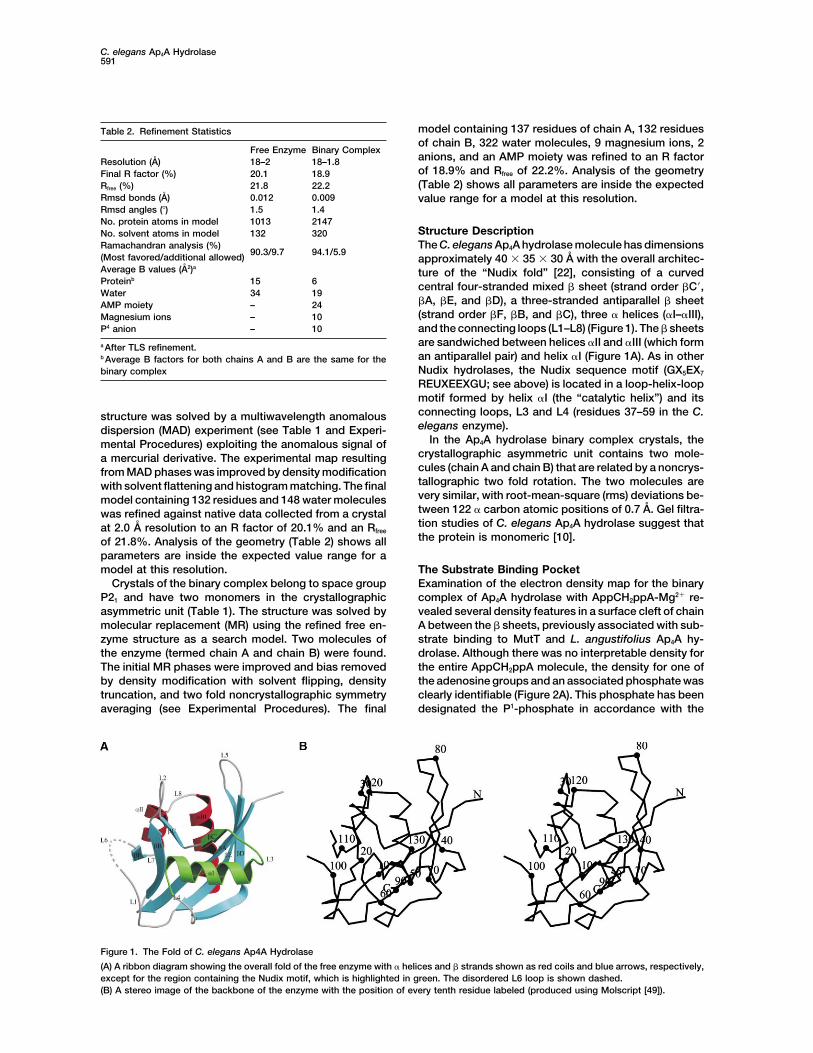
Structure DescriptionNo. solvent atoms in model 132 320Ramachandran analysis (%)
90.3/9.7 94.1/5.9The C. elegans Ap4A hydrolase molecule has dimensions
(Most favored/additional allowed) approximately 40 35 30 A with the overall architec-Average B values (A2)a
ture of the “Nudix fold” [22], consisting of a curvedProteinb 15 6 central four-stranded mixed � sheet (strand order �C�,Water 34 19
�A, �E, and �D), a three-stranded antiparallel � sheetAMP moiety – 24(strand order �F, �B, and �C), three � helices (�I–�III),Magnesium ions – 10
P4 anion – 10 and the connecting loops (L1–L8) (Figure 1). The � sheetsare sandwiched between helices �II and �III (which forma After TLS refinement.an antiparallel pair) and helix �I (Figure 1A). As in otherb Average B factors for both chains A and B are the same for theNudix hydrolases, the Nudix sequence motif (GX5EX7binary complexREUXEEXGU; see above) is located in a loop-helix-loopmotif formed by helix �I (the “catalytic helix”) and itsconnecting loops, L3 and L4 (residues 37–59 in the C.structure was solved by a multiwavelength anomalouselegans enzyme).dispersion (MAD) experiment (see Table 1 and Experi-
In the Ap4A hydrolase binary complex crystals, themental Procedures) exploiting the anomalous signal ofcrystallographic asymmetric unit contains two mole-a mercurial derivative. The experimental map resultingcules (chain A and chain B) that are related by a noncrys-from MAD phases was improved by density modificationtallographic two fold rotation. The two molecules arewith solvent flattening and histogram matching. The finalvery similar, with root-mean-square (rms) deviations be-model containing 132 residues and 148 water moleculestween 122 � carbon atomic positions of 0.7 A. Gel filtra-was refined against native data collected from a crystaltion studies of C. elegans Ap4A hydrolase suggest thatat 2.0 A resolution to an R factor of 20.1% and an Rfreethe protein is monomeric [10].of 21.8%. Analysis of the geometry (Table 2) shows all
parameters are inside the expected value range for amodel at this resolution. The Substrate Binding Pocket
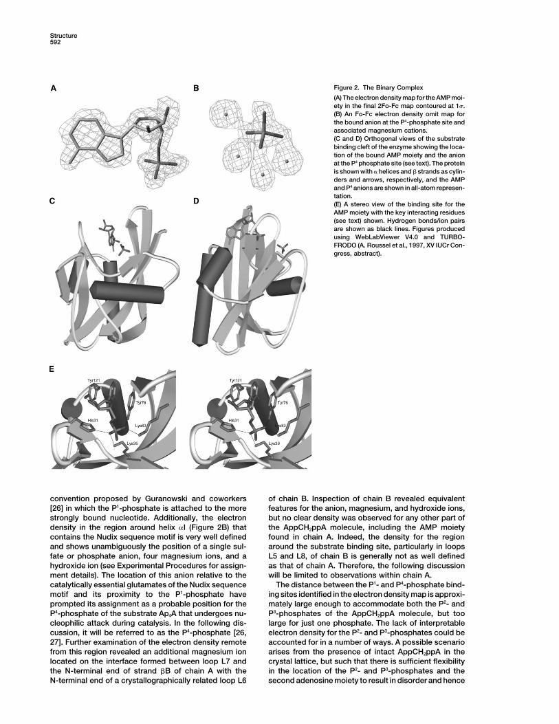
Examination of the electron density map for the binaryCrystals of the binary complex belong to space groupP21 and have two monomers in the crystallographic complex of Ap4A hydrolase with AppCH2ppA-Mg2� re-
vealed several density features in a surface cleft of chainasymmetric unit (Table 1). The structure was solved bymolecular replacement (MR) using the refined free en- A between the � sheets, previously associated with sub-
strate binding to MutT and L. angustifolius Ap4A hy-zyme structure as a search model. Two molecules ofthe enzyme (termed chain A and chain B) were found. drolase. Although there was no interpretable density for
the entire AppCH2ppA molecule, the density for one ofThe initial MR phases were improved and bias removedby density modification with solvent flipping, density the adenosine groups and an associated phosphate was
clearly identifiable (Figure 2A). This phosphate has beentruncation, and two fold noncrystallographic symmetryaveraging (see Experimental Procedures). The final designated the P1-phosphate in accordance with the
Figure 1. The Fold of C. elegans Ap4A Hydrolase
(A) A ribbon diagram showing the overall fold of the free enzyme with � helices and � strands shown as red coils and blue arrows, respectively,except for the region containing the Nudix motif, which is highlighted in green. The disordered L6 loop is shown dashed.(B) A stereo image of the backbone of the enzyme with the position of every tenth residue labeled (produced using Molscript [49]).
Structure592
Figure 2. The Binary Complex
(A) The electron density map for the AMP moi-ety in the final 2Fo-Fc map contoured at 1�.(B) An Fo-Fc electron density omit map forthe bound anion at the P4-phosphate site andassociated magnesium cations.(C and D) Orthogonal views of the substratebinding cleft of the enzyme showing the loca-tion of the bound AMP moiety and the anionat the P4 phosphate site (see text). The proteinis shown with � helices and � strands as cylin-ders and arrows, respectively, and the AMPand P4 anions are shown in all-atom represen-tation.(E) A stereo view of the binding site for theAMP moiety with the key interacting residues(see text) shown. Hydrogen bonds/ion pairsare shown as black lines. Figures producedusing WebLabViewer V4.0 and TURBO-FRODO (A. Roussel et al., 1997, XV IUCr Con-gress, abstract).
convention proposed by Guranowski and coworkers of chain B. Inspection of chain B revealed equivalentfeatures for the anion, magnesium, and hydroxide ions,[26] in which the P1-phosphate is attached to the more
strongly bound nucleotide. Additionally, the electron but no clear density was observed for any other part ofthe AppCH2ppA molecule, including the AMP moietydensity in the region around helix �I (Figure 2B) that
contains the Nudix sequence motif is very well defined found in chain A. Indeed, the density for the regionaround the substrate binding site, particularly in loopsand shows unambiguously the position of a single sul-
fate or phosphate anion, four magnesium ions, and a L5 and L8, of chain B is generally not as well definedas that of chain A. Therefore, the following discussionhydroxide ion (see Experimental Procedures for assign-
ment details). The location of this anion relative to the will be limited to observations within chain A.The distance between the P1- and P4-phosphate bind-catalytically essential glutamates of the Nudix sequence
motif and its proximity to the P1-phosphate have ing sites identified in the electron density map is approxi-mately large enough to accommodate both the P2- andprompted its assignment as a probable position for the
P4-phosphate of the substrate Ap4A that undergoes nu- P3-phosphates of the AppCH2ppA molecule, but toolarge for just one phosphate. The lack of interpretablecleophilic attack during catalysis. In the following dis-
cussion, it will be referred to as the P4-phosphate [26, electron density for the P2- and P3-phosphates could beaccounted for in a number of ways. A possible scenario27]. Further examination of the electron density remote
from this region revealed an additional magnesium ion arises from the presence of intact AppCH2ppA in thecrystal lattice, but such that there is sufficient flexibilitylocated on the interface formed between loop L7 and
the N-terminal end of strand �B of chain A with the in the location of the P2- and P3-phosphates and thesecond adenosine moiety to result in disorder and henceN-terminal end of a crystallographically related loop L6
C. elegans Ap4A Hydrolase593
no interpretable electron density. Given the well-defined of the P4-phosphate (Figure 3). The fourth magnesiumion lies out of the plane of the other three and 3.2 Aelectron density of the AMP moiety and the proposed P4-
phosphate, this seems unlikely. A more likely scenario is away from the nearest of them. Each magnesium ishexacoordinated, with ligands provided by the P4-phos-that the substrate analog has been turned over in the
active site, as it is not totally refractory to hydrolysis phate, the main chain carboxyl of Lys36, the side chaincarboxyl groups of Glu52, Glu56, and Glu103 (from theby enzymes of this type [28, 29]. In this scenario, the
observed AMP moiety is part of either the ATP product, Nudix sequence motif), a hydroxide ion, and nine othersolvent molecules presumed to be water (Figure 3).where its P2- and P3-phosphates are disordered in the
crystal lattice, or the AMP product itself, which has oc- Thus, the orientation of a bound P4-phosphate is quiteconstrained through its interactions with the magnesiumcupied the binding site after the ATP has diffused out.
The anion at the P4-phosphate site is then most likely ions. The necessity for precise binding of a P4-phos-phate by the enzyme in order to correctly orientate itsto be a phosphate or sulfate ion carried over from the
purification of the enzyme [25]. substrate for nucleophilic attack is consistent with theinability of Ap4A hydrolase to catalyze the hydrolysis ofAMP Moiety Binding Pocket
Analysis of the AMP moiety in the binary complex re- an Ap3A substrate [10, 32].Studies on the metal requirements of the plant-typeveals that the molecule sits in a groove on the protein
surface formed by the two � sheets orientated such that B. bacilliformis hydrolase [33] suggest a requirementfor only two catalytic magnesium ions. Therefore, thethe adenine moiety is directed toward helices �II and
�III and the phosphate toward the “catalytic helix” �I observation of four magnesium ions (and one hydroxideion) in the region of the “catalytic” helix in the C. elegans(Figures 2C and 2D). The nucleotide binds in a closed
conformation (Figure 2A), with the ribose sugar found enzyme is unexpected. Crystals of the binary complexonly grew in the presence of 200 mM MgCl2, far in excessin the C2�-endo C3�-exo conformer and the adenine ring
in the anti conformation. of the optimum 3–5 mM concentration for catalysis [10],and thus the additional ions may represent an artifactThe adenine ring binds in a pocket formed by the side
chains of Tyr76 located at the C-terminal end of strand of crystallization. It is not possible to predict with confi-dence which of the four magnesium ions might be bound�D and Tyr121 situated in loop L8. The two phenolic
rings of the tyrosines sandwich the adenine ring, forming in an authentic substrate complex in vivo. However, thestructure does support the role of residues Glu52, Glu56,extensive - stacking interactions, burying approxi-
mately 130 A2 (50%) of the solvent-accessible surface and Glu103 as metal coordinating ligands, and mutationof these residues to glutamines results in 103-, 105-,area of the adenine ring (Figure 2E). This double-stack-
ing arrangement has also been observed in several other and 40-fold reductions in Kcat, respectively (A.G.McL.,unpublished data).nucleotide binding proteins, including the unrelated
mammalian cap binding translation initiation factor [30] Second Nucleotide Binding SiteExamination of the region beyond the P4-phosphate re-and vaccinia virus mRNA-cap-dependent 2�-O-methyl-
transferase [31]. The N6 nitrogen of the adenine group veals, in the absence of significant conformationalchanges, no clear binding site for the second adenosineextends into a pocket formed by the side chains of
Thr33, Leu74, Val85, Tyr87, Ala119, and Met124, where moiety. Ap4A hydrolase will hydrolyze a range of Np4Xsubstrates [2, 32], and this is consistent with the lackit forms a direct hydrogen bond to the central of three
bound water molecules. The size of the pocket and the of a defined binding site for the X moiety. This wouldsuggest that the interactions of the protein with the Np4lack of base-specific interactions are consistent with
the ability of the enzyme to hydrolyze a variety of di- moiety of the substrate are sufficient to stabilize thesubstrate and orientate the P4-phosphate for nucleo-nucleoside polyphosphate substrates [32]. The 2� hy-
droxyl of the ribose moiety makes a hydrogen bond with philic attack, as previously described [27].P2-P3 Phosphate Binding Regionthe side chain hydroxyl group of Tyr121. In addition, the
ribose oxygen O4 makes a van der Waals contact with There was no interpretable electron density for eitherthe P2- or P3-phosphates. However, inspection of thethe side chain phenolic ring of Tyr76 (Figure 2E). Stabili-
zation of the P1-phosphate moiety on the enzyme is protein in this region did reveal that Lys83 is positionedsuch that it could stabilize the P2- as well as the P1-achieved via a series of hydrogen bond/salt bridges
between the phosphate oxygens and the side chain NZ phosphate, and residues His38, Lys79, and Tyr27 mightnitrogen of Lys36 and Lys83, the side chain hydroxyl participate in phosphate stabilization either by directgroup of Tyr76, and the side chain imidazole ring N�2 interaction or via metal coordination. This latter sugges-of His31 (Figure 2E). tion is prompted by studies of the metal requirementsP4-Phosphate Binding Site of the plant-type B. bacilliformis Ap4A hydrolase thatThe proposed P4-phosphate forms only one direct inter- have shown one further metal ion to bind the Ap4A sub-action with the protein via a hydrogen bond between strate in addition to the two catalytically required metalone of its oxygens and the main chain amide of His38. ions [33].However, it does contact several water molecules andmagnesium ions. In total, four magnesium ions were
Substrate Analog-Inducedfound in the catalytic site above helix �I (Figure 3). ThreeConformational Changesof the magnesium ions are positioned so as to form anSuperposition of the structures of the free and binaryisosceles triangle (inter-ion distances 3.1 and 3.5 A) atcomplex forms of Ap4A hydrolase shows that they havethe base of the P4-phosphate such that each one of
three magnesium ions is ligated by one of the oxygens a very similar overall conformation. The value for the
Structure594
Figure 3. The Binding Site for the P4-Phos-phate
(A) A stereo view of the P4-phosphate bind-ing site above the “catalytic helix” �I showingthe bound anion depicted as a phosphate instick representation, associated Mg2� ca-tions (green spheres), hydroxide ion (purplesphere), water molecules (red spheres), andcoordinating protein side chain ligand net-work (see text). The protein backbone isshown in dark green.(B) Detailed view of the anion coordinationscheme showing the novel magnesium clus-ter with atoms colored as in (A) and ligandbonds shown as black lines with distancesin angstroms marked. (Produced using Web-LabViewer v4.0.)
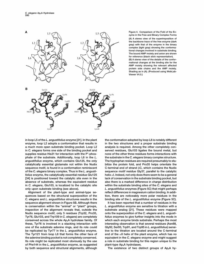
rmsd of 101 out of 138 � carbon positions (residues though the trigger for the conformational change hasyet to be determined. The side chain of His31 rotates2–24, 32–75, 82–94, and 106–136) is 0.4 A. The largest
changes in main chain positions occur in three loop approximately 90 about its �1 dihedral angle. This reori-entation results in the side chain imidazole ring N�2regions (Figure 4A). First, the loops L2 (residues 25–31)
and L5 (residues 74–82) that flank the adenosine binding atom coming into hydrogen bonding distance of the O1oxygen of the P1-phosphate. Examination of the resi-site undergo small conformational changes in order to
accommodate substrate binding. Second, the disor- dues of the Nudix sequence motif reveals that, withthe exception of Glu56, none of the residues shows adered loop, L6 (residues 93–106), which had no interpret-
able density in the free enzyme structure, becomes significant shift. Glu56, however, does shift the positionof its side chain carboxyl group to optimize its coordina-ordered upon substrate and metal binding, with well-
defined density unambiguously outlining its path. The tion geometry to two of the four magnesium ions locatedin the catalytic site. The only other significant shifts inordered L6 loop is stabilized via the coordination of
the O�1 and O�2 atoms of Glu103 to two of the four side chain positions occur in regions where the crystalcontacts differ between the two crystal forms.magnesium ions located in the catalytic site. Closer ex-
amination of the side chain positions within the sub-strate binding site reveals several significant alterations Comparison with L. angustifolius Ap4A Hydrolase
Superposition of the L. angustifolius plant-type Ap4A(Figure 4B). A rotation of approximately 90 about the�1 dihedral angle of Tyr121 occurs with an associated hydrolase with the structures of both the free and sub-
strate analog bound C. elegans animal-type Ap4A hy-shift in the side chain of Tyr76 such that the planes ofthe two phenolic rings lie parallel and at a distance of drolase both gave rmsds of 1.6 A over 91 matched �
carbon positions (Figure 5A). Analysis of this superposi-approximately 7 A and results in the formation of thebinding pocket for an adenine ring of the substrate. tion reveals the two folds are closely equivalent. The
major differences in the structures are a 7 residue inser-Movement of the Tyr121 side chain is therefore essentialfor localization of an adenine ring on the enzyme, al- tion in loop L6 and an extra, ill-defined helix (helix �II)
C. elegans Ap4A Hydrolase595
Figure 4. Comparison of the Fold of the En-zyme in the Free and Binary Complex Forms
(A) A stereo view of the superimposition ofthe backbone trace of the free enzyme (darkgray) with that of the enzyme in the binarycomplex (light gray) showing the conforma-tional changes involved in substrate binding.The bound AMP moiety and anion are shownfor reference (black stick representation).(B) A stereo view of the details of the confor-mational changes at the binding site for theAMP moiety showing the relevant affectedprotein side chains and the AMP moiety.Shading as in (A). (Produced using WebLab-Viewer V4.0.)
in loop L5 of the L. angustifolius enzyme [21]. In the plant the conformation adopted by loop L8 is notably differentin the two structures and a proper substrate bindingenzyme, loop L2 adopts a conformation that results in
a much more open substrate binding pocket. Loop L2 analysis is required. Among the other completely con-served residues, Glu103 ligates the bound metal, butin C. elegans forms one side of the binding pocket and
supplies residue His31 for interaction with the P1-phos- none of the other three residues forms interactions withthe substrate in the C. elegans binary complex structure.phate of the substrate. Additionally, loop L6 in the L.
angustifolius enzyme, which contains Glu125, the only The tryptophan residues are required presumably to sta-bilize the protein fold, and Pro35 helps orientate thecatalytically essential glutamate not within the Nudix
sequence motif, is found in a conformation reminiscent C-terminal end of strand �C, which contains the Nudixsequence motif residue Gly37, parallel to the catalyticof the C. elegans binary complex. Thus in the L. angusti-
folius enzyme, the catalytically essential residue Glu125 helix �I. Indeed, not only does there seem to be a generallack of conservation in the substrate binding pocket, but[34] is positioned toward the catalytic site even in the
absence of substrate, whereas the equivalent residue also there is a marked difference in charge distributionwithin the substrate binding sites of the C. elegans andin C. elegans, Glu103, is localized to the catalytic site
only upon substrate binding (see above). L. angustifolius enzymes (Figure 5C) that might perhapsreflect differences in magnesium cation binding. In addi-Alignment of the plant-type and animal-type se-
quences based on the structural superposition of the tion, there are noticeably more polar residues in thebinding site of the L. angustifolius enzyme (Figure 5C).C. elegans and L. angustifolius structures results in the
sequence alignment shown in Figure 5B. Although there It has been reported that a number of residues in theL. angustifolius enzyme are sensitive to titration with ais conservation within the “animal” or “plant” groups,
inspection of the alignment reveals that, outside the substrate analog [21]. These residues were mappedonto the superposition of the C. elegans and L. angusti-Nudix sequence motif, only 5 residues (Trp32, Pro35,
Tyr76, Glu103, and Trp109 in C. elegans) are completely folius enzymes to give further insights into the mode inwhich each enzyme binds substrate. Perhaps the mostconserved across the whole Ap4A hydrolase family. Of
these, Tyr76 is involved in a stacking interaction with interesting observation is that several residues (Asn88,Gly92, Ser93, Trp91, and Trp95 in L. angustifolius) sensi-one of the substrate adenine rings, and its role could
be replicated by Tyr77 in the L. angustifolius enzyme. tive to the titration are located around the C-terminalend of the �II helix of the plant enzyme, which has noThe Tyr121 from loop L8 that forms the other face of
the adenine binding pocket is not completely conserved. equivalent in the C. elegans enzyme and might suggesta role in substrate binding for this region unique to theIts role might be replicated most obviously by the use
of Phe144 in the L. angustifolius enzyme, as suggested plant-type Ap4A hydrolases.The existence of two distinct groups of Ap4A hy-by both sequence and structural alignments, although
Structure596
Figure 5. A Comparison between the C. elegans and L. angustifolius Ap4A Hydrolases and Other Family Members
(A) A stereo view of the superimposition of the backbone trace of the binary complex form of the C. elegans enzyme (blue) with that of theL. angustifolius enzyme (green).(B) A structure-based sequence alignment of the members of the two groups, animal-type and plant-type, of Ap4A hydrolase. Residues havebeen highlighted on the basis of the Nudix consensus sequence (reverse type face), conservation across all species outside the Nudix motif(red box), and conservation among animal-type (blue box) or plant-type (green box) sequences.(C) Surface representation of the C. elegans and L. angustifolius Ap4A hydrolases showing the difference in shape and distribution of chargewithin the substrate binding site. Positively charged (including His), negatively charged, and polar residues are colored blue, red, and green,respectively, while the rest are gray. The conserved residues within each group (animal-type or plant-type) as well as the Nudix motif residues(underlined) are labeled. (Figures produced using WebLabViewer V4.0.)
C. elegans Ap4A Hydrolase597
drolases raises the question of their evolution. Whether candidate for the catalytic base. Work on the L. angusti-folius Ap4A hydrolase has shown an inversion of stereo-the lack of conservation in the substrate binding pocket
and elsewhere in the sequences outside the Nudix motif chemistry upon hydrolysis [36], consistent with an exopattern of attack by the activated water [26]. However,is a reflection of divergence of an ancestral Ap4A hy-
drolase or the convergence of separate Nudix hy- in the absence of an intact substrate or analog and thepresence of the unexpectedly large number of bounddrolases toward the same substrate, Ap4A, remains un-
clear. magnesium ions, it is not possible to predict with confi-dence which, if any, of the bound solvent moleculesmight act as the attacking nucleophile.Comparison with Other Nudix Hydrolases
There are structures available for two other membersBiological Implicationsof the Nudix hydrolase family, the NMR structure of the
E. coli nucleoside triphosphate pyrophosphohydrolase,The enzyme Ap4A hydrolase catalyzes the breakdownMutT, with and without a nonhydrolysable substrate an-of Ap4A, a ubiquitous molecule produced mainly as aalog [20, 23], and the crystal structure of E. coli orf209by-product of protein synthesis. The asymmetric cleav-ADP-ribose pyrophosphatase (ADPRase), with and with-age of Ap4A carried out by the C. elegans enzyme stud-out ADP-ribose [22]. Both of these enzymes catalyzeied here results in the production of ATP and AMP, whichthe hydrolysis of the P1-P2 linkage of their respectivecan be recycled into the cellular stored energy pool.substrates. (In this discussion, the �, �, and � phos-Thus, the enzyme may have a simple housekeepingphates of the substrates described in the literature forfunction, but the Ap4A molecule has been implicated inMutT and ADPRase have been redefined as P1, P2, andplaying a role in one or more essential cellular pro-P3 for consistency with the Ap4A hydrolase description.)cesses, including DNA replication, metabolic stress, andThey both have a very similar overall fold to the C. ele-apoptosis, and thus the hydrolase may have a moregans Ap4A hydrolase, although the dimeric nature ofimportant function in the careful regulation of the intra-ADPRase results in a binding pocket that contains resi-cellular levels of Ap4A. In particular, the ratio of Ap4A todues from both subunits and a noticeably different orien-Ap3A has been linked to the choice between differentia-tation for the substrate.tion and apoptosis in cultured cell lines. The C. elegansIn the structures of the C. elegans Ap4A hydrolase andAp4A hydrolase belongs to the Nudix family and is theMutT, the bound adenosine moiety of their substratefirst monomeric member to have its crystal structureanalogs lies against the equivalent � strands (�A, �D,determined. The structure of the enzyme both free andand �E in Ap4A hydrolase and �A, �C, and �D in MutT),in a binary complex shows how a conformational changealthough in MutT it is exposed to solvent on the otheraccompanies substrate binding and how one nucleosideface, whereas in Ap4A hydrolase the adenine ring isand the P1- and P4-phosphates are bound. The confor-buried in a pocket. The relative positions of the P2-phos-mational change brings key conserved residues intophate in MutT and putative P4-phosphate of Ap4A hy-position to enable catalysis to occur. The structure re-drolase are very similar, lying close to the catalytic helixveals how the enzyme can discriminate between di-�I and coordinated by magnesium ions bound by resi-nucleoside tetraphosphate and triphosphate substratesdues from the Nudix sequence motif. In contrast, theand hence can help specifically in maintaining the Ap4A/bound ADP-ribose substrate in the ADPRase complexAp3A ratio. Furthermore, the lack of any direct, base-resembles a “horseshoe” [22] and is thus orientatedspecific contacts to the bound nucleoside and the ab-such that the direction of its P1-P2 phosphate linkage issence of an obvious second nucleoside binding siteopposite to that of the substrate analog in the MutToffer an explanation for the ability of the enzyme to actcomplex. Nonetheless, the diphosphate links that un-upon a range of nucleoside tetraphosphate-containingdergo hydrolysis in all three enzymes are positionedsubstrates. Structure and sequence comparisons showvery similarly in space relative to the catalytic helix �Ihow the C. elegans enzyme and other eukaryotic andand the residues of the Nudix sequence motif. Thus, thearchaeal Ap4A hydrolases form a subclass with struc-basic structural architecture provided by the commontural characteristics distinct from those of the plant andmixed �/� fold of these enzymes has been modified soproteobacterial enzymes.as to present the appropriate diphosphate bonds in the
correct positions to the catalytic machinery of the NudixExperimental Proceduresresidues for hydrolysis.
Protein CrystallizationC. elegans Ap4A hydrolase was expressed and purified as describedCatalytic Mechanism of C. eleganselsewhere [25]. Crystals were grown in the presence and absenceAp4A Hydrolaseof AppCH2ppA-Mg2� by the hanging drop method of vapor diffusion.
The proposed mechanism of hydrolysis of the diphos- The free enzyme crystals belong to space group C2221 with cellphate link catalyzed by Ap4A hydrolase requires the acti- dimensions a � 62.8 A, b � 72.9 A, and c � 61.1 A and were grown
with well solutions containing 32% (w/v) PEG4000, 0.2 M ammoniumvation of a bound solvent molecule by a base to facilitateacetate and 100 mM sodium citrate (pH 5.6). VM calculations sug-its nucleophilic attack on the P4-phosphate of the sub-gested a solvent content of 43% for a monomer in the asymmetricstrate [26, 27]. Examination of the region around theunit. Heavy-atom derivatives of these crystals were prepared byputative P4-phosphate in the binary complex, the muta-soaking the crystals in 2 mM ethyl mercury phosphate (EMP) for 2
tion data on the C. elegans and L. angustifolius Ap4A hr. The Ap4A hydrolase binary complex was produced by additionhydrolases, and data on E. coli MutT (A.G.McL., unpub- of 10 mM AppCH2ppA and 10 mM magnesium chloride to the C.
elegans enzyme. The crystals were grown by equilibration againstlished data) [28, 35] suggests Glu56 as the most likely
Structure598
well solutions of 30% (w/v) PEG4000, 0.2 M magnesium chloride, structure of the free enzyme as a search model. The two subunitsin the asymmetric unit were successfully located from a solutionand Tris-HCl (pH 8.5) and belong to space group P21 with cell dimen-
sions a � 57.6 A, b � 36.8 A, and c � 68.9 A with a � � 114.2. VM with a correlation of 58.5 and an R factor of 43.7%. Analysis of thisMR solution in TURBO-FRODO revealed consistent crystal packing.calculations suggested a solvent content of 40% for two monomers
in the asymmetric unit. For data collection, both crystal forms were A 3Fo-2Fc electron density map was calculated on the MR solutionusing data from 20–2 A resolution that indicated a correct solutioncooled straight from the drop in a stream of nitrogen gas at 100 K.had been found but showed poor electron density around severalloop regions. Therefore, the calculated phases were subjected toData Collection and Processingdensity modification within the program CNS [48], which includedA full three-wavelength MAD data experiment was performed on asolvent flipping, density truncation, and two fold NCS averaging. Thesingle EMP-soaked free enzyme crystal, and data were collectedresulting density-modified map showed significant improvements,to a minimum Bragg spacing of 2.0 A using a Mar Research CCDwith loop regions now clearly defined. The model was rebuilt to fitscanner at the ESRF station BM14. Three wavelengths were chosenthe observed density and then submitted to restrained maximum-near the Hg absorption edge (Table 1) based on an X-ray fluores-likelihood refinement using REFMAC. Iterative cycles of model build-cence scan of the frozen crystal in order to maximize the f�� compo-ing and refinement were used to construct a model representing 137nent (�1, peak), to minimize the f� component (�2, inflexion), andand 132 of the 138 expected residues (with 93% of these residuesto maximize �f� (�3, remote). The data for each wavelength wereassigned unambiguously) in chain A and chain B, respectively. Atprocessed individually and scaled in such a way as to preservethis point, density within the expected substrate binding sites wasanomalous signal using the HKL Suite of programs [37]. Native dataaccounted for in chains A and B by the addition of a phosphate ionfor the free enzyme were collected from a single frozen crystal on(a sulfate ion can also be equally well refined at this position), fouran ADSC Quantum 4 CCD camera at the Daresbury SRS station 9.6magnesium ions (assigned on the basis of their hexacoordinationto 2.0 A resolution. Details of the data collection statistics are givenwith excellent octahedral geometry and short ligand bonds of aver-in Table 1.age length 2.1 A), and a hydroxide ion (assigned as the most appro-Data for the binary complex were collected from a single frozenpriate species present able to coordinate three magnesium ionscrystal both on a Mar345 image plate scanner mounted on a Rigakusimultaneously with bond lengths of approximately 2.1 A) as wellRU200 X-ray generator with Yale focusing mirror optics to 2.0 Aas the addition of an AMP molecule to the binding-site of chainresolution and subsequently on an ADSC Quantum 4 CCD cameraA. The model was then further refined, and a total of 320 solventat the Daresbury SRS station 9.6 to 1.8 A resolution. Data for eachmolecules were added by cycles of REFMAC and ARP/wARP. Atdataset were processed using the HKL Suite of programs. Detailsthis time, the higher resolution data became available (1.8 A), andof the data collection statistics are given in Table 1.subsequent refinement was done using this data. In the final stages,one additional magnesium ion was added remote from either sub-Phasing, Model Building, and Structure Refinementstrate binding site, and TLS parameterisation was used to accountof the Free Enzyme Structurefor the overall anisotropic motion of each protein chain in the asym-The positions of four mercury atoms were found in the asymmetricmetric unit [45]. Tight control was maintained of the geometric pa-unit of the EMP-soaked free enzyme data using the direct methodsrameters of the model including the main chain torsion angles, asprocedure, as implemented in the program SnB [38]. Data for theassessed by Ramachandran plot analysis using the program PRO-peak wavelength were scaled and processed with the DREAR pro-CHECK [46]. No restraints were used for the magnesium-ligandgram package [39] to derive the renormalized E values correspond-distances. The refined temperature factors of the bound ligandsing to the anomalous differences. These E values were then submit-were similar to those of the surrounding protein molecule prior toted to SnB using the program’s standard parameters. Analysis of theTLS parameterization, and thus all the ligand molecules are consid-minimum function values showed a bimodal distribution indicatingered as being fully occupied in both chains A and B. There was noconvergence to a solution.interpretable electron density for residues Met1 of chain A and theThe mercury atom parameters were then refined, and initialfirst three N-terminal residues (Met1, Val2, and Val3), Thr9, Gly80,phases were calculated in the program MLPHARE [40] following theand Lys81 of chain B.pseudo-MIR procedure [41]. The phases were further improved by
solvent flattening and histogram matching using structure factorAcknowledgmentsamplitudes from the �3 (remote) dataset with the program DM [42].
The resulting 2.0 A map was of excellent quality, with readily identifi-We are grateful for the support of Hassan Belrhali and the stationable side chain density, and therefore these modified phases werescientists on beamline BM14 at the ESRF and station 9.6 at theused in the automated model building procedure implemented byCLRC Daresbury SRS laboratory and for travel funds provided bythe program ARP/wARP [43]. This procedure produced a polyalan-EMBL under the EC Human Capital and Mobility Program. H.M.A.ine chain trace corresponding to 75% of the main chain, and thisis the recipient of a postgraduate scholarship from the Egyptianinitial model then formed the basis of a manual build in the programgovernment. Work in the laboratories of J.B.R. and A.G.McL. isTURBO-FRODO (A. Roussel et al., 1997, XV IUCr Congress, ab-supported by the BBSRC and The Wellcome Trust. J.B.R. is a Royalstract). The resulting model was subjected to restrained maximum-Society Olga Kennard Fellow.likelihood refinement at a resolution of 2.0 A, in the program
REFMAC [44], against data collected from a native crystal. Iterativecycles of phase combination of the partial structure phases and Received: October 24, 2001
Revised: January 9, 2002those from the MAD experiment, model building, and refinement,which in the later stages was performed using TLS parameterisation Accepted: January 23, 2002to account for the overall anisotropic motion of the protein [45],were used to construct a model representing 132 residues of the References138 expected residues, with 92% of these residues assigned unam-biguously. ARP/wARP also added a total of 109 solvent molecules 1. McLennan, A.G. (1999). The MutT motif family of nucleotideautomatically during the refinement process, and tight control was phosphohydrolases in man and human pathogens (review). Int.maintained over the geometric parameters of the model, including J. Mol. Med. 4, 79–89.the main chain torsion angles as assessed by Ramachandran plot 2. Guranowski, A. (2000). Specific and nonspecific enzymes in-analysis using the program PROCHECK [46]. There was no interpret- volved in the catabolism of mononucleoside and dinucleosideable electron density for residues Ser26, Glu104, Gln105, and the polyphosphates. Pharmacol. Ther. 87, 117–139.final two C-terminal residues, Gly137 and Phe138. 3. Cartwright, J.L., Britton, P., Minnick, M.F., and McLennan, A.G.
(1999). The IalA invasion gene of Bartonella bacilliformis en-codes a (di)nucleotide polyphosphate hydrolase of the MutTMolecular Replacement of the Binary Complex
The structure of the binary complex was solved by molecular re- motif family and has homologs in other invasion bacteria. Bio-chem. Biophys. Res. Commun. 256, 474–479.placement (MR) with the program AMoRe [47], using the refined
C. elegans Ap4A Hydrolase599
4. Conyers, G.B., and Bessman, M.J. (1999). The gene, ialA, asso- AMPCPP-M2� complex and mechanism of its pyrophosphohy-drolase action. Biochemistry 36, 1199–1210.ciated with the invasion of human erythrocytes by Bartonella
bacilliformis, designates a nudix hydrolase activity on dinucleo- 24. Blackburn, G.M., Guo, M.-J., and McLennan, A.G. (1992). Syn-thetic structural analogues of dinucleoside polyphosphates. Inside 5�-polyphosphates. J. Biol. Chem. 274, 1203–2354.
5. Guranowski, A., Jakubowski, H., and Holler, E. (1983). Catabo- Ap4A and Other Dinucleoside Polyphosphates, A.G. McLennan,ed. (Boca Raton, Florida: CRC Press), pp. 305–342.lism of diadenosine 5�,5���-P1,P4-tetraphosphate in prokaryotes.
Purification and properties of diadenosine 5�,5���-P1,P4-tetra- 25. Bailey, S., Sedelnikova, S.E., Blackburn, G.M., Abdelghany,H.M., McLennan, A.G., and Rafferty, J.B. (2002). Crystallizationphosphate (symmetrical) pyrophosphohydrolase from Esche-
richia coli K12. J. Biol. Chem. 258, 14784–14789. of a complex of Caenorhabditis elegans diadenosine tetraphos-phate hydrolase and a non-hydrolysable substrate-analogue,6. Plateau, P., Fromant, M., Brevet, A., Gesquiere, A., and Blan-
quet, S. (1985). Catabolism of bis(5�-nucleosidyl) oligophos- AppCH2ppA. Acta Crystallogr. D58, 526–528.26. Guranowski, A., Brown, P., Ashton, P.A., and Blackburn, G.M.phates in Escherichia coli: metal requirements and substrate
specificity of homogeneous diadenosine 5�,5���-P1,P4-tetra- (1994). Regiospecificity of the hydrolysis of diadenosine poly-phosphates catalyzed by three specific pyrophosphohydro-phosphate pyrophosphohydrolase. Biochemistry 24, 914–922.
7. Bessman, M.J., Frick, D.N., and O’Handley, S.F. (1996). The lases. Biochemistry 33, 235–240.27. McLennan, A.G., Prescott, M., and Evershed, R.P. (1989). Identi-MutT proteins or ‘nudix’ hydrolases, a family of versatile, widely
distributed, “housecleaning” enzymes. J. Biol. Chem. 271, fication of point of specific enzymic cleavage of P1,P4-bis(5�-adenosyl) tetraphosphate by negative ion FAB mass spectrom-25059–25062.
8. Koonin, E.V. (1993). A highly conserved sequence motif defining etry. Biomed. Environ. Mass Spectrom. 18, 450–452.28. Guranowski, A., Starzynska, E., Taylor, G.E., and Blackburn,the family of MutT-related proteins from eubacteria, eukaryotes
and viruses. Nucleic Acids Res. 21, 4847. G.M. (1989). Studies on some specific Ap4A-degrading enzymeswith the use of various methylene analogues of P1,P4-bis-(5�,5���-9. Maksel, D., Guranowski, A., Ilgoutz, S.C., Moir, A., Blackburn,
M.G., and Gayler, K.G. (1998). Cloning and expression of diade- adenosyl) tetraphosphate. Biochem. J. 262, 241–244.29. McLennan, A.G., Taylor, G.E., Prescott, M., and Blackburn, G.M.nosine 5�,5���-P1,P4-tetraphosphate hydrolase from Lupinus an-
gustifolius L. Biochem. J. 329, 313–319. (1989). Recognition of ���-substituted and ��, ����-disubsti-tuted phosphonate analogues of bis(5�-adenosyl) tetraphos-10. Abdelghany, H.D., Gasmi, L., Cartwright, J.L., Bailey, S., Raf-
ferty, J.B., and McLennan, A.G. (2001). Cloning, characterization phate by the bis(5�-nucleosidyl)-tetraphosphate pyrophospho-hydrolases from Artemia embryos and Escherichia coli.and crystallization of a diadenosine 5�-5���-P1,P4-tetraphos-
phate pyrophosphohydrolase from Caenorhabditis elegans. Biochemistry 28, 3868–3875.30. Marcotrigiano, J., Gingras, A.C., Sonnenberg, N., and Burley,Biochim. Biophys. Acta, in press.
11. Goerlich, O., Foeckler, R., and Holler, E. (1982). Mechanism of S.K. (1997). Cocrystal structure of the messenger RNA 5� cap-binding protein (eIF4E) bound to 7-methyl-GDP. Cell 89, 951–synthesis of adenosine(5�)tetraphospho(5�)adenosine (AppppA)
by aminoacyl-tRNA synthetases. Eur. J. Biochem. 126, 135–142. 961.31. Hodel, A.E., Gershon, P.D., and Quiocho, F.A. (1998). Structural12. Baxi, M.D., and Vishwanatha, J.K. (1995). Diadenosine poly-
phosphates: their biological and pharmacological significance. basis for sequence nonspecific recognition of 5�-capped mRNAby a cap modifying enzyme. Mol. Cell 1, 443–447.J. Pharmacol. Toxicol. Methods 33, 121–128.
13. Kisselev, L.L., Justesen, J., Wolfson, A.D., and Frolova, L.Y. 32. Guranowski, A., and Sillero, A. (1992). Enzymes cleaving di-nucleoside polyphosphates. In Ap4A and Other Dinucleoside(1998). Diadenosine oligophosphates (ApnA), a novel class of
or foe? Pharmacol. Ther. 87, 73–89. 33. Conyers, G.B., Wu, G., Bessman, M.J., and Mildvan, A.S. (2000).Metal requirements of a diadenosine pyrophosphatase from15. Vartanian, A., Prudovsky, I., Suzuki, H., DalPra, I., and Kisselev,
L. (1997). Opposite effects of cell differentiation and apoptosis Bartonella bacilliformis: magnetic resonance and kinetic studiesof the role of Mn2�. Biochemistry 39, 2347–2354.on Ap3A/Ap4A ratio in human cell cultures. FEBS Lett. 415,
160–162. 34. Maksel, D., Gooley, P.R., Swarbrick, J.D., Guranowski, A.,Gange, C., Blackburn, G.M., and Gayler, K.R. (2001). Character-16. Vartanian, A., Alexandrov, I., Prudowski, I., McLennan, A., and
Kisselev, L. (1999). Ap4A induces apoptosis in cultured human ization of active-site residues in diadenosine tetraphosphatehydrolase from Lupinus angustifolius. Biochem. J. 357, 399–405.cells. FEBS Lett. 456, 175–180.
17. Murphy, G.A., Halliday, D., and McLennan, A.G. (2000). The Fhit 35. Lin, J., Abeygunawardana, C., Frick, D.N., Bessman, M.J., andMildvan, A.S. (1996). The role of Glu57 in the mechanism of thetumor suppressor protein regulates the intracellular concentra-
tion of diadenosine triphosphate but not diadenosine tetraphos- Escherichia coli MutT enzyme by mutagenesis and hetero-nuclear NMR. Biochemistry 35, 6715–6726.phate. Cancer Res. 60, 2342–2344.
18. Mitchell, S.J., and Minnick, M.F. (1995). Characterization of a 36. Dixon, R.M., and Lowe, G. (1989). Synthesis of (Rp,Rp)-P1,P4-Bis(5�-adenosyl)-1[17O,18O2]tetraphosphate from (Sp,Sp)-P1,P4-two-gene locus from Bartonella bacilliformis associated with
the ability to invade human erythrocytes. Infect. Immun. 63, Bis(5�-adenosyl)-1[thio-18O2], 4[thio-18O2]tetraphosphate withretention at phosphorus and the stereochemical course of1552–1562.
19. Anderson, B.E., and Neumann, M.A. (1997). Bartonella spp. as hydrolysis by the unsymmetrical Ap4A phosphodiesterase fromlupin seeds. J. Biol. Chem. 264, 2069–2074.emerging human pathogens. Clin. Microbiol. Rev. 10, 203–219.
20. Abeygunawardana, C., Weber, D.J., Gittis, A.G., Frick, D.N., Lin, 37. Otwinowski, Z., and Minor, W. (1997). Processing of X-ray Dif-fraction Data Collected in Oscillation Mode (San Diego: Aca-J., Miller, A.F., Bessman, M.J., and Mildvan, A.S. (1995). Solution
structure of the MutT enzyme, a nucleoside triphosphate pyro- demic Press).38. Weeks, C.M., DeTitta, G.T., Hauptman, H.A., Thuman, P., andphosphohydrolase. Biochemistry 34, 14997–15005.
21. Swarbrick, J.D., Bashtannyk, T., Maksel, D., Zhang, X., Black- Miller, R. (1994). Structure solution by minimal-function phaserefinement and Fourier filtering. II. Implementation and applica-burn, G.M., Gayler, K.R., and Gooley, P.R. (2000). The three-
dimensional structure of the Nudix enzyme diadenosine tetra- tions. Acta Crystallogr. A 50, 210–220.39. Blessing, R.H., Guo, D.Y., and Langs, D.A. (1998). Intensity sta-phosphate hydrolase from Lupinus angustifolius L. J. Mol. Biol.
302, 1165–1177. tistics and normalization. In Direct Methods for Solving Macro-molecular Structures, NATO ASI Series Volume, Series C: Math-22. Gabelli, S.B., Bianchet, M.A., Bessman, M.J., and Amzel, L.M.
(2001). The structure of ADP-ribose pyrophosphate reveals the ematica and Physical Sciences, Volume 507, S. Fortier, ed.(Dordrecht, The Netherlands: Kluwer Academic Publishers), pp.structural basis for the versatility of the Nudix family. Nat. Struct.
Biol. 8, 467–472. 47–71.40. Otwinowski, Z. (1991). Maximum likelihood refinement of heavy23. Lin, J., Chitrananda, J.L., Frick, D.N., Bessman, M.J., and Mild-
van, A.S. (1997). Solution structure of the quaternary MutT-M2�- atom parameters. In Proceedings of the CCP4 Study Weekend,
Structure600
W. Wolf, P.R. Evans, and A.G.W. Leslie, eds. (Warrington, UK:SERC Daresbury Laboratory), pp. 80–88.
41. Ramakrishnan, V., and Biou, V. (1997). Treatment of multiwave-length anomalous diffraction data as a special case of multipleisomorphous replacement. In Methods in Enzymology: Macro-molecular Crystallography, Volume 276, C.W. Carter, and R.M.Sweet, eds. (San Diego: Academic Press), pp. 538–557.
42. Cowtan, K. (1994). “DM”: an automated procedure for phaseimprovement by density modification. Joint CCP4 and ESF-EACBM Newsletter on Protein Crystallography 31, pp. 34–38.
43. Lamzin, V.S., and Wilson, K.S. (1993). Automated refinement ofprotein models. Acta Crystallogr. D 49, 129–149.
44. Murshudov, G.N., Vagin, A.A., and Dodson, E.J. (1997). Refine-ment of macromolecular structures by the maximum-likelihoodmethod. Acta Crystallogr. D 53, 240–255.
45. Winn, M.D., Isupov, M., and Murshudov, G.N. (2001). Use ofTLS parameters to model anisotropic displacement in macro-molecular refinement. Acta Crystallogr. D 57, 122–133.
46. Laskowski, R.A., Macarthur, M.W., Moss, D.W., and Thornton,J.M. (1993). PROCHECK: a program to check the stereochemi-cal quality of protein structures. J. Appl. Crystallogr. 26, 283–291.
47. Navazza, J. (1994). AmoRe: an automated package for molecu-lar replacement. Acta Crystallogr. A 47, 392–400.
48. Brunger, A.T., and Warren, G.L. (1998). Crystallography andNMR system (CNS): a new software system for macromolecularstructure determination. Acta Crystallogr D 54, 905–921.
49. Kraulis, P. (1991). MOLSCRIPT: a program to produce bothdetailed and schematic plots of protein structures. J. Appl. Crys-tallogr. 24, 946–950.
Accession Numbers
Coordinates of the free enzyme and binary complex have beendeposited in the RCSB with accession codes 1KT9 and 1KTG.