This article was downloaded by: [University Of South Australia Library] On: 26 August 2012, At: 01:30 Publisher: Taylor & Francis Informa Ltd Registered in England and Wales Registered Number: 1072954 Registered office: Mortimer House, 37-41 Mortimer Street, London W1T 3JH, UK Diatom Research Publication details, including instructions for authors and subscription information: http://www.tandfonline.com/loi/tdia20 THE CURRENT STATUS OF SOME VERY SMALL FRESHWATER DIATOMS OF THE GENERA STEPHANODISCUS AND CYCLOSTEPHANOS Hannelore Håkansson a & Hedy Kling b a Department of Quaternary Geology, University of Lund, Tornavägen 13, S-223 63, Lund, Sweden b Department of Fisheries and Oceans, Freshwater Institute, Winnipeg, MB, R3T 2N6, Canada Version of record first published: 31 Oct 2011 To cite this article: Hannelore Håkansson & Hedy Kling (1990): THE CURRENT STATUS OF SOME VERY SMALL FRESHWATER DIATOMS OF THE GENERA STEPHANODISCUS AND CYCLOSTEPHANOS , Diatom Research, 5:2, 273-287 To link to this article: http://dx.doi.org/10.1080/0269249X.1990.9705119 PLEASE SCROLL DOWN FOR ARTICLE Full terms and conditions of use: http://www.tandfonline.com/page/terms-and-conditions This article may be used for research, teaching, and private study purposes. Any substantial or systematic reproduction, redistribution, reselling, loan, sub-licensing, systematic supply, or distribution in any form to anyone is expressly forbidden. The publisher does not give any warranty express or implied or make any representation that the contents will be complete or accurate or up to date. The accuracy of any instructions, formulae, and drug doses should be independently verified with primary sources. The publisher shall not be liable for any loss, actions, claims, proceedings, demand, or costs or damages whatsoever or howsoever caused arising directly or indirectly in connection with or arising out of the use of this material.
Transcript
This article was downloaded by: [University Of South Australia Library]On: 26 August 2012, At: 01:30Publisher: Taylor & FrancisInforma Ltd Registered in England and Wales Registered Number: 1072954 Registeredoffice: Mortimer House, 37-41 Mortimer Street, London W1T 3JH, UK
Diatom ResearchPublication details, including instructions for authors andsubscription information:http://www.tandfonline.com/loi/tdia20
THE CURRENT STATUS OF SOME VERYSMALL FRESHWATER DIATOMS OFTHE GENERA STEPHANODISCUS ANDCYCLOSTEPHANOSHannelore Håkansson a & Hedy Kling ba Department of Quaternary Geology, University of Lund, Tornavägen13, S-223 63, Lund, Swedenb Department of Fisheries and Oceans, Freshwater Institute,Winnipeg, MB, R3T 2N6, Canada
Version of record first published: 31 Oct 2011
To cite this article: Hannelore Håkansson & Hedy Kling (1990): THE CURRENT STATUS OF SOME VERYSMALL FRESHWATER DIATOMS OF THE GENERA STEPHANODISCUS AND CYCLOSTEPHANOS , DiatomResearch, 5:2, 273-287
To link to this article: http://dx.doi.org/10.1080/0269249X.1990.9705119
PLEASE SCROLL DOWN FOR ARTICLE
Full terms and conditions of use: http://www.tandfonline.com/page/terms-and-conditions
This article may be used for research, teaching, and private study purposes. Anysubstantial or systematic reproduction, redistribution, reselling, loan, sub-licensing,systematic supply, or distribution in any form to anyone is expressly forbidden.
The publisher does not give any warranty express or implied or make any representationthat the contents will be complete or accurate or up to date. The accuracy of anyinstructions, formulae, and drug doses should be independently verified with primarysources. The publisher shall not be liable for any loss, actions, claims, proceedings,demand, or costs or damages whatsoever or howsoever caused arising directly orindirectly in connection with or arising out of the use of this material.
Department of Quaternary Geology, University of Lund, Tornavagen 13, S-223 63 Lund, Sweden
Hedy Kling
Department of Fisheries and Oceans, Freshwater Institute, Winnipeg, MB R3T 2N6, Canada
The morphology of some very small Stephamdiscus and Cyclostephanos species from Canada, USA and Europe has been studied by light and electron microscopy. The possible influence of the environment on the variation of those characters often used in the diagnosis of species is discussed and an attempt is made to improve understanding of the features used to characterize such species as Cyclostephanos tholiformis Stoermer, H&ansson & Theriot, C . delicatus (Genkal) comb. nov. and S . cf. rninutulus (Kiitz.) Cleve & Moller. Two new species are described Stephanodiscus binatus H&ansson & Kling sp. nov. and S. nipigonensis Kling & H&ansson sp. nov. Similarities and differences to original material are further shown and discussed.
INTRODUCTION
Small cenmc diatoms (2- 14 pm), particularly those in the genera Cyclotella (Kiitz.) Brebisson, Cyclostephanos Round, and Stephanodiscur Ehrenb. are often prominent in plankton and sediment samples. Because of their size, attempts to identify these organisms without the aid of electron microscopy is generally unsatisfactory. If either the margin or the central area is indiscernible, positive identification even to the “generic” level is often impossible.
It has been suggested that three criteria in particular may be useful for species differentiation: the topography of the valve face - whether it is flat or undulating (HAkansson 1976, Stoermer & H h s - son 1984); the presence or absence of a valve face fultoportula (Stoermer & Hhnsson 1984, Geissler 1986, Hthnsson & Hickel 1986); and the depth of the mantle (Round 1981). These charac- ters have also been discussed elsewhere and it has been suggested that their variability could be a combined result of interrelated effects of size, genetics and environmental factors (Geissler 1970, 1986, Kobayasi et al. 1985, Theriot et al. 1988). Theriot et al. (1987) emphasized the importance of the rimoportula as a diagnostic feature and Theriot & Kociolek (1986) discussed the possible diag- nostic significance of the pattern of the cribra. Stoermer & Hikansson (1983) pointed to the fact that
Dia
tom
Res
earc
h 19
90.5
:273
-287
. dow
nloa
ded
from
ww
w.ta
ndfo
nlin
e.co
m
274 H. HAKANSSON AND H. KLING
the characters found in Cycfostephanos aizmasii (Hust.) Stoermer & HAkansson are intermediate be- tween those used to differentiate Stephanodiscus and Cyclostephanos.
From our increased knowledge of the characteristic features of different taxa we are able in this paper to describe more fully the species Cycfostephanos thofiformis Stoermer, HAkansson & Theriot (1987), transfer S. deficatus Genkal (1985) to the genus Cyclostephanos, and describe two new species. We also compare some centric diatom species found in American, Canadian and European waters with original type material.
MATERIAL AND METHODS
Some of the material is from the same Canadian lakes described in previous publications (Kling & Hmnsson 1988, HAkansson & Kling 1989). Other material is from midwest America (Stoermer et a f . 1987) or is new from Europe (HAkansson collection).
All samples were treated with 35% hydrogen peroxide to eliminate organic material. Stubs for scanning electron microscopy (SEM) and slides for light microscopy (LM) were made from the cleaned material. Light micrographs were taken at the Department of Quaternary Geology, University of Lund, Sweden, with a Zeiss Photomicroscope I11 using interference contrast and a magnification of 1000~. SEM micrographs were taken at the Freshwater Institute, Winnipeg on a Cambridge 100-S90 microscope and at the Department of Zoology, University of Lund, Sweden on a Jeol-330 micro- scope. Reference material is permanently housed at both institutes.
Valva circularis, in pa te centrali lacunata vel scutata, 5-9 pm d i m . Striarum radialium quaeque prope centrum valvae ex unica serie areolarum, prope frontis a limbo confinium ex 2-5 seriebus constituta. Interfasciculi paulum elevati in fronte passim ramificati, in circulo frontem terminante spinis ubique armati. Fultoportula sub quaque quarta vel quinta spina per tubulum e limbo exiens, aerundem unica saltem in quaqua fronte, lacunata sicut scutata, eccentrica sita, omnes binis pork satelliticis munitae. Rimoportula unica labiis sessilibus per tubum conspicuum juxta spinam exiens. Limbus non profundus, inter basem spinae et marginem 1-3 areolas exhibens.
Holotypus: M 215, Hallstatter See, Austria (leg. R. Schmidt). Collection H&ansson, Department of Quaternary Geology, Lunds University, Sweden
Isotypus: M 216, Hallstatter See, Austria (leg. R. Schmidt). Collection H%kansson, Department of Quaternary Geology, Lunds University, Sweden; WPG 006, Collection H. Kling, Freshwater Institute, Winnipeg, Canada. Cana 35000
Type locality: Hallstatter See, Austria
Figs 1-8. Stephanodiscus bimfus sp. nov. Fig. 1. LM, scale bar = 10 pm. Figs 2-8. SEM, scale bars = 1 pm (Figs 2,4-8), or 2 pm (Fig. 3). Figs 2 4 , 8. Valve exterior with irregularly arranged areolae in the central zone, eccentric valve face fultoportula, and the conspicuous tube of the rimoportula beside a spine (Figs 2 and 4, arrows). Figs 4, 5. Valve interior with eccentric valve face fultoportula and marginal fultoportulae with two satellite pores; note also the sessile rimoportula and, especially in Fig. 6, the external tubulus of the rimoportula together with the adjacent spine. Fig. 7. Frustules with lacunate and scutate valve face where the short mantle can be seen. (Figs 1-6 from Hawk Lake, 7 . 8 from Hallstatter Lake).
Dia
tom
Res
earc
h 19
90.5
:273
-287
. dow
nloa
ded
from
ww
w.ta
ndfo
nlin
e.co
m
STEPHANODISCUS AND CYCLOSTEPHAh'OS 275
Dia
tom
Res
earc
h 19
90.5
:273
-287
. dow
nloa
ded
from
ww
w.ta
ndfo
nlin
e.co
m
276 H. HAKANSSON AND H. KLING
Valves circular, central part of valve lacunate or scutate. Diameter 5-9 pm. Radial striae uniseriate but with somewhat irregularly arranged areolae in the central region of the valve face, becoming multiseriate, with 2-5 rows of areolae, towards the valve face/valve mantle junction (Figs 2-4, 8). Interfascicles slightly raised with occasional branching on the valve face (Fig. 2). Spines present on every interfascicle, in a ring at the valve facehalve mantle junction (Figs 2,s).
A fultoportula opens through a short tube beneath every fourth to fifth spine (Fig. 2). At least one eccentric valve face fultoportula can be found in both lacunate and scutate valves (Figs 2-4,
All fultoportulae have two satellite pores (Figs 5, 6). A single rimoportula with sessile internal labia (Figs 5, 6) and a conspicuous external tube is situated on an interfascicle beside a spine on the valve face/valve mantle junction (Figs 2, 4 & 6, arrowed). The mantle is shallow, with one to three areolae between a spine base and the valve mantle edge.
8).
Occurrence: Hallstatter See, Austria: Hawk Lake, Northwest Territories (Canada); L. Nipigon, Ontario (Canada). These are all oligotrophic lakes.
The mean size (5.5 pm) of the specimens from Hallstiitter See and Lake Nipigon were identical while those from Hawk Lake were slightly larger (7.7 pm). The Hawk Lake specimens were very abundant at depths of 12-16 cm in a core from the lake, while the Nipigon and Hallstiitter See specimens were from recent plankton samples.
This species can be easily confused with S. perforatus Genkal & Kuzmin and S. minutulus (Kutz.) Cleve & Moller, but there are differences. S. perforatus is flat while both S. binatus and S . minutulus are undulate. The significance of the topography of the valve face (flat or undulate) has been dis- cussed elsewhere (Kobayasi et al. 1985). HAkansson (1976) used it as a differentiating feature in her investigation of Swedish Stephanodiscus. We think that S. hantzschii Grunow is a very good example of a taxon that always has a flat valve-face, regardless of the habitat (Figs 36-38). The undulation in S. minutulus, which can be seen in the original material (Fig. 33), also appears to be a reliable character.
The central fultoportula is very distinct in S. binatus and the distance between the interfascicles is broader than in S. minutulus; furthermore, there are always more than two rows of areolae in the outer region of the fascicles in S. binatus, in contrast to S. minutulus. There are three satellite pores around the marginal fultolportulae, although Round (1981, fig. 24) found only two. The most striking feature of S. binatus is the presence of paired processes at one point along the valve face/valve mantle junction: a spine and the external tubulus of the rimoportula emerge very close together (Figs 2 & 6, arrows). The description and figures of S. perforatus given by Genkal & Kuzmin (1978) do not describe these special features, even though they seem to be a good criteria for species differentiation. The original material of S. minutulus is eroded, but there is no trace of paired processes like those in S. binatus.
Valve circularis, in parte centrali valde lacunata vel scutata, 6-20 pm diam. Striarum radialium quaeque prope centrum valvae ex unica serie areolarum, p r o p frontis a limbo confinium ex fasciculo biseriato vel quadriseriato constituta. Interfasciculi plani vel paulum elevati, in circulo
Figs 9-15. Stephanodiscus nipigonemis sp. nov. Figs 11, 12. LM. scale bar (Fig. 11) = 10 pm. Figs 9, 10, 13-15. SEM, scale bars = 2 pn. Figs 9, 13, 15. Valve exterior with the opening of the eccentric fultoportula (arrow “a”); Figs 13 and 15 show the continuous deposit of silicate in form of processes fixed to the mantle edge. The short tube-like opening of the rimoportula is visible in Figs 9 & 15 (arrow “b”). Figs 10, 14. Valve interior with the domed cribra covering the areolae, all fultoportulae with three satellite pores and the rimoportula with sessile labia (Fig. 14, mow).
Dia
tom
Res
earc
h 19
90.5
:273
-287
. dow
nloa
ded
from
ww
w.ta
ndfo
nlin
e.co
m
STEPHANODISCUS AND CYCLOSTEPHANOS 277
Dia
tom
Res
earc
h 19
90.5
:273
-287
. dow
nloa
ded
from
ww
w.ta
ndfo
nlin
e.co
m
278 H. HAKANSSON AND H. KLING
frontem terminante spinis vel nodulis humilibus (saepe deficientibus) armati. Fultoportula sub quoque tertio vel quarto interfasciculo per tubulum vel nodulum e limbo exiens, aerundem unica inconspicua in quaque fronte, lacunata sicut scutata, eccentrice sita, omnes temis pons satelliticis cinctae. Rimoportula unica labiis sessilibus per tubulum locum spinae occupantem exiens. Limbus non profundus, inter basem spinae et marginem 3-5 areolas exhibens, ad marginem perpetue silicificatus.
Holotypus: M 243, H&ansson Collection, Department of Quaternary Geology, Lund, Sweden. Isotype material: WPG 007, Collection H. Kliig, Freshwater Institute, Winnipeg, Canada. Cana
Type locality: Lake Nipigon, Ontario, Canada. 35001
Valves circular, central part of the valve strongly lacunate or scutate, diameter 6-20 p. Striae radiating in uniseriate rows of areolae from the valve centre, becoming biseriate to quadriseriate fascicles towards the valve face/valve mantle junction (Figs 9, 10, 14, 15). Interfascicles, flat to slightly raised. Spines or small nodules (often lacking) situated on interfascicles, in a ring at the valve face/valve mantle junction (Figs 13, 15). A fultoportula opens onto the mantle through a short tube or nodule beneath every third to fourth interfascicle (Figs 9, 13, 15). A single inconspicuous valve face fultoportula is located eccentrically on both the lacunate and scutate valves (Figs 9, 10, 14, 15 arrowed “a”). All the fultoportulae have three satellite pores. A single rimoportula with a sessile labium (Figs 10, 14) opens through a short tube in the position of a spine on an interfascicle at the valve facehalve mantle junction (Fig. 9 and 15 arrowed “b”). The mantle is shallow, with space for only 3-5 areolae from the position of the spine base to the mantle edge. Continuous silicate deposition can be seen in form of processes fixed to the edge of the mantle (Figs 13-15).
Occurrence: Lake Nipigon, Ontario (Canada), Trout Lake, Ontario (Canada), Big Lake, Northwest Territories (Canada). Large oligotrophic lakes. pH 7.3-8.4. Plankton often severely phosphorus limited. Si: P 500-1000 1.
This species is very similar to S. oregonica (Ehrenb.) HAkansson and was recorded previously as Stephanodiscus sp. A in Hhnsson & Kling (1989). Our experience has shown that the lack of spines is quite characteristic of this species. In their investigation of Cyclosfephanos dubius (Fricke) Round, Hickel & Hkansson (1987) found valves both with and without spines; the initial cells did not have spines though this may be a general feature of initial cells (see Round 1982). It would be very easy to conclude that the morphotype I1 (the fine structural form, without spines in Hickel & Hkansson 1987) is the first vegetative valve after auxospore formation, but some specimens of the coarse
Figs 16-21. Cyclosfephanos delicatus comb. nov. Figs 17a, b. LM, scale bar (17a) = 10 pm. Figs 16, 18-21. SEM, scale bars = 1 pm (Figs 16, 18-21) or 2 p (Fig. 17). Fig. 16. Valve exterior with the raised opening of the eccentric valve face fultoportula, the depression of this from the sibling cell and the conspicuous opening of the rimoportula beside a spine (arrowed). Fig. 17. Oblique girdle view of a frustule showing the irregularly areolate, shallow mantle and the openings of the marginal fultoportulae beneath every third to fourth spine. Fig. 19 shows in more detail the mantle, the spines and the openings of the marginal fultoportulae with a ring of rib-like structures around them (arrows “b”) and the opening of the rimoportula (arrow “a”) beside a spine. Fig. 18. Valve interior showing the eccentric fultoportula with two satellite pores, the marginal fultoportulae with three satellite pores, and the rimoportula with sessile labia. Figs 20 & 21 show in more detail the branching of the interfascicles; they begin and end at the valve face/valve mantle junction. The marginal fultoportulae have three satellite pores (Fig. 21, arrow).
Dia
tom
Res
earc
h 19
90.5
:273
-287
. dow
nloa
ded
from
ww
w.ta
ndfo
nlin
e.co
m
STEPHANODISCUS AND CYCLOSIEPHANOS 279
Dia
tom
Res
earc
h 19
90.5
:273
-287
. dow
nloa
ded
from
ww
w.ta
ndfo
nlin
e.co
m
280 H. HAKANSSON AND H. KLING
structured form (morphotype I) without spines were also present. Stephanodiscus conspicueporus Stoermer, HAkansson & Theriot is another example of a species without spines. It is found in the nu- trient-enriched, nearshore waters of Lake Michigan. The question that arises here is whether presence or absence of spines is genetically or environmentally determined, or whether it can result from both.
Emended description: Cells cylindrical, single or in colonies. Valves circular with a small central part lacunate or scutate, diameter 6 to 14 pm. Radial striae uniseriate at the valve centre, becoming biseriate to quadriseriate fascicles towards the valve face/valve mantle junction (Fig. 16); these are barely discernible in the light microscope (Figs 17% b). An annulus surrounds some areolae in the very centre of the valve face. The interfascicles are slightly raised and end at a spine at the valve face/valve mantle junction. The spines are supported by a ring of ribs (Fig. 19, arrowed “b”) and form a ring at the valve face/valve mantle junction. A single strongly raised fultoportula occurs near the valve centre, while diametrically opposite is a depression corresponding to the fultoportula of the sibling valve (Fig. 16). The areolae on the mantle are irregularly arranged (Fig. 19). Internal branching of the interfascicles occurs at the valve face/valve mantle junction (branching does not continue down the mantle; Fig. 20). Cribra are domed in the valve centre, become flattened near the margin, and are depressed on the mantle. A fultoportula occurs below every fourth to sixth spine (Figs 16-18). A single rimoportula with a sessile internal labium and inconspicuous external opening occurs on an interfascicle slightly beside and below a spine (Figs 18, 19 arrowed “a”). Fig. 21 shows the insertion of the rimoportula just at, but not continuing onto one of the interfascicle branches. The mantle fultoportulae have three satellite pores internally, and the central fultoportula have two satellite pores (Figs 18,21 arrowed).
Occurrence: Lazy Lagoon, Iowa (USA), Red River, Manitona (Canada) and North Dakota (USA), Lake Winnipeg, Manitoba (Canada). Highly eutrophic water with high pH and conductivity.
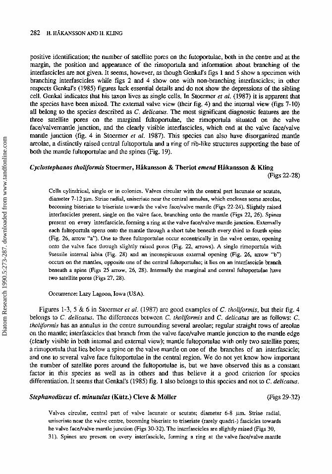
The characteristics of the cribra (particularly on the mantle) as well as the inconspicuous external opening of the rimoportula, which lacks an external tube, made us decide to transfer Stephanodiscus delicatus Genkal to the genus Cyclostephanos. Co-occurrence of several Stephanodiscus species can easily lead to misinterpretation of their characteristic features. Some of these diatoms can live both as short chains or as single cells, and the question always arises as to whether these two forms belong to the same species. By re-studying Lazy Lagoon material we found that it can be difficult to distinguish between C. tholiformis and C. delicatus in the LM, and it seems possible that both Genkal(l985) and Stoermer et al. (1987) may have mixed the two m a . The pictures and descriptions of S. delicatus and C. tholiformis are confusing. Genkal(l985) does not give a detailed enough description to make a
~~ ~~ ~~
Figs 22-28. Cyclostephanos tholiformis. Figs 23,24. LM, scale bar (Fig. 23) = 10 pm. Figs 22.25-28. SEM, scale bars = 1 pm. Fig. 22. Valve exterior with regularly inserted spines, clearly visible branching of the interfascicles onto the mantle, and raised openings (arrowed) of the valve face fultoportulae. Fig. 26 shows in detail the opening of one of the marginal fultoportulae (arrow “a”) and the opening of the rimoportula (mow “b”) on one of the interfascicle branches. Fig. 25. Valve interior, showing the valve face fultoportulae with two satellite pores, and raised interfascicles. Figs 27 & 28 show in detail the branching interfascicles, the marginal fultoportulae with two satellite pores and the rimoportula with sessile labia.
Dia
tom
Res
earc
h 19
90.5
:273
-287
. dow
nloa
ded
from
ww
w.ta
ndfo
nlin
e.co
m
.VEPHANODISCUS AND CYCLO.VEPHAN0S 28 1
Dia
tom
Res
earc
h 19
90.5
:273
-287
. dow
nloa
ded
from
ww
w.ta
ndfo
nlin
e.co
m
282 H. HAKANSSON AND H. KLING
positive identification; the number of satellite pores on the futoportulae, both in the centre and at the margin, the position and appearance of the rimoportula and information about branching of the interfascicles are not given. It seems, however, as though Genkal’s figs 1 and 5 show a specimen with branching interfascicles while figs 2 and 4 show one with non-branching interfascicles; in other respects Genkal’s (1985) figures lack essential details and do not show the depressions of the sibling cell. Genkal indicates that his taxon lives as single cells. In Stoermer et al. (1987) it is apparent that the species have been mixed. The external valve view (their fig. 4) and the internal view (figs 7-10) all belong to the species described as C. delicatus. The most significant diagnostic features are the three satellite pores on the marginal fultoportulae, the rimoportula situated on the valve face/valvemantle junction, and the clearly visible interfascicles, which end at the valve face/valve mantle junction (fig. 4 in Stoermer et al. 1987). This species can also have disorganized mantle areolae, a distinctly raised central fultoportula and a ring of rib-like structures supporting the base of both the mantle fultoportulae and the spines (Fig. 19).
Cells cylindrical, single or in colonies. Valves circular with the central part lacunate or scutate, diameter 7-12 p. Striae radial, uniseriate near the central annulus, which encloses some areolae, becoming biseriate to triseriate towards the valve face/valve mantle (Figs 22-24). Slightly raised interfascicles present, single on the valve face, branching onto the mantle (Figs 22, 26). Spines present on every interfascicle, forming a ring at the valve face/valve mantle junction. Externally each fultoportula opens onto the mantle through a short tube beneath every third to fourth spine (Fig. 26, arrow “a”). One to three fultoportulae occur eccenmically in the valve centre, opening onto the valve face through slightly raised pores (Fig. 22, arrows). A single rimoportula with 9sessile internal labia (Fig. 28) and an inconspicuous external opening (Fig. 26, arrow “b”) occurs on the mantles, opposite one of the central fultoportulae; it lies on an interfascicle branch beneath a spine (Figs 25 arrow, 26, 28). Internally the marginal and central fultoportulae have two satellite pores (Figs 27,28).
Occurrence: Lazy Lagoon, Iowa (USA).
Figures 1-3, 5 & 6 in Stoermer et al. (1987) are good examples of C. tholiformis, but their fig. 4 belongs to C. delicatus. The differences between C. tholiformis and C. delicatus &e as follows: C. tholiformis has an annulus in the centre surrounding several areolae; regular straight rows of areolae on the mantle; interfascicles that branch from the valve face/valve mantle junction to the mantle edge (clearly visible in both internal and external view); mantle fultoportulae with only two satellite pores; a rimoportula that lies below a spine on the valve mantle on one of the branches of an interfascicle; and one to several valve face fultoportulae in the central region. We do not yet know how important the number of satellite pores around the fultoportulae is, but we have observed this as a constant factor in this species as well as in others and thus believe it a good criterion for species differentiation. It seems that Genkal’s (1985) fig. 1 also belongs to this species and not to C. delicatus.
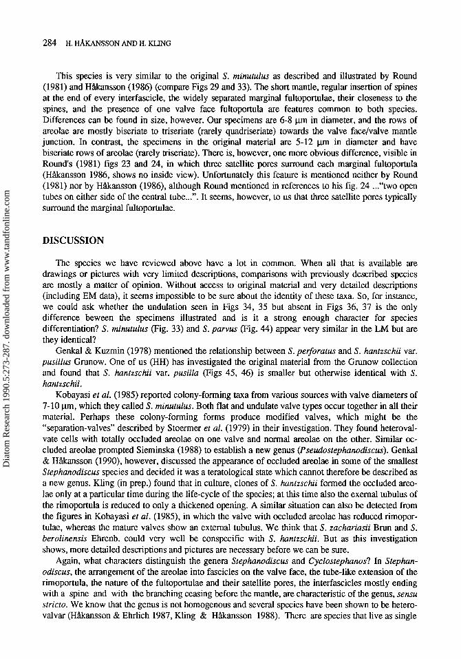
Valves circular, central part of valve lacunate or scutate; diameter 6-8 pm. Striae radial, uniseriate near the valve centre, becoming biseriate to trisenate (rarely quadri-) fascicles towards he valve face/valve mantle junction (Figs 30-32). The interfascicles are slightly raised (Figs 30, 31). Spines are presenl on every interfascicle, forming a ring at the valve face/valve mantle
Dia
tom
Res
earc
h 19
90.5
:273
-287
. dow
nloa
ded
from
ww
w.ta
ndfo
nlin
e.co
m
STEPHANODISCUS AND CYCLOSTEPHANOS 283
Figs 29-32. Stephanodiscus cf. minutulus. Fig. 29. LM, scale bar = 10 pm. Figs 30-32. SM, scale bars = 1 pm. Figs 30, 31. Valve exterior with regularly inserted spines, shallow mantle, the very short tube-like opening of the rimoportula (mowed), and one valve face fultoportula. Fig. 32. Valve interior with the valve face fultoportula and the marginal fultoportulae, all with two satellite pores.
junction. A fultoportula opens through a short tube on the mantle below every fourth to fifth spine (Fig. 30). A single slightly raised fultoportula is situated eccentrically on the valve face (Figs 30, 31). All fultoportulae have two satellite pores (Fig. 32). A rimoportula, with sessile internal labia (Fig. 32), opens through a very short tube or a raised thickening in a position where a spine would normally be, on an interfascicle (between two mantle fultoportulae) at the valve face/valve mantle junction (Figs 30, 31 arrows). The mantle is narrow (there are 2-3 areolae between base of a spine and the mantle edge). The mantle fultoportulae are very near the spines (no areolae between), 1-2 areolae from the mantle edge.
Occurrence: L885 (Canada) highly eutrophic. High pH and conductivity
Dia
tom
Res
earc
h 19
90.5
:273
-287
. dow
nloa
ded
from
ww
w.ta
ndfo
nlin
e.co
m
284 H. HAKANSSON AND H. KLING
This species is very similar to the original S. minutulus as described and illustrated by Round (1981) and Hhnsson (1986) (compare Figs 29 and 33). The short mantle, regular insertion of spines at the end of every interfascicle, the widely separated marginal fultoportulae, their closeness to the spines, and the presence of one valve face fultoportula are features common to both species. Differences can be found in size, however. Our specimens are 6-8 pm in diameter, and the rows of areolae are mostly biseriate to triseriate (rarely quadriseriate) towards the valve face/valve mantle junction. In contrast, the specimens in the original material are 5-12 pm in diameter and have biseriate rows of areolae (rarely triseriate). There is, however, one more obvious difference, visible in Rounds (1981) figs 23 and 24, in which three satellite pores surround each marginal fultoportula (HAkansson 1986, shows no inside view). Unfortunately this feature is mentioned neither by Round (1981) nor by HAkansson (1986), although Round mentioned in references to his fig. 24 ...“ two open tubes on either side of the central tube...”. It seems, however, to us that three satellite pores typically surround the marginal fultoportulae.
DISCUSSION
The species we have reviewed above have a lot in common. When all that is available are drawings or pictures with very limited descriptions, comparisons with previously described species are mostly a matter of opinion. Without access to original material and very detailed descriptions (including EM data), it seems impossible to be sure about the identity of these m a . So, for instance, we could ask whether the undulation seen in Figs 34, 35 but absent in Figs 36, 37 is the only difference beween the specimens illustrated and is it a strong enough character for species differentiation? S. minutulus (Fig. 33) and S. parvus (Fig. 44) appear very similar in the LM but are they identical?
Genkal & Kuzmin (1978) mentioned the relationship between S. perforatus and S . hantzschii var. pusillus Grunow. One of us (HH) has investigated the original material from the Grunow collection and found that S. huntzschii var. pusilla (Figs 45, 46) is smaller but otherwise identical with S. hantzschii.
Kobayasi et al. (1985) reported colony-forming m a from various sources with valve diameters of 7-10 pm, which they called S. minutulus. Both flat and undulate valve types occur together in all their material. Perhaps these colony-forming forms produce modified valves, which might be the “separation-valves” described by Stoermer et al. (1979) in their investigation. They found heteroval- vate cells with totally occluded areolae on one valve and normal areolae on the other. Similar oc- cluded areolae prompted Sieminska (1988) to establish a new genus (Pseudostephanodiscus). Genkal & HAkansson (1990), however, discussed the appearance of occluded areolae in some of the smallest Stephanodiscus species and decided it was a teratological state which cannot therefore be described as a new genus. Kling (in prep.) found that in culture, clones of S. hantzschii formed the occluded areo- lae only at a particular time during the life-cycle of the species; at this time also the exemal tubulus of the rimoportula is reduced to only a thickened opening. A similar situation can also be detected from the figures in Kobayasi et al. (1985), in which the valve with occluded areolae has reduced rimopor- tulae, whereas the mature valves show an external tubulus. We think that S. zachariasii Brun and S. berolinensis Ehrenb. could very well be conspecific with S. huntzschii. But as this investigation shows, more detailed descriptions and pictures are necessary before we can be sure.
Again, what characters distinguish the genera Stephanodiscus and Cyclostephanos? In Stephan- odiscus, the arrangement of the areolae into fascicles on the valve face, the tube-like extension of the rimoportula, the nature of the fultoportulae and their satellite pores, the interfascicles mostly ending with a spine and with the branching ceasing before the mantle, are characteristic of the genus, sensu stricto. We know that the genus is not homogenous and several species have been shown to be hetero- valvar (HAkansson & Ehrlich 1987, Kling & HAkansson 1988). There are species that live as single
Dia
tom
Res
earc
h 19
90.5
:273
-287
. dow
nloa
ded
from
ww
w.ta
ndfo
nlin
e.co
m
STEPHANODISCUS AND CYCLOSTEPHANOS 285
Figs 33-46. LM, scale bar (Fig. 33) = 1 pm. Fig. 33. S. rninurulus; original material, Kiitzing collection, BM 17995. Figs 34, 35. S. cf. medius; material on BM 17995. Figs 36-38. S. hantzschii. Typenmaterial, Rabenhorst 1104. Figs 39, 40. S. zachuriasii; original slide from the Brun collection. Figs 41, 42. S. berolinensis; original drawings from the Ehrenberg collection. Fig. 43. S. vestibulus. Fig. 44. S . parvus; Cleve & Moller slide collection 266. Figs 45,46. S. hantzschii var. pusillus; Grunow collection 2036.
cells, others that form short chains or other types of colony. Some species have a regular, others an irregular structure. The primary characters of the genus Cyclosrephanos are: tghe chamber-like arrangement in the marginal zone (Round 1982); the simple external opening of the rimoportula (Theriot er al. 1987); and the flattened cribra of the areolae on the valve mantgle (Theriot & Kociolek 1986). Several species that have been transferred from the genus Stephanodiscus to the genus Cyclosrephanos, as well as other more recently described species, do not have the chamber-like
Dia
tom
Res
earc
h 19
90.5
:273
-287
. dow
nloa
ded
from
ww
w.ta
ndfo
nlin
e.co
m
286 H. HAKANSSON AND H. KLING
arrangement in the marginal zone. Stoermer & HAkansson (1984) discussed the possibility of this being an intermediate evolutionary step between Stephanodiscus and Cyclostephanos in which the chamber-like structure is missing and all other features are the same. In these “intermediate” species the interfascicles are raised and costa-like, especially internally, and branch before reaching or on the valve mantle. Perhaps there are sufficient grounds for establishing another genus. Some species have “typical” interfascicle features, e.g. C. costatilimbus and C . delicatus, in which the interfascicles either do not branch or have branches that end shortly after being initiated. More careful investigations, especially culture studies, are necessary.
ACKNOWLEDGEMENTS
We want to thank Drs A. Podzorski, S. Lawrence, E. Theriot, F. Round and D. Mann for valuable comments on the manuscript and for correcting the language. We are greatly indebted to Dr T. Christensen for the Latin diagnosis.
REFERENCES
GEISSLER, U. (1970). Die Variabilitat der Schalenmerkmale bei den Diatomeen. Nova Hedwigia, 19, 623-773. GEISSLER, U. (1986). Experimental investigations on the variability of frustule characteristics of several
freshwater diatoms. In: Proceedings of the 8th International Diatom Symposium (M. Ricard, ed.), 59-66. Koeltz, Koenigstein
GENKAL, S. I. (1985). Novyj vid is roda Stephanodiscus Ehr. (Bacillariophyceae). Nov. Syst. Nis. Rust., 22, 30- 32
GENKAL, S. I. & KUZMIN, G. V. (1978). New taxa of the genus Stephanodiscus Ehr. Botanicheskij Zhurnal,
GENKAL, S . I. & HAKANSSON, H. (1990). The problem of distinguishing the newly described diatom genus Pseudostephanodiscus Sieminska. Diatom Research, 5 , 15-23
HAKANSSON, H. (1976). Die Struktur und Taxonomie einiger StephanodiscusArten aus eutrophen Seen Schwedens. Botmiska Notiser, 129,25-34
HAKANSSON, H. (1986). A taxonomic reappraisal of some Stephanodiscus species (Bacillariophyta). British Phycological Journal, 21,25-37
HAKANSSON, H. & EHRLICH, A. (1987). Stephanodiscus galileenris sp. nov. from Holocene subsurface sediments of Lake Kinneret, Israel. Diatom Research, 2, 15-21
HAKANSSON, H. & HICKEL, B. (1986). The morphology and taxonomy of the diatom Stephanodiscus neoastraea sp. nov. British Phycological Journal, 21,3943
HAKANSSON, H. & KLING, H. (1989). A light and electron microscope study of previously described and new Stephanodiscus species (Bacillariophyceae) from central and northern Canadian lakes with ecological notes on the species. Didom Research, 4,269-288
HICKEL, B. & HAKANSSON, H. (1987). Dimorphism in Cyclostephanos dubius (Bacillariophyta) and the morphology of initial valves. Diatom Research, 2 , 3 5 4 6
KLING, H. (in press). Culture studies of the Stephanodiscus hantzschii-complex. KLING, H. & HAKANSSON, H. (1988). A light and electron microscope study of Cyclotellu species
(Bacillariophyceae) from Central and Northern Canadian Lakes. Diatom Research, 3,55-82 KOBAYASI, H., KOBAYASHI, H. & IDEI, M. (1985). Fine structure and taxonomy of the small and tiny
Stephanodiscus (Bacillariophyceae) species in Japan. 3. Co-occurrence of Stephanodiscus minutulus (Kutz.) Round and S. parvus Stoerm. & H&. Japanese Journal of Phycology, 33,293-300
ROUND, F. E. (1981). The diatom genus Stephanodiscus: an electron microscopic view of the classical species. Archiv fur Protistenkunde, 124,455470
63,1309-1312
Dia
tom
Res
earc
h 19
90.5
:273
-287
. dow
nloa
ded
from
ww
w.ta
ndfo
nlin
e.co
m
STEPHANODISCUS AND CYCLOSTEPHANOS 287
ROUND. F. E. (1982). Auxospore structures. initial valves and the development of populations of Stephamdiscus in Farmoor Reservoir. A d s of Botany, 49,447459
SIEMINSKA, J. (1988). Pseudostephanodiscus gen. n. (Bacillariophyceae). Archivfir Protistenkunde, 135, 183- 185
STOERMER, E. F. & HkANSSON, H. (1983). An investigation of the morphological structure and taxonomic relationships of Stephanodiscus damasii Hustedt. Bacillaria, 6, 245-255
STOERMER, E. F. & HAKANSSON, H. (1984). Stephanodiscusparvus: validation of an enigmatic and widely misconstrued taxon. Nova Hedwigia, 39,497-51 1
STOERMER, E. F., HAKANSSON, H. & THERIOT, E. C. (1987). Cyclostephanos species newly reported from North America: C. tholiformis sp. nov. and C. costatilimbus comb. nov. British Phycological Journal, 22, 349-358
STOERMER, E. F., KINGSTON, J. C. & SICKO-GOAD, L. (1979). The morphology and taxonomic relationships of Stephanodiscus binderanus var. oestrupii (A. Cl.) A. C1. Nova Hedwigia, Beihgt 64, 65-78
THERIOT, E. & KOCIOLEK. J. P. (1986). Two new Pliocene species of Cyclostephanos (Bacillariophyceae) with comments on the classification of the freshwater Thalassiosiraceae. Journal of Phycology, 22, 121-128
THERIOT, E., H k W S S O N , H. & STOERMER, E. F. (1988). Morphometric analysis of Stephanodiscus alpinus (Bacillariophyceae) and its morphology as an indicator of lake trophic status. Phycologia, 27, 485- 493
THERIOT, E., STOERMER, E. F. & HAKANSSON, H. (1987). Taxonomic intepretatrion of the rimoportula of freshwater genera in the centric diatom family Thalassiosiraceae. Diatom Research, 2,251-265