1Department of Biology, Saint Louis University, Saint Louis, MO 63103, USA 2Department of Molecular Microbiology & Immunology, Saint Louis University, Saint Louis, MO 63103, USA
Nonhealing diabetic foot ulcers are a significant complication of diabetes. They develop due to peripheral neuropathy, and may take weeks or months to close. A failure to heal may necessitate lower limb amputation causing significant morbidity and mortality. Wound healing impairment in diabetic patients is associated with delays in immune cell migration and altered macrophage activation. These processes are orchestrated by the cytokine milieu in the wound manifested by an upregulation of proinflammatory cytokines and downregulation of anti-inflammatory cytokines. This review examines the current knowledge of cytokine expression (IL-1β, IL-6, IL-10, TNF-α, TGF-β, and C-reactive proteins as well as the chemokines CCL2 and CXCL12) and explores potential cytokine immunotherapy to aid healing.
Practice Points
● The complex orchestration of events in normal wound healing is guided by the specific temporal and spatial expression of cytokines and chemokines in the wound; however, the exact coordination of these events is not completely understood.
● The expression of cytokines and chemokines is altered in diabetic wounds, which results in persistent inflammation and impaired healing.
● Blocking the activity of individual proinflammatory cytokines (IL-1β, TNF-α, C-reactive protein) has improved diabetic wound healing in both animal models and humans.
● Diabetic wound healing may also be improved by increasing the expression of anti-inflammatory cytokines (IL-10 and TGF-β).
● The administration of chemokines (or drugs that induce chemokine expression) may improve leukocyte migration and healing in the wound.
● Because of the complexity of wound healing, a multifaceted approach targeting multiple cytokines (thereby altering the cytokine milieu of the diabetic wound) may be most effective for improved healing.
BackgroundDiabetes mellitus is a prevalent metabolic disease that is characterized by chronic hyperglycemia and long-term complications such as retinopa-thy, hypertension and abnormal lipid metabo-lism [1]. In addition, other complications such as peripheral neuropathy and impaired wound healing lead to chronic foot ulcers, a major cause of diabetes-associated lower limb amputations.
The wound healing process is a complex orchestration of events including inflamma-tion, angiogenesis and extracellular matrix growth and remodeling [2]. The impaired heal-ing observed in diabetic wounds correlates with decreased keratinocyte, fibroblast and immune cell migration into the wound and reduced endothelial cell angiogenesis [3,4]. The movement and function of these cells in the wound are controlled by the local cytokine milieu. In par-ticular, the cytokine milieu plays an important role in differentiating macrophages into subsets that exist on a functional spectrum ranging from proinflammatory (classically activated M1) to anti-inflammatory and healing (alternatively activated M2) macrophages. Diabetic wounds have been linked to both increased proinflam-matory cytokine production and an increased ratio of classically to alternatively activated mac-rophages (M1/M2) [5]. This review focuses on cytokines that are differentially expressed in dia-betic wounds, and it highlights research aimed at altering the cytokine milieu in the diabetic wound as an aid to healing.
interleukins●● iL-1β
IL-1β is an important inflammatory molecule produced by blood monocytes and tissue mac-rophages [6]. This cytokine is activated via cas-pase-1 cleavage in the secretory lysosome, after caspase-1 activation by the NALP3 inflamma-some. This cycle allows IL-1β to amplify its own secretion via its initiation of the assembly of the NALP3 inflammasome. Along with activat-ing the inflammasome, IL-1β also stimulates inflammation via increasing mobilization of leukocytes from the bone marrow and secretion of acute-phase proteins from the liver [7].
Obese patients may have a sustained release of IL-1 β from adipose tissue, and this could have broad effects based on the wide distribution of the IL-1 receptor. Indeed, elevated IL-1β has been implicated in the development of insulin resistance and aberrant healing in diabetes [8].
Human diabetic foot ulcers have increased levels of IL-1β, and these levels decrease as the ulcers heal [9]. Using skin explants, topical treatment of IL-1 on normal tissue correlated with increased CXCR2 expression and delayed wound clo-sure [10]. Isolated wound macrophages from dia-betic humans and db/db mice display increased IL-1β and NALP3 inflammasome components through 10 days of healing, and blocking of the inflammasome correlated with improved healing in these wounds [11].
Over the past decade, researchers have aimed at blocking the effects of IL-1β in the hopes of decreasing the chronically inflamed condition of diabetic wounds. Anakinra (Kineret) is an IL-1 receptor antagonist (IL1Ra) commonly used for treating rheumatoid arthritis (RA). Due to the implications of IL-1β in pancreatic beta cell destruction, much work has been done in using IL1Ra to combat diabetic pathogen-esis [12]. Indeed, IL-1Ra and anti-IL-1β antibody treatments have correlated with improved beta cell functionality in Type 2 diabetic patients, indicating the importance of this cytokine in this disease [13,14]. In wound healing, Anakinra was shown to be effective at decreasing IL-6 and TNF-α protein levels in wound tissue during the first 48 h postwounding of normal mice [15]. Similarly, both db/db mice injected with IL-1β-neutralizing antibodies and IL-1R knockout mice display wounds with a decreased M1/M2 ratio, decreased IL-6 and TNF-α gene expression and improved healing [16]. Thus, the blocking of IL-1β function in diabetic wounds with either neutralizing antibodies or the receptor antago-nist may have potential in the improvement of diabetic wound healing.
●● iL-6IL-6 is a cytokine secreted by T lymphocytes and macrophages and is critically important in host defense [17]. Adipose tissue, particularly vis-ceral fat, is also a significant source of IL-6 [18]. IL-6 stimulates acute-phase protein release from the liver, production of neutrophils in the bone marrow and supports the proliferation of B lymphocytes. It also influences recruitment of leukocytes through the stimulation of IL-8 and MCP-1 secretion from endothelial cells [19]. As such, this cytokine represents a significant portion of the inflammatory response.
Diabetic insulin resistance and β-cell inflam-mation are associated with increased levels of IL-6 [20]. Rabbits treated with the toxic glucose
analog alloxan monohydrate display signifi-cantly elevated blood glucose, delayed wound healing and significantly higher wound expres-sion levels of IL-6 and its receptor GP130 com-pared with controls [21]. Hyperglycemia has been shown to significantly elevate IL-6 expression in a dose-dependent manner in macrophages isolated from normal mice, and macrophages isolated from streptozotocin (STZ)-injected and db/db mice corroborate this effect [22]. Diabetic patients with foot ulcers displayed significantly higher levels of circulating acute-phase proteins and IL-6 than those without foot ulcers [23]. Both glucose concentration and wound chro-nicity seem to have strong correlation with the increased IL-6 expression observed in diabetic foot ulcers.
IL-6-deficient mice have impaired mac-rophage infiltration and delayed wound heal-ing, which was not observed in mice lacking the α-subunit of the IL-6 receptor alone [24]. However, blocking the IL-6 receptor has been shown to successfully decrease inflammation in models of corneal alkali burns, indicating a potential benefit in its use in chronic wounds [25]. This may be explained by a need for some level of IL-6 in the normal wound healing process combined with deleterious effects when IL-6 is overexpressed in chronic wounds. Tocilizumab (Actemra) is an anti-IL-6 receptor antibody that has been effective in improving blood glucose levels in patients with Type 2 diabetes [26]. In a study of joint surgeries between RA patients treated with nonbiologic antirheumatic drugs and those treated with Tocilizumab, the latter group experienced significant depression of post-operative fever and plasma C-reactive protein (CRP) [27]. This suggests an efficient suppression of the inflammatory effects of IL-6 in humans, but it has yet to be tested in a model of diabetic wound healing.
In addition to antibody treatment of IL-6, nat-ural remedies may represent a potential option for lowering its expression in diabetic wound healing. Curcumin, one of the main ingredi-ents in turmeric, has been shown to signifi-cantly decrease circulating plasma levels of IL-6 in STZ-injected mice over 7 weeks [28]. Indeed, use of curcumin-loaded poly (ε-caprolactone) nanofibers resulted in significantly lower levels of IL-6 release in vitro from lipopolysaccharide-stimulated macrophages and improved wound healing in STZ-injected mice [29]. These results suggest that the reduction in circulating IL-6
levels, whether by antibody-mediated or natural treatments, could be a key tool in the field of diabetic wound healing.
●● iL-10Unlike IL-1β and IL-6, IL-10 is an anti-inflam-matory cytokine. In vivo, it is mostly secreted by T helper cells, regulatory T cells, macrophages and dendritic cells [30,31]. The IL-10R is mainly expressed on immune cells, predominantly macrophages, where it will inhibit the release of proinflammatory mediators, suppress anti-gen presentation and enhance phagocytosis [32]. IL-10R signaling is essential for the generation of anti-inflammatory macrophages that regu-late mucosal defense in mice and humans [33]. Interestingly, regulatory T-cell-mediated sup-pression of T helper 17 (Th17)-induced inflam-mation is mediated by IL-10 secretion [34]. Due to the correlation between autoimmune diseases and Th17 cells, IL-10 involvement in diabetes with regards to this lymphocyte subset should be investigated.
There is an association between obesity, Type 2 diabetes and low circulating levels of IL-10 [35]. IL-10 protein expression is lower in isolated mac-rophages from db/db mice compared with db/+ mice over 7 days postwounding [36]. In addi-tion, STZ-injected rats have shown significantly lower protein levels of this cytokine in the tissue through 7 days postwounding compared with controls [37]. Human diabetic foot ulcers have decreased expression of IL-10, particularly in the keratinocytes and endothelial cells at the wound margins [38]. Unlike the high expression of pro-inflammatory cytokines, the expression of this anti-inflammatory cytokine in diabetic wounds is quite low, and its paucity may contribute to the development of chronic nonhealing wounds.
There are several methods available for increasing IL-10 expression. Highly purified eicosapentaenoic acid increased IL-10 expres-sion in monocytes derived from obese patients with dyslipidemia [39]. Recently, the topical application of curcumin on wounds of STZ-injected rats correlated with increased IL-10 mRNA and protein, increased wound contrac-tion and improved granulation tissue forma-tion [40]. Lentiviral-mediated IL-10 overexpres-sion in mice has been shown to correlate with decreased scar formation and reduced proin-flammatory cytokine production, such as IL-6 and MCP-1 [41]. Another potential gene delivery vector is N-acyl low-molecular weight chitosan.
Diabetes Manag. (2015) 5(6)528
Review DeClue & Shornick
future science group
Nanomicelles of this vector designed to express IL-4 and IL-10 were injected intramuscularly into STZ-injected mice, which resulted in sig-nificantly higher serum levels of these cytokines and decreased proinflammatory cytokines [42]. Whether by topical treatments or gene therapy, increasing IL-10 in diabetic wounds suggests an interesting option in improvement of healing.
Noninterleukin cytokines●● TNF-α
TNF-α is a proinflammatory cytokine that stimulates inflammation at low levels and inhib-its extracellular matrix synthesis at high lev-els [43]. While the main source of this cytokine is macrophages, it can also be produced by adipose tissue, neurons, mast cells and lym-phocytes [44,45]. In combination with IL-1β and IL-6, it can stimulate the acute-phase response, act as a potent neutrophil chemoattractant and stimulate the classical activation of macrophages. This latter function occurs through its bind-ing with TNFR2, which activates the MAPK and NF-κB signaling pathways [46]. In vitro, TNF-α also stimulates apoptosis of fibroblasts, keratinocytes and endothelial cells [47,48]. This occurs through its binding with TNFR1, which stimulates downstream mitochondrial cyto-chrome C release, caspase 9 activation and apo-ptosome formation. TNF-α frequently opposes the proliferative activity of TGF-β, presumably through the inactivation of Smad 2/3 [49]. While TNFR1 is fairly ubiquitous in distribution, TNFR2 is mainly relegated to immune cells, limiting its proinflammatory effects to leuko-cytes. Consequently, TNF-α signaling is quite complex and sometimes antagonistic, allowing it to perform many functions in the process of wound healing.
STZ-injected rats displayed significantly higher levels of serum TNF-α by day 4 post-wounding. Oral supplementation with camel undenatured whey protein reduced both TNF-α expression in the wound and time to wound closure [50]. Diabetic patients have also shown a significant upregulation in serum TNF-α dur-ing high blood glucose events compared with a relatively little change observed in normal patients [51]. In addition, TNF-α has also been observed to be elevated in the serum of obese patients [52]. Acute hyperglycemia appears to trig-ger a much stronger upregulation of TNF-α in diabetics, lending further credence to the overall chronic inflammatory state seen in this disease.
Anti-TNF-α neutralizing antibodies adminis-tered to ob/ob mice significantly improved heal-ing and reduced inflammation and the numbers of viable macrophages at the wound site [53]. Etanercept (Enbrel) is a TNF receptor:IgG1 F
c
fusion protein that has been shown to signifi-cantly decrease TNF-α activity in the wounds of chronic leg ulcers [54]. It has also been effective in db/db mouse wounds, where it significantly decreased fibroblast apoptosis and increased new matrix formation [55]. Infliximab (Remicade) is an anti-TNF-α neutralizing antibody treatment that is used to treat autoimmune diseases such as RA and Crohn’s disease. It has been found to be successful at healing human chronic leg ulcers, healing 9 of 14 ulcers by more than 75% within just 8 weeks of use [56]. As of yet, Infliximab has not been tested in any model of diabetic wound healing [57].
●● TGF-βTGF-β acts as a chemoattractant for neutro-phils and monocytes early in healing, as well as stimulates monocyte-to-macrophage differentia-tion, proliferation of fibroblasts and subsequent extracellular matrix synthesis later in the pro-cess [58]. Upon injury, it is secreted by platelets, keratinocytes, resident macrophages and fibro-blasts [59]. As such, TGF-β experiences a biphasic expression during normal wound healing that peaks within a few hours and again at 5 days postinjury [60]. This cytokine elicits its effects via binding, heterodimerization and phosphoryla-tion of its receptor, which then phosphorylates Smad proteins that translocate to the nucleus and regulate expression of target genes by bind-ing to the promoter elements [61].
A reduction in TGF- β expression has been observed in both human wounds and rodent models of wound healing. Human nonhealing venous ulcers have shown suppressed TGF-β signaling via downregulated TGF-βR and atten-uation of Smad signaling, and STZ-injected mice have reduced wound tissue expression of TGF-β on day 4 postwounding [62,63]. In a rat model of Type 2 diabetes, decreased TGF-β signaling was associated with delayed wound closure. In vitro analysis of isolated diabetic dermal fibroblast demonstrated reduced TGF-β RII signaling and fibroblast migration [64]. Similarly, human diabetic foot ulcers have decreased expression of both TGF-β and its receptor in the wound [65]. Importantly, the concentration of this cytokine in the wounds of Type 2 diabetic patients
529
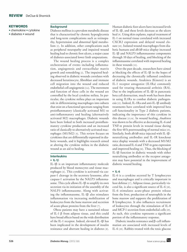
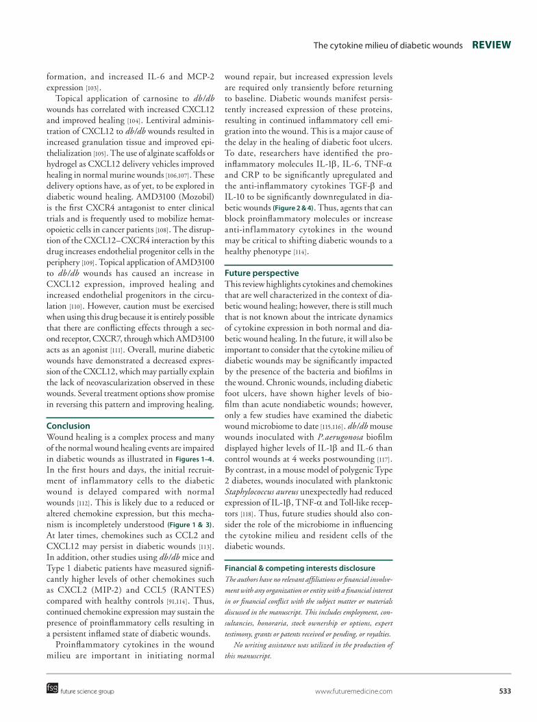
Figure 1. early events in normal wound healing include the production of chemokines: CCL2 by keratinocytes and CXCL12 by endothelial cells. These molecules recruit polymorphonuclear cells and macrophages into the wound bed. At this stage, the macrophages have an M1 phenotype that is characterized by production of the proinflammatory cytokines IL-1β, IL-6 and TNF-α.
The cytokine milieu of diabetic wounds Review
future science group www.futuremedicine.com
correlates with decreased levels of matrix met-alloproteinases and a concomitant increase of tissue inhibitor of matrix metalloproteinases [66]. As a consequence, the lower concentration of TGF-β found in diabetic wounds may delay wound healing by preventing the growth of the proliferative phase and allowing the disintegra-tion of the extracellular matrix by the matrix metalloproteinases.
Studies have shown that intradermal injec-tion of a TGF-β-expressing plasmid significantly improves wound closure, collagen synthesis and angiogenesis in db/db mice [67,68]. In a blinded study of human diabetic neuropathic ulcers, applications of TGF-β correlated with improved healing compared with controls alone [69]. It has been suggested that because the latent form TGF-β binds to the extracellular matrix, this form may be a more effective in vivo therapy than
active TGF-β due to its increased half-life [70]. This would indicate that treatment with a drug, such as nitric oxide, that can activate the latent form may be a more appealing alternative. For instance, the application of ointment from the Momordica charantia fruit has improved wound closure, increased granulation tissue formation and increased TGF-β detection in STZ-injected rats [71,72]. Accordingly, drugs that could increase endogenous levels of active TGF-β may be more effective than gene therapy approaches.
●● C-reactive proteinCRP is a highly conserved acute-phase protein of hepatic origin that acts as a pattern recog-nition receptor. Structurally, it consists of five identical protomers that each contains a phos-phocholine binding site, which together form a pentraxin around a central core. Functionally,
Early normal response
CCL2
CCL2
CCL2 CCL2
CCL2
TNF-α
TNF-α
TNF-α
IL-1β
IL-1β
IL-1β
IL-6
IL-6
IL-6
Mac
Mac
Mac
PMN
PMN
PMN
PMN
CXCL12
CXCL12
CXCL12CXCL12
Endothelium
Diabetes Manag. (2015) 5(6)530
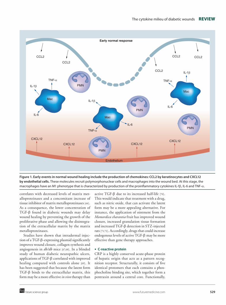
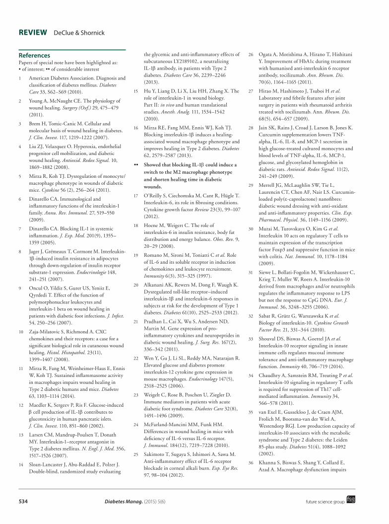
Figure 2. in the later stages of normal wound healing there is reepithelialization, angiogenesis and the rebuilding of extracellular matrix fibers. The macrophages display a more M2 phenotype through the production of the anti-inflammatory cytokines IL-10 and TGF-β. Fibroblasts also produce TGF-β.
Review DeClue & Shornick
future science group
it can activate the classical complement system, stimulate phagocytosis and bind to immuno-globin receptors [73]. This occurs via its binding to phosphocholine expressed on the surface of dying cells and bacteria. It is also able to upregu-late adhesion molecules expressed on endothelial cells and increase the release of proinflammatory cytokines such as IL-1β, IL-6 and TNF-α [74,75]. As CRP is regulated transcriptionally by IL-6 and IL-1β, it can result in a cyclic amplification of inflammation.
Both Type 1 and Type 2 diabetic patients have significantly elevated CRP in their plasma [76,77]. In addition, Type 1 diabetic patients with micro-vascular complications have higher CRP levels compared with otherwise healthy Type 1 dia-betics [78]. Diabetic humans with foot ulcers also display significantly higher CRP in their serum when compared with wounded nondia-betic patients or diabetics without foot ulcers,
and CRP levels were significantly lower for those diabetic patients with healed ulcers [79]. This indicates the potential of CRP as a clinical biomarker for diabetic foot ulcer healing.
Statin therapy has been known to signifi-cantly decrease circulating CRP levels, as well as improve normal wound healing [80,81]. Both injection and topical application of Simvastatin (Zocor) on db/db mice have correlated with enhanced angiogenesis and improved healing through the first week postwounding [82,83]. Likewise, Pravastatin (Pravachol) treatment of STZ-injected mice has resulted in significantly higher wound breaking strength, hydroxyproline content and eNOS expression over 10 days post-wounding [84]. Finally, Atorvastatin (Lipitor) has improved healing in STZ-injected rats over 14 days postwounding, as well as decreased serum CRP levels and prevented new diabetic foot ulcers in Type 2 diabetic patients [85,86]. In order
Late normal response
TGF-β
TGF-β
TGF-β
TGF-β
IL-10IL-10
Mac
Mac
Endothelium
Angiogenesis
531
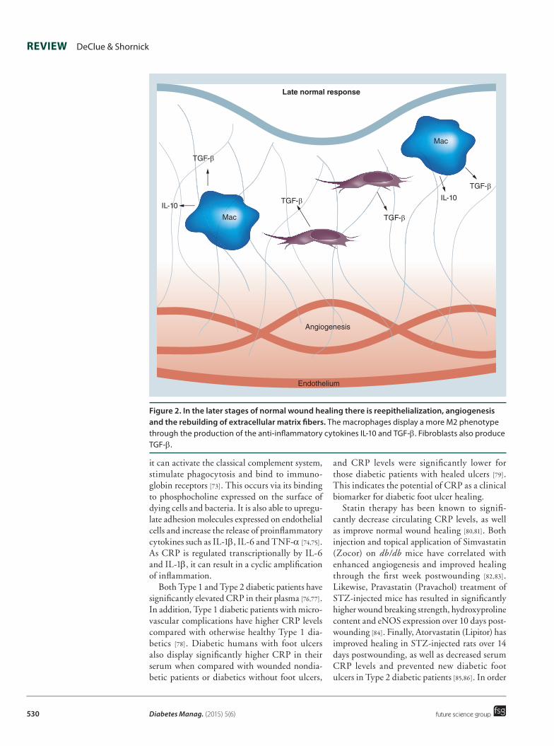
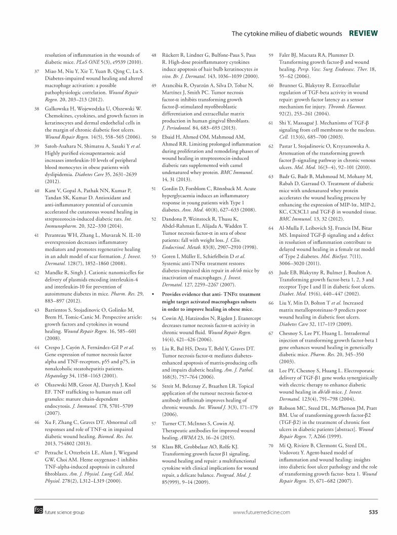
Figure 3. Normal events are impaired in the early stages of diabetic wound healing. Both chemokine production and recruitment of phagocytes to the wound are reduced.
The cytokine milieu of diabetic wounds Review
future science group www.futuremedicine.com
to reduce adverse systemic effects from statin treatment, topical application seems to be the optimal delivery route for improved diabetic wound healing.
Chemokines●● CCL2 (MCP-1)
CCL2, or MCP-1, is a CC family chemokine that is secreted by keratinocytes and acts as a che-moattractant for macrophages and T cells [87]. Along with its chemotactic function, this chemokine also stimulates growth factor produc-tion from recently emigrated leukocytes, stimu-lates fibroblast activity via mast cell-derived IL-4 and increases motility of endothelial cells [88]. Within the wound, CCL2 displays the highest expression during the first 24 h postwounding, with levels dropping off after the first week [89].
CCL2-deficient mice exhibit delayed heal-ing, decreased angiogenesis and collagen pro-duction and delayed macrophage migration into the wound [90]. Db/db murine wounds have a lower expression of CCL2 than controls within 24 h postwounding, but a higher expression of
this chemokine after 13 days [91]. Bone marrow-derived macrophages from db/db mice showed a significant decrease in chemotaxis to CCL2 and impaired scratch wound closure compared with controls, despite expressing normal levels of CCR2, the receptor for CCL2 [92]. This sug-gests that, in addition to expressing lower levels of CCL2, diabetic wounds may also contain immune cells that are less responsive to its signal.
Recently, it was observed that the local injec-tion of CCL2 upon wounding restored its defi-cient expression within 24 h, promoted re-epi-thelialization and improved monocyte homing in db/db wounds [93]. Interestingly, low-intensity vibrational treatment of db/db mice resulted in improved healing, augmented angiogenesis and significantly increased CCL2 wound expres-sion by day 7 postwounding compared with nonvibrated diabetic controls [94]. This type of mechanical treatment has been supported based on improved blood flow, but the exact mecha-nisms have yet to be elucidated. STZ-injected mice have shown improved wound healing by day 5 postwounding, increased leukocyte
Early diabetic response
TNF-α
IL-1β
IL-6
Mac
PMN
PMN
CXCL12
Endothelium
CCL2CCL2
Diabetes Manag. (2015) 5(6)532
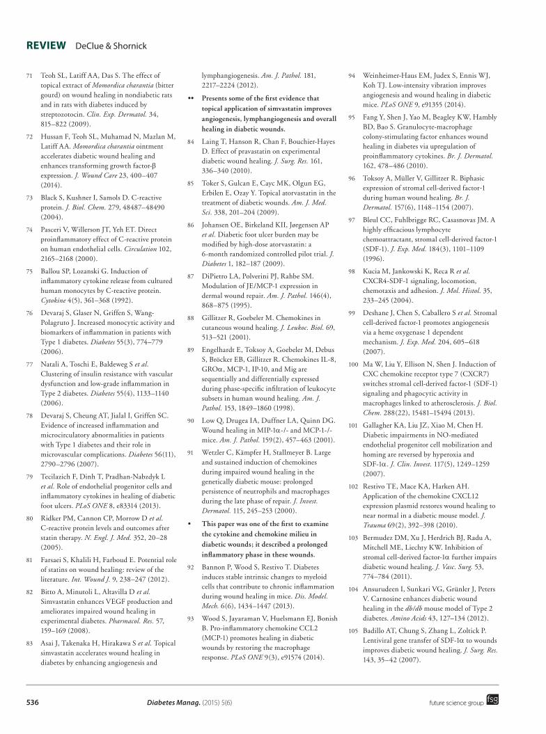
Figure 4. in the later stages of diabetic wound healing, there is persistent production of chemokines (CCL2 and CXCL12) and an associated continued recruitment of neutrophils and macrophages. The macrophages have reduced expression of M2 markers and continue to produce proinflammatory cytokines (IL-1β, IL-6 and TNF-α).
Review DeClue & Shornick
future science group
intravasation and significantly increased CCL2 expression within 24 h in conjunction with local administration of GM-CSF [95]. This study sug-gests that CCL2 may be an intermediary for the improved leukocyte migration and healing observed in wounds treated with some growth factors.
●● CXCL12 (SDF-1)CXCL12, or SDF-1, is a CXC family chemokine expressed by endothelial cells and fibroblasts [96]. Its receptor, CXCR4, is expressed by lympho-cytes and monocytes [97]. CXCR4 signaling is involved in trafficking and homing of hemat-opoietic stem and progenitor cells, as well as tumor metastasis [98]. Importantly, CXCL12 is a potent angiogenic factor that stimulates endothelial migration in a VEGF-independent manner [99]. The recent discovery of CXCR7, a
second receptor for CXCL12, has shed new light on potential additional functions. While not expressed on normal blood leukocytes, CXCR7 has been found on macrophages in pathologi-cal conditions. It may be involved in switching macrophages to a proinflammatory phenotype and increasing phagocytic activity [100].
STZ-injected mice show decreased expres-sion of CXCL12 through 9 days postwounding compared with controls, and the injection of reconstituted CXCL12 has improved healing in these wounds [101]. Db/db mice also display decreased levels of this chemokine through 7 days postwounding, and the insertion of a plasmid expressing CXCL12 correlated with improved healing [102]. In contrast, the use of a CXCL12 antagonist exacerbated impaired heal-ing in db/db wounds. This was evidenced by decreased angiogenesis and granulation tissue
Late diabetic response
CCL2CCL2
CCL2
CCL2
TNF-α
TNF-α
TNF-α
IL-1β
IL-1β
IL-1β
IL-6
IL-6
IL-6
Mac Mac
Mac
PMN
PMNPMN
PMN
PMN
PMN
CXCL12 CXCL12
Endothelium
TNF-α
IL-1β
IL-6
MacTNF-α
IL-1β
IL-6Mac
533
The cytokine milieu of diabetic wounds Review
future science group www.futuremedicine.com
formation, and increased IL-6 and MCP-2 expression [103].
Topical application of carnosine to db/db wounds has correlated with increased CXCL12 and improved healing [104]. Lentiviral adminis-tration of CXCL12 to db/db wounds resulted in increased granulation tissue and improved epi-thelialization [105]. The use of alginate scaffolds or hydrogel as CXCL12 delivery vehicles improved healing in normal murine wounds [106,107]. These delivery options have, as of yet, to be explored in diabetic wound healing. AMD3100 (Mozobil) is the first CXCR4 antagonist to enter clinical trials and is frequently used to mobilize hemat-opoietic cells in cancer patients [108]. The disrup-tion of the CXCL12–CXCR4 interaction by this drug increases endothelial progenitor cells in the periphery [109]. Topical application of AMD3100 to db/db wounds has caused an increase in CXCL12 expression, improved healing and increased endothelial progenitors in the circu-lation [110]. However, caution must be exercised when using this drug because it is entirely possible that there are conflicting effects through a sec-ond receptor, CXCR7, through which AMD3100 acts as an agonist [111]. Overall, murine diabetic wounds have demonstrated a decreased expres-sion of the CXCL12, which may partially explain the lack of neovascularization observed in these wounds. Several treatment options show promise in reversing this pattern and improving healing.
ConclusionWound healing is a complex process and many of the normal wound healing events are impaired in diabetic wounds as illustrated in Figures 1–4. In the first hours and days, the initial recruit-ment of inf lammatory cells to the diabetic wound is delayed compared with normal wounds [112]. This is likely due to a reduced or altered chemokine expression, but this mecha-nism is incompletely understood (Figure 1 & 3). At later times, chemokines such as CCL2 and CXCL12 may persist in diabetic wounds [113]. In addition, other studies using db/db mice and Type 1 diabetic patients have measured signifi-cantly higher levels of other chemokines such as CXCL2 (MIP-2) and CCL5 (RANTES) compared with healthy controls [91,114]. Thus, continued chemokine expression may sustain the presence of proinflammatory cells resulting in a persistent inflamed state of diabetic wounds.
Proinflammatory cytokines in the wound milieu are important in initiating normal
wound repair, but increased expression levels are required only transiently before returning to baseline. Diabetic wounds manifest persis-tently increased expression of these proteins, resulting in continued inflammatory cell emi-gration into the wound. This is a major cause of the delay in the healing of diabetic foot ulcers. To date, researchers have identified the pro-inflammatory molecules IL-1β, IL-6, TNF-α and CRP to be significantly upregulated and the anti-inflammatory cytokines TGF-β and IL-10 to be significantly downregulated in dia-betic wounds (Figure 2 & 4). Thus, agents that can block proinflammatory molecules or increase anti-inf lammatory cytokines in the wound may be critical to shifting diabetic wounds to a healthy phenotype [114].
Future perspectiveThis review highlights cytokines and chemokines that are well characterized in the context of dia-betic wound healing; however, there is still much that is not known about the intricate dynamics of cytokine expression in both normal and dia-betic wound healing. In the future, it will also be important to consider that the cytokine milieu of diabetic wounds may be significantly impacted by the presence of the bacteria and biofilms in the wound. Chronic wounds, including diabetic foot ulcers, have shown higher levels of bio-film than acute nondiabetic wounds; however, only a few studies have examined the diabetic wound microbiome to date [115,116]. db/db mouse wounds inoculated with P.aerugonosa biofilm displayed higher levels of IL-1β and IL-6 than control wounds at 4 weeks postwounding [117]. By contrast, in a mouse model of polygenic Type 2 diabetes, wounds inoculated with planktonic Staphylococcus aureus unexpectedly had reduced expression of IL-1β, TNF-α and Toll-like recep-tors [118]. Thus, future studies should also con-sider the role of the microbiome in influencing the cytokine milieu and resident cells of the diabetic wounds.
Financial & competing interests disclosureThe authors have no relevant affiliations or financial involve-ment with any organization or entity with a financial interest in or financial conflict with the subject matter or materials discussed in the manuscript. This includes employment, con-sultancies, honoraria, stock ownership or options, expert testimony, grants or patents received or pending, or royalties.
No writing assistance was utilized in the production of this manuscript.
Diabetes Manag. (2015) 5(6)534
Review DeClue & Shornick
future science group
ReferencesPapers of special note have been highlighted as: • of interest; •• of considerable interest
1 American Diabetes Association. Diagnosis and classification of diabetes mellitus. Diabetes Care 33, S62–S69 (2010).
2 Young A, McNaught CE. The physiology of wound healing. Surgery (Oxf.) 29, 475–479 (2011).
3 Brem H, Tomic-Canic M. Cellular and molecular basis of wound healing in diabetes. J. Clin. Invest. 117, 1219–1222 (2007).
4 Liu ZJ, Velazquez O. Hyperoxia, endothelial progenitor cell mobilization, and diabetic wound healing. Antioxid. Redox Signal. 10, 1869–1882 (2008).
5 Mirza R, Koh TJ. Dysregulation of monocyte/macrophage phenotype in wounds of diabetic mice. Cytokine 56 (2), 256–264 (2011).
6 Dinarello CA. Immunological and inflammatory functions of the interleukin-1 family. Annu. Rev. Immunol. 27, 519–550 (2009).
7 Dinarello CA. Blocking IL-1 in systemic inflammation. J. Exp. Med. 201(9), 1355–1359 (2005).
8 Jager J, Grémeaux T, Cormont M. Interleukin-1β-induced insulin resistance in adipocytes through down-regulation of insulin receptor substrate-1 expression. Endocrinologie 148, 241–251 (2007).
9 Oncul O, Yildiz S, Gurer US, Yeniiz E, Qyrdedi T. Effect of the function of polymorphonuclear leukocytes and interleukin-1 beta on wound healing in patients with diabetic foot infections. J. Infect. 54, 250–256 (2007).
10 Zaja-Milatovic S, Richmond A. CXC chemokines and their receptors: a case for a significant biological role in cutaneous wound healing. Histol. Histopathol. 23(11), 1399–1407 (2008).
11 Mirza R, Fang M, Weinheimer-Haus E, Ennis W, Koh TJ. Sustained inflammasome activity in macrophages impairs wound healing in Type 2 diabetic humans and mice. Diabetes 63, 1103–1114 (2014).
12 Maedler K, Sergeev P, Ris F. Glucose-induced β cell production of IL-1β contributes to glucotoxicity in human pancreatic islets. J. Clin. Invest. 110, 851–860 (2002).
13 Larsen CM, Mandrup-Poulsen T, Donath MY. Interleukin-1–receptor antagonist in Type 2 diabetes mellitus. N. Engl. J. Med. 356, 1517–1526 (2007).
14 Sloan-Lancaster J, Abu-Raddad E, Polzer J. Double-blind, randomized study evaluating
the glycemic and anti-inflammatory effects of subcutaneous LY2189102, a neutralizing IL-1β antibody, in patients with Type 2 diabetes. Diabetes Care 36, 2239–2246 (2013).
15 Hu Y, Liang D, Li X, Liu HH, Zhang X. The role of interleukin-1 in wound biology. Part II: in vivo and human translational studies. Anesth. Analg. 111, 1534–1542 (2010).
16 Mirza RE, Fang MM, Ennis WJ, Koh TJ. Blocking interleukin-1β induces a healing-associated wound macrophage phenotype and improves healing in Type 2 diabetes. Diabetes 62, 2579–2587 (2013).
17 O’Reilly S, Ciechomska M, Cant R, Hügle T. Interleukin-6, its role in fibrosing conditions. Cytokine growth factor Review 23(3), 99–107 (2012).
18 Hoene M, Weigert C. The role of interleukin-6 in insulin resistance, body fat distribution and energy balance. Obes. Rev. 9, 20–29 (2008).
19 Romano M, Sironi M, Toniatti C et al. Role of IL-6 and its soluble receptor in induction of chemokines and leukocyte recruitment. Immunity 6(3), 315–325 (1997).
20 Alkanani AK, Rewers M, Dong F, Waugh K. Dysregulated toll-like receptor–induced interleukin-1β and interleukin-6 responses in subjects at risk for the development of Type 1 diabetes. Diabetes 61(10), 2525–2533 (2012).
21 Pradhan L, Cai X, Wu S, Andersen ND, Martin M. Gene expression of pro-inflammatory cytokines and neuropeptides in diabetic wound healing. J. Surg. Res. 167(2), 336–342 (2011).
22 Wen Y, Gu J, Li SL, Reddy MA, Natarajan R. Elevated glucose and diabetes promote interleukin-12 cytokine gene expression in mouse macrophages. Endocrinology 147(5), 2518–2525 (2006).
23 Weigelt C, Rose B, Poschen U, Ziegler D. Immune mediators in patients with acute diabetic foot syndrome. Diabetes Care 32(8), 1491–1496 (2009).
24 McFarland-Mancini MM, Funk HM. Differences in wound healing in mice with deficiency of IL-6 versus IL-6 receptor. J. Immunol. 184(12), 7219–7228 (2010).
25 Sakimoto T, Sugaya S, Ishimori A, Sawa M. Anti-inflammatory effect of IL-6 receptor blockade in corneal alkali burn. Exp. Eye Res. 97, 98–104 (2012).
26 Ogata A, Morishima A, Hirano T, Hishitani Y. Improvement of HbA1c during treatment with humanised anti-interleukin 6 receptor antibody, tocilizumab. Ann. Rheum. Dis. 70(6), 1164–1165 (2011).
27 Hirao M, Hashimoto J, Tsuboi H et al. Laboratory and febrile features after joint surgery in patients with rheumatoid arthritis treated with tocilizumab. Ann. Rheum. Dis. 68(5), 654–657 (2009).
28 Jain SK, Rains J, Croad J, Larson B, Jones K. Curcumin supplementation lowers TNF-alpha, IL-6, IL-8, and MCP-1 secretion in high glucose-treated cultured monocytes and blood levels of TNF-alpha, IL-6, MCP-1, glucose, and glycosylated hemoglobin in diabetic rats. Antioxid. Redox Signal. 11(2), 241–249 (2009).
29 Merrell JG, McLaughlin SW, Tie L, Laurencin CT, Chen AF, Nair LS. Curcumin-loaded poly(ε-caprolactone) nanofibres: diabetic wound dressing with anti-oxidant and anti-inflammatory properties. Clin. Exp. Pharmacol. Physiol. 36, 1149–1156 (2009).
30 Murai M, Turovskaya O, Kim G et al. Interleukin 10 acts on regulatory T cells to maintain expression of the transcription factor Foxp3 and suppressive function in mice with colitis. Nat. Immunol. 10, 1178–1184 (2009).
31 Siewe L, Bollati-Fogolin M, Wickenhauser C, Krieg T, Muller W, Roers A. Interleukin-10 derived from macrophages and/or neutrophils regulates the inflammatory response to LPS but not the response to CpG DNA. Eur. J. Immunol. 36, 3248–3255 (2006).
32 Sabat R, Grütz G, Warszawska K et al. Biology of interleukin-10. Cytokine Growth Factor Rev. 21, 331–344 (2010).
33 Shouval DS, Biswas A, Goettel JA et al. Interleukin-10 receptor signaling in innate immune cells regulates mucosal immune tolerance and anti-inflammatory macrophage function. Immunity 40, 706–719 (2014).
34 Chaudhry A, Samstein RM, Treuting P et al. Interleukin-10 signaling in regulatory T cells is required for suppression of Th17 cell-mediated inflammation. Immunity 34, 566–578 (2011).
35 van Exel E, Gussekloo J, de Craen AJM, Frolich M, Bootsma-van der Wiel A, Westendorp RGJ. Low production capacity of interleukin-10 associates with the metabolic syndrome and Type 2 diabetes: the Leiden 85-plus study. Diabetes 51(4), 1088–1092 (2002).
36 Khanna S, Biswas S, Shang Y, Collard E, Azad A. Macrophage dysfunction impairs
535future science group www.futuremedicine.com
The cytokine milieu of diabetic wounds Review
resolution of inflammation in the wounds of diabetic mice. PLoS ONE 5(3), e9539 (2010).
37 Miao M, Niu Y, Xie T, Yuan B, Qing C, Lu S. Diabetes-impaired wound healing and altered macrophage activation: a possible pathophysiologic correlation. Wound Repair Regen. 20, 203–213 (2012).
38 Galkowska H, Wojewodzka U, Olszewski W. Chemokines, cytokines, and growth factors in keratinocytes and dermal endothelial cells in the margin of chronic diabetic foot ulcers. Wound Repair Regen. 14(5), 558–565 (2006).
39 Satoh-Asahara N, Shimatsu A, Sasaki Y et al. Highly purified eicosapentaenoic acid increases interleukin-10 levels of peripheral blood monocytes in obese patients with dyslipidemia. Diabetes Care 35, 2631–2639 (2012).
40 Kant V, Gopal A, Pathak NN, Kumar P, Tandan SK, Kumar D. Antioxidant and anti-inflammatory potential of curcumin accelerated the cutaneous wound healing in streptozotocin-induced diabetic rats. Int. Immunopharm. 20, 322–330 (2014).
41 Peranteau WH, Zhang L, Muvarak N. IL-10 overexpression decreases inflammatory mediators and promotes regenerative healing in an adult model of scar formation. J. Invest. Dermatol. 128(7), 1852–1860 (2008).
42 Mandke R, Singh J. Cationic nanomicelles for delivery of plasmids encoding interleukin-4 and interleukin-10 for prevention of autoimmune diabetes in mice. Pharm. Res. 29, 883–897 (2012).
43 Barrientos S, Stojadinovic O, Golinko M, Brem H, Tomic-Canic M. Perspective article: growth factors and cytokines in wound healing. Wound Repair Regen. 16, 585–601 (2008).
44 Crespo J, Cayón A, Fernández-Gil P et al. Gene expression of tumor necrosis factor alpha and TNF-receptors, p55 and p75, in nonalcoholic steatohepatitis patients. Hepatology 34, 1158–1163 (2001).
45 Olszewski MB, Groot AJ, Dastych J, Knol EF. TNF trafficking to human mast cell granules: mature chain-dependent endocytosis. J. Immunol. 178, 5701–5709 (2007).
46 Xu F, Zhang C, Graves DT. Abnormal cell responses and role of TNF-α in impaired diabetic wound healing. Biomed. Res. Int. 2013, 754802 (2013).
47 Petrache I, Otterbein LE, Alam J, Wiegand GW, Choi AM. Heme oxygenase-1 inhibits TNF-alpha-induced apoptosis in cultured fibroblasts. Am. J. Physiol. Lung Cell. Mol. Physiol. 278(2), L312–L319 (2000).
48 Rückert R, Lindner G, Bulfone-Paus S, Paus R. High-dose proinflammatory cytokines induce apoptosis of hair bulb keratinocytes in vivo. Br. J. Dermatol. 143, 1036–1039 (2000).
49 Arancibia R, Oyarzún A, Silva D, Tobar N, Martínez J, Smith PC. Tumor necrosis factor-α inhibits transforming growth factor-β-stimulated myofibroblastic differentiation and extracellular matrix production in human gingival fibroblasts. J. Periodontol. 84, 683–693 (2013).
50 Ebaid H, Ahmed OM, Mahmoud AM, Ahmed RR. Limiting prolonged inflammation during proliferation and remodeling phases of wound healing in streptozotocin-induced diabetic rats supplemented with camel undenatured whey protein. BMC Immunol. 14, 31 (2013).
51 Gordin D, Forsblom C, Rönnback M. Acute hyperglycaemia induces an inflammatory response in young patients with Type 1 diabetes. Ann. Med. 40(8), 627–633 (2008).
52 Dandona P, Weinstock R, Thusu K, Abdel-Rahman E, Alijada A, Wadden T. Tumor necrosis factor-α in sera of obese patients: fall with weight loss. J. Clin. Endocrinol. Metab. 83(8), 2907–2910 (1998).
53 Goren I, Müller E, Schiefelbein D et al. Systemic anti-TNFα treatment restores diabetes-impaired skin repair in ob/ob mice by inactivation of macrophages. J. Invest. Dermatol. 127, 2259–2267 (2007).
54 Cowin AJ, Hatzirodos N, Rigden J. Etanercept decreases tumor necrosis factor-α activity in chronic wound fluid. Wound Repair Regen. 14(4), 421–426 (2006).
55 Liu R, Bal HS, Desta T, Behl Y, Graves DT. Tumor necrosis factor-α mediates diabetes-enhanced apoptosis of matrix-producing cells and impairs diabetic healing. Am. J. Pathol. 168(3), 757–764 (2006).
56 Streit M, Beleznay Z, Braathen LR. Topical application of the tumour necrosis factor-α antibody infliximab improves healing of chronic wounds. Int. Wound J. 3(3), 171–179 (2006).
57 Turner CT, McInnes S, Cowin AJ. Therapeutic antibodies for improved wound healing. AWMA 23, 16–24 (2015).
58 Klass BR, Grobbelaar AO, Rolfe KJ. Transforming growth factor β1 signaling, wound healing and repair: a multifunctional cytokine with clinical implications for wound repair, a delicate balance. Postgrad. Med. J. 85(999), 9–14 (2009).
60 Brunner G, Blakytny R. Extracellular regulation of TGF-beta activity in wound repair: growth factor latency as a sensor mechanism for injury. Thromb. Haemost. 92(2), 253–261 (2004).
61 Shi Y, Massagué J. Mechanisms of TGF-β signaling from cell membrane to the nucleus. Cell. 113(6), 685–700 (2003).
62 Pastar I, Stojadinovic O, Krzyzanowska A. Attenuation of the transforming growth factor β-signaling pathway in chronic venous ulcers. Mol. Med. 16(3–4), 92–101 (2010).
63 Badr G, Badr B, Mahmoud M, Mohany M, Rabah D, Garraud O. Treatment of diabetic mice with undenatured whey protein accelerates the wound healing process by enhancing the expression of MIP-1α, MIP-2, KC, CX3CL1 and TGF-β in wounded tissue. BMC Immunol. 13, 32 (2012).
64 Al-Mulla F, Leibovich SJ, Francis IM, Bitar MS. Impaired TGF-β signaling and a defect in resolution of inflammation contribute to delayed wound healing in a female rat model of Type 2 diabetes. Mol. BioSyst. 7(11), 3006–3020 (2011).
65 Jude EB, Blakytny R, Bulmer J, Boulton A. Transforming growth factor-beta 1, 2, 3 and receptor Type I and II in diabetic foot ulcers. Diabet. Med. 19(6), 440–447 (2002).
66 Liu Y, Min D, Bolton T et al. Increased matrix metalloproteinase-9 predicts poor wound healing in diabetic foot ulcers. Diabetes Care 32, 117–119 (2009).
67 Chesnoy S, Lee PY, Huang L. Intradermal injection of transforming growth factor-beta 1 gene enhances wound healing in genetically diabetic mice. Pharm. Res. 20, 345–350 (2003).
68 Lee PY, Chesnoy S, Huang L. Electroporatic delivery of TGF-β1 gene works synergistically with electric therapy to enhance diabetic wound healing in db/db mice. J. Invest. Dermatol. 123(4), 791–798 (2004).
69 Robson MC, Steed DL, McPherson JM, Pratt BM. Use of transforming growth factor-β2 (TGF-β2) in the treatment of chronic foot ulcers in diabetic patients [abstract]. Wound Repair Regen. 7, A266 (1999).
70 Mi Q, Riviere B, Clermont G, Steed DL, Vodovotz Y. Agent-based model of inflammation and wound healing: insights into diabetic foot ulcer pathology and the role of transforming growth factor- beta 1. Wound Repair Regen. 15, 671–682 (2007).
Diabetes Manag. (2015) 5(6)536
Review DeClue & Shornick
future science group
71 Teoh SL, Latiff AA, Das S. The effect of topical extract of Momordica charantia (bitter gourd) on wound healing in nondiabetic rats and in rats with diabetes induced by streptozotocin. Clin. Exp. Dermatol. 34, 815–822 (2009).
72 Hussan F, Teoh SL, Muhamad N, Mazlan M, Latiff AA. Momordica charantia ointment accelerates diabetic wound healing and enhances transforming growth factor-β expression. J. Wound Care 23, 400–407 (2014).
73 Black S, Kushner I, Samols D. C-reactive protein. J. Biol. Chem. 279, 48487–48490 (2004).
74 Pasceri V, Willerson JT, Yeh ET. Direct proinflammatory effect of C-reactive protein on human endothelial cells. Circulation 102, 2165–2168 (2000).
75 Ballou SP, Lozanski G. Induction of inflammatory cytokine release from cultured human monocytes by C-reactive protein. Cytokine 4(5), 361–368 (1992).
76 Devaraj S, Glaser N, Griffen S, Wang-Polagruto J. Increased monocytic activity and biomarkers of inflammation in patients with Type 1 diabetes. Diabetes 55(3), 774–779 (2006).
77 Natali A, Toschi E, Baldeweg S et al. Clustering of insulin resistance with vascular dysfunction and low-grade inflammation in Type 2 diabetes. Diabetes 55(4), 1133–1140 (2006).
78 Devaraj S, Cheung AT, Jialal I, Griffen SC. Evidence of increased inflammation and microcirculatory abnormalities in patients with Type 1 diabetes and their role in microvascular complications. Diabetes 56(11), 2790–2796 (2007).
79 Tecilazich F, Dinh T, Pradhan-Nabzdyk L et al. Role of endothelial progenitor cells and inflammatory cytokines in healing of diabetic foot ulcers. PLoS ONE 8, e83314 (2013).
80 Ridker PM, Cannon CP, Morrow D et al. C-reactive protein levels and outcomes after statin therapy. N. Engl. J. Med. 352, 20–28 (2005).
81 Farsaei S, Khalili H, Farboud E. Potential role of statins on wound healing: review of the literature. Int. Wound J. 9, 238–247 (2012).
82 Bitto A, Minutoli L, Altavilla D et al. Simvastatin enhances VEGF production and ameliorates impaired wound healing in experimental diabetes. Pharmacol. Res. 57, 159–169 (2008).
83 Asai J, Takenaka H, Hirakawa S et al. Topical simvastatin accelerates wound healing in diabetes by enhancing angiogenesis and
lymphangiogenesis. Am. J. Pathol. 181, 2217–2224 (2012).
84 Laing T, Hanson R, Chan F, Bouchier-Hayes D. Effect of pravastatin on experimental diabetic wound healing. J. Surg. Res. 161, 336–340 (2010).
85 Toker S, Gulcan E, Cayc MK, Olgun EG, Erbilen E, Ozay Y. Topical atorvastatin in the treatment of diabetic wounds. Am. J. Med. Sci. 338, 201–204 (2009).
86 Johansen OE, Birkeland KII, Jørgensen AP et al. Diabetic foot ulcer burden may be modified by high-dose atorvastatin: a 6-month randomized controlled pilot trial. J. Diabetes 1, 182–187 (2009).
87 DiPietro LA, Polverini PJ, Rahbe SM. Modulation of JE/MCP-1 expression in dermal wound repair. Am. J. Pathol. 146(4), 868–875 (1995).
88 Gillitzer R, Goebeler M. Chemokines in cutaneous wound healing. J. Leukoc. Biol. 69, 513–521 (2001).
89 Engelhardt E, Toksoy A, Goebeler M, Debus S, Bröcker EB, Gillitzer R. Chemokines IL-8, GROα, MCP-1, IP-10, and Mig are sequentially and differentially expressed during phase-specific infiltration of leukocyte subsets in human wound healing. Am. J. Pathol. 153, 1849–1860 (1998).
90 Low Q, Drugea IA, Duffner LA, Quinn DG. Wound healing in MIP-1α-/- and MCP-1-/- mice. Am. J. Pathol. 159(2), 457–463 (2001).
91 Wetzler C, Kämpfer H, Stallmeyer B. Large and sustained induction of chemokines during impaired wound healing in the genetically diabetic mouse: prolonged persistence of neutrophils and macrophages during the late phase of repair. J. Invest. Dermatol. 115, 245–253 (2000).
92 Bannon P, Wood S, Restivo T. Diabetes induces stable intrinsic changes to myeloid cells that contribute to chronic inflammation during wound healing in mice. Dis. Model. Mech. 6(6), 1434–1447 (2013).
93 Wood S, Jayaraman V, Huelsmann EJ, Bonish B. Pro-inflammatory chemokine CCL2 (MCP-1) promotes healing in diabetic wounds by restoring the macrophage response. PLoS ONE 9(3), e91574 (2014).
94 Weinheimer-Haus EM, Judex S, Ennis WJ, Koh TJ. Low-intensity vibration improves angiogenesis and wound healing in diabetic mice. PLoS ONE 9, e91355 (2014).
95 Fang Y, Shen J, Yao M, Beagley KW, Hambly BD, Bao S. Granulocyte-macrophage colony-stimulating factor enhances wound healing in diabetes via upregulation of proinflammatory cytokines. Br. J. Dermatol. 162, 478–486 (2010).
96 Toksoy A, Müller V, Gillitzer R. Biphasic expression of stromal cell-derived factor‐1 during human wound healing. Br. J. Dermatol. 157(6), 1148–1154 (2007).
97 Bleul CC, Fuhlbrigge RC, Casasnovas JM. A highly efficacious lymphocyte chemoattractant, stromal cell-derived factor-1 (SDF-1). J. Exp. Med. 184(3), 1101–1109 (1996).
98 Kucia M, Jankowski K, Reca R et al. CXCR4-SDF-1 signaling, locomotion, chemotaxis and adhesion. J. Mol. Histol. 35, 233–245 (2004).
99 Deshane J, Chen S, Caballero S et al. Stromal cell-derived factor-1 promotes angiogenesis via a heme oxygenase 1 dependent mechanism. J. Exp. Med. 204, 605–618 (2007).
100 Ma W, Liu Y, Ellison N, Shen J. Induction of CXC chemokine receptor type 7 (CXCR7) switches stromal cell-derived factor-1 (SDF-1) signaling and phagocytic activity in macrophages linked to atherosclerosis. J. Biol. Chem. 288(22), 15481–15494 (2013).
101 Gallagher KA, Liu JZ, Xiao M, Chen H. Diabetic impairments in NO-mediated endothelial progenitor cell mobilization and homing are reversed by hyperoxia and SDF-1α. J. Clin. Invest. 117(5), 1249–1259 (2007).
102 Restivo TE, Mace KA, Harken AH. Application of the chemokine CXCL12 expression plasmid restores wound healing to near normal in a diabetic mouse model. J. Trauma 69(2), 392–398 (2010).
103 Bermudez DM, Xu J, Herdrich BJ, Radu A, Mitchell ME, Liechty KW. Inhibition of stromal cell-derived factor-1α further impairs diabetic wound healing. J. Vasc. Surg. 53, 774–784 (2011).
104 Ansurudeen I, Sunkari VG, Grünler J, Peters V. Carnosine enhances diabetic wound healing in the db/db mouse model of Type 2 diabetes. Amino Acids 43, 127–134 (2012).
105 Badillo AT, Chung S, Zhang L, Zoltick P. Lentiviral gene transfer of SDF-1α to wounds improves diabetic wound healing. J. Surg. Res. 143, 35–42 (2007).
537future science group www.futuremedicine.com
The cytokine milieu of diabetic wounds Review
106 Rabbany SY, Pastore J, Yamamoto M et al. Continuous delivery of stromal cell-derived factor-1 from alginate scaffolds accelerates wound healing. Cell Transplant. 19, 399–408 (2010).
107 Henderson PW, Singh SP, Krijgh DD et al. Stromal-derived factor-1 delivered via hydrogel drug-delivery vehicle accelerates wound healing in vivo. Wound Repair Regen. 19, 420–425 (2011).
108 Domanska UM, Kruizinga RC, Nagengast WB. A review on CXCR4/CXCL12 axis in oncology: no place to hide. Eur. J. Cancer. 49, 219–230 (2013).
109 Shepherd RM, Capoccia BJ, Devine SM, DiPersio J. Angiogenic cells can be rapidly mobilized and efficiently harvested from the blood following treatment with AMD3100. Blood 108(12), 3662–3667 (2006).
110 Nishimura Y, Ii M, Qin G, Hamada H, Asai J. CXCR4 antagonist AMD3100 accelerates
impaired wound healing in diabetic mice. J. Invest. Dermatol. 132(3), 711–720 (2012).
111 Kalatskaya I, Berchiche YA, Gravel S, Limberg BJ, Rosenbaum JS, Heveker N. AMD3100 is a CXCR7 ligand with allosteric agonist properties. Mol. Pharmacol. 75, 1240–1247 (2009).
112 Ochoa O, Torres F, Shireman P. Chemokines and diabetic wound healing. Vascular 15(6), 350–355 (2007).
113 Chatzigeorgiou A, Harokopos V. The pattern of inflammatory/anti-inflammatory cytokines and chemokines in Type 1 diabetic patients over time. Ann. Med. 42(6), 426–438 (2010).
114 Donath MY. Targeting inflammation in the treatment of Type 2 diabetes: time to start. Nat. Rev. Drug Discov. 13, 465–476 (2014).
115 Davis SC, Martinez L, Kirshner R. The diabetic foot: the importance of biofilms and wound bed preparation. Curr. Diab. Rep. 6(6), 439–445 (2006).
116 James GA, Swogger E, Wolcott R et al. Biofilms in chronic wounds. Wound Repair Regen. 16, 37–44 (2008).
117 Zhao G, Usui ML, Underwood RA. Time course study of delayed wound healing in a biofilm-challenged diabetic mouse model. Wound Repair Regen. 20(3), 342–352 (2012).
118 Nguyen K, Seth A, Hong S et al. Deficient cytokine expression and neutrophil oxidative burst contribute to impaired cutaneous wound healing in diabetic, biofilm-containing chronic wounds. Wound Repair Regen. 21(6), 833–841 (2013).