Physiol. Res. 43: 65-73, 1994 The Dehydrating Function of the Descending Colon in Relationship to Crypt Function R.J. NAFTALÍN Physiology Group , Biomedical Sciences, King’s College, Strand , London , Great Britain Received December 16, 1993 Accepted January 3, 1994 Summary Very high pressure is required to generate hard faeces — 5-10 atmospheres. This is much more than can be supplied by the mechanical force from the muscular wall of the colon. Osmotic pressure (at least 200 mOsm) can generate the necessary suction forces required to consolidate faeces. The colon has a hypertonic absórbate (net above plasma — 500 mOsm) in uiuo. Fluorescence imaging of perifused rat descending colonic mucosa shows high steady state Na+ concentrations (600 mM) in the intercryptal extracellular space and low [Na + ] present in the crypt lumen. This [Na + ] distribution generates an osmotic pressure gradient across the crypt luminal wall resulting in a fluid inflow into the crypt lumen. Direct observation using confocal fluorescence microscopy of FITC dextran (mol. wt. 10 000) shows that there is concentration polarisation of the dextran in the upper 30 % of the crypt lumen. The time course and steady state distribution of concentration polarisation of fluorescent dyes within the crypt lumen permit an estimation of the fluid convection rate along the length of the crypt lumen. This is sufficient to account for the majority of fluid absorption by the colon. Observation of the suction force on agarose gels by rat descending colon in vivo shows that the colon generates up to 4 000 cm H2 O suction pressure on the stiff gels, this is accompanied by a hypertonic absórbate from the gels of 800 mOsm. Disruption of the colonic musoca by bile salts reduces the suction pressure to about 40 cm H2 O. Key words Colon - Eryths - Absorption - Water - Faecal dehydration Introduction The main functions of the mature human large intestine are to make hard faeces from the small intestinal digesta, chylous fluid and thereafter, to store and eventually evacuate the faeces. Between 1.5 1 and 2 1 of chylous fluid pass into the large intestine in 24 h. This contains salts at approximately the same concentrations as plasma. No substantial loss of solids occurs during passage along its length. At the ileo- caecal valve, the ratio of fluid to solid weight is about 20:1, i.e. chylous fluid is 95 % water (Greenberger 1990, Johnson 1991). The water content of normal hard, formed faeces is < 68 %, i.e. the ratio of water to solids in faeces is 2:1 and about 100 ml of fluid is excreted in faeces per day. Electrolyte input and output of the colon The salt content of faeces is variable, the [Na +] in faecal water can fall low as 25 mM and faecal [K+] is surprisingly high, 50-70 mM. There is exchange of luminal Cl" for secreted bicarbonate anion [HCO3- ]. Consequently faecal [Cl- ], 15-20 mM, is much lower than present in plasma, but faecal bicarbonate concentration (30 mM) is slightly higher than found in the plasma. The average rate of colonic Na+ absorption is 70 p.mol cm-2 day-1; and [Na +] in the colonic absórbate is — 175 mM, i.e. the average tonicity of colonic absórbate is at least — 350 mOsm. The Na + absorption rate from small intestine is 450 //mol cm-2 day-1. The average [Na +] in the small intestinal absórbate is 135 mM.
Transcript
Physiol. Res. 43: 65-73, 1994
The Dehydrating Function of the Descending Colon in Relationship to Crypt Function
R.J. NAFTALÍN
Physiology Group, Biomedical Sciences, King’s College, Strand, London , Great Britain
Received December 16, 1993 Accepted January 3, 1994
SummaryVery high pressure is required to generate hard faeces — 5 -1 0 atmospheres. This is much more than can be supplied by the mechanical force from the muscular wall of the colon. Osmotic pressure (at least 200 mOsm) can generate the necessary suction forces required to consolidate faeces. The colon has a hypertonic absórbate (net above plasma — 500 mOsm) in uiuo. Fluorescence imaging of perifused rat descending colonic mucosa shows high steady state Na+ concentrations (600 mM) in the intercryptal extracellular space and low [Na + ] present in the crypt lumen. This [Na + ] distribution generates an osmotic pressure gradient across the crypt luminal wall resulting in a fluid inflow into the crypt lumen. Direct observation using confocal fluorescence microscopy of FITC dextran (mol. wt. 10 000) shows that there is concentration polarisation of the dextran in the upper 30 % of the crypt lumen. The time course and steady state distribution of concentration polarisation of fluorescent dyes within the crypt lumen permit an estimation of the fluid convection rate along the length of the crypt lumen. This is sufficient to account for the majority of fluid absorption by the colon. Observation of the suction force on agarose gels by rat descending colon in vivo shows that the colon generates up to 4 000 cm H2 O suction pressure on the stiff gels, this is accompanied by a hypertonic absórbate from the gels of 800 mOsm. Disruption of the colonic musoca by bile salts reduces the suction pressure to about 40 cm H2 O.
The main functions of the mature human large intestine are to make hard faeces from the small intestinal digesta, chylous fluid and thereafter, to store and eventually evacuate the faeces. Between 1.5 1 and 2 1 of chylous fluid pass into the large intestine in 24 h. This contains salts at approximately the same concentrations as plasma. No substantial loss of solids occurs during passage along its length. At the ileo- caecal valve, the ratio of fluid to solid weight is about 20:1, i.e. chylous fluid is 95 % water (Greenberger 1990, Johnson 1991). The water content of normal hard, formed faeces is < 68 %, i.e. the ratio of water to solids in faeces is 2:1 and about 100 ml of fluid is excreted in faeces per day.
Electrolyte input and output of the colon
The salt content of faeces is variable, the [Na + ] in faecal water can fall low as 25 mM and faecal [K+] is surprisingly high, 50-70 mM. There is exchange of luminal Cl" for secreted bicarbonate anion [HCO3- ]. Consequently faecal [Cl- ], 15-20 mM, is much lower than present in plasma, but faecal bicarbonate concentration (30 mM) is slightly higher than found in the plasma.
The average rate of colonic Na+ absorption is 70 p.mol cm-2 day-1; and [Na + ] in the colonic absórbate is — 175 mM, i.e. the average tonicity of colonic absórbate is at least — 350 mOsm. The Na + absorption rate from small intestine is 450 //mol cm-2 day-1. The average [Na + ] in the small intestinal absórbate is 135 mM.
66 Naftalin Vol. 43
In summary, the osmotic concentration of mobile salts within faecal water is hypotonic to plasma; this is caused by the hypertonic NaCl concentration in the absorbed fluid from the colonic lumen (Curran and Schwartz 1960, Duthie et al. 1964, Powell 1979, Bleakman and Naftalin 1990)
Regional variation in the rate of colonic fluid absorption
In studies on human colon functions, comparison of tritiated water absorption by the proximal and distal segments indicated that the distal segment comprised approximately 20 % of the total colon surface area. However, the distal test segment only accounted for 5 -7 % of total sodium, chloride, or water absorption; in contrast, 17-20 % of total potassium or bicarbonate secretion occurred there (Debongnie and Phillips 1978, Devroede and Phillips 1969).
Similar studies on fluid absorption from faeces in situ, show that fluid absorption diminishes rapidly along the length of the colon in pigs, sheep and cattle. In sheep the rate diminishes continuously along the entire length of the colon (Hecker and Grovum 1975). A problem with this analysis is that some of the diminished rate of fluid absorption in the distal colon could result from consolidation of the luminal contents, rather than from a change in the rate of absorption.
Mechanics o f faeces formation
Faeces can be regarded as a water saturated soil, consisting of particulate matter and fluid. The particulate matter making up the solid structure of the soil is known as the soil skeleton. The water, termed pore-water, occupies the spaces between the soil skeleton. If pressure is applied to a soil, this initially increases pore-water pressure, which produces hydraulic pressure gradients between the soil and its surroundings. The result is a flow of water out of the soil as the particulate matter is compressed or consolidated. As water flows out of the soil, the pore- water pressure subsides and the load is taken up by the soil skeleton. Consolidation is completed when the pore-water pressure has reached zero and all the stress is taken up by the soil skeleton.
The size of particles, which make up the soil skeleton, determine the size of the water pores (porosity) and, hence, the water permeability of the soil. The larger the particle size, the higher the permeability and the more rapidly is consolidation achieved (e.g. sand). Whereas, the smaller the particle size, the lower the water permeability and the longer is required for consolidation to occur (e.g. clay) (Bolton 1979).
Translated into terms relating to faecal dehydration - the solids in chylous fluid are quickly converted into coliform bacteria of relatively uniform
size (0.5 pm in diameter), similar to the particle size of the fine clay. An exponential relationship exists between the force required to compress water out of faeces and its steady state water content (McKie et al. 1990). Reduction of faecal water content from 95 % to about 80 % is relative easy, requiring a force of only = 5 kPa (50 cm H2O). To reduce the water content of faeces further to < 60 % is much harder (hard faeces with a clay consistency) requiring ~ 1000 kPa (= 1 0 atmospheres).
During transit through the caecum, the faecal water content is reduced to 80 %, i.e. the ratio of water : solids falls from 20 : 1 to 4 : 1; thus, nearly 75 % of the water entering the colon is removed by the high capacity, low power, caecal fluid transport system. Further reduction to a faecal water content of 60 % occurs in the descending colon, via a low capacity, high power absorptive system (Hecker and Grovum 1975).
The maximal radial compressive pressure generated by human colonic smooth muscle can rise transiently to = 100 mm Hg (15 kPa), but generally is less than 10 mm Hg. This is about 500x less than the force required to generate hard faeces. The compressive forces exerted by the colonic muscle wall would require about nine years to reduce faeces to 60 % water! The only other force available to the colon that can be used to dehydrate faeces is osmotic pressure.
The osmotic pressure generated by 100 mM NaCl across an ideal semi-permeable membrane is ~ 5 atmospheres; at 37 °C 1 mOsm = 26 cm H2O pressure.
Relationship of colonic mucosal stmcture to its mechanical function
The colonic mucosa consists of a flat mucosal sheet into which many blind-ended tubules (crypts of Lieberkuhn) are inserted. No villi project above the colonic surface. In human colonic mucosa there are 15 000-20 000 crypts cm-2 (Lacy 1991). Approximately 40 % of the cells on the luminal surface of the colon are superficial crypt cells; the remaining 60 % are surface tall columnar epithelial cells. 75-80 % of all colonocytes are associated with the crypts and only 10-15 % of all colonocytes are surface absorptive cells.
Standing gradient hypothesis and water absorption applied to colonic crypts
Water flow across almost all epithelia is driven by hydraulic pressure gradients. The equation defining water flow,
Jw - Lp(AP-crAn)where Jw is the water flow, cm3 per cm-2 of membrane, AP is the hydrostatic pressure gradient across the membrane (cm H2O); An is the osmotic
1994 Dehydrating Function of Descending Colon 67
pressure gradient; where II = RTc and RT, the product of the gas constant and the absolute temperature at 37 °C = 26 cm H20/mM, is the conversion factor which transforms a solute concentration difference (mM) to hydrostatic pressure (cm H2O), Lp is the coefficient relating pressure (osmotic and hydrostatic) across the epithelium to water flow (cm s " 1 cm H 20-1).
The reflection coefficient, a is a measure of the mobility of the osmotic solute(s) relative to solvent within the membrane water conducting pores. If the membrane pores have a smaller radius than the solute, then the solute is impermeant (o - 1) and the full (ideal) osmotic pressure of the solute gradient is exerted; if the pores are much larger than the solutes, they do not retard the flow of osmotic solute relative to water (a = 0) and no osmotic pressure is generated.
The solute of greatest influence on water flow across mammalian epithelia is Na+ because it is present in greatest abundance in the extracellular fluid and is subject to Na+ pump action. The Na+-K+ pump is absent from the apical membranes and is present in the basolateral membranes of colonic mucosa (Gramlich et al. 1990). This asymmetric distribution of Na+ pump sites leads to: a) the net flow of Na+ across the cells from apical to basal surfaces, b) depletion of Na+ from the epithelial cell cytosol leading to a low intracellular Na+ concentration (10-15 mM), and c) accumulation of Na+ in the extracellular fluid surrounding the basolateral surfaces of the cell. This generates a higher concentration of Na+ than is present in the luminal solution and hence to a transepithelial osmotic pressure gradient.
Hypertonic absorption
The osmotic pressure gradient across a tissue which transports fluid and water
A c = C(submucosal fluid) — C(luminal fluid) is determined by the osmotic concentration (tonicity) of the fluid absórbate emerging from the basolateral pole of epithelial membranes.
The absórbate tonicity (mM) C* = Js/Jw where Js is the rate of solute flow (mol cm-2 s_1).
Since Jw = Lp( AP - o(C* - C)RT) it follows that in a steady state
C* = Js/Lp(A P-a(C*-C)RT)Inspection of the above equations shows that
the absórbate tonicity is related to Js/Jw, the ratio of solute flow and water flow. The lower the hydraulic conductance Lp, the higher is the absórbate concentration.
Additionally, the net flow of solute across a membrane, Js depends on the pump rate, Jp and the leak permeability, Ps
Js = Jp- P s(C*-C)
Application to the colon
Overall, the colonic mucosa is nearly as permeable as the small intestine to both salt and water (Bleakman and Naftalin 1990). However, from its capacity to generate a hypertonic absórbate, it can be deduced that there are discrete regions within the colon which are very impermeant to both salt and water. The Na+ pump activity associated with colonic crypt cells raises the [Na + ] of the pericryptal extracellular solution. Because little water flow is induced across the low Lp transcryptal route, a large osmotic pressure gradient is generated across the crypt wall.
Fig. 1A diagram showing the heterogeneous hydraulic flow pathways across the colonic mucosa.
Fluorescence imaging of extracellular Na +
Na+ can be visualised in isolated colonic mucosa using fluorescence microscopy with a probe (SBFI) which binds Na+. Uncomplexed (non- fluorescent) SBFI equilibrates uniformly between the tissue extracellular fluid and bathing solution. High densities of Na+ pump activity are localized to discrete regions, the basolateral membranes of the surface and crypt mucosa (Gramlich et al. 1990). Hence, Na+ is non-uniformly distributed within the extracellular fluid. The fluorescent SBFI.Na+ complex accumulates where Na+ is accumulated (Naftalin and Pedley 1990, Pedley and Naftalin 1993) and hence the discrete regions of accumulated Na+ can be visualised.
In isolated pieces of descending rat colonic mucosa, bathed in saline solutions containing SBFI, the extracellular fluid surrounding the crypts shows up as bright rings of fluorescence (see Fig. 2). The image shows that the pericryptal extracellular fluid in the
Vol. 4368 Naftalin
upper 150 /<m of the intercryptal region fluid contains [Na + ] at ~ 4x higher concentration than is present in the external bathing solution, i.e. the osmolarity of pericryptal extracellular fluid is ~ 1200 mOsm. Further examination shows that [Na + ] in the lower 150 jum of the crypt is close to isotonic. The [Na + ] inside the crypt
lumen is less than found in the luminal bathing solution. Following exposure to the secretagogue, theophylline, the apical membranes become leaky to NaCl and the accumulated NaCl in the intercryptal spaces falls rapidly (within 2 -4 min) to the isotonic level (Pedley and Naftalin 1993).
F ig -2A. Extracellular Na+ SBFI fluorescence surrounding rat descending colonic crypts. The highly fluorescent regions are in the intercryptal extracellular regions in the upper 50-100 //-m of the mucosa. No high Na+ fluorescence is seen in the crypt lumen. The maximal Na+ concentration is ~ 600 mM.B. The same piece of rat descending colonic mucosa as in 2A after exposure to theophylline for 6 min. The extracellular Na+ SBFI fluorescence has virtually disappeared as theophylline opens cAMP- and Ca2+-dependent C l- conductance in the apical membranes of the mucosa which permits NaCl leakage from the lateral intracellular spaces to the luminal bathing solution.
1994 Dehydrating Function of Descending Colon 69
The Na+ distribution within the tissue as illustrated by fluorescence microscopy is consistent with the following view: a) Na+ entering the crypt lumen is absorbed by the crypt epithelium, b) this depletes the crypt luminal solution of Na + , c) the
[Na+| in the solution adjacent to the basolateral surface of the crypt is raised and this hypertonic pericryptal solution acts as a source of osmotic pressure across the crypt wall.
— • — luminal HTC dextran
external HTC dextran
0 50 100 150 200 250
distance
Fig. 3A. A confocal microscopic depth scan through rat descending colonic crypts superfused with Tyrode solution containing a fluorescent impermeant probe F1TC labelled dextran (mol. wt. 10 000). Each successive frame reading (left to right) is 40 ¡urn deeper than the previous one. The maximal fluorescence is seen to be between 60 and 80 ¿am from the crypt luminal surface. The dye permeates across the crypt wall via paracellular pathways. Hence, it tends to accumulate in the pericryptal space. No fluorescence is seen below a depth of 200 /um.B. The mean fluorescence density of the luminal contents of 6 -7 crypts equilibrated to steady state with FITC dextran (0-200 gm depth). The data are taken from scans similar to those shown in Fig. 3A. The line shows that the maximal fluorescence intensity within the lumen at a depth of 60 gm is ~ 5-fold greater than at the crypt opening. The line with open symbols shows a depth scan of the pericryptal fluorescence.
70 Naftalín Vol. 43
Evidence for fluid absorption by colon crypts
The direction and velocity of a convective stream of water within the crypts can be observed using a high molecular weight, water soluble fluorescence probe, e.g. fluorescein isothiocyanate (FITC) labelled dextran (mol. wt. 10 000), which is impermeable to cells, but which readily enters the crypt lumen by free diffusion from the external bathing solution, or via a convective stream set up by fluid tension from within the crypt lumen.
The convective stream can be visualised because of the concentration polarisation phenomenon.
Convective diffusion
Convective flow of an impermeant solute into a blind-ended tube, whose walls absorb the solvent, leads to accumulation of the reflected solute within the lumen to a higher concentration than in the outer bathing solution. This phenomenon is called concentration polarisation due to convective-diffusion.
Solute accumulates as an exponential function of the ratio exp(v x/D), the Peclet number; where v is the convective velocity (cm s“ 1); x is the linear distance along the crypt (cm), and D is the diffusion coefficient of the solute in the fluid (cm2 s_1).
Steady state solute accumulation within the tube lumen increases as v and x increase and decreases as D increases. By following the rate of dye accumulation and its concentration profile along the length of the crypt, it is possible to estimate both the convective velocity of fluid, v at the crypt opening, and the profile of the velocity decrease along the length of the crypt (Pedley and Naftalin, 1993, 1994).
Direct observation of convective diffusion in colonic crypts
Using a confocal fluorescence microscope gives both a very high resolution fluorescence image and a very low depth of field at the plane of focus. An advantage of the latter property is that only the fluorescent light in the plane of focus is observed, out- of-focus signals do not interfere with the image. By scanning at different depths through a tissue, it is possible to build up a 3-D image of the fluorescence distribution within the tissue.
This facility is used here to demonstrate the steady state concentration profile of FITC dextran along the length of the crypt lumen. The maximal concentration of dye is ~ 7-fold higher than in the external bathing solution. The maximum luminal concentration is achieved ~ 60 /um from the crypt opening. However, because the dye leaks at a slow rate through the upper part of the crypt wall and leaks at a high rate at the bottom of the crypt where new cells are formed which have loose immature intercellular
junctions, the dye fails to accumulate in the lower 100 /um of the crypt. Estimates of the rate of fluid inflow at the crypt entrance and along the length of the crypt indicate that almost all the convected fluid is absorbed from the upper 100-150 /<m of the crypt lumen. Thereafter the convective velocity falls to zero.
The total fluid inflow into the crypts per 1 cm2 of mucosal surface area can be estimated from, the product of v, the fluid convective velocity at the crypt entrance, n, the number of crypts and a, the average cross sectional area of each crypt lumen.
Hence as v = ~ lx l0~3 cm s -1, n = 20 000 cm-2, and a = n (5x10~4)2 cm2
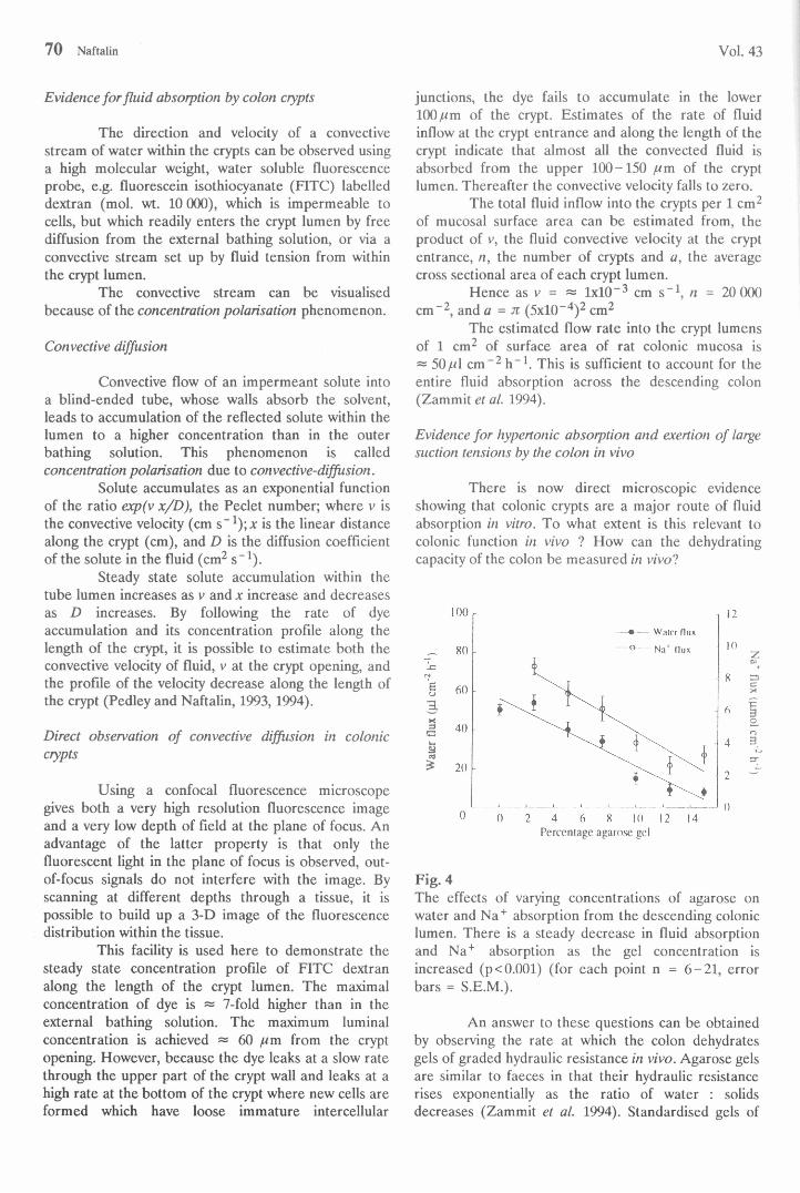
The estimated flow rate into the crypt lumens of 1 cm2 of surface area of rat colonic mucosa is ~ 50/i\ cm-2 h -1. This is sufficient to account for the entire fluid absorption across the descending colon (Zammit et al. 1994).
Evidence for hypertonic absorption and exertion of large suction tensions by the colon in vivo
There is now direct microscopic evidence showing that colonic crypts are a major route of fluid absorption in vitro. To what extent is this relevant to colonic function in vivo ? How can the dehydrating capacity of the colon be measured in vivo?
n
u .a£
Percentage agarose gel
zZ2CX
3oo3Kjsr
Fig. 4The effects of varying concentrations of agarose on water and Na+ absorption from the descending colonic lumen. There is a steady decrease in fluid absorption and Na+ absorption as the gel concentration is increased (p< 0.001) (for each point n = 6-21, error bars = S.E.M.).
An answer to these questions can be obtained by observing the rate at which the colon dehydrates gels of graded hydraulic resistance in vivo. Agarose gels are similar to faeces in that their hydraulic resistance rises exponentially as the ratio of water : solids decreases (Zammit et al. 1994). Standardised gels of
1994 Dehydrating Function of Descending Colon 7 1
varying strengths, calibrated for hydraulic conductance (Lp) were inserted into the descending colon of anaesthetised rats. The capacity of the colon to dehydrate and desalinate the gels was measured by monitoring water and salt loss from the gels over 1 -2 h. As the gel strength increases, the rates of fluid and Na+ absorption from the gels diminishes (Fig. 4). However, the ratio of Na+ absorption : water absorption increases, i.e. the tonicity of the colonic absórbate from the luminal gels is increased as the hydraulic resistance of luminal load is raised (Fig. 5).
Fig. 5The estimated suction pressure (cm H2O) and hypertonicity (cm H2O) of the descending colonic absórbate as a function of agarose concentration in the luminal solution (for each point n = 6-21).
The suction pressure (Fig. 5) exerted by the colon on the gels is estimated directly from the relationship P = Jw/Lp. The efficiency of conversion of the osmotic pressure to suction pressure rises as the luminal resistance increases. The suction pressure exerted by the colonic mucosa increases as the gel strength is increased, the maximal pressure exerted ~ 4 000 cm H2O on 15 % gels.
These experiments show the following points:a) When there is low luminal hydraulic resistance, i.e. liquid faeces in the lumen, fluid absorption is relatively rapid and nearly isotonic and the mucosa exerts a low suction pressure.b) When there is a high luminal hydraulic resistance, i.e. there is solid faeces in the lumen, the descending colon produces a very hypertonic absórbate and high suction pressure which acts to dehydrate the faeces.c) Agents, such as the bile salt, deoxycholic acid, which increase the leakiness of the junctions between the
cells, prevent water absorption by preventing the accumulation of a hypertonic pericryptal solution,d) The colonic mucosa adjusts to the large variation in luminal hydraulic load by increasing the tight- junctional hydraulic and solute flow resistance when the luminal hydraulic resistance increases. This increases the efficiency of conversion of osmotic pressure to suction pressure within the crypt lumen and probably also increases the crypt luminal resistance by narrowing the lumen, thereby decreasing the Lp of the transcryptal route for water flow.
Although the ileum has a much higher rate of fluid absorption than the colon, it is very poor at absorbing fluid against a large hydraulic resistance. A 2.5 % gel agarose reduces fluid absorption by the ileum by 75 % but has no effect on fluid absorption by the colon. This prevents chylous fluid within the ileum from ever becoming dehydrated.
How can the colonic crypts withstand the high suction pressures within the lumen?
Although the suction pressure exerted by the colonic crypts on the luminal contents is huge, it acts at the microscopic level of the crypt lumen. Macroscopic effects on faeces are only observed because of the very large numbers of crypts acting in parallel.
Laplace’s law plays an important role in tissue mechanics, for a cylinder P = r/r ; where P = lumen pressure, r = wall tension and r = lumen radius.
Fluid tension develops within the crypt lumen, because of fluid withdrawal from the lumen across the crypt wall. This leads to a reduction in luminal pressure and ultimately to inward collapse of the crypt lumen. The functional diameter of the collapsed crypt then decreases to < 0.1 .pm (Bleakman and Naftalin 1990).
Thus, a pressure of -500 kPa exerted in a cylinder of lumen radius 0.1 ^m and outer radius 30 ¡urn exerts a tension of 0.05 N m '1 at the inner surface of the crypt and ~ 10 N m '1 at the outer circumference of the crypt cells.§
The narrow-bore cylindrical crypt lumen lined with wedge-shaped cells is well-suited to withstand huge compressive forces and is also an effective way of converting osmotic pressure into fluid tension. The seal between the crypt luminal opening and the solid faeces in the colonic lumen is maintained by a surface layer of mucus covering the surfaces of the colon and faeces. This mucus permits the faeces to slide across the luminal surface, whilst suction is maintained.
High fluid tension develops because fluid in a capillary is not subject to cavitation even at tensions as large as 10 MPa.§§
^The wall tension in the aorta, 2 cm radius with mean blood pressure = 100 mm Hg (13.4 kPa), is 260 Nm 1 (Smaje et at. 1980).§§Very high fluid tensions have been observed elsewhere. Tree roots and octopus suckers can also generate negative fluid tensions (Pickard 1981, Smith 1991).
72 Naftalín Vol. 43
Control o f colonic absorption
The orthodox view is that "crypts are secretory" and in the small intestine the absorption takes place exclusively in the villus. In the colon, the relatively trivial absorptive function is considered to be undertaken by the surface mucosa. We have already seen that these views are incorrect. How did they arise?
The evidence for the secretory function of crypts arises from an experiment in which isolated colonic mucosa is exposed to the secretagogue prostaglandin E2. Net fluid is expelled from the lumens of isolated crypts into the mucosal bathing solution following exposure to the secretagogue (Welsh et al. 1982). The main objections to interpreting these data
to mean that the colonic crypts are normally secretory are as follows:a) there is a contractile layer of cells surrounding the basal surface of colonic crypts which, when induced to contract by PGE2, will cause a shortening of the crypt length and a decrease in crypt diameter - thus squeezing the fluid contents out of the lumen (Joyce et al. 1987), andb) this phenomenon is transient and could be useful in ejecting accumulated solid materials from the crypt lumen.
Thus a secretagogue-induced fluid and electrolyte secretion is completely compatible with absorption during normal colonic function, but it is not the normal mode of functioning of colonic crypts.
References
BLEAKMAN D., NAFTALÍN R.J.: Hypertonic fluid absorption from rabbit descending colon in vitro. Am. J. Physiol. 258: G377-G390,1990.
BOLTON Vi.: A Guide to Soil Mechanics. London, Macmillan, 1979, pp. 27-37 and 157-187.CURRAN P.F., SCHWARTZ G.F.: Na, Cl and water transport by rat colon./. Gen. Physiol. 43: 555-571, 1960.DEBONGNIE J.C., PHILLIPS S.F.: Capacity of human colon to absorb fluid. Gastroenterology 74:698-703, 1978.DEVROEDE G.F., PHILLIPS S.F.: Studies of the perfusion technique for colonic absorption. Gastroenterology
56: 92-100,1969.DUTHIE H.L., WATTS J.M., DE DOMBAL F.T., GALLIGHER J.C.: Serum electrolytes and colonic transfer of
water and electrolytes in chronic ulcerative colitis. Gastroenterology 47: 525 - 530, 1964.GRAMLICH T.L., HENNIGAR R A , SPICER S.S., SCHULTE B.A.: Immunohistochemical localization of
sodium-potassium-stimulated adenosine triphosphatase and carbonic anhydrase in human colon and colonic neoplasm. Arch. Pathol. Lab. Med. 114: 415-419, 1990.
GREENBERGER N.J.: Gastrointestinal Disorders: A Pathophysiologic Approach. 4th ed. Year Book Medical Publishers, Chicago, 1990.
HECKER J.F., GROVUM W.L.: The rates of passage of digesta and water absorption along the large intestines of sheep, cows and pigs. Austr. J. Biol. Sci. 28:161 -187, 1975.
JOHNSON J.R.: Gastrointestinal Physiology. 4th ed. Mosby Year Book, St. Louis, 1991.JOYCE N.C., HAIRE M.F., PALADE G.E.: Morphologic and biochemical evidence for a contractile cell network
within the rat intestinal mucosa. Gastroenterology 92: 68-81, 1987.LACY E.R.: Functional morphology of the large intestine. In: The Handbook of Physiology, Section 6, The
Gastrointestinal System, Vol. IV, M. Field, R.A. Frizzell (eds), American Physiological Society, Bethesda, 1991, pp. 121-195.
McKIE A.T., POWTRIE W., NAFTALÍN R.J.: Mechanical aspects of rabbit fecal dehydration. Am. J. Physiol. 258: G391-G394, 1990.
NAFTALÍN R.J., PEDLEY K.C.: Video enhanced imaging of the fluorescent N a+ probe SBFI indicates that colonic crypts absorb fluid by generating a hypertonic interstitial fluid. FEBS Letters 260: 187-194, 1990.
PEDLEY K.C., NAFTALÍN R.J.: Evidence from fluorescence microscopy and comparative studies that rat, ovine and bovine colonic crypts are absorptive./. Physiol. Loud. 460: 525-547, 1993.
PEDLEY K.C., NAFTALÍN R.J.: Confocal microscopic study of concentration polarization of fluorescein isothiocyanate dextran (mol. weight 10,000) within the lumens of rat descending colon crypts in vitro. /. Physiol. Lond. 1994 in press.
PICKARD W.F.: The ascent of sap in plants. Prog. Biophys. Mol. Biol. 37:181-229, 1981.POWELL D.W.: Transport in Large Intestine. In: Membrane Transport in Biology, G. GIESBISCH, D.C.
TOSTESON, H.H. USSING (eds), IVB Transport Organs, Berlin, Springer-Verlag, 1978, pp. 781-809..SMAJE L.H., FRASER PA., CLOUGH G.: The distensibility of single capillaries and venules in the cat
mesentery. Microvasc. Res. 20: 358 - 370, 1980.SMITH A.M.: Negative pressure generated by octopus suckers: a study of the tensile strength of water in nature.
/. Exp. Biol. 157: 257 - 271, 1991.
1994 Dehydrating Function of Descending Colon 73
WELSH M.J., SMITH P.L., FROMM M., FRIZZELL R.A.: Crypts are the site of intestinal fluid and electrolyte secretion. Science 218:1219 -1221, 1982.
ZAMMIT P.S., MENDIZABAL M.V., NAFTALIN R.J.: Effects on fluid, Na+ and K+ flux of varying luminal hydraulic resistance in rat colon in vivo. J. Physiol. Lond. 1994 in press