Dehydrogenase GRD1 Represents a Novel Component of theCellulase Regulon in Trichoderma reesei (Hypocrea jecorina)�†§
Andre Schuster, Christian P. Kubicek, and Monika Schmoll*Research Area of Gene Technology and Applied Biochemistry, Institute for Chemical Engineering, Vienna University of Technology,
Gumpendorfer Strasse 1a/1665, A-1060 Vienna, Austria
Received 6 March 2011/Accepted 8 May 2011
Trichoderma reesei (Hypocrea jecorina) is nowadays the most important industrial producer of cellulase andhemicellulase enzymes, which are used for pretreatment of cellulosic biomass for biofuel production. In thisstudy, we introduce a novel component, GRD1 (glucose-ribitol dehydrogenase 1), which shows enzymaticactivity on cellobiose and positively influences cellulase gene transcription, expression, and extracellularendo-1,4-�-D-glucanase activity. grd1 is differentially transcribed upon growth on cellulose and the inductionof cellulase gene expression by sophorose. The transcription of grd1 is coregulated with that of cel7a (cbh1)under inducing conditions. GRD1 is further involved in carbon source utilization on several carbon sources,such as those involved in lactose and D-galactose catabolism, in several cases in a light-dependent manner. Weconclude that GRD1 represents a novel enhancer of cellulase gene expression, which by coregulation with themajor cellulase may act via optimization of inducing mechanisms.
Plant cell walls, which predominantly consist of cellulosicmaterial, represent one of the most abundant renewable en-ergy sources on earth. Natural recycling processes, which in-volve fungi as essential degraders of cellulosic biomass, arecrucial for the environmental carbon cycle. One of the mostefficient fungi in this respect is Trichoderma reesei (anamorphof Hypocrea jecorina), which is of high industrial importancebecause of this very characteristic. This ascomycete is able toproduce and secrete up to 100 g of extracellular protein perliter (7), an attribute which is mainly exploited for the produc-tion of highly efficient cellulase mixtures. These enzymes arewidely used in industry (3, 19, 20). The most promising appli-cation nowadays, however, is the use of cellulases for the pro-duction of second-generation biofuels. Since T. reesei is themost prolific producer of cellulases, the components of itscellulase system have been subject to extensive research on thestructure, induction, expression, and regulation of the produc-tion of these enzymes (32, 53).
Cellulases are induced by cellulose, but by the late 1950s, itwas already found that soluble carbon sources can also inducecellulases (39). It has been reported that sophorose (37, 61),cellobiose (38), lactose (39), L-sorbose (29), L-arabitol, and afew additional substrates (40) can act as inducers of cellulasegene expression in T. reesei. Sophorose (2-O-�-glucopyranosyl-D-glucose), a transglycosylation product of cellobiose, is a verypowerful inducer of cellulases (37, 61) which is converted fromcellobiose by beta-glucosidase (68). However, upon induction
by sophorose and lactose, only an incomplete array of cellu-lases is produced (42, 62). Additionally, it was reported thatdespite seemingly similar conditions of cellulase induction, dif-ferential gene expression occurs on cellulose and sophorose(52).
While the regulation of cellulase gene expression on differ-ent carbon sources and in the presence of numerous inducingcompounds has been studied for decades now, the influence oflight on this process was only discovered a few years ago (51).Although the initially detected effect of light on the transcrip-tion of cellulase genes was only about 50%, later studies in-volving different signaling components revealed up to 10-folddifferences between parental and mutant strains in depen-dence on light (54, 57). Important components involved inlight-modulated cellulase gene transcription are ENV1 (51)and the photoreceptors BLR1 and BLR2 (6). These consider-able effects of light require controlled light conditions, i.e., theelimination of random light or dark pulses during cultivation,for the analysis of cellulase gene expression in T. reesei, whichwe also routinely apply.
In this study, we characterize a novel dehydrogenase whichis strongly upregulated upon induction by sophorose versusgrowth on cellulose and is coregulated with cel7a (cbh1).GRD1 (glucose-ribitol dehydrogenase 1) exhibits enzymaticactivity on cellobiose and positively affects cellulase gene tran-scription, expression as well as extracellular endo-1,4-�-D-glu-canase activity. The finding that the effects of GRD1 are in partdependent on the light status is in agreement with light-mod-ulated regulation of cellulase gene expression.
MATERIALS AND METHODS
Strains, plasmids, and culture conditions. T. reesei strain QM9414 (ATCC26921) and a T. reesei �tku70 strain, which is defective in the nonhomologousend-joining pathway (24), were used in the present study. For Northern analyses,T. reesei was grown in liquid culture in 200 ml Mandels Andreotti minimalmedium (36) supplemented with 0.1% (wt/vol) peptone to induce germinationand with 1% (wt/vol) microcrystalline cellulose (catalog no. 14204; Serva, Heidel-berg, Germany) as a carbon source under controlled light conditions. For culti-
* Corresponding author. Mailing address: Vienna University ofTechnology, Institute of Chemical Engineering, Research Area Mo-lecular Biotechnology, Gumpendorfer Strasse 1a/1665, 1060 Vienna,Austria. Phone: 43 1 58801 166552. Fax: 43 1 58801 17299. E-mail:[email protected].
† Supplemental material for this article may be found at http://aem.asm.org/.
� Published ahead of print on 20 May 2011.§ The authors have paid a fee to allow immediate free access to
this article.
4553
Dow
nloa
ded
from
http
s://j
ourn
als.
asm
.org
/jour
nal/a
em o
n 24
Dec
embe
r 20
21 b
y 11
2.15
1.10
4.56
.
vation in light, daylight neon lamps were used and set to 1,800 lx (25 �molphotons m�2 s�1). Harvesting of dark-grown cultures was done under safe redlight (red, E27, 15-W Philips PF712E darkroom lamp) in order to preventrandom light pulses. After cultivation of QM9414 in darkness for analysis onglycerol (24 h), glucose (17 h), or lactose (20 h) and harvesting of the darkcontrol sample at time point 0 (18), strains were grown in light for 15, 30, 60, and120 min and harvested by filtration. The duration of precultivation in darkness(24, 17, or 20 h, respectively) was chosen according to the growth of the strainsin order to ensure comparable biomass production. For the replacement exper-iments, T. reesei was grown as described above on glycerol. After 24 h of culti-vation in darkness, the mycelia were harvested, washed, and transferred intofresh Mandels Andreotti minimal medium containing 1.5 mM sophorose (no.35208.01 Serva, Heidelberg, Germany) as the carbon source. After 2, 4, and 5 hof cultivation in constant light or constant darkness, mycelia were harvested,washed with tap water, and frozen in liquid nitrogen.
Escherichia coli JM109 was used for the propagation of vector molecules andDNA manipulations (71).
Rapid subtraction hybridization for analysis of differential expression. Isola-tion of total RNA, preparation of cDNA, comparative cDNA hybridization, andreverse Northern blotting were performed as described earlier (27, 52, 56).Briefly, parental strain QM9414 was grown on Mandels Andreotti minimal me-dium with 1% (wt/vol) cellulose as carbon source for 48 h or pregrown on 1%(wt/vol) glycerol for 24 h, replaced into medium containing 1.5 mM sophorosefor cellulase induction, and harvested after 5 h of cultivation as described earlier(52). After isolation of total RNA, cDNA was prepared using the CreatorSMART cDNA library construction kit (Clontech, TakaraBio United States),digested with EcoRII (all restriction enzymes used were purchased from Fer-mentas, Vilnius, Lithuania), and purified. Amounts of 100 ng of tester cDNA(QM9414/sophorose) and 3 �g of driver cDNA (QM9414/cellulose) were usedfor rapid subtraction hybridization. Putatively differentially expressed cDNAfragments were cloned and transformed into E. coli JM109. PCR fragmentsamplified from these clones were tested for differential expression by reverseNorthern blotting.
Construction of the T. reesei (�tku70) �grd1 strain. To construct a �grd1 strainof T. reesei, the 3� region of grd1 was amplified using primers pDelGRD13F(sequences of all oligonucleotides used in this study are listed in Table 1) andpDelGRD13R, and the PCR product was digested with SalI and EcoRV. The 5�region was amplified using primers pDelGRD15F and pDelGRD15R, and thePCR product was digested with Acc65I and SalI. The pBluescript vector (Strat-agene, La Jolla, CA) was digested with EcoRV and Acc65I. The prepared 3� and5� fragments were purified and ligated simultaneously into the pBluescript vectorto obtain pBgrd135. This plasmid was digested with SalI, dephosphorylated usingshrimp alkaline phosphatase (Fermentas), and ligated to a pyr4 fragment excisedfrom pBpyr4 (pBluescript containing the pyr4 fragment excised from pFG1integrated into the SalI site [22]) with SalI to obtain pDELgrd1P (see Fig. S1Ain the supplemental material). For transformation, pDELgrd1P was linearizedwith EcoRV and 10 �g of linearized vector was used for the transformation ofprotoplasts of the �tku70 strain as described previously (23).
Deletion strains were tested for integration of the construct by PCR usingprimers grd1cDNAF and grd1cDNAR, which bind outside the region to bedeleted. Successful deletion resulted in the amplification of a longer fragment(2,805 bp) than amplification from DNA of the parental strain (1,303 bp) (datanot shown).
For complementation of �grd1 with the grd1 wild-type gene, we amplified the
entire gene, including flanking sequences, using primers pDelgrd15F andpDelgrd13R. This PCR product was ligated into pGEMT (Promega, Madison,WI) to obtain grd1pGEMT. The fragment was excised from grd1pGEMT usingNotI and ligated into the NotI site of the vector pBSamds (55) to obtainpBgrd1Re. After retransformation of �grd1, integration was confirmed usingprimers grd1cDNAF and grd1cDNAR as described above.
Construction of a T. reesei grd1 overexpression strain. To construct a grd1overexpression strain of T. reesei, plasmid pLH1hph (26) was used as the back-bone vector. The hph open reading frame was excised by digestion with NsiI andXbaI to obtain pLH1. A fragment was amplified using primers grd1anti1F andgrd1anti1R, which were used to introduce a multiple cloning site and digestedwith PstI and NheI and ligated into the NsiI-XbaI (for creation of correspondingends to PstI and NheI)-digested pLH1 vector to obtain pLH1mcs. Primergrd1anti1R contains five additional restriction sites (SpeI, SalI, DraI, BglII, andStuI) for the introduction of a new multiple cloning site. From pBSXH (51), thehph (pki1p::hph::cbh2t) cassette was excised by digestion with XhoI, Klenow fill-inwas applied to create a blunt end, and the resulting fragment was digested withBamHI. The vector pLH1mcs was digested with BamHI and SmaI and ligatedwith the digested hph fragment to obtain pLH1mcsHPH. This vector was di-gested with StuI, purified, and religated in order to remove the grd1 antisensefragment and leave the multiple cloning site intact. The resulting vector(paGPDhph) contains a gpd1 (glycerol 3-phosphate) promoter and terminator,hygromycin B resistance gene (hph), and a multiple cloning site (see Fig. S1B inthe supplemental material). The open reading frame of grd1 was amplified usingPhusion polymerase (Finnzymes, Espoo, Finland) and primers grd1cDNAF andgrd1cDNAR. The PCR product was ligated into the StuI site of paGPDhph toobtain pagrd1hph (see Fig. S1B in the supplemental material). After confirma-tion of the correct orientation of the inserted fragment, the resulting vector waslinearized by digestion with NarI and used for transformation into the genome ofparental strain QM9414 as described previously (23). Mutant strains were testedfor integration of the construct by PCR using primers grd1cDNAF and TgpdR(26) or grd1cDNAR and PgpdF (26). These primers bind inside the gpd1 pro-moter or terminator region or in the grd1 region. Successful integration resultedin a specific band (1,824 bp or 2,195 bp, respectively), and no amplification waspossible with DNA from the parental strain (data not shown).
Biolog Phenotype MicroArray technique. T. reesei strains were grown on 2%(wt/vol) malt extract agar plates at 28°C for 1 week until sporulation. Inoculawere extracted from these plates. The assay was performed as described previ-ously (12, 13). The Biolog Phenotype MicroArray microplates were incubated for72 h in constant light or constant darkness. Analyses were repeated at least twicefor each strain.
Phylogenetic analysis. Clustal X 2.0.7 (33) was used for the alignment ofamino acid sequences. MEGA 4.0.2 (64) was used for phylogenetic analysis,using the minimum evolution method and 500 bootstrap replications as a test ofphylogeny. Protein sequences were obtained from the Joint Genome Institute T.reesei, Trichoderma viride, and Trichoderma atroviride genome databases (http://genome.jgi-psf.org/). Sequences of proteins encoded by the genomes of Neu-rospora crassa, Aspergillus spp., Fusarium spp., and Magnaporthe grisea wereretrieved from their respective genome databases at the Broad Institute (http://www.broadinstitute.org/science/data).
Nucleic acid isolation and hybridization. Fungal mycelia were harvested byfiltration, washed with tap water, frozen, and ground in liquid nitrogen. Forextraction of DNA, mycelium powder was suspended in buffer A (1.2 M NaCl, 5mM EDTA, 0.1 M Tris-HCl, pH 8.0), incubated for 20 min at 65°C, cooled on
a Restriction sites introduced to facilitate cloning are underlined.
4554 SCHUSTER ET AL. APPL. ENVIRON. MICROBIOL.
Dow
nloa
ded
from
http
s://j
ourn
als.
asm
.org
/jour
nal/a
em o
n 24
Dec
embe
r 20
21 b
y 11
2.15
1.10
4.56
.
ice, mixed with 1 volume phenol/chloroform/isoamyl alcohol (49:49:2 [vol/vol/vol]), and centrifuged (21,000 � g for 15 min). Following an extraction with 1volume of chloroform/isoamyl alcohol (24:1 [vol/vol]), DNA was precipitatedwith 1 volume of isopropanol and washed with 70% (vol/vol) ethanol. Total RNAwas isolated by the guanidinium thiocyanate method (10). Standard methods (50,56) were used for electrophoresis, blotting, and hybridization of nucleic acids.
cDNA preparation and RT-PCR. Total RNA was treated with DNase I (Fer-mentas, Vilnius, Lithuania). cDNA was prepared using a RevertAID H minusfirst-strand cDNA synthesis kit (Fermentas). GoTaq polymerase (Promega,Madison, WI) was used for subsequent PCR amplification. Primers tef1F andtef1R were used for amplification of tef1 for 25 cycles, and primers grd1cDNAFand grd1cDNAR for amplification of grd1 for 35 cycles.
Biomass determination. Parental strain QM9414 and the �grd1 strain weregrown in liquid medium with cellulose as the carbon source as described above.Mycelia were ground in liquid nitrogen, and the powder was suspended in 0.1 NNaOH. This suspension was sonicated three times for 1 min and incubated for3 h at room temperature. Samples were centrifuged for 10 min at 10,000 rpm,and the supernatants were transferred to new reaction tubes. The protein con-centration reflecting biomass content was measured using a Bio-Rad proteinassay (Bio-Rad Laboratories, Hercules, CA).
Determination of cellulase activity in culture filtrates. The parental strainQM9414 and the �grd1 and grd1S (constitutively expressing GRD1) strains weregrown as described above. Culture filtrates were used for the analysis of endo-1,4-�-D-glucanase activity with the aid of an azo-CM-cellulose kit (no. S-ACMC,Megazyme, Wicklow, Ireland), which was used according to the manufacturer’sinstructions.
Protein recovery from culture filtrates and Western blotting. A 500-�l volumeof culture filtrate from cultivation on Mandels Andreotti minimal medium with1% (wt/vol) microcrystalline cellulose in constant light and constant darkness for72 h and 96 h was precipitated by adding 2 ml methanol, 500 �l chloroform, and1.5 ml water and mixed well. After centrifugation for 5 min at 8,000 rpm, theupper phase was discarded. Thereafter, 1.5 ml methanol was added and mixedwell and the mixture centrifuged again for 5 min. Then the supernatant wasdiscarded, and the pellet was dried at room temperature and redissolved in 50 �lsterile water.
SDS-PAGE and Western blotting were performed according to standard pro-tocols (50). Proteins were blotted on a nitrocellulose membrane (Hybond-CExtra, Amersham Biosiences, United Kingdom) by semidry electroblotting. Foranalysis of the expression of cellulases, antibodies against the major cellulaseCEL7A (46) and a horseradish peroxidase-conjugated anti-mouse IgG (Pro-mega, Madison, WI) were used.
Production and purification of a GST-GRD1 fusion protein. To construct aglutathione S-transferase (GST) fusion expression vector, the grd1 open readingframe was amplified from cDNA prepared from QM9414 grown on lactose byPCR using primers GRD1GSTR and GRD1GSTF. The resulting PCR productwas cloned into the BamHI-XhoI sites of the pGEX-4T2 (Stratagene) vector toobtain GRD1GSTpGex. This vector was transformed into E. coli BL21 (Strat-agene). The resulting E. coli strain was grown on selective LB medium to anoptical density at 600 nm (OD600) of 0.600. Production of the heterologousprotein was induced using 0.1 mM (final concentration) isopropyl-�-D-thiogalac-topyranoside (IPTG; Fermentas), and the culture was harvested by centrifuga-tion after 3 h of growth under inducing conditions. The heterologous protein waspurified using glutathione-Sepharose 4B according to the manufacturer’s proto-col (GE Bioscience, Uppsala, Sweden). Purification and production of the GSTfusion protein was confirmed by SDS-PAGE. The concentration of the resultingrecombinant protein was determined with the Bio-Rad protein assay (Bio-Rad).
Determination of enzyme specificity. GRD1 enzymatic activity was determinedspectrophotometrically essentially as described earlier (48). Briefly, we measuredthe change in absorbance at 340 nm for NADH and NADPH oxidation or NAD�
and NADP� reduction of the respective substrate by GRD1 at 30°C. Concen-trations of 100 mM substrate and 0.25 mM NADH/NADPH and 100 �g ofpurified GST protein were used in the assay. Activities are given as specificactivities in nkat/mg protein.
For purification of the enzyme assay, we used Vivaspin 2 columns with a 3,000molecular-mass-cutoff polyethersulfone (PES) membrane (Sartorius Stedim Bio-tech, Cedex, France). Amounts of 1 ml of reaction mixture were added to thecolumns and centrifuged for 10 min at 10,000 rpm in a fixed-angle rotor at 20°C.For high-performance liquid chromatography (HPLC) analysis, we used anAminex HPX-87H column (Bio-Rad) with 10 mM H2SO4 as the mobile phase ata flow rate of 0.5 ml min�1 at 40°C. The concentration of substrate was calculatedfrom a calibration curve established with 1 mM, 10 mM, 50 mM, and 100 mMconcentrations of the respective substrate.
RESULTS
Characterization of a putative cellulase induction-associ-ated gene. A cDNA screening experiment comparing genesdifferentially transcribed during growth on cellulose or afterthe induction of cellulase gene expression by sophorose (52)revealed that these two conditions are indeed not entirelycomparable. Several genes were found to be differentially reg-ulated on these two carbon sources, which is in agreement withearlier studies showing that the cellulase mixtures secretedupon induction by these carbon sources are not identical (42,62). We therefore conducted a similar experiment aimed atidentifying genes specifically transcribed in the presence ofsophorose, using cDNA from cultures grown in the presence ofsophorose (5 h after replacement) as the tester and cDNAresulting from cellulose-grown mycelia as the driver. This anal-ysis revealed several transcripts to be more abundant upongrowth in the presence of sophorose than with cellulose as thecarbon source (see Table S1 in the supplemental material).Among those, the gene represented by STCD21 and STCD55,a putative glucose-ribitol dehydrogenase (TR_123946), wasfound most frequently (9 fragments targeted this gene). En-zymes of this group (EMBL-EBI InterPro domain IPR002347)are tetrameric and catalyze the oxidation of D-glucose withoutprior phosphorylation to D-�-gluconolactone. Because of itspresumed function as a glucose-ribitol dehydrogenase, wenamed this gene grd1 (glucose-ribitol dehydrogenase 1).
Grd1 comprises a 1,205-bp open reading frame interruptedby one intron and encodes a protein of 304 amino acids. It hasno N-terminal signal peptide and is predicted to be located inthe cytoplasm (PSORTII; http://www.psort.org/).
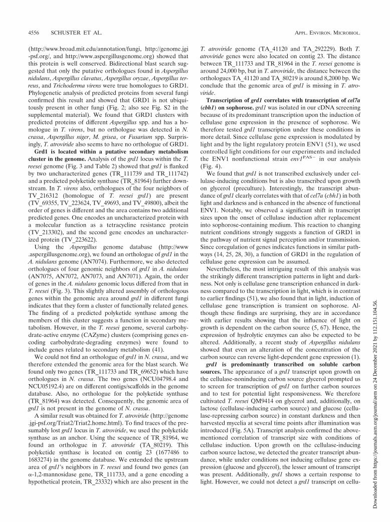
GRD1 is a conserved but not ubiquitous dehydrogenase infungi. Searching the T. reesei genome for genes encoding pro-teins with characteristics similar to those of GRD1, we foundnine additional models comprising the same domains(IPR002198, IPR002347, and IPR016040), hence indicatingthat the T. reesei genome comprises 10 putative glucose-ribitoldehydrogenases (Fig. 1). However, although GRD1 seems tobe conserved in several fungal species, we could not find acharacterized orthologue of GRD1 in fungi. Nevertheless,BLAST searches against several fungal genome databases
FIG. 1. Phylogenetic analysis of short-chain dehydrogenases usingthe minimum evolution method and 500 bootstrap replications as testof phylogeny. Phylogenetic tree obtained by MEGA 4.0.2. The se-quences were obtained from the JGI T. reesei genome database, ver-sion 2.0 (http://genome.jgi-psf.org/Trire2/Trire2.home.html). The scalebar reflects evolutionary distance.
VOL. 77, 2011 GRD1, A NOVEL DEHYDROGENASE IN T. REESEI 4555
Dow
nloa
ded
from
http
s://j
ourn
als.
asm
.org
/jour
nal/a
em o
n 24
Dec
embe
r 20
21 b
y 11
2.15
1.10
4.56
.
(http://www.broad.mit.edu/annotation/fungi, http://genome.jgi-psf.org/, and http://www.aspergillusgenome.org) showed thatthis protein is well conserved. Bidirectional blast search sug-gested that only the putative orthologues found in Aspergillusnidulans, Aspergillus clavatus, Aspergillus oryzae, Aspergillus ter-reus, and Trichoderma virens were true homologues to GRD1.Phylogenetic analysis of predicted proteins from several fungiconfirmed this result and showed that GRD1 is not ubiqui-tously present in other fungi (Fig. 2; also see Fig. S2 in thesupplemental material). We found that GRD1 clusters withpredicted proteins of different Aspergillus spp. and has a ho-mologue in T. virens, but no orthologue was detected in N.crassa, Aspergillus niger, M. grisea, or Fusarium spp. Surpris-ingly, T. atroviride also seems to have no orthologue of GRD1.
Grd1 is located within a putative secondary metabolismcluster in the genome. Analysis of the grd1 locus within the T.reesei genome (Fig. 3 and Table 2) showed that grd1 is flankedby two uncharacterized genes (TR_111739 and TR_111742)and a predicted polyketide synthase (TR_81964) further down-stream. In T. virens also, orthologues of the four neighbors ofTV_216312 (homologue of T. reesei grd1) are present(TV_69355, TV_223624, TV_49693, and TV_49800), albeit theorder of genes is different and the area contains two additionalpredicted genes. One encodes an uncharacterized protein witha molecular function as a tetracycline resistance protein(TV_213302), and the second gene encodes an uncharacter-ized protein (TV_223622).
Using the Aspergillus genome database (http://www.aspergillusgenome.org), we found an orthologue of grd1 in theA. nidulans genome (AN7074). Furthermore, we also detectedorthologues of four genomic neighbors of grd1 in A. nidulans(AN7075, AN7072, AN7073, and AN7071). Again, the orderof genes in the A. nidulans genomic locus differed from that inT. reesei (Fig. 3). This slightly altered assembly of orthologousgenes within the genomic area around grd1 in different fungiindicates that they form a cluster of functionally related genes.The finding of a predicted polyketide synthase among themembers of this cluster suggests a function in secondary me-tabolism. However, in the T. reesei genome, several carbohy-drate-active enzyme (CAZyme) clusters (comprising genes en-coding carbohydrate-degrading enzymes) were found toinclude genes related to secondary metabolism (41).
We could not find an orthologue of grd1 in N. crassa, and wetherefore extended the genomic area for the blast search. Wefound only two genes (TR_111733 and TR_69652) which haveorthologues in N. crassa. The two genes (NCU04798.4 andNCU05192.4) are on different contigs/scaffolds in the genomedatabase. Also, no orthologue for the polyketide synthase(TR_81964) was detected. Consequently, the genomic area ofgrd1 is not present in the genome of N. crassa.
A similar result was obtained for T. atroviride (http://genome.jgi-psf.org/Triat2/Triat2.home.html). To find traces of the pre-sumably lost grd1 locus in T. atroviride, we used the polyketidesynthase as an anchor. Using the sequence of TR_81964, wefound an orthologue in T. atroviride (TA_80219). Thispolyketide synthase is located on contig 23 (1677486 to1683274) in the genome database. We extended the upstreamarea of grd1’s neighbors in T. reesei and found two genes (an�-1,2-mannosidase gene, TR_111733, and a gene encoding ahypothetical protein, TR_23332) which are also present in the
T. atroviride genome (TA_41120 and TA_292229). Both T.atroviride genes were also located on contig 23. The distancebetween TR_111733 and TR_81964 in the T. reesei genome isaround 24,000 bp, but in T. atroviride, the distance between theorthologues TA_41120 and TA_80219 is around 8,2000 bp. Weconclude that the genomic area of grd1 is missing in T. atro-viride.
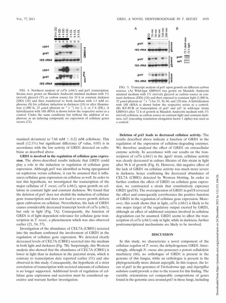
Transcription of grd1 correlates with transcription of cel7a(cbh1) on sophorose. grd1 was isolated in our cDNA screeningbecause of its predominant transcription upon the induction ofcellulase gene expression in the presence of sophorose. Wetherefore tested grd1 transcription under these conditions inmore detail. Since cellulase gene expression is modulated bylight and by the light regulatory protein ENV1 (51), we usedcontrolled light conditions for our experiments and includedthe ENV1 nonfunctional strain env1PAS� in our analysis(Fig. 4).
We found that grd1 is not transcribed exclusively under cel-lulase-inducing conditions but is also transcribed upon growthon glycerol (preculture). Interestingly, the transcript abun-dance of grd1 clearly correlates with that of cel7a (cbh1) in bothlight and darkness and is enhanced in the absence of functionalENV1. Notably, we observed a significant shift in transcriptsizes upon the onset of cellulase induction after replacementinto sophorose-containing medium. This reaction to changingnutrient conditions strongly suggests a function of GRD1 inthe pathway of nutrient signal perception and/or transmission.Since coregulation of genes indicates functions in similar path-ways (14, 25, 28, 30), a function of GRD1 in the regulation ofcellulase gene expression can be assumed.
Nevertheless, the most intriguing result of this analysis wasthe strikingly different transcription patterns in light and dark-ness. Not only is cellulase gene transcription enhanced in dark-ness compared to the transcription in light, which is in contrastto earlier findings (51), we also found that in light, induction ofcellulase gene transcription is transient on sophorose. Al-though these findings are surprising, they are in accordancewith earlier results showing that the influence of light ongrowth is dependent on the carbon source (5, 67). Hence, theexpression of hydrolytic enzymes can also be expected to bealtered. Additionally, a recent study of Aspergillus nidulansshowed that even an alteration of the concentration of thecarbon source can reverse light-dependent gene expression (1).
grd1 is predominantly transcribed on soluble carbonsources. The appearance of a grd1 transcript upon growth onthe cellulase-noninducing carbon source glycerol prompted usto screen for transcription of grd1 on further carbon sourcesand to test for potential light responsiveness. We thereforecultivated T. reesei QM9414 on glycerol and, additionally, onlactose (cellulase-inducing carbon source) and glucose (cellu-lase-repressing carbon source) in constant darkness and thenharvested mycelia at several time points after illumination wasintroduced (Fig. 5A). Transcript analysis confirmed the above-mentioned correlation of transcript size with conditions ofcellulase induction. Upon growth on the cellulase-inducingcarbon source lactose, we detected the greater transcript abun-dance, while under conditions not inducing cellulase gene ex-pression (glucose and glycerol), the lesser amount of transcriptwas present. Additionally, grd1 shows a certain response tolight. However, we could not detect a grd1 transcript on cellu-
4556 SCHUSTER ET AL. APPL. ENVIRON. MICROBIOL.
Dow
nloa
ded
from
http
s://j
ourn
als.
asm
.org
/jour
nal/a
em o
n 24
Dec
embe
r 20
21 b
y 11
2.15
1.10
4.56
.
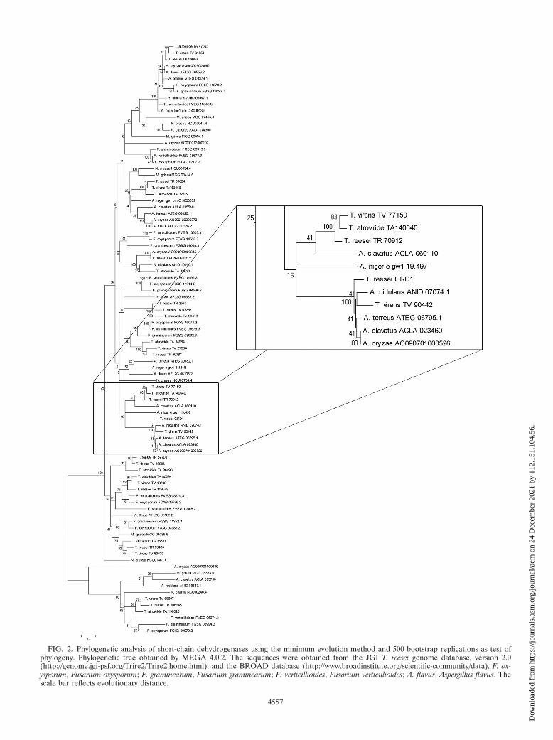
FIG. 2. Phylogenetic analysis of short-chain dehydrogenases using the minimum evolution method and 500 bootstrap replications as test ofphylogeny. Phylogenetic tree obtained by MEGA 4.0.2. The sequences were obtained from the JGI T. reesei genome database, version 2.0(http://genome.jgi-psf.org/Trire2/Trire2.home.html), and the BROAD database (http://www.broadinstitute.org/scientific-community/data). F. ox-ysporum, Fusarium oxysporum; F. graminearum, Fusarium graminearum; F. verticillioides, Fusarium verticillioides; A. flavus, Aspergillus flavus. Thescale bar reflects evolutionary distance.
4557
Dow
nloa
ded
from
http
s://j
ourn
als.
asm
.org
/jour
nal/a
em o
n 24
Dec
embe
r 20
21 b
y 11
2.15
1.10
4.56
.
lose using Northern blots, which confirms the validity of ourinitial cDNA screening approach for genes transcribed moreabundantly on sophorose than on cellulose. We therefore ap-plied RT-PCR, which revealed transcription of grd1 also upongrowth on cellulose for 72 h in light and darkness, albeit at verylow levels (Fig. 5B).
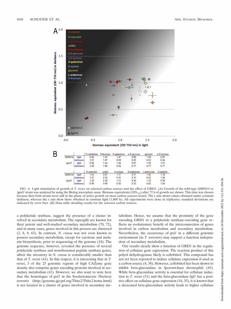
Carbon source utilization varies between parental strainand �grd1 strain in light and in darkness. In order to inves-tigate the function of GRD1 in T. reesei, we constructed adeletion mutant of grd1 in a strain defective in the nonhomol-ogous-end-joining pathway (24). The growth of the �grd1strain on plates is indistinguishable from the growth of theparental strain QM9414. Since grd1 is not transcribed onlyunder cellulase-inducing conditions, we intended to gain fur-ther insight as to the putative function of this dehydrogenaseby the use of Biolog Phenotype MicroArrays (13), which allowthe analysis of growth on 95 carbon sources. We tested thegrowth of the �grd1 strain and the parental strain in light anddarkness after 72 h of cultivation (Fig. 6; also see Fig. S3 in thesupplemental material).
Besides an only minor difference in growth on the cellulase
inducer cellobiose (38), we found that GRD1 positively influ-ences growth on �-D-lactose. Interestingly, we also found dif-ferences in growth between the parental strain and the mutanton several intermediates of lactose and D-galactose catabolism(32, 44, 58), specifically, D-galactose, D-fructose, D-xylose, xyli-tol, D-mannitol, and L-arabinose. In some cases, these differ-ences were dependent on the light status (Fig. 6). Since lactoseis an important inducer of cellulase gene expression (59), thisinfluence of GRD1 on growth on these intermediates is inaccordance with the hypothesis that GRD1 is involved in in-ducer modification at an as-yet-unknown metabolic level.
GRD1 shows enzymatic activity with cellobiose. The puta-tive function of GRD1 prompted us to investigate the substratespecificity of GRD1 in more detail. We used a GRD1-GSTfusion protein expressed for this purpose from cDNA in E. coliwithout trypsin digestion, because it was shown that cleavageof the GST moiety of the fusion protein could lead to aninactivation of the protein (58).
The enzymatic activity was tested on D-glucose, D-sorbitol,D-xylose, xylitol, D-galactose, L-arabinose, maltose, D-galactitol,L-arabitol, D-arabitol, D-mannitol, D-ribitol, cellobiose, andsophorose as substrates and with NADH, NAD�, NAD(P)H,or NAD(P)� as the cofactor. Specific activity was only detectedon cellobiose (52.9 nkat/mg protein, with NADPH as cofactor).The reaction mixtures from the assays with a positive result(cellobiose), as well as those with D-glucose as the negativecontrol, were purified to remove high-molecular-weight com-pounds and monitored by HPLC analysis. We found that theretention time of glucose was 12 min and that of cellobiose was10 min. As a negative control for our samples, we used areaction mixture similar to that used in the assay but withoutincubation. This control was immediately purified to removethe GST fusion protein and, hence, prevent the enzymaticreaction.
For D-glucose, as expected, no conversion was detected. Incontrast, we could detect a consumption of cellobiose. Afterincubation, the substrate concentration was decreased fromthe reference concentration of 8.71 mM 0.26 mM (mean
FIG. 3. Schematic representation of the genomic locus of grd1. Scaffold or position numbers are represented as specified in the respectivegenome databases (http://genome.jgi-psf.org/Trire2/Trire2.home.html and http://www.aspgd.org/). Genes encoding orthologues of GPD1 areshown in white, and orthologues of the polyketide synthase are shown in gray. Protein identification numbers are given in Table 2. The schemeis drawn to scale.
TABLE 2. Proteins encoded by genes found around the genomiclocus of GRD1 and its orthologues
a See Fig. 2.b Protein identification numbers are from the respective genome databases
(http://genome.jgi-psf.org/Trire2/Trire2.home.html and http://www.aspgd.org/).
4558 SCHUSTER ET AL. APPL. ENVIRON. MICROBIOL.
Dow
nloa
ded
from
http
s://j
ourn
als.
asm
.org
/jour
nal/a
em o
n 24
Dec
embe
r 20
21 b
y 11
2.15
1.10
4.56
.
standard deviation) to 7.66 mM 0.22 mM cellobiose. Thissmall (12.1%) but significant difference (P value, 0.05) is inaccordance with the low activity of GRD1 detected on cello-biose as described above.
GRD1 is involved in the regulation of cellulase gene expres-sion. The above-described results indicate that GRD1 couldplay a role in the induction or regulation of cellulase geneexpression. Although grd1 was identified as being upregulatedon sophorose versus cellulose, it can be assumed that it influ-ences cellulase gene expression on cellulose as well. In order totest this hypothesis, we investigated the transcription of themajor cellulase of T. reesei, cel7a (cbh1), upon growth on cel-lulose in constant light and constant darkness. We found thatthe deletion of grd1 does not abolish the induction of cellulasegene transcription and does not lead to severe growth defectsupon cultivation on cellulose. Nevertheless, the lack of GRD1causes considerably decreased transcript levels of cel7a (cbh1),but only in light (Fig. 7A). Consequently, the function ofGRD1 is of light-dependent relevance for cellulase gene tran-scription in T. reesei, a phenomenon which was also observedearlier (21, 54, 57).
Investigation of the abundance of CEL7A (CBH1) secretedinto the medium confirmed the involvement of GRD1 in theregulation of cellulase gene expression. We detected clearlydecreased levels of CEL7A (CBH1) secreted into the mediumin both light and darkness (Fig. 7B). Surprisingly, this Westernanalysis also showed that the abundance of CEL7A (CBH1) islower in light than in darkness in the parental strain, which iscontrary to transcription data reported earlier (51) and alsoobserved in this study. Consequently, the hypothesis of a strictcorrelation of transcription with secreted amounts of cellulasesis no longer supported. Additional levels of regulation of cel-lulase gene expression and secretion must be considered op-erative and warrant further investigation.
Deletion of grd1 leads to decreased cellulase activity. Theresults described above indicate a function of GRD1 in theregulation of the expression of cellulose-degrading enzymes.We therefore analyzed the effect of GRD1 on extracellularenzyme activity. In accordance with our results on the tran-scription of cel7a (cbh1) in the �grd1 strain, cellulase activitywas clearly decreased in culture filtrates of this strain in lightafter 96 h of growth (Fig. 8). However, this negative effect ofthe lack of GRD1 on cellulase activity was much more severein darkness, hence confirming the decreased abundance ofCEL7A (CBH1) detected by Western blotting. In order tofurther confirm the effect of GRD1 on cellulase gene expres-sion, we constructed a strain that constitutively expressesGRD1 (grd1S). The overexpression of GRD1 in grd1S reversedthe effect and consequently corroborates the positive functionof GRD1 in the regulation of cellulase gene expression. More-over, this result shows that in light, cel7a (cbh1) is likely to beone major target of the regulatory output exerted by GRD1,although an effect of additional enzymes involved in cellulosedegradation can be assumed. GRD1 seems to affect the tran-scription of cel7a (cbh1) only in light, while in darkness, furtherposttranscriptional mechanisms are likely to be involved.
DISCUSSION
In this study, we characterize a novel component of thecellulase regulon of T. reesei, the dehydrogenase GRD1. Inter-estingly, although N. crassa also possesses a potent cellulolyticmachinery (66), no orthologue of GRD1 is present in thegenome of this fungus, while an orthologue is present in thephylogenetically more distant aspergilli. In this respect, the lo-cus of grd1 in the genomes of Trichoderma spp. and Aspergillusnidulans could provide a clue to the reason for this finding. Thevariable orientations yet comparable compositions of genesfound in the genomic area around grd1 in these fungi, including
FIG. 4. Northern analysis of cel7a (cbh1) and grd1 transcription.Strains were grown on Mandels Andreotti minimal medium with 1%(wt/vol) glycerol (Y) as carbon source for 24 h in constant darkness(DD) (18) and then transferred to fresh medium with 1.5 mM so-phorose (S) for cellulase induction in darkness (18) or after illumina-tion (1,800 lx, 25 �mol photons m�2 s�1) for 2, 4, or 5 h (DL). Ahybridization with 18S rRNA is shown below the respective series as acontrol. Under the same conditions but without the addition of so-phorose as an inducing compound, no expression of cellulase genesoccurs (21).
FIG. 5. Transcript analysis of grd1 upon growth on different carbonsources. (A) Wild-type QM9414 was grown on Mandels Andreottiminimal medium with 1% (wt/vol) glycerol as carbon source in con-stant darkness (DD) (18) and then exposed to constant light (1,800 lx,25 �mol photons m�2 s�1) for 15, 30, 60, and 120 min. A hybridizationwith 18S rRNA is shown below the respective series as a control.(B) RT-PCR of transcription of grd1 and tef1 in wild-type strainQM9414 after 72 h of growth in Mandels Andreotti medium with 1%(wt/vol) cellulose as carbon source in constant light and constant dark-ness. tef1 (encoding translation elongation factor 1 alpha) was used asa control.
VOL. 77, 2011 GRD1, A NOVEL DEHYDROGENASE IN T. REESEI 4559
Dow
nloa
ded
from
http
s://j
ourn
als.
asm
.org
/jour
nal/a
em o
n 24
Dec
embe
r 20
21 b
y 11
2.15
1.10
4.56
.
a polyketide synthase, suggest the presence of a cluster in-volved in secondary metabolism. The aspergilli are known fortheir potent and well-studied secondary metabolism (70, 72),and in many cases, genes involved in this process are clustered(2, 8, 9, 63). In contrast, N. crassa was not even known topossess secondary metabolism, except for carotene and mela-nin biosynthesis, prior to sequencing of the genome (18). Thegenome sequence, however, revealed the presence of severalpolyketide synthase and nonribosomal peptide synthase genes,albeit the inventory in N. crassa is considerably smaller thanthat of T. reesei (41). In this respect, it is interesting that in T.reesei, 5 of the 25 genomic regions of high CAZyme genedensity also comprise genes encoding proteins involved in sec-ondary metabolism (41). However, we also want to note herethat the homologue of grd1 in the Sordariomycete Thielaviaterrestris (http://genome.jgi-psf.org/Thite2/Thite2.home.html)is not located in a cluster of genes involved in secondary me-
tabolism. Hence, we assume that the proximity of the geneencoding GRD1 to a polyketide synthase-encoding gene re-flects an evolutionary benefit of the interconnection of genesinvolved in carbon metabolism and secondary metabolism.Nevertheless, the occurrence of grd1 in a different genomicenvironment (in T. terrestris) may support a function indepen-dent of secondary metabolism.
Our results clearly show a function of GRD1 in the regula-tion of cellulase gene expression. The reaction product of thispolyol dehydrogenase likely is cellobiitol. This compound hasnot yet been reported to induce cellulase expression if used asa carbon source (4, 38). However, cellobiitol has been shown toinhibit beta-glucosidase in Sporotrichum thermophile (45).While beta-glucosidase activity is essential for cellulase induc-tion in T. reesei (31) and the beta-glucosidase bgl1 has a posi-tive effect on cellulase gene expression (16, 35), it is known thata decreased beta-glucosidase activity leads to higher cellulase
FIG. 6. Light stimulation of growth of T. reesei on selected carbon sources and the effect of GRD1. (A) Growth of the wild-type QM9414 or�grd1 strain was analyzed by using the Biolog microplate assay. Biomass equivalents (OD750) after 72 h of growth are shown. This time was chosenbecause then both strains were still in the phase of active growth on most carbon sources tested. The y axis shows values obtained under constantdarkness, whereas the x axis show those obtained in constant light (1,800 lx). All experiments were done in triplicates; standard deviations areindicated by error bars. (B) Data table detailing results for the selected carbon sources.
4560 SCHUSTER ET AL. APPL. ENVIRON. MICROBIOL.
Dow
nloa
ded
from
http
s://j
ourn
als.
asm
.org
/jour
nal/a
em o
n 24
Dec
embe
r 20
21 b
y 11
2.15
1.10
4.56
.
activities by enabling the maintenance of a larger pool of theinducing disaccharide (58). On the other hand, the transglyco-sylation product of cellobiose, sophorose, induces an intracel-lular beta-glucosidase (34). Thus, GRD1 could act as a regu-lator of beta-glucosidase activity in order to prevent inhibitoryglucose concentrations in the cell.
The coregulation of grd1 with cel7a (cbh1), together with theenzymatic activity of grd1 on the degradation product ofCBH1, cellobiose, could be responsible for intracellular sens-ing of cellulase efficiency and adjustment of cellulase levels andbeta-glucosidase activity to optimize energy-efficient substrateutilization. In this respect also, the expression of GRD1 undernoninducing conditions is useful, since the different transcriptsize under these conditions indicates activation upon receptionof the cellulose-derived signal. Most of the hydrolysis of cello-biose to glucose occurs outside the cell, but nevertheless, anuptake mechanism for cellobiose has been confirmed in T.reesei (17). These findings corroborate a specific signaling func-tion of this compound for the presence of cellulose outside thecell. The lack of GRD1 would thus cause increasing glucoseand decreased cellobiose levels (reflecting decreased amountsof cellulose) in the medium and, hence, in the cell (as producedby beta-glucosidase), which would cause decreased cellulasegene expression. We therefore propose a model in whichGRD1 represents an intracellular sensor of extracellular cel-lulolytic activity that adjusts beta-glucosidase activity and,hence, inducer (sophorose) and/or repressor (glucose) abun-dance.
The regulation of cellulase gene expression in T. reesei hasbeen subject to extensive research for decades. Especially afterthe cloning of the major cellulase of T. reesei, cbh1 (60, 65),elucidation of the mechanism of cellulase induction and regu-lation was studied at the molecular level. A few years thereaf-ter, comparison of mRNA and transcript levels in severalTrichoderma strains and under different conditions led to thewell-founded assumption that the regulation of cellulase gene
expression occurs at a pretranslational level (15, 43, 47). Untilrecently, light was not considered an influencing factor in cel-lulase gene expression. In this study, we report the surprisingdiscovery that the assumption of strictly transcriptional regu-lation of cellulase gene expression does not hold true for theapplication of different light conditions. Several reasons forthis phenomenon are conceivable. On one hand, light-depen-dent splicing has been shown in fungi (49, 69), but if thismechanism were at work, additional bands resulting from(probably nonfunctional) truncated versions of CBH1 shouldhave been detected on Western blots for light-grown cultures,which was not the case. However, clock regulation of ribosomebiogenesis (but not transcriptional regulation of ribosomegenes) has been shown and, hence, provides a means for post-transcriptional regulation of clock-controlled genes (11). Be-cause of the crucial importance of light for resetting of thecircadian clock, such a mechanism is likely to also be affectedby light, which would provide a reasonable explanation forposttranscriptional regulation of cellulase gene expression bylight.
Interestingly, this effect does not seem to be limited to thecellulase genes themselves. From our data, the question alsoarises as to how the effect of GRD1 on cellulase gene tran-scription and, obviously, extracellular cellulase activity can bedependent on the light status. The beneficial effect of GRD1on cellulase gene transcription in light is very clear; the light-dependent differences in the abundance of secreted CEL7A(CBH1) in the parental strain and the �grd1 strain are lesspronounced. Moreover, decreased abundance of CEL7A(CBH1) is seen not only in light but also in darkness. Conse-quently, the mechanisms regulating cellulase gene expressionat a posttranscriptional level are likely to be influenced byupstream signaling pathways, one of which signals the presence(or increased abundance) of the product of the enzymaticreaction of GRD1.
We conclude not only that components of signal transduc-tion pathways are responsible for the maintenance of balancedlight responses but that metabolic enzymes, such as GRD1, canalso have a light-dependent effect. The lack of GRD1 leads to
FIG. 8. Biomass-specific cellulase activity of wild-type QM9414strain and �grd1 and grd1S strains during growth in liquid cultures onMandels Andreotti minimal medium with 1% (wt/vol) microcrystallinecellulose as carbon source for 72 h and 96 h in constant light (1,800 lx,25 �mol photons m�2 s�1) or constant darkness. Data from at leasttwo independent experiments were combined for each value. Errorbars show standard deviations.
FIG. 7. (A) Northern analysis of cel7a (cbh1) transcription. Strainswere grown on Mandels Andreotti minimal medium with 1% (wt/vol)microcrystalline cellulose as carbon source for 72 h and 96 h in con-stant light (LL) (1,800 lx, 25 �mol photons m�2 s�1) or constantdarkness (DD). Representative hybridizations with 18S rRNA areshown below the respective series. (B) Western blot analysis of CEL7Afrom culture filtrates of wild-type QM9414 and �grd1 strains duringgrowth in liquid cultures on Mandels Andreotti minimal medium with1% (wt/vol) microcrystalline cellulose as carbon source for 72 h and96 h in constant light (1,800 lx, 25 �mol photons m�2 s�1) or constantdarkness. Equal amounts of culture filtrate were used.
VOL. 77, 2011 GRD1, A NOVEL DEHYDROGENASE IN T. REESEI 4561
Dow
nloa
ded
from
http
s://j
ourn
als.
asm
.org
/jour
nal/a
em o
n 24
Dec
embe
r 20
21 b
y 11
2.15
1.10
4.56
.
altered light responsiveness of cel7a (cbh1) transcription and,hence, affects the interpretation and output of signals trans-mitted under the conditions tested. Consequently, a distincteffect of light pulses on cellulase gene expression should alsobe considered, with experiments aimed at the analysis of met-abolic genes not supposed to be part of a signal transductioncascade.
ACKNOWLEDGMENTS
Our work was supported by grants from the Austrian Science Fund(FWF; P-20004, P-21072, and V152-B20) to M.S.
REFERENCES
1. Atoui, A., et al. 2010. Cross-talk between light and glucose regulation con-trols toxin production and morphogenesis in Aspergillus nidulans. FungalGenet. Biol. 47:962–972.
2. Brown, D. W., T. H. Adams, and N. P. Keller. 1996. Aspergillus has distinctfatty acid synthases for primary and secondary metabolism. Proc. Natl. Acad.Sci. U. S. A. 93:14873–14877.
3. Buchert, J., et al. 1998. Applications of Trichoderma reesei enzymes in thepulp and paper industry, p. 343–363. In G. E. Harman and C. P. Kubicek(ed.), Trichoderma and Gliocladium, vol. 2. Taylor and Francis, London,United Kingdom.
4. Canevascini, G., M. Coudray, R. Southgate, and H. Meier. 1979. Inductionand catabolite repression of cellulase synthesis in the thermophilic fungusSporotrichum thermophile. Microbiology 110:291–303.
5. Carlile, M. J. 1965. The photobiology of fungi. Annu. Rev. Plant Physiol.Plant Mol. Biol. 16:175–202.
6. Castellanos, F., et al. 2010. Crucial factors of the light perception machineryand their impact on growth and cellulase gene transcription in Trichodermareesei. Fungal Genet. Biol. 47:468–476.
7. Cherry, J. R., and A. L. Fidantsef. 2003. Directed evolution of industrialenzymes: an update. Curr. Opin. Biotechnol. 14:438–443.
8. Chiang, Y. M., et al. 2009. A gene cluster containing two fungal polyketidesynthases encodes the biosynthetic pathway for a polyketide, asperfuranone,in Aspergillus nidulans. J. Am. Chem. Soc. 131:2965–2970.
9. Chiang, Y. M., et al. 2008. Molecular genetic mining of the Aspergillussecondary metabolome: discovery of the emericellamide biosynthetic path-way. Chem. Biol. 15:527–532.
10. Chomczynski, P., and N. Sacchi. 1987. Single-step method of RNA isolationby acid guanidinium thiocyanate-phenol-chloroform extraction. Anal.Biochem. 162:156–159.
11. Dong, W., et al. 2008. Systems biology of the clock in Neurospora crassa.PLoS One 3:e3105.
12. Druzhinina, I. S., M. Komon-Zelazowska, L. Atanasova, V. Seidl, and C. P.Kubicek. 2010. Evolution and ecophysiology of the industrial producer Hypo-crea jecorina (Anamorph Trichoderma reesei) and a new sympatric agamospe-cies related to it. PLoS One 5:e9191.
13. Druzhinina, I. S., M. Schmoll, B. Seiboth, and C. P. Kubicek. 2006. Globalcarbon utilization profiles of wild-type, mutant, and transformant strains ofHypocrea jecorina. Appl. Environ. Microbiol. 72:2126–2133.
14. Eisen, M., P. Spellman, P. Brown, and D. Botstein. 1998. Cluster analysis anddisplay of genome-wide expression patterns. Proc. Natl. Acad. Sci. U. S. A.95:14863–14868.
15. el-Gogary, S., A. Leite, O. Crivellaro, D. Eveleigh, and H. El-Dorry. 1989.Mechanism by which cellulose triggers cellobiohydrolase I gene expression inTrichoderma reesei. Proc. Natl. Acad. Sci. U. S. A. 86:6138–6141.
16. Fowler, T., and R. D. Brown. 1992. The bgI1 gene encoding extracellular�-glucosidase from Trichoderma reesei is required for rapid induction of thecellulase complex. Mol. Microbiol. 6:3225–3235.
17. Fritscher, C., R. Messner, and C. P. Kubicek. 1990. Cellobiose metabolismand cellobiohydrolase I biosynthesis by Trichoderma reesei. Exp. Mycol. 14:405–415.
18. Galagan, J. E., et al. 2003. The genome sequence of the filamentous fungusNeurospora crassa. Nature 422:859–868.
19. Galante, Y., A. De Conti, and R. Monteverdi. 1998. Application ofTrichoderma enzymes in the food and feed industries, p. 327–342. In G. E.Harman and C. P. Kubicek (ed.), Trichoderma and Gliocladium, vol. 2.Taylor and Francis, London, United Kingdom.
20. Galante, Y., A. De Conti, and R. Monteverdi. 1998. Application ofTrichoderma enzymes in the textile industry, p. 311–326. In G. E. Harmanand C. P. Kubicek (ed.), Trichoderma and Gliocladium, vol. 2. Taylor andFrancis, London, United Kingdom.
21. Gremel, G., M. Dorrer, and M. Schmoll. 2008. Sulphur metabolism andcellulase gene expression are connected processes in the filamentous fungusHypocrea jecorina (anamorph Trichoderma reesei). BMC Microbiol. 8:174.
22. Gruber, F., J. Visser, C. Kubicek, and L. Graaff. 1990. Cloning of the
Trichoderma reesei pyrG gene and its use as a homologous marker for ahigh-frequency transformation system. Curr. Genet. 18:447–451.
23. Gruber, F., J. Visser, C. P. Kubicek, and L. H. de Graaff. 1990. The devel-opment of a heterologous transformation system for the cellulolytic fungusTrichoderma reesei based on a pyrG-negative mutant strain. Curr. Genet.18:71–76.
24. Guangtao, Z., et al. 2009. Gene targeting in a nonhomologous end joiningdeficient Hypocrea jecorina. J. Biotechnol. 139:146–151.
25. Harmer, S., et al. 2000. Orchestrated transcription of key pathways inArabidopsis by the circadian clock. Science 290:2110–2113.
26. Hartl, L., C. P. Kubicek, and B. Seiboth. 2007. Induction of the gal pathwayand cellulase genes involves no transcriptional inducer function of the ga-lactokinase in Hypocrea jecorina. J. Biol. Chem. 282:18654–18659.
27. Jiang, H., D. Kang, D. Alexandre, and P. Fisher. 2000. RaSH, a rapidsubtraction hybridization approach for identifying and cloning differentiallyexpressed genes. Proc. Natl. Acad. Sci. U. S. A. 97:12684–12689.
28. Kasuga, T., et al. 2005. Long-oligomer microarray profiling in Neurosporacrassa reveals the transcriptional program underlying biochemical and phys-iological events of conidial germination. Nucleic Acids Res. 33:6469–6485.
29. Kawamori, M., Y. Morikawa, and S. Takasawa. 1985. Inductive formation ofcellulases by L-sorbose in Trichoderma reesei. Appl. Microbiol. Biotechnol.22:235–236.
30. Kim, S., et al. 2001. A gene expression map for Caenorhabditis elegans. Sci.STKE 293:2087–2092.
31. Kubicek, C. 1987. Involvement of a conidial endoglucanase and a plasma-membrane-bound �-glucosidase in the induction of endoglucanase synthesisby cellulose in Trichoderma reesei. Microbiology 133:1481–1487.
32. Kubicek, C. P., M. Mikus, A. Schuster, M. Schmoll, and B. Seiboth. 2009.Metabolic engineering strategies for the improvement of cellulase produc-tion by Hypocrea jecorina. Biotechnol. Biofuels 2:19.
33. Larkin, M., et al. 2007. Clustal W and Clustal X version 2.0. Bioinformatics23:2947–2948.
34. Loewenberg, J. R. 1984. Sophorose induction of an intracellular b-glucosi-dase in Trichoderma. Arch. Microbiol. 137:53–57.
35. Mach, R., et al. 1995. The bgl1 gene of Trichoderma reesei QM9414 encodesan extracellular, cellulose-inducible-glucosidase involved in cellulase induc-tion by sophorose. Mol. Microbiol. 16:687–697.
36. Mandels, M., and R. E. Andreotti. 1978. Problems and challenges in thecellulose to cellulase fermentation. Process Biochem. 13:6–13.
37. Mandels, M., F. W. Parrish, and E. T. Reese. 1962. Sophorose as an inducerof cellulase in Trichoderma viride. J. Bacteriol. 83:400–408.
38. Mandels, M., and E. T. Reese. 1960. Induction of cellulase in fungi bycellobiose. J. Bacteriol. 79:816–826.
39. Mandels, M., and E. T. Reese. 1957. Induction of cellulase in Trichodermaviride as influenced by carbon sources and metals. J. Bacteriol. 73:269–278.
40. Margolles-Clark, E., M. Ihnen, and M. Penttila. 1997. Expression patterns often hemicellulase genes of the filamentous fungus Trichoderma reesei onvarious carbon sources. J. Biotechnol. 57:167–179.
41. Martinez, D., et al. 2008. Genome sequencing and analysis of the biomass-degrading fungus Trichoderma reesei (syn. Hypocrea jecorina). Nat. Biotech-nol. 26:553–560.
42. Messner, R., F. Gruber, and C. Kubicek. 1988. Differential regulation ofsynthesis of multiple forms of specific endoglucanases by Trichoderma reeseiQM9414. J. Bacteriol. 170:3689–3693.
43. Messner, R., E. Kubicek-Pranz, A. Gsur, and C. Kubicek. 1991. Cellobio-hydrolase II is the main conidial-bound cellulase in Trichoderma reesei andother Trichoderma strains. Arch. Microbiol. 155:601–606.
44. Metz, B., R. de Vries, S. Polak, V. Seidl, and B. Seiboth. 2009. The Hypocreajecorina (syn. Trichoderma reesei) lxr1 gene encodes a D-mannitol dehydro-genase and is not involved in L-arabinose catabolism. FEBS Lett. 583:1309–1313.
45. Meyer, H., and G. Canevascini. 1981. Separation and some properties of twointracellular �-glucosidases of Sporotrichum (Chrysosporium) thermophile.Appl. Environ. Microbiol. 41:924–931.
46. Mischak, H., et al. 1989. Monoclonal antibodies against different domains ofcellobiohydrolase I and II from Trichoderma reesei. Biochim. Biophys. Acta990:1–7.
47. Morawetz, R., F. Gruber, R. Messner, and C. Kubicek. 1992. Presence,transcription and translation of cellobiohydrolase genes in severalTrichoderma species. Curr. Genet. 21:31–36.
48. Pail, M., et al. 2004. The metabolic role and evolution of L-arabinitol 4-de-hydrogenase of Hypocrea jecorina. Eur. J. Biochem. 271:1864–1872.
49. Ruiz-Roldan, M., V. Garre, J. Guarro, M. Marine, and M. Roncero. 2008.Role of the White collar 1 photoreceptor in carotenogenesis, UV resistance,hydrophobicity and virulence of Fusarium oxysporum. Eukaryot. Cell 7:1227–1230.
50. Sambrook, J., E. F. Fritsch, and T. Maniatis. 1989. Molecular cloning: alaboratory manual, 2nd ed. Cold Spring Harbor Laboratory Press, ColdSpring Harbor, NY.
51. Schmoll, M., L. Franchi, and C. P. Kubicek. 2005. Envoy, a PAS/LOVdomain protein of Hypocrea jecorina (anamorph Trichoderma reesei), mod-
4562 SCHUSTER ET AL. APPL. ENVIRON. MICROBIOL.
Dow
nloa
ded
from
http
s://j
ourn
als.
asm
.org
/jour
nal/a
em o
n 24
Dec
embe
r 20
21 b
y 11
2.15
1.10
4.56
.
ulates cellulase gene transcription in response to light. Eukaryot. Cell4:1998–2007.
52. Schmoll, M., and C. P. Kubicek. 2005. ooc1, a unique gene expressed onlyduring growth of Hypocrea jecorina (anamorph: Trichoderma reesei) on cel-lulose. Curr. Genet. 48:126–133.
53. Schmoll, M., and C. P. Kubicek. 2003. Regulation of Trichoderma cellulaseformation: lessons in molecular biology from an industrial fungus. A review.Acta Microbiol. Immunol. Hung. 50:125–145.
54. Schmoll, M., A. Schuster, N. Silva Rdo, and C. P. Kubicek. 2009. TheG-alpha protein GNA3 of Hypocrea jecorina (anamorph Trichoderma reesei)regulates cellulase gene expression in the presence of light. Eukaryot. Cell8:410–420.
55. Schmoll, M., C. Seibel, D. Tisch, M. Dorrer, and C. P. Kubicek. 2010. Anovel class of peptide pheromone precursors in ascomycetous fungi. Mol.Microbiol. 77:1483–1501.
56. Schmoll, M., S. Zeilinger, R. Mach, and C. Kubicek. 2004. Cloning of genesexpressed early during cellulase induction in Hypocrea jecorina by a rapidsubtraction hybridization approach. Fungal Genet. Biol. 41:877–887.
57. Seibel, C., et al. 2009. Light-dependent roles of the G-protein alpha subunitGNA1 of Hypocrea jecorina (anamorph Trichoderma reesei). BMC Biol. 7:58.
58. Seiboth, B., C. Gamauf, M. Pail, L. Hartl, and C. P. Kubicek. 2007. TheD-xylose reductase of Hypocrea jecorina is the major aldose reductase inpentose and D-galactose catabolism and necessary for beta-galactosidaseand cellulase induction by lactose. Mol. Microbiol. 66:890–900.
59. Seiboth, B., B. S. Pakdaman, L. Hartl, and C. P. Kubicek. 2007. Lactosemetabolism in filamentous fungi: how to deal with an unknown substrate.Fungal Biol. Rev. 21:42–48.
60. Shoemaker, S., et al. 1983. Molecular cloning of exo-cellobiohydrolase iderived from Trichoderma Reesei strain L27. Nat. Biotechnol. 1:691–696.
61. Sternberg, D., and G. Mandels. 1979. Induction of cellulolytic enzymes inTrichoderma reesei by sophorose. J. Bacteriol. 139:761–769.
62. Sternberg, D., and G. Mandels. 1980. Regulation of the cellulolytic system inTrichoderma reesei by sophorose: induction of cellulase and repression ofbeta-glucosidase. J. Bacteriol. 144:1197–1199.
63. Szewczyk, E., et al. 2008. Identification and characterization of the asper-thecin gene cluster of Aspergillus nidulans. Appl. Environ. Microbiol. 74:7607–7612.
64. Tamura, K., J. Dudley, M. Nei, and S. Kumar. 2007. MEGA4: molecularevolutionary genetics analysis (MEGA) software version 4.0. Mol. Biol. Evol.24:1596–1599.
65. Teeri, T., I. Salovuori, and J. Knowles. 1983. The molecular cloning of themajor cellulase gene from Trichoderma reesei. Nat. Biotechnol. 1:696–699.
66. Tian, C., et al. 2009. Systems analysis of plant cell wall degradation by themodel filamentous fungus Neurospora crassa. Proc. Natl. Acad. Sci. U. S. A.106:22157–22162.
67. Tisch, D., and M. Schmoll. 2010. Light regulation of metabolic pathways infungi. Appl. Microbiol. Biotechnol. 85:1259–1277.
68. Vaheri, M., M. Leisola, and V. Kauppinen. 1979. Transglycosylation prod-ucts of cellulase system of Trichoderma reesei. Biotechnol. Lett. 1:41–46.
69. Vargovic, P., R. Pokorny, U. Holker, M. Hofer, and L. Varecka. 2006. Lightaccelerates the splicing of srh1 homologue gene transcripts in aerial myceliaof Trichoderma viride. FEMS Microbiol. Lett. 254:240–244.
70. Wortman, J., et al. 2009. The 2008 update of the Aspergillus nidulans genomeannotation: a community effort. Fungal Genet. Biol. 46:S2–S13.
71. Yanisch-Perron, C., J. Vieira, and J. Messing. 1985. Improved M13 phagecloning vectors and host strains: nucleotide sequences of the M13mp18 andpUC19 vectors. Gene 33:103–119.
72. Yu, J. H., and N. Keller. 2005. Regulation of secondary metabolism infilamentous fungi. Annu. Rev. Phytopathol. 43:437–458.
VOL. 77, 2011 GRD1, A NOVEL DEHYDROGENASE IN T. REESEI 4563