The development of 12th to 14th day foetusesfollowing reimplantation of pre- and early-primitivestreak-stage mouse embryos
R. S. P. BEDDINGTONSir William Dunn School of Pathology, South Parks Road, Oxford, OX1 3RE,U.K.
SUMMARYThis paper describes a technique for transplanting early postimplantation mouse embryos
from their own implantation site to a decidua in another pregnant mouse. Evidence is providedthat this procedure is compatible with their continued development. At a low frequency both 6thand 7th day embryos can re-establish themselves and continue apparently normal developmentand placentation for at least 6-8 days.
INTRODUCTION
Normal pregnancy following the transfer of early mammalian embryos to theoviduct or uterus of suitably primed females is now a routine occurrence in manyspecies including man (Whittingham, 1979; Edwards & Steptoe, 1983). Thistechnique has proved invaluable in a number of diverse studies of mammaliandevelopment including the identification of maternal effects (McLaren, 1981), theanalysis of cell lineages (Gardner, 1978), and the genetic manipulation of embryos(Seidel, 1983; Gordon, 1983). However, normal implantation and developmenthave only been reported when embryos are transferred before the stage at whichthey naturally implant in the uterus. Consequently, many profitable avenues ofinvestigation applicable to preimplantation embryos are not feasible at laterstages. As a result relatively little is known about the dramatic developmentalchanges, including the formation of the foetal tissues, which occur soon afterimplantation.
For this reason attempts have been made to reimplant egg-cylinder and early-primitive-streak-stage embryos. This paper describes the most successful strategyemployed to date and provides the first evidence that postimplantationmammalian embryos can continue normal development and form an apparentlyfunctional chorioallantoic placenta when transferred from one pregnant female toanother.
MATERIALS AND METHODSThe basic protocol for the reimplantation experiments, adopted after a series of pilot studies
using donor embryos and recipient females of various ages, was the transfer of egg-cylinder-stage embryos into normally pregnant females whose own litters were 24 h less advanced thanthe donor embryos. In all experiments host and donor embryos were distinguishable bydifferences in glucose phosphate isomerase (GPI) allozymes and by the presence or absence ofretinal pigmentation at the time of analysis.
AnimalsIn the majority of experiments albino outbred PO strain (Pathology, Oxford) female mice
previously mated to PO males, belonging to the same closed colony homozygous for the Gpi-la
allele of GPI, were used as recipients. Donor embryos were obtained from matings between POfemales and agouti C3H/HeH males (Harwell). These embryos, therefore, were Gpi-la/Gpi-lb
and carried a dominant pigment marker. In a few experiments reciprocal transfers wereperformed. In one series of experiments PO donor embryos were transferred to PO recipientswhich had been mated to agouti male mice carrying a translocation (T(l;2)5Ca) (Harwell) whichresults in approximately 50 % embryo mortality after implantation due to genomic imbalance(Carter, Lyon & Phillips, 1956). The surviving host embryos from these matings were Gpi-la/Gpi-lb and pigmented.
All mice were maintained on a regime of 14 h light/10 h dark with the midpoint of the darkperiod being 19.00 h. The morning on which a copulation plug was detected was designated thefirst day of gestation.
Recovery and transfer of embryosDonor embryos were dissected from the uterus on either the 6th (egg-cylinder formation) or
7th (early-primitive-streak stage) day of gestation. Dissections were undertaken in PB1 medium(Whittingham & Wales, 1969) containing 10 % (v/v) foetal calf serum instead of bovine serumalbumin. The conceptuses were removed in their entirety so that both the ectoplacental coneand Reichert's membrane remained intact. 6th and 7th day embryos were transferred into 5thand 6th day recipients respectively.
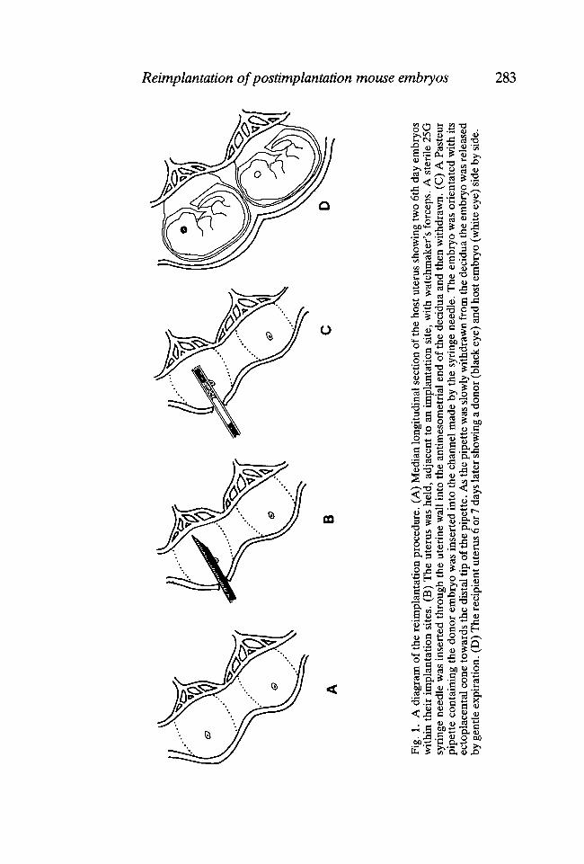
Recipients were anaesthetized by a single intraperitoneal injection of Avertin (01 ml 5g - 1
body weight). A small midventral incision was made through the skin and body wall of the lowerabdomen and the uterus was gently pulled through the opening. Donor embryos weretransferred into the implantation sites (Fig. 1) with the aid of a dissecting microscope (Wild) andfibre-optic illumination (Volpi). Initially, a channel was made through the antimesometrialuterine muscle into the centre of the decidua with a sharp 25-Gauge syringe needle (Microlance,Becton-Dickinson). The embryos were inserted in a small volume of PB1 + 10% FCS using afine hand-drawn siliconized (Repelcote) Pasteur pipette, braked with paraffin oil (Boots U.K.,Ltd) and containing a marker air bubble. Each implantation site received a single donor embryotransferred in the appropriate orientation such that the ectoplacental cone was directedmesometrially. A maximum of nine transfers was made to any individual recipient. For 5th dayrecipients it was necessary to inject 0-3 ml of a 5 % (w/v) solution of Pontamine Sky Blue(Hopkins & Williams) into the tail vein prior to surgery in order to pin point the implantationsites (Psychoyos, 1961).
Identification and evaluation of reimplanted embryosRecipients were killed on the 12th, 13th or 14th day of gestation (i.e. 6 to 8 days after
transfer). The excised uterus was placed in phosphate-buffered saline (PBS) and allimplantation sites which had received a donor embryo were dissected. All conceptuses, whetherhost or donor, normal or abnormal, were recovered. The presence or absence of heartbeat andvisceral yolk sac (VYS) circulation was recorded and the approximate developmental stage anddegree of normality assessed. Retinal pigmentation was used to distinguish normal host anddonor embryos and later their origin was confirmed and the identity of abnormal or retarded
embryos established by GPI electrophoresis (see below). A note was made as to whetherfoetuses were twinned or single within an implantation site.
Grossly normal donor foetuses, together with their placentae, were fixed in formol-acetic-alcohol and processed for routine histology. Foetuses were removed briefly from 70 %alcohol during dehydration, lightly blotted and weighed. In addition their crown-rump lengthswere measured and their somite numbers counted. Subsequently, serial histological sections(7/an) were prepared and stained with haemalum and eosin. Twelfth to 14th day PO embryosand 13th and 14th day (C3H/HeH,xPO) embryos were treated similarly and used as controls forassessing the developmental normality of reimplanted embryos.
ElectrophoresisThe VYS, or part of it, from any putative donor embryo, including all embryos which were
developmentally retarded or resorbing, was frozen in a small volume of PBS. Grossly abnormalconceptuses and membrane vesicles were frozen intact. Electrophoresis was carried out usingTitan III plates (Helena Laboratories) according to the protocol described by McLaren & Buehr(1981), except that the concentration of the tris-glycine bridge buffer was reduced to 0-025 %.The plates were stained using the method of Eicher & Washburn (1978).
RESULTS
Transfer of 7th day embryos into 6th day recipients
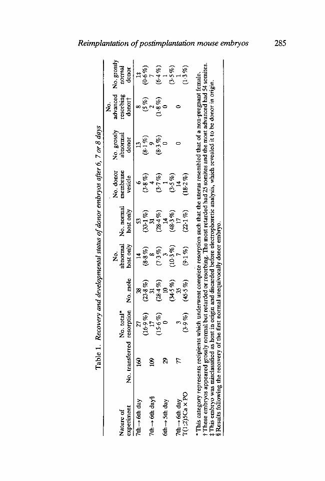
The incidence of recovery and the extent of development of donor embryos aregiven in Table 1. In all, eight donor embryos formed grossly normal foetusescomplete with placenta, vigorous heartbeat and VYS circulation. The first of theseto be recovered was misclassified, due to a failure to detect retinal pigmentation onthe 12th day of gestation, and discarded as a normal host embryo. Subsequently,GPI analysis showed it to have been donor in origin. The remaining sevenreimplanted conceptuses were available for more rigorous scrutiny. A comparisonof their weights, crown-rump lengths and somite numbers is shown in Table 2. Inall cases the initial 24 h advantage of donor embryos was not maintained butinstead their gross morphology resembled the developmental stage of their moreadvanced host littermates. However, there is no evidence that this retardationwith respect to their chronological age increased between the 12th and 14th days ofgestation and it may, therefore, have occurred soon after transfer. Reimplantedembryos which were twinned with host embryos in a single implantation site, andinvariably had what have been called 'fused placentae' (McLaren & Michie,1959a), tended to be smaller than those which developed alone (Table 2).
Examination of histological sections revealed that five of the seven recognizedreimplanted foetuses were indistinguishable from controls (Fig. 2). All foetalorgans appeared normal both in size and morphology. This was true also of theirplacentae which contained abundant nucleated red blood cells in the labyrinthinefoetal capillaries. The two remaining reimplanted foetuses that were studied werenot wholly normal. In one the abnormality was trivial in so far as only the right eyewas slightly defective. The optic cup appeared normal and lens induction hadoccurred but the overlying surface epithelium remained thickened, possibly dueto some mechanical constraint in utero interfering with ocular growth. Thecontralateral eye was normal and in all other respects this embryo resembled those
Tab
le 1
. R
ecov
ery
and
deve
lopm
enta
l sta
tus
of d
onor
em
bryo
s af
ter 6, 7
or
8 da
ys
3 1
Nat
ure
of
No.
tot
al*
expe
rim
ent
No.
tra
nsfe
rred
re
sorp
tion
No.
mol
e
No.
abno
rmal
host
onl
yN
o. n
orm
alho
st o
nly
No.
don
orm
embr
ane
vesi
cle
No.
gro
ssly
abno
rmal
dono
r
No.
adva
nced
N
o. g
ross
lyre
sorb
ing
norm
aldo
norf
do
nor
7th-
>6t
hday
§
6th-
»5th
day
7th
-• 6
th d
ayT
(l;2
)5C
axP
O
160
109 29 77
27(1
6-9
%)
17(1
5-6
%)
0 3(3
-9 %
)
38(2
3-8
%)
31(2
84%
)10
(34-
5 %
)35
(45-
5 %
)
14(8
-8 %
)8
(7-3
%)
3(1
0-3
%)
7(9
-1 %
)
53(3
3-1%
)31
(28-
4%)
14(4
8-3
%)
17(2
24%
)
6(3
-8 %
)4
(3-7
%)
1(3
-5 %
)14
(18-
2%)
13(8
-1 %
)9
(8-3
%)
0 0
(5%
)2
(1-8
%)
0 0
It(0
-6%
)7
(6-4
%)
1(3
-5 %
)1
(1-3
%)
* T
his
cate
gory
rep
rese
nts
reci
pien
ts w
hich
und
erw
ent
com
plet
e re
sorp
tion
such
that
the
ute
rus
rese
mbl
ed t
hat
of a
non
-pre
gnan
t fe
mal
e,t
The
se e
mbr
yos
appe
ared
gro
ssly
nor
mal
but
ret
arde
d or
res
orbi
ng. T
he m
ost r
etar
ded
had
25 so
mite
s an
d th
e m
ost a
dvan
ced
had
54 so
mite
s.tT
his
em
bryo
was
mis
clas
sifie
d as
hos
t in
ori
gin
and
disc
arde
d be
fore
ele
ctro
phor
etic
ana
lysi
s, w
hich
rev
eale
d it
to b
e do
nor
in o
rigi
n.§
Res
ults
fol
low
ing
the
reco
very
of
the
first
nor
mal
une
quiv
ocal
ly d
onor
em
bryo
.
to oo
286 R. S. P. BEDDINGTON
LJJ
Reimplantation of postimplantation mouse embryos 287
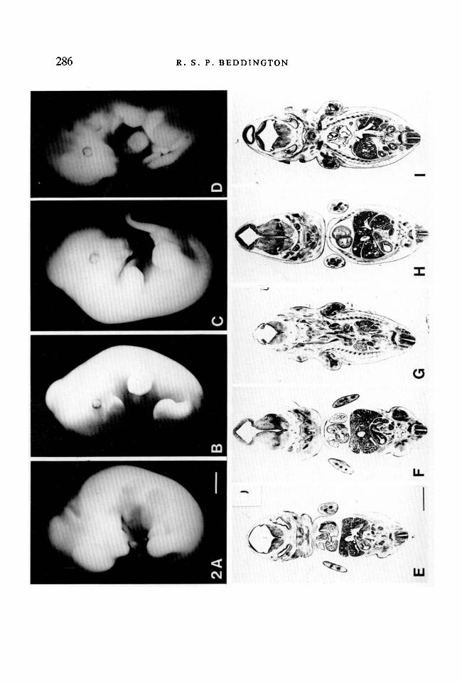
described above (Figs 2F & 2G). The seventh embryo had an abnormal heartwhich was manifest as an enlarged pericardial cavity and reduced thickening of themyocardium (Fig. 2D). Presumably, as a consequence of cardiac malfunctionthere was noticeable oedema in other tissues.
Transfer of 6th day embryos into 5th day recipients
This regime was compatible with continued development of the transferredembryo but the incidence of successful reimplantation was reduced (Table 1). Thesingle foetus recovered after 8 days was entirely normal and all measuredparameters were commensurate with control conceptuses (Table 2).
The effect of increased host embryo mortality
It was confirmed from inspection of implantation sites that litters produced frommating normal females to males heterozygous for the T(l;2)5Ca translocationsuffer approximately 50 % mortality by mid-gestation. About half the embryos inthese litters appear retarded on the 7th day although they may survive in varyingdegrees of disorganization up to recognizable early somite stages. It wasanticipated that the transfer of normal 7th day embryos to such recipients wouldincrease the incidence of reimplantation due to the absence of host competition in50 % of implantation sites. Unfortunately, the results show that there is no benefitto reimplantation but rather an increase in the frequency of resorption sites ormoles (Table 1). Only one donor embryo appeared grossly normal after 7 days andit was somewhat smaller and less advanced than its littermates (Table 2).
DISCUSSION
Implantation in the mouse begins on the 5th day of gestation and by the 7th daythe uterine epithelium of the crypt has disappeared and the trophoblast has erodedthe decidual tissue as far as the maternal blood vessels. This results in the primaryand secondary trophoblast giant cells of the conceptus lying in intimate contactwith the endometrium (Snell & Stevens, 1966; Potts, 1969). At this point, wellhidden from direct observation, the embryo embarks on the complex process ofgastrulation. This gross rearrangement of the embryo essentially lays thefoundation, providing both the architecture and the building material, fororganogenesis and subsequent foetal development. It is during gastrulation thatone would like to have direct access to the embryo, without forfeiting thepossibility of its continued development, in order to determine the origin ofparticular foetal tissues and their early lineage relationships one with another.
Fig. 2. Control embryos and reimplanted embryos recovered 7 days after transfer. (A)Control PO embryo on the 13th day of gestation (Bar = lmm). (B,C) Normalreimplanted embryos. (D) Reimplanted embryo with an abnormal heart. (E) Frontalsection of a control 13th day PO embryo. Inset: Sagittal section of a 7th day(C3H/Heh x PO) embryo at the time of transfer. (F,G) Frontal sections of thereimplanted embryo with an abnormal eye. All other organ systems appear normal. (H,I)Frontal sections of embryo depicted in Fig. C.
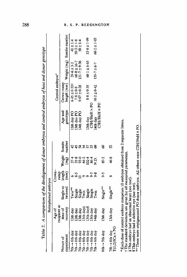
00 OO
Tab
le 2
. A
com
pari
son
of t
he d
evel
opm
ent
of d
onor
em
bryo
s an
d co
ntro
l em
bryo
s of
hos
t and
don
or g
enot
ype
Nat
ure
ofex
peri
men
t
7th-
>6t
hday
7th-
>6t
hday
7th-
»6th
day
7th-
>6t
hday
7th-
>6t
hday
7th
-»6t
h da
y7t
h-»6
thda
y
6th-
»5th
day
7th-
> 6
th d
ayT
(l;2
)5C
axP
O
Rei
mpl
ante
d em
bryo
s
Age
of
reci
pien
t at
tim
e of
reco
very
12th
day
12th
day
13th
day
13th
day
i13
th d
ay§
13th
day
14th
day
13th
day
13th
day
Sing
le o
rtw
inne
d
Tw
in**
Sing
leSi
ngle
Tw
inSi
ngle
Sing
leT
win
Sing
le**
Sing
le**
Cro
wn-
rum
ple
ngth
(mm
)
6 6-5
10 9 9 9-5
9-8
9-5
8
Wei
ght
(mg)
27-4
31-8
93-0
64-2
102-
486
-4N
.D.
80-2
46-8
Som
itenu
mbe
r42 45 54 54 57 55 60 60 52
Age
and
geno
type
12th
day
PO
13th
day
PO
14th
day
PO
13th
day
C3H
/HeH
x P
O14
th d
ayC
3H/H
eH x
PO
Con
trol
em
bryo
s*
Cro
wn-
rum
ple
ngth
(m
m)
6-45
±0-
55f
7-8
±0-
89-
97 ±
0-28
8-8
±0-
35
10-2
±0-
42
Wei
ght
(mg)
29-6
±3-
268
-8 ±
16-7
131-
7 ±
9-36
68-1
±6-
65
139-
7 ±
6-7
Som
ite n
umbe
r41
±1-
450
-3 ±
1-8
58 ±
1-4
53-8
±1-
99
60-2
± 1
-03
• 03 W O a O H O
* E
ach
clas
s of
con
trol
em
bryo
com
pris
es 1
0 em
bryo
s ob
tain
ed f
rom
2 s
epar
ate
litt
ers.
fTh
e st
anda
rd d
evia
tion
s ar
e gi
ven
for
all m
easu
red
para
met
ers.
tTh
is e
mbr
yo h
ad a
n ab
norm
al h
eart
(se
e te
xt).
§Thi
s em
bryo
had
a d
efec
tive
eye
(see
tex
t).
** T
hese
wer
e un
pigm
ente
d P
O d
onor
em
bryo
s. A
ll ot
hers
wer
e C
3H/H
eH x
PO
.
Reimplantation of postimplantation mouse embryos 289
Egg cylinders, or their component parts, will continue to grow and differentiatein extrauterine sites although morphogenesis and consequently tissue associationsare seldom normal in such circumstances (Grobstein, 1952; Diwan & Stevens,1976; Skreb & Svajger, 1975; Svajger, Levak-Svajger, Kostovic-Knezevic &Bradamante, 1981). Therefore ectopic grafts are of only limited use in establishingthe sequence of events in the embryo itself. Postimplantation culture techniques(New, 1978), restricted mainly to the rat, can support consistently normal growthand development of whole embryos, excluding the parietal yolk sac, overrelatively short periods (24-48 h depending on the stage at explantation). Themost prolonged development sustained in vitro has been the growth of rat embryosfrom the onset of gastrulation to the 30- to 40-somite stage. However,development under the conditions used was seldom entirely normal over thisperiod and those embryos which survived suffered considerable retardation ingrowth (Buckley, Steele & New, 1978). In general, the limited duration ofguaranteed normal development in culture has allowed only crude estimation tobe made of tissue fate and potency in the egg cylinder (Beddington, 1981, 1982,1983).
Clearly, these problems would be overcome if postimplantation embryos couldbe manipulated in vitro and subsequently returned to the uterus to continuenormal development. With such an end in mind it is at least encouraging that 6thand 7th day embryos can resume normal growth and development, and undergoapparently effective placentation, after transfer from their own decidua into one ina different pregnant female. Manipulation would, of course, involve puncturingReichert's membrane and although the importance of an intact Reichert'smembrane for reimplantation has not been tested it is known that injectingthrough this membrane in vivo is compatible with normal development (Weissman,Papaioannou & Gardner, 1978; Jaenisch, 1980).
The extensive development and growth seen in the few successful reimplants(Table 2; Fig. 2) surpasses that achieved in vitro. This is probably attributable totheir ability to form a functional placenta since it is accepted that growth in culturedeclines after the stage at which it is thought that the placenta first contributes tonutritional and respiratory exchange (New, 1978). However, no reimplantedembryos have yet been born (0/98 transfers; unpublished observations) althoughit is still unclear whether this is due to placental failure in the final week ofpregnancy or complications during parturition itself.
Although the results demonstrate the feasibility of reimplantation the frequencyof successful operations is too low for reimplantation to be seen as an immediatelyuseful procedure. One can only speculate as to why so few reimplant. Obviously,much depends on the accuracy of transfer and certainly practice alone ledto an increased rate of donor embryo recovery. Some embryos may not remainwithin the host decidua whilst others may be located sufficiently eccentricallythat mechanical or other influences prevent their normal development andplacentation. The high frequency of resorptions, host as well as donor, may becaused by undue damage to either embryo or decidua during transfer. Certain
290 R. S. P. BEDDINGTON
physiological elements may also play a role. For example, earlier studies havedemonstrated that twinning and fused placentae lead to an increase in foetalmortality and decrease in foetal weight (McLaren & Michie, 1959a). Similarly,more than eight implantations in a uterine horn results in an elevated midgestationdeath rate (McLaren & Michie, 195%). Thus, the artificial increase in litter sizetogether with the higher probability of twinning introduced by the reimplantationtechnique may contribute to the failure of some donor embryos. There is someindication that donor embryos which develop as twins with the resident hostembryo do not fare as well as those whose host embryo resorbs (Table 2).
The attempt to overcome the possible disadvantages of overcrowding by theintroduction of lethal genomic imbalance into half the host embryos via theT(l;2)5Ca translocation was a conspicuous failure. However, although theaffected host embryos start to deteriorate at the egg-cylinder stage they maysurvive, albeit in an abnormal or disorganized fashion, until the early somite stage.It is possible that resorption of the host embryo at this more advanced stage mayjeopardize the development of any adjacent donor embryo. Alternatively, theimplantation sites induced by the mutant embryos may not be wholly normal.Certainly trophectoderm-vesicle-induced decidua, which appear histologicallynormal, (Gardner, 1972) do not support extensive development of transferredpostimplantation embryos, even if the contralateral uterine horn containsnormally developing host embryos (unpublished observations). Unfortunately,therefore, it still remains to find a means of increasing the frequency of successfulreimplantation so that the feasibility of producing viable postimplantation mousechimaeras can be assessed.
I would like to thank Professor R. L. Gardner for his discussion of the manuscript and Miss J.Green for technical assistance. I am also grateful to Dr M. F. Lyon for providing mice carryingthe T(l;2)5Ca translocation. This work was supported by a Lister Institute Research Fellowshipand by the Imperial Cancer Research Fund.
REFERENCESBEDDINGTON, R. S. P. (1981). An autoradiographic analysis of the potency of embryonic
ectoderm in the 8th day postimplantation mouse embryo. J. Embryol. exp. Morph. 64, 87-104.BEDDINGTON, R. S. P. (1982). An autoradiographic analysis of tissue potency in different regions
of the embryonic ectoderm during gastrulation in the mouse. /. Embryol. exp. Morph. 69,265-285.
BEDDINGTON, R. S. P. (1983). The origin of the foetal tissues during gastrulation in the rodent. InDevelopment in Mammals Vol. 5 (ed. M. H. Johnson), pp. 1-32. Oxford: Elsevier.
BUCKLEY, S. K. L., STEELE, C. E. & NEW, D. A. T. (1978). In vitro development of earlypostimplantation rat embryos. DevlBiol. 65, 396-403.
CARTER, T. C , LYON, M. F. & PHILLIPS, R. J. S. (1956). Further genetic studies of eleventranslocations in the mouse. /. Genet. 54, 462-473.
DIWAN, S. B. & STEVENS, L. C. (1976). Development of teratomas from ectoderm of mouse eggcylinders. /. natn. Cancer Inst. 57, 937-942.
EDWARDS, R. G. & STEPTOE, P. C. (1983). Current status of in-vitro fertilisation and implantationof human embryos. Lancet ii, 1265-1269.
Reimplantation of postimplantation mouse embryos 291EICHER, E. M. & WASHBURN, L. L. (1978). Assignment of genes to regions of mouse
chromosomes. Proc. natn. Acad. Set, U.S.A. 75, 946-950.GARDNER, R. L. (1972). An investigation of inner cell mass and trophoblast tissue following their
isolation from the mouse blastocyst. /. Embryol. exp. Morph. 28, 279-312.GARDNER, R. L. (1978). The relationship between cell lineage and differentiation in the early
mouse embryo. In Results and Problems in Cell Differentiation Vol. 9 (ed. W. J. Gehring), pp.205-241. New York: Springer-Verlag.
GORDON, J. W. (1983). Transgenic mice: A new and powerful experimental tool in mammaliandevelopmental genetics. Developmental Genetics 41, 1-20.
GROBSTEIN, C. (1952). Intraocular growth and differentiation of clusters of mouse embryonicshields cultured with and without primitive endoderm and in the presence of possibleinductors. /. exp. Zool. 119, 355-380.
JAENISCH, R. (1980). Retroviruses and embryogenesis: microinjection of Moloney leukaemiavirus into midgestation mouse embryos. Cell 19,181-188.
MCLAREN, A. (1981). Analysis of maternal effects on development in mammals. J. Reprod. Fert.62 Suppl., 591-596.
MCLAREN, A. & BUEHR, M. (1981). GPI expression in female germ cells of the mouse. Genet.Res. Camb. 37, 303-309.
MCLAREN, A. & MICHIE, D. (1959a). Experimental studies on placental fusion in mice. /. exp.Zool. 141, 47-73.
MCLAREN, A. & MICHIE, D. (19596). Superpregnancy in the mouse: I. Implantation and foetalmortality after induced superovulation in females of various ages. /. exp. Biol. 36, 281-300.
NEW, D. A. T. (1978). Whole-embryo culture and the study of mammalian embryos duringorganogenesis. Biol. Rev. 53, 81-122.
POTTS, M. (1969). The ultrastructure of egg implantation. In Advances in Reproductive PhysiologyVol. 4 (ed. A. McLaren), pp. 241-263. London: Logos Press Ltd.
PSYCHOYOS, A. (1961). Permeabilite capillaire et decidualisationuterine. C. r. hebd. Seanc. Acad.ScL, Paris 264, 956-958.
SEIDEL, G. E. (1983). Mammalian oocytes and preimplantation embryos as methodologicalcomponents. Biol. Reprod. 28, 36-49.
SKREB, N. & SVAJGER, A. (1975). Experimental teratomas in rats. In Teratomas and Differentiation(ed. M. I. Sherman & D. Solter), pp. 83-99. London: Academic Press.
SNELL, G. D. & STEVENS, L. C. (1966). Early embryology. In Biology of the Laboratory Mouse (ed.E. L. Green), pp. 205-245. New York: McGraw-Hill.
SVAJGER, A., LEVAK-SVAJGER, B., KOSTOVIC-KNEZEVIC, L. & BRADAMANTE, Z. (1981). Mor-phogenetic behaviour of the rat embryonic ectoderm as a renal homograft. /. Embryol. exp.Morph. 65 (Suppl.), 243-267.
WEISSMAN, I., PAPAIOANNOU, V. E. & GARDNER, R. L. (1978). Fetal hematopoietic origins of theadult hematolymphoid system. In Differentiation of Normal and Neoplastic Hematopoietic Cells.Cold Spring Harbor Conferences in Cell Proliferation Vol 5 Book A (ed. B. Clarkson, P. A.Marks & J. E. Till), pp. 33-47. Cold Spring Harbor Laboratory.
WHnnNGHAM, D. G. (1979). In-vitro fertilization, embryo transfer and storage. Br. med. Bull.35, 105-111.
WHITTINGHAM, D. E. & WALES, R. G. (1969). Storage of two-cell mouse embryos in vitro. Aust. J.biol. Sci. 22, 1065-1068.