THE DEVELOPMENT OF NEUTROPHILIC POLYMORPHONUCLEAR LEUKOCYTES IN HUMAN BONE MARROW ORIGIN AND CONTENT OF AZUROPH1L AND SPECIFIC GRANULES* BY DOROTHY FORD BAINTON,$ M.D., JOAN L. ULLYOT,§ M.D., A~D MARILYN G. FARQUHAR,][ PH.D. (From the Department of .Pathology, University of California School of Medicine, San Francisco, Califarnla 94122) (Received for publication 27 May 1971) Previous electron microscopic and cytochemical studies from this laboratory (1-3) have demonstrated that rabbit neutrophilic polymorphonuclear leukocytes (PMN) ~ contain two types of granules, azurophils and specifics, which have separate origins and are different in nature. The large, dense azurophil granules represent a special type of primary lysosome containing peroxidase and lysosomal or digestive enzymes. They are produced early in development, during the promyelocyte stage, 2 and arise from the concave face of the Golgi complex. The smaller, less dense specific granules represent an entirely different secretory product which contains alkaline phosphatase and lacks lysosomal enzymes and peroxidase. They are produced later in develop- ment, during the myelocyte stage, and arise from the opposite or convex face of the Golgi complex. Recently Baggiolini, Hirsch, and de Duve have prepared fractions of azurophil and specific granules from rabbit PMN by zonal sedimentation (4) and isopycnic centrifugation (5) and have confirmed by biochemical assay our cytochem- ical findings concerning the enzyme content of the two granule types. It now remains to be determined whether the findings obtained in rabbit PMN pertain to other species, especially the human. To date there has been no * This investigation was supported by Grants A IVI 10486 and AM 15399 from the National Institute of Arthritis and Metabolic Diseases, U.S. Public Health Service, and by Cancer Re- search Funds of the University of California. $ Recipient of a U.S. Public Health Service Career Award (AM 11902) from the National Institute of Arthritis and Metabolic Diseases. § Special Research Fellow, U.S. Public Health Service Fellowship GM 44073, National Institute of General Medical Sciences. II Recipient of a U.S. Public Health Service Career Award (GM 25,109) from the National Institute of General Medical Sciences. Present address: The Rockefeller University, New York 10021. 1Abbreviations used in this paper: ER, endoplasmic reticulum; PMN, neutrophilic poly- morphonuclear leukocytes. 2 In our earlier papers, the term "progranulocyte" was used to designate this stage of de- velopment, but we will employ the equivalent and currently more widely used term "promye- locyte" hereafter. THE JOURNAL OF EXPERIMENTAL MEDICINE • VOLUME 134, 1971 907 Downloaded from http://rupress.org/jem/article-pdf/134/4/907/1084415/907.pdf by guest on 20 December 2021
Transcript
T H E D E V E L O P M E N T O F N E U T R O P H I L I C P O L Y M O R P H O N U C L E A R L E U K O C Y T E S I N H U M A N B O N E M A R R O W
ORIGIN AND CONTENT OF AZUROPH1L AND SPECIFIC GRANULES*
BY DOROTHY FORD BAINTON,$ M.D., JOAN L. ULLYOT,§ M.D., A~D MARILYN G. FARQUHAR,][ PH.D.
(From the Department of .Pathology, University of California School of Medicine, San Francisco, Califarnla 94122)
(Received for publication 27 May 1971)
Previous electron microscopic and cytochemical studies from this laboratory (1-3) have demonstrated that rabbit neutrophilic polymorphonuclear leukocytes (PMN) ~ contain two types of granules, azurophils and specifics, which have separate origins and are different in nature. The large, dense azurophil granules represent a special type of primary lysosome containing peroxidase and lysosomal or digestive enzymes. They are produced early in development, during the promyelocyte stage, 2 and arise from the concave face of the Golgi complex. The smaller, less dense specific granules represent an entirely different secretory product which contains alkaline phosphatase and lacks lysosomal enzymes and peroxidase. They are produced later in develop- ment, during the myelocyte stage, and arise from the opposite or convex face of the Golgi complex. Recently Baggiolini, Hirsch, and de Duve have prepared fractions of azurophil and specific granules from rabbit PMN by zonal sedimentation (4) and isopycnic centrifugation (5) and have confirmed by biochemical assay our cytochem- ical findings concerning the enzyme content of the two granule types.
I t now remains to be determined whether the findings obtained in rabbi t P M N per ta in to other species, especially the human. To da te there has been no
* This investigation was supported by Grants A IVI 10486 and AM 15399 from the National Institute of Arthritis and Metabolic Diseases, U.S. Public Health Service, and by Cancer Re- search Funds of the University of California.
$ Recipient of a U.S. Public Health Service Career Award (AM 11902) from the National Institute of Arthritis and Metabolic Diseases.
§ Special Research Fellow, U.S. Public Health Service Fellowship GM 44073, National Institute of General Medical Sciences.
II Recipient of a U.S. Public Health Service Career Award (GM 25,109) from the National Institute of General Medical Sciences. Present address: The Rockefeller University, New York 10021.
1 Abbreviations used in this paper: ER, endoplasmic reticulum; PMN, neutrophilic poly- morphonuclear leukocytes.
2 In our earlier papers, the term "progranulocyte" was used to designate this stage of de- velopment, but we will employ the equivalent and currently more widely used term "promye- locyte" hereafter.
THE JOURNAL OF EXPERIMENTAL MEDICINE • VOLUME 134, 1971 907
Dow
nloaded from http://rupress.org/jem
/article-pdf/134/4/907/1084415/907.pdf by guest on 20 Decem
ber 2021
908 DEVELOPMENT Or HUMAN PMN
general agreement on the nature or numbers of granule types present in human
P M N ; two, three, and even four granule types have been described in various electron microscopic studies (6-20). The present s tudy represents an a t tempt to
establish the nature and number of P M N granule types in the human using an approach similar to that used previously for the rabbit, with the following modification: in this work we have taken advantage of the fact that myeloper- oxidase is localized exclusively in azurophil granules and we have utilized this enzyme as a cytochemical marker to facilitate identification of azurophils. This modified approach was necessary when it became apparent that morphologic criteria (size, shape, density) were not sufficient to distinguish granule types in the human. Our results, already reported in abstract form elsewhere (21-23), in- dicate that although human P M N granules are morphologically more hetero- geneous than those of the rabbit, the situation is basically the same in the two
species with respect to the existence, mode of origin, and enzyme content of azurophil and specific granules.
Materials and Methods
Collection of Tissues.--Bone marrow was obtained from the sterna or ribs of two normal volunteers and 30 hematologically normal patients undergoing cardiothoracic surgery. Venous blood was obtained in polyethylene syringes wet with aqueous heparin, and the leukocytes were concentrated in one of four ways: (a) dextran sedimentation (24), (b) ammonium chloride lysis of red blood cells (25), (c) buffy coat disc of Anderson (26), or (d) the buffy coat tubes of Kaplow (27). Cover glass smears and cell suspensions were processed as previously described (2, 3) unless otherwise stated.
Methods for Morphologic Studles.--Fixation was carried out in one of the following solutions: (a) 1.5% distilled glutaraldehyde (28) in 0.1 ~ sodium cacodylate-HC1 buffer ([)H 7.4) with 1% sucrose for 16 hr at 4°C or 6 hr at 22°C, (b) 1% formaldehyde-3% glutaraldehyde with CaCI2 (29) for 4 hr at 22°C, or (e) glutaraldehyde-OsO4 for 1 hr at 4°C (15). The cells were then packed by centrifugation at ~I0,000 g, postfixed for 1 hr at 4°C in 1% Os04 in acetate- Veronal buffer (pH 7.4) with 5% sucrose added, treated with buffered 0.5% uranyl acetate (30) for 1 hr at 22°C, dehydrated in ethanol, carried through propylene oxide, and embedded in Araldite. In some cases propylene oxide was avoided by infiltrating specimens with increas- ing concentrations of Araldlte mixed with 100% ethyl alcohol (31), or embedding according to Spurr's technique (32).
Methods for Cytochemical Studies.--Fixation was carried out for 10 rain-16 hr in either glu- taraldehyde at 4°C or formaldehyde-glutaraldehyde at 22°C as described above. Cells were
A bbreviotion for figures: ag, azurophil granule (spherical) ag', azurophil granule (ellipsoid) ca, centriole er, rough-surfaced endoplasmic reticulum £, glycogen G Golgi complex Gc, Golgi cisternae ia, immature azurophil granule is, immature specific granule
M, mature PMN or band m, mitochondrion n, nucleus nu, nucleolus P, promyelocyte pn, perinuclear cisterna r, ribosome sg, specific granule re, vesicles Y, myelocyte
Dow
nloaded from http://rupress.org/jem
/article-pdf/134/4/907/1084415/907.pdf by guest on 20 Decem
ber 2021
D. F. BAINTON~ J. L. ULLYO'r~ AND M. G. FARQUILAR 909
incubated in the following media containing 5% sucrose: (a) modified Gomori's medium (33), pH 5.0, for acid phosphatase, using fl-glycerophosphate (Sigma Chemical Co., St. Louis, Mo.; Grade I) as substrate; (b) Goldfischer's medium (34), pH 5.5, for arylsulfatase, using p-nitro- catechol sulfate, followed by treatment with 2% ammonium sulfide (35); (c) Graham and
MARROW (development, 14 days)
mye~ob~
~-~'"~ ~ metamyelocyte band cell
S mature pmn
BLOOD (transit, average ~10 hr)
> TISSUES (function,~l-2 days}
Fro. 1. Diagrammatic representation of PMN life cycle and stages of PMN maturation. The myeloblast is a relatively undifferentiated cell with a large oval nucleus, large nucleoli, and cytoplasm lacking granules. I t originates from a precursor pool of elusive stem cells and is followed by two secretory stages: the promyelocyte and the myelocyte. During each of these stages a distinct type of secretory granule is produced: azurophils (solid black) formed only during the promyelocyte 2 stage, and specific granules (light forms) produced during the myelo- cyte stage. The metamyelocyte and band forms are nonproliferating, nonsecretory stages which develop into the mature PMN. The latter is characterized by a multilobulated nucleus and cytoplasm containing primarily glycogen and granules. Out of every 100 nucleated cells in bone marrow, 2% are myeloblasts, 5% promyelocytes, 12% myelocytes, 22% metamyelocytes and bands, and 20% mature PMN, giving a total of ~60% developing PMN (39). The times indi- cated for the various compartments were determined by isotope labeling techniques (see refs. 40-42).
Karnovsky's medium (36), pH 7.6, for peroxidase; (d) Gomori's medium for alkaline phos- phatase (pH 9.2), using fl-glycerophosphate followed by treatment with 2% lead nitrate (3, 37). Methods for the other enzyme incubations and controls, as well as those for subsequent processing and microscopy, were as previously described (2, 3), with the exception that KCN
Dow
nloaded from http://rupress.org/jem
/article-pdf/134/4/907/1084415/907.pdf by guest on 20 Decem
ber 2021
910 DEVELOPMENT OF HUMAN PMN
inhibition of peroxidase activity- was tested in a closed system after preincubation of the cells with KCN (0.01 M) at pH 6.0 for 30 rain (38).
Histochemical Staining on Smears of Bone Marrow.--Cover glass smears were prepared, fixed, stained, and incubated and processed for enzyme histochemistry exactly as described in a previous paper (2) unless otherwise stated.
RESULTS
Background Information
The PMN leukocyte of the blood is a highly specialized, nondividing "end" cell with a short life-span (see Fig. 1). The bulk of its life cycle is spent in the marrow, where it proliferates, differentiates, and is stored for a few days. The mature cell is then released into the blood and circulates briefly before migrat- ing into the tissues where it functions as a mobile phagocyte.
P M N development in the bone marrow has classically been divided into six stages (myeloblast, promyelocyte, myelocyte, metamyelocyte, band, and ma- ture PMN) distinguished in Romanovsky-stained smears of bone marrow (see Fig. 1) on the basis of cell size, nuclear morphology, and granule content. Two of these stages are recognized mainly by the nature of their granules: the pro- myelocyte, which contains a single population of large, reddish-purple "azuro- phil ''a granules, and the myelocyte, which contains a mixed granule population consisting of smaller, pinkish "specific" granules, along with azurophils which become less conspicuous. Up to now, it has not been established whether human P M N contain a single type of granule which alters its staining properties during maturation, or whether, as is now known for the rabbit, the granules represent entirely different products formed in two successive waves of secretory activity. To resolve this question, we have focussed our attention on the promyelocyte and myelocyte, the stages of development during which the granules appear.
Effects of Preparatory Techniques on Granule Morphology
Our initial electron microscopic studies revealed that human PMN granules cannot be divided into distinct types on the basis of morphology alone. We tried numerous variations in methodology (see Materials and Methods), of which none was entirely satisfactory, and in accord with the experience of others (12-14), several were found to affect granule morphology profoundly. In partic- ular, after propylene oxide, the otherwise dense content of the granules of the promyelocyte (azurophils) appeared irregularly extracted, and after uranyl acetate (in block or on grid), the smaller, generally less dense granules that arise in the myelocyte (specifics) appear darker. Cells fixed in formaldehyde-glu-
a The term "azurophil ," introduced by Michaelis, refers to an affinity for an "azure" or "sky-blue" dye and not to the color of the granules, which stain metachromatical ly, i.e., red- dish-purple. T hey have also been called "unripened," "undifferentiated," or "nonspecific" granules.
Dow
nloaded from http://rupress.org/jem
/article-pdf/134/4/907/1084415/907.pdf by guest on 20 Decem
ber 2021
D. F. BAINTON, 3. L. IILLYOT, AND M. G. FARQUI-IAR 911
taraldehyde usually contain a higher proportion of elongated or dumbbell- shaped granules than those fixed in glutaraldehyde alone, presumably owing to better preservation of the elongated forms. Fixation with a mixture of glu- taraldehyde and OsO4 (15) gave results similar to those seen with glutaralde- hyde followed by OsO4.
The best results in tissue prepared for morphology alone were obtained by fixation in glutaraldehyde followed by OsO4, and by omission of both uranyl acetate and propylene oxide during subsequent processing. With this method, two main types of granules could be distinguished: one larger and denser, and the other smaller and less dense. However, in mature PMN there were many granules of intermediate size and density which were difficult to classify (see Fig. 2). Moreover, such embeddings (carried out without propylene oxide) were difficult to section except in the case of those embedded according to Spurr's technique (32).
We were able to avoid the above-mentioned difficulties and obtain excellent differential contrast among granules by fixing cells in glutaraldehyde and react- ing them for peroxidase, after which they could be processed routinely (uranyl acetate staining in block, alcohol-propylene oxide dehydration, and Epon or Araldite embedding). Since, as in the case of the rabbit, peroxidase is present in all the granules of promyelocytes (i.e. in all azurophils), the peroxidase reaction product renders the azurophil granules very dense and easily distinguishable from granules formed during the myelocyte stage, which are peroxidase-nega- tive. Accordingly, the following observations on developmental stages of human PMN are based on such preparations reacted for peroxidase in which the pres- ence of enzyme reaction product serves as a marker and stabilizer of the azuro- phils.
Stages of Neutrophil Maturation
Myeloblast.--The earliest cell of the neutrophilic series is the myeloblast, a relatively small (~-40 u) undifferentiated cell with a high nuclear:cytoplasmic ratio and prominent nucleoli. The scant cytoplasm is devoid of granules but contains abundant free polysomes and mitochondria. Annulate lamellae, con- sidered characteristic of embryonic cells and reported in blasts from mouse marrow (43), are occasionally seen. Very early promyelocytes resemble the myeloblast, but contain a few azurophil granules and a larger Golgi complex (Fig. 3).
PMN Promyelocyte (Figs. 4-8).--The promyelocyte can be recognized by its large size (~-~ 15/z), rounded nucleus, and population of variable but frequently large numbers of peroxidase-positive granules which correspond to the azuro- phil granules seen by light microscopy. I t has a large Golgi region and moderate amounts of rough endoplasmic reticulum (ER). Fig. 4 illustrates a typical cell profile.
Dow
nloaded from http://rupress.org/jem
/article-pdf/134/4/907/1084415/907.pdf by guest on 20 Decem
ber 2021
FIG. 2. Mature PMN from human bone marrow. Several lobes of the nucleus are seen (n 1 -- n3), and numerous granules, as well as beta particles of glycogen (g), are scattered throughout the cytoplasm. A few mitochondria (m) and a small Golgi complex (Gc) are also visible. Some of the granules present are large and dense (ag), whereas others are small and less dense (sg). However, many granules are intermediate in size and density. Elongated forms, including football and dumbbe]l shapes, are also present (arrows). This great variability in size, shape, and density renders identification of granule types by morphologic criteria difficult and unreliable. The glycogen is well preserved but membranes show poor contrast in this type of preparation in which there was no staining with uranyl acetate in block (cf. Fig. 11).
The insets depict internal structure within the large, dense (azurophil) granules. Inset a shows a spherical granule (ia) containing concentric half-rings. This type of internal structure was only observed occasionally in azurophil granules of normal mature PMN. Inset b illus- trates the crystalline lattice with periodicity of ~ 80 A which is commonly seen in football or ellipsoid forms (ag'). A cross-section (X) of the ellipsoid form and an immature specific granule (is) are also present in this field taken from a PMN myelocyte. Tissue was fixed in glutaralde- hyde, 22°C for 6 hr, postfixed in osmium, dehydrated in alcohol, and embedded in Araldite with omission of propylene oxide. The sections were stained with uranyl acetate and alkaline lead. Fig. 2, X 17,000; a, X 45,000; b, X 45,000.
Dow
nloaded from http://rupress.org/jem
/article-pdf/134/4/907/1084415/907.pdf by guest on 20 Decem
ber 2021
D. ~'. BAINTON, J, L. IYLLYOT, AND M. G. I~ARQU~AR 913
Figures 3-11 are electron micrographs of developing PMN from normal human bone mar- row reacted for peroxidase.
FIo. 3. Early PMN promyelocyte. The nucleus (n) with its prominent nucleolus (nu) occu- pies the bulk of this very immature cell. The surrounding cytoplasm contains a few azurophil granules (ag), a large Golgi complex (G), several mitochondria (m), scanty rough endoplasmic reticulum (er), and many free polysomes (r). A centriole (ce) is present in the Golgi region.
All of the azurophil granules (ag) appear dense, since they are strongly reactive for peroxi- dase. The secretory apparatus [i.e. the perinuclear cisterna (pn), rough endoplasmic reticulum (er), and some of the Golgi cisternae (Gc)] is also reactive, although less so than the granules, a point which is better illustrated in Figs. 4 and 5.
Specimen fixed in glutaraldehyde for 16 hr at 4°(2, incubated in the peroxidase medium of Graham and Karnovsky for 1 hr at 22°C, postfixed in Os04, treated in block with uranyl ace- tare, dehydrated in ethanol, infiltrated with propylene oxide, and embedded in Araldite. Sec- tion stained for 1 min with lead citrate. X 21,000.
Al though all the azurophil granules are reactive for peroxidase, they va ry in size and shape and in the amount and degree of compact ion of peroxidase reac- tion product . Two main shapes can be identified: round and football-shaped. Tlae predominant form is round ( d i a m e t e r ' = ~ 500 m/x); i ts contents appear flocculent and less compact in immature granules, bu t become more homogene- ous and dense as the granules ma tu re and undergo condensation. Footbal l -
Dow
nloaded from http://rupress.org/jem
/article-pdf/134/4/907/1084415/907.pdf by guest on 20 Decem
ber 2021
F•. 4. PMN promyelocyte, reacted for peroxidase. This cell is the largest (N15 #) of the neutrophilic series. I t has a sizable, slightly indented nucleus (n), a prominent Golgi region (G), and cytoplasm packed with peroxidase-positive azurophil granules (ag). Note the two general shapes of azurophil granules, spherical (ag) or ellipsoid (ag'). The majority are spheri-
Dow
nloaded from http://rupress.org/jem
/article-pdf/134/4/907/1084415/907.pdf by guest on 20 Decem
ber 2021
D. F. BAINTON, ~[. L. IYLLYOT, AND M. G. FARQUHAR 915
shaped forms (300 X 900 m#) are less common. They frequently contain crystal- line inclusions with a periodicity of ~-~ 80 A, oriented parallel to their long axis (Figs. 6-7). Round profiles with a central core also showing a periodic struc- ture (Fig. 8) are presumed to represent "footballs" cut in cross-section. The secretory apparatus of the promylelocyte (i.e. rough ER, including the peri- nuclear cisterna, and Golgi cisternae) also contains peroxidase reaction product which is less concentrated than that in the azurophil granules (Fig. 5). At times the innermost Golgi cisterna along the concave surface of the Golgi stack is more reactive than the others, as seen in Fig. 5. Promyelocytes are frequently observed in various stages of mitosis during which reaction product is present throughout the entire ER.
P M N Myelocyte (Figs. 9 and 10).--The myelocyte is smaller (10 #) than the promyelocyte and can be recognized by its indented nucleus, prominent Golgi complex, and mixed population of granules, which includes variable numbers of smaller, peroxidase-negative granules, as well as the large peroxidase-positive azurophil granules. These new granules, which accumulate throughout the myelocyte stage and eventually come to outnumber the azurophils, correspond to the specific granules. They vary in size and shape but typically occur as spheres ( ~ 200 m~) or rods (130 X 1000 m/~); the latter are often cigar- or dumbbell-shaped. Despite their variability in shape, they all have a similar con- tent which is homogeneous, of low density, and devoid of peroxidase activity. Hence in these respects they appear to represent a single granule population.
The Golgi complex of the myelocyte is large, and in contrast to that of the promyelocyte, is not reactive for peroxidase. Immature specific granules appear to bleb off its outer cisternae along the convex Golgi surface (Fig. 10). The pres- ence of a prominent Golgi complex containing forming specific granules dis- tinguishes the myelocyte from later, nonsecretory stages.
I t should be emphasized that elements of the secretory apparatus of the myelocyte were never seen to contain peroxidase. The only peroxidase-positive elements present at this and subsequent stages are the azurophil granules.
cal, with a homogeneous matrix, but a few ellipsoid forms containing crystalloids (best seen in Figs. 6-8) are also present. Many of the spherical forms (arrows) have a dense periphery and a lighter core, owing presumably to incomplete penetration of substrate into the compact centers of mature granules.
Peroxidase reaction product is visible in less concentrated form within all compartments of the secretory apparatus [endoplasmic reticulum (er), perinuclear cisterna (pn), and Golgi cis- ternae (Gc)]. No reaction product is seen in the cytoplasmic matrix, mitochondria (m), or nu- dells (n).
The inset depicts a portion of another promyelocyte at higher magnification, showing to better advantage flocculent deposits of peroxidase reaction product in the rough ER (er) in- cluding the perinuclear cisterna (prO. Ribosomes do not show up in this section, which was lightly stained with lead.
Specimen fixed in glutaraldehyde for 10 rain at 4°C and subsequently processed exactly as in Fig. 3. Fig. 4, X 15,000, inset, X 34,000.
Dow
nloaded from http://rupress.org/jem
/article-pdf/134/4/907/1084415/907.pdf by guest on 20 Decem
ber 2021
Dow
nloaded from http://rupress.org/jem
/article-pdf/134/4/907/1084415/907.pdf by guest on 20 Decem
ber 2021
D. F. BAINTON, J. L. ULLYOT, AND NL G. FARQUHAR 917
Among the latter, in addition to the usual large azurophil forms, occasional clusters of much smaller (200 m/z) peroxidase-positive granules can be found (see inset, Fig. 9).
I t appears that, as in the case of the rabbit, production of peroxidase-positive azurophil granules ends in the promyelocyte, and the beginning of the myelo- cyte stage is marked by the production of a second population of granules which are peroxidase negative. I t is of interest that cells are frequently observed which contain mature azurophils but no specific granules, have the indented nuclear configuration of the myelocyte, lack peroxidase in the E R or Golgi cisternae, and have a large Golgi apparatus which does not contain forming granules of either type. Such cells are presumed to be in a hiatus between the two waves of secre- tory activity. Similar cells have been described by Ackerman (44) in the cat. Mitoses have also been observed during the myelocyte stage; it can be seen that the granules are distributed in approximately equal numbers to daughter cells.
Later Stages (Bone Marrow).--The metamyelocyte, band, and mature P M N are nondividing, nonsecretory stages which are identified by their nuclear morphology, mixed granule population, inactive Golgi region (Fig. 11), and accumulation of glycogen particles (Fig. 2). The number of granules present at these stages is quite large; granule counts carried out on sections passing through the Golgi region indicated the presence of an average of 200-300/cell profile, with approximately twice as many specifics as azurophils. As in the myelocyte, peroxidase activity is present only in azurophil granules. Dur ing these stages the azurophils become somewhat smaller and more oval (compare azurophils in Fig. 4 to those in Figs. 9 and 11), a change which probably occurs due to progressive concentration of the granule contents, and which may be related to the diminished azurophilia noted in Wright 's-stained smears (see
Figs. 5-8 illustrate portions of PMN promyelocytes. FIG. 5. Golgi region of PMN promyelocyte. At this stage, peroxidase reaction product is
present within [a] all cisternae of the rough ER (er), including transitional elements (arrow), and the perinuclear cisterna (not shown here); [b] clusters of smooth vesicles located at the periphery of the Golgi complex (re); [c] all cisternae of the Golgi complex (Go); and [d] all im- mature (ia) and mature azurophil granules (ag). Most of the mature granules (ag) appear uni- formly dense due to the presence of reaction product throughout. Immature granules are larger and their contents less compact (ia). Note that peroxidase is more concentrated in the cisternae along the concave surface of the Golgi complex, suggesting that, as in the case of the rabbit (1), azurophil granules arise from this Golgi face (see ~').
FIGS. 6-8. Azurophil granules of promyelocytes occur in two main forms. The majority are spherical, with a dense, homogeneous matrix (ag in Fig. 5). Others are ellipsoid or football- shaped (Figs. 6 and 7) with a crystalline substructure which may be partially obscured in ma- ture granules by the dense peroxidase reaction product (Fig. 7) but is clearly visible in the im- mature ellipsoid (Fig. 6) or in preparations not incubated for peroxidase (Fig. 2 b). Round granule profiles with a central periodicity (Fig. 8) are presumed to represent "footballs" cut perpendicular to the crystal axis.
Specimen preparation as in Fig. 4. Fig. 5, )< 56,000; Figs. 6-7, )< 63,000; Fig. 8, >( 90,000.
Dow
nloaded from http://rupress.org/jem
/article-pdf/134/4/907/1084415/907.pdf by guest on 20 Decem
ber 2021
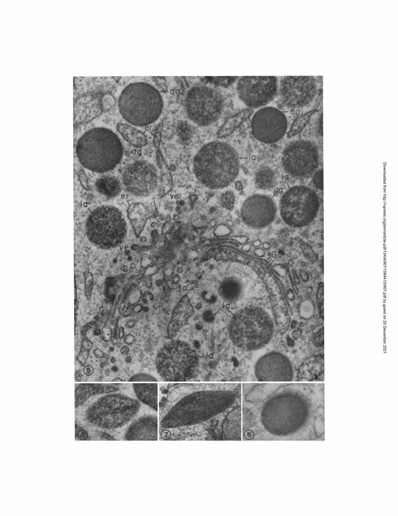
FIG. 9. PMN myelocyte. At this stage the cell is smaller (~10 #) than the promyelocyte (see Fig. 4), the nucleus is more indented, and the cytoplasm contains two different types of granules: [1] large, peroxidase-positive azurophils (ag), and [2] the generally smaller specific granules (sg), which do not stain for peroxidase. A number of immature specifics (is), which are larger, less compact, and more irregular in contour than mature granules, are seen in the Golgi region (G). Note that peroxidase reaction product is present only in azurophil granules,
Dow
nloaded from http://rupress.org/jem
/article-pdf/134/4/907/1084415/907.pdf by guest on 20 Decem
ber 2021
D. !~'. BAINTON, .]. L. ULLYOT, AND M. G. FAP.QITHAR. 919
Fig. 12). A few of the azurophils take on a rough dumbbell-shape (see Figs. 9 and 11) similar to the form common among specifics, emphasizing the necessity of using peroxidase to identify the azurophils in more mature neutrophils.
Blood PMN.- -The mature P M N of the blood appear generally similar to the mature P M N in the marrow when cells are collected using the Kaplow buffy- coat tube, a method involving a minimal delay in fixation (~-~ 5 rain) and mini- real t rauma to the cells. When other currently available methods of isolating blood P M N are used, which involve longer delays before fixation as well as chemical or osmotic t rauma (heparin, ethylenediaminetetraacefic acid [EDTA], NH4C1 lysis, or dextran), more variation is observed in the size, shape, and numbers of granules present, and in the intensity of the peroxidase reaction in azurophils.
Cytochemicat Studies on Granule Contents
The results with peroxidase preparations indicated clearly the existence of two distinct types of granules which are produced at different times during the development of human PMN. Hence the next step was to establish the nature of their contents other than peroxidase. Accordingly, we carried out combined histochemical staining on bone marrow smears and electron micro- scopic cytochemistry on bone marrow cells in suspension, as in our earlier studies on the rabbit (2, 3).
Light Microscopy of Bone Marrow Smears.--Tests for six enzymes and basic protein were carried out on smears of bone marrow cells. When available, both azo-dye and metal-salt techniques were used. As in the rabbit (2), we compared the distribution and intensity of each reaction to the distribution of azurophil and specific granules in developing neutrophils. Thus, if a given granule enzyme is confined to the azurophils, promyelocytes should contain numerous reactive granules and mature cells, progressively fewer. Conversely, if an enzyme is present only in specifics, mature cells should show many reactive granules and promyelocytes, none. If the enzyme is present in both granule types, all stages after the myeloblast should contain numerous reactive gran- ules.
Our results are given in Table I and are illustrated in Figs. 12-17. Smears
and is not seen in the rough ER (er) (which nonetheless has a content of moderate density), perinudear cisterna (pn), or Golgi cisternae (Go) (which appear empty). This is in keeping with the fact that azurophil production has ceased, and only peroxidase-negative specifics are produced during the myelocyte stage.
The inset, a portion of a myelocyte, depicts a duster of peroxidase-positive granules, most of which are smaller and more pleomorphic than the surrounding specifics (sg) and azurophils (ag). These are presumed to represent azurophil variants, since they appear during the pro- myelocyte stage. The presence of such granule variants emphasizes the need for using criteria other than size and shape for identifying PMN granules in the human. Specimen preparation as in Fig. 4. Fig. 9, X 20,000; inset, X 41,000.
Dow
nloaded from http://rupress.org/jem
/article-pdf/134/4/907/1084415/907.pdf by guest on 20 Decem
ber 2021
920 DEVELOPMENT OF HUM_AN PMN
Fro. 10. Higher power view of the Golgi region of PMN myelocyte similar to the cell shown in Fig. 9. As in the preceding figure, peroxidase reaction is seen in azurophils (ag) but not in specific granules (st). No reaction product is seen in the ER (er), perinuclear cisterna (pn), Golgi cisternae (Gc), or newly formed granules (is), but the ER has a content of moderate density. The stacked, smooth-surfaced Golgi cisternae (Gc) are oriented around the centriole (ce). Note that the outer cisternae have a content of intermediate density (arrows) which is similar to the content of the specific granules. The images are less suggestive than in the rabbit (1), but they are compatible with the view that specific granules arise from the convex face of the Golgi complex in both species. Specimen fixed in formaldehyde-glutaraldehyde for 1 hr at 22°C and thereafter prepared as that in Fig. 3. X 33,000.
incubated for peroxidase, pH 7.6 (Fig. 13), acid phosphatase, pH 5.0 (Fig. 15), arylsulfatase, pH 5.5 (Fig. 16), fl-galactosidase, pH 4.5, or 5'-nucleotidase, pH 4.0, showed numerous reactive granules in promyelocytes, whereas later stages contained fewer reactive granules. Hence these four lysosomal enzymes, like peroxidase, appear to be confined to azurophil granules.
A different pat tern of distribution was found for one enzyme tested, alkaline phosphatase, pH 9.2 (Fig. 14). Promyelocytes showed no reactive granules,
Dow
nloaded from http://rupress.org/jem
/article-pdf/134/4/907/1084415/907.pdf by guest on 20 Decem
ber 2021
FIG. 11. Mature PMN, reacted for peroxidase. The cytoplasm is filled with granules; the smaller peroxidase-negative specifics (sg) are more numerous, azurophils (ag) having been re- duced in number by cell divisions after the promyelocyte stage. Some small, irregularly- shaped azurophil granule variants are also present (arrow) (see Fig. 9). The nucleus is con- densed and lobulated (n 1 -- n4), the Golgi region (G) is small and lacks forming granules, the ER (er) scanty, and mitochondria (m) few. Note that the cytoplasm of this cell has a rather ragged, moth-eaten appearance due to the fact that the glycogen, which is normally present (cf. Fig. 2), has been extracted in this preparation by staining in block with uranyl acetate.
The insets depict portions of the cytoplasm of mature PMN reacted for peroxidase. Inset a demonstrates that the peroxidase-positive azurophils (ag) can be easily distinguished from the unreactive specifics (sg). Note that one of the specifics is quite elongated (--d000 m/z). Inset b illustrates the narrow connection between two lobes (n 1 -- n 2) of the PMN nucleus. Specimen preparation as in Fig. 3. Inset, specimen preparation as in Fig. 4. Fig. l l , )< 21,000; inset a, X 36,000; inset b, X 14,000.
Dow
nloaded from http://rupress.org/jem
/article-pdf/134/4/907/1084415/907.pdf by guest on 20 Decem
ber 2021
922 DEVELOPMENT OF ttUMAN PMN
TABLE I Distribution of Reactive Granules in Human Promyelocytes and Mature PMN
Test pH Number of reactive granules
Promyelocyte Mature PMN
Peroxidase 7.6 Many Few Acid phosphatase 5.0 Many Few 5'-nucleotidase 4.0 Many Few /3-galactosidase 4.5 Many Few Arylsulfatase 5.5 Many Few Alkaline phosphatase 9.2 None Many Fast green FCF 8.1 Many Many Biebrich scarlet 9.9 Many Many
myelocytes contained a variable number, and later stages contained many. This distribution indicates that alkaline phosphatase is localized in specific granules.
Two tests for basic protein, fast green FCF (pH 8.2) and Biebrich scarlet (pH 9.9), yielded intense staining of all stages (promyelocytes, myelocytes and more mature PMN) (Fig. 17), indicating that both azurophil and specific granules contain basic proteins.
Electron Microscopy.--Cytochemical techniques were used to determine directly the intracellular localization of those of the above enzymes for which ultrastructural methods are available, i.e., arylsulfatase, alkaline phosphatase, and acid phosphatase. As in the case of the rabbit, we found that the enzyme activity present within mature PMN granules was latent to demonstration by metal-salt techniques (3). However, the presence of a given enzyme within a given granule population was inferred when reaction product was demonstrable in immature granules and in the secretory apparatus at the stage during which granules in question are being formed. Thus, enzyme reactivity within ER and Golgi elements at the promyelocyte stage indicates that the enzyme is being packaged into azurophils, whereas localization of reactivity in these compartments during the myelocyte stage suggests packaging into specifics.
Arylsulfalase.--Reaction product was generally scanty but was found con- sistently in promyelocytes, where it appeared as a punctate density and was localized in immature azurophils (both round and football forms) and within occasional rough ER and Golgi cisternae (Fig. 18). After the promyelocyte (myelocyte and later stages), reaction product was restricted to some of the granules and did not occur in the secretory apparatus. The identity of the reactive granules could not be established with certainty in this type of prep- aration (not reacted for peroxidase), but they were presumed to be azurophils. The fact that arylsulfatase activity was absent from immature specifics and from ER and Golgi cisternae of myelocytes suggests that this enzyme is restricted to aznrophil granules.
Dow
nloaded from http://rupress.org/jem
/article-pdf/134/4/907/1084415/907.pdf by guest on 20 Decem
ber 2021
D. F. BAINTON, J. L. ULLYOT, AND M. G. FARQUHAR 923
Figs. 12-17 are light micrographs of bone marrow smears X 950. Fig. 12 shows the various stages of maturation with Wright's stain. Figs. 13-16 show the results of tests for various granule enzymes which appear to be confined to either azurophil (Figs. 13, 15, 16) or specific (Fig. 14) granules.
Fro. 12. Wright's stain, air-dried. Azurophil granules appear large and stain reddish-purple in the promyelocyte (P), but their numbers diminish and staining characteristics are altered (see Discussion) in the mature cells (M). Compare the size and color of the "lilac" granules seen in the mature cell (M) to the larger reddish-purple granules in the promyelocyte (P). Note the pink blush of the cytoplasm of the mature PMN (M). The large pink area in the cytoplasm of the PMN myelocyte (Y) marks the beginning of specific granule formation and is called "the sunrise of neutrophilia."
FIGs. 13, 15-16. In all these figures, promyelocytes (P) contain more reactive granules than do mature cells (M). Fig. 13, peroxidase, fixed in glutaraldehyde and incubated in the medium of Graham and Karnovsky (pH 7.6). Fig. 15, acid phosphatase, fixed in acetone and incubated in Burstone's medium (pH 5.0) and counterstained with hematoxylin. Fig. 16, aryl- sulfatase, fixed in glutaraldehyde and incubated in Goldfischer's medium (pH 5.5). This dis- tribution of reactivity corresponds to that of azurophil granules.
FIG. 14. Alkaline phosphatase, fixed in formol-calcium, incubated in Kaplow's medium (pH 9.2), and counterstained with hematoxylin. The large promyelocyte (P) is unreactive, but mature cells (M) stain intensely. This distribution of reactivity corresponds to that of the specific granules.
FIG. 17. Biebrich scarlet (pH 9.9), methyl alcohol fixation. All stages after the myelohlast, i.e. promyelocytes (P), myelocytes (Y), and mature cells (M), contain numerous stained gran- ules.
Dow
nloaded from http://rupress.org/jem
/article-pdf/134/4/907/1084415/907.pdf by guest on 20 Decem
ber 2021
924 DEVELOPNIENT OF HUMAN PMN
Alkal ine Phosphatase . - -React ion product for phosphatase ac t iv i ty a t p H 9.2 was confined to the myelocyte stage, where i t appeared in Golgi cisternae (Fig. 19) and within scat tered specific granules, both rounded (Fig. 20) and elongated (Fig. 21) forms. No reaction product was seen in promyelocytes,
Fro. 18. PMN promyelocyte reacted for arylsulfatase. Deposits of dense lead sulfide are present in small amounts in the Golgi cisternae (Gc) and in some adjacent immature azurophil granules (ia). Three of the larger immature azurophil forms (ia') are rimmed by dense reaction product. Two of these granules also appear to contain concentric rings; this aspect can be seen better in the inset, which is also an immature azurophil granule (ia'). Two mature azurophil granules (ag) do not contain reaction product. Note that fine particles cover the background in this preparation, which was treated in block with (NH~)2S. Specimen fixed for 30 min at 4°C in glutaraldehyde, incubated for 4 hr at 37°C in Goldfischer's medium (pH 5.5), washed in 2% (NH4)2S, postfixed in OsO4, treated for 10 min in block with uranyl acetate, and em- bedded in Araldite. Section stained 1 min with lead citrate. Fig. 18 and inset X 29,000.
confirming the observations obtained by light microscopy tha t alkaline phos- phatase appears relat ively late in neutrophil development and is restr icted to specific granules. The enzyme was not demonstrable in any organelles, including the granules in stages after the myelocyte. As a l ready mentioned, the enzyme ac t iv i ty present in mature granules is la tent to demonstra t ion by metal -sa l t techniques in this type of prepara t ion (3).
Dow
nloaded from http://rupress.org/jem
/article-pdf/134/4/907/1084415/907.pdf by guest on 20 Decem
ber 2021
D. F. BAINTON, J. L. ULLYOT, AND M. G. FARQUJ:IAR 925
A c i d Phospha tase . - -D i s t r ibu t ion of reaction product for this enzyme was more complex than the above. In accordance with the light microscopic findings, the strongest reaction was found in promyelocytes, in which all compar tments of the secretory apparatus , as well as some immature azurophil granules
Fins. 19-21. Alkaline phosphatase preparations of PMN myelocytes. The morphologic preservation is generally poor due to the high pH (9.2) of the incubation medium. Lead phos- phate reaction product is present in most of the Golgi cisternae (Gc) and in several of the im- mature specific granules (is). The somewhat denser, mature specific granules (sg) are unreac- tive, as are the azurophil granules (ag), which commonly appear pale and extracted. Reaction product can be seen in both round (is) and elongated (is') immature specific granules. Specimen preparation as for Fig. 4, except that incubation was carried out in Gomori's medium, pH 9.2, for 2 hr (Figs. 19 and 20) or 16 hr (Fig. 21) at 37°C, and was followed by treatment with lead nitrate. Sections stained 1 min with lead citrate. Fig. 19, X 46,000; Fig. 20, X 40,000; Fig. 21, X 44,000.
(including the ellipsoid forms), were reactive. In the myelocyte stage, reaction product was far less abundant bu t was nevertheless present in a few fragments of rough ER, in the Golgi cisternae, and in small vesicles (presumably im- mature specific granules) which are located near the Golgi region. We found a similar reaction previously in P M N myelocytes of the rabbi t (3) and suggested tha t this phosphatasic ac t iv i ty may represent an enzyme different from lyso-
Dow
nloaded from http://rupress.org/jem
/article-pdf/134/4/907/1084415/907.pdf by guest on 20 Decem
ber 2021
926 DEVELOPMENT OF HUMAN PMN
somal acid phosphatase, since such activity has been demonstrated in Golgi cisternae and around forming secretory granules in many different types of cells (see references 3, 45, 46). Stages beyond the myelocyte, including blood PMN, did not contain reaction product.
Table I I summarizes our conclusions on the localization of enzymes within PMN granules, based on the combined results of histochemical staining of bone marrow smears and electron microscopic cytochemistry. Acid hydrolases, as well as peroxidase, appear to be localized in azurophil granules and absent from specifics. Alkaline phosphatase, which is not a lysosomal enzyme, is found in the specifics. The number of reactive granules and the amount of reaction product present vary with the enzymes tested. This variability may reflect actual differences in the amount of enzyme present, or could be explained on the basis of technical difficulties associated with the cytochemical techniques.
TABLE II Results of Analysis of Enzyme Content of Human PMN Granules
* Acid phosphatase reaction product was seen in some immature specific granules. See text for discussion.
Controls and Comments.--Peroxidase activity within the secretory apparatus of promyelocytes was heaviest after brief (10-15 min) fixation in glutaraldehyde at 4°C. With longer fixation, or with formaldehyde-glutaraldehyde, this activity was diminished but the reaction in azurophil granules was not affected. Reaction product appears more dense if staining with uranyl acetate in block is omitted, but membranes, particularly those of the Golgi complex, are less well defined.
No reaction product was observed in marrow or blood PMN incubated in peroxidase medium lacking either H202 or diaminobenzidine (36). Addition of KCN and/or aminotriazole (pH 7.4) (inhibitors of peroxidase) to the incubation medium eliminated the reaction in ER and Golgi cisternae but did not affect the reaction in azurophils. However, preincubation in 0.01 M KCN at pH 6 for 30 rain, followed by incubation in peroxidase medium in a closed system, pH 7.6, with 0.01 M KCN added (38), inhibited the reaction in azuro- phil granules as well as the secretory apparatus.
Dow
nloaded from http://rupress.org/jem
/article-pdf/134/4/907/1084415/907.pdf by guest on 20 Decem
ber 2021
D. F. BAINTON, J. L. ULLYOT, AND 31. G. FARQUHAR 927
Control preparations incubated with substrate omitted from the medium were consistently unreactive. NaF added to the acid phosphatase medium in- hibited the reaction completely.
DISCUSSION
The present study of human bone marrow has shown that developing PMN undergo two successive waves of secretory activity resulting in the production and accumulation of two types of granules with different contents. The azuro- phils, which represent the first generation of granules, are formed during the promyelocyte stage. They occur in two different forms, rounded and football- shaped, the latter with crystalline inclusions, and contain peroxidase and several acid hydrolases. The second generation of granules, the specifics, are formed later in development, during the myelocyte stage. They are on the average smaller than the azurophils, with homogeneous content, and are spherical or elongated. They contain alkaline phosphatase, but lack lysosomal enzymes and peroxidase, and therefore represent a different type of secretory product. The specifics are the predominant granule of mature PMN, since the number of azurophil granules per cell is reduced during the myelocyte stage by mitoses, whereas the specifics are formed continually throughout this stage and progres- sively accumulate.
These findings indicate that the situation in the human is basically similar to that in the rabbit with respect to the existence of two types of granules, their mode of formation, and nature of their contents. The differences between the two species seem to be largely in the granule packaging: the human makes more, smaller, and more heterogeneous granules than does the rabbit. Thus, rabbit azurophils are large (~-~ 800 m#) spherical granules, whereas those of the human are smaller ( ~ 500 m-), more variable in size, occur in two different shapes (spherical or ellipsoid), and have a content which is easily extracted during processing. Specific granules of rabbit and man are more similar in density and content and in the fact that they occur in two different shapes (round or elongated), except that in the human they are much smaller in diameter and are frequently more elongated. In both species, specifics out- number azurophils, but the human averages 150 specifics and 75 azurophils/cell profile for a ratio of 2:1; whereas, the corresponding figures in the rabbit are smaller, i.e. 54 and 16 specific and azurophil granules, respectively, for a ratio of 3:1. I t should be pointed out, however, that since the diameter of the rabbit granules is much larger, the estimated collective volume of each type of secretory product in the two species is similar. Observations carried out so far on other species [rat (47), cat (44), guinea pig (48), and horse (49)] indicate that PMN granule morphology, particularly that of the azurophils, is characteristic for a given species. PMN are similar in this respect to certain other cell types [eosinophilic (50) and basophilic (48) leukocytes and pancreatic beta cells (51)]
Dow
nloaded from http://rupress.org/jem
/article-pdf/134/4/907/1084415/907.pdf by guest on 20 Decem
ber 2021
928 DEVELOPMENT OF HUMAN PMN
in which granule morphology is so distinctive that it serves to identify the species of origin.
Our findings are in accord with the reports of others that strong peroxidase activity is characteristic of the human azurophil (10, 16-19) at all stages of PMN development. We have also stressed the necessity of using peroxidase as a marker for azurophils because of the difficulty of distinguishing azurophil granules from specifics in the human and in some other species, e.g., the cat (44) and, according to our preliminary observations, the rat, and the Minnesota miniature pig. Moreover, in the human the density, size, and shape of the granules appear to be readily influenced by the preparatory methods such as fixation, dehydration, and staining. Some of the additional "granule types" reported in other studies of human PMN (6-9, 11-15, 20) may be produced artifactually by variations in methodology. Most investigators have described one granule type that is large and commonly pale, and another smaller and denser. The large, pale granules are undoubtedly azurophils which have been extracted by propylene oxide. Elongated variants with crystalline inclusions corresponding to our "footballs" have also been described (9, 12-14, 16, 17); Daems and van der Ploeg (9) and Dunn et al. (16) found peroxidase activity in such granules, whereas Breton-Gorius and Guichard (17) reported negative results.
One further species difference is that in the human, unlike the rabbit, it is possible to demonstrate peroxidase within the entire secretory apparatus of the promyelocyte from the onset of granule production, and thus identify the compartments involved in the segregation of azurophil granule contents (17, 22, 23). The fact that reaction product is present in progressively increasing concentration within the ER, Golgi cisternae, and azurophil granules indicates that the pathway for secretion and condensation of peroxidase conforms in general to that described for secretory proteins in the pancreas (52) and other cell types (see ref. 53). However, in this instance, as in the neutrophilic promye- tocyte of the cat (44) and in the rabbit and rat eosinophilic myelocyte (53, 54), the participation of the Golgi cisternae in the concentration and packaging process has been demonstrated. The abrupt disappearance of peroxidase from the secretory apparatus at the end of the promyelocyte stage in the human marks the point at which azurophil production ceases and the myelocyte stage begins. This provides clear cytochemical evidence of the change in secretory product (from azurophil to specific) that takes place during neutrophilic maturation.
Up to now, the relationship between the azurophil and specific granules has been unclear, because the large reddish-purple azurophils so prominent in the early neutrophil precursors in smears of normal marrow are not observed after the myelocyte stage. The electron microscopic demonstration that two distinct granule types persist in mature PMN suggests that the relatively large "lilac"
Dow
nloaded from http://rupress.org/jem
/article-pdf/134/4/907/1084415/907.pdf by guest on 20 Decem
ber 2021
D. F. ]3AINTON, J. L. ULLYOT, AND M. G. FARQUttAR 929
granules visible in mature cells with the light microscope are, in fact, azurophils with altered staining characteristics. Maturation and increasing concentration of azurophil granule contents may lead to decreased adsorption of dye molecules and lessened metachromasia, particularly if maturation is associated with complex formation between stainable acid mucosubstances and basic enzymes, as suggested by Spicer and his coworkers (55, 56). 4 Since most of the granules are in a size range (~-~ 200 m/z) at the limit of resolution of the light microscope, they probably cannot be distinguished individually, but are responsible for the pink background color of neutrophils during and after the myelocyte stage.
Heterogeneity in the enzyme content of P M N granules is well documented in the rabbit and suggested by previous histochemical work in the human. Ackerman (57) noted that peroxidase, acid phosphatase, and arylsulfatase appear early in P M N development, whereas alkaline phosphatase appears later. Our studies confirm these findings and demonstrate that the early and late appearance of these enzymes is associated with their packaging into azurophil and specific granules, respectively. Just as peroxidase reaction is used in clinical hematology to mark an earlier stage of development, i.e. differ- entiation into the granulocytic series, alkaline phosphatase may serve as a further useful index of neutrophil maturation (differentiation into myelocytes) as well as a marker for the specific granule.
Biochemical assays of a total "granule fraction" prepared from human PMN have indicated that these granules contain a complement of enzymes similar to that of rabbit PMN [i.e. peroxidase (58), acid phosphatase, /5- glucuronidase (59, 60)]. Recently Olsson (61), using a silica-gel density gradient, was able to separate this common granule fraction from human neutrophils into two subfractions: a heavy fraction composed of denser and larger granules which contained acid phosphatase,/~-galactosidase, and/~-glucuronidase; and a more heterogeneous, lighter fraction containing all the alkaline phosphatase. Our results indicate this same distribution of enzymes between azurophils and specifics, suggesting that these granule types constitute Olsson's "heavy" and "light" fractions, respectively. I t should be emphasized that there is at present no evidence that human PMN contain a third granule population with distinc- tive chemical contents, as has been suggested in the case of the rabbit (4, 5, 37). ~ However, the existence of subpopulations of the two main granule types
4 In previous work on rabbit PMN, we were able to stain azurophil granules at all stages of PMN development, including the mature PMN of blood and exudate, by using azure A at pH 5 after heat fixation or brief fixation in acetone (see Fig. 1 of ref. 2). Thus far, we have been un- successful in applying the same stain to human azurophils.
Even in the rabbit the existence of a third granule population is open to question since we did not find any evidence of such granules in our morphological (1) or cytochemical (3) PMN preparations. In addition, in collaboration with Baggiolini and de Duve, we recently have ex- amined PMN granule fractions by electron microscopical cytochemistry and have found that
Dow
nloaded from http://rupress.org/jem
/article-pdf/134/4/907/1084415/907.pdf by guest on 20 Decem
ber 2021
930 DEVELOPMENT OF HUMAN PMN
cannot be ruled out on the basis of data obtained by either cell fractionafion or cytochemical staining, since according to our present findings, except in the case of peroxidase, only a portion of the granules of either type gave a positive reaction with any of the tests.
The demonstration of distinct chemical differences between azurophil and specific granules, coupled with the persistence of both types in mature PMN, suggests that the different granules may have independent functions. This implication is supported by recent evidence (63) that azurophil and specific granules of the rabbit discharge their contents into phagocytic vacuoles in sequence, the specifics preceding the azurophils. Since as far as we have been able to determine, granules of human PMN parallel those of the rabbit in enzyme content, though differing in morphology, it seems likely that the more extensive data available on the rabbit may eventually prove applicable to the human. Thus, lysozyme is known to be present in human granules (59), and the recent studies of rabbit granules by Baggiolini et al. have shown that the bulk of the lysozyme activity (4, 5) and all of the lactoferrin (64) are associated with the specifics. If this proves to be the case with human specifics as well, present evidence suggests that azurophils have both antibacterial and digestive functions and therefore qualify as modified primary lysosomes, whereas specifics lack lysosomal enzymes and have so far been demonstrated to have only anti- bacterial activity and alkaline phosphatase. In fact, their contents and their exact role in phagocytosis remain largely unknown.
SUMMARY
Neutrophilic leukocytes (PMN) and their precursors from normal human marrow and blood were examined by histochemical staining and by electron microscopy and cytochemistry in order to determine the origin and nature of their cytoplasmic granules. Human neutrophils contain two basic types of granules, azurophils and specifics, which differ in morphology, contents, and time of origin. Azurophils are large and may be spherical or ellipsoid, the latter with a crystalline inclusion. They are produced in the first secretory stage (promyelocyte), contain peroxidase and various lysosomal enzymes, and thus correspond to modified primary lysosomes. Specifics are smaller, may be spherical or elongated, and are formed during a later secretory stage (myelo- cyte). They lack tysosomal enzymes and contain alkaline phosphatase and basic protein; their contents remain largely undetermined. Specifics outnumber
the acid phosphatase activityin their C fraction (which is supposed to contain tertiary granules [5]) is localized in at least three different organelles: Golgi cisternae, phagocytic vacuoles, and small pleomorphic granules. The Golgi cisternae appear to be derived largely from PMN, but the latter two elements resemble phagocytic vacuoles and azurophil granules (62), respectively, of monocytes which are present in small numbers (~2%) in the starting material (peritoneal exudates).
Dow
nloaded from http://rupress.org/jem
/article-pdf/134/4/907/1084415/907.pdf by guest on 20 Decem
ber 2021
D. F. BAINTON, J. L. ULLYOT, AND M. G. ~AlZQUHAK 931
azurophils in the mature PMN because of reduction in numbers of azurophils per cell by ceil division in the myelocyte stage. The findings indicate that the situation is basically the same as described previously in the rabbit, insofar as the origin, enzymic activity, and persistence in the mature cell o[ the two types (azurophil and specific) of granules are concerned. The main difference between PMN of the two species is in the morphology (size, shape, and density) of the granules, especially the azurophils.
We wish to acknowledge the excellent technical assistance of Mrs. Jean Sarris, Miss Irene Rudolf, and Miss Yvonne Jacques. We also wish to thank Drs. Clement A. Finch and Benson Roe and their associates for procurement of bone marrows.
REFERENCES
1, Bainton, D. F., and M. G. Farquhar. 1966. Origin of granules in polymorpho- nuclear leukocytes: two types derived from opposite faces of the Golgi complex in developing granulocytes. J. Cell Biol. 28:277.
2. Bainton, D. F., and M. G. Farquhar. 1968. Differences in enzyme content of azurophil and specific granules of polymorphonuclear leukocytes. I. Histo- chemical staining of bone marrow smears. J. Cell Biol. 89:286.
3. Bainton, D. F., and M. G. Farquhar. 1968. Differences in enzyme content of azurophil and specific granules of polymorphonuclear leukocytes II. Cyto- chemistry and electron microscopy of bone marrow cells. J. Cell Biol. 39"299.
4. Baggiolini, M., J. G. Hirsch, and C. de Duve. 1969. Resolution of granules from rabbit heterophil leukocytes into distinct populations by zonal sedimentation J. Cell Biol. 40:829.
5. Baggiolini, M., J. G. ttirseh, and C. de Duve. 1970. Further biochemical and morphological studies of granule fractions from rabbit heterophil leukocytes. J. Cell Biol. 45:586.
6. Wetzel, B. K. 1970. The comparative fine structure of normal and diseased mam- malian granulocytes. In Regulation of Hematopoiesis. A. S. Gordon, editor Appleton-Century-Crofts, New York. 2:819.
7. Bessis, M., and J. Thiery. 1961. Electron microscopy of human white blood cells and their stem cells. Int, Rev. Cytol. 19.:199.
8. Capone, R. J., E. L. Weinreb, and G. B. Chapman. 1964. Electron microscope studies on normal human myeloid elements. Blood. 23:300
9. Daems, W. Th.. and M. van der Ploeg. 1966. On the heterogeneity of human neutrophilic leucocyte granules. In Electron Microscopy. R. Uyeda, editor. Maruzen Co., Ltd., Tokyo. 9.:83.
10 Enomoto, T., and T Kitani. 1966 Electron microscopic studies on peroxidase and acid phosphatase reaction in human leukocytes (in normal and leukemic cells and on the phagocytosis). Acta Haematol. Jap. 29:554.
11. Kondo, K., J. Yoshitake, and K. Takemura. 1966. The fine structure of Auer bodies. J. Electron Microsc. 15:237.
12. Breton-Gorius, J. 1966. Structures p6riodiques dans les granulations 6osinophiles et neutrophiles des leucocytes polynucl6aires du sang de l'homme. Nouv. Rev. Ft. H~matol. 6:195.
Dow
nloaded from http://rupress.org/jem
/article-pdf/134/4/907/1084415/907.pdf by guest on 20 Decem
ber 2021
932 DEVELOPMENT OF HUMAN PMN
13. Watanabe, I., S. Donahue, and N. Hoggatt. 1967. Method for electron micro- scopic studies of circulating human leukocytes and observations on their fine structure. J. Ultrastruct. Res. 20:366.
14. Daems, W. Th. 1968. On the fine structure of human neutrophilic leukocyte granules. J. Ultrastruct. Res. 24:343.
15. Hirsch, J. G., and M. E. Fedorko. 1968. Ultrastructure of human leukocytes after simultaneous fixation with glutaraldehyde and osmium tetroxide and "postfixation" in uranyl acetate. J. Cell Biol. 38:615.
16. Dunn, W. B., J. H. Hardin, and S. S. Spicer. 1968. Ultrastructural localization of myeloperoxidase in human neutrophil and rabbit heterophil and eosinophil leukocytes. Blood. 32:935.
17. Breton-Gorius, J., and J. Guichard. 1969. Etude au microscope 61ectronique de la localisation des peroxydases dans les cellules de la moelle osseuse humaines. Nouv. Rev. Ft. HOmatol. 9:678.
18. Baehner, R. L., M. J. Karnovsky, and M. L. Karnovsky. 1969. Degranulation of leukocytes in chronic granulomatous disease. J. Clin. Invest. 48:187.
19. McCall, C. E., I. Katayama, R. S. Cotran, and M. Finland. 1969. Lysosomal and ultrastructural changes in human "toxic" neutrophils during bacterial infec- tion. J. Exp. Med. 129:267.
20. Scott, R. E., and R. G. Horn. 1970. Ultrastructural aspects of neutrophil granulo cyte development in humans. Lab. Invest. 9.3:202.
21. Bainton, D. F., and M. G. Farquhar. 1969. Nature of human neutrophilic leuko- cyte granules (PMN). Fed. Proc. 28:617.
22. Bainton, D. F., J. L. Ullyot, and M. G. Farquhar. 1970. Formation and content of human neutrophilic leukocyte (PMN) granules. In Proceedings of the X I I I International Congress of Hematology, Munich. In press.
23. Ullyot, J. L., D. F. Bainton, and M. G. Farquhar. 1970. Cytochemical studies of human neutrophillc leukocyte granules. J. Histochem. Cytochem. 18:681.
24. Fallon, H. J., E. Frei, I I I , ]. D. Davidson, J. S. Trier, and D. Burk. 1962. Leuko- cyte preparations from human blood: evaluation of their morphologic and metabolic state. J. Lab. Clin. Med. 59:779.
25. Douglas, S. D., W. C. Davis, and H. H. Fudenberg. 1969. Granulocytopathies: pleomorphism of neutrophil dysfunction. Amer. J. Med. 46:901.
26. Anderson, D. R. 1965. A method of preparing peripheral leukocytes for electron microscopy. J. Ultrastruct. Res. 13:263.
27. Kaplow, L. S. 1969. Buffy coat preparatory tube. Amer. Y. Clin. Pathol. 51:806. 28. Sabatini, D. D., K. Bensch, and R. J. Barrnett. 1963. Cytochemistry and elec-
tron microscopy. The preservation of cellular ultrastructure and enzymatic activity by aldehyde fixation. J. Cell Biol. 17:19.
29. Karnovsky, M. J~ 1965. A formaldehyde-glutaraldehyde fixative of high osmo- lality for use in electron microscopy. J. Cell Biol. 27:137A. (Abstr.)
30. Farquhar, M. G., and G. E. Palade. 1965. Cell junctions in amphibian skin. J. Cell Biol. 26:263.
31. Smith, R. E., and W. It. Fishman. 1969. p-(Acetoxymercuric) aniline diazotate, a reagent for visualizing the naphthol AS-BI product of acid hydrolase action at the level of the light and electron microscope. J. ttistochem. Cytochem. 17:1.
Dow
nloaded from http://rupress.org/jem
/article-pdf/134/4/907/1084415/907.pdf by guest on 20 Decem
ber 2021
D. F. BAINTON, J. L. ULLYOT, AND M. G. FARQUItAR 933
32. Spurr, A. R. 1969. A low-viscosity epoxy resin embedding medium for electron microscopy. J. Ultrastruct. Res. 9.6:31.
33. Barka, T., and P. J. Anderson. 1962. Histochemical methods for acid phospha- tase using hexazonium pararosanilin as coupler. J. ttistochem. Cytochem. 10:741.
34. Goldfischer, S. 1965. The cytochemical demonstration of lysosomal aryl sulfatase activity by light and electron microscopy. J. ttistochem. Cytochem. 13:520.
35. Holtzman, E., and R. Dominitz. 1968. Cytochemical studies of lysosomes, Golgi apparatus and endoplasmic reticulum in secretion and protein uptake by adrenal medulla cells of the rat. J. ttistochem. Cytochem. 16:320.
36. Graham, R. C., and M. J. Karnovsky. 1966. The early stages of absorption of injected horseradish peroxidase in the proximal tubules of mouse kidney: ultrastructural cytochemistry by a new technique. J. Itistochem. Cytochem: 14:291.
37. Wetzel, B. K., S. S. Spicer, and R. G. Horn. 1967. Fine structural localization of acid and alkaline phosphatases in cells of rabbit blood and bone marrow. J. ttistochem. Cytochem. 15:311.
38. Strum, J. M., and M. J. Karnovsky. 1971. Aminotriazole goiter: fine structure and localization of thyroid peroxidase activity. Lab. Invest. 9.4:1.
39. Cartwright, G. E. 1968. Diagnostic Laboratory Hematology. Grune and Strat- ton, Inc., New York. 187.
40. Athens, J. W. 1969. Granulocyte kinetics in health and disease. In Human Tumor Cell Kinetics. S. Perry, editor. Nat. Cancer Inst. Monogr. 30:135.
41. Boggs, D. R. 1967. The kinetics of neutrophilic leukocytes in health and in disease. Seminars ttematol. 4:359.
42. Cronkite, E. P., and P. C. Vincent. 1970. Granulocytopoiesis. Myeloproliferative Disorders of Animals and Man. AEC Symp. Set. 19:3.
43. Berman, I., and C. C. Stice. 1970. Annulate lamellae in an undifferentiated mouse marrow cell. Tissue Cell 2:11.
44. Ackerman, G. A. 1968. Ultrastructure and cytochemistry of the developing neutrophil. Lab. Invest. 19:290.
45. Smith, R. E., and M. G. Farquhar. 1966. Lysosome function in the regulation of the secretory process in cells of the anterior pituitary gland. J. Cell Biol. 31:319.
46. Farquhar, M. G. 1969. Lysosome function in regulating secretion: disposal of secretory granules in cells of the anterior pituitary gland. In Lysosomes in Biology and Pathology. Dingle and Fell, editors. North Holland Publishing Co., Amsterdam. 2:462.
47. Yamada, E., and R. Yamauchi. 1966. Some observations on the cytochemistry and morphogenesis of the granulocytes in the rat bone marrow as revealed by electron microscopy. Acta ttaematol. Jap. 29:530.
48. Terry, R. W., D. F. Bainton, and M. G. Farquhar. 1969. Formation and struc- ture of specific granules in basophilic leukocytes of the guinea pig. Lab. Invest.
21:65. 49. Takikawa, K., and H. Ohta. 1966. On the nature of neutrophilic granules. Acta
t f ae matol. J a p. $9:571.
Dow
nloaded from http://rupress.org/jem
/article-pdf/134/4/907/1084415/907.pdf by guest on 20 Decem
ber 2021
934 D E V E L O P M E N T OF H U M A N PMN
50. Miller, F., E. de Harven, and G. E. Palade. 1966. The structure of eosinophil leukocyte granules in rodents and in man. J. Cell Biol. 31:349.
51. Lacy, P. E. 1961. Electron microscopy of the beta cell of the pancreas. Amer. J. Med. 31:851.
52. Palade, G. E. 1966. Structure and function at the cellular level. J. Amer. Med. Ass. 198:815.
53. Bainton, D. F., and M. G. Farquhar. 1970. Segregation and packaging of granule enzymes in eosinophilic leukocytes. J. Cell Biol. 45:54.
54. Miller, F., and V. Herzog. 1969. Die Lokalisation von Peroxydase und saurer Phosphatase in eosinophilen Leukocyten w~hrend der Reifung. Z. Zellforsch. Mikrosk. Anat. 97:84.
55. Dunn, W. B., and S. S. Spicer. 1969. Histochemical demonstration of sulfated mucosubstances and cationic proteins in human granulocytes and platelets. J. ttistochem. Cytochem. 17:668.
56. Hardin, J. H., and S. S. Spicer. 1971. Ultrastructural localization of dialyzed iron-reactive mucosubstance in rabbit heterophils, basophils, and eosinophils. J. Cell Biol. 48:368.
57. Ackerman, G. A. 1964. Histochemical differentiation during neutrophil develop- ment and maturation. Ann. N.Y . Acad. Sci. 113:537.
58. Schultz, J., R. Corlin, F. Oddi, K. Kaminker, and W. Jones. 1965. Myeloperoxi- dase of the leucocyte of normal human blood. I I I . Isolation of the peroxidase granule. Arch. Biochem. Biophys. 111:73.
59. Hirschhorn, R., and G. Weissmann. 1965. Isolation and properties of human leukocyte lysosomes in vitro. Proc. Soc. Exp. Biol. Med. 119:36.
60. Chodirker, W. B., G. N. Bock, and J. H. Vaughan. 1968. Isolation of human PMN leukocytes and granules: observations on early blood dilution and on heparin. J. Lab. Clin. Med. 71:9.
61. Olsson, I. 1969. Isolation of human leukocyte granules using colloidal silica- polysaccharide density gradients. Exp. Cell Res. 54:325.
62. Nichols, B. A., D. F. Bainton, and M. G. Farquhar. 1971. Differentiation of monocytes: origin, nature, and fate of their azurophil granules. J. Cell Biol. 50:498.
63. Bainton, D. F. 1970. Sequential discharge of PMN leukocyte granules during phagocytosis of microorganisms. J. Cell Biol. 47:11A. (Abstr.)
64. Baggiolini, M., C. de Duve, P. L. Masson, and 3. F. Heremans. 1970. Association of lactoferrin with specific granules in rabbit heterophil leukocytes. J. Exp. Med. 131:559.
Dow
nloaded from http://rupress.org/jem
/article-pdf/134/4/907/1084415/907.pdf by guest on 20 Decem