35
The Development of Novel Contrast Agents Robert E. Lenkinski PhD Department of Radiology Bench Meets Bedside

The Development of Novel Contrast Agents
Robert E. Lenkinski PhD Department of Radiology
Bench Meets Bedside

Topics
Non-targeted agents Activatable agents Targeted and responsive agents
Non Targeted Contrast Agents
• FDA approved agents are widely used clinically in CT and MRI
• Micro-bubbles are less widely used in US


“Gadolinium” is widely used to delineate tumor borders

For Gd(H2O)93+, tauM= 1.24 ns
GdDTPA2- ταυM = 303 ns GdDOTA- tauM = 244 ns
MRI contrast agents rely upon rapid exchange of Gd3+-bound water with bulk water
0
10
20
30
40
50
60
-12 -10 -8 -6 -4log τMR
ela
xiv
ity, m
M-1
s-1
τR = 0.3 ns
τR = 30 ns
τR = 3 ns
The optimal inner-sphere water lifetime is ~20-30 ns

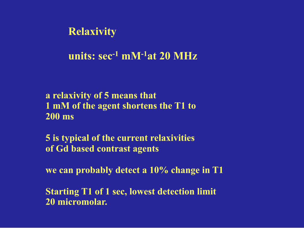
Relaxivity units: sec-1 mM-1at 20 MHz
a relaxivity of 5 means that 1 mM of the agent shortens the T1 to 200 ms 5 is typical of the current relaxivities of Gd based contrast agents we can probably detect a 10% change in T1 Starting T1 of 1 sec, lowest detection limit 20 micromolar.
Activatable agents Optical (fluorescence)
MRI/MRS based agents

Schematic of some key steps involved in a molecular imaging study.
Michelle L. James, and Sanjiv S. Gambhir Physiol Rev 2012;92:897-965
©2012 by American Physiological Society

Key molecular imaging modalities used for preclinical and/or clinical applications.
Michelle L. James, and Sanjiv S. Gambhir Physiol Rev 2012;92:897-965
©2012 by American Physiological Society
http://www.utsouthwestern.edu/research/ core-facilities/sair/index.html

Schematic diagram of the synthesis (a) and activation (b) of the developed reporter probe.
Ching-Hsuan Tung et al. Cancer Res 2000;60:4953-4958
©2000 by American Association for Cancer Research
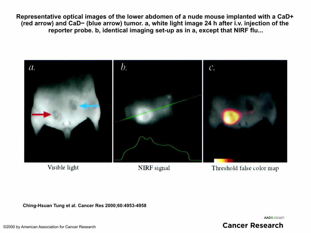
Representative optical images of the lower abdomen of a nude mouse implanted with a CaD+ (red arrow) and CaD− (blue arrow) tumor. a, white light image 24 h after i.v. injection of the
reporter probe. b, identical imaging set-up as in a, except that NIRF flu...
Ching-Hsuan Tung et al. Cancer Res 2000;60:4953-4958
©2000 by American Association for Cancer Research


Robert E. Lenkinski , John V. Frangioni, and Elena Vinogradov
Departments of Radiology and Medicine Beth Israel Deaconess Medical Center
Harvard Medical School
Contrast Agent Development for the Visualization of Micro-
calcifications

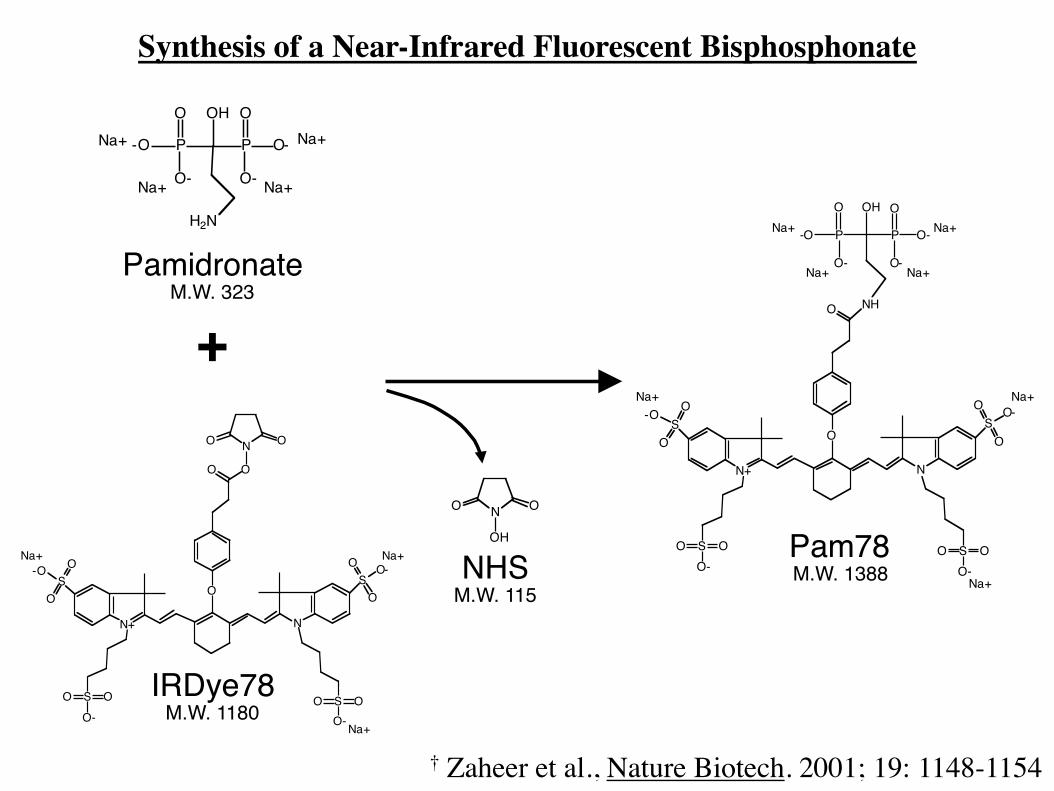
+
IRDye78M.W. 1180
PamidronateM.W. 323
H2N
P
OH O
O-
O-P
O
O-
-ONa+ Na+
Na+ Na+
O ON
OH
Na+
SO
O-O
S OOO-
O
O
SO OO-
N+
SO
O O-
N
NH
P
OH O
O-
O-P
O
O-
-O
Na+
Na+
Na+ Na+
Na+ Na+
NHSM.W. 115
Na+
O ON
SO
O-O
S OOO-
O
O O
SO OO-
N+
SO
O O-
N
Na+
Na+
Pam78M.W. 1388
Synthesis of a Near-Infrared Fluorescent Bisphosphonate
† Zaheer et al., Nature Biotech. 2001; 19: 1148-1154
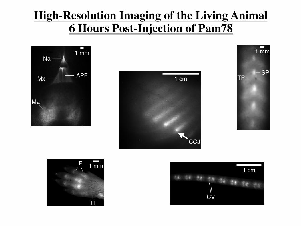
1 mmNa
Ma
Mx APF 1 cm
CCJ
1 mm
SPTP
1 mmP
H
1 cm
CV
High-Resolution Imaging of the Living Animal 6 Hours Post-Injection of Pam78

Raman spectroscopy shows two kinds of micro-calcifications
Calcium oxalate-primarily benign arising from
ductal secretions Hyroxyapatite -primarily malignant necrotic
mineralized cells


Published in: Kumar R. Bhushan; Preeti Misra; Fangbing Liu; Sanjeev Mathur; Robert E. Lenkinski; John V. Frangioni; J. Am. Chem. Soc. 2008, 130, 17648-17649. DOI: 10.1021/ja807099s Copyright © 2008 American Chemical Society

\
Published in: Kumar R. Bhushan; Preeti Misra; Fangbing Liu; Sanjeev Mathur; Robert E. Lenkinski; John V. Frangioni; J. Am. Chem. Soc. 2008, 130, 17648-17649. DOI: 10.1021/ja807099s Copyright © 2008 American Chemical Society
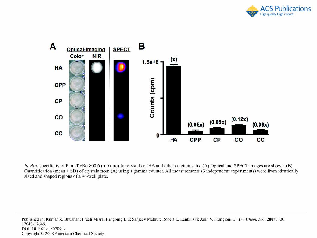
In vitro specificity of Pam-Tc/Re-800 6 (mixture) for crystals of HA and other calcium salts. (A) Optical and SPECT images are shown. (B) Quantification (mean ± SD) of crystals from (A) using a gamma counter. All measurements (3 independent experiments) were from identically sized and shaped regions of a 96-well plate.
Published in: Kumar R. Bhushan; Preeti Misra; Fangbing Liu; Sanjeev Mathur; Robert E. Lenkinski; John V. Frangioni; J. Am. Chem. Soc. 2008, 130, 17648-17649. DOI: 10.1021/ja807099s Copyright © 2008 American Chemical Society

In vivo imaging of rat breast cancer microcalcification. (A) Intraoperative NIR-fluorescence imaging and (B) SPECT/CT imaging. Arrows mark location of breast cancer microcalcification. (C) Blood clearance and (D) biodistribution of Pam-Tc-800 6a compared to 99mTc MDP. Figure data are representative of 3 independent experiments.
Published in: Kumar R. Bhushan; Preeti Misra; Fangbing Liu; Sanjeev Mathur; Robert E. Lenkinski; John V. Frangioni; J. Am. Chem. Soc. 2008, 130, 17648-17649. DOI: 10.1021/ja807099s Copyright © 2008 American Chemical Society


Micro-injection Model

Figure 2. In vivo MRI study: HA crystals were implanted subcutaneously at right ;lank of a mouse. CT scan (right) was performed to locate implanted HA crystals (arrow). UTE MR imaging was performed before (top left) and after injection of Gd-‐DOTA-‐Ser-‐PAM and after 6h clearance (bottom left).
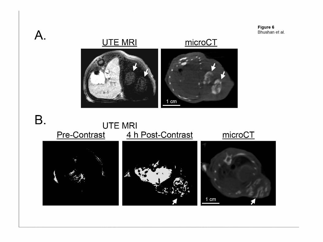

We have developed a gadolinium-based, MR-compatible contrast agent specific for hydroxyapatite, the calcium salt most commonly associated with malignant calcification. We employed a ultra-short echo time (UTE) pulse sequence, and characterized the sensitivity,specificity of and relaxivity of this agent in vitro and in vivo.
We have demonstrated contrast-enhanced detection of hydroxyapatite by UTE MRI in a syngeneic rat model of breast cancer microcalcification.
Conclusions

Schematic of some key steps involved in a molecular imaging study.
Michelle L. James, and Sanjiv S. Gambhir Physiol Rev 2012;92:897-965
©2012 by American Physiological Society


