jo u r nal homep age: www.elsev ier .com/ locate /vetpar
hort Communication
he diagnosis and management of a case of leishmaniosis in aog imported to Australia
atthew P. Besta,∗, Amanda Ashb, Jemma Bergfeldc, Janine Barrettd
Brisbane Veterinary Specialist Centre, Cnr Old Northern and Keong Roads, Albany Creek, Queensland 4035, AustraliaSchool of Veterinary and Life Sciences, Murdoch University, South Street, Western Australia 6150, AustraliaAustralian Animal Health Laboratory, CSIRO Animal, Food and Health Sciences, Private Bag 24, Geelong, Victoria 3220, AustraliaBiosecurity Queensland, Department of Agriculture, Fisheries and Forestry, 80 Ann Street, Brisbane, Queensland 4000, Australia
r t i c l e i n f o
rticle history:eceived 31 October 2013eceived in revised form 16 March 2014ccepted 27 March 2014
eywords:ustralia
a b s t r a c t
This case study discusses in detail for the first time the diagnosis and management of acase of leishmaniosis in a dog imported to Australia. The dog presented with epistaxisand a non-regenerative anaemia five years after being imported from Europe. Protozoawere identified within macrophages in bone marrow and splenic cytology. A Leishmaniaindirect fluorescent antibody test was performed and was positive while an Ehrlichia canisantibody test was negative. Polymerase chain reaction of the ITS-1 and ITS-2 regions ofskin, lymph node, spleen and bone marrow were all positive for Leishmania infantum. The
mphotericin Bogeishmania infantumCR
dog was treated with amphotericin B with a strong clinical response. The importance ofthorough diagnostics in non-endemic areas, particularly Australia, is discussed. Treatmentwith amphotericin B is discussed. Vigilance, disease reporting and response frameworksare recommended for non-endemic areas.
Canine leishmaniosis is a zoonotic tropical disease with widespread global distribution (Solano-Gallego et al.,009) and an expanding geographical range (Gonzalezt al., 2010; Gramiccia, 2011). It is a significant zoonoticisease with World Health Organisation global burden dis-ase estimates placing Leishmania second for mortalitynd fourth for morbidity amongst human tropical diseasesMathers et al., 2007). Diagnostic options for canine leish-
Please cite this article in press as: Best, M.P., et al., The diagnoimported to Australia. Vet. Parasitol. (2014), http://dx.doi.org/1
aniosis have increased in number and accuracy in recentears but definitive diagnosis in dogs still requires sev-ral, concordant test results (Solano-Gallego et al., 2009;
Gramiccia, 2011). Opinions on optimal and ethical treat-ment vary with compulsory culling still current in somecountries, whereas increasingly sophisticated treatmentsare employed elsewhere (Gramiccia, 2011; Dantas-Torreset al., 2012). While some question the ability to truly curecanine leishmaniosis (Ribeiro et al., 2008; Manna et al.,2009; Torres et al., 2011) clinical cures can be achieved withsome dogs no longer infectious through their biologicalvectors (da Silva et al., 2012).
In Australia there is no recognised endemic zoonoticLeishmania species present. A small number of cases havebeen recorded in imported dogs but to date none of thesehave been published. An endemic Australian Leishmaniaspecies in a captive population of red kangaroos (Maco-
sis and management of a case of leishmaniosis in a dog0.1016/j.vetpar.2014.03.032
pus rufus) (Rose et al., 2004) and in several species ofwild macropods (Dougall et al., 2009) has been discov-ered recently. This creates a need for rigorous diagnostictesting to identify the causative species if leishmaniosis is
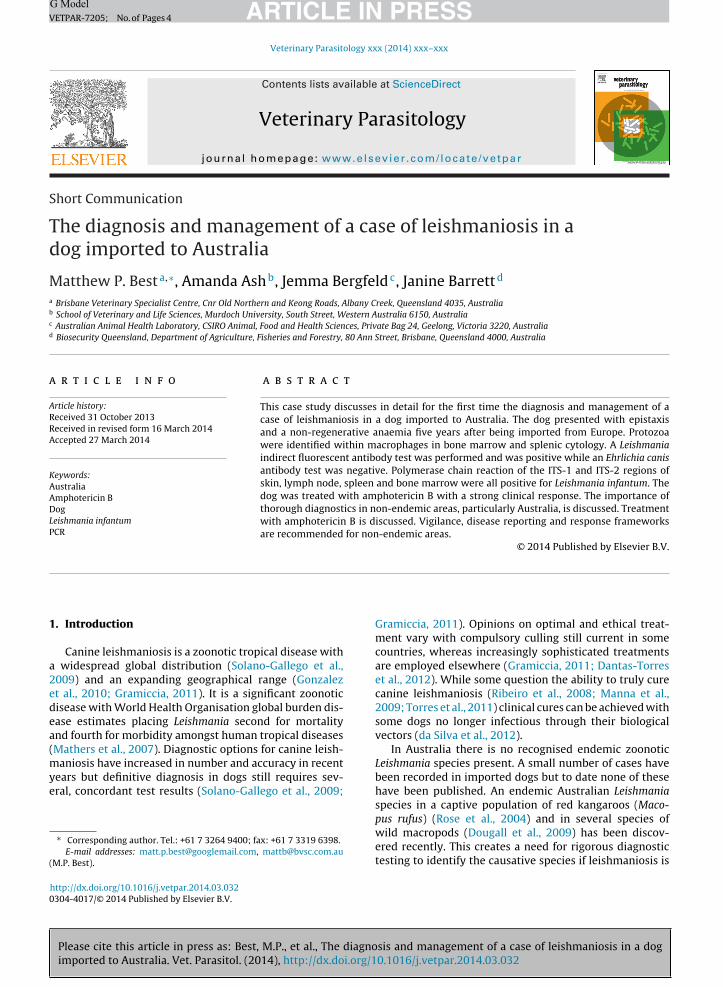
Fig. 1. Cytology of a splenic macrophage containing protozoal organisms
ARTICLEVETPAR-7205; No. of Pages 4
2 M.P. Best et al. / Veterinar
suspected in Australia. While phlebotomine sandflies arepresent in Australia there are no known competent bio-logical vectors of the non-Australian Leishmania speciespresent. A day-flying midge, species Forcipomyia (Lasiohe-lea) spp., has been implicated as the vector for endemicAustralian Leishmania (Dougall et al., 2011) and this is thefirst non-sandfly insect strongly implicated as a naturalvector of any Leishmania species. In light of these findingsa thorough disease plan should be in place for suspectedcases of Leishmania in Australia; including comprehensivediagnostic testing, clear treatment goals, monitoring ofresponse to treatment and measures to minimise the riskof transmission to other dogs and people. While Australiais in a unique situation with regards to Leishmania, manyconsiderations are likely applicable to other extralimitalareas.
2. Case report
A nine year old female neutered husky dog waspresented with a three month history of bilateral mucop-urulent nasal discharge, which had progressed to minorepistaxis three weeks prior to presentation, and major epis-taxis three days prior to presentation. Previous medicalhistory included three years of presumed atopic dermati-tis which had been treated with courses of prednisoloneand cephalexin. The dog had recently developed bilat-eral symmetrical alopecia and calcinosis cutis, attempts towithdraw the prednisolone had resulted in collapse 48 hlater. The dog was born in Portugal and had lived in theUK, during which time the dog had travelled back andforth from Portugal to the UK. In 2007 at the age of fivethe dog was emigrated to Australia and, after the requiredperiod of quarantine, had moved to a Brisbane suburb andresided there for the last four years, with no history of widertravel within Australia. Serological testing for Leishamaniavia IFAT or ELISA was required prior to import to Australiaat this time. The precise test information for this dog is notknown but presumably the result was negative.
Clinical examination revealed no obvious structural,pigmentation or sensation changes around the nasal cavitywith normal symmetrical airflow. The mucous membraneswere pale with a rapid refill. There was widespread non-pruritic symmetrical alopecia with calcinosis cutis andsmall open sores. Hepatomegaly and mild peripheral lym-phadenopathy were appreciated.
An abdominal ultrasound showed a large hyperechoicliver containing a cavitated and partially mineralised mass,suspected to be a benign lesion, while the adrenal glandswere small and inconspicuous. A biochemistry panel wasperformed (Chem17 + lytes4, Catalyst Dx; IDEXX) whichrevealed a hepatopathy with an ALP 1514 U/L (referenceinterval (RI): 23–212 U/L), ALT 143 U/L (RI: 10–100 U/L),GGT 42 U/L (RI: 0–7 U/L), a mild hyperglobulinemia at46 g/L (RI: 25–45 g/L) and a slightly decreased creatinine at42 �mol/L (RI: 44–159 �mol/L). An activated clotting time(VetScan I-STAT; Abaxis) was 106 s (RI: 90–130 s). The PCV
Please cite this article in press as: Best, M.P., et al., The diagnoimported to Australia. Vet. Parasitol. (2014), http://dx.doi.org/1
was 15% (RI: 35–57%) while a smear examination suggesteda non-regenerative anaemia with morphologically normalred and white cells. A single macrophage at the feath-ered edge of a blood smear was observed with suspected
within the cytoplasm. Organisms are approximately 2 �m in diameter,with a blue nucleus, a single dark purple kinetoplast and a pale bluecytoplasm. Bar = 10 �m.
intracytoplasmic organisms. Cystocentesis was performedand the urine had a specific gravity of 1.013 with an activesediment from which multisensitive Escherichia coli wasisolated.
The dog was sedated for bone marrow aspiration anda nasal computed tomograph. Bone marrow cytologyshowed adequate populations of all cell-line precur-sors with approximately 10% of cells present beingmacrophages with the cytoplasm distended by populationsof protozoa with 2 �m diameters, a dark nucleus, a singlebar-shaped kinetoplast and pale-blue cytoplasm. Nasal CTwas unremarkable with no mass effect or erosive processdetected.
Following notification of and discussion with theDepartment of Agriculture, Forestry and Fisheries Queens-land, extensive sampling was performed to allow confir-mation of the diagnosis prior to treatment. This includedcollection of multiple aliquots of EDTA blood and serum,skin punch biopsies, skin scrapes, thick and thin smears ofsplenic aspirates and lymph node core biopsies. These sam-ples were tested at the Australian Animal Health Laboratoryand Murdoch University. Protozoal organisms were iden-tified on cytology of the bone marrow and in the splenicaspirates (Fig. 1). Histology of the skin and lymph nodedid not identify any organisms. A commercially available(VRMD, 2011) Leishmania infantum indirect fluorescentantibody test (IFAT), considered genus specific (OIE, 2008),was performed and was positive at 1:6400 and negativeat 1:12,800 indicating a high Leishmania antibody titre. AnEhrlichia canis antibody test (Protatek immunofluorescenceantibody slide test) was negative. PCR analysis using theribosomal ITS-1 and ITS-2 regions (Tai et al., 2000) wasperformed on the skin, popliteal lymph node, peripheralblood and bone marrow samples. All samples were posi-tive by PCR and sequenced nucleotides were found to be a100% match with published sequences of L. infantum (NCBI
sis and management of a case of leishmaniosis in a dog0.1016/j.vetpar.2014.03.032
Accession numbers AJ634355/AJ634339).The affected dog had three companions that closely
cohabited in an outdoors run with close contact during theday and night with biting insects known to be present. All
hree companion dogs, two huskies and a malamute, wereegative on L. infantum IFAT.
At 28 days following initial presentation treatmentas begun and a deltamethrin-impregnated collar was
btained (Scalibor; Intervet). Amphotericin B lipid complexas given intravenously at 50 mg per dose (approximately
.5 mg/kg) diluted to 1 mg/ml in 5% dextrose in water andiven over one hour. Creatinine, ALT and electrolytes wereonitored, a 10 ml/kg fluid bolus was given and maropitant
Cerenia; Zoetis) was administered prior to each treat-ent. An acute increase in ALT up to 702 U/L was recorded
ollowing the second treatment with normal bile acid stim-lation test results. This reduced rapidly with supportiveare including SAMe (Denosyl; Ceva) and a short treatmentelay. Following the fourth dose acute kidney injury was
ndicated by a creatinine of 872 �mol/L, resulting in sevenays hospitalisation on fluid therapy prior to dischargeor home care with an esophagostomy tube placed. Fur-her treatment was deferred for 53 days after which timereatinine was back within normal limits and treatmentas resumed with a similar protocol to the initial round
f treatment, except daily doses of 25 mg amphotericin Bere given for four consecutive days. This treatment was
olerated well with a moderate increase in ALT to 384 U/Lbserved but no renal compromise or clinically apparentide effects noted. The total dose of amphotericin B givenas approximately 15 mg/kg.
One month following completion of the treatment bloodCR was negative for L. infantum and allopurinol wastarted at a dose of 15 mg/kg orally twice daily.
. Discussion
Leishmaniosis presents a major diagnostic and manage-ent challenge wherever it occurs. In endemic areas the
pproach to this disease varies widely and remains con-entious (Dantas-Torres et al., 2012). In non-endemic areashere are sporadic case reports of disease (Slappendel andeske, 1999) but even where travel-related disease is rela-ively common, such as in the United Kingdom, the diseases often not closely monitored, is likely under-reported andhe risks of transmission are poorly understood (Shaw et al.,009).
In this case thorough diagnostics excluded potentiallternative infections, such as E. canis which can cross reacterologically (Ferreira et al., 2007; Marcondes et al., 2011),rimarily through the use of PCR analysis. Appropriateesting will depend upon geographic location and shoulde conducted in under the consultation or supervisionf the relevant government authority as Leishmania is anIE-listed disease (OIE, 2013). Monitoring of disease inci-ence allows protection of public health and response tohanges in the disease prevalence or transmission. Whereeishmaniosis is not a notifiable disease its prevalence isnderreported (Shaw et al., 2009) and the risk of failing toetect a new or low density competent vector is increased.
Please cite this article in press as: Best, M.P., et al., The diagnoimported to Australia. Vet. Parasitol. (2014), http://dx.doi.org/1
The decision to treat is controversial. In this case treat-ent was performed due to the lack of known local vectorshich made epidemic veterinary disease and zoonotic dis-
ase highly unlikely.
PRESSology xxx (2014) xxx–xxx 3
Amphotericin B is not recommended in endemic areasdue to concerns about side effects and induction of humanresistance. It was used in this case because of its excellentavailability and low cost compared to other suitable agentsin Australia, the low risk of development of resistance, thelow likelihood of zoonotic transmission and the patients’clinical suitability for this medication with a low serumcreatinine prior to treatment.
Please consult the supplementary material providedelectronically with this article for in depth discussionand review of the literature discussing the major issuesof canine leishmaniosis in extralimital areas; includingdiagnosis, the decision to treat, the risks of transmission,treatment options and disease monitoring. The decisionsmade in this specific case are further discussed in this mate-rial.
In conclusion the increasing incidence of leishmaniosisin non-endemic areas is challenging owners, veterinari-ans and government authorities. These authors identify aneed for clear, locally relevant guidelines directing diag-nosis, treatment and management of suspected cases. Thiscase report provides an example of such case managementand highlights issues of particular relevance to Australia,such as the local status of Leishmania, incidence of caninetravel to endemic areas, local climate and possible localvectors. These guidelines would be recommended for allgovernments in non-endemic Leishmania areas.
Conflicts of interest
The authors have no conflicts of interest concerning thework reported in this paper.
Acknowledgements
The authors thank Ross Lunt and serology staff at CSIROAAHL for their work on the Leishmania and Ehrlichia serol-ogy and Susan Boyd (IDEXX Laboratories, Brisbane, currentaddress QML Laboratories, Brisbane, Australia) for makingthe original cytological diagnosis of Leishmania based onbone marrow cytology.
Appendix A. Supplementary data
Supplementary material related to this article can befound, in the online version, at http://dx.doi.org/10.1016/j.vetpar.2014.03.032.
References
Dantas-Torres, F., Solano-Gallego, L., Baneth, G., Ribeiro, V.M., de Paiva-Cavalcanti, M., Otranto, D., 2012. Canine Leishmaniosis in the old andnew worlds: unveiled similarities and differences. Trends Parasitol.28, 531–538.
da Silva, S.M., Amorim, I.F.G., Ribeiro, P.R., Azevedo, E.G., Demicheli, C.,Melo, M.N., Tafuri, W.L., Gontijo, N.F., Michalick, M.S.M., Frézard, F.,2012. Efficacy of combined therapy with liposome-encapsulated meg-lumine antimoniate and allopurinol in treatment of canine visceralleishmaniasis. Antimicrob. Agents Chemother. 56, 2858–2867.
sis and management of a case of leishmaniosis in a dog0.1016/j.vetpar.2014.03.032
Dougall, A., Shilton, C., Low Choy, J., Alexander, B., Walton, S., 2009. Newreports of Australian cutaneous leishmaniasis in Northern Australianmacropods. Epidemiol. Infect. 137, 1516–1520.
Dougall, A., Alexander, B., Holt, D.C., Harris, T., Sultan, A.H., Bates, P.A.,Rose., K., Walton, S.F., 2011. Evidence incriminating midges (Diptera:
Ceratopogonidae) as potential vectors of Leishmania in Australia. Int.J. Parasitol. 41, 571–579.
Ferreira, E.dC., de Lana, M., Carneiro, M., Reis, A.B., Paes, D.V., da Silva, E.S.,Schallig, H., Gontijo, C.M.F., 2007. Comparison of serological assaysfor the diagnosis of canine visceral leishmaniasis in animals pre-senting different clinical manifestations. Vet. Parasitol. 146, 235–241.
Gonzalez, C., Wang, O., Strutz, S.E., González-Salazar, C., Sánchez-Cordero,V., Sarkar, S., 2010. Climate change and risk of leishmaniasis in NorthAmerica: predictions from ecological niche models of vector andreservoir species. PLoS Negl. Trop. Dis. 4, e585.
Gramiccia, M., 2011. Recent advances in leishmaniosis in pet animals: epi-demiology, diagnostics and anti-vectorial prophylaxis. Vet. Parasitol.181, 23–30.
Manna, L., Vitale, F., Reale, S., Picillo, E., Neglia, G., Vescio, F., Gravino, A.E.,2009. Study of efficacy of miltefosine and allopurinol in dogs withleishmaniosis. Vet. J. 182, 441–445.
Marcondes, M., Biondo, A.W., Gomes, A.A.D., Silva, A.R.S., Vieira, R.F.C.,Camacho, A.A., Quinn, J., Chandrashekar, R., 2011. Validation of a Leish-mania infantum ELISA rapid test for serological diagnosis of Leishmaniachagasi in dogs. Vet. Parasitol. 175, 15–19.
Mathers, C.D., Ezzati, M., Lopez, A.D., 2007. Measuring the burden ofneglected tropical diseases: the global burden of disease framework.PLoS Negl. Trop. Dis. 1, e114.
M.N., Fézard, F., Michalick, M.S., 2008. Reduced tissue parasitic loadand infectivity to sand flies in dogs naturally infected by Leishmania(Leishmania) chagasi following treatment with a liposome formula-tion of meglumine antimoniate. Antimicrob. Agents Chemother. 52,2564–2572.
Rose, K., Curtis, J., Baldwin, T., Mathis, A., Kumar, B., Sakthianandeswaren,A., Spurck, T., Low Choy, L., Handman, E., 2004. Cutaneous leishmani-asis in red kangaroos: isolation and characterisation of the causativeorganisms. Int. J. Parasitol. 34, 655–664.
Shaw, S.E., Langton, D.A., Hillman, T.J., 2009. Canine leishmaniosis in theUnited Kingdom: a zoonotic disease waiting for a vector? Vet. Para-sitol. 163, 281–285.
Slappendel, R.J., Teske, E., 1999. A review of canine leishmaniasis pre-senting outside endemic areas. In: Killick-Kendrick, R. (Ed.), CanineLeishmaniasis: An Update. Proceedings of the Canine LeishmaniasisForum. Barcelona, Spain, pp. 54–59.
Solano-Gallego, L., Koutinas, A., Miró, G., Cardoso, L., Pennisi, M.G., Ferrer,L., Bourdeau, P., Oliva, G., Baneth, G., 2009. Directions for the diagno-sis, clinical staging, treatment and prevention of canine leishmaniosis.Vet. Parasitol. 165, 1–18.
Tai, N.o.E., Osman, O.f., Fari, M.E., Presber, W., Schonian, G., 2000. Geneticheterogeneity of ribosomal internal transcribed spacer in clinical sam-ples of Leishmania donovani spotted on filter paper as revealed bysingle-strand conformation polymorphisms and sequencing. Trans. R.Soc. Trop. Med. Hyg. 94, 575–579.
Torres, M., Bardagí, M., Roura, X., Zanna, G., Ravera, I., Ferrer, L., 2011. Longterm follow-up of dogs diagnosed with leishmaniosis (clinical stage
sis and management of a case of leishmaniosis in a dog0.1016/j.vetpar.2014.03.032
II) and treated with meglumine antimoniate and allopurinol. Vet. J.188, 346–351.
VRMD, 2011. Leishmania infantum FA substrate slide. http://www.vmrd.com/pdfLibrary/12-well%20Slides/Leishmania/LSH 12 well slideSLD-IFA-LSH P090211-002 130210.pdf