64
The ECHO Suggests Pulmonary Hypertension. What Next? Dr. Jonathon Langridge, B.Sc., MD, FRCPC Cardiovascular Respiratory Conference, 2016
The ECHO Suggests Pulmonary
Hypertension. What Next?
Dr. Jonathon Langridge, B.Sc., MD, FRCPC
Cardiovascular Respiratory Conference, 2016

I have no conflicts of interest to declare
1) Review definition and classification of PH
2) Discuss presentation and diagnostic approach
3) Overview of treatment strategies
(Recently published: 2015 ESC/ERS Guidelines)
Definition
PH refers to elevated pulmonary arterial pressure
Pressure elevation in pulmonary arterial system
alone (increased resistance or flow) OR
2° to pressure elevation in pulmonary venous
system
PH defined as mPAP ≥ 25 mmHg at rest
Upper limit of normal mPAP 20 mmHg
mPAP 21-24 mmHg considered “borderline”:
- Unclear clinical significance
- May progress to significant disease
(especially idiopathic, family hx, CTD)
-Warrants monitoring
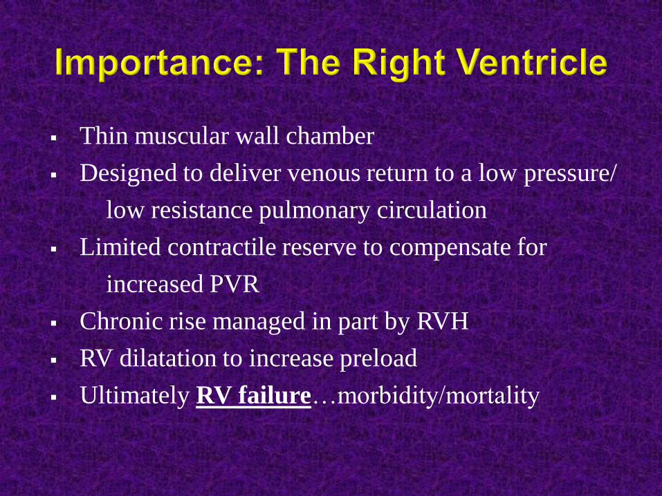
Thin muscular wall chamber
Designed to deliver venous return to a low pressure/
low resistance pulmonary circulation
Limited contractile reserve to compensate for
increased PVR
Chronic rise managed in part by RVH
RV dilatation to increase preload
Ultimately RV failure…morbidity/mortality
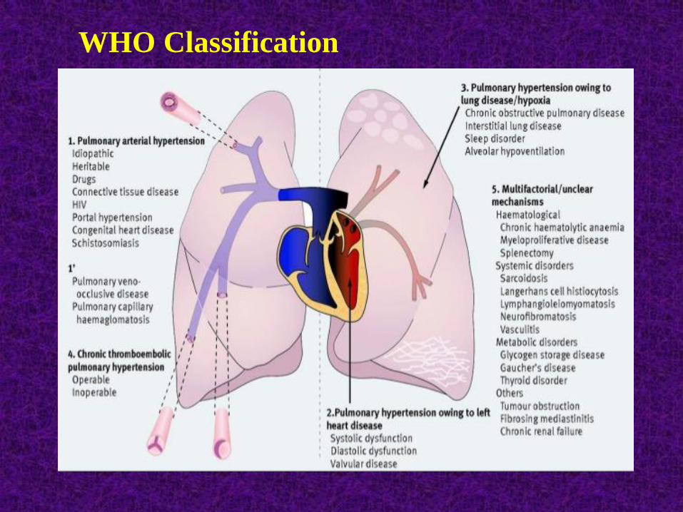
Classification
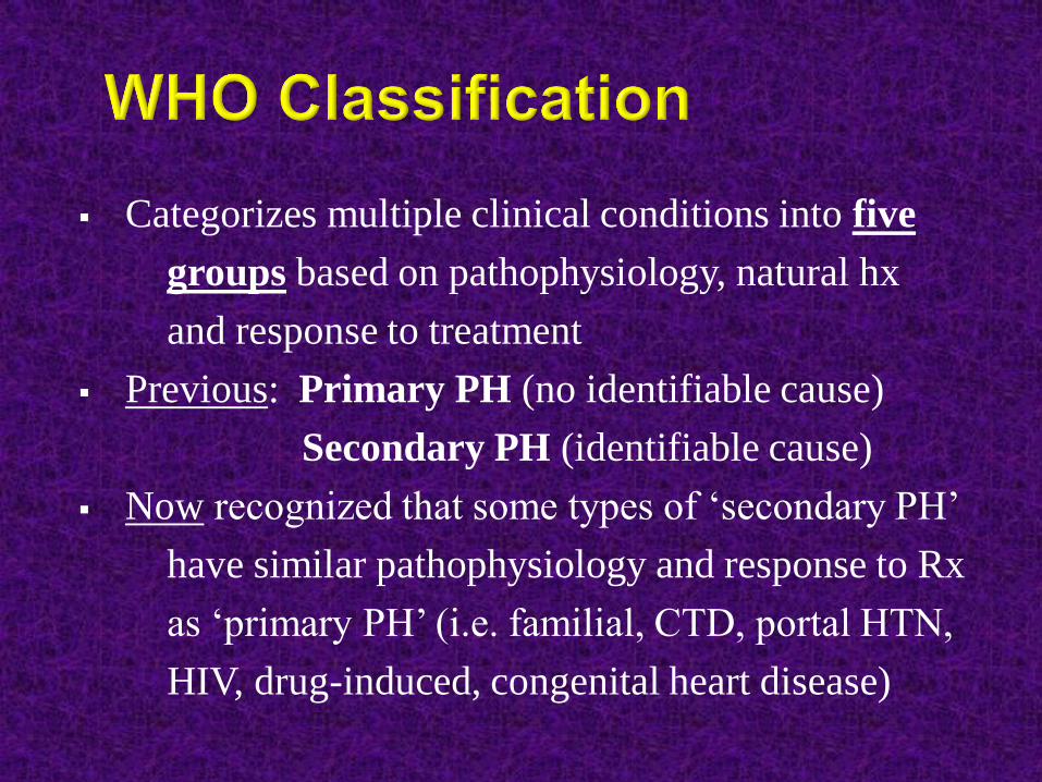
Categorizes multiple clinical conditions into five
groups based on pathophysiology, natural hx
and response to treatment
Previous: Primary PH (no identifiable cause)
Secondary PH (identifiable cause)
Now recognized that some types of ‘secondary PH’
have similar pathophysiology and response to Rx
as ‘primary PH’ (i.e. familial, CTD, portal HTN,
HIV, drug-induced, congenital heart disease)
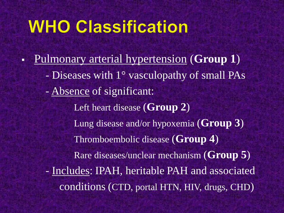
Pulmonary arterial hypertension (Group 1)
- Diseases with 1° vasculopathy of small PAs
- Absence of significant:
Left heart disease (Group 2)
Lung disease and/or hypoxemia (Group 3)
Thromboembolic disease (Group 4)
Rare diseases/unclear mechanism (Group 5)
- Includes: IPAH, heritable PAH and associated
conditions (CTD, portal HTN, HIV, drugs, CHD)
Group 1: Pulmonary arterial hypertension (PAH)
Group 2: PH owing to left heart disease
Group 3: PH owing to lung disease and/or hypoxemia
Group 4: Chronic thromboembolic PH (CTEPH)
Group 5: PH with unclear/multifactorial mechanisms
Presentation
Symptoms of PH nonspecific – often leads to delay
in diagnosis
Mainly related to progressive RV dysfunction
Initial symptoms typically induced by exertion
Progressive exertional dyspnea most common
Chest pain, syncope, fatigue
With overt RV failure – abdominal distension,
ankle edema
Less common symptoms due to mechanical
complications: hemoptysis, wheeze, hoarseness
Symptoms of associated conditions
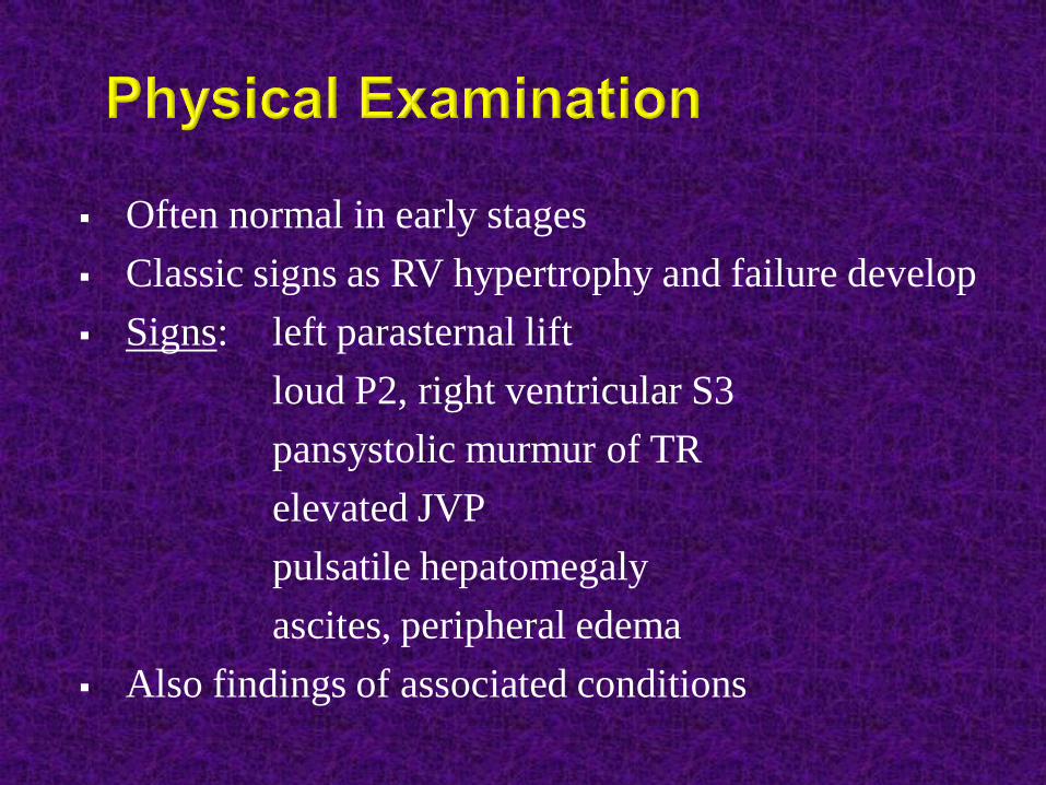
Often normal in early stages
Classic signs as RV hypertrophy and failure develop
Signs: left parasternal lift
loud P2, right ventricular S3
pansystolic murmur of TR
elevated JVP
pulsatile hepatomegaly
ascites, peripheral edema
Also findings of associated conditions
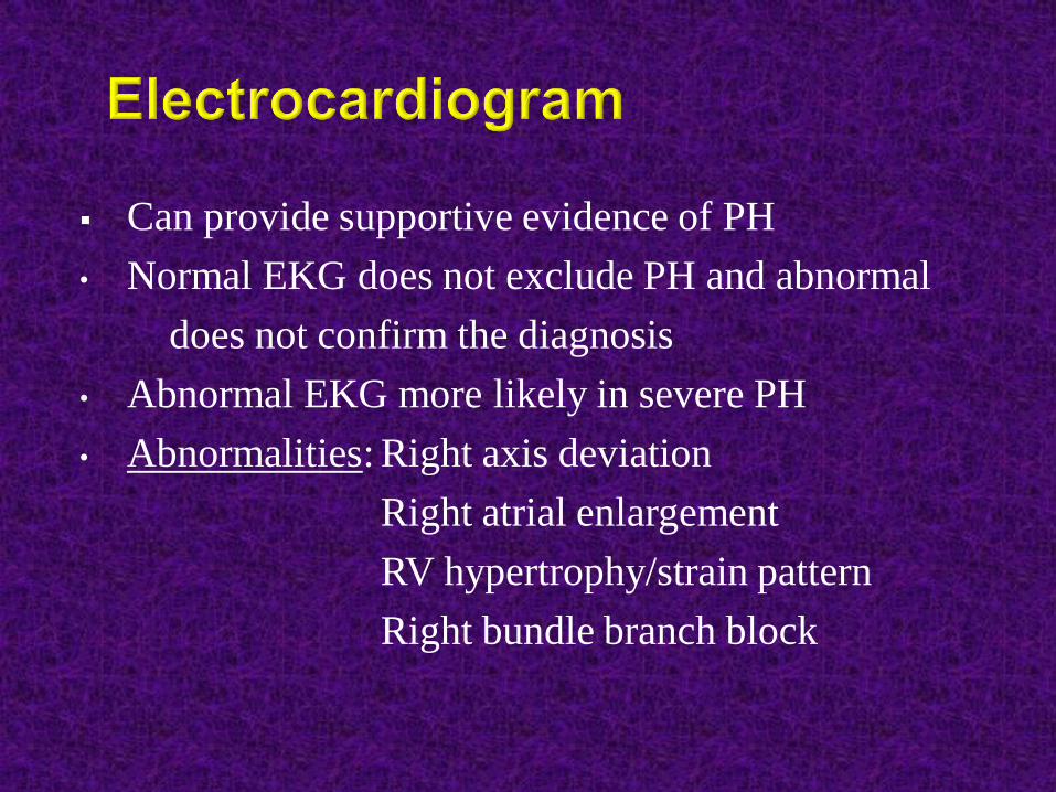
Can provide supportive evidence of PH
• Normal EKG does not exclude PH and abnormal
does not confirm the diagnosis
• Abnormal EKG more likely in severe PH
• Abnormalities: Right axis deviation
Right atrial enlargement
RV hypertrophy/strain pattern
Right bundle branch block
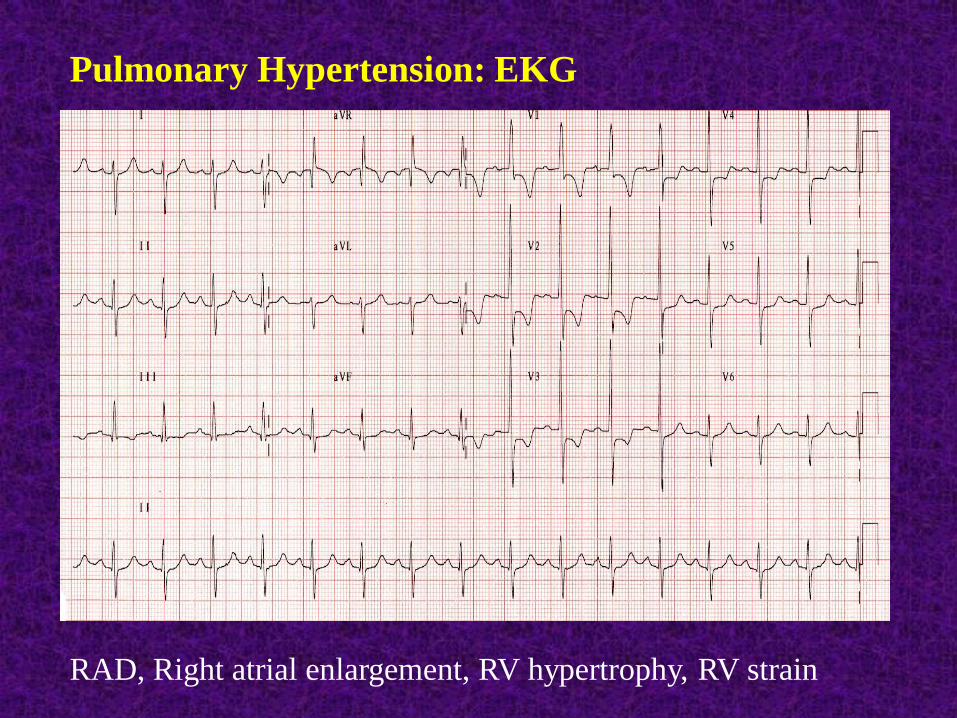
Pulmonary Hypertension: EKG
RAD, Right atrial enlargement, RV hypertrophy, RV strain
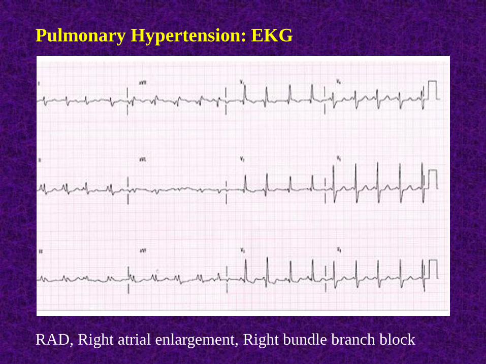
Pulmonary Hypertension: EKG
RAD, Right atrial enlargement, Right bundle branch block
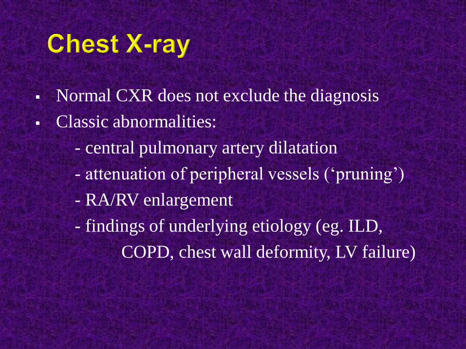
Normal CXR does not exclude the diagnosis
Classic abnormalities:
- central pulmonary artery dilatation
- attenuation of peripheral vessels (‘pruning’)
- RA/RV enlargement
- findings of underlying etiology (eg. ILD,
COPD, chest wall deformity, LV failure)
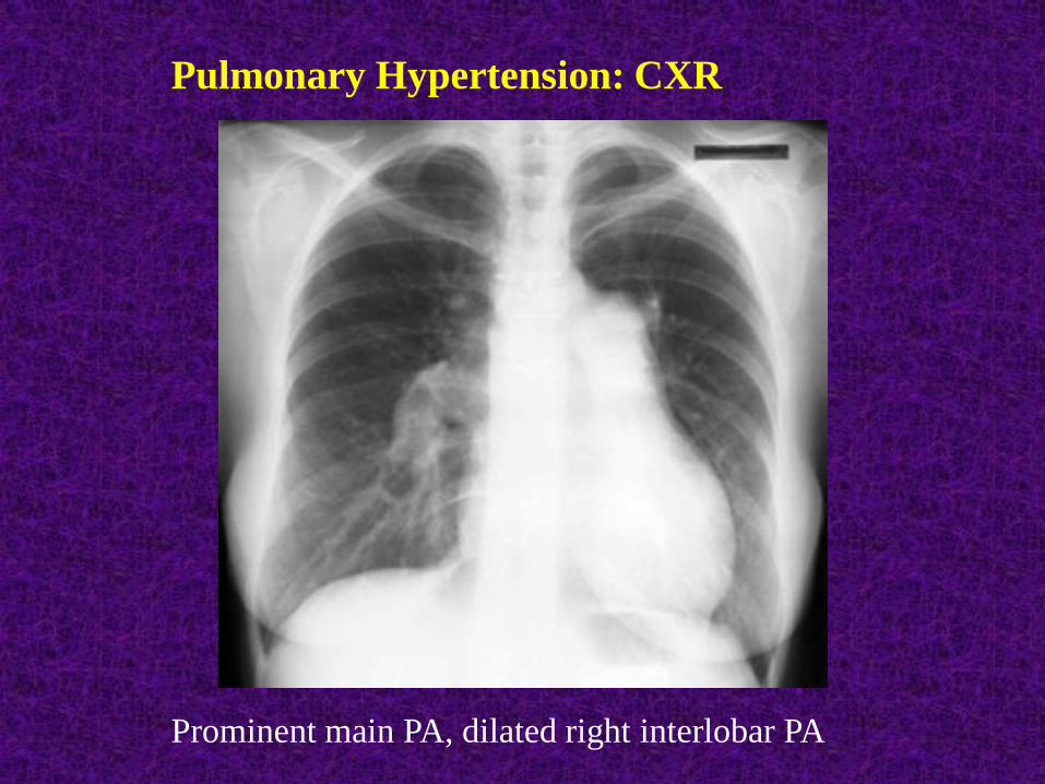
Pulmonary Hypertension: CXR
Prominent main PA, dilated right interlobar PA
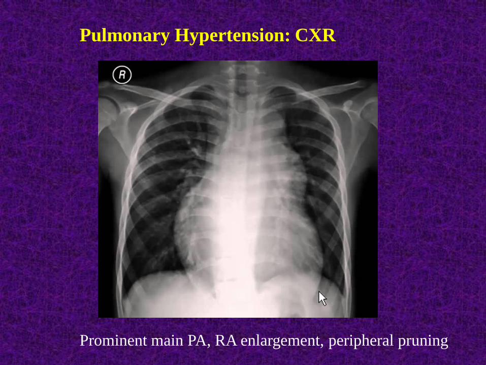
Pulmonary Hypertension: CXR
Prominent main PA, RA enlargement, peripheral pruning
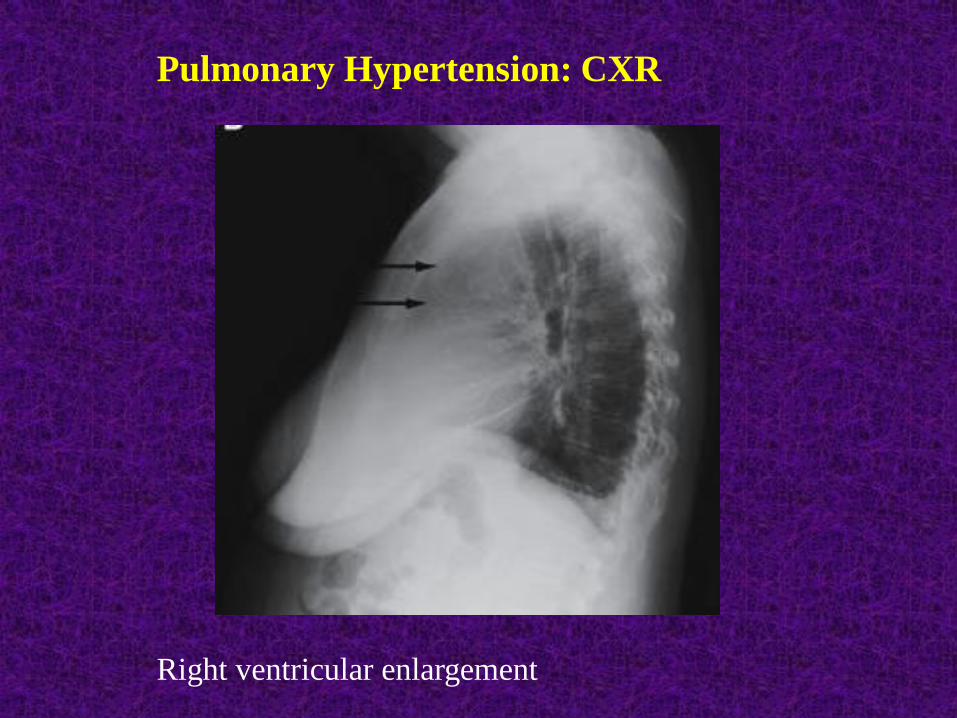
Pulmonary Hypertension: CXR
Right ventricular enlargement
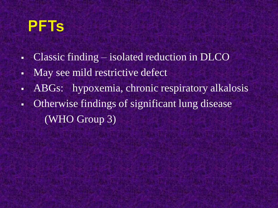
Classic finding – isolated reduction in DLCO
May see mild restrictive defect
ABGs: hypoxemia, chronic respiratory alkalosis
Otherwise findings of significant lung disease
(WHO Group 3)
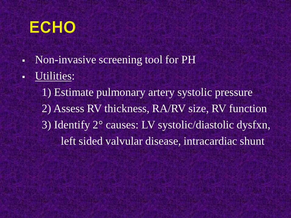
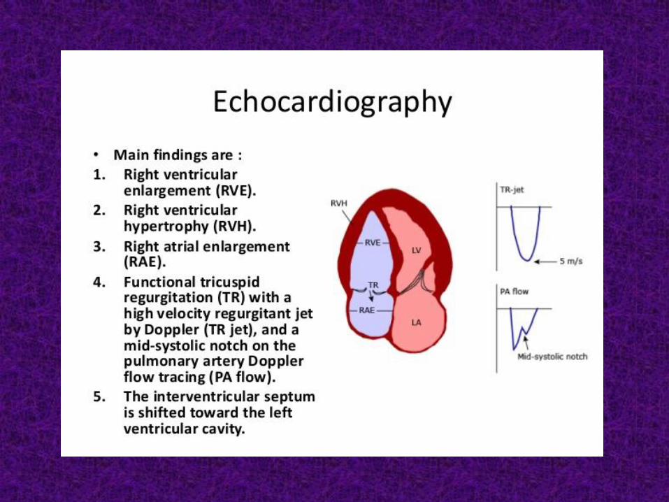
Non-invasive screening tool for PH
Utilities:
1) Estimate pulmonary artery systolic pressure
2) Assess RV thickness, RA/RV size, RV function
3) Identify 2° causes: LV systolic/diastolic dysfxn,
left sided valvular disease, intracardiac shunt
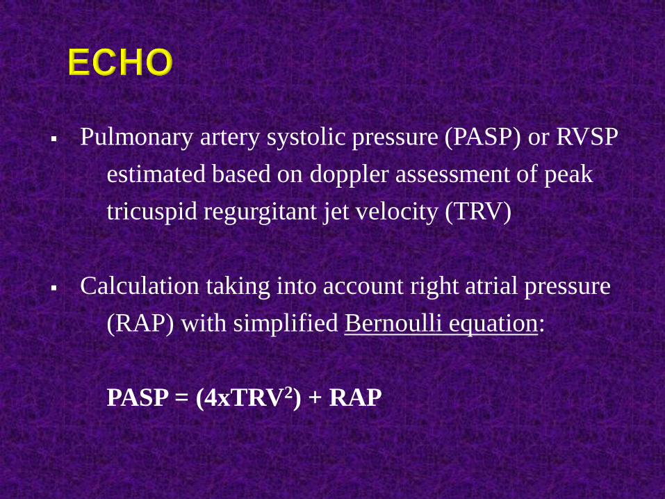
Pulmonary artery systolic pressure (PASP) or RVSP
estimated based on doppler assessment of peak
tricuspid regurgitant jet velocity (TRV)
Calculation taking into account right atrial pressure
(RAP) with simplified Bernoulli equation:
PASP = (4xTRV2) + RAP
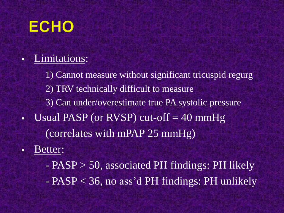
Limitations:
1) Cannot measure without significant tricuspid regurg
2) TRV technically difficult to measure
3) Can under/overestimate true PA systolic pressure
Usual PASP (or RVSP) cut-off = 40 mmHg
(correlates with mPAP 25 mmHg)
Better:
- PASP > 50, associated PH findings: PH likely
- PASP < 36, no ass’d PH findings: PH unlikely
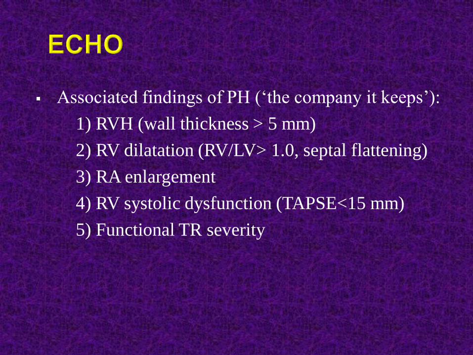
Associated findings of PH (‘the company it keeps’):
1) RVH (wall thickness > 5 mm)
2) RV dilatation (RV/LV> 1.0, septal flattening)
3) RA enlargement
4) RV systolic dysfunction (TAPSE<15 mm)
5) Functional TR severity
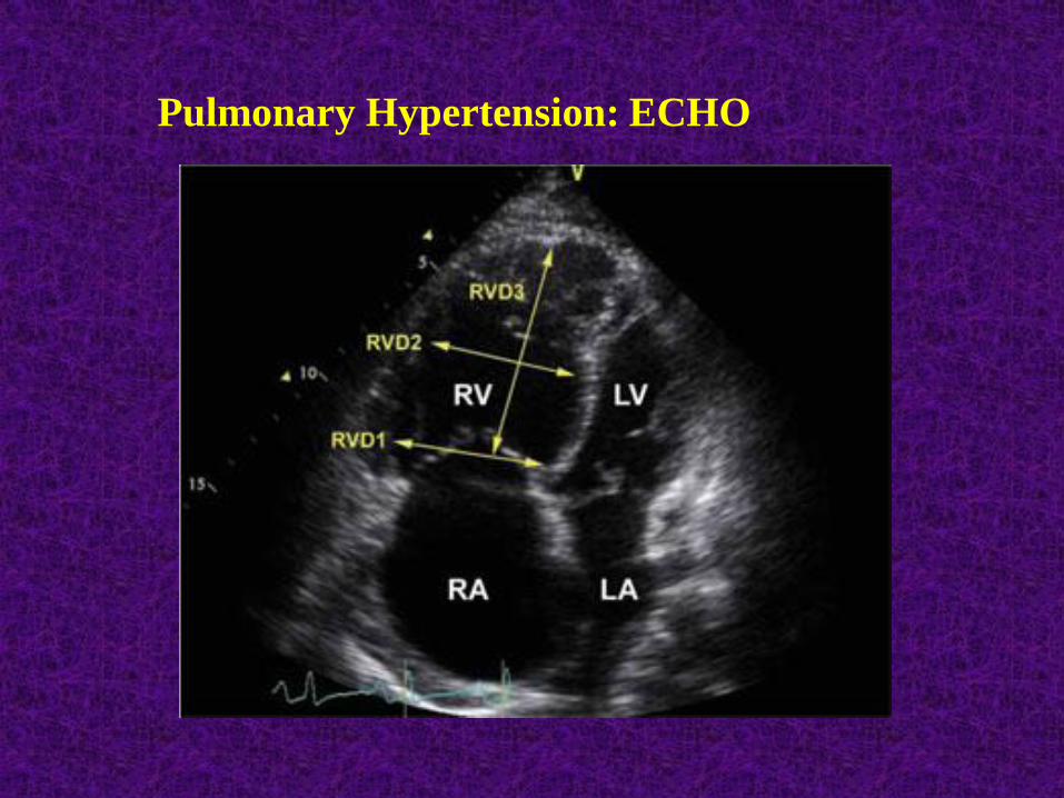
Pulmonary Hypertension: ECHO
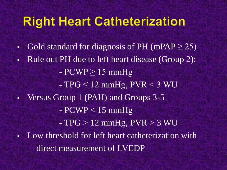
Gold standard for diagnosis of PH (mPAP ≥ 25)
Rule out PH due to left heart disease (Group 2):
- PCWP ≥ 15 mmHg
- TPG ≤ 12 mmHg, PVR < 3 WU
Versus Group 1 (PAH) and Groups 3-5
- PCWP < 15 mmHg
- TPG > 12 mmHg, PVR > 3 WU
Low threshold for left heart catheterization with
direct measurement of LVEDP

Diagnostic Approach
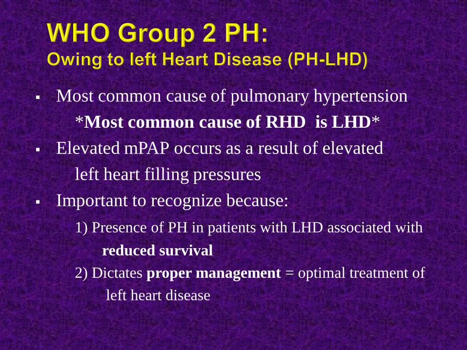
Most common cause of pulmonary hypertension
*Most common cause of RHD is LHD*
Elevated mPAP occurs as a result of elevated
left heart filling pressures
Important to recognize because:
1) Presence of PH in patients with LHD associated with
reduced survival
2) Dictates proper management = optimal treatment of
left heart disease
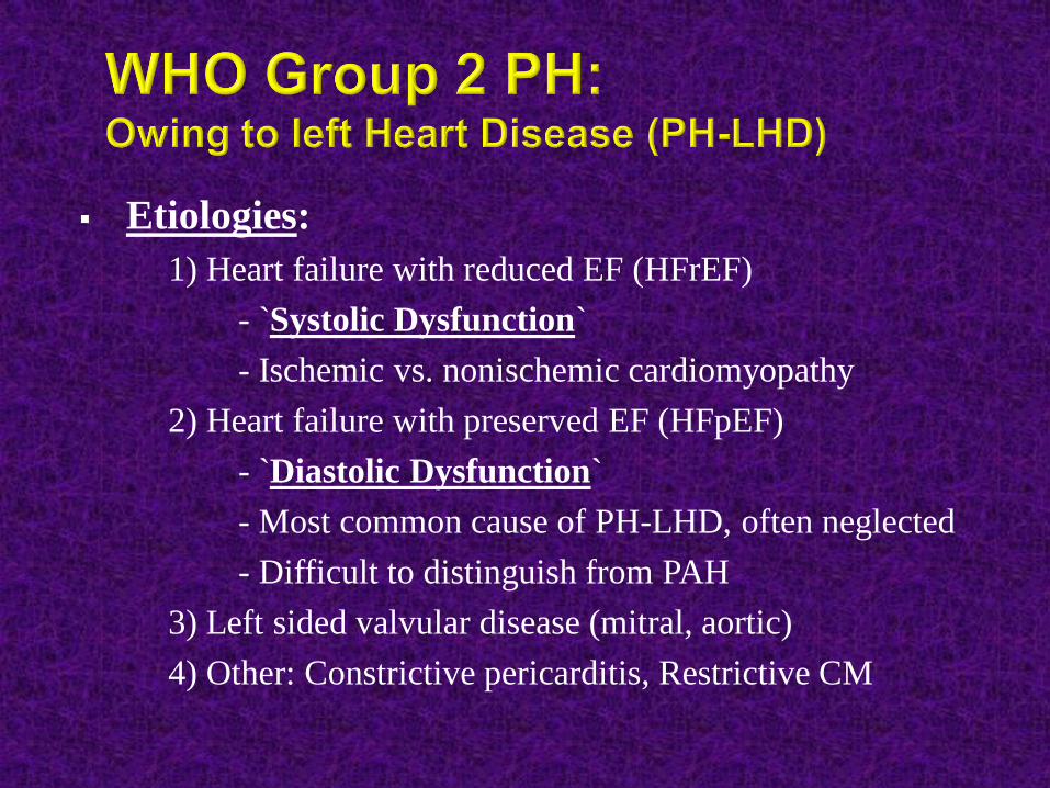
Etiologies:
1) Heart failure with reduced EF (HFrEF)
- `Systolic Dysfunction`
- Ischemic vs. nonischemic cardiomyopathy
2) Heart failure with preserved EF (HFpEF)
- `Diastolic Dysfunction`
- Most common cause of PH-LHD, often neglected
- Difficult to distinguish from PAH
3) Left sided valvular disease (mitral, aortic)
4) Other: Constrictive pericarditis, Restrictive CM
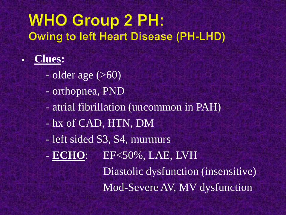
Clues:
- older age (>60)
- orthopnea, PND
- atrial fibrillation (uncommon in PAH)
- hx of CAD, HTN, DM
- left sided S3, S4, murmurs
- ECHO: EF<50%, LAE, LVH
Diastolic dysfunction (insensitive)
Mod-Severe AV, MV dysfunction
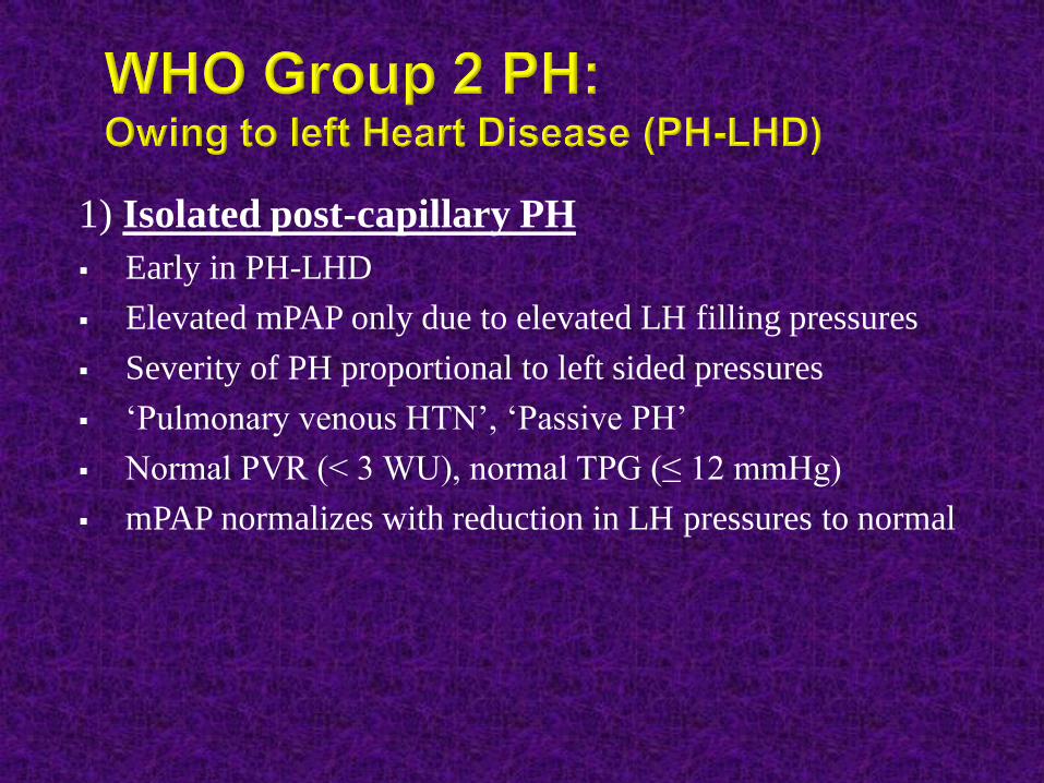
1) Isolated post-capillary PH
Early in PH-LHD
Elevated mPAP only due to elevated LH filling pressures
Severity of PH proportional to left sided pressures
‘Pulmonary venous HTN’, ‘Passive PH’
Normal PVR (< 3 WU), normal TPG (≤ 12 mmHg)
mPAP normalizes with reduction in LH pressures to normal
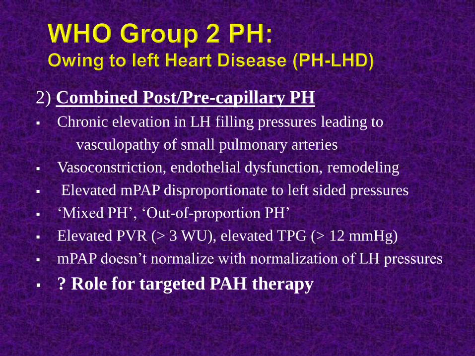
2) Combined Post/Pre-capillary PH
Chronic elevation in LH filling pressures leading to
vasculopathy of small pulmonary arteries
Vasoconstriction, endothelial dysfunction, remodeling
Elevated mPAP disproportionate to left sided pressures
‘Mixed PH’, ‘Out-of-proportion PH’
Elevated PVR (> 3 WU), elevated TPG (> 12 mmHg)
mPAP doesn’t normalize with normalization of LH pressures
? Role for targeted PAH therapy
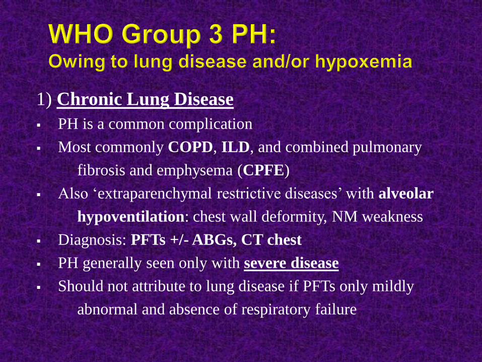
1) Chronic Lung Disease
PH is a common complication
Most commonly COPD, ILD, and combined pulmonary
fibrosis and emphysema (CPFE)
Also ‘extraparenchymal restrictive diseases’ with alveolar
hypoventilation: chest wall deformity, NM weakness
Diagnosis: PFTs +/- ABGs, CT chest
PH generally seen only with severe disease
Should not attribute to lung disease if PFTs only mildly
abnormal and absence of respiratory failure
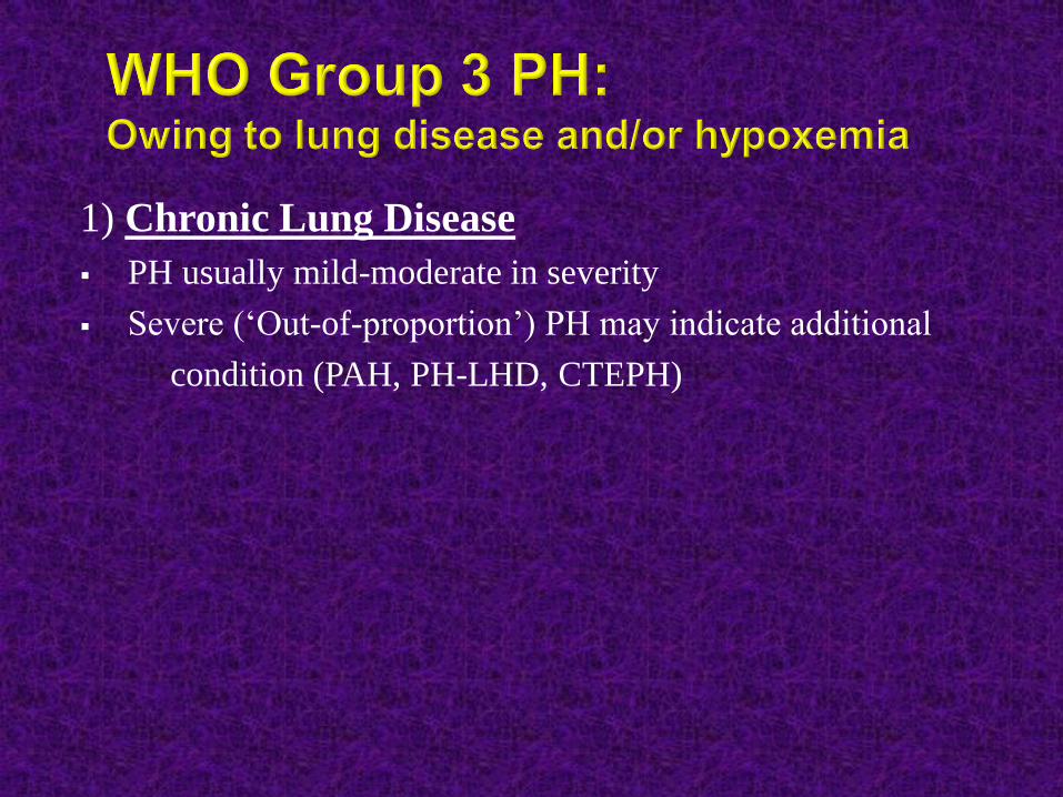
1) Chronic Lung Disease
PH usually mild-moderate in severity
Severe (‘Out-of-proportion’) PH may indicate additional
condition (PAH, PH-LHD, CTEPH)
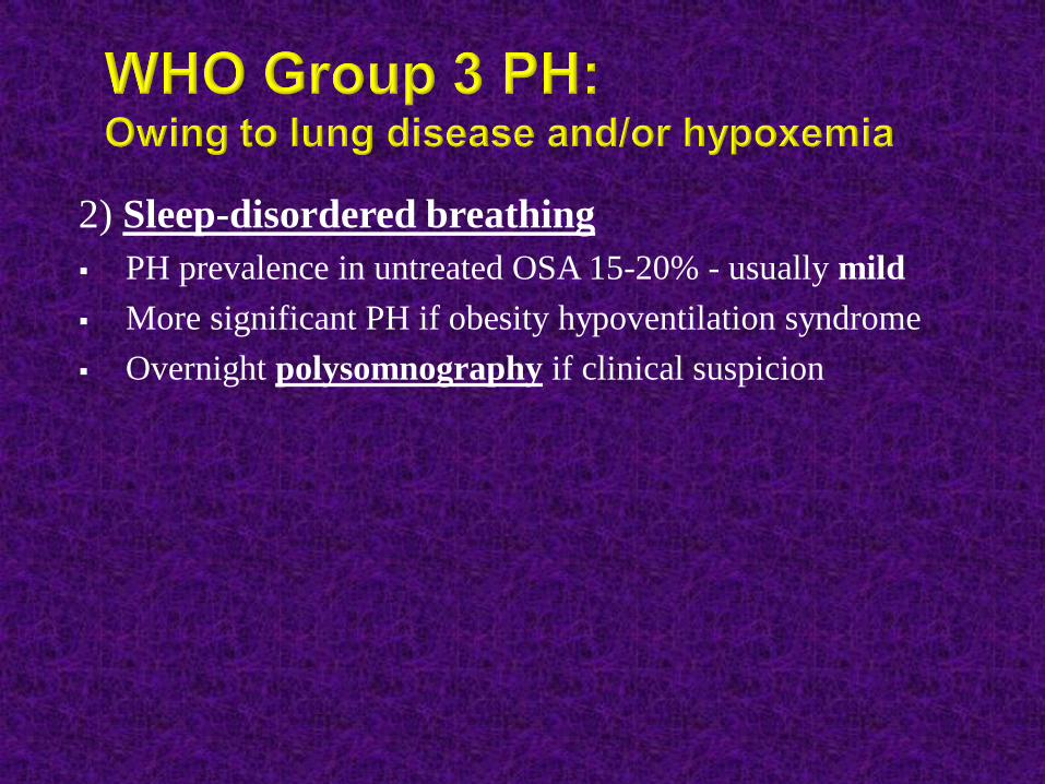
2) Sleep-disordered breathing
PH prevalence in untreated OSA 15-20% - usually mild
More significant PH if obesity hypoventilation syndrome
Overnight polysomnography if clinical suspicion
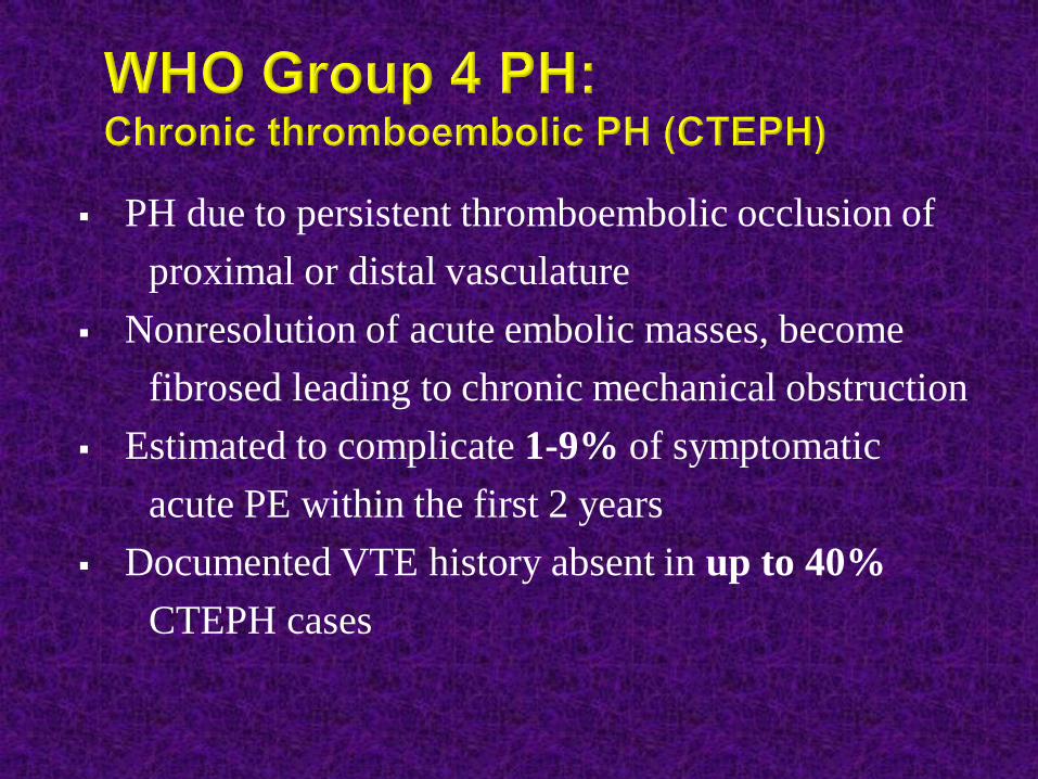
PH due to persistent thromboembolic occlusion of
proximal or distal vasculature
Nonresolution of acute embolic masses, become
fibrosed leading to chronic mechanical obstruction
Estimated to complicate 1-9% of symptomatic
acute PE within the first 2 years
Documented VTE history absent in up to 40%
CTEPH cases
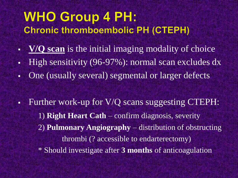
V/Q scan is the initial imaging modality of choice
High sensitivity (96-97%): normal scan excludes dx
One (usually several) segmental or larger defects
Further work-up for V/Q scans suggesting CTEPH:
1) Right Heart Cath – confirm diagnosis, severity
2) Pulmonary Angiography – distribution of obstructing
thrombi (? accessible to endarterectomy)
* Should investigate after 3 months of anticoagulation
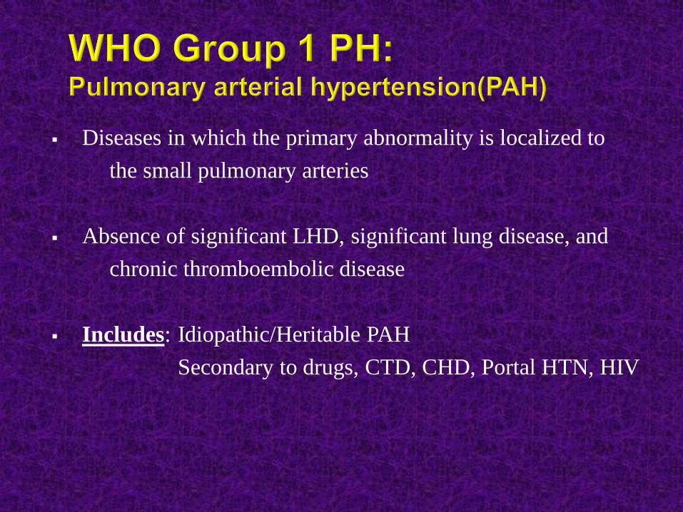
Diseases in which the primary abnormality is localized to
the small pulmonary arteries
Absence of significant LHD, significant lung disease, and
chronic thromboembolic disease
Includes: Idiopathic/Heritable PAH
Secondary to drugs, CTD, CHD, Portal HTN, HIV
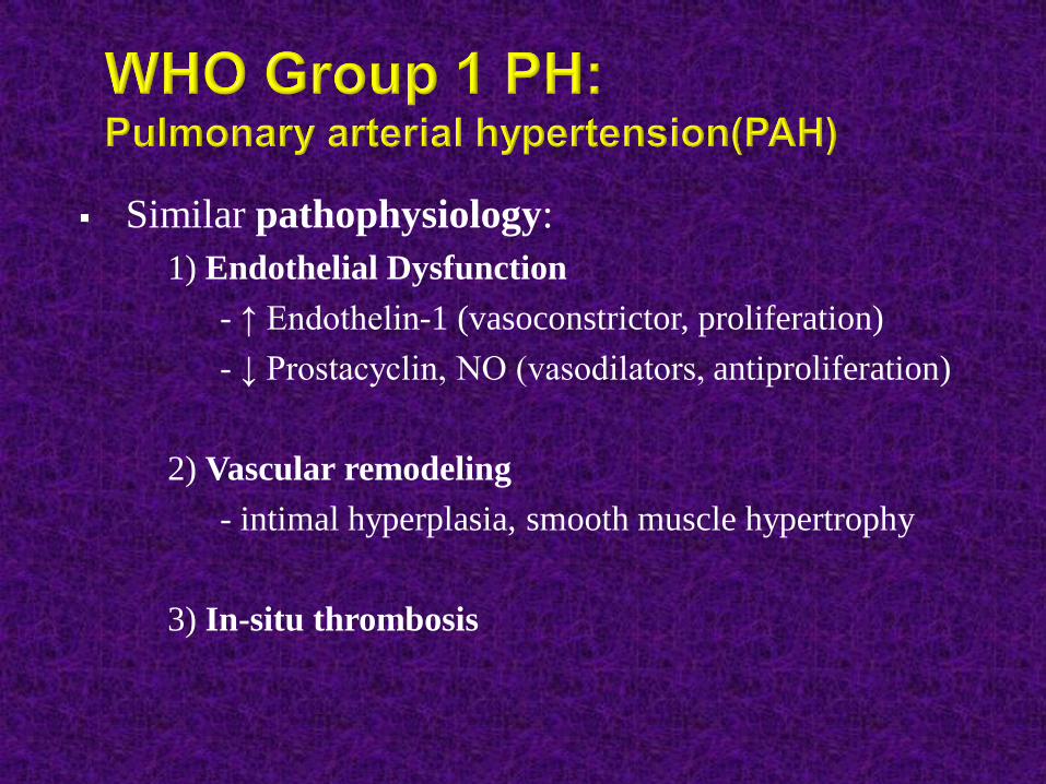
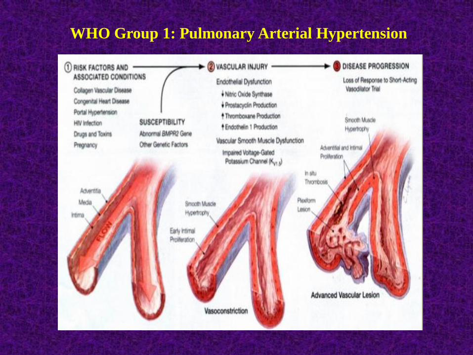
Similar pathophysiology:
1) Endothelial Dysfunction
- ↑ Endothelin-1 (vasoconstrictor, proliferation)
- ↓ Prostacyclin, NO (vasodilators, antiproliferation)
2) Vascular remodeling
- intimal hyperplasia, smooth muscle hypertrophy
3) In-situ thrombosis
WHO Group 1: Pulmonary Arterial Hypertension
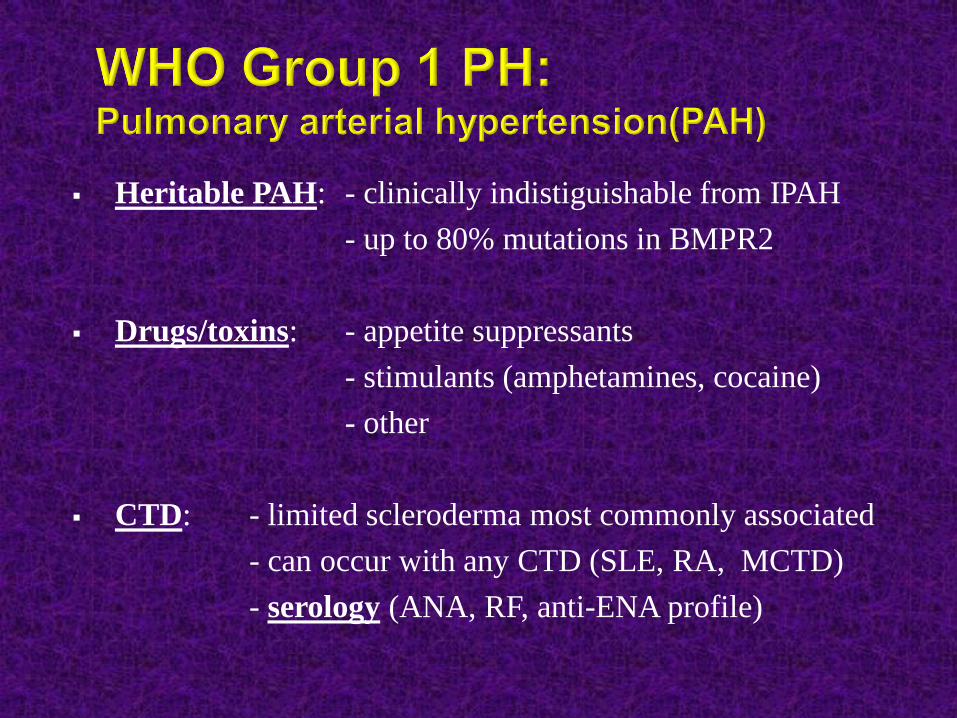
Heritable PAH: - clinically indistiguishable from IPAH
- up to 80% mutations in BMPR2
Drugs/toxins: - appetite suppressants
- stimulants (amphetamines, cocaine)
- other
CTD: - limited scleroderma most commonly associated
- can occur with any CTD (SLE, RA, MCTD)
- serology (ANA, RF, anti-ENA profile)
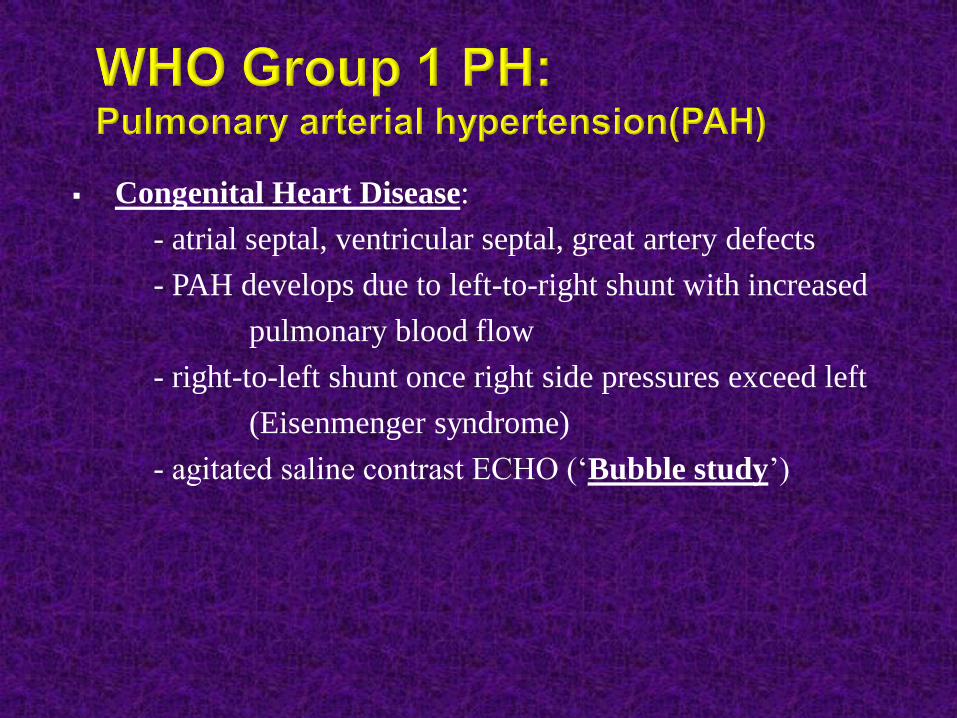
Congenital Heart Disease:
- atrial septal, ventricular septal, great artery defects
- PAH develops due to left-to-right shunt with increased
pulmonary blood flow
- right-to-left shunt once right side pressures exceed left
(Eisenmenger syndrome)
- agitated saline contrast ECHO (‘Bubble study’)
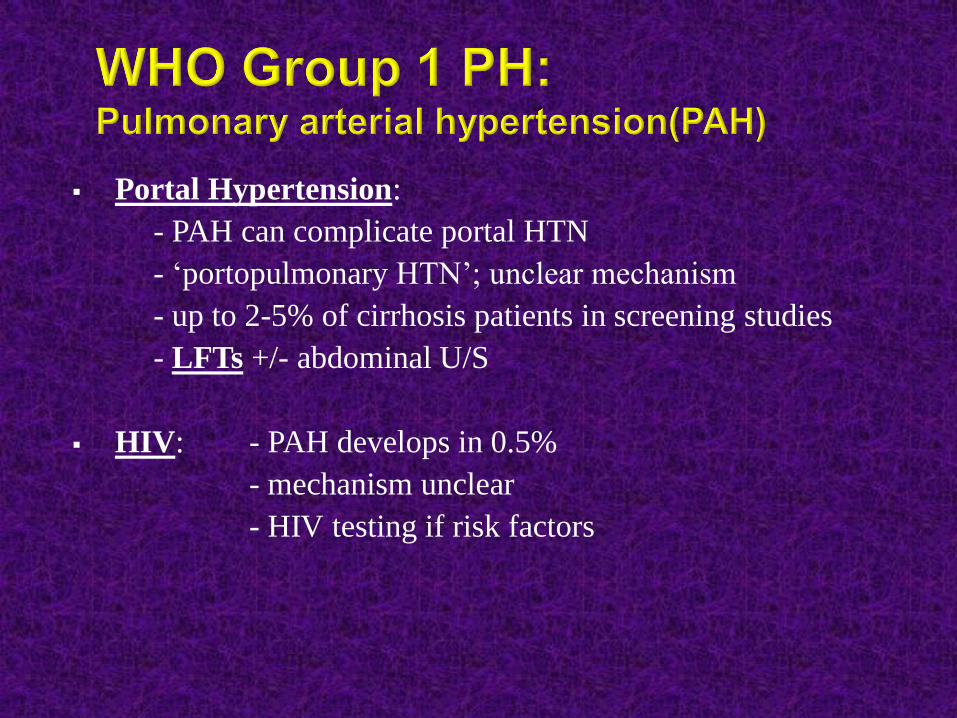
Portal Hypertension:
- PAH can complicate portal HTN
- ‘portopulmonary HTN’; unclear mechanism
- up to 2-5% of cirrhosis patients in screening studies
- LFTs +/- abdominal U/S
HIV: - PAH develops in 0.5%
- mechanism unclear
- HIV testing if risk factors
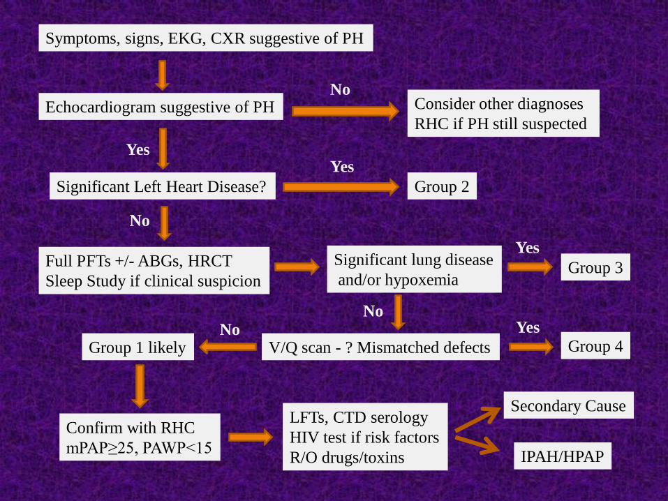
Symptoms, signs, EKG, CXR suggestive of PH
Echocardiogram suggestive of PH
Significant Left Heart Disease? Group 2
Full PFTs +/- ABGs, HRCT
Sleep Study if clinical suspicion
Consider other diagnoses
RHC if PH still suspected
Significant lung disease
and/or hypoxemiaGroup 3
V/Q scan - ? Mismatched defects Group 4Group 1 likely
Confirm with RHC
mPAP≥25, PAWP<15
LFTs, CTD serology
HIV test if risk factors
R/O drugs/toxins
Secondary Cause
IPAH/HPAP
No
Yes
No
Yes
Yes
NoYesNo

Treatment Strategies
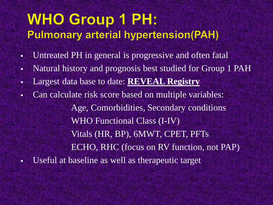
Untreated PH in general is progressive and often fatal
Natural history and prognosis best studied for Group 1 PAH
Largest data base to date: REVEAL Registry
Can calculate risk score based on multiple variables:
Age, Comorbidities, Secondary conditions
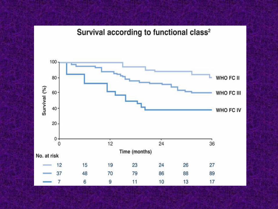
WHO Functional Class (I-IV)
Vitals (HR, BP), 6MWT, CPET, PFTs
ECHO, RHC (focus on RV function, not PAP)
Useful at baseline as well as therapeutic target
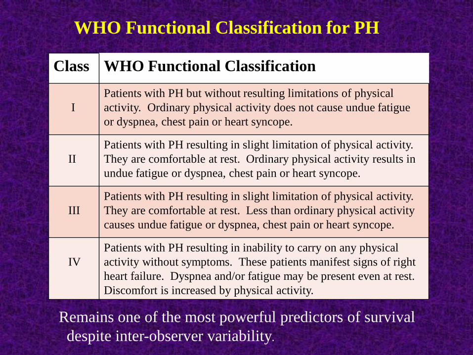
Class WHO Functional Classification
I
Patients with PH but without resulting limitations of physical
activity. Ordinary physical activity does not cause undue fatigue
or dyspnea, chest pain or heart syncope.
II
Patients with PH resulting in slight limitation of physical activity.
They are comfortable at rest. Ordinary physical activity results in
undue fatigue or dyspnea, chest pain or heart syncope.
III
Patients with PH resulting in slight limitation of physical activity.
They are comfortable at rest. Less than ordinary physical activity
causes undue fatigue or dyspnea, chest pain or heart syncope.
IV
Patients with PH resulting in inability to carry on any physical
activity without symptoms. These patients manifest signs of right
heart failure. Dyspnea and/or fatigue may be present even at rest.
Discomfort is increased by physical activity.
Remains one of the most powerful predictors of survival
despite inter-observer variability.
WHO Functional Classification for PH
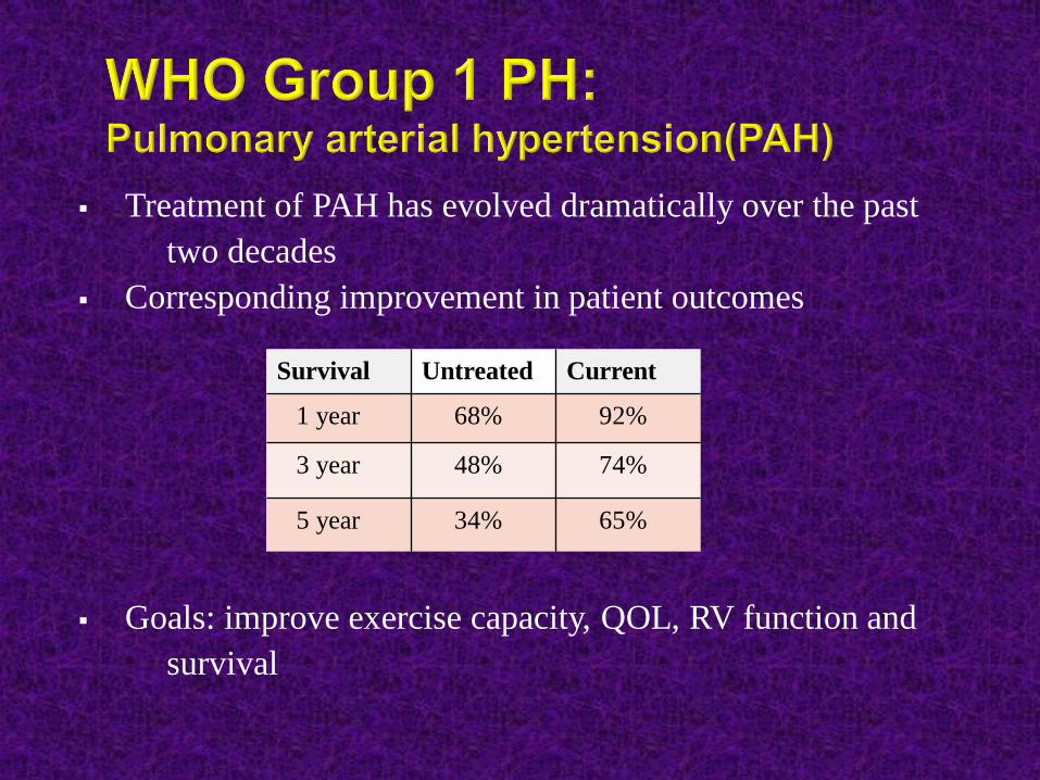
Treatment of PAH has evolved dramatically over the past
two decades
Corresponding improvement in patient outcomes
Goals: improve exercise capacity, QOL, RV function and
survival
Survival Untreated Current
1 year 68% 92%
3 year 48% 74%
5 year 34% 65%
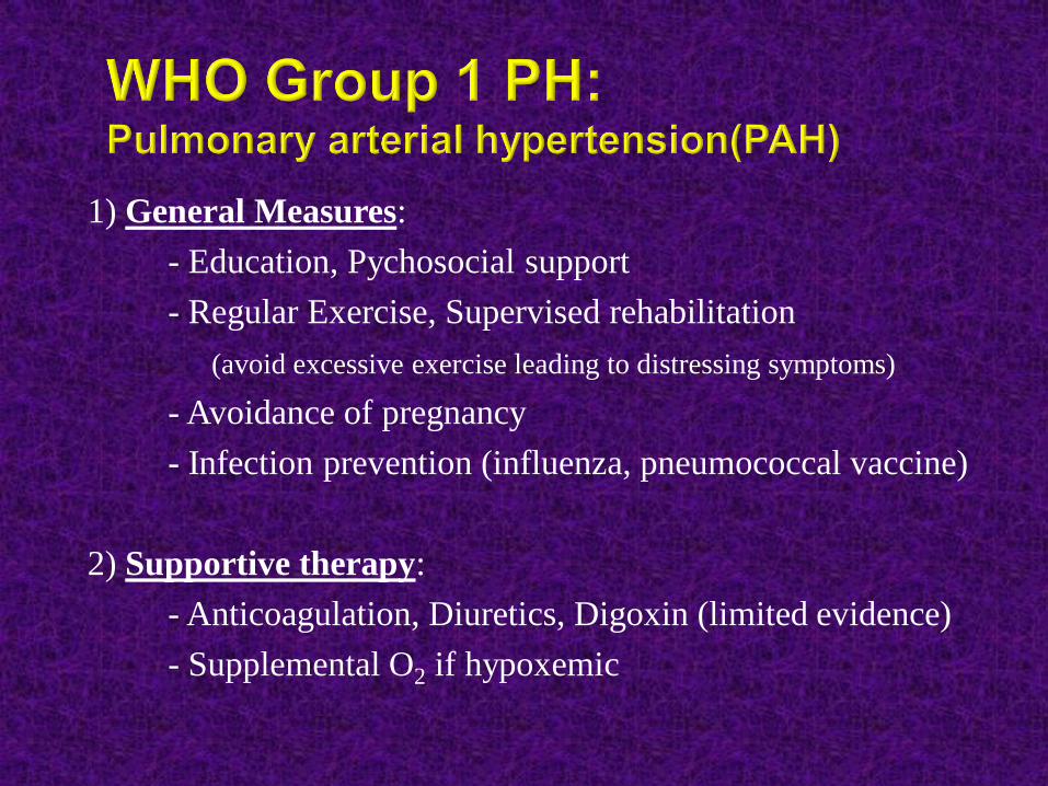
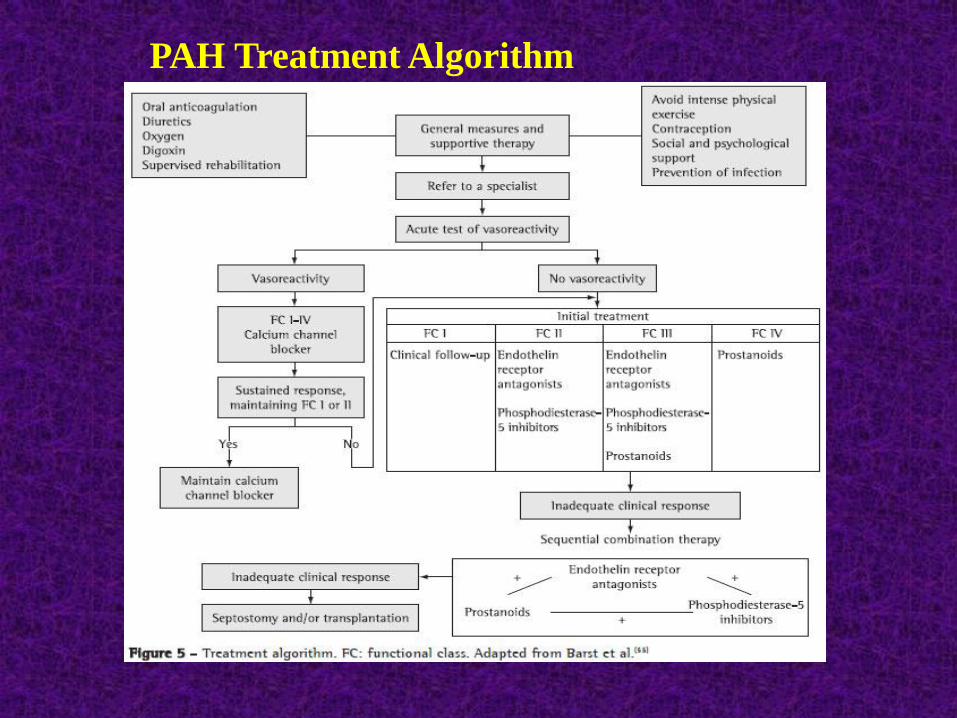
1) General Measures:
- Education, Pychosocial support
- Regular Exercise, Supervised rehabilitation
(avoid excessive exercise leading to distressing symptoms)
- Avoidance of pregnancy
- Infection prevention (influenza, pneumococcal vaccine)
2) Supportive therapy:
- Anticoagulation, Diuretics, Digoxin (limited evidence)
- Supplemental O2 if hypoxemic
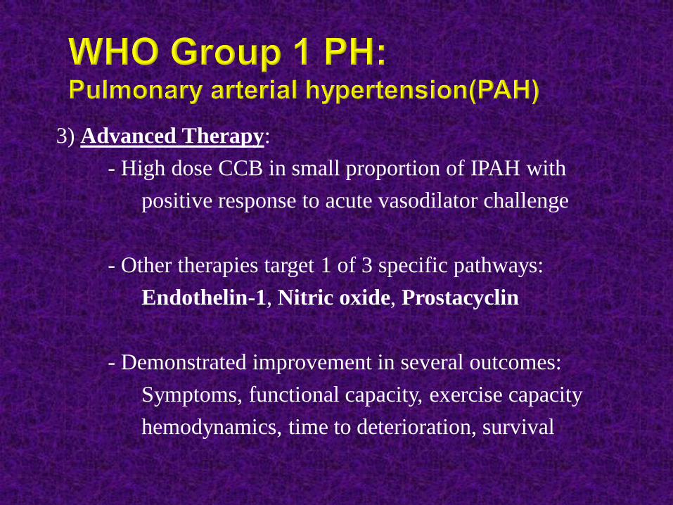
3) Advanced Therapy:
- High dose CCB in small proportion of IPAH with
positive response to acute vasodilator challenge
- Other therapies target 1 of 3 specific pathways:
Endothelin-1, Nitric oxide, Prostacyclin
- Demonstrated improvement in several outcomes:
Symptoms, functional capacity, exercise capacity
hemodynamics, time to deterioration, survival
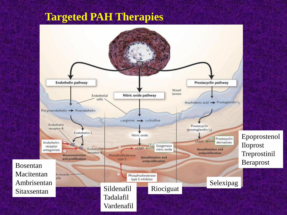
Bosentan
Macitentan
Ambrisentan
Sitaxsentan Sildenafil
Tadalafil
Vardenafil
Riociguat
Epoprostenol
Iloprost
Treprostinil
Beraprost
Selexipag
Targeted PAH Therapies
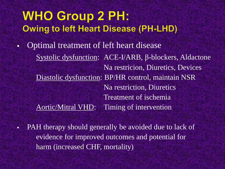
Optimal treatment of left heart disease
Systolic dysfunction: ACE-I/ARB, β-blockers, Aldactone
Na restricion, Diuretics, Devices
Diastolic dysfunction: BP/HR control, maintain NSR
Na restriction, Diuretics
Treatment of ischemia
Aortic/Mitral VHD: Timing of intervention
PAH therapy should generally be avoided due to lack of
evidence for improved outcomes and potential for
harm (increased CHF, mortality)
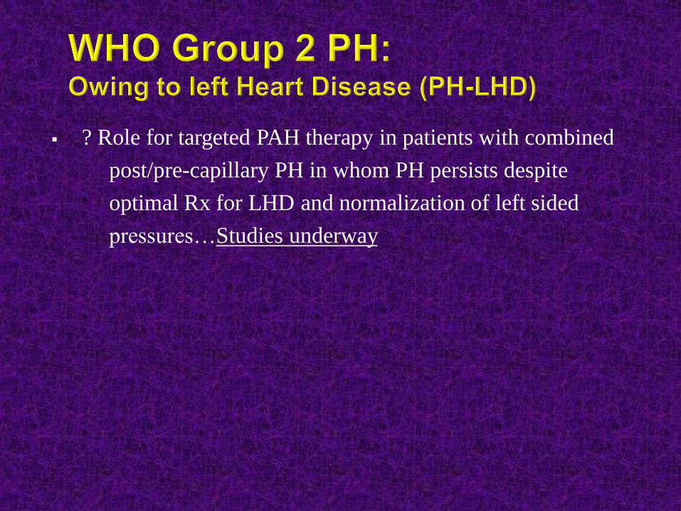
? Role for targeted PAH therapy in patients with combined
post/pre-capillary PH in whom PH persists despite
optimal Rx for LHD and normalization of left sided
pressures…Studies underway
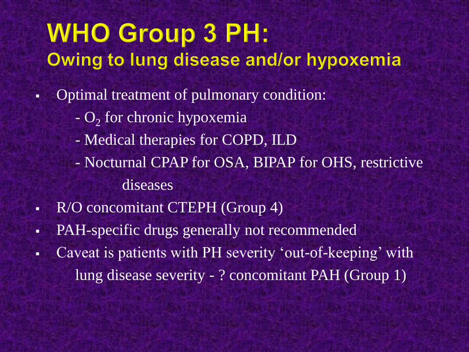
Optimal treatment of pulmonary condition:
- O2 for chronic hypoxemia
- Medical therapies for COPD, ILD
- Nocturnal CPAP for OSA, BIPAP for OHS, restrictive
diseases
R/O concomitant CTEPH (Group 4)
PAH-specific drugs generally not recommended
Caveat is patients with PH severity ‘out-of-keeping’ with
lung disease severity - ? concomitant PAH (Group 1)
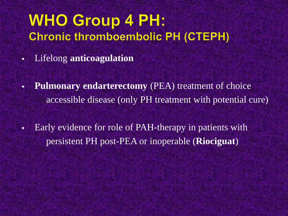
Lifelong anticoagulation
Pulmonary endarterectomy (PEA) treatment of choice
accessible disease (only PH treatment with potential cure)
Early evidence for role of PAH-therapy in patients with
persistent PH post-PEA or inoperable (Riociguat)
Thank-you