The e¡ect of hexadecaprenyl diphosphate on phospholipid membranes Tadeusz Janas a ; *, Teresa Janas b , Krystyna Walin ¤ska a a Department of Biophysics, Pedagogical University, Monte Cassino 21 B, 65-561 Zielona Go ¤ra, Poland b Department of Physics, Technical University, Podgo ¤rna 50, 65-246 Zielona Go ¤ra, Poland Received 6 January 2000; accepted 24 January 2000 Abstract In the present study we investigated phospholipid bilayer membranes and phospholipid vesicles made from dioleoyl- phosphatidylcholine (DOPC) or its mixture with the phosphate ester derivative of long-chain polyprenol (hexadecaprenyl diphosphate, C 80 -PP) by electrophysiological and transmission electron microscopy (TEM) techniques. The membrane conductance-temperature relationships and the membrane breakdown voltage have been measured for different mixtures of C 80 -PP/DOPC. The current-voltage characteristics, the membrane conductance, the activation energy of ion migration across the membrane and the membrane breakdown voltage were determined. Hexadecaprenyl diphosphate decreases the membrane conductance, increases the activation energy and the membrane breakdown voltage for the various values of C 80 - PP/DOPC mole ratio. The analysis of TEM micrographs shows several characteristic structures, which have been described. The data indicate that hexadecaprenyl diphosphate modulates the surface curvature of the membranes by the formation of aggregates in liquid-crystalline phospholipid membranes. The properties of modified membranes can result from the presence of the negative charges in the hydrophilic part of C 80 -PP molecules and can be modulated by the concentration of this compound in membranes. We suggest that the dynamics and conformation of hexadecaprenyl diphosphate in membranes depend on the transmembrane electrical potential. ß 2000 Elsevier Science B.V. All rights reserved. Keywords : Phospholipid membrane ; Polyprenyl diphosphate ; Membrane conductance ; Membrane breakdown voltage ; Activation energy ; Transmission electron microscopy 1. Introduction Polyisoprenols are natural products and deriva- tives of a common C 5 isoprene unit. Their phosphor- yl derivatives, polyprenyl phosphates and dolichyl phosphates, function mainly as carriers of glycosyl units across membranes in the glycosylation reac- tions [1^4]. The occurrence of phosphate esters of polyisoprenols in membrane fractions from prokary- otic and eukaryotic cells is frequently reported [4^7]. Unicellular eukaryotes, fungi, animal and some plant tissues contain phosphoryl derivatives of K-saturated polyisoprenols (dolichyl monophosphates, dolichyl diphosphates) [6,7]. Bacterial membranes and leaves of some plants contain K-unsaturated polyisoprenols (polyprenols) and their phosphoryl derivatives [5,8,9]. The peptidoglycan layer of bacterial cell walls is 0005-2736 / 00 / $ ^ see front matter ß 2000 Elsevier Science B.V. All rights reserved. PII:S0005-2736(00)00154-1 * Corresponding author. Fax: +48 (68) 3265449; E-mail : [email protected]Biochimica et Biophysica Acta 1464 (2000) 273^283 www.elsevier.com/locate/bba
Transcript
The e¡ect of hexadecaprenyl diphosphate on phospholipid membranes
Tadeusz Janas a;*, Teresa Janas b, Krystyna Walinska a
a Department of Biophysics, Pedagogical University, Monte Cassino 21 B, 65-561 Zielona Gora, Polandb Department of Physics, Technical University, Podgorna 50, 65-246 Zielona Gora, Poland
Received 6 January 2000; accepted 24 January 2000
Abstract
In the present study we investigated phospholipid bilayer membranes and phospholipid vesicles made from dioleoyl-phosphatidylcholine (DOPC) or its mixture with the phosphate ester derivative of long-chain polyprenol (hexadecaprenyldiphosphate, C80-PP) by electrophysiological and transmission electron microscopy (TEM) techniques. The membraneconductance-temperature relationships and the membrane breakdown voltage have been measured for different mixtures ofC80-PP/DOPC. The current-voltage characteristics, the membrane conductance, the activation energy of ion migration acrossthe membrane and the membrane breakdown voltage were determined. Hexadecaprenyl diphosphate decreases themembrane conductance, increases the activation energy and the membrane breakdown voltage for the various values of C80-PP/DOPC mole ratio. The analysis of TEM micrographs shows several characteristic structures, which have been described.The data indicate that hexadecaprenyl diphosphate modulates the surface curvature of the membranes by the formation ofaggregates in liquid-crystalline phospholipid membranes. The properties of modified membranes can result from the presenceof the negative charges in the hydrophilic part of C80-PP molecules and can be modulated by the concentration of thiscompound in membranes. We suggest that the dynamics and conformation of hexadecaprenyl diphosphate in membranesdepend on the transmembrane electrical potential. ß 2000 Elsevier Science B.V. All rights reserved.
Polyisoprenols are natural products and deriva-tives of a common C5 isoprene unit. Their phosphor-yl derivatives, polyprenyl phosphates and dolichyl
phosphates, function mainly as carriers of glycosylunits across membranes in the glycosylation reac-tions [1^4]. The occurrence of phosphate esters ofpolyisoprenols in membrane fractions from prokary-otic and eukaryotic cells is frequently reported [4^7].Unicellular eukaryotes, fungi, animal and some planttissues contain phosphoryl derivatives of K-saturatedpolyisoprenols (dolichyl monophosphates, dolichyldiphosphates) [6,7]. Bacterial membranes and leavesof some plants contain K-unsaturated polyisoprenols(polyprenols) and their phosphoryl derivatives[5,8,9].
The peptidoglycan layer of bacterial cell walls is
0005-2736 / 00 / $ ^ see front matter ß 2000 Elsevier Science B.V. All rights reserved.PII: S 0 0 0 5 - 2 7 3 6 ( 0 0 ) 0 0 1 5 4 - 1
Biochimica et Biophysica Acta 1464 (2000) 273^283www.elsevier.com/locate/bba
biosynthesized using a lipid carrier undecaprenyl di-phosphate to assemble and transport the disaccha-ride-pentapeptide precursor [4,8]. Similar lipid-linkedcycles are involved in the biosynthesis of bacteriallipopolysaccharides and eukaryotic glycoproteins,the latter involving the structurally related dolichylphosphate as a lipid carrier. The lengthy polyisopren-oid chain seems an important property for the lipidacceptors, and this probably relates to their ability to£uidize locally the membrane bilayer [4,10].
The molecule of hexadecaprenyl diphosphate (C80-PP) consists of a hydroxyl group, which is esteri¢edwith two phosphate groups: a hydrophilic part withnegative charges, and a hydrophobic part, a longunsaturated isoprenyl chain, mainly of poly-cis con-¢guration. This molecule is composed of 16 isopreneunits with the structure gt2c12KPP where g is anisoprene residue farthest from the esteri¢ed hydroxylgroup, t is a trans-isoprene residue, c is a cis-isopreneresidue, K is an unsaturated, terminal cis-isopreneresidue and P is the phosphate group [11]. Long-chain polyprenols isolated from plant photosynthetictissues of Spermatophyta contain di-trans-, poly-cis-prenols with the general structure gt2cnKOH, wherethe number of isoprene units n varies usually from 6to 30^40, depending on the plant species [9]. Thephosphoryl derivatives of these long-chain poly-cis-prenols have been present in small amounts in leavesof many species of Gymnospermae and Angiosper-mae [5].
In the present study we investigated bilayer lipidmembranes (BLM) and lipid vesicles made from dio-leoylphosphatidylcholine (DOPC) or its mixture withlong-chain polyprenol by electrophysiological andtransmission electron microscopy (TEM) techniques.The lipid bilayer has often served as an experimentalmodel of biological membranes [12^18] due to itssimilarity to biological membranes. The current-volt-age characteristics, the membrane conductance-tem-perature relationships and the membrane breakdownvoltage have been measured for DOPC and di¡erentmixtures of C80-PP/DOPC. The membrane conduc-tance for DOPC and C80-PP/DOPC bilayers, themembrane hydrophobic thickness and the activationenergy of ion migration across these membranes havebeen determined. Structures of lipid vesicles preparedfrom DOPC and C80-PP/DOPC mixtures have beenstudied.
2. Materials and methods
2.1. Chemicals
DOPC (1,2-dioleoyl-sn-glycero-3-phosphocholine)was purchased from Sigma. It gave a single spot onSilica Gel TLC plates (Merck) in chloroform/metha-nol/water (65:25:4, v/v/v) and in chloroform/metha-nol/acetic acid/water (50:30:8:4, v/v/v). Hexadeca-prenol (C80) was isolated from leaves of Picea abies[19]. It gave a single spot on Silica Gel G TLC plates(Merck) in ethyl acetate/toluene (5:95, v/v) and onRP-18 HP TLC plates (Merck) in acetone. Hexade-caprenyl diphosphate (C80-PP) (Fig. 1) was made bychemical phosphorylation of hexadecaprenol basedon the method of Danilov et al. [20]. It gave a singlespot on Silica Gel G TLC plates (Merck) in chloro-form/methanol/water (65:25:4, v/v/v). n-Decane andbutanol were purchased from Aldrich and Fisher,respectively.
2.2. Membrane formation
Bilayer lipid membranes in the form of hemi-spheres were formed according to the technique de-scribed previously [10] on a Te£on capillary tube inunbu¡ered (pH 6) aqueous solution of 0.1 M and0.2 M NaCl (inside and outside, respectively).DOPC and C80-PP/DOPC mixtures used for mem-brane formation were dissolved in n-decane/butanol(3:1, v/v) to obtain a concentration of 10 mg of lipidper ml of solvent. The area of the macrovesicularbilayer lipid membrane was about 50 mm2.
2.3. Electrical measurements
Silver chloride electrodes were used to detect the
Fig. 1. The structure of hexadecaprenyl diphosphate (C80-PP):gt2c12KPP, where g is an isoprene residue farthest from the es-teri¢ed hydroxyl group, t is a trans-isoprene residue, c is a cis-isoprene residue, K is an K-unsaturated, terminal cis-isopreneresidue and P is the phosphate group.
BBAMEM 77823 9-3-00
T. Janas et al. / Biochimica et Biophysica Acta 1464 (2000) 273^283274
electric potentials and the currents. The area of themembrane, S, was determined by an optical measure-ment of membrane dimensions (the precision 0.03mm). The temperature, T (the precision of measure-ments 0.1 K), was controlled by water circulatingfrom an external bath. Electrical conductance ofthe membrane, G, was calculated from current-volt-age characteristics (the precision of voltage and cur-rent measurements: 0.1 mV and 0.01 nA, respec-tively). To obtain the values of the breakdownvoltage, VB, the applied voltage was increased by ascan rate of 10 mV s31. The membrane rupture wasre£ected by a rapid increase of current [10]. The ac-tivation energy (the temperature-dependent part) ofion migration across the membrane, EA, was deter-mined from Arrhenius plots of normalized conduc-tance of bilayer lipid membranes [21]:
�ln��G=C�=�G0=C0��� � �EA=R���1=T�3�1=T0�� �1�where ln[(G/C)/(G0/C0)] is the normalized conduc-tance of the membrane, G0 and C0 are membraneconductance and membrane capacitance, respec-tively, at temperature T0, R is the gas constant.The normalization of membrane conductance (withrespect to the membrane capacitance measured si-multaneously) corrects any variations in the bilayerconductance which are due to variations in bilayerthickness or bilayer area. The capacitance of themembrane, C, was determined (the precision of mea-surement 0.1 nF) from recorded membrane dischargecurves [22]. Dielectric constant equal to 2.1 was as-sumed for the calculations of the hydrophobic thick-ness of the bilayer capacitor.
2.4. Electron microscopy
Lipid vesicles were prepared from DOPC or themixture of C80-PP/DOPC. Small amounts of lipids(0.3 mg of DOPC or 0.112 mg/0.2 mg of C80-PP/DOPC respectively, for mole ratio 0.2) mixed inchloroform were dried under nitrogen at 45³C untila thin ¢lm of dry lipid formed on the wall of the testtube. The lipid suspensions were obtained by addi-tion of water from a Millipore water system. Theconcentration of lipid was 1 mg per ml of water.Lipids were hydrated at 45³C for 24 h. The vesicledispersions were obtained by vortexing hydrated lip-ids using a laboratory vibrator (Janke and Kunkel,
IKA Labortechnik VF 2) for 5 min at moderatemodes.
A droplet of vesicle dispersion was put on a micro-scope copper grid (400 mesh) covered previously withFormvar membrane and a thin evaporated layer ofcarbon. The samples were dried at room temperatureand negatively stained by depositing a drop of 1%aqueous uranyl acetate solution for 1 min, then gridswere rinsed in water from a Millipore water system.The dried grids were again covered with a thinevaporated layer of carbon. Samples were analyzedin the transmission electron microscope JEOL JEM1200EX at 80 kV.
3. Results
The behavior of hexadecaprenyl diphosphate/DOPC membranes as a function of applied potentialwas studied by performing voltammetric experimentsand lipid vesicles prepared from C80-PP/DOPC wereanalyzed in the transmission electron microscopy.
As presented in Fig. 2, the current-voltage charac-teristics are symmetric and linear for values of thepotential in the range 320 to +20 mV. The values ofthe curve slope are smaller for C80-PP/DOPC bi-layers in comparison with the slope for DOPC mem-branes.
Fig. 2. Current-voltage steady-state characteristics of bilayer lip-id membranes versus the C80-PP/DOPC mole ratio. Experimentswere performed at 25 þ 1³C.
BBAMEM 77823 9-3-00
T. Janas et al. / Biochimica et Biophysica Acta 1464 (2000) 273^283 275
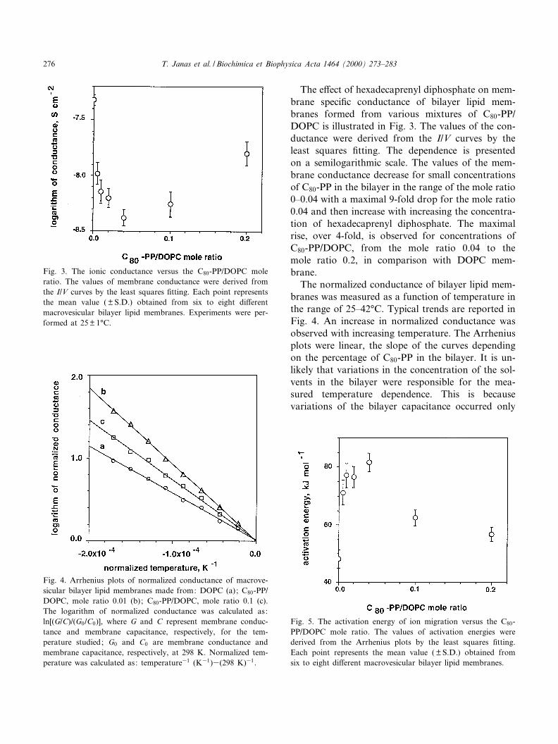
The e¡ect of hexadecaprenyl diphosphate on mem-brane speci¢c conductance of bilayer lipid mem-branes formed from various mixtures of C80-PP/DOPC is illustrated in Fig. 3. The values of the con-ductance were derived from the I/V curves by theleast squares ¢tting. The dependence is presentedon a semilogarithmic scale. The values of the mem-brane conductance decrease for small concentrationsof C80-PP in the bilayer in the range of the mole ratio0^0.04 with a maximal 9-fold drop for the mole ratio0.04 and then increase with increasing the concentra-tion of hexadecaprenyl diphosphate. The maximalrise, over 4-fold, is observed for concentrations ofC80-PP/DOPC, from the mole ratio 0.04 to themole ratio 0.2, in comparison with DOPC mem-brane.
The normalized conductance of bilayer lipid mem-branes was measured as a function of temperature inthe range of 25^42³C. Typical trends are reported inFig. 4. An increase in normalized conductance wasobserved with increasing temperature. The Arrheniusplots were linear, the slope of the curves dependingon the percentage of C80-PP in the bilayer. It is un-likely that variations in the concentration of the sol-vents in the bilayer were responsible for the mea-sured temperature dependence. This is becausevariations of the bilayer capacitance occurred only
Fig. 3. The ionic conductance versus the C80-PP/DOPC moleratio. The values of membrane conductance were derived fromthe I/V curves by the least squares ¢tting. Each point representsthe mean value ( þ S.D.) obtained from six to eight di¡erentmacrovesicular bilayer lipid membranes. Experiments were per-formed at 25 þ 1³C.
Fig. 4. Arrhenius plots of normalized conductance of macrove-sicular bilayer lipid membranes made from: DOPC (a); C80-PP/DOPC, mole ratio 0.01 (b); C80-PP/DOPC, mole ratio 0.1 (c).The logarithm of normalized conductance was calculated as:ln[(G/C)/(G0/C0)], where G and C represent membrane conduc-tance and membrane capacitance, respectively, for the tem-perature studied; G0 and C0 are membrane conductance andmembrane capacitance, respectively, at 298 K. Normalized tem-perature was calculated as: temperature31 (K31)3(298 K)31.
Fig. 5. The activation energy of ion migration versus the C80-PP/DOPC mole ratio. The values of activation energies werederived from the Arrhenius plots by the least squares ¢tting.Each point represents the mean value ( þ S.D.) obtained fromsix to eight di¡erent macrovesicular bilayer lipid membranes.
BBAMEM 77823 9-3-00
T. Janas et al. / Biochimica et Biophysica Acta 1464 (2000) 273^283276
slowly whereas the variations with temperature re-ported in our paper occurred rapidly. These resultsare in accordance with the observations reported bySmith et al. [21].
The relationship between the value of activationenergy of ion transport across the membrane, EA,and the percentage of hexadecaprenyl diphosphatein macrovesicular bilayers is shown in Fig. 5. Thevalues of activation energies were derived from theArrhenius plots by the least squares ¢tting. Arrhe-nius plots were linear in the temperature range 25^42³C (data not shown). For smaller concentrationsof hexadecaprenyl diphosphate in the membrane, anincrease of EA was observed in comparison withDOPC bilayers. For higher concentrations of C80-PP in the membrane a decrease of the value of EA
was observed. The EA value increases from 48 þ 3 kJmol31 for DOPC bilayers to the maximal value,EAmax = 82 þ 3.1 kJ mol31, for bilayers preparedfrom the mixture C80-PP/DOPC, mole ratio equalsto 0.02, and then decreases to 57 þ 2.6 kJ mol31 forthe value of C80-PP/DOPC mole ratio equals to 0.2.The values of the activation energy for electrical con-duction with DOPC membranes found in our studyare a little higher than those for egg lecithin/choles-terol membranes (35 þ 2 kJ mol31) [21].
Fig. 6 illustrates the e¡ect of hexadecaprenyl di-
phosphate on the breakdown voltage of the mem-brane, VB. The membrane electromechanical stability(proportional to the value of VB) is modulated by thepresence of hexadecaprenyl diphosphate in the bi-layer lipid membrane. The increase of VB is observedfor small concentrations up to the mole ratio 0.01 ofC80-PP in the membrane, and than, for higher con-centrations of C80-PP in the membrane, a slight de-crease in the value of VB is observed. The value ofmembrane breakdown voltage increases from203 þ 10 mV for DOPC bilayers to the maximalvalue VBmax = 273 þ 22 mV for the bilayer pre-pared from the C80-PP/DOPC mixture at 0.01 moleratio.
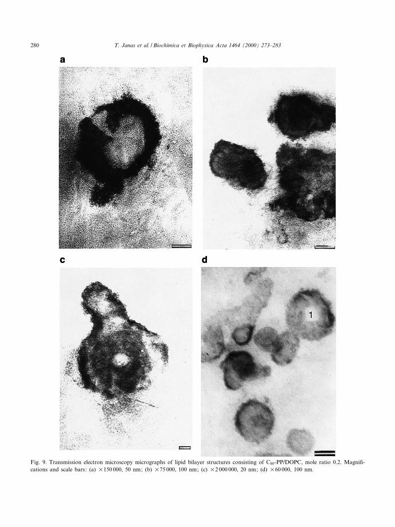
Lipid vesicles prepared from DOPC and C80-PP/DOPC mixtures (mole ratios 0.01 and 0.2), were an-alyzed in the transmission electron microscope. Ex-amples of these vesicles are shown in Figs. 7^9. Theywere chosen from about 100 micrographs and repre-sent the typical tendency of the modi¢cation of thevesicle structure. Fig. 7 presents typical DOPC dis-
Fig. 6. The membrane breakdown voltage versus the C80-PP/DOPC mole ratio. Each point represents the mean value( þ S.D.) obtained from six to eight di¡erent macrovesicular bi-layer lipid membranes. Experiments were performed at25 þ 1³C.
Fig. 7. The transmission electron microscopy micrograph of lip-id bilayer structures consisting of DOPC. Magni¢cationU100 000; the bar represents 50 nm.
BBAMEM 77823 9-3-00
T. Janas et al. / Biochimica et Biophysica Acta 1464 (2000) 273^283 277
BBAMEM 77823 9-3-00
T. Janas et al. / Biochimica et Biophysica Acta 1464 (2000) 273^283278
persion of spherical vesicles of various sizes in therange from approx. 40 nm to approx. 300 nm. Fig.8a^f show lipid vesicles prepared from C80-PP/DOPC, mole ratio 0.01. The shapes of smallervesicles presented in Fig. 8a are rather regular withdiameters from 100 nm to 150 nm. Among the spher-ical vesicles there are some irregular vesicles withsimilar diameters. The spherical vesicles are shownin Fig. 8b. The diameters of these regular vesiclesare approx. 170^300 nm. Among the regular vesiclesprepared from C80-PP/DOPC, mole ratio 0.01, thereare some elongated, multi-bent structures (Fig. 8c,d).The micrograph presented in Fig. 8c shows someregular (structures 1 and 2) and some elongatedvesicles (structures 3 and 4). Fig. 8d shows the large,elongated and bent vesicle (structure 1). The dimen-sions of this vesicle range from 200 nm to 2600 nm.Elongated vesicles in Fig. 8e,f are bent and they aregeometrically similar to the torus. The part of struc-ture 1 in Fig. 8e is similar to the handle of the jug.Structures 2 and 3 are also torus-like structures and
their dimensions range from 100 nm to 300 nm. Themicrograph presented in Fig. 8f shows a large, elon-gated torus-like structure with many `handles'. Thelength of this structure is about 700 nm.
A new kind of structure, prepared from C160/DOPC mixture, mole ratio 0.2, can be seen in Fig.9a^d. A small bottle-like vesicle is presented in Fig.9a. The light characteristic shape can be observedinside of the vesicle. The membranes of this vesicleare grainy. The dimensions of the vesicle range from160 nm to 200 nm. Among the vesicles presented inFig. 9b there are also some vesicles with grainy mem-branes. Structure 1 is similar to the budding struc-ture. Structure 2 seems to be the e¡ect of the fusionof two vesicles. Inside of these vesicles (structures 1and 2) are seen dark, circular shapes. The vesiclepresented in Fig. 9c is irregular and a buddingvesicle. Membranes are grainy and the internal struc-ture is heterogeneous. The width of this vesicle isabout 170 nm. Some vesicles with grainy membranesare presented in Fig. 9d. One of them (structure 1)
Fig. 8. Transmission electron microscopy micrographs of lipid bilayer structures consisting of C80-PP/DOPC, mole ratio 0.01. Magni¢-cations and scale bars: (a) U60 000, 100 nm; (b) U150 000, 50 nm; (c) U25 000, 200 nm; (d) U20 000, 200 nm; (e) U75 000, 100nm; (f) U100 000, 50 nm.
BBAMEM 77823 9-3-00
T. Janas et al. / Biochimica et Biophysica Acta 1464 (2000) 273^283 279
Fig. 9. Transmission electron microscopy micrographs of lipid bilayer structures consisting of C80-PP/DOPC, mole ratio 0.2. Magni¢-cations and scale bars: (a) U150 000, 50 nm; (b) U75 000, 100 nm; (c) U2 000 000, 20 nm; (d) U60 000, 100 nm.
BBAMEM 77823 9-3-00
T. Janas et al. / Biochimica et Biophysica Acta 1464 (2000) 273^283280
seems to be a torus-like structure. Numerous granu-lations are seen inside of these vesicles.
The electron micrographs show that lipid vesiclesprepared from DOPC are regular whereas lipidvesicles prepared from C80-PP/DOPC, besides regu-lar structures, form several irregular characteristicstructures.
4. Discussion
Our investigations show that the lipid bilayersmodi¢ed by hexadecaprenyl diphosphate (C80-PP)modulate electrical, transport and mechanical prop-erties of these membranes. The conformationalchanges of a negatively charged lipid molecule canbe induced electrostatically, by changing membranesurface charge density [23]. The molecule of hexade-caprenyl diphosphate contains a long, hydrophobicisoprenyl chain and two phosphate groups and itseems that changes of modi¢ed membranes aremainly caused by a negative electrical charge of thephosphate groups. All the studied membranes werein the liquid-crystalline state, because the phase tran-sition temperature for dioleoylphosphatidylcholinebilayers is below 320³C [24] and the hydrocarbonchain of hexadecaprenyl diphosphate contains sev-eral double bounds in cis-conformation [11]. Nophase transition of K-saturated polyprenol/DOPC bi-layers could be detected between 12³C and 82³C [25].
With respect to the electrical properties, the mea-surements showed that hexadecaprenyl diphosphateincreases the activation energy for ion migration andmembrane breakdown voltage. Furthermore thepresence of hexadecaprenyl diphosphate in the phos-pholipid bilayer results in changes in the slopes of I/V curves and also in a decrease of membrane speci¢cconductance. The membrane speci¢c conductance(Fig. 3) increases above the C80-PP/DOPC, mole ra-tio 0.04, although values of conductance of modi¢edmembranes are smaller than the values of conduc-tance of DOPC bilayers. McCloskey and Troy [26]noted that neutral spin-labeled K-saturated polypren-yl phosphate-dolichyl phosphate (Dol-P) did not ag-gregate at concentrations smaller than 0.03. In ourcase the aggregation of hexadecaprenyl diphosphateseems to occur at concentrations higher than 0.04.The aggregation leads to domain formation with
phosphate groups in the hydrophilic part of themembrane. Similar properties of Dol-P were pre-sented by Valtersson et al. [25]. The authors sug-gested the formation of Dol-P-hexagonal structuresin the lipid vesicle dispersion. The decrease in mem-brane conductance at smaller C80-PP concentrationscan result from a restriction of the movement ofDOPC molecules in bilayers. The following increasein membrane conductance can arise from a creationof ionic pathways in the boundary region of C80-PPaggregates. Di¡erences in properties of modi¢ed bi-layers for various concentrations of C80-PP in mem-branes have also been con¢rmed in the case of inves-tigations of the activation energy of ion migrationacross the membrane, the breakdown voltage andthe membrane thickness. The large increase, over2-fold, in the activation energy upon incorporationof the C80-PP is most interesting. The values of theactivation energy of bilayers formed from lecithin/cholesterol [21] were found to be similar to thosefor DOPC bilayers investigated by us. The depen-dence of the activation energy on the percentage ofC80-PP in the bilayer (Fig. 4) has a maximum at C80-PP/DOPC, mole ratio 0.04, at the same concentra-tions as for the minimum of membrane conductance(Fig. 3). The value of the hydrophilic-lipophilic bal-ance (HLB) of hexadecaprenyl diphosphate is ex-pected to be smaller than 1 and it seems to in£uencethe permeability of C80-PP/DOPC bilayers for ions.
Hexadecaprenyl diphosphate increases the value ofthe breakdown voltage of DOPC bilayers. This phe-nomenon re£ects the stabilization e¡ect of the inter-action of the potential gradient with the C80-PPheadgroup on DOPC bilayers. As shown in Fig. 6,the values of breakdown voltage increase substan-tially for C80-PP/DOPC, mole ratio range 0^0.04,and then decrease, although the values of the break-down voltage are higher than in the case of DOPCbilayers. The inclusion of long chained hydrophobicchains in lipid bilayers is known to increase theirstability [27]. This is probably related to the increasein the breakdown voltage in the case of our study.The observed increase in the breakdown voltage canalso be related to the decrease in the size of pores. Asanalyzed in the paper of Smith et al. [21] the increasein the activation energy is related to the decrease inthe radius of the transmembrane pore. For the valueof the activation energy equals 18 kJ/mole the au-
BBAMEM 77823 9-3-00
T. Janas et al. / Biochimica et Biophysica Acta 1464 (2000) 273^283 281
thors estimated the minimum pore radius to be about1 nm. For C80-PP/DOPC bilayers, with the activa-tion energy for ion transport about 4^5-fold bigger,the minimal pore radius can be estimated to be in therange 0.2^0.3 nm. The action of C80-PP seems there-fore to e¡ect the formation of these pores or dramat-ically reduce their size.
Hexadecaprenyl diphosphate molecules consist ofa long unsaturated, mainly poly-cis con¢gurationisoprenoid chain with phosphate groups bonded tothe unsaturated K-isoprene residue. The cis geometryenables the chain to be more compact and fold into ashorter length than poly-trans-isoprenoids. Hexade-caprenyl diphosphate molecules seem to orient in themembrane with their phosphate headgroups at thehydrophilic interface. Similar properties were exper-imentally detected by McCloskey and Troy [26], Val-tersson et al. [25], and de Ropp et al. [17] in the caseof lipid bilayers modi¢ed by dolichyl phosphate.Contrary to the behavior of C80-PP, polyprenyl alco-hols increase the membrane speci¢c conductance,and all values of conductance of modi¢ed mem-branes are higher than the value of conductance ofDOPC bilayers, decrease activation energy of ionmigration, breakdown voltage and membrane thick-ness [10]. The aggregation of neutral polyisoprenoidin phospholipid membranes was observed even atrelative concentrations less than 0.005 [17]. Theseaggregates can modulate the permeability and stabil-ity of polyisoprenol-phospholipid membranes.
The dispersions of vesicles prepared from DOPCor C80-PP/DOPC mixtures were investigated bytransmission electron microscopy. Analysis of lipidvesicles in TEM indicates the changes of the mem-brane elasticity for liposomal membranes modi¢edby hexadecaprenyl diphosphate, respectively to theconcentrations of C80-PP in bilayers. In the case ofC80-PP/DOPC dispersions both spherical and non-spherical vesicles were observed. There were multi-budding structures, torus-like structures, elongatedvesicles, and fused vesicles. Vesicles were smooth orrough. The membrane texture of rough vesicles wasgrainy. On the basic of these micrographs we inferthat hexadecaprenyl diphosphate changes the mem-brane £uidity and elasticity and modulates the sur-face curvature of modi¢ed lipid membranes, espe-cially for relative concentrations of C80-PP in themembrane higher than 0.03. The e¡ect of hexadeca-
prenyl diphosphate on the surface curvature of themembranes can result from its molecular shape witha considerable hydrophobic part in comparison withthe small hydrophilic part with the phosphategroups. The variety of structures of phosphatidylcho-line vesicles has been reported by Klo«sgen et al. [28].They demonstrated the existence of superstructuresof phospholipid bilayers. The existence of the super-structure can be regarded as the evidence that lipidsmay directly in£uence the activity of biological mem-branes by changing their £uid-crystalline properties.Some functions of the phosphate ester derivatives oflong-chain polyprenols in bilayers are connected withthe modi¢cation of some properties of the mem-branes.
The data obtained by electrophysiological investi-gations and analysis by transmission electron micros-copy show that electrical, mechanical and transportproperties of lipid membranes change under the in-£uence of hexadecaprenyl diphosphate. The resultsindicate that hexadecaprenyl diphosphate can modu-late the surface curvature of the membranes by theformation of microdomains. The properties of modi-¢ed membranes can result from the presence of thenegative charges in the hydrophilic part of hexade-caprenyl diphosphate molecules and are modulatedby the concentration of this compound in mem-branes. We suggest that the dynamics and conforma-tion of the phosphate ester derivatives of long-chainpolyprenols in membranes depend on the transmem-brane electrical potential.
Acknowledgements
The authors would like to thank Prof. TadeuszChojnacki and Prof. Ewa Sèwiezçewska from the De-partment of Lipid Biochemistry and Biophysics, Pol-ish Academy of Sciences, Warsaw, for stimulatingdiscussion and for making possible the isolationand puri¢cation of polyprenols in the laboratoriesof the Department of Lipid Biochemistry, and Prof.Elzçbieta Wyroba with coworkers for making possiblethe electron microscopy investigations in the NenckiInstitute of Experimental Biology, Warsaw. Thiswork was carried out within the research projectNo. 6 PO4A 014 10 supported by the State Commit-tee for Scienti¢c Research in 1996^1998.
BBAMEM 77823 9-3-00
T. Janas et al. / Biochimica et Biophysica Acta 1464 (2000) 273^283282
References
[1] F.W. Hemming, in: J.W. Porter, S.L. Spurgeon (Eds.), Bio-synthesis of Isoprenoid Compounds, John Wiley, New York,1983, pp. 305^354.
[2] G. Van Duijn, C. Valtersson, T. Chojnacki, A.J. Verkleij, G.Dallner, B. De Kruij¡, Biochim. Biophys. Acta 861 (1986)211^223.