The effect of bodily illusions on clinical pain:a systematic review and meta-analysisEva Boescha, Valeria Bellana,b,c, G. Lorimer Moseleya,d, Tasha R. Stantona,d,*
AbstractThis systematic review and meta-analysis critically examined the evidence for bodily illusions to modulate pain. Six databases weresearched; 2 independent reviewers completed study inclusion, risk of bias assessment, and data extraction. Included studiesevaluated the effect of a bodily illusion on pain, comparing results with a control group/condition. Of the 2213 studies identified, 20studies (21 experiments) were included. Risk of bias was high due to selection bias and lack of blinding. Consistent evidence of paindecrease was found for illusions of the existence of a body part (myoelectric/Sauerbruch prosthesis vs cosmetic/no prosthesis;standardized mean differences 5 21.84, 95% CI 5 22.67 to 21.00) and 4 to 6 weeks of mirror therapy (standardized meandifferences 5 21.11, 95% CI 5 21.66 to 20.56). Bodily resizing illusions had consistent evidence of pain modulation (in thedirection hypothesized). Pooled data found no effect on pain for 1 session of mirror therapy or for incongruent movement illusions(except for comparisons with congruent mirrored movements: incongruent movement illusion significantly increased the odds ofexperiencing pain). Conflicting results were found for virtual walking illusions (both active and inactive control comparisons). Singlestudies suggest no effect of resizing illusions on pain evoked by noxious stimuli, no effect of embodiment illusions, but a significantpain decrease with synchronous mirrored stroking in nonresponders to traditional mirror therapy. There is limited evidence tosuggest that bodily illusions can alter pain, but some illusions, namely mirror therapy, bodily resizing, and use of functionalprostheses show therapeutic promise.
A growing body of evidence points to a complex relationshipbetween pain and disruptions of other bodily perceptions of thepainful part. First, structural and functional differences betweenpeople with and without pain, both cortically and subcortically,include areas clearly involved in bodily awareness and percep-tion.20,26,50 Second, distortions of bodily perception most ofteninvolve the body part feeling larger than it really is,41,30 withbehavioral hand size estimation tasks confirming this alteredperception.41 Similar distortions can be evoked experimentally byanaesthetizing the area, a procedure known to alter responseprofiles of primary somatosensory cortex neurones,4 or bycutaneous stimulation,23 which suggests that cortical andperceptual dysfunction might simply reflect peripheral disturban-ces. However, inducing the illusion that a body part is enlargedincreases movement-evoked swelling in people with complex
regional pain syndrome (CRPS),48 and treatments that targetthese functional brain changes, such as graded motor imageryand sensorimotor retraining, reduce pain,40,43,51 which suggeststhe link may be bidirectional. Importantly, reductions in painappear to be coincident with restoration of functional corticalrepresentation.50
One way to manipulate perception, and thus experimentallyevaluate the relationship between pain and perception, is throughillusions. Recent studies have found that illusions can alter painlevels in conditions such as osteoarthritis (OA), CRPS, andneuropathic pain.44,48,52 Furthermore, some illusions have beenused to interrogate the idea that pathological pain results fromamismatch betweenmotor intention andmotor output.34,46 Overa decade of investigations into the potential utility of using illusionsto modulate pain have yielded sometimes sophisticated andcostly treatments,7 but, with the exception of mirror therapy,18,6
there appears to have been no attempt to take stock, synthesize,and critically evaluate what is now a substantial literatureevaluating illusions and pain. As with any treatment that may beprovided to patients, it is imperative to understand the currentevidence supporting its use. Thus this systematic review andmeta-analysis aimed to determine the current evidence concern-ing the effects of bodily illusions on both acute and chronic pain.
2. Methods
2.1. Data sources
A systematic search strategy in MEDLINE, EMBASE, PsycINFO,CINAHL, Amed and PubMed was used to identify studiesevaluating the effect of bodily illusions on pain (from relative dateof inception toFebruary 28, 2014). Search strategiesweremodified
Sponsorships or competing interests that may be relevant to content are disclosed
at the end of this article.
a Sansom Institute for Health Research and PainAdelaide Consortium, University of
South Australia, Adelaide, Australia, b Department of Psychology, University of
Milano-Bicocca, Milan, Italy, c NeuroMI—Milan Center for Neuroscience, Milan,
Italy, d Neuroscience Research Australia, Randwick, Australia
*Corresponding author. Address: School of Health Sciences, University of South
to meet the specific requirements of each database (See Table 1for Medline search strategy). Reference lists of potentially eligiblestudies and relevant systematic reviewswere hand searched. Last,4 experts in the field (Dr Alberto Gallace, Dr Roger Newport, DrDiana Torta, Dr Martin Diers) were contacted to identify anyadditional eligible studies that may have been missed by thesearch. This systematic review was guided by principles from theCochrane Collaboration of Systematic reviews and the PreferredMethods of Reporting of Systematic Review and Meta-Analyses(PRISMA) statement.39 We conducted this review using an a prioriprotocol (available from T.R.S. upon request).
2.2. Study selection
Studies were eligible for inclusion if they recruited participants withan acute or chronic painful condition, if they evaluated the effect ofa bodily illusion on pain, and if they compared illusion results withcontrol condition or to a healthy (pain-free) control group, and ifthey provided results for a quantitative measure of pain.Specifically, studies had to use illusions that altered the perceptionof the painful body part. No restriction was placed on language.
Studies were excluded if they recruited only healthy pain-freecontrols or if the illusion did not alter the perception of the body (eg,the illusion only altered perception of the environment or the illusionwas a used merely as a distraction). Studies that evaluatedcombination treatment (ie, illusions and another nonillusory activetreatment in one person) were not included, unless the controlgroup also received the nonillusory active treatment (such that thesole effect of illusion could be determined). All types of studydesigns, except case studies, were considered eligible for inclusion.
2.2.1. Definition of a bodily illusion
A bodily illusion was defined as a phenomenon in which anexternal stimulus is interpreted by the neural system in such awaythat the resultant perception of the body is significantly differentfrom reality. This may include alterations to the size/shape,location, movement, or ownership (eg, the rubber hand illusion[RHI]5) of the painful body part. Additionally, this includes illusionsof pain-free, normal function of the body part and/or illusoryexistence of an amputated body part (ie, mirror therapy). Thisdefinition of a bodily illusion was determined by consultation with3 experts in the field (Dr Alberto Gallace, Dr Martin Diers, Dr RogerNewport).
2.3. Study inclusion
The titles and abstracts of all studies retrieved by the search wereinitially screened by 2 independent assessors (E.B., V.B.) and any
discrepancies were discussed. If consensus was unable to bereached, a third independent assessor (T.R.S.) was consulted.Following this initial screen, the full text of potentially eligiblestudies were formally evaluated for inclusion using an identicalprocess and using a custom-designed, piloted inclusion form.
2.4. Risk of bias assessment
The risk of bias was assessed by 2 independent reviewers usingcustom-designed piloted forms that included assessment ofselection bias, detection bias, blinding, statistical methods,reporting bias, performance bias, and other forms of relevantbias (eg, the presence of concomitant treatment). The Strength-ening the Reporting of Observational Studies in Epidemiology(STROBE) guidelines61 were used to inform risk of bias assess-ment for cross-sectional repeated measures and observationalstudies. Randomized controlled trials (RCT) were assessed withadditional questions on allocation concealment and adequatesequence generation on the basis of the Cochrane risk of biasguidelines.29 Assessment of bias related to crossover effects andrandomization of test condition was completed for repeatedmeasures studies.
2.5. Data extraction
Two independent reviewers used a customized piloted dataextraction form to retrieve the following information from includedstudies: study design (ie, case–control, repeated within-subjectmeasures, RCT), sample size, demographics of participants (eg,age, gender) and control groups (if applicable), type and nature ofcontrol (ie, control condition vs separate control group; placeboor inactive control vs active intervention), inclusion and exclusioncriteria for participants, source of participants, type and durationof illusion, credibility of illusion, other clinical information (eg,concomitant conditions, time since amputation). Quantitativepainmeasures (ie, intensity of pain, duration of pain relief, numberof participants with pain) were extracted including the baselinescores (where reported), postintervention scores, and painscores from all follow-up time points. If applicable, change scoresfor pain measures were also extracted. For pain outcomes,measures of central tendency (mean or median) and measures ofdispersion (SD, interquartile range, 95% confidence intervals[95% CI]) were extracted for each group/testing condition. Ifincluded studies provided insufficient information, study authorswere contacted up to 3 times. If no response was received after 3attempts, the data were considered unobtainable.
2.6. Data synthesis and analysis
Included studies were grouped according to the type of illusionand the similarity of the illusion they used: (1) Bodily resizingillusions (ie, technology was used to alter the visual size of thebody part); (2) mirror therapy; (3) illusions of virtual walking (use ofa mirror and video projector set-up to induce an illusion ofwalking); (4) illusions of a new limb (functional prostheses used inamputees to cause a feeling of possessing the limb); (5) illusionsof ownership (rubber hand illusion); and (6) illusions of in-congruent movement (use of a mirror and bilateral limbmovement; arms move in opposite directions and this inducesan incongruence between vision and the actual movement [eg,reflected image of nonpainful arm moving upwards, but hiddenpainful arm actually moving downwards]).
When 2 or more studies evaluated 1 type of illusion and useda comparable illusion and control condition, pooling of data using
Table 1
Medline search strategy.
Medline search:
exp illusion/ OR illusion*.mp OR rubber hand illusion*.mp OR mirror therap*.mp OR
perceptual distortion/ OR size perception/ OR tendon vibration.mp OR virtual reality.
mp OR visual illusion.mp OR body schema.mp OR Body Image/ OR sensation/ OR
multisensory integration.mp OR sensory motor.mp OR sensorimotor.mp OR
integration.mp OR incongruence.mp
AND
pain/ OR chronic pain/ OR acute pain/ OR experimental pain.mp OR neuropathic
pain.mp OR Pain Threshold/ OR complex regional pain syndrome.mp OR Reflex
Sympathetic Dystrophy/ OR Complex Regional Pain Syndrome/ OR phantom limb
pain.mp OR Phantom Limb/ OR phantom pain.mp
March 2016·Volume 157·Number 3 www.painjournalonline.com 517
Copyright � 2015 by the International Association for the Study of Pain. Unauthorized reproduction of this article is prohibited.
Revman 5.0 software11 was considered. When similar controlconditions were used within 1 study (ie, Ref. 48) the mostconservative effect estimate was used for pooling. Furthermore,when numerous pain outcomes were provided within 1 study, themost commonly recognized pain outcome measure (decideda priori) was used for pooling.
For nominal level pain outcome measures, odds ratios (OR)were calculated. For pooling of between-subject study designs,the Manzel–Haetzel random effects analysis was used. Forwithin-subject study designs (ie, repeatedmeasures, cross-over),the natural logarithm of the ORs (lnOR) and its standard error (SE[lnOR]) were inputted into Revman using the generic inverse-variance (GIV) function, so as to allow pooling (as per CochraneCollaboration’s recommendations for crossover studydesigns29). Calculation of SE (lnOR) was completed using themarginal probabilities of success method.2,17 When adequatedata were not available to calculate SE (lnOR), correlationcoefficients from similar studies (with similar comparisons) wereimputed; in these cases, sensitivity analyses were performedusing correlations 60.1.29
For continuous measures of pain outcomes, all pooling useda GIV random effects model to calculate standardized meandifferences (SMD; used due to differences between studies inpain measurement scales). SMDs were calculated by dividing themean difference in posttreatment pain scores between illusorycondition and nonillusory control condition by the pooled SD.When studies used change scores, the mean difference of thepre-/postchange scores between illusory and nonillusory con-ditions was divided by the pooled SD of the difference scores. Forwithin-subject study designs, we used the postconditioncorrelation coefficients, calculated from individual patient data,to calculate the standard error of the SMD (SE [SMD]).29 Whenadequate information was not provided/available, a correlationcoefficient, taken from a similar study for which individual patientdata were present, was imputed. The robustness of thisimputation was evaluated by reanalyzing these data with thecorrelation coefficient increased and decreased by 0.1.29 For allcomparisons involving within-subject study designs, the SMD(and its SE) were entered into Revman using the GIV method.29
When necessary, SMD and SE (SMD) for between-group studydesigns were calculated using Revman. Last, in meta-analyseswith significant pooled effect estimates, we calculated theabsolute treatment effect by multiplying the SMD by a represen-tative between-subject SD for an appropriate pain scale as perestablished recommendations.29
Interpretation of effect estimates (SMD) was according toCohen (small # 0.2; moderate 5 0.5; large $ 0.8).12 Heteroge-neity of pooled studies was assessed using the x2 test and I2
statistic. When the x2 had P , 0.10, statistically significantheterogeneity was considered present; when I2 . 60%, sub-stantial heterogeneity was considered present.29 Where appro-priate, data were presented as effect estimates (SMD/OR and the95% CI). For ease of clinical interpretation, significant effectestimate data were also presented as absolute change in pain(note: the scales differ for each study, therefore we present theresults as percentage change). For each stage of the reviewprocess, kappa values (k) were calculated to evaluate chance-corrected agreement between raters.
3. Results
The search strategy resulted in a total of 2213 citations, of which20 studies (21 experiments; Ref 58 reported 2 experiments) metthe eligibility criteria and were included in the present
review.7–10,13,15,19,31,33–35,37,44,48,52,54,57–59,63 See Figure 1 fora flow chart of this process and Table 2 for details of theseincluded studies. Authors of 12 studies were contacted to retrievenecessary pain outcomes or within-subject correlations (forpooling): some/all of the missing information was provided for 8studies,13,15,19,44,48,52,54,63 necessary data were unable to beprovided in 3 studies33–35 and 1 author was unable to becontacted.7 Agreement between raters was k 5 0.794 at thescreening stage and k 5 0.096 at the inclusion stage. The latterlow agreement score reflected rater differences in the perceivedeligibility of virtual reality studies; these differences were resolvedupon clarification of inclusion criteria.
3.1. Study design
Included studies utilized a variety of study designs. Six studiesused an RCT study design7–10,37,59; 2 studies were non–randomized control studies63,19; 1 study used a cross-sectional study design31; 1 study a 3 3 3 mixed design.54
The remaining 10 studies (11 experiments) used repeated-measure study designs,13,15,33–35,44,48,52,57,58 involvingwithin-subject comparisons.
3.2. Type of health condition
Eligible studies evaluated a variety of chronic pain conditions (ie,pain persisting for at least 3 months36) including neuropathic painin paraplegic patients44,59; phantom limb pain (PLP)7,10,19,31,57,58,63; fibromyalgia only (FMS33); whiplash associateddisorder (WAD13); FMS and CRPS34; CRPS only35,48,54; CRPSpoststroke8,9; upper extremity pain poststroke37; OA of thehand52; upper back pain.15 No studies assessed the effect ofbodily illusions in acute/subacute pain conditions (ie, pain for lessthan 3 months).
3.3. Type of illusion and control conditions
Three studies evaluated the effect on pain of bodily resizingillusions, (ie, altering the perceived size of the painful bodypart15,48,52); 8 studies evaluated the effect of mirrortherapy,7–10,19,35,37,57 2 studies evaluated virtual walking (usingmirror set-up44,59); 2 studies creating the illusion of an existingbody part (using functional prostheses in amputees31,63); 2studies (3 experiments) evaluated the effect of embodiment (viasynchronous stroking of a rubber hand or body57,58); 3 studiesevaluated incongruent movement illusions (ie, incongruencebetween vision and proprioception using a mirror set-up33,34).All studies compared the effect of bodily illusions on pain witha nonillusory control condition, illusory control condition, or both.Three studies additionally compared the pain group’s results withthose from a healthy pain-free control group13,15,33 and 1 studycompared the pain group’s results with those from a separateclinical pain control group.54
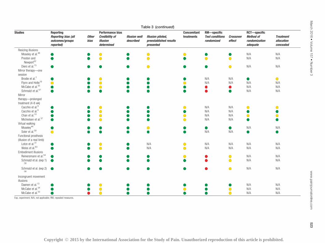
3.4. Risk of bias
All studies had ahigh risk of bias (Table 3). Sample sizewas small inmost and only 3 studies performed an a priori power calcula-tion.8,13,37 Participants were blinded in 25% of experiments(5 studies8,13,33,59,63) and assessors in only 20% of experiments(4 studies8,31,37,59). In 33% of experiments, credibility of the illusionwas evaluated and deemed credible15,44,54,57–59; and only 33% ofexperiments with a repeated-measures study design adequatelycontrolled for crossover effects.13,44,48,57 None of the 6 RCTs
518 E. Boesch et al.·157 (2016) 516–529 PAIN®
Copyright � 2015 by the International Association for the Study of Pain. Unauthorized reproduction of this article is prohibited.
included in the review had a low risk of bias—blinding ofparticipants was not possible inmost studies. Agreement betweenraters ranged between k5 0.151 and k5 1.0. Only 2 categories,“standardized diagnosis” and “incomplete outcome reporting,”were below k5 0.6. Of all bias ratings, 4.5% required consultationwith the third assessor.
3.5. Outcomes: effect of illusions on pain
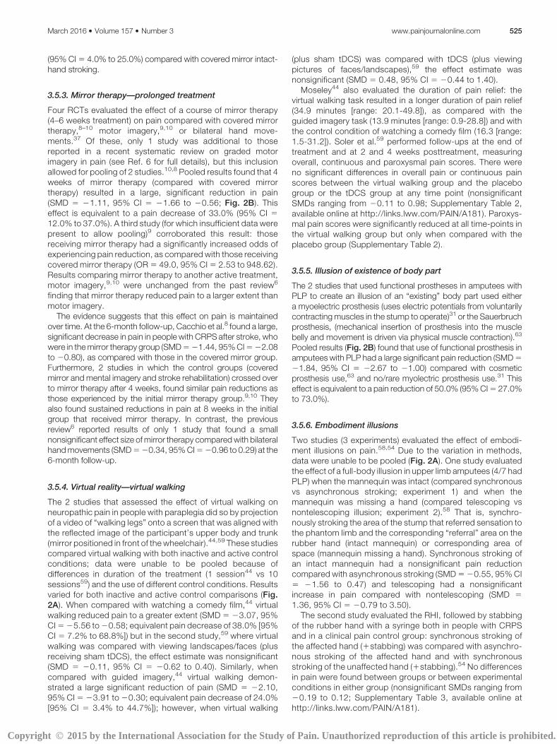
For all studies using continuous outcomes, the individual effectsizes are shown in Figure 2A.
3.5.1. Resizing illusions
Various methods were used to induce resizing illusions:binoculars were used to magnify/minify hand size48; specializedvideo equipment was used to stretch/shrink hand size (congruentvision and touch used to induce the illusion)52; altered video wasused to enlarge/downscale back size.15 All 3 studies assessedpain intensity (preillusion to postillusion) and 2 reported on thechange of “clinical” (ie, preexisting) pain intensity.48,52 However,because of differences in the type of illusion (magnify whole bodypart vs stretch one area of the body part), population studied andhypothesized direction of effect, these studies were unable to bepooled.
In studies that evaluated change in preexisting clinical painintensity, illusions that reduced the size of the affected body partconsistently reduced pain compared with controls (Fig. 2). In
people with CRPS, a visual illusion that decreased the overall sizeof the affected hand significantly reduced pain comparedwith no-resizing of the painful body part (SMD520.87, 95%CI521.43to20.32). This corresponds to a pain increase of 9.5% above thepain induced by handmovements alone (95%CI5 7% to 18.5%).Similarly, in people with hand OA, a multisensory illusion(combining touch and vision) that “shrunk” the affected painfuljoint, significantly reduced pain compared with a resizing illusionon a nonpainful area of the hand (SMD520.59, 95%CI520.95to20.23). This is an equivalent pain decrease of 24.1%, (95% CI5 9.0% to 37.3%). On the contrary, illusions that increased thesize of the affected body part had differential effects in CRPS andOA, but findings were in the directions hypothesized. Comparedwith no-resizing of the painful body part, visual illusions thatincreased the apparent size of the CPRS-affected handsignificantly increased pain above that induced by hand move-ments alone (SMD5 0.54, 95% CI5 0.04-1.05; equivalent painincrease of 14.0%, 95%CI of 5.2% to 23.0%). In OA,multisensoryillusions that “stretched” the size of the painful joint, significantlydecreased pain compared with resizing illusions of a nonpainfulpart of the hand (SMD 5 21.12, 95% CI 5 21.55 to 20.70;equivalent pain decrease of 34.7%, 95% CI of 21.8% to 48.3%).
One study investigated pain intensity and unpleasantness ofexternally applied nociceptive stimuli (pressure and electrical)during illusory resizing of the back in people with chronic backpain.15 Neither illusions of an enlarged back nor a downscaledback had an effect on pain intensity or unpleasantness for eithernociceptive stimulus (vs watching a normal-sized back;
Figure 1. Flow chart detailing the screening and inclusion process (www.prisma-statement.org).
March 2016·Volume 157·Number 3 www.painjournalonline.com 519
Copyright � 2015 by the International Association for the Study of Pain. Unauthorized reproduction of this article is prohibited.
nonsignificant SMDs ranging from20.08 to 0.12 and20.05 to20.10, respectively; Supplementary Table 1, available online athttp://links.lww.com/PAIN/A181). Comparisons between thosewith back pain and a healthy control sample found conflictingresults for pain intensity/unpleasantness over the 3 conditionsand 2 types of nociceptive stimuli (Supplementary Table 1,available online at http://links.lww.com/PAIN/A181).
One study also evaluated the time to return to pretask painlevels.43 During illusory hand magnification in CRPS, the time toreturn to pretask pain levels was significantly longer (P 5 0.03)than it was for both illusory reduction of the hand and no-illusioncontrol conditions. During illusory reduction of hand size, time topretask pain return was significantly shorter than no-illusioncontrol conditions (P 5 0.03).
3.5.2. Mirror therapy—1 session
Four studies evaluated the effect of 1 session of mirror therapyon pain, of which 3 could be pooled. These 3 studies
compared mirror therapy (movement of intact limb) withcovered mirror therapy (mirror obscured by cloth; in CRPS35
and in PLP7) or with no treatment in PLP19 and founda nonsignificant pooled effect estimate (SMD 5 0.17, 95% CI521.72 to 2.06; Fig. 2B), suggesting no effect of 1 session ofmirror therapy. The individual and pooled results were un-changed based on sensitivity analyses (See SupplementaryFigure 1, available online at http://links.lww.com/PAIN/A181).
The fourth study explored illusory touch (using a mirror therapyset-up) in upper limb amputees with PLP that were previouslyunresponsive to traditional movement-based mirror therapy.Illusory touch was induced through synchronous stroking of thestumpwith mirrored stroking of the referred sensation location onthe intact hand andwas comparedwithmirroredmovements andwith covered mirror intact-hand stroking.57 In both comparisons,illusory stroking reduced pain levels (SMD521.65, 95% CI522.89 to 20.42 and SMD 5 25.13, 95% CI 5 28.99 to 21.28,respectively). This is an equivalent pain decrease of 17.0% (95%CI5 4.0% to 29%) compared with mirror movements and 14.0%
Figure 2. (A) Effect estimates for studies evaluating continuous outcomes of pain (standardizedmean differences and 95%confidence intervals). (B) Pooled effectson pain of 1 session of mirror therapy (compared with coveredmirror therapy or no intervention), prolongedmirror therapy (compared with coveredmirror therapy),and functional prostheses (compared with no/cosmetic prosthesis). For 1 session of the mirror therapy, the SD of the difference scores29 was imputed for Brodieet al.7 using a correlation of 0.70 for pre-/posttreatment pain results. This correlation was taken from studies with similar pre-post data.
Active, active control condition; AS, Asynchronous stroking; BHM, bilateral hand movements; CMT, covered mirror therapy; EMI, explicit motor imagery;inactive, inactive control condition; IT, illusory touch (using traditional mirror box set-up); MT, mirror therapy; NS, nonsignificant; RHI, rubber hand illusion; S,significant; SS, synchronous stroking; stab, threatening stimuli (stabbing).
524 E. Boesch et al.·157 (2016) 516–529 PAIN®
Copyright � 2015 by the International Association for the Study of Pain. Unauthorized reproduction of this article is prohibited.
(95% CI5 4.0% to 25.0%) compared with covered mirror intact-hand stroking.
3.5.3. Mirror therapy—prolonged treatment
Four RCTs evaluated the effect of a course of mirror therapy(4–6 weeks treatment) on pain compared with covered mirrortherapy,8–10 motor imagery,9,10 or bilateral hand move-ments.37 Of these, only 1 study was additional to thosereported in a recent systematic review on graded motorimagery in pain (see Ref. 6 for full details), but this inclusionallowed for pooling of 2 studies.10,8 Pooled results found that 4weeks of mirror therapy (compared with covered mirrortherapy) resulted in a large, significant reduction in pain(SMD 5 21.11, 95% CI 5 21.66 to 20.56; Fig. 2B). Thiseffect is equivalent to a pain decrease of 33.0% (95% CI 512.0% to 37.0%). A third study (for which insufficient data werepresent to allow pooling)9 corroborated this result: thosereceiving mirror therapy had a significantly increased odds ofexperiencing pain reduction, as compared with those receivingcovered mirror therapy (OR5 49.0, 95% CI5 2.53 to 948.62).Results comparing mirror therapy to another active treatment,motor imagery,9,10 were unchanged from the past review6
finding that mirror therapy reduced pain to a larger extent thanmotor imagery.
The evidence suggests that this effect on pain is maintainedover time. At the 6-month follow-up, Cacchio et al.8 found a large,significant decrease in pain in people with CRPS after stroke, whowere in themirror therapy group (SMD521.44, 95%CI522.08to 20.80), as compared with those in the covered mirror group.Furthermore, 2 studies in which the control groups (coveredmirror and mental imagery and stroke rehabilitation) crossed overto mirror therapy after 4 weeks, found similar pain reductions asthose experienced by the initial mirror therapy group.9,10 Theyalso found sustained reductions in pain at 8 weeks in the initialgroup that received mirror therapy. In contrast, the previousreview6 reported results of only 1 study that found a smallnonsignificant effect size ofmirror therapy comparedwith bilateralhandmovements (SMD520.34, 95%CI520.96 to 0.29) at the6-month follow-up.
3.5.4. Virtual reality—virtual walking
The 2 studies that assessed the effect of virtual walking onneuropathic pain in people with paraplegia did so by projectionof a video of “walking legs” onto a screen that was aligned withthe reflected image of the participant’s upper body and trunk(mirror positioned in front of the wheelchair).44,59 These studiescompared virtual walking with both inactive and active controlconditions; data were unable to be pooled because ofdifferences in duration of the treatment (1 session44 vs 10sessions59) and the use of different control conditions. Resultsvaried for both inactive and active control comparisons (Fig.2A). When compared with watching a comedy film,44 virtualwalking reduced pain to a greater extent (SMD 5 23.07, 95%CI525.56 to20.58; equivalent pain decrease of 38.0% [95%CI 5 7.2% to 68.8%]) but in the second study,59 where virtualwalking was compared with viewing landscapes/faces (plusreceiving sham tDCS), the effect estimate was nonsignificant(SMD 5 20.11, 95% CI 5 20.62 to 0.40). Similarly, whencompared with guided imagery,44 virtual walking demon-strated a large significant reduction of pain (SMD 5 22.10,95% CI523.91 to20.30; equivalent pain decrease of 24.0%[95% CI 5 3.4% to 44.7%]); however, when virtual walking
(plus sham tDCS) was compared with tDCS (plus viewingpictures of faces/landscapes),59 the effect estimate wasnonsignificant (SMD 5 0.48, 95% CI 5 20.44 to 1.40).
Moseley44 also evaluated the duration of pain relief: thevirtual walking task resulted in a longer duration of pain relief(34.9 minutes [range: 20.1-49.8]), as compared with theguided imagery task (13.9 minutes [range: 0.9-28.8]) and withthe control condition of watching a comedy film (16.3 [range:1.5-31.2]). Soler et al.59 performed follow-ups at the end oftreatment and at 2 and 4 weeks posttreatment, measuringoverall, continuous and paroxysmal pain scores. There wereno significant differences in overall pain or continuous painscores between the virtual walking group and the placebogroup or the tDCS group at any time point (nonsignificantSMDs ranging from 20.11 to 0.98; Supplementary Table 2,available online at http://links.lww.com/PAIN/A181). Paroxys-mal pain scores were significantly reduced at all time-points inthe virtual walking group but only when compared with theplacebo group (Supplementary Table 2).
3.5.5. Illusion of existence of body part
The 2 studies that used functional prostheses in amputees withPLP to create an illusion of an “existing” body part used eithera myoelectric prosthesis (uses electric potentials from voluntarilycontractingmuscles in the stump to operate)31 or the Sauerbruchprosthesis, (mechanical insertion of prosthesis into the musclebelly and movement is driven via physical muscle contraction).63
Pooled results (Fig. 2B) found that use of functional prosthesis inamputees with PLP had a large significant pain reduction (SMD521.84, 95% CI 5 22.67 to 21.00) compared with cosmeticprosthesis use,63 and no/rare myolectric prosthesis use.31 Thiseffect is equivalent to a pain reduction of 50.0% (95%CI5 27.0%to 73.0%).
3.5.6. Embodiment illusions
Two studies (3 experiments) evaluated the effect of embodi-ment illusions on pain.58,54 Due to the variation in methods,data were unable to be pooled (Fig. 2A). One study evaluatedthe effect of a full-body illusion in upper limb amputees (4/7 hadPLP) when the mannequin was intact (compared synchronousvs asynchronous stroking; experiment 1) and when themannequin was missing a hand (compared telescoping vsnontelescoping illusion; experiment 2).58 That is, synchro-nously stroking the area of the stump that referred sensation tothe phantom limb and the corresponding “referral” area on therubber hand (intact mannequin) or corresponding area ofspace (mannequin missing a hand). Synchronous stroking ofan intact mannequin had a nonsignificant pain reductioncompared with asynchronous stroking (SMD520.55, 95%CI5 21.56 to 0.47) and telescoping had a nonsignificantincrease in pain compared with nontelescoping (SMD 51.36, 95% CI 5 20.79 to 3.50).
The second study evaluated the RHI, followed by stabbingof the rubber hand with a syringe both in people with CRPSand in a clinical pain control group: synchronous stroking ofthe affected hand (1stabbing) was compared with asynchro-nous stroking of the affected hand and with synchronousstroking of the unaffected hand (1stabbing).54 No differencesin pain were found between groups or between experimentalconditions in either group (nonsignificant SMDs ranging from20.19 to 0.12; Supplementary Table 3, available online athttp://links.lww.com/PAIN/A181).
March 2016·Volume 157·Number 3 www.painjournalonline.com 525
Copyright � 2015 by the International Association for the Study of Pain. Unauthorized reproduction of this article is prohibited.
Three studies evaluated the effect of incongruent movementillusions, which were hypothesized to induce and/or increasepain.13,33,34 In these studies, the mirror provided a reflectedimage of the nonpainful body part; the painful body part washidden from view behind the mirror. Participants moved thepainful and the nonpainful body part in opposite directionscreating incongruence between vision (reflected image of arm inthe mirror) and proprioceptive feedback. The results were varied.
Only 2 studies33,34 evaluated exacerbation of preexistingsymptoms and performed similar within-subject comparisonsand thus were pooled (Fig. 3). In people with pain (CRPS orFMS34 or FMS33), there was a significant increase in the odds ofexperiencing pain during incongruent movement illusions, ascompared with congruent movements using amirror (OR5 1.67;95% CI 5 1.25-2.24), but no differences when compared withincongruent movements without visual cue (whiteboard; OR 50.83; 95% CI 5 0.57-1.19), or congruent movements withwhiteboard (OR 5 1.07; 95% CI 5 0.91-1.27). Sensitivityanalyses imputing correlations 60.1 did not alter these results.In people with CRPS or FMS, incongruent movement illusionsresulted in higher reports of severe pain (17%) than congruentmirror movements (4%) and incongruent/congruent movementswithout visual cue (both 0%).34
In the third study,13 however, participants with WAD (andwithout arm pain) were not more likely to develop pain in thearm during the incongruent movement illusion than duringcontrol conditions (congruent mirror, Incongruent white-board, congruent whiteboard; nonsignificant ORs rangingfrom 1.00 to 1.12; Supplementary Table 4, available online athttp://links.lww.com/PAIN/A181). People with WAD only hadincreased odds of developing arm pain during the incongruentmovement illusion compared with general movement alone,ie, in which no mirror or whiteboard was used.
3.5.7.2. Comparison with healthy controls
Two studies also compared the pain population to healthycontrols33,13; neither study found that incongruent movementillusions selectively increased pain to a greater extent ina painful population than in healthy controls (ie, pain increasealso occurred during control conditions). In the first study,people with FMS did not have increased odds of experiencingpain compared with healthy controls during an incongruentmovement illusion (OR5 3.90; 95% CI5 0.93 to 16.31) or witha congruent mirror control condition (OR 5 1.99, 95% CI 50.51 to 7.71)33 but had increased odds of experiencing painduring both whiteboard control conditions (incongruent: OR512.60, 95%CI5 1.48 to 107.54; congruent: OR5 10.67, 95%CI 5 1.24 to 91.98). In the second study,13 people with WADhad increased odds of experiencing arm pain compared withhealthy controls, but this occurred for all conditions (congruentmirror/whiteboard: OR 5 66.60, 95% CI 5 3.78 to 1173.63;incongruent mirror/whiteboard: OR5 59.59, 95% CI5 3.38 to1050.18), including 2 movement conditions without view of thewhiteboard or mirror, suggesting that it was merely movementin people with WAD that was pain provoking.
4. Discussion
Weevaluated the current evidence for the effect of bodily illusionsonpain. With the caveat that all studies had a high risk of bias,consistent evidence emerged: a decrease in pain was imparted byfunctional prosthetic use (illusory limb presence); illusory reductionin the apparent size of the body part; synchronous touch; andprolongedmirror therapy treatment. Illusory increase in theapparentsize of the body part consistently modulated pain in thehypothesized direction (direction varied between conditions).Inconsistent effects on pain were found for virtual walking. Noeffects on pain were found for 1 session of mirror therapy, forembodiment illusions and for most incongruence illusions, althoughincongruentmovement illusions had greater odds of increasing pain
Figure 3. Pooled effects for incongruent movement illusions (compared with nonillusory control conditions) on pain. Imputed correlations were calculated fromDaenen et al. 201213 due to use of identical comparisons (0.716 for mirror incongruence vs mirror congruence comparison, 0.657 for mirror incongruence vswhiteboard incongruence comparison, and 0.716 for mirror incongruence vs whiteboard congruence comparison).
526 E. Boesch et al.·157 (2016) 516–529 PAIN®
Copyright � 2015 by the International Association for the Study of Pain. Unauthorized reproduction of this article is prohibited.
than congruent mirroredmovements did. Our findings are limited tochronic pain—no studies evaluated acute pain.
4.1. Resizing illusions
The coexistence of cortical misrepresentation of the body andperceptual size dysfunction in chronic painful conditions37,43
underpins the investigation of resizing illusions. Changing how thepainful body part looks may induce changes in corticalrepresentation56 and thus affect pain. We found limited evidencefor bodily resizing illusions (3 studies, n 5 48), but the evidencesuggests that the type of condition, perceptual dysfunction andillusion (general vs targeted) may be important. For example,people with CRPS often report that their affected arm feels biggerthan the healthy arm30,41,49; illusions that magnified the entireCRPS-affected hand increased pain and illusions that “min-imized” the hand decreased pain.48 Conversely, people with OAperceive their hand to be smaller than healthy controls do24 andstretching illusions centered on the painful joint had a larger effecton pain than shrinking illusions did.52 Perhaps pain relief dependson normalizing the perceptual dysfunction. Moreover, it isinteresting that visual resizing of the whole hand did not affectOA pain, but multisensory illusions did.52 Clearly more work isneeded to elucidate these findings. Last, resizing illusions inchronic back pain did not alter intensity or unpleasantness of painevoked by experimental nociceptive stimuli,15 raising thepossibility of differential effects on chronic pain vs acutenociceptive processing.
4.2. Mirror therapy
Mirror therapy is thought to reduce pain by providing pain-free visualfeedback of normal limb movement. The effect has been attributedto removing incongruence between motor intention and sensoryfeedback for the painful (or phantom) limb,27,32 but cognitivemechanisms associated with threat appraisal have also beenproposed.45 There is limited evidence that 1 session of mirrortherapy does not reduce pain,7,19,35 but limited-to-moderateevidence that prolonged mirror therapy does, at least whencompared with inactive control conditions8–10 or explicit motorimagery.9,10 Nonsignificant effects for 1 session of mirror therapymay reflect reducedpower (n518 vsn530 for prolonged therapy),or alternatively, a cumulative effect of prolonged mirror therapy.
Interestingly, nonresponders to traditional mirror therapy hadsignificant pain reduction with congruent tactile and visualinformation (synchronous stroking), applied using the traditionalmirror box set-up.57 Perhaps the presence of multisensorycongruent information is most important to the effects on pain,rather than themodality of themultisensory components. Indeed,that tactile input could be as effective as traditional mirror therapyhas been proposed previously.45 It is interesting to speculate thattactile multisensory information might avoid the associativepairing of movement with pain14,60 and thus be less likely totrigger nociception, the latter also being a premise to gradedmotor imagery.43
4.3. Virtual walking illusions
Virtual walking aims to create the illusion of normal leg function.Again, evidence is limited (2 studies, n5 44): 1 small randomizedrepeated-measures study44 found significant pain reductions buta larger RCT59 found no effect. Although the studies differed oninactive control conditions—Soler et al.59 used a double-sham(sham tDCS and sham illusion), whereas Moseley44 compared
with a comedy film to control for distraction—that virtual walkinggroup of Soler et al. also received sham tDCS suggests that thesedifferences are not likely relevant. It may be that the samplesdiffered in other ways or that virtual walking does not add an effectabove and beyond nonspecific effects of treatment. However,that virtual walking illusions were more effective at relieving painthan guided imagery,44 which has known efficacy compared withplacebo,22 but not tDCS with the motor cortex,59 suggests thatcomparison with common treatments for chronic pain isnecessary before clinical implementation.
4.4. Functional prosthesis—illusory existence of a missingbody part
Although the creation of illusory existence of a missing body partwas not the primary aim of these studies,31,63 they clearly inducesuch an experience. Limited evidence (2 studies, n 5 35)suggests that use of functional prostheses reduces PLP. Thelarge between-group difference in the time wearing the prosthe-sis (22 vs 5 years)31 and the significant between-group agedifference,63 suggests caution in interpreting these studies’findings.
4.5. Embodiment illusions
Embodiment illusions are hypothesized to influence pain through“replacing” the real, painful body part with an artificial counter-part.25 It is certainly intuitively attractive: we might be able toreduce pain by “projecting it” away from our body. That the RHIinduces limb-specific changes in temperature regulation47 andhistamine reactivity1 raises the possibility of modulation ofnociception at a tissue or spinal level as well. Evidence existsthat embodiment modulates physiological responses to painfulstimuli: decreased arousal responses occur with high levels ofself-identification with an avatar.55 However, in healthy volun-teers, the evidence is conflicting as to whether the RHI does28 ordoes not38 decrease experimentally induced thermal pain.Perhaps the experimental methodology used for embodiment isessential to the modulatory effects on pain. Alternatively, perhapsthe pain is not decreased in intensity or unpleasantness butsimply felt elsewhere: indeed, participants report that the evokedpain is felt in the rubber hand, not the stimulated hand.38 Ourreview found that embodiment does not modulate pain—full-body embodiment illusion58 did not decrease pain and RHIcombined with threat (stabbing)54 did not increase pain,suggesting that embodiment and pain modulation may beseparate processes.
4.6. Incongruent movement illusions
Incongruent movement illusions have been used to test thehypothesis that incongruence between motor intention andmotor action causes or exacerbates pain.27,32 We found limitedevidence (3 studies, n 5 87) against this idea—incongruenceillusions did not selectively cause or exacerbate pain in patientswhen compared with control conditions or healthy controls. Thesole exception was that incongruence illusions weremore likely toresult in pain exacerbation than mirrored, congruent move-ments,33,34 although it is interesting to speculate that the lattermay constitute a dosage of traditional mirror therapy. Thatperforming opposing movements with the arms, but withoutvisual feedback, aggravated pain in FMS patients (more so than inhealthy controls)33 might simply reflect motor or biomechanicaldemands of the task.
March 2016·Volume 157·Number 3 www.painjournalonline.com 527
Copyright � 2015 by the International Association for the Study of Pain. Unauthorized reproduction of this article is prohibited.
The evidence is primarily limited to short-duration bodily illusions(eg, 20 seconds-15 minutes). Repeated/prolonged interventionsmight be required to decrease pain53: studies evaluating at least 4weeks of mirror therapy8–10,37 all reported significant painreduction. Moreover, evidence suggests a positive effect ofgraded motor imagery,40,42,43 which incorporates 2 weeks ofintensive mirror therapy training, in people with CRPS or PLP.
4.8. Strengths and limitations
We emphasized rigor by conforming to the gold-standardapproach to meta-analytical reviews.39,29 We used a sensitivesearch strategy, hand searching references and consultingexperts; yet, it remains possible that we missed eligible studies.We developed, in collaboration with field experts, clear constraintsaround what bodily illusions entailed, but we recognize that theresults may have varied if our definition did. The wide variety ofmethodologies, types of illusions, and patient groups limitedpooling and thus meta-analytical power. Although we required allstudies to have a control condition/group to minimize the risk ofsignificant results because of nonspecific effects of treatment, thehigh risk of bias of included studies (lack of blinding) raises thepossibility that pain relief may have been mediated, at least in part,by expectation.3,16 IncludedRCTsgenerally blinded the assessors,which strengthens our confidence in these findings.
4.9. Future research
Robust and suitably powered RCTs are needed. Furthermore,understanding the underlying mechanisms of the illusions wouldfacilitate refinement of those that show promise. For example,a common feature of bodily illusions is that they are multisensoryin nature, raising the possibility that the conditions in which theseillusions are effective may have deficits in multisensory in-tegration. Indeed, the lack of therapeutic success of traditionalmirror therapy in amputees with telescoped limbs (ie, when visiondoes not match what they perceive),21 but the efficacy ofsynchronous touch (ie, vision of touch matches referredsensation areas)57 suggests that this is a relevant consideration.Preliminary evidence that illusory touch improves sensation inperipheral neuropathy62 further supports this idea. Clearly morework is needed to clarify these relationships.
4.10. Conclusion
This review found promising effects on pain for resizing illusions,functional prosthetic use for PLP, and mirror therapy, suggestingthat evaluation of repeated treatment is warranted. Due to thelimited evidence base, caution must be employed in prematurelydismissing other illusion methodologies. Further studies withlarger samples and varying dosages are essential before solidconclusions can be drawn.
Conflict of interest statement
T. R. Stanton received travel and accommodation support fromEli Lilly Ltd for a Western Canada speaker’s tour (September2014); this was unrelated to the present topic. G. L. Moseleyconsults for Pfizer, Kaiser Permanente, Providence Health,NOIgroup Australasia and Workers’ Compensation Boards inAustralia and North America. G. L. Moseley receives royaltiesfrom his published books (Explain Pain, Explain Pain handbook,Graded Motor Imagery handbook, Painful Yarns). E. Boesch andV. Bellan have no conflict of interests to declare.
E. Boesch was supported by a University of South Australia(UniSA) Division of Health Sciences Honours Scholarship anda UniSA School of Health Sciences Conference Scholarship. V.Bellan was supported by a postgraduate scholarship from theUniversity of Milano-Bicocca. G. L. Moseley was supported bya National Health and Medical Research Council ResearchFellowship (ID1061279). T. R. Stanton was supported bya Canadian Institute for Health Research Postdoctoral TrainingFellowship (ID223354) and National Health & Medical ResearchCouncil Early Career Fellowship (ID 1054041).
Acknowledgements
The authors thank Carolyn Berryman for her valuable helpselecting and including studies; Neil O’Connell for his statisticalassistance with the meta-analytical techniques; Dr AlbertoGallace, Dr Roger Newport, Dr Diana Torta, Dr Martin Diers fortheir valuable help in identifying missed studies and Dr AlbertoGallace, Dr Roger Newport, Dr Martin Diers for establishinga consensus about the definition of bodily illusion.
Appendix A. Supplemental Digital Content
Supplemental Digital Content associated with this article can befound online at http://links.lww.com/PAIN/A181.
Article history:Received 3 June 2015Received in revised form 15 October 2015Accepted 5 November 2015Available online 14 November 2015
References
[1] Barnsley N, McAuley JH, Mohan R, Dey A, Thomas P, Moseley GL. Therubber hand illusion increases histamine reactivity in the real arm. Curr Biol2001;21:R945–946.
[2] Becker MP, Balagtas CC. Marginal modelling of binary cross-over data.Biometrics 1993;49:997–1009.
[3] Benedetti F, Mayberg HS, Wager TD, Stohler CS, Zubieta JK.Neurobiological mechanisms of the placebo effect. J Neurosci 2005;25:10390–402.
[4] Bjorkman A, Weibull A, Rosen B, Svensson J, Lundborg G. Rapid corticalreorganisation and improved sensitivity of the hand following cutaneousanaesthesia of the forearm. Eur J Neurosci 2009;29:837–44.
[5] Botvinik M, Cohen J. Rubber hands “feel” touch that eyes see. Nature1998;391:756.
[6] Bowering KJ, O’Connell NE, Tabor A, Catley MJ, Leake HB, Moseley GL,Stanton TR. The effects of graded motor imagery and its components onchronic pain: a systematic review andmeta-analysis. J Pain 2013;14:3–13.
[7] Brodie EE, Whyte A, Niven CA. Analgesia through the looking-glass? Arandomized controlled trial investigating the effect of viewing a “virtual”limb upon phantom limb pain, sensation andmovement. Eur J Pain 2007;11:428–36.
[8] Cacchio A, De Blasis E, De Blasis V, Santilli V, Spacca G. Mirror therapy incomplex regional pain syndrome type 1 of the upper limb in strokepatients. Neurorehabil Neural Repair 2009;23:792–9.
[9] Cacchio A, De Blasis E, Necozione S, di Orio F, Santilli V. Mirror therapyfor chronic complex regional pain syndrome type 1 and stroke. N Engl JMed 2009;361:634–6.
[10] Chan BL, Witt R, Charrow AP, Magee A, Howard R, Pasquina PF,Heilman KM, Tsao JW. Mirror therapy for phantom limb pain. N Engl JMed 2007;357:2206–7.
[11] Cochrane Collaboration. Review Manager (Revman). Book ReviewManager (Revman). City: Nordic Cochrane Centre, 2011.
[12] Cohen J. Statistical power analysis for the behavioural science. Hillsdale,MJ: Lawrence Erlbaum Associates, 1998.
[13] Daenen L, Nijs J, Roussel N, Wouters K, Van Loo M, Cras P.Sensorimotor incongruence exacerbates symptoms in patients with
528 E. Boesch et al.·157 (2016) 516–529 PAIN®
Copyright � 2015 by the International Association for the Study of Pain. Unauthorized reproduction of this article is prohibited.
chronic whiplash associated disorders: an experimental study.Rheumatology 2012;51:1492–9.
[14] De Jong JR, Vlaeyen JW, Onghena P, Cuypers C, Den Hollander M,Ruijgrok J. Reduction of pain-related fear in complex regional painsyndrome type I: the application of graded exposure in vivo. PAIN 2005;116:264–75.
[15] Diers M, Zieglgansberger W, Trojan J, Drevensek AM, Erhardt-Raum G,Flor H. Site-specific visual feedback reduces pain perception. PAIN 2013;154:890–6.
[16] Eippert F, Bingel U, Schoell ED, Yacubian J, Klinger R, Lorenz J, BuchelC. Activation of the opioidergic descending pain control system underliesplacebo analgesia. Neuron 2009;63:533–43.
[18] Ezendam D, Bongers RM, Jannink MJ. Systematic review of theeffectiveness of mirror therapy in upper extremity function. DisabilRehabil 2009;31:2135–49.
[19] Flinn SR, Hotle AC. A case series report on amputees with pro digit handprostheses receiving mirror therapy. J Hand Ther 2011;24:390–1.
[20] Flor H, Braun C, Elbert T, Brinbaumer N. Extensive reorganization ofprimary somatosensory cortex in chronic back pain patients. NeurosciLett 1997;224:5–8.
[21] Foell J, Bekrater-Bodmann R, Diers M, Flor H. Mirror therapy for phantomlimb pain: brain changes and the role of body representation. Eur J Pain2014;18:729–39.
[22] Fors EA, Sexton H, Gotestam KG. The effect of guided imagery andamitriptyline on daily fibromyalgia pain: a prospective, randomized,controlled trial. J Psychiatr Res 2002;36:179–87.
[23] Gandevia SC, Phegan CML. Perceptual distortions of the human bodyimage produced by local anaesthesia, pain and cutaneous stimulation.J Physiol 1999;514:609–16.
[24] Gilpin HR, Moseley GL, Stanton TR, Newport R. Evidence for distortedmental representation of the hand in osteoarthritis. Book Evidence fordistorted mental representation of the hand in osteoarthritis. City, 2014.p. 258–63.
[25] Giummarra MJ, Georgiou-Karistianis N, Nicholls ME, Gibson SJ,Bradshaw JL. The phantom in the mirror: a modified rubber-handillusion in amputees and normals. Perception 2010;39:103–18.
[26] Gwilym SE, Filippini N, Douaud G, Carr AJ, Tracey I. Thalamic atrophyassociated with painful osteoarthritis of the hip is reversible afterarthroplasty. Arthritis Rheum 2010;62:2930–40.
[27] Harris AJ. Cortical origin of pathological pain. Lancet 1999;354:1464–6.[28] Hegedus G, Darnai G, Szolcsany T, Feldmann A, Janszky J, Kallai J. The
[29] Higgins JPT, Green S. Cochrane handbook for systematic reviews ofinterventions. In: Higgins JPT, Altman DG, Sterne JAC, editors. Bookcochrane handbook for systematic reviews of interventions. City: TheCochrane Collaboration, 2011.
[30] Lewis JS, Kersten P, McCabe CS, McPherson KM, Blake DR. Bodyperception distrubance: a contribution to pain in complex regional painsyndrome (CRPS). PAIN 2007;133:111–19.
[31] Lotze M, Grodd W, Birbaumer N, Erb M, Huse E, Flor H. Does use ofmyoelectric prosthesis prevent cortical reorganization and phantom limbpain? Nat Neurosci 1999;2:501–2.
[32] McCabe CS. Simulating sensory disturbances in healthy controls-implications for pathology. Rheumatology 2005;42:63.
[33] McCabe CS, Cohen H, Blake DR. Somaesthetic disturbances infibromyalgia are exaggerated by sensory-motor conflict: implications forchronicity of the disease? Rheumatology 2007;46:1587–92.
[35] McCabe CS, Haigh RC, Ring EFJ, Halligan PW, Wall PD, Blake DR. Acontrolled pilot study of the utility of mirror visual feedback in the treatmentof complex regional pain syndrome (type 1). Rheumatology 2003;42:97–101.
[36] Merskey H, Bogduk N. Classification of chronic pain: descriptions ofchronic pain syndromes and definitions of pain terms. Seattle, WA: IASPPress, 1994.
[37] Michielsen ME, Selles RW, Van Der Geest JN, Eckhardt M, Yavuzer G,Stam HJ, Smits M, Ribbers GM, Bussmann JB. Motor recovery andcortical reorganization after mirror therapy in chronic stroke patients:a phase II randomized controlled trial. Neurorehabil Neural Repair 2011;25:223–33.
[38] Mohan R, Jensen KB, Petkova VI, Dey A, Barnsley N, Ingvar M, McAuleyJH, Moseley GL, Ehrsson HH. No pain relief with the rubber hand illusion.PLoS One 2012;7:1–7.
[39] Moher D, Liberati A, Tetzlaff J, Altman DG, Group TP. Preferred reportingitems for systematic reviews and meta-analyses: the PRISMA statement.PLoS Med 2009;6:1–28.
[40] Moseley GL. Graded motor imagery is effective for long-standingcomplex regional pain syndrome: a randomised controlled trial. PAIN2004;108:192–8.
[41] Moseley GL. Distorted body image in complex regional pain syndrome.Neurology 2005;65:773.
[42] Moseley GL. Is successful rehabilitation of complex regional painsyndrome due to sustained attention to the affected limb? Arandomised clinical trial. PAIN 2005;114:54–61.
[43] Moseley GL. Graded motor imagery for pathologic pain: a randomisedcontrolled trial. Neurology 2006;67:2129–34.
[44] Moseley GL. Using visual illusion to reduce at-level neuropathic pain inparaplegia. PAIN 2007;130:294–8.
[45] Moseley GL, Gallace A, Spence C. Is mirror therapy all it is cracked up tobe? Current evidence and future directions. PAIN 2008;138:7–10.
[46] Moseley GL, McCormick K, Hudson M, Zalucki N. Disrupted corticalproprioceptive representation evokes symptoms of peculiarity,foreignness and swelling, but not pain. Rheumatology 2006;45:196–200.
[47] Moseley GL, Olthof N, Venema A, Don S, Wijers M, Gallace A, Spence C.Psychologically induced cooling of a specific body part caused by theillusory ownership of an artificial counterpart. Proc Natl Acad Sci U S A2008;105:13169–73.
[48] Moseley GL, Parsons TJ, Spence C. Visual distortion of a limb modulatesthe pain and swelling evoked by movement. Curr Biol 2008;18:R1047–8.
[49] Peltz E, Seifert F, Lanz S, Mueller R, Mayhoefner C. Impaired hand sizeestimation in CRPS. J Pain 2011;12:1095–101.
[50] Pleger B, Ragert P, Schwenkreis P, Forster A, Wilimzig C, Dinse H,Volkmar N, Maier C, Tegenthoff M. Patterns of cortical reorganizationparallel impaired tactile discrimination and pain intensity in complexregional pain syndrome. NeuroImage 2006;32:503–10.
[51] Pleger B, Tegenthoff M, Ragert P, Forster A, Dinse HR, Maier C.Sensorimotor returning in complex regional pain syndrome parallels painreduction. Ann Neurol 2005;57:425–9.
[52] Preston C, Newport R. Analgesic effects of multisensory illusions inosteoarthritis. Rheumatology 2011;50:2314–15.
[53] Ramachandran VS, Rogers-Ramachandran D. Synaesthesia in phantomlimbs induced with mirrors. Proc Biol Sci 1996;263:377–86.
[54] Reinersmann A, Landwehrt J, Krumova EK, Petersburs J, Ocklenburg S,Gunturkun O, Maier C. The rubber hand illusion in complex regional painsyndrome: preserved ability to integrate a rubber hand indicates intactmultisensory integration. PAIN 2013;154:1519–27.
[55] RomanoD, Pfeiffer C,Maravita A, BlankeO. Illusory self-identification withan avatar reduces arousal responses to painful stimuli. Behav Brain Res2014;261:275–81.
[56] Schaefer M, Flor H, Heinze HJ, Rotte M. Morphing the body: illusoryfeeling of an elongated arm affects somatosensory homunculus.Neuroimage 2007;36:700–5.
[57] Schmalzl L, Ragno C, Ehrsson HH. An alternative to traditional mirrortherapy. Illusory touch can reduce phantom limb pain when illusorymovement does not. Clin J Pain 2013;29:e10–18.
[58] Schmalzl L, Thomke E, Ragno C, Nilseryd M, Stockselius A, Ehrsson HH.“Pulling telescoped phantoms out of the stump”: manipulating theperceived position of phantom limbs using a full-body illusion. Front HumNeurosci 2011;5:1–12.
[59] Soler D, Kumru H, Pelayo R, Vidal J, Tormos JM, Fregni F, Navarro X,Pascual-Leone A. Effectiveness of transcranial direct current stimulationand visual illusion on neuropathic pain in spinal cord injury. Brain 2010;133:2565–77.
[60] Vlaeyen JW, Linton SJ. Fear-avoidancemodel of chronicmusculoskeletalpain: 12 years on. PAIN 2012;153:1144–7.
[61] von Elm E, Altman DG, Egger M, Pocock SJ, Gotzsche PC,Vandenbroucke JP. Strengthening the reporting of observationalstudies in epidemiology (STROBE) statement: guidelines for reportingobservational studies. Br Med J 2007;335:806–8.
[62] Wand BM, Stephens SE, Manqharam EI, George PJ, Bulsara MK,O’Connell NE, Moseley GL. Illusory touch temporarily improves sensationin areas of chronic numbness: a brief communication. NeurorehabilNeural Repair 2014;28:797–9.
[63] Weiss T, Miltner WH, Adler T, Bruckner L, Taub E. Decrease in phantomlimb pain associated with prosthesis-induced increased use of anamputation stump in humans. Neurosci Lett 1999;272:131–4.
March 2016·Volume 157·Number 3 www.painjournalonline.com 529
Copyright � 2015 by the International Association for the Study of Pain. Unauthorized reproduction of this article is prohibited.


![The rubber hand illusion in action - Melbourne School of … · 2019. 10. 25. · Bodily illusions Body image Body schema Perception Action ... cation (pp. 136–157)]. However, it](https://static.documents.pub/doc/80x56/60fe61cb174c7f13ed4ba1b5/the-rubber-hand-illusion-in-action-melbourne-school-of-2019-10-25-bodily.jpg)


