THE EFFECT OF EXTERNAL CONSTRICTION OF A BLOOD VESSEL ON BLOOD FLOW1 R. E. SHIPLEY AND D. E. GREGG From the Department of Medicine, Western Reserve University, Cleveland, Ohio Received for publication January 14, 1944 In many situations in which blood flow through a vessel is under consideration, it is frequently a matter of practical importance to estimate the reduction in blood flow which will result when the lumen of a blood vessel is partially ob- structed or locally reduced by compression. As examples, a vessel lumen may be reduced from natural causes by external compression (e.g., extravascular muscular contraction, neoplastic masses), intrinsic pathologic changes (sclerotic plaques, scars), or artificially reduced by vessel clamps (Goldblatt clamps and other devices), flow measuring instruments (thermostromuhr, electromagnetic flow recorder), etc. Considering the diversified fields in which such information might be of practical value, little physiological work has been published which deals with the relationship of vessel bore to volume flow. Interest in this problem was aroused during the course of recent experimental studies in this laboratory on the accuracy of the thermostromuhr method for measuring blood flow (1, 2). It was observed that the application of a loosely fitting thermostromuhr unit to an artery frequently caused an appreciable de- crease in the flow through the artery as measured by a rotameter (3). While it did not seemlikely at the time that the lumen was markedly reduced by appli- cation of the thermostromuhr, no vessel diameter or wall thickness measurements were made from which values of lumen area could later be computed. These observations suggested the possibility that a relatively small external constriction of an artery could, under physiological conditions, induce an appreciable reduc- tion in flow through a vessel. The study of vessel constriction and its flow limiting effects involves con- sideration of the various factors which determine the rate of blood flow through a given vessel segment. An expression of the interrelationship of these factors is given in Poiseuille’s law which states that the rate of flow through small tubes is directly proportional to the 4th power of the radius of the lumen and to the pressure drop across the two ends and inversely proportional to the viscosity of the blood and the length of the tube. Thus, for any vessel segment which is locally constricted, the accompanying reduction in blood flow will become greater as 1, the lumen is decreased; 2, the length of the segment constricted is increased, and 3, the viscosity of the blood is increased. If, in a given case, these factors remain fixed, the rate of flow through the constriction will be governed entirely by the pressure difference across the two ends of the con- stricted segment. The factors which determine this pressure difference in an 1 The expenses of this investigation were defrayed by a grant from the Commonwealth Fund. 289 by 10.220.33.6 on April 3, 2017 http://ajplegacy.physiology.org/ Downloaded from
Transcript
THE EFFECT OF EXTERNAL CONSTRICTION OF A BLOOD VESSEL ON BLOOD FLOW1
R. E. SHIPLEY AND D. E. GREGG
From the Department of Medicine, Western Reserve University, Cleveland, Ohio
Received for publication January 14, 1944
In many situations in which blood flow through a vessel is under consideration, it is frequently a matter of practical importance to estimate the reduction in blood flow which will result when the lumen of a blood vessel is partially ob- structed or locally reduced by compression. As examples, a vessel lumen may be reduced from natural causes by external compression (e.g., extravascular muscular contraction, neoplastic masses), intrinsic pathologic changes (sclerotic plaques, scars), or artificially reduced by vessel clamps (Goldblatt clamps and other devices), flow measuring instruments (thermostromuhr, electromagnetic flow recorder), etc. Considering the diversified fields in which such information might be of practical value, little physiological work has been published which deals with the relationship of vessel bore to volume flow.
Interest in this problem was aroused during the course of recent experimental studies in this laboratory on the accuracy of the thermostromuhr method for measuring blood flow (1, 2). It was observed that the application of a loosely fitting thermostromuhr unit to an artery frequently caused an appreciable de- crease in the flow through the artery as measured by a rotameter (3). While it did not seem likely at the time that the lumen was markedly reduced by appli- cation of the thermostromuhr, no vessel diameter or wall thickness measurements were made from which values of lumen area could later be computed. These observations suggested the possibility that a relatively small external constriction of an artery could, under physiological conditions, induce an appreciable reduc- tion in flow through a vessel.
The study of vessel constriction and its flow limiting effects involves con- sideration of the various factors which determine the rate of blood flow through a given vessel segment. An expression of the interrelationship of these factors is given in Poiseuille’s law which states that the rate of flow through small tubes is directly proportional to the 4th power of the radius of the lumen and to the pressure drop across the two ends and inversely proportional to the viscosity of the blood and the length of the tube. Thus, for any vessel segment which is locally constricted, the accompanying reduction in blood flow will become greater as 1, the lumen is decreased; 2, the length of the segment constricted is increased, and 3, the viscosity of the blood is increased. If, in a given case, these factors remain fixed, the rate of flow through the constriction will be governed entirely by the pressure difference across the two ends of the con- stricted segment. The factors which determine this pressure difference in an
1 The expenses of this investigation were defrayed by a grant from the Commonwealth Fund.
intact artery are 1, the blood pressure on the upstream side of the constriction; 2, the resistance to flow through the constricted segment, and 3, the peripheral resistance of the arteries and vascular bed distal to the constriction.
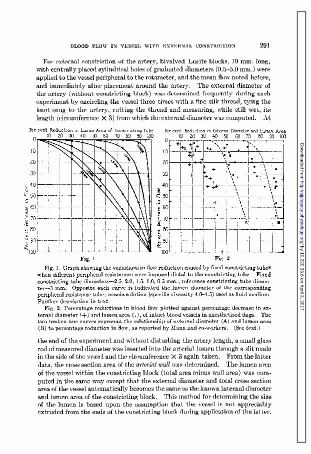
Before proceeding to intact vessels in the animal, experimental determinations of flow through various constrictions were first made in an artificial system in which t,he above factors could be more easily varied or controlled. The ap- paratus used consisted of a reservoir tank (30 cm. in diameter) to the bottom of which was connected a rubber hose of large diameter (I.D. = 2 cm.). A metal tube, 10 mm. in length with a cylindrical lumen of 0.5, 1.0, 1.5, 2.0, 2.5 or 3.0 mm., was inserted into an adapter fitted to the distal end of the hose. A periph- eral resistance (PR) was introduced distal to the constricting tube and separated from it by a 10 cm. length of large diameter hose (20 mm. I.D.). For the PR mechanism, one of a similar set of metal tubes 20 mm. long (I.D. ranging from 0.5 to 3.0 mm.) was used. Acacia solution with a viscosity approximating that of blood (4) (specific viscosity 4.0-4.5) was allowed to flow by gravity at a con- stant hydrostatic pressure, the outflow from the PR tube emerged into a vessel of the same fluid and the overflow was measured with a graduate and stop watch.
In figure 1 are plotted percentage reductions in lumen area of the constricting tube versus percentage decrease in flow when different peripheral resistances were imposed distal to the constricting tube. The internal diamet,ers of the PR tubes are indicated beside the respective curves. In the high PR range (small bore PR tubes), the constricting tubes limit flow very little until the percentage decrease in lumen area is very high. When the PR is low (large bore PR tubes), the constricting tubes are much more effective in limiting flow.
From these experimental data it is evident that the PR mechanism at the end of the flow circuit (distal constriction) plays an extremely important role in de- termining the extent of flow limitation caused by the constricting tlube (proximal constriction). One would anticipate quite similar variations in flow reduction in an in viva preparation where the PR of a given vascular bed may undergo consid- erable change through vasodilatation or constriction. Accordingly, experiments were extended to anesthetized dogs in which were studied the effects of localized arterial constriction upon the flow of blood within an artery.
For this study, the common carotid artery was chosen because of its accessibil- ity and length. Dogs, weighing W-20 kgm., were anesthetized2, given antico- agulants3, and a large rotamet*er (3) with large bore cannulae inserted between t*he cut ends of one common carotid artery. Mean blood pressure was recorded by a mercury manometer connected to the rotameter. For a number of determina- tions the peripheral resistance of the bed was temporarily lowered, either by nitroglycerine injection (0.00016 gram) into the same artery (via a side tube on the rotameter), or by occluding the artery for 2-3 minutes prior to a det#ermina- tion, thereby producing in the latter case an ischemic dilatation within the corresponding bed. All of the experiments were of short duration and the condi- tion of the dogs was excellent throughout.
2 Sodium pentobarbital, 20 mgm. per kilo. 3 Heparin, 100 U. per kilo and pontamine fast pink, 150 mgm. per kilo.
BLOOD FLOW IN VESSEL WITH EXTERNAL CONSTRICTION 291
For external constriction of the artery, bivalved Lucite blocks, 10 mm. long, with centrally placed cylindrical holes of graduated diameters (0.5-5.0 mm.) were applied to the vessel peripheral to the rotameter, and the mean flow noted before, and immediately after placement around the artery. The external diameter of the artery (without constricting block) was determined frequently during each experiment by encircling the vessel three times with a fine silk thread, tying the knot snug to the artery, cutting the thread and measuring, while still wet, its length (circumference X 3) from which the external diameter was computed. At
Per cent Reduction tn Lumen Arca of Constric,tinq Tube
10 20 30 40 50 60 70 80 90 100 Per cent Reduction in External Diameter and Lumen Area A 10 20 30 40 50 60 70 80 90 100
70
20 30
90
00 Fig. 1 Fig. 2
Fig. 1. Graph showing the variations in flow reduction caused by fixed constricting tubes when different peripheral resistances were imposed distal to the constricting tube. Fixed constricting tube diameters -2.5, 2.0, 1.5, 1.0, 0.5 mm.; reference constricting tube diame- ter-3 mm. Opposite each curve is indicated the lumen diameter of the corresponding peripheral resistance tube; acacia solution (specific viscosity 4.0-4.5) used as fluid medium. Further description in text.
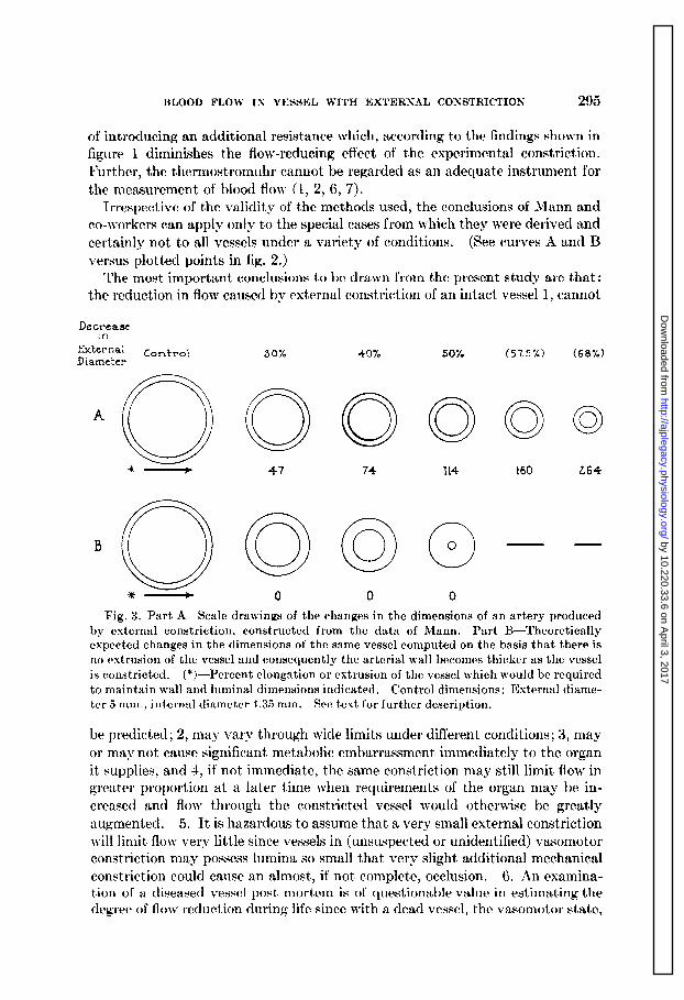
Fig. 2. Percentage reductions in blood flow plotted against percentage decrease in ex- ternal diameter (+) and lumen area (.), of intact blood vessels in anesthetized dogs. The two broken line curves represent the relationship of external diameter (A) and lumen area (B) to percentage reduction in flow, as reported by Mann and co-workers. (See text.)
the end of the experiment and without disturbing the artery length, a small glass rod of measured diameter was inserted into the arterial lumen th .rough a slit made in the side of the vessel and the circumference X 3 again taken. From the latter data, the cross section area of the arterial wall was determined. The lumen area of the vessel within th .e constricting block (total area minus wall area) was com- puted in the same way except that the external diameter and total cross section area of the vessel automatically becomes the same as the known internal diameter and lumen area of the constricting block. This method for determining the size of the lumen is based upon the assumption that the vessel is not appreciably extruded from the ends of the constricting block during application of the latter.
Such would diminish the cross section area of the arterial wall and thereby in- crease relatively the lumen area. However, in actual practice, direct observation of the vessel at the point of emergence from the constricting block revealed no visible extrusion or elongation during application of the constricting blocks.4
Of four similar animal experiments, the combined results of two typical ones are presented in figure 2 in which are plotted the percentage reductions in flow versus percentage reductions in external diameter (+) and lumen area (0) of the artery. Each point indicates the percentage change from the control to the mechanically constricted state of the artery. In these experiments, the blood flow ranged from 140 to 828 cc. per minute under different conditions and mean blood pressure remained constant (within 2-3 mm. Hg) during the actual deter- minations.
In contrast to the orderly sequence of changes in flow observed with the gravity system (fig. 1), the same relationships recorded in viva with successive constrictions were decidedly irregular. Scattering of the points was observed even though the PR of the bed was not intentionally altered. When nitroglycer- ine was given or hyperemia induced, the points were even more irregular with a proportionately greater decrease in flow in relation to the percentage constriction.
DISCUSSION. The relationship of flow, peripheral resistance, and applied pres- sure to the added resistance of luminal constriction can be most simply illustrated by reference to the somewhat analogous flow of current in an electrical circuit.
According to Ohm’s law, the flow of current is determined by voltage (pressure)
Resistance l
Substituting hemodynamic equivalents, the rate of flow of blood through a given artery will be proportional to the applied pressure divided by the total resistance peripheral to that point in the vessel. As a working example, let us say 2 cc./sec. will flow through an artery when the applied pressure is 120 mm. Hg and the
120 peripheral resistance is 60 arbitrary units-then 6. = 2. Assuming that a given
localized constriction of the artery will add 20 units of resistance, then the flow
will decrease by 25 per cent to 1.5 cc./min. 120
15 m+= l
. If at another
time, the peripheral bed is made to dilate so that its PR becomes 40 units, then
the control flow will be 3 cc./min. . The addition of the same local
4 A direct test of the method was made by observing the changes in arterial bore by means of a low power microscope fitted with an ocular micrometer. A recently isolated artery segment, tied to a cannula with a plane glass window at the end, was held in place with its long axis parellel to that of the microscope and inflated with 100 mm. Hg pressure. By transilluminating the arterial wall, the bore of the vessel could be visualized and meas- ured at a focal plane 1.5 cm. from the end of the artery segment. Constricting blocks were applied at this level and the changes in bore observed directly. Close agreement between the values obtained by direct observation and those which were computed from circum- ference and wall area measurements permitted the use of the indirect method in the experi- ments to be presented here.
BLOOD FLOW IN VESSEL WITH EXTERNAL CONSTRICTION 293
constriotion (20 units of resistance) will now decrease the flow by 33 per cent to 2 -
cc./min. 120 -----=2. + 20 >
In other words, the effectiveness of a given local con-
striction in reducing the flow will vary under different conditions of the peri- pheral bed and the flow reduction may be large or small depending upon whether the resistance offered by the constriction is large or small in proportion to the total resistance (i.e., constriction resistance + peripheral resistance).
It is evident from the wide scattering of points in figure 2 that there exists no fixed relationship between percentage reduction in flow and percentage reduction in the luminal or external dimensions of a vessel in viva. One would not expect that the orderly sequence of changes resulting from the introduction of a single variable in an artificial system (as in fig. 1) would be reproduced in the living animal since, for the same vessel, the blood pressure, peripheral resistance and
TABLE 1 Relationship of changes produced in the dimensions of the same vessel by external constriction
rate of flow will undergo physiological variations from time to time. These factors will also vary from animal to animal and among different vessels in the same animal.
Of equal and perhaps greater importance is the fact that the wall area, external diameter, and lumen diameter may be considerably different from vessel to vessel, and, in the case of the latter two factors, even for the same vessel under different vasomotor conditions. The relationship of changes in external diameter to changes in lumen area (and flow) may therefore be equally variable. The magni- tude of variation in this relationship is illustrated in table 1 in which are presented the calculated percentage and absolute changes in the dimensions of a single vessel which will result from a 10 per cent reduction in external diameter with different pre-existing vasomotor states. These data illustrate (and mathematical con- siderations dictate) that as the external vessel diameter progressively diminishes (as in the case of active vasoconstriction) the same percentage (10 per cent) decrease in the existing external diameter will cause an increasingly gpeater per- centage decrease in the existing lumen area (cf. italicized figures, table 1).
An additional and equally unpredictable factor which may alter the ultimate influence of a localized constriction upon blood flow is the response of the periph- eral bed. When the flow to the bed is reduced by the constriction, the peripheral vessels may dilate as a result of the associated ischemia and the flow may tend to increase, t,he combined resultant of which will be a new equilibrium. For this reason it is probable that the amounts by which blood flow was reduced in the foregoing animal experiments are less than those which would be recorded if it were possible to prevent the partial compensation (by dilatation) for the ischemia of the bed peripheral to the constriction.
The foregoing considerations reveal that it is impossible to predict within rather wide limits the amount of flow reduction which will result when an intact vessel is constricted by external means. However, from a physiological stand- point, a reduction of the lumen of a vessel may be of little functional importlance to the vascular bed supplied by that vessel when the rate of blood flow is already low, but on the other hand, the same constriction may seriously limit the blood supply to the same bed just at the time when the requirements of the latter are greatest and flow would otherwise be much greater.
Many of the findings, interpretations and conclusions presented here differ from those reported by Mann, Herrick, Essex and Baldes (5). These investiga- tors studied the effects of vessel constriction in an artificial system and iv2 vice,
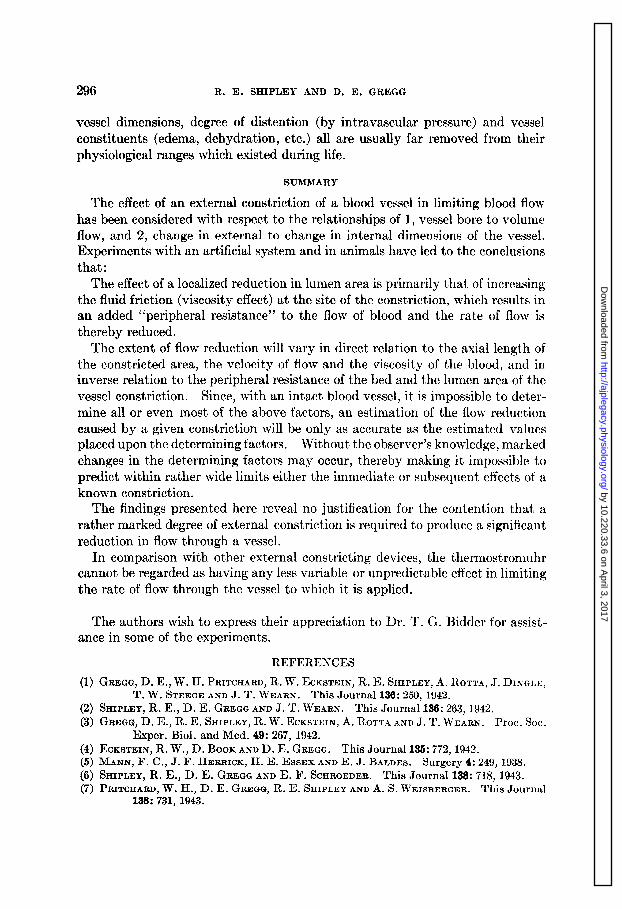
and concluded that blood flow through a vessel is not reduced by a significant amount unless the cross section area of the lumen is reduced by 50-70 per cent. Unfortunately, certain differences in the procedures and methods of study make it impossible to compare much of their data with that presented here. As ex- amples, 1, in their experiments with an artificial circulation system, a peripheral resistance was maintained by capillary tubes or a “satisfact,ory mechanism.” The use of a single, arbitrarily chosen peripheral resistance will not reveal the wide range and variability of the flow reductions which can be caused by a given con- striction. (See family of curves, fig. 1.) 2. In their communication, a progressive external constriction of a vessel in the living animal is reported to have caused the lumen to decrease in size as illustrated in the scale drawings of figure 3A. Wit,h the use of constricting units 10 mm. long, it is difficult to conceive of a mechanism by which a, the wall thickness would remain tlhe same throughout the various stages of constriction, and b, the external diameter of their vessel could be reduced 57 per cent and 68 per cent without obliteration of the lumen and cessation of flow. In figure 3B are shown the successive changes in the dimensions of t,he same vessel which have been calculated mathematically in accord with geometric principles. 3. In the same experiments, the flow reductions attributed t,o the application of constricting units cannot be considered maximal since the ther- mostromuhr itself (used in the intact vessel experiments to measure the flow in the artery) may have already constricted the lumen to such an extent5 that blood flow is initially reduced by a significant amount. Its effect would be t/hat
5 6‘ . . . successful measurement of blood flow necessitates the application of [thermo- stromuhr] units which have internal diameters somewhat less than the external diameters of the blood vessels under investigation.” -Mann, Herrick, Essex and Baldes (5).
BLOOD FLOW Ix VESSEL WITH EXTERTLrAL CONSTRICTION 295
of introducing an additional resistance which, according to the findings shown in figure 1 diminishes the flow-reducing effect of the experimental constriction. Further, the thermostromuhr cannot be regarded as an adequate instrument for the measurement of blood flow (1, 2, 6, 7).
Irrespective of the validity of the methods used, the conclusions of Mann and co-workers can apply only to the special cases from which they were derived and certainly not to all vessels under a variety of conditions. (See curves A and B versus plotted points in fig. 2.)
The most important conclusions to be drawn from the present study are that: the reduction in flow caused bv external constriction of an intact vessel 1, cannot, -’
External Diameter
Control
A
0 *- -
30%
0
47
0
0
40%
0
74
0 0
0
50%
0
114
0 0
0
(57.5%) (68%)
0 0 0
i60 264
Fig. 3. Part A-Scale drawings of the changes in the dimensions of an artery produced by external constriction, constructed from the data of Mann. Part B-Theoretically expected changes in the dimensions of the same vessel computed on the basis that there is no extrusion of the vessel and consequently the arterial wall becomes thicker as the vessel is constricted. (*)-Percent elongation or extrusion of the vessel which would be required to maintain wall and luminal dimensions indicated. Control dimensions : External diame- ter 5 mm., internal diameter 4.35 mm. See text for further description.
be predicted; 2, may vary t!hrough wide limits under different conditions; 3, may or may not cause significant metabolic embarrassment immediately to the organ it supplies, and 4, if not immediate, the same constriction may still limit flow in greater proportion at a later time when requirements of t,he organ may be in- creased and flow through the constricted vessel would otherwise be greatly augmented. 5. It is hazardous to assume that a very small external constriction will limit flow very little since vessels in (unsuspected or unidentified) vasomotor constriction may possess lumina so small that very slight additional mechanical constriction could cause an almost, if not complete, occlusion. 6. An examina- tion of a diseased vessel post mortem is of questionable value in estimating the degree of flow reduction during life since with a dead vessel, the vasomotor state,
vessel dimensions, degree of distention (by intravascular pressure) and vessel constituents (edema, dehydration, etc.) all are usually far removed from their physiological ranges which existed during life.
SUMMARY
The effect of an external constriction of a blood vessel in limiting blood flow has been considered with respect to the relationships of 1, vessel bore to volume flow, and 2, change in external to change in internal dimensions of the vessel. Experiments with an artificial system and in animals have led to the conclusions that:
The effect of a localized reduction in lumen area is primarily that of increasing the fluid friction (viscosity effect) at the site of the constriction, which results in an added “peripheral resistance” to the flow of blood and the rate of flow is thereby reduced.
The extent of flow reduction will vary in direct relation to the axial length of the constricted area, the velocity of flow and the viscosity of the blood, and in inverse relation to the peripheral vessel constriction . Since, with
resistance of the bed and the lumen area of the an intact blood vessel, it is impossible to deter-
mine all or even most of the above factors, an estimation of the flow reduction caused by a given constriction will be only as accurate as the estimated values placed upon the determining factors. Without the observer’s knowledge, marked changes in the determining factors may occur, thereby making it impossible to predict within rather wide limits either the immediate or subsequent effects of a known constriction.
The findings presented here reveal no justification for the contention that a rather marked degree of external constriction is required to produce a significant reduction in flow through a vessel.
In comparison with other external constricting devices, the thermostromuhr cannot be regarded as having any less variable or unpredictable effect in limiting the rate of flow through the vessel to which it is applied.
The authors wish to express their appreciation to Dr. T. G. Bidder for assist- ance in some of the experiments.
REFERENCES
(1) GREGG, D. E., W. H. PRITCHARD, R. W. ECKSTEIN, R. E. SHIPLEY, A. ROTTA, J. DINGLE, T. W. STEEGE AND J. T. WEARN. This Journal 136: 250, 1942.
(2) SHIPLEY, R. E., D. E. GREGG AND J. T. WEARN. This Journal 136: 263, 1942. (3) GREGG, D. E., R. E. SHIPLEY, R. W. ECKSTEIN, A. ROTTA AND J. T. WEARN. Proc. Sot.
Exper. Biol. and Med. 49: 267, 1942. (4) ECKSTEIN, R. W., D. BOOK AND D. E. GREGG. This Journal 136: 772,1942. (5) MANN, F. C., J. F. HERRICK, H. E. ESSEX AND E. J. BALDES. Surgery 4: 249, 1938. (6) SHIPLEY, R. E., D. E. GREGG AND E. F. SCHROEDER. This Journal 138: 718, 1943. (7) PRITCHARD, W. H., D. E. GREGG, R. E. SHIPLEY AND A. S. WEISBERGER. This Journal