AN ABSTRACT OF THE THESIS OF Seeviga Skultab for the degree of Master of Science in Entomology presented on March 16, 1983 Title: THE EFFECT OF PHOTQPERIOD AND TEMPERATURE ON OVARIAN DEVELOPMENT AND FAT PRODUCTION IN CULEX PEUS SPEISER (DIPTERA: CULICIDAE) Abstract approved: Redacted for Privacy Bruce F. Eldridge" The effect of photoperiod and temperature on ovari- an follicle development and fat production was studied in a colonized population of CuZex peus Speiser from Philomath, Oregon. Females were subjected to simulated fall conditions of photoperiod and temperature. Under a combination of short photoperiod and low temperature, there were vari- ous effects on their physiological activities such as the retardation of follicular development, a reduction in the blood-feeding rate and the occurrence of hyper- trophic fat. In the laboratory, conditions of a short day length photoperiod (8hL:16hD) and cool temperatures (15°C) to which females were subjected from the pupal stage to eight days after emergence influenced the development of follicles, and resulted in the ovaries remaining in a diapause condition. Under conditions of
Transcript
AN ABSTRACT OF THE THESIS OF
Seeviga Skultab for the degree of Master of Science
in Entomology presented on March 16, 1983
Title: THE EFFECT OF PHOTQPERIOD AND TEMPERATURE ON
OVARIAN DEVELOPMENT AND FAT PRODUCTION IN CULEX PEUS
SPEISER (DIPTERA: CULICIDAE)
Abstract approved:Redacted for Privacy
Bruce F. Eldridge"
The effect of photoperiod and temperature on ovari-
an follicle development and fat production was studied
in a colonized population of CuZex peus Speiser from
Philomath, Oregon.
Females were subjected to simulated fall conditions
of photoperiod and temperature. Under a combination of
short photoperiod and low temperature, there were vari-
ous effects on their physiological activities such as
the retardation of follicular development, a reduction
in the blood-feeding rate and the occurrence of hyper-
trophic fat. In the laboratory, conditions of a short
day length photoperiod (8hL:16hD) and cool temperatures
(15°C) to which females were subjected from the
pupal stage to eight days after emergence influenced the
development of follicles, and resulted in the ovaries
remaining in a diapause condition. Under conditions of
16 hour photophases and 25°C, females showed an increase
in follicle size over time. Females exhibited a marked
reduction of blood-feeding activity in response to a
combination of short photophases (8 hours) and cool
temperatures (15°C). Blood-fed females held under sim-
ulated fall conditions developed a considerable amount
of fat reserve while non-blood-fed females, maintained
under the same conditions, and females taking a blood-
meal at warmer temperatures had significantly less fat.
It was concluded that daylength is an important
factor controlling the follicular development of females
of C. peus. Pupae and adults were exposed to combin-
ations of 12 photoperiods (photophases of 9.5, 10, 10.5,
11, 11.5, 12, 12.5, 13, 13.5, 14, 14.5 and 15 hours) and
a temperature of 18°C. Follicle size gradually increas-
ed as photophase was lengthened. At photophases be-
tween 9.5 and 12.5 hours the follicles remained small
and the sharp increase was seen at photophases of 13
hours or more. Experimental study showed that less than
13 hours of light per day stimulated the entire popu-
lation to enter ovarian diapause.
Field collections of larvae made in 1981 showed
that adult activity decreased in September. With the
retardation of follicle development, suppression of blood-
feeding drive and formation of hypertrophic fat in re-
sponse to simulated fall conditions, it was concluded
that the northern population of C. peus undergoes
ovarian diapause each fall as inseminated adult fe-
males.
The Effect of Photoperiod and Temperature on OvarianDevelopment and Fat Production in Culex peus
Speiser (Diptera: Culicidae)
by
Seeviga Skultab
A THESIS
submitted to
Oregon State University
in partial fulfillment ofthe requirements for the
degree of
Master of Science
Completed March 16, 1983
Commencement June 1983
APPROVED:
Redacted for PrivacyProfessor of Dep"artmerit'of Entomol
Redacted for Privacy
Head of Department of Entomology
Redacted for Privacy
(Dean of Graduate /school
Date thesis is presented March 16, 1983
Typed by Jane A. Tuor for Seeviga Skultab
Acknowledgement
I am indebted to the Royal Thai Government for
financial support through my graduate education at
Oregon State University.
I am grateful to Dr. Bruce F. Eldridge, my major
professor and thesis advisor, for his overall guidance,
many helpful suggestions and criticisms throughout
the study. His generous advice, assistance, valuable
training and friendship are appreciated. Without his
help, this study could not have successfully been com-
pleted.
I wish to thank Dr. Rod Frakes, Dr. Jo-Ann Leong
and Dr. Michael Burgett for serving on my committee.
I would like to express my deepest appreciation
to my mother, Mrs. Srinual; and my brother, Kasom, for
their love, self-sacrifice, encouragement and patience.
I also would like to extend my sincere appreciation
to Mr. Somsak Saengtharatip for his love, understanding
and encouragement.
Special thanks must go to Mrs. Myrtle Skinner,
Mrs. Solveig Meeker, Mr. Leon Pimentel and "John,"
who have provided an enjoyable environment in which to
help me make it through my graduate studies.
Finally, thank you to all friends for giving me
merry and joy during these past years.
This thesis is dedicated to my father dearest,
Group Captain Virach Skultab M.D., for giving me the
inspiration, motivation, confidence and love that has
IV. RESULTS 30Effect of Photoperiod and Temperature on
Ovarian Development 30Experiment Ia 30Experiment Ib 41Experiment Ic 49
Effect of Photoperiod and Temperature onFat Production 51
Experiment II 51Field Collections of Mosquitoes 53
V. DISCUSSION 57
VI. CONCLUSIONS 71
VII. BIBLIOGRAPHY 73
LIST OF FIGURES
Figure
1 Distribution of Culex peus in NorthAmerica, north of Mexico
2 Female Culex peus mosquito
3 Inside a programmed incubator showingan illumination source, a wire-woundresistor, a pan of fresh water and twoexperimental globes
4
5
6
7
8
9
Rate of adult eclosion for Culex peusat indicated photoperiods and temper-atures
Rate of ovarian follicle developmentfor Culex peus at indicated photo-periods and temperatures
Follicles of Culex peus mosquito rearedat 15°C under 8hL:16hD, 17 days post-adult-emergence
Follicles of CuZex peus mosquitoreared at 15°C under 16hL:8hD, 17 dayspost-adult-emergence
Follicles of Culex peus mosquito rearedat 25°C under 8hL:16hD, 17 days post-adult-emergence
Follicles of Cuies peus mosquito rearedat 25°C under 16hL:8hD, 17 days post-adult-emergence
10 Ovarian follicle length of Culex peusmaintained at 18°C and 12 differentphotoperiods
11 Ratio of follicle: germarium length ofCulex peus maintained at 18°C and 12different photoperiods
Page
7
8
24
36
38
39
39
40
40
45
46
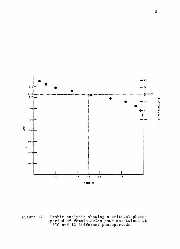
12 Probit analysis showing a critical photo-period of female CuZex peus maintainedat 18°C and 12 different photoperiods 48
LIST OF FIGURES - CONTINUED
Figure Pa e
13 Abundance of adult CuZex peus mos-quitoes reared from collections atPhilomath log pond, Oregon, April-November, 1981 56
LIST OF TABLES
Table Page
1
2
3
4
5
6
7
8
9
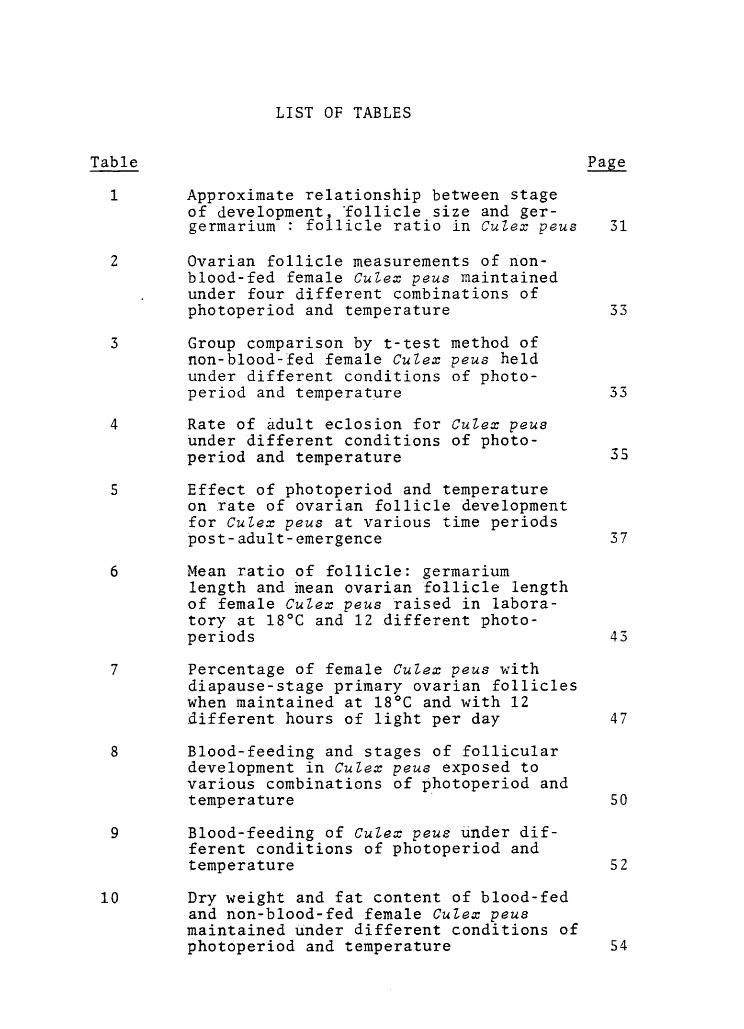
Approximate relationship between stageof development, follicle size and ger-germarium : follicle ratio in Culex peus 31
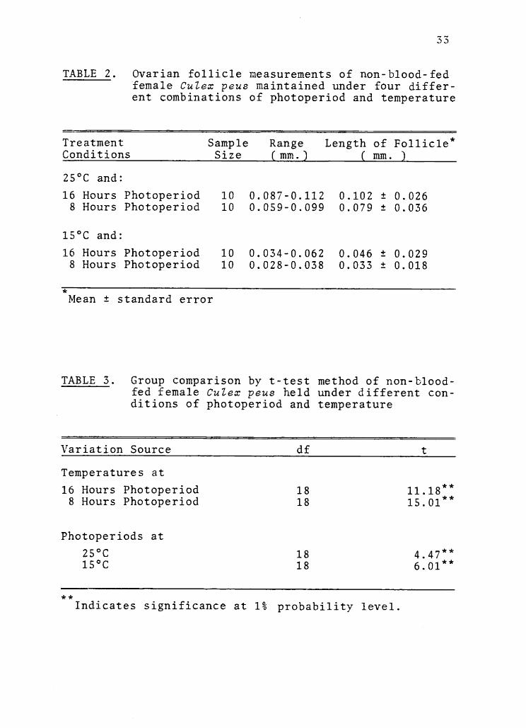
Ovarian follicle measurements of non-blood-fed female Culex peus maintainedunder four different combinations ofphotoperiod and temperature
Group comparison by t-test method ofnon-blood-fed female Culex peus heldunder different conditions of photo-period and temperature
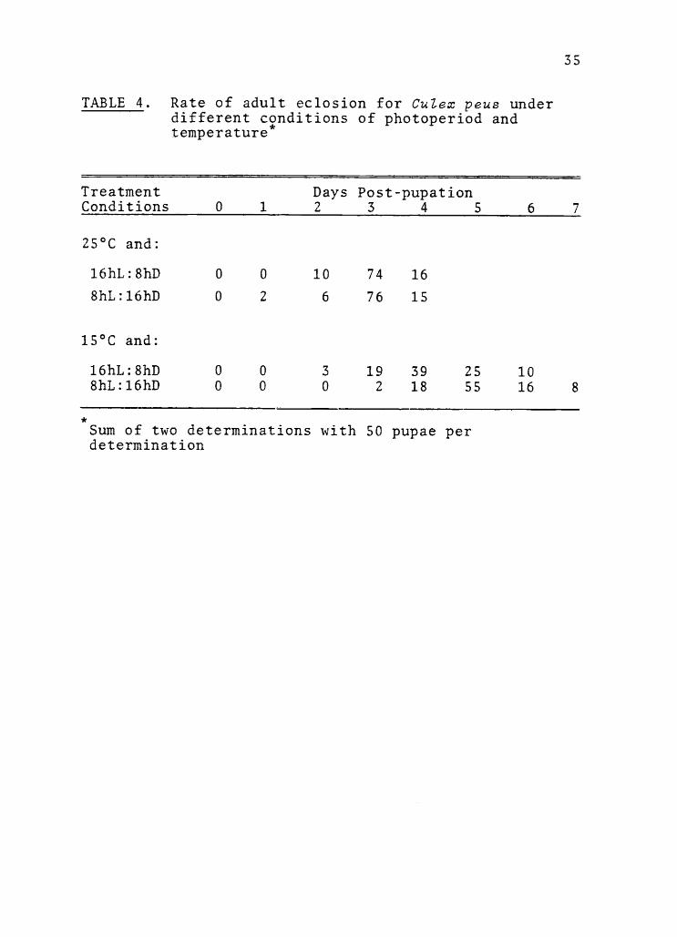
Rate of adult eclosion for Culex peusunder different conditions of photo-period and temperature
Effect of photoperiod and temperatureon rate of ovarian follicle developmentfor Culex peus at various time periodspost-adult-emergence
Mean ratio of follicle: germariumlength and mean ovarian follicle lengthof female Culex peus raised in labora-tory at 18°C and 12 different photo-periods
Percentage of female CuLex peus withdiapause-stage primary ovarian follicleswhen maintained at 18°C and with 12different hours of light per day
Blood-feeding and stages of folliculardevelopment in Culex peus exposed tovarious combinations of photoperiod andtemperature
Blood-feeding of Cities peus under dif-ferent conditions of photoperiod andtemperature
10 Dry weight and fat content of blood-fedand non-blood-fed female Culex peusmaintained under different conditions ofphotoperiod and temperature
33
33
35
37
43
47
50
52
54
THE EFFECT OF PHOTOPERIOD AND TEMPERATURE ON OVARIANDEVELOPMENT ANDFAT PRODUCTION IN CULEX PEUS
SPEISER (DIPTERA: CULICIDAE)
INTRODUCTION
Culex (Culex) peus Speiser is a common mosquito
occurring in western parts of North, Central and South
America. It ranges from Colombia and Venezuela north
to the southwestern part of the state of Washington
(Knight and Stone,1977; Darsie and Ward, 1981). The
status of the species as a pest and as a human and
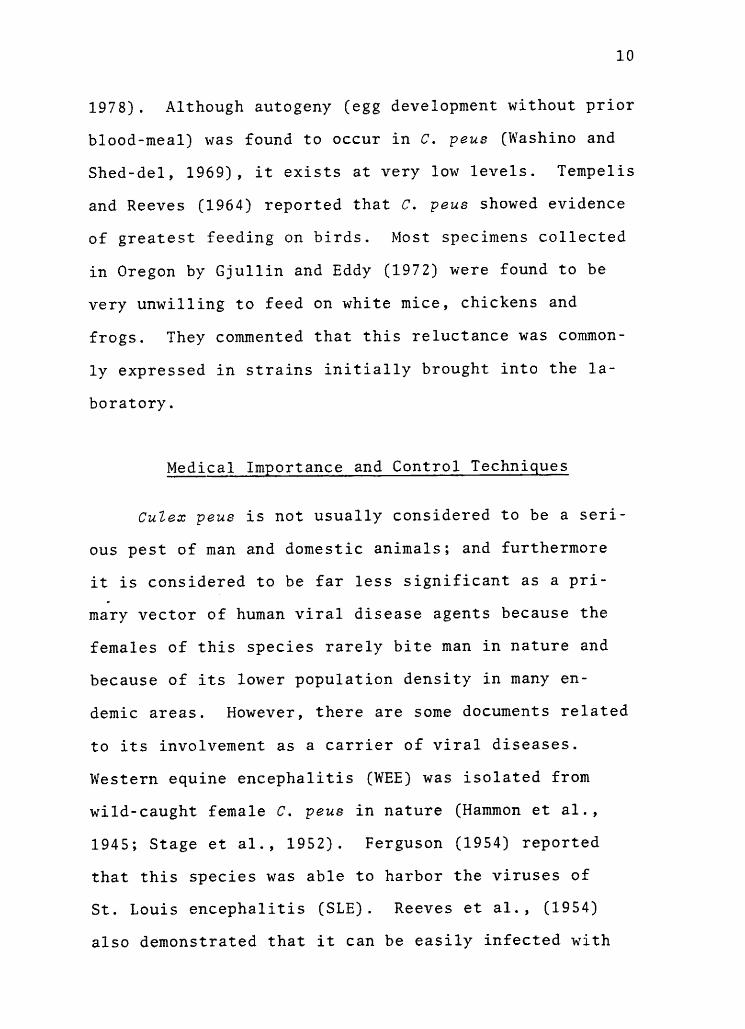
animal disease vector is largely unknown. However,
considerable information is available concerning con-
trol of the species. The common name of C. peus, the
banded foul-water mosquito, stems from its appearance
and the fact that larvae are often found in relatively
polluted water. C. peus shares the characteristic of
a white-banded proboscis with CuZex tarsalis Coquillett,
but the two species can be distinguished from each
other by examination of the ventral surface of the ab-
domen and the outer surface of the rear femora and ti-
biae.
The mosquitoes of the genus Culex are known to sur-
vive winters in cooler portions of the temperate zone
as inactive adult females, assumed to be in a state of
diapause. Diapausing mosquitoes normally show reduc-
tion in blood-feeding drive and cessation of ovarian
2
follicle development, and restrict their feeding to a
carbohydrate diet, resulting in accumulation of body
fat which will be depleted gradually as hibernation
proceeds. These physiological alterations occur in
several Culex species, which undergo a true diapause
as reproductive adult females (Eldridge,1968; Sanburg
and Larsen, 1973; Eldridge et al., 1976, 1979b). The
role of environmental factors in triggering and termin-
ating diapause in Culex mosquitoes is unclear. Sev-
eral studies have been done to investigate the influ-
ence of photoperiod and temperature on ovarian develop-
ment in Culex mosquitoes based on either behavioral or
physiological aspects or both. Most results indicate
that a combination of short daily photophases and cool
temperatures induce ovarian diapause. However, this
phenomenon is not evident in all species of Culex mos-
quitoes.
Among vector species, the fact that the adult fe-
male overwinters has raised the possibility of them
serving as overwinter hosts of disease pathogens. The
role of Culex mosquitoes as overwinter carriers of path-
ogenic viruses for man and animals has been discussed
in several studies. Although hibernating mosquitoes
have a lower rate of metabolism than active ones, an
energy source is nevertheless needed for overwinter
survival. They obtain this energy from fat stored in
3
the form of hypertrophied abdominal fat bodies. Ordin-
arily, blood-fed female mosquitoes do not develop fat
bodies and it has been assumed that cessation of blood-
feeding is necessary to hibernation. Furthermore, it
is the immature stages which are sensitive to short
photophases, and once the adult form emerges, the physi-
ological status of hibernation is established. Such
females would not serve as overwinter virus reservoirs,
since they would not take prehibernation blood-meals.
Gonotrophic dissociation (whereby a prehibernation
blood-meal results in fat production rather than egg
production) has been demonstrated to occur in the genus
Cules (Eldridge, 1966, 1968), and the possibility thus
exists that diapausing mosquitoes may function as a
reservoir for arboviruses throughout the winter. Such
female mosquitoes would become infected following a
blood-meal containing an arbovirus and undergo gono-
trophic dissociation. The energy derived from the
blood-meal would be used in fat production rather than
ovarian development. The isolation of St. Louis en-
cephalitis virus from overwintering Culex pipiens is
evidence of this possibility (Bailey et al., 1978).
Many studies have been done on species of great
importance, i.e. C. tarsalis, well-known as a primary
vector of viral diseases of man and animals (Hender-
son et al., 1979; Walters and Smith, 1980). The less
4
significant species, C. peus, was the choice of this
study because of its possible role as a secondary
vector of viral diseases and because of the scant
knowledge of its winter biology. Existing information
concerning C. peus is mostly about its systematics,
distribution and control; not many details are avail-
able about its biology and almost nothing concerning
its overwintering habits. Although little is known
about the vector competence of C. peus for arboviruses,
females of this species have been shown to be infected
with western equine encephalitis (WEE) and St. Louis
encephalitis (SLE) in nature. Thus this species is a
potential overwinter host for these viruses. Very few
collections of C. peus have been made during winter,
but they are assumed to overwinter as adult females
(Bohart and Washino, 1978).
Since so little is known about the winter biology
of C. peus, I chose to study various aspects of its
phenology in both field and laboratory populations. To
determine when populations were active in nature, I
studied collection records of larvae and adults made
from log ponds near Corvallis. I also studied adult
eclosion rates and blood-feeding activities in labora-
tory populations under different conditions of photo-
period and temperature. Retardation of development of
the ovaries and body fat production are phenomena
5
associated with hibernation; therefore, I conducted
experimental studies of ovarian follicle growth and
fat formation under various combinations of photo-
period and temperature to determine if C. peus under-
goes reproductive diapause and can survive through sim-
ulated winter conditions.
The specific objectives of these studies were:
(1) To observe the effect of photoperiod and tem-
perature on ovarian development in C. peus and,
specifically, to determine whether or not the combin-
ation of short photophase and cool temperature induces
ovarian diapause in this species.
(2) To determine, if C. peus proves to be photo-
period sensitive, the photoperiod required to induce
ovarian diapause at a selected temperature in non-blood-
fed female mosquitoes.
(3) To determine the development of body fat in
response to those combinations of light and temperature
in both blood-fed and sugar-fed female mosquitoes in
the laboratory.
I hope these studies will contribute to the event-
ual understanding of the bionomics of this species.
Figure 5. Rate of ovarian follicle development forCulex peus at indicated photoperiods andtemperatures
21
3 9
Figure 6. Follicles of Culex peus mosquito reared at15°C under 8hL:16hD, 17 days post-adult-emergence (at 40X magnification)
Figure 7. Follicles of Culex peus mosquito reared at15°C under 16hL:8hD, 17 days post-adult-emergence (at 40X magnification)
4--)
cd1
(1) .--i
cd rd(I) cd
.1--)
O V) e--,4-) 0 gr-1 p oHCr (A 4-JEra >-, cdO cd UE "Ij H
t-I-I
CO1---- r-tr-I g
0) bi3)1, ^ C13
C:1 EN rgCI) vC)
'`d-
4-1-1 oo alO ....."
Na)a) -lc)
r--i gUH
ti (..)r-I 0O Lr)
r..14 Cs.1
CO
a);-4
bO.,-1u.
4-)CO
i
I'd 4-)
1-4
cd rd(I) cd$-4 1
+-)O U) r-.4-) 0 g1-1 p, oCr V) -I-I
O C'd Urd F1
4-4Cl) c--- .1-1
1-1 g(1) aPt.J -, cd
Czi EEi 4a) coN . c:,
i-.1 -4.(3 ,4
v.")4-1 r---I cd
O ,-.....
V) (1)
(1) '0r-I gUH1-1 (..)r-1 0O u")
1.1-1 (`-3
01
41
while follicles of females maintained at 25°C and eight
hours photoperiod developed to stage I-II with a mean
follicle length of 0.088 mm. The ovarian follicle length
was slightly longer under conditions of 15°C/16:8LD than
under 25°C/8:16LD conditions, the average follicle length
and Christopher's stage were 0.094 mm. and IIa, respec-
tively. Under short photoperiod and cool temperature
conditions, the follicles had developed slightly to a
mean of 0.055 mm. but never exceeded stage N (pre-rest-
ing stage). The correlation between follicle size and
Christopher's stage was quite high (r=0.91).
When expressed as follicle:germarium ratio, the
-data agree closely with those expressed as follicle
length (Table 5). The correlation between follicle size
and follicle:germarium length ratio was 0.92.
Experiment Ib
An experiment was conducted to determine the duration
of light required to induce ovarian diapause at 18°C
(i.e. to see if a "critical" photophase could be deter-
mined). Pupae were subjected to 12 different photo-
periods (9.5, 10, 10.5, 11, 11.5, 12, 12.5, 13, 13.5,
14, 14.5, and 15 hours of light per day). Only four
treatment combinations could be programmed simultaneous-
ly. Each treatment was tested twice. The second
batches of pupae were placed into the incubators
42
two days after the first groups. Emerged adult females
were provided only 10% sucrose solution. On the eighth
day after the peak of the emergence of adults, females
from each treatment were removed, frozen and dissected
for measurement of length of ovarian follicle and as-
sociated germarium. The method of dissection was the
same as in the previous experiment. This procedure was
repeated until all 12 treatments had been done.
The result of the dissection is shown in Table 6.
Follicle sizes increased with the increasing duration of
light per day. Females exposed to 13 hour or greater
photophases had ovaries with a mean follicle length of
greater than 0.075 mm. and a follicle:germarium length
ratio over 1.5:1.0. On the other hand, females subject-
ed to less than 13 hour photophases had a mean follicle
length of less than 0.075 mm. and ratio of follicle:ger-
marium length not exceed 1.5:1.0. The range and mean
values of ovarian follicle lengths and of ratios are
shown graphically in Figure 10 and 11. Table 7 shows
the percentages of female having diapause-stage ovaries
under different photoperiod conditions at 18°C. Pro-
bit analysis performed on a microcomputer (Figure 12)
indicated that 50% of the populations entered diapause
when held under 13.051 hours of light per day (95%
confidence limits were 13.432 and 12.687 respectively).
TABLE 6. Mean ratio of follicle : germarium length and mean ovarian follicle lengthof female Culex peus raised in laboratory at 18°C and 12 different photo-periods
Figure 10. Ovarian follicle length of Culex peusmaintained at 18°C and 12 differentphotoperiods
3.0
2.5
z2.Q
eg
2
U1.5
O
O0
ex 1.0
* *
Y 0.56 + 0.164 X
r 0.91"
1
9 10 11 12
PHOTOPER 100 ( HOURS )
13
46
14 15
Indicates significance at 1% probability level.
Figure 11. Ratio of follicle : germarium length ofCulex peus maintained at 18°C and 12 dif-ferent photoperiods
47
TABLE 7. Percentage of female Culex peus with diapause-stage primary ovarian follicles when main-tained at 18°C and with 12 different hours oflight per day
Duration of Light No. FemalesPer Day
o Female withlDiapause-stage
Follicles
9 h
10 h
10 h
11 h
11 h
12 h
12 h
13 h
13 h
14 h
14 h
15 h
30 min
30 min
30 min
30 min
30 min
30 min
10 100
10 100
10 100
10 80
10 100
10 100
10 100
10 30
10 40
10 0
10 10
10 10
1 Determinations based on Spielman and Wong (1973b); thefollicle was determined in diapause if follicle length:its germarium length ratio was 1.5 or less
48
1.15
1.1151.10
1.05
00 0.95
0.90
0.85
0.80
1111111
.11 11111=4.01
0.2 0.4 0.5 0.6 0.8
PROSITS
15
14
13.050613
12
11
10
Figure 12. Probit analysis showing a critical photo-period of female Culex peus maintained at18°C and 12 different photoperiods
49
Experiment Ic
The general pattern of ovarian development in mos-
quitoes consists of two periods. The first period of
oocyte growth called previtellogenic development occurs
prior to yolk deposition and extends from the "pre-
resting" to the "resting" stage. The second period
called vitellogenic development extends from the
"resting" stage to full development of eggs. General-
ly, follicles of non- blood -fed females will not develop
to mature eggs (except in autogenous strains). This
experiment was conducted to observe the development of
the follicle of Culex peus after a blood-feeding under
four combinations of photoperiod (8 and 16 hours) and
temperature (15°C and 25°C). Each treatment was run
twice. Females from each conditioning incubator were
placed into a small screened cage and were fed on a
Japanese quail on the eighth day after their emergence.
All blood-feeding trials were conducted overnight at
20°C in a separate room. Blood-fed and non-blood-fed
females were segregated from each other and were held
in their respective incubators. When digestion had been
completed, which last about eight days, both blood-fed
and non-blood-fed females were dissected for determin-
ation of follicular development. The results are shown
in Table 8.
50
TABLE 8. Blood-feeding and stages of follicular devel-opment in Culex peus exposed to various com-binations of photoperiod and temperature
**Percentage differences followed by different letters
are significant at the 1% probability level by X2.
Difference between percentages followed by the sameletter are not significant
were lower at the lower temperature. At 25°C, more than
50% of the females tested took a blood-meal under both
photoperiod conditions. There was no significant dif-
ference between photoperiods at the higher temperature,
whereas, at the lower temperature (15°C), the percentage
of blood-feeding among females held under short photo-
phases was significantly lower than that observed for
long photophase females.
After the blood-meal had been completely digested,
the females were processed for determination of fat
production by extraction with petroleum ether. They
were oven-dried at 40°C, weighed, ether-extracted,
53
re-dried and re-weighed as discussed in the "Materials
and Methods" section. The difference between pre- and
post-extraction weights represented the extracted fat
of the mosquitoes. The mean dry weights, the mean
weights of extracted fat and the mean percentages of
fat for each of the eight groups are shown in Table 10.
The differences among groups in dry weight were sig-
nificant at the 1% probability level (F=4.86"). The
differences in amount and percentages of fat between
blood-fed and non-blood-fed females were not significant
at the 1% probability level except for a significantly
greater amount of fat under conditions of eight hours
photoperiod at cool temperature. At 15°C, however,
females which took a blood-meal under 16 hours photo-
phase showed a significantly lower amount and percent-
age of fat formation than females which took a blood-
meal under 8 hours photoperiod. Non-blood-fed females
held at 15°C also showed a significant difference in
percentage of fat between the groups exposed to short
and long photoperiods at the 1% probability level.
Field Collections of Mosquitoes
Field collections of mosquito larvae were made
from alog pond in Philomath, Oregon to determine the
natural occurrence of Culex peus. The results of the
TABLE 10. Dry weight and fat content of blood-fed and non-blood-fed female Culex peusmaintained under different conditions of photoperiod and temperature
*Mean ± standard errorDifferences between percentages followed by different letters are significant at the1% probability level by t-test method. Difference between percentages followed by thesame letter are not significant.
55
field collections including the duration of natural
daylight plus civil twilight are shown in Figure 13.
C. peas larvae first appeared in late April. They
reached peak abundance in August. Larvae had disap-
peared from the log pond by Mid-November.
3.0
2.0
1.0
56
Hours of daylight (including civil twilight)
16.40 13.30r
11
A I 1
II/11 1 1 1\
,4 n v
i \i ii It
Al
.
I Av \
I 1 1 1 1
7 15 22 7 15
1 1 I 1 I I 1 1 1 1 1 J 1
22 7 15 22 7 15 22 7 15 22 7 15 22
\
I 1 J fi 1 17 15 22 7 15 22
APRIL MAY JUNE JULY AUGUST SEPTEMBER OCTOBER NOVEMBER
Figure 13. Abundance of adult Culex peus reared fromcollections at Philomath log pond, Oregon,April-November, 1981
DISCUSSION
The primary objective of this research was to de-
velop some details of the winter biology of Culex peus
since the knowledge about hibernation of this species
is scant. The studies were designed to observe the ef-
fects of photoperiod and temperature on the development
of ovarian follicle and of body fat.
photoperiod and temperature have proven to have
an effect on ovarian development in several species of
CuZex mosquitoes, for example, Culex pipiens pallens
(Wang, 1979), Culex restuans and CuZex pipiens (Eldridge
et al., 1976; Madder, 1981). Eldridge (1966, 1968)
found that ovarian development in C. pipiens was sup-
pressed by a combination of low temperature and short
photoperiod. The results here show that a combination
of 15°C and eight hour photophases resulted in the
ovaries of C. peus remaining in a diapause condition.
Diapause seems likely to be induced by short photophases
and modified by cool temperature since the effect of
temperature on follicle size differed significantly
under photoperiod conditions of 8hL:16hD; follicles
of females maintained at a temperature of 25°C were
twice as long as those of females maintained at 15°C.
Danielevskii (1961) pointed out that diapausing females
during late summer and fall resulted from the response
to shortening daily light that was enhanced by cooler
58
temperature. Sanburg and Larsen (1973) also reported
in their study of C. pipiens pipiens that at 22°C, fol-
licle size increased when females were reared under
15 hour photophases but not under those of 10 hours.
In this experiment, follicle size increased over time
at 25°C, regardless of photoperiod. At 15°C, however,
photoperiod influences on ovarian follicle development
were evident. The follicles of females exposed to
16 hour photophases developed slower and were smaller
after the first nine days of adult life but they de-
veloped to the resting stage (stage I-II) after 21
days. On the other hand, follicles of females held
under eight hour photophases never developed beyond
stage N and remained in the diapause stage. Therefore,
follicle development of C. peas appeared to be arrested
by a combination of 15°C and eight hours photoperiod.
Experiments on the effect of photoperiod and
temperature on rate of adult eclosion revealed that
both photoperiod and temperature to which pupae were
subjected appeared to influence the timing of emergence
of adults. Since temperature is known to directly af-
fect rate of eclosion, the differences observed be-
tween the two photoperiod treatments at 15°C suggest
subtle differences in temperature even though the ex-
perimental treatments are designed to avoid this. In-
candescent lamps used in the incubators provided a more
natural illumination source than fluorescent lamps
59
even though the latter are cooler and freer from com-
plicating secondary heat effects. However, to avoid
the fluctuation of temperature, wire-wound power heat
compensating resistors were installed in the incubators.
Another possible factor which cannot be ruled out is
the disruption of circadian rhythms established under
insectary lighting conditions. Since the larvae were
reared under 16 hour photophases, when the pupae were
moved to the 8hL:16hD treatment conditions, they may have
required some time to adapt to the new rhythm, and
thus, activities such as eclosion in the eight hour
photophase groups were delayed. The results of the
experiment reported here are similar to those reported
by Eldridge et al., (1976) in C. restuans and Culex
salinarius. They found that at a temperature of 25°C
under 16 hours of light per day, both species under-
went 50% of adult eclosion by day two post-pupation,
while in this study those of C. peus underwent eclosion
by day 2.5 under both short and long photophases. At
15°C, however, adult eclosion occurred later in the
three species. Under 16 hour photophases, 50% of
eclosion of C. restuans had occurred by the same day
as that of C. peus (day 3.5), whereas, in C. peus ex-
posed to 15°C and eight hour photophases it occurred
a day later (day 4.5). Photoperiod is believed to in-
sert its action on the brain (Adkisson, 1966; Mansingh,
60
1971; Beck, 1980). It is possible that ecdysone which
is secreted from the prothoracic gland and is necessary
for differentiation to the adult stage was suppressed
by an inappropriate photoperiod of short daily light.
This physiological event needs more investigation.
The methods used to determine diapausing follicles
in these experiments were based on the follicle length
along with the ratio of follicle:germarium length.
The use of these ratios to distinguish diapausing from
non-diapausing females varies from worker to worker.
Spielman and Wong (1973b) proposed the ratio of 1.5:1.0
or less in classifying diapausing follicle. Madder
(1981), on the other hand, used the ratio of no more
than 2.0:1.0. However, he commented that the differ-
ence in the ratio value may be due to the inclusion of
the connecting channel between follicle and its germar-
ium in the measurement of the germarium by Spielman,
thus reducing the ratio. Both the follicle length
and the ratio are not definitely reliable in consider-
ing diapausing and non-diapausing status because some
non-diapausing females have been reported to have a
ratio of less than 2.0:1.0 and vice versa (Madder,
1981). Also, there is no reason to believe that ratios
for one species will necessarily hold for other species
of Culex. I found that in C. peus, some resting stage
follicles (stage I-II of Kawai, 1969) had a ratio of
61
1.2:1.0. Determination of stage of follicular develop-
ment along with follicular measurement is suggested.
Individual difference and uneven size of follicles,
called "mosaic" as reported by Danielevskii (cited in
Eldridge, 1968), were also observed in C. peus as
follicles of different stage of development were found
at the same time and within the same ovary. This
uneven growth was seldom found but it is apparently
more commonly seen in vitellogenic stage of develop-
ment.
The pupal stage of C. pipiens (Eldridge, 1968)
and C. tarsalis (Harwood and Halfhill, 1964) was identi-
fied to be sensitive to photoperiod. The author could
not confirm this observation in C. peus since none of
the experiments were designed to test this, but it is
likely to be so. My initial results suggested only
that C. peus was sensitive to photoperiod and tempera-
ture. Attempting to determine a critical photoperiod
required subjecting females of this species to a
series of photoperiods at a single temperature (18°C).
The response of follicle size to photoperiod was not
linear. Also, graphical results show a wide range of
follicle lengths and ratios at almost every photoperiod
tested. The difference may be due to individual vari-
ation since many other factors were uniform in each
treatment. Rearing techniques, like crowding, food
62
rations, and salinity of media, which are known to af-
fect growth, rhythm and synchrony of pupation and em-
ergence (Nayar, 1968; Nayar and Sauerman, 1970) might
have some effect on variation of individual responses.
However, pupae were randomly divided so that such vari-
ation should have been confused among groups. It is
obvious that follicle lengths increased as photophases
increased. The correlation between photoperiod and
follicle size is 0.88. Again, the ratio of follicle:
germarium length in addition to follicle length was
used to consider diapause status in the mosquitoes.
The correlation between photoperiod and the ratio is
quite high (r=0.91). The data indicated that at 18°C,
diapause in C. peus was induced by the duration of
less than 13 hours of light per day. This observa-
tion agrees closely with the findings of Spielman
and Wong (1973b) that at 18°C, nearly all female C.
pipiens entered diapause when subjected to no more
than 12 hour photophases.
A linear regression fitted in Figure 6 expressed
the photo-periodic response of C. peus. The shape of
the response curve here was similar to that studied
by Sanburg and Larsen (1973) in C. pipiens pipiens,
namely, follicle size was larger at longer photophases.
The critical photoperiod of C. peus, based on the data
obtained from this experiment and analyzed by a probit
63
analysis, was 13.051 hours of light per day. Spielman
and Wong (1973b) found that at 18°C, the critical
photoperiod of C. pipiens was a 13 hour photophase.
Variation in critical photoperiod is not uncommon
since Bradshaw (1976, 1977) reported that critical
photoperiod was closely correlated with latitude
and altitude. That is, critical photoperiod will
be lengthened at the higher latitudes. The colonies
of C. pipiens studied by Spielman and Wong (1973b)
and of C. peus studied by me were collected at
latitudes of approximately 42°N and 44°N, respective-
ly.
Prehibernating female mosquitoes of several Culex
species showed a reduction of blood-feeding activity
in response to conditioning by short photoperiod and
low temperature (Eldridge, 1965, 1972, 1979; Oda, 1971).
Reduction of blood-feeding drive is a characteristic
used as a criterion for diapause in many Culex mos-
quitoes. C. pipiens and C. restuans were reported to
have suppression of blood-feeding under simulated fall
conditions of short photoperiod and cool temperature,
and ovaries of females which happened to take blood-
meals would remain in a diapause state if the post-
feeding temperature was still low (Eldridge et al.,
1972, 1979). Eldridge (1965) demonstrated the effect
of crowding of adult females on blood-feeding in
64
C. pipiens where by blood-feeding decreased with an
increase in density. This factor did not occur in
these blood-feeding trials in C. peus due to small
number of females in the feeding cages. Also, the
factor of age was eliminated by using females of the
same age. However, they varied in size. The author
could not confirm whether carbohydrate diet had an
influence on blood-feeding response in C. peus as it
did in C. pipiens and C. quinquefasciatus (Eldridge,
1965). Adult female C. peus were provided with 10%
sucrose solution from the first day of emergence in
all treatments and throughout the experiment. On
the eighth day post-adult-emergence, blood-feeding
trials were conducted overnight. Under simulated fall
conditions of short photophase and cool temperature,
females were reluctant to take blood and exhibited
a preference for feeding on sugar-water. The concen-
tration of sugar-water was not considered to affect
the rate of blood-feeding since Eldridge (1965) demon-
strated that in C. pipiens and C. quinquefasciatus
blood-feeding rate did not vary after feeding on a
series of sucrose solutions ranging from 5% to 50%.
A factor that might cause reduction in blood-feeding
is the duration of maintenance of females on sugar-
water, even though female C. pipiens (Eldridge, 1965)
and C. p. pallens (Hosoi, 1954) with dilated abdomens
65
containing sucrose solution were observed to take full
blood-meals. Other possible sources of variation in-
clude the size of the test cage and defensiveness of
the quail hosts. However, the results reported here
indicate that a combination of low temperature and
short photoperiod suppressed the blood-feeding re-
sponse of C. peus. Nevertheless, one female showed
evidence of taking blood under a combination of 15°C
and eight hour photophases. This suggests the pos-
sibility that this species may take blood-meals, at
least at low frequencies, in the fall in nature. Fe-
males which took a blood-meal at warmer temperature
and at cool temperature but under long photophases,
either fully or partially, developed mature eggs after
the blood-meal had been completely digested.
Wallis (1959) reported that blood is not necessary
for fat formation and hibernation in C. restuans, since
females preferred feeding upon sucrose solutions late
in the summer. However, C. restuans took blood and
exhibited gonotrophic dissociation (the phenomenon where-
by a blood-meal results in fat body production rather
than ovarian development) in response to fall conditions
of photoperiod and temperature. Gonotrophic dissoci-
ation could not be demonstrated in C. peus. The female
that took blood after conditioning under eight hour
photophases and at 15°C formed a considerable amount of
66
fat reserve but was not examined for follicular develop-
ment. However, sucrose-fed females, maintained under
the same conditions, and which had not taken a blood-
meal previously developed less body fat. It appears
evident that the increased amount of fat was derived
from the blood-meal taken. This is strong circumstan-
tial evidence of gonotrophic dissociation, but more
blood-fed females which have been held under short
photophase and low temperature conditions need to be
examined. At warm temperatures (25°C), percentages
of fat extracted were slightly higher in non-blood-
fed females than in blood-fed ones and follicle ex-
amination revealed that females which took a blood-
meal developed their follicles to mature eggs or at
least to stage Va (of Kawai, 1969), while the follicles
of non-blood-fed females did not develop past stage
IIb (of Kawai, 1969).
The results of this study indicate that C. peus
exhibits ovarian diapause in response to a combination
of short daily photophases and low temperature. It
is interesting to compare these results with the works
of Eldridge et al., (1968, 1976) on the effect of tem-
perature and photoperiod on ovarian development in C.
quinquefasciatus and C. salinarius. The approximate
northernmost limits of C. quinquefasciatus, C. peus
and C. salinarius are 42°N, 47°N and 48°N latitude,
67
respectively. Eldridge and his coworkers performed
their studies of C. quinquefasciatus on a laboratory
colony colonized from larvae collected in Florida and
of C. salinarius colonized from Maryland, both of which
are below 40°N latitude. I conducted these studies
of C. peus with materials collected at about 44.6°N
latitude. Although the three species have basically
southern ranges, the important distinction is that
C. quinquefasciatus and C. salinarius do not show ovarian
diapause in response to fall photoperiod conditions.
Presumably, this difference is due to the geographic
variation as discussed above and variation among and
within species, since Culex taraslis reared in the
laboratory from females collected near Corvallis under-
go ovarian diapause, whereas, those from two California
areas do not (Eldridge 1983, personal communication).
Thus it seems likely that ovarian diapause would only
be evident in the northern populations of the range
of C. peus but this needs further investigation.
Direct evidence of hibernation of C. peus in nature
is still lacking. Although females of C. peus were
collected in Planada and Berkeley, California in late
Janaury (Freeborn and Bohart, 1951), it can not be de-
termined whether this species actually survived an en-
tire winter at these locations. To confirm how suc-
cessfully C. peus can utilize their fat reserves and
68
hibernate, the survival period after feeding activity
should be ovserved and, especially, the interactions
between photoperiod and thermoperiod should be studied.
Beck (1983) reported that the close relationship of
thermophase temperatures which occur during photophase
and cryophase temperatures during scotophase are of
importance in the determination of diapause, since
the incidence of diapause in several insect species
was influenced by the cryophase temperatures coincid-
ing with the scotophase.
Field collections of larvae showed that adult
activity of C. peus declined in September coinciding
with the shortening of day light and decreasing of tem-
perature. Their first appearance of larval populations
in April suggest that at this latitude (44°N) this
species hibernates here, but overwintering C. peus have
never been recovered in Oregon. The results of this
study suggest that the physiological response to en-
vironmental factors are consistent with a species which
overwinters as inseminated adult females.
The question of the possibility of C. peus serving
as an overwinter host for arboviruses of medical or
veterinary importance remains unresolved. Evidence of
infected blood-meals by other Culex mosquitoes during
fall season was reported in several documents (Kokernot
et al., 1969; Dalrymple et al., 1972). Bailey et al.,
(1978) reported that two strains of St. Louis encephalitis
69
virus (SLE) were isolated from hibernating C. pipiens
females during winter. Japanese encephalitis (JE) has
also been isolated from Culex tritaeniorhynchus and
WEE from C. tarsalis (Eldridge, 1981). This evidence
suggests that such females would take viremic blood-
meals in fall and that the virus would persist in the
mosquitoes, or, alternatively that the overwinter gen-
eration of mosquitoes would become infected transo-
varially from the summer, parental generation. There
is also evidence which points to the possibility of
prehibernation blood-meals in C. pipiens. In the
laboratory, an increase in temperature from 15°C to
25°C for 72-84 hours resulted in blood-feeding of pre-
viously hibernating females C. pipiens and a proportion
of those females taking blood had undeveloped ovaries
(Eldridge and Bailey, 1979). This finding suggests
the possibility of prehibernating C. pipiens females
harboring viral diseases while overwintering as adult
females. More research is needed on blood-feeding
habits of C. peus in nature i.e. seasonal feeding
patterns and possible seasonal shifts in host range.
Under laboratory conditions, C. tarsalis, a vector of
WEE and SLE viruses, fed both on birds and on mammals,
while C. peus exhibited a strong preference of feeding
on avian hosts (Nelson et al., 1976). It is interest-
ing to wonder whether C. peus would feed on mammals
70
when birds are scarce as hosts, as they would be in
late fall, since Nelson and his colleagues conducted
their experiments using double-feedings (baits with
a jackrabbit and either a chicken or a pheasant).
Wild-caught C. peus females have yielded isolations
of WEE virus in nature (Hammon et al., 1945; Stage
et al., 1952). Ferguson (1954) also reported the abil-
ity of this species to harbor the virus of St. Louis
encephalitis (SLE). Since C. peus showed evidence
of some blood-feeding under simulated fall conditions
in the laboratory, questions about prehibernating fe-
males taking a blood-meal in nature and the possibility
of viruses surviving in hibernating mosquito vectors
of this species are of interest.
71
CONCLUSIONS
1. Three characteristics were considered in determin-
ation of reproductive diapause in Culex peus.
They are failure of ovarian development, retard-
ation of blood-feeding drive and formation of hy-
pertrophic fat in response to short photoperiod
and low temperature. C. peus expresses ovarian
diapause after exposure of pupal stage to the
eighth day of adult life to a combination of eight
hour photophases and cool (15°C) temperatures.
2. Diapausing females have ovarian follicles in the
pre-resting stage (stage N of Kawai, 1969) with
a mean follicle length of 0.055 mm. and a ratio
of follicle:germarium length 1.1 : 1.0.
3. At 18°C, at least 13 hours of light per day are
required for normal follicle development, while
a shorter duration of daily light stimulates
females to enter ovarian diapause.
4. Simulated fall conditions not only retard fol-
licular development but also delay adult eclosion,
suppress blood-feeding and result in the accumu-
lation of body fat.
5. Gonotrophic dissociation may exist in C. peus
since an increased amount of fat was shown to
derive from a blood-meal in female held under
72
diapause-inducing conditions. More confirmation
is needed to prove failure of ovarian follicle
development following a blood-meal.
6. In these experiments, the correlation between
the length of follicle and that of its associ-
ated germarium was relatively high (r=0.96).
To use the follicle : germarium length ratio in
determining ovarian diapause is a better way
compared to the use of follicle size since the
correlation between photoperiod and the ratio
(r=0.91) was higher than that between photo-
period and follicle size (r=0.88).
7. The role of this species as a vector of viral
diseases is still unknown but it should be con-
sidered since it shows the possibility of taking
blood-meals under simulated fall conditions and
exhibiting ovarian diapause.
8. Future research should be undertaken to understand
the ability of this species in taking infected
blood-meals, remaining in diapause condition, hi-
bernating successfully and transmitting viral
agents.
73
BIBLIOGRAPHY
Adkisson, P. L. 1966. Internal Clocks and InsectDiapause. Science. 154:234-241.
Arntfield, P. W., W. J. Gallaway and R. A. Brust.1982. Blood feeding in overwintering Culex tar -salis (Diptera: Culcidae) from Manitoba. Can.Entomol. 114:85-86.
Bailey, C. L., B. F. Eldridge, D. G. Hayes, D. M. Watts,R. F. Tammariello and J. M. Dalrymple. 1978.Isolation of St. Louis encephalitis virus fromoverwintering of CuZex pipiens mosquitoes. Science.199:1346-1349.
Beck, S. D. 1980. Insect Photoperiodism. AcademicPress, New York. 387 pp.
Beck, S. D. 1983. Insect Thermoperiodism. Ann. Rev.Entomol. 28:91-108.
Bohart, R. M. and R. K. Washino. 1978. Mosquitoesof California. Division of Agricultural Sciences,University of California, Berkeley. 153 pp. 3rdedition.
Bradshaw, W. E. 1976. Geography of photoperiodic re-sponse in diapausing mosquito. Nature. 262:384-386.
Bradshaw, W. E. and L. P. Lounibos. 1977. Evolution ofdormancy and its photoperiodic control in pitcher-plant mosquitoes. Evolution. 31:546-567.
Breland, 0. P. 1957. Variation in the larvae of CuZexstigmatosoma Dyar with notes on similar species(Diptera: Culicidae). Ent. Soc. Amer. Ann. 50:175-178.
Burgess, L. and J. G. Rempel. 1966. The stomodaealnervous system, the neurosecretory system, and thegland complex in Aedes aegypti (L.)(Diptera: Culi-cidae). Can. J. Zool. 44:731-765.
Buxton, P. A. 1935. Changes in the composition ofadult CuZex pipiens during hibernation. Parasi-tol. 27:263-265.
Carpenter, S. J. and W. J. LaCasse. 1955. Mosquitoesof North America (North of Mexico). University ofCalifornia Press. 360 pp.
74
Chapman, H. C. 1959. Overwintering larval populationsof Culex erythrothorax in Nevada. Mosq. News. 19:244-246.
Danilevskii, A. S. and Y. I. Glinyanaya. 1958. Thedependence of the gonotrophic cycle and imaginaldiapause of blood-sucking mosquitoes on variationin day-length. (In Russian) Uchenye Zap. LeningradGosud. Univ. No. 240, Ser. Biol. Nauk, 46:34-51.Cited in Eldridge, B.F. 1968.
Danilevskii, A. S. 1961. Photoperiod and seasonaldevelopment of insects, 1965 translated by J. John-ston. Oliver and Boyd, Edinburgh. 283 pp.
Danks, H. V. 1978. Modes of seasonal adaptation in theinsects. I. Winter survival. Can. Entomol. 110:1167-1205.
Darsie, R. F. Jr. and R. A. Ward. 1981. Identificationand Geographical distribution of the mosquitoesof North America, North of Mexico. Supplements toMosquito Systematics. American Mosquito ControlAssociation. Fresno, California. 313 pp.
Dyar, H. G. 1922. The Mosquitoes of the United States.Proc. U.S. Nat. Mu. #2447. 62:23.
Eldridge, B. F. 1965. The influence of environmentalfactors on blood-feeding and hibernation in mos-quitoes of the Culex pipiens Complex. Ph.D. Thesis,Purdue University, Indiana, 97 pp.
Eldridge, B. F. 1966. Environmental control of ovar-ian development in mosquitoes of the CuZex pipienscomplex. Science. 151:826-828.
Eldridge, B. F. 1968. The effect of temperature andphotoperiod on blood-feeding and ovarian develop-ment in mosquitoes of the Culex pipiens complex.Amer. J. Med. Hyg. 17:133-140.
Eldridge, B. F., C. L. Bailey, and M. D. Johnson. 1972.A preliminary study of the geographic distributionand overwintering of Culex restuans Theobald andCuZex salinarius Coquillett (Diptera: Culicidae).J. Med. Ent. 9:233-238.
Eldridge, B. F., M. D. Johnson and C. L. Bailey. 1976.Comparative studies of two North American mosquitospecies: Culex restuans and Culex saZinarius: re-sponse to temperature and photoperiod in the lab-oratory. Mosq. News. 36:506-513.
75
Eldridge, B. F. 1979. A note on the origin and pro-nunciation of the name Culex peus Speiser. Mos-quito Systematics. 11:289-290.
Eldridge, B. F. and C. L. Bailey. 1979. Experimentalhibernation studies in Culex pipiens (Diptera:Culicidae): Reactivation of ovarian developmentand blood-feeding in prehibernating females. J.Med. Ent. 15:462-467.
Eldridge, B. F. 1981. Vector maintenance of pathogensin adverse environments (with special reference tomosquito maintenance of arboviruses). In Vectorsof Disease Agents: Interactions with Plants, Ani-mals, and Man. edited by J. J. McKelvey, Jr.,B. F. Eldridge and K. Maramorosch. Praeger Pub-lishers, New York. 229 pp.
Eldridge, B. F. and J. Callicrate. 1982. Efficacy ofBacillus thuringiensis var. israelensis de Barjacfor mosquito control in a western Oregon log pond.Mosq. News. 42:102-105.
Ferguson, F. F. 1954. Biological factors in the trans-mission of arthropod-borne virus encephalitides.Public Health Monograph #23, U.S. Department ofHealth, Education and Welfare, Public Health Ser-vice, 37 pp.
Francy, D. B., and W. A. Rush, M. Montoya, D. S. Inglishand R. A. Bolin. 1981. Transovarial transmissionof St. Louis encephalitis virus by Culex pipienscomplex mosquitoes. Amer. J. Trop. Med. Hyg. 30:699-705.
Freeborn, S. B. and R. M. Bohart. 1951. The mosquitoesof California. Bulletin of the California InsectSurvey, University of California Press, Berkeleyand Los Angeles. 1:58-59.
Georghiou, G. P., R. N. Norland, M. S. Mulla and M. K.Hawley. 1975. Studies on a new mosquito larvicide0-2, 5 di chloro-4-methylthiophenyl-o, o-diethylphosphorothioate. Proc. Pap. Ann. Conf. Calif.Mosq. Control Assoc. 43:72.
Gillett, J. D. 1971. Mosquitoes. Weidenfeld andNicolson, London. 274 pp.
Gjullin, C. M. and G. W. Eddy. 1972. The Mosquitoesof the Northwestern United States. Technical Bul-letin #1447. 111 pp.
76
Gwadz, R. W. and A. Spielman. 1973. Corpus allatumcontrol of ovarian development in Aedes aegypti.J. Insect Physiol. 19:1441-1448.
Hammon, W. M., W. C. Reeves and P. Galindo. 1945.Epidemiologic studies of Encephalitis in theSan Joaquin Valley of California, 1943, with theisolation of viruses from mosquitoes. Amer. J.Hyg. 42:299-306.
Hammon, W. M. and W. C. Reeves. 1947. Interepidemicstudies on arthropod-borne virus encephalitidesand poliomyelitis in Kern County, California andthe Yakima Valley, Washington, 1944. Amer. J.Hyg. 46:326-335.
Harwood, R. F. and E. Halfhill. 1964. The effect ofphotoperiod on fat body and ovarian developmentof Culex tarsalis. Ann. Ent. Soc. Amer. 57:596-600.
Harwood, R. F. and M. T. James. 1979. Entomology inHuman and Animal Health. MacMillan PublishingCo., Inc., New York. 548 pp. 7th edition.
Hazelrigg, J. 1976. Laboratory rate of predation ofseparate and mixed sexes of adult Notonecta uni-fasciata on 4th instar larvae of Culex peus.Proc. Pap. Ann. Conf. Calif. Mosq. Control Assoc.44:57-59.
Henderson, L. P., R. A. Brust and F. C. Wong. 1979.Biological transmission of Western Encephalomye-litis virus by Culex tarsalis Coquillett. Mosq.News. 39:385-390.
Hosoi, T. 1954. Egg production in Culex pipiens pal-lens Coquillett. IV. Influence of breeding con-ditions on wing length, body weight, and follicleproduction. Jap. J. Med. Sci. Biol. 7:129-134.
Hudson, J. E. 1978. Overwintering sites and ovariandevelopment of some mosquitoes in Central Alberta,Canada. Mosq. News. 38:570-579.
Jakob, W. L., D. B. Francy and S. A. Taylor. 1980.Studies of male offspring from overwinteringCulex pipiens complex mosquitoes. Mosq. News.40:523-526.
77
Jordan, R. G. 1980. Embryonic diapause in three popu-lations of the western tree hole mosquito, Aedessierrensis. Ann. Ent. Soc. Amer. 73:357-359.
Kawai, S. 1969. Studies on the follicular develop-ment and feeding activity of the females ofCulex tritaeniorhynchus with special referenceto those in autumn. Trop. Med. 11:145-169.
Keener, G. G. 1952. Observations on overwinteringof Culex tarsalis Coquillett (Diptera, Culcidae)in western Nebraska. Mosq. News 12:205-209.
Knight, K. L. and A. Stone. 1977. A Catalog of theMosquitoes of the World. The Thomas Say Founda-tion. 5:611 pp. 2nd edition.
Larsen, J. R. and D. Bodenstein. 1959. The humoralcontrol of egg maturation in the mosquito. J.
Experimental Zoology. 140:343-381.
Larsen, J. R. and A. Broadbent. 1968. The neuro-secretory cells of the brain of Aedes aegyptiin relation to larval molt, metamorphosis andovarian development. Trans. Amer. Microsc. Soc.87:395-410.
Lea, A. 0. 1972. Regulation of egg maturation in themosquito by the neurosecretory system: the roleof the corpus cardiacum. Gen. Comp. Endocrinol.Suppl. 3:602-608.
Madder, D. J. 1981. Biological studies on Culex pipiensL. and Culex restuans Theobald (Diptera: Culici-dae) in Southern Ontario. Ph.D. Thesis, The Un-iversity of Guelph, Ontario, Canada. 117 pp.
Magnarelli, L. A. 1979. Diurnal nectar-feeding ofAedes cantator and Aedes sollicitans (Diptera:Culicidae). Environ. Entomol. 8:949-955.
Mansingh, A. 1971. Physiological classification ofdormancies in insects. Can. Entomol. 103:983-1009.
Meola, R. W. and R. S. Petralia. 1980. Juvenile hor-mone induction of biting behavior in Cuiex mos-quitoes. Science. 209(4464):1548-1550.
Mulla, M. S. and H. A. Darwazeh. 1975. Activity andlongevity of insect growth regulators againstmosquitoes. J. Econ. Entomol. 68:791-794.
78
Mulla, M. S., B. A. Federici and H. A. Darwazeh. 1980.Effectiveness of the bacterial pathogen Bacillusthuringiensis serotype H-14 against mosquito lar-vae. Proc. Pap. Ann. Conf. Calif. Mosq. VectorControl Assoc. Inc. 48:25-27.
Myers, C. M. 1964. Identification of Culex (Culex)larvae in California. Pan-Pac. Ent. 40:13-18.
Nayar, J. K. 1968. Biology of CuZex nigripalpus Theo-bald (Diptera: Culicidae). Part 1: Effects ofrearing conditions on growth and the diurnalrhythm of pupation and emergence. J. Med. Ent.5:39-46.
Nayar, J. K. and D. M. Sauerman, Jr. 1970. A com-parative study of growth and development in Flori-da mosquitoes. Part 1: Effects of environmentalfactors on ontogenetic timings, endogenous diur-nal rhythm and synchrony of pupation and emergence.J. Med. Ent. 7:163-174.
Oda, T. 1971. On the effect of the photoperiod andtemperature on the feeding activity and folliculardevelopment of Culex pipiens pailens females.Trop. Med. (Japan) 13:200-204.
Patterson, R. S., B. J. Smittle and R. T. DeNeve. 1969.Feeding habits of male southern house mosquitoeson P32 labeled and unlabeled plants. J. Econ.Entomol. 62:1455-1458.
Reeves, W. C., R. C. Herold, L. Rosen, B. Brookman andW. McD. Hammon. 1954. Studies on avian malariain vectors and hosts of encephalitis in Kern County,California. II. Infections in mosquito vectors.Amer. J. Trop. Med. Hyg. 3:696-703.
Reeves, W. C., R. E. Bellamy and R. P. Serivani. 1958.Relationship of mosquito vectors to winter survi-val of encephalitis viruses. I. Under natural con-ditions. Amer. J. Hyg. 67:78-89.
Rosen, L. and W. C. Reeves. 1954. Studies on avianmalaria in vectors and hosts of encephalitis inKern County, California. III. The comparative vec-tor ability of some of the local culicine mosquitoes.Amer. J. Trop. Med. Hyg. 3:704-708.
Rush, W. A. 1962. Observations on an overwinteringpopulation of CuZex tarsalis with notes on otherspecies. Mosq. News. 22:176-181.
79
Rush, W. A., J. M. Brennan and C. M. Eklund. 1958.A natural hibernation site of the mosquito Culextarsalis Coquillett in the Columbia River Basin,Washington, Mosq. News. 18:288-293.
Sanburg, L. L. and J. R. Larsen. 1973. Effect ofphotoperiod and temperature on ovarian develop-ment in Culex pipiens pipiens. J. Insect Physiol.19:1173-1190.
Saunders, D. S. 1976. The Biological Clock of Insects.in The Insects. 1977. Sci. Amer. 63-70.
Schaeffer, C. H. and R. K. Washino. 1974. Lipid con-tents of some overwintering adult mosquitoes col-lected from different parts of northern California.Mosq. News. 34:207-210.
Shelton, R. M. 1973. The effects of temperature ondevelopment of eight mosquito species. Mosq.News. 33:1-12.
Shroyer, D. A. and G. B. Craig. 1980. Egg hatchabil-ity and diapause in Aedes triseriatus (Diptera:Culicidae) temperature and photoperiod inducedlatencies. Ann. Ent. Soc. Amer. 73:39-43.
Slaff, M. and W. J. Crans. 1981. The activity andphysiological status of pre- and post-hibernatingCulex salinarius (Diptera: Culicidae) populations.J. Med. Ent. 18:65-68.
Spielman, A. 1974. Effect of synthetic juvenile hor-mone on ovarian diapause of Culex pipiens mos-quitoes. J. Med. Ent. 11:223-225.
Spielman, A. and J. A. Wong. 1973a. Studies onautogeny in natural populations of CuZex pipiens.III. Midsummer preparation for hibernation inautogenous populations. J. Med. Ent. 10:319-324.
Spielman, A. and J. A. Wong. 1973b. Environmentalcontrol of ovarian diapause in Culex pipiens.Ann. Ent. Soc. Amer. 66:905-907.
Stage, H. H., C. M. Gjullin and W. W. Yates. 1952.Mosquitoes of the Northwestern States. UnitedStates Department of Agriculture. AgricultureHandbook #46. 75-78.
80
Stone, A. 1958. Types of mosquitoes described byC. F. Adams in 1903. J. Kans. Ent. Soc. 31:
235-237.
Suleman, M. and W. K. Reisen. 1979. Culex quinque-fasciatus Say: Life table characteristics ofadults reared from wild-caught pupae from NorthWest Frontier Province, Pakistan. Mosq. News.39:756-762.
Tate, P. and M. Vincent, 1936. The biology of autogen-ous and anautogenous races of CuZex pipiens L.(Diptera: Culicidae). Parasitol. 28:115-145.
Tekle, A. 1960. The physiology of hibernation andits role in the geographical distribution ofpopulations of the Culex pipiens Complex. Amer.J. Trop. Med. Hyg. 9:321-330.
Tempelis, C. H. and W. C. Reeves. 1964, Feeding habitsof one Anopheline and three Culicine mosquitoesby the precipitin test. J. Med. Ent. 1:148-151.
Wallis, R. C. 1959. Diapause and fat body formationby Culex restuans Theobald (Diptera: Culicidae).Proc. Ent. Soc. Wash. 61:219-222.
Walters, L. L. and T. A. Smith. 1980. Bio-Ecologicalstudies of Culex mosquitoes in a focus of Westernequine and St. Louis encephalitis virus transmis-sion (New River Basin, Imperial Valley, California).I. Larval ecology and trends of adult dispersal.Mosq. News. 40: 227-235.
Wang, R. L. 1979. Observations on the influence ofphotoperiod on diapause of Culex pipiens pallensCoquillett. Acta Ent. Sinica. 22:294-300. InChina. Engl. Sum.
Washino, R. K. and F. Shad-Del. 1969. Autogeny inCulex peus Speiser. Mosq. News. 19:493-494.
Williams, C. M. and P. L. Adkisson. 1964. Physio-logy of insect diapause. XIV. An endocrinemechanism for the photoperiodic control of pupaldiapause in the oak silkworm, Antheraea pernyi.Biol. Bull. 127:511-525.
Yu, H. S. and E. F. Legner. 1976. Regulation ofAquatic Diptera by Planaria. Entomophaga. 21:


![The Effect Of Photoperiod And Temperature On The[1]](https://static.documents.pub/doc/80x56/5599963f1a28abf7278b4721/the-effect-of-photoperiod-and-temperature-on-the1.jpg)


![EARLY FLOWERING3 Redundancy Fine-Tunes Photoperiod … · EARLY FLOWERING3 Redundancy Fine-Tunes Photoperiod Sensitivity1[OPEN] Andrew J.S. Rubenach, Valérie Hecht, Jacqueline K.](https://static.documents.pub/doc/80x56/5f70a7e86c02c415f04ab3da/early-flowering3-redundancy-fine-tunes-photoperiod-early-flowering3-redundancy-fine-tunes.jpg)
