Page 1
University of ConnecticutOpenCommons@UConn
Master's Theses University of Connecticut Graduate School
12-11-2013
The Effect of Varying Frequencies of MechanicalVibration on the Rate of Orthodontic ToothMovement in MiceThomas G. DobieUniversity of Connecticut School of Medicine and Dentistry, [email protected]
This work is brought to you for free and open access by the University of Connecticut Graduate School at OpenCommons@UConn. It has beenaccepted for inclusion in Master's Theses by an authorized administrator of OpenCommons@UConn. For more information, please [email protected] .
Recommended CitationDobie, Thomas G., "The Effect of Varying Frequencies of Mechanical Vibration on the Rate of Orthodontic Tooth Movement in Mice"(2013). Master's Theses. 523.https://opencommons.uconn.edu/gs_theses/523
Page 2
The Effect of Varying Frequencies of Mechanical Vibration on the Rate of Orthodontic Tooth Movement in Mice
Thomas G. Dobie, D.D.S., M.S.
D.D.S, Louisiana State University School of Dentistry
M.S., Tulane University School of Medicine
A Thesis
Submitted in Partial Fulfillment of the
Requirements for the Degree of
Master of Dental Science
At the
University of Connecticut
2013
Page 3
ii
APPROVAL PAGE
Masters of Dental Science Thesis
The Effect of Varying Frequencies of Mechanical Vibration on the Rate of Orthodontic Tooth Movement in Mice
Presented by
Thomas G. Dobie, D.D.S., M.S. Major Advisor_____________________________________________________
Ravindra Nanda, B.D.S., M.D.S., Ph.D. Associate Advisor__________________________________________________
Flavio Uribe, D.D.S., M.D.S. Associate Advisor__________________________________________________
Ivo Kalajzic, M.D., Ph.D.
University of Connecticut
2013
Page 4
iii
TABLE OF CONTENTS Page
TITLE PAGE i
APPROVAL PAGE ii
TABLE OF CONTENTS iii
ABSTRACT 1
BACKGROUND 3
Anatomy, Biological Responses, and Orthodontic Tooth Movement 3
Tooth Movement Models 8
RATIONALE 13
HYPOTHESIS 13
SPECIFIC AIMS 14
MATERIALS AND METHODS 14
Study Design 14
Method for Orthodontic Force Application 15
Application of Mechanical Vibration 16
Wellness Monitoring and Euthanasia 17
Micro-CT Analysis and Tooth Movement Measurements 17
STATISTICAL ANALYSIS 19
RESULTS 19
Tooth movement 20
DISCUSSION 20
CONCLUSIONS 24
Page 5
iv
FIGURES 26
TABLES 31
REFERENCES 35
Page 6
1
ABSTRACT
Objective: The aim of this study is to utilize the Orthodontic Tooth Movement (OTM)
model in a mouse in order to study the mechanical vibrational effects in bone.
Specifically, we wish to test various frequencies of vibration under an orthodontic force
to determine whether or not an increase in OTM is seen. We also intend to further
investigate the role of osteoclasts in OTM and how they interplay with increasing tooth
movement.
Materials and Methods: Fifty-eight male CD1 mice were randomly placed into 1 of 8
groups. Three of these groups were part of the experimental subset which all received an
orthodontic force in conjunction with either 5Hz, 10Hz or 20Hz vibration. The 5 control
groups consisted of matching vibrations groups without the presence of an orthodontic
force, along with a baseline control group and an orthodontic force only group. The
orthodontic force application consists of a 10g Ni-Ti closed-coil spring connecting the
maxillary right first molar and the maxillary central incisors, which is kept in place with
steel ligatures at either end for a total of 14 days. During this time period, any mice that
were part of a group that required mechanical vibration were then exposed to a vibratory
force from a Bose Transducer to the occlusal surface of the molar every 3 days for 15-
minute sessions. All animals were then sacrificed and underwent micro-CT analysis
followed by histological staining for identification of osteoclasts in the area surrounding
the maxillary first molar.
Page 7
2
Results: After 14 days of orthodontic force application, there was no difference in tooth
movement between the different experimental groups. However, the maximum tooth
movement was observed in the spring +5Hz group and was least in the spring only group.
Micro-CT analysis showed a statistically significant decrease in bone volume fraction
(BV/TV) when control groups were compared with experimental groups; however,
differences in bone volume and tissue density were statistically insignificant between
different experimental groups.
Conclusion: From the findings of this study, we can conclude that mechanical vibration
has no statistically significant effects on the amount of orthodontic tooth movement seen
in a mouse model. However, we are currently increasing the sample size in hopes that a
certain frequency will show a preferential finding.
Page 8
3
BACKGROUND
Anatomy, Biological Responses, and Orthodontic Tooth Movement
Orthodontic treatment in recent years generally approximates about two years of fixed
appliance therapy in order to complete treatment. During this time all biological
processes in close proximity can be affected in some fashion, with detrimental effects in
some cases. While in treatment, patients have an increased susceptibility to periodontal
disease, dental caries and root desorption, all of which become more severe if treatment
time is prolonged. As a consequence, anything that can help reduce orthodontic
treatment time is both beneficial for patient and practitioner. Due to the fact that
Orthodontic Tooth Movement (OTM) is the result of gradual remodeling (cycle of
apposition and resorption) of supporting alveolar bone, factors affecting this cycle could
modulate the rate of tooth movement [1].
The attempt to shorten the patient’s treatment time can be divided into 2 main categories:
pharmacological and mechanical. Local or systemic administration of biological factors
[2, 3] such as parathyroid hormone (PTH) [4], thyroxin [5], Vitamin D3, [1,25 (OH)2D3]
[6] and prostaglandins [7] have been investigated in prior experiments. The problem with
such a systemic approach to accelerating tooth movement, however, lies in the numerous
adverse reactions, such as, local pain [8], severe root resorption [9], and other drug-
induced side effects. For this reason, the trend has turned towards finding a physical or
mechanical approach in the hopes that side effects can be avoided. These approaches
include, but are not limited to: electrical currents [10, 11], magnets [12], laser beams [13]
and various types of vibration at different frequencies [14-16].
Page 9
4
With respect to vibration, however, the literature is limited, contradictory, and there are
many areas that are still lacking sufficient information. At the current time there have
been experiments in various animal models and even in humans, but there is still a
disparity in the effects seen, the type of vibratory stimuli utilized and no one has
elucidated the ideal frequency for optimal response, if such a frequency even exists. The
types of vibration that have been looked at thus far include: whole-body vibration, pulsed
electromagnetic field driven vibration, resonance vibration, and mechanical vibration.
Studies that involve whole-body vibration have been done in both animals and humans.
Christiansen and Silva studied the effect of this type of vibratory stimuli on forty adult
mice using a frequency of 45 Hz with varying magnitudes of force for fifteen minutes per
day for a total of 5 weeks. They were able to find an increase in Orbicular bone volume
in the experimental vibration group, however it was not dose-dependent [17]. Rubin et
al. turned their investigation over to humans where they carried out a 1-year prospective,
randomized, double-blinded, placebo-controlled clinical trial on seventy post-menopausal
women. In these subjects they administered whole-body vibration at a frequency of 30
Hz with 0.2 grams of magnitude for twenty minutes per day. They found an inhibition of
bone loss in both the spine and the femur with more significant findings in subjects
whose body mass was lower [18]. It appears from these studies that low-magnitude, high
frequency vibration for relatively short durations has an anabolic potential for bone,
namely, it increased the number and width of trabeculae as well as enhancing the
stiffness and strength of cancellous bone [18]. Due to the fact that the same molecular
mechanisms involving bone turnover, specifically modeling and remodeling are similar
Page 10
5
to those that are required for OTM, it makes sense that applying a vibratory stimuli might
have an effect on the rate of tooth movement.
Various studies have looked at applying a pulsed electromagnetic field (PEMF) in order
to create a vibratory stimulus. As far back as 1987, Stark and Sinclair looked at applying
PEMF in forty male Hartley guinea pigs, where they applied 12 grams of orthodontic
force and looked at the effects of a 25 Hz PEMF for ten days. They found a significantly
increased rate and total amount of tooth movement along with a significant increase in
bone and matrix deposition and in the number of osteoclasts present [16]. Darendeliler,
Sinclair and Kusy in 1995 also studied PEMF but incorporated a samarium-cobalt magnet
as well. In their study they looked at a frequency of 15 Hz while applying an orthodontic
force of 15 grams. At the end of the 10-day experiment, they found that the amount of
tooth movement in the magnet and PEMF groups was significantly greater than that of
the group with orthodontic force alone [12]. They hypothesized that the increase in the
rate of OTM was due to a reduction of the initial lag phase which follows force
application [12]. Again in 2007, Darendeliler et al. looked at the effects of PEMF and
neodymium-iron-born magnets using forty-four Wistar rats. This time they applied 25
grams of orthodontic force with a frequency of 30 Hz. Their results showed a
significantly greater tooth movement in the group exposed to the PEMF [14].
Nishimura et al. looked at the effects on orthodontic tooth movement in rats utilizing
resonance vibration (vibration with a continuously changing frequency) applied to the
dentition[15]. In their 21-day study they used forty-two male Wistar rats, which were
divided into two groups. A 0.012 Nickel-Titanium (Ni-Ti) expansion spring provided an
orthodontic force of 12.8 grams, while resonance vibration (60 ± 8 Hz) was applied to the
Page 11
6
occlusal surface of 1st molars for 8 minutes, one time per week. At the completion of the
experiment, they found that the amount of tooth movement in the vibration group was
significantly greater (15%). Histologically, they found that on day 3 there was enhanced
Receptor Activator of Nuclear factor kappa-B ligand (RANKL) expression by osteoclasts
and fibroblasts, and a significantly increased number of osteoclasts present (1.7x control)
on day 8 [15].
Ultrasonic vibration has also been studied with its effects on OTM. Ohmae et al. looked
at 5 adult male beagle dogs where they bilaterally extracted maxillary first premolars.
They then applied an 80-gram force using a sectional archwire between the canine and
first premolar in order to close the extraction space. During this time, one side was
exposed to a homo-directional ultrasonic vibration for 2 minutes, two times per week for
a total of 8 – 10 weeks. They too found a significantly greater amount of tooth
movement in the teeth exposed to ultrasonic vibration [19].
During the last few years a company by the name of AcceleDentTM
has produced a device
that can be used in humans in order to apply a vibratory force of 30 Hz to the dentition.
To date they have conducted two studies. The first was a non-controlled experiment in
14 subjects where they used the appliance for 20 minutes per day for a total of 6 months.
While they had no control to compare their results to, they postulated that the 3mm per
month that they saw in the maxilla and the 2.1mm per month in the mandible is greater
when compared with the accepted norm of approximately 1mm per month often seen
clinically [20]. Following these findings they then conducted a prospective, randomized,
blinded, sham-controlled clinical trial on 39 subjects at the University of Texas at San
Antonio, which found promising results, but is yet to be published in the orthodontic
Page 12
7
literature. They found significantly greater tooth movement during the aligning phase
(106%) and significantly greater tooth movement during space closure (38%).
Since the release of this commercially publicized vibratory apparatus there has been an
increase in interest in the effects of vibration on OTM. Another company has produced a
similar product, named the “Tooth Masseuse”, however it’s price point is considerably
cheaper, on the order of 25 fold less. This company has mainly advocated its use for
reducing the pain associated with orthodontic tooth movement, but recently a prospective
randomized clinical trial by Miles et al. was performed in order to assess the Tooth
Masseuse’s ability to increase the rate of OTM as well as alleviate the patient’s
discomfort. As with all studies, again a different frequency and force magnitude is
produced by this machine, namely 111 Hz and 6 grams for 20 minutes per day. They,
however, found that the appliance was unsuccessful in having any effect on either aspect
and concluded that at least at this frequency, the application of mechanical vibration has
no clinical advantage [21].
Recently at the University of Connecticut Health Center a project was undertaken where
the effects of mechanical vibration on OTM was studied in 37 female Sprague Dawley
rats. In this experiment an orthodontic force of 25 grams and two different frequencies
were applied: 30 Hz and 60 Hz. The vibratory force was applied for 10 minutes two-
times per week for a total duration of 14 days. The results of this study, however, are
very different from that seen in prior experiments. Rather than an increase in the amount
of OTM, they saw a significantly reduced amount of tooth movement (50%) in the 30 Hz
group, along with a significantly greater number of apoptotic cells. These findings pose a
very different outlook on the effect of vibration on OTM, and thus further investigation is
Page 13
8
clearly needed. The results of these findings were utilized as a pilot study for a
continuation of another experiment by the same researchers where they looked
specifically at 30 Hz only with a force of 0.4 grams and keeping all other parameters the
same in 26 female Sprague-Dawley rats. Their findings were again inhibitory in nature,
showing a significant reduction in the amount of orthodontic tooth movement when 30
Hz vibration was applied [22].
It is clear from all of these studies, that the application of a vibratory stimulus does have
an effect on the metabolism of bone, and thus could play a role in the rate of OTM.
However, there is still much to be learned in this field, and further research is clearly
needed in order to further understand this phenomenon. The purpose of this study is to
assess the effects of varying frequencies of mechanical vibration on the rate of
orthodontic tooth movement in a mouse model.
Tooth Movement Models
Historically, several animal models have been designed to study tissue responses to
mechanical loading during orthodontic tooth movement. Primate, dog and cat models
have been reported in pioneering histological studies using light microscopy [23, 24] and
electron microscopy [23, 25]. The limitations related to the use of these animal models
are directly due to their similarity and applicative value to humans. The rat model
proposed by Waldo in 1954 [26] had increased levels of experimental control over other
animal models and has become the investigative workhorse for unraveling the processes
of mechanotransduction and alveolar bone remodeling in orthodontic tooth movement
[27]. Today, rats are the most commonly used animal models, accounting for over half of
Page 14
9
all orthodontic tooth movement animal studies [27]. Compared with most other animals,
the use of the rat has several advantages: they are relatively inexpensive, which allows
using large samples; they can be housed for long periods of time; histological preparation
of the rat is easier than other models; there is greater availability of antibodies required
for cellular and molecular biological techniques, and they are larger than mice, which
makes it easier to place orthodontic appliances. The rat does have its own limitations
however: denser alveolar bone as compared to humans; lack of osteons and less
abundant osteoid tissue; structural dissimilarities in the arrangement of PDL fibers and
the supporting structures, and tissue development during root formation and tissue
changes as a result of orthodontic treatment appear to be faster in rats than in humans,
although their principal mechanisms are the same [27].
Rat models have enabled a diverse scope of orthodontic research, ranging from
measuring proliferation rates of periodontal cells under load, to assessing the effects of
prostaglandins, bisphosphonates and leukotrienes on tooth movement [7, 28, 29].
In Ren et al.’s systematic review of the 153 (57% of the total tooth movement models)
studies done on rats over the past twenty years, however, it was found that the majority of
the experimental models utilized poorly designed force systems that lacked control over
force level consistency over the duration of tooth movement [27].
Only three methods met Ren’s inclusion criteria for a good model [27]. Ren’s inclusion
criteria were: a force magnitude of less than 20cN; mesial movement of molars; an
experimental duration greater than 2 weeks; and no extra experimental conditions, such
as drug intervention. Most of the studies failed to take into account the physiology of the
rat (i.e. natural distal drift of the molars and the continual eruption of the incisors), or the
Page 15
10
orthodontic appliance design was faulty. The distal drift of the molars underestimates the
amount of mesial movement of the molars and the continual eruption of the incisors can
lead to a minimized control of force direction. The appliance design can be considered
poor when it does not take into account the 50-fold reduction in the rat’s molar root
surface area compared to humans, or the appliance simply lacks a constant and continual
force [27].
Pavlin et al. were the first to develop a mouse model back in 2000, where they performed
experiments to test the load conditions that would generate an optimal biological
response of paradental tissues [30, 31]. They used an elastomeric “o-ring” tied between
maxillary incisors and the first molar, and a red elgiloy (alloy of nickel and cobalt) open
coil spring (0.0056” x 0.022”, Rocky Mountain Orthodontics, Denver, CO) tied and
bonded to the same teeth, respectively. It was found that the coil spring had considerable
advantages over the “o-ring.” Firstly, bonding of a coil spring to the molar and the
incisors eliminates contact of the appliance with gingival tissues, greatly reducing the risk
of tissue irritation [30, 31]. This correlates with the criticisms of Charles Waldo, whom in
1954, was among the first pioneers responsible for the advent of the rat model. His
method, known as the Waldo method, utilized an orthodontic intermaxillary elastic,
which was stretched and inserted into the interproximal space just cervical to the contact
area between the molars of rats [26]. This method has been criticized due to the unknown
force decay of the elastic. Springs have proven to be more reliable because they are able
to deliver a reproducible force of 10 +-2cN over a range of 3-15mm of activation [27].
Secondly, the spring has a lower force/deflection rate (F/∆) as compared to an
elastomeric. These two major factors allow for a more precise and reproducible
Page 16
11
application of a low level force, which also remains more constant compared with that
delivered by an elastomeric “o-ring.”
King [32], Keeling [33], and Nixon [34] in the 1990’s produced the only 3 articles that
met all of Ren’s criteria for an ideal rat model [27]. Forces of 20, 40, and 60cN were
used in all 3 articles. These studies were criticized for having an initial constant force,
but not reactivating it, and forces of 40 and 60cN being too high. The appliance
consisted of a 9 mm length of closed coil spring (0.006” Hi-T; arbor diameter: 0.022”,
Unitek, Monrovia, Calif.) suspended between a cleat bonded to the occlusal surface of the
maxillary first molars and the lateral surface of the maxillary incisors. Initial force values
were measured by suspending known weights from the anterior end of these coils before
fixation to the incisors. Tooth movement was based from enlarged cephalograms, and
was measured from the position of a reproducible landmark on the molar cleat with
respect to either zygomatic amalgam implants, or a barbed broach placed submucosally
on the palate. Palatally placed barbed broaches represented a more reliable, less
traumatic, and more easily executed superpositional landmark than zygomatic amalgams
which had a 79% appliance success rate with many animals ending up losing too much
weight. All of these factors contributed to poor overall animal care [27, 32-34].
In 2004, Ren’s model was fabricated due to the shortcomings of the rat models used from
1981-2002, and was used as a spilt-mouth design. This design compensated for the
physiological distal drift of the molars, growth of the snout and concomitant forward
movement of the incisors, and the continuous eruption and possible distal tipping of the
incisors. In this model stainless steel ligature wires with a diameter of 0.2 mm were bent
to enclose all three maxillary molars as one unit. To this ligature wire a Sentalloy®
Page 17
12
closed coil spring (Ni Ti, 10 cN, wire diameter 0.22 mm, eyelet diameter 0.56 mm, GAC,
New York, USA) was attached to deliver a reproducible force of 10 ± 2 cN over a range
of 3-15 mm of activation. A transverse hole was drilled through the alveolar bone and
both maxillary incisors at the mid-root level using a drilling bur (D0205, Dentsply). A
stainless steel ligature wire (diameter 0.3 mm, Dentaurum) was inserted through the hole.
Bonding was applied until the buccal and palatal wires were entirely embedded in the
bonding material, after which it was light cured. It was activated and subsequently
attached to the ligature wire through the snout and the incisors [27].
Most recently, in 2006, Yoshimatsu et al used a variation of the Ren model, but instead
used Ni-Ti coil springs [35] in order to further develop the mouse model for OTM. Their
mouse model included a Ni-Ti closed coil spring, with the wire diameter of 0.15mm, and
a coil diameter 0.9mm. The appliance was inserted between the maxillary incisor and the
first molar on the left side. It was fixed with a 0.1mm wire around each tooth using a
dental adhesive agent (Superbond; Sunmedical Shiga, Japan). To prevent detachment
from the maxillary incisors during the experiment, a shallow groove, 0.5mm from the
gingiva, was made on the maxillary incisor every 4 days, and the wire was reattached at
the new groove. According to the manufacturer’s database, the force level of the coil
spring after activation was approximately 10g. The maxillary left molar was used as the
experimental side, and the right as the control, taking into account the distal molar drift
that would naturally occur [35].
Page 18
13
RATIONALE
It has been well documented that high-frequency, low-magnitude vibration has an
anabolic effect on bone, namely an increase in trabecular bone, but much is unknown
about the specific mechanisms involved during this modeling and re-modeling process.
Previous studies in animal models have shown that vibration can increase OTM when
coupled with an orthodontic force, but an optimal frequency and an optimal force of the
vibration has not been established. Most vibrational studies regarding OTM have been
conducted in guinea pigs and rats, which are genetically similar to humans but not as
close as mice. Therefore the purpose behind this study is to utilize the OTM model in a
mouse in order to study vibrational effects in bone. Specifically we wish to test various
frequencies of vibration under an orthodontic force that is equivalent to the force used
clinically in humans to determine whether or not an increase in OTM is seen. We also
intend to further investigate the role of osteoclasts in OTM and how they interplay with
increasing tooth movement
HYPOTHESIS
We hypothesize that mechanical vibration will increase the rate of OTM when applied
directly to the dentition. As a result, greater amounts of tooth movement will be seen
when the teeth are directly measured utilizing µ-CT images taken from the mice,
following their euthanization. We also hypothesize that an increase in vibrational
frequency will cause an increase in osteoclast number based on a dose-response, which
will be evaluated by utilizing specific immunohistological analyses.
Page 19
14
Null Hypothesis 1: There will be no difference in the amount of OTM in the
experimental vibration groups versus the force-only control.
SPECIFIC AIMS
Specific Aim 1: To utilize the current in vivo mouse model for OTM to measure
the difference in the amount of tooth movement when varying
the frequency of vibration under a constant force.
Specific Aim 2: To determine the optimal frequency of vibration during OTM for
maximal osteoclast recruitment and proliferation.
Specific Aim 3: To quantify and compare the bone volume, tissue density and
bone volume fraction between different control and
experimental groups.
MATERIALS AND METHODS
Study Design
All experimental procedures were performed at the University of Connecticut Health
Center under the strict guidelines of an approved protocol (ACC# 100340-0115) for
animal experimentation. The study consisted of 58 male CD1 mice (12 weeks old),
which were randomly placed into 1 of 8 groups (3 experimental / 5 control). In each
group the procedure will be applied to the right side of the maxilla.
The following are the 3 experimental groups:
(1) Spring + 5hz Vibration (n = 10)
(2) Spring + 10hz Vibration (n = 10)
Page 20
15
(3) Spring + 20hz Vibration (n = 10)
The following are the 5 control groups:
(1) No Spring + No Vibration (n = 5)
(2) No Spring + 5hz Vibration Only (n = 5)
(3) No Spring + 10hz Vibration Only (n = 5)
(4) No Spring + 20hz Vibration Only (n = 5)
(5) Spring Only (n = 8)
Method for Orthodontic Force Application
Animals were anesthetized with an intraperitoneal injection of ketamine and xylazine
(6µL/g body-weight). A custom mouth-prop was fabricated from 0.036 mm SS wire and
was placed between the maxillary and mandibular incisors in order to hold the mouth
open.
Mice that were subjected to an orthodontic force had a Nickel-Titanium (Ni-Ti) coil-
spring placed between the central incisors and the maxillary right first molar.
Specifically, a low force/deflection rate Ni-Ti closed coil-spring (G&H wires,
Indianapolis, IN) was placed and activated 1.5mm delivering a continual force of
approximately 10g. The force/deflection rate (F/∆) for the spring was determined in
order to calibrate the amount of force produced by activation of the spring.
Prior to appliance delivery, Ni-Ti coil spring appliances were pre-fabricated consisting of
two separate segments of 0.004 mm stainless-steel (SS) ligature wire, one connected to
either end of the Ni-Ti coil spring (wrapped around two coils).
Page 21
16
In order to connect the spring appliances, one end of the 0.004mm SS ligature wire was
threaded through the contact between the first and second right maxillary molars, and
then cinched tightly around the molar below its height of counter. The spring was then
activated to the incisors where the other 0.004mm SS ligature wire was cinched tightly
around both maxillary central incisors. To prevent any dislodging, the wire around the
incisors was secured using composite resin (Transbond XT Light Cure Adhesive Paste,
3M Unitek, Monrovia, CA), which was cured using a commercial unit (LEDemetron 1,
Dentsply). Finally, the mandibular incisors were reduced slightly in length to try and
reduce the amount of appliance breakage when the mice were eating [35].
After appliance insertion the mice were allowed to recover in the presence of an
incandescent light for warmth and the animals were then returned to their cages once full
ambulation and self-cleansing had returned. The appliance was checked every 3 days,
and additional bonding material was added if necessary. The duration of the experiment
was 14 days.
Application of Mechanical Vibration
Following adequate induction of general anesthesia using a mixture of ketamine and
xylazine (described above), a custom mouth-prop fabricated from 0.017” x 0.025”
Titanium Molybdenum Alloy (TMA) wire was placed between the maxillary and
mandibular incisors in order to hold the mouth open. At this point, a feedback-loop
controlled, electromechanical actuator (Model 3230, Bose/EnduraTec, Minnetonka, MN)
was utilized in order to apply unilateral mechanical vibration to the occlusal surface of
the maxillary right first molar along the long axis of the tooth. Loading protocols for
Page 22
17
individual animals consisted of 15 minutes of mechanical vibration, at 5, 10 or 20 Hertz
(cycles/second) depending on the group the mouse was assigned to. Mechanical
vibration was applied at three-day intervals (day: 1, 4, 7, 10, 13).
Wellness Monitoring and Euthanasia
Depending on the group the mice were randomly assigned to, they were exposed to
orthodontic force, mechanical vibration or the combination of the two, or no treatment at
all. Prior to any experimentation, all mice were acclimated to a 12-hour light/dark cycle
for at least 1 week.
All animals were housed under normal laboratory conditions and were a fed soft dough
diet (Bio-Serve Frenchtown, NJ) and water ad libitum. In order to monitor the food intake
during the experiment, all mice were weighed every 3 days. Any mouse that lost more
than 20% body-weight was sacrificed and excluded from the study.
Upon completion of the experiment (day 14), all mice were euthanized by CO2 inhalation.
All animal experimental procedures were in compliance with the guidelines set forth in
the Guide for Care and Use of Laboratory Animals [36].
Micro-CT Analysis and Tooth Movement Measurements
Following euthanasia, at day 14, the mice were decapitated and cleansed of soft tissues.
The skulls were then placed in 10% neutral buffered Formalin for seven days at +4°C
with constant agitation, upon which time they were sent for radiographic imaging.
Specifically, three-dimensional images were obtained using a micro-focus X-ray
computed tomography (micro-CT) machine. All micro-CT imaging and subsequent
Page 23
18
analysis was performed by the Micro-CT facility, located in The Medical Arts and
Research Building (MARB) at the University of Connecticut Health Center.
Scanning was performed at 55 kV and 145 amps, collecting 1,000 projections per rotation
at 300 millisecond integration times. Three-dimensional images were then constructed
using standard convolution and back projection algorithms with Shepp and Logan
filtering and rendered within a 12.3 mm field of view at a discrete density of 578,704
voxels/mm 3 (isometric 12 mm voxels).
The images obtained were then utilized to determine the amount of orthodontic tooth
movement by measuring the distance between the maxillary first and second molars. The
two points that were used were the most distal point of the first molar (M1) and the most
mesial point of the second molar (M2), with the difference (M1-M2 distance) being the
total distance the tooth moved. These measurements were made in the sagittal plane
along the path of the tooth movement, which was located by determining which image
plane showed the most root structure.
The initial separation distance (day 0) was 0 mm in all groups, which means that the most
convex surfaces of both molars were in contact with each other prior to the application of
any orthodontic force.
The region of interest for the analysis of bone volume, tissue density and bone volume
fraction (BV/TV) consisted of a square region that extended 200 µm from the mesial
surface of the disto-buccal and disto-lingual roots of the right maxillary first molars.
Page 24
19
STATISTICAL ANALYSIS
Simple descriptive statistics were used to summarize the data. Outcomes examined in the
experimental groups included inter-molar distance, bone volume, tissue density and bone
volume fraction (BV/TV), whereas the outcome examined in the control groups: Control
1: (No Spring + No Vibration); Control 2: (No Spring + 5hz Vibration); Control 3: (No
Spring + 10hz); Control 4: (No Spring + 20hz vibration) included bone volume, tissue
density and Bone Volume Fraction (BV/TV).
Considering the small sample size, non-parametric tests were used to examine the
association between the outcome variables and treatment groups: Spring Only (control);
Spring + 5hz; Spring + 10hz; and Spring + 20hz. Kolmogorov-Smirnov test was used to
examine the distribution of outcome variables and the distribution was assessed both in
the control and treatment groups. Kruskal Wallis tests were used to compare the
outcomes across treatment groups. Pairwise comparisons between different groups were
conducted using the Mann-Whitney U test.
All statistical tests were two sided and a p-value of <0.05 was deemed to be statistically
significant for the Kruskal Wallis test. Considering the multiple pairwise comparisons
used, in-order to minimize Type 1 errors, Bonferroni corrections were used.
RESULTS
All the mice, except five were included in the study. All five of the excluded mice were
removed primarily due to the loss of the orthodontic appliance. All mice included in the
study remained healthy and had a slight increase in body weight.
Page 25
20
Tooth movement
After 14 days of orthodontic force application, there was no difference (statistically
insignificant) in tooth movement between different experimental groups. However, the
maximum tooth movement was observed in Spring + 5Hz group and was least in Spring
Only group (Spring + 5Hz > Spring + 10Hz > Spring + 20Hz > Spring Only).
Micro-CT analysis showed a statistically significant decrease in bone volume fraction
(BV/TV) when control groups (No Spring + No Vibration [base line], No Spring + 5Hz,
No Spring + 10Hz and No Spring + 20Hz) were compared with experimental groups
(Spring + 5Hz, Spring + 10Hz, Spring + 20Hz and Spring Only), however, between the
four different control groups and four different experimental groups there was no
statistical difference in bone volume fraction. Among the control groups, the bone
volume of baseline control was significantly greater (P<0.05) than No spring + 20 Hz
group, however, the tissue density was not different (P>0.05) between the control groups.
Similarly, the differences in bone volume and tissue density were statistically
insignificant between different experimental groups.
DISCUSSION
In this study our aim was to elucidate the effects that mechanical vibration might have on
orthodontic tooth movement. The reason for investigating this topic is that in the
literature there has been a great disparity in the reported findings regarding the effects of
vibration. Both on a macroscopic and microscopic level, confounding results have been
seen. One of the reasons for this might be due to the vast differences in research
Page 26
21
protocols tested, frequencies utilized, differing or even un-reported force levels applied,
and of course the obvious differences seen between the various animal models used.
The reason for investigating the effects of mechanical vibration on orthodontic tooth
movement clearly stems from the very foundation that supports the dentition, which is the
surrounding alveolar bone.
As far back 1885, Wilhelm Roux spoke of the dynamic ability of bone to adapt to the
forces that act upon it, even though to most people this novel concept is unfortunately
remembered as Wolff’s Law, even if he was not the originator of this idea [37]. Being
that the dentition is incased in bone, it makes very logical sense that any effects on the
alveolar bone might have a direct effect on the speed at which teeth are able to move
through this bone. From this point onwards, however, is where the disparity in the
literature begins.
Most people are familiar with the phenomenon associated with loss of bone mass in
astronauts who are exposed to prolonged periods of zero gravity. The concept behind
this medical condition is the lack of mechanical loading of the bones due to the lack of
gravity present, and thus the bone mass decreases. In accordance with this, the very
opposite phenomenon is seen with gymnasts who exhibit a much greater loading of
bones, and thus have been found to have greater bone density in their long bones.
When Umemura et al. looked at jumping frogs in 1997, he found that increasing the
amount of jumps per day did not have any effect on how much bone was produced as
long as the frog jumped 5 times in a day when compared to more than one hundred times
[38]. As a result he concluded that duration of loading was not a factor. Lanyon in 1984
took it one step further and investigated birds and postulated that bones are able to
Page 27
22
respond to mechanical stimuli and thus become desensitized easily [39]. With this
concept in mind, Rubin et al. in 2002, and again in 2004 investigated the effects of low-
magnitude high frequency vibration on trabecular bone formation and found that a
significant increase in density was seen [40] [18].
From these aforementioned studies, as well as many others, it is now very widely
accepted that vibration has an anabolic effect on bone [18]. With this being said, it seems
almost logical that if bone density were to increase as a result of mechanical vibration,
then orthodontic tooth movement would in fact decrease as a result, however, opposite
findings have been found by various researchers, namely Darendeliler in 1995 [12] and
2007 [14], as well as one of the more recent findings by Nishimura et al. in 2008 [15].
One of the most logical ways to elucidate the reason for these findings would be to
evaluate what is going on at the cellular level and see if there is an up-regulation or
down-regulation of osteoclasts which are the most important cells involved in bone
turnover. However, exploration into this area has produced even more confounding
results.
When looking at the effects on osteoclasts, studies have shown that vibration can cause
an increase [15], a decrease [41], or even no effect at all on osteoclast numbers [42].
Thus, in our study the aim was to not only determine if mechanical vibration has an effect
on orthodontic tooth movement, but if it does, at what frequency does it have the greatest
effect. Following these results, the idea was to then look at the bone at a microscopic
level to try and understand the biology behind the results.
In this study 5Hz, 10Hz and 20Hz were all evaluated with the same vertical force of 5g
applied to the occlusal surface of the maxillary right first molars. These various
Page 28
23
frequencies were applied to mice in both the experimental groups (concurrent orthodontic
force of 10g) and the control groups (no orthodontic force) in order to see if there was
any difference in the effects not only on the amount of tooth movement, but also on the
actual bone itself.
While there was no difference in the amount of tooth movement between the various
experimental groups, there was a trend that was seen: Spring + 5Hz > Spring + 10Hz >
Spring + 20Hz > Spring only, which seems to follow the concept that vibration has an
anabolic effect on bone, in the fact that as the frequency increases there is a concomitant
decrease in the amount of tooth movement seen. However, all of the vibration groups did
show more tooth movement than an orthodontic force on its own. From these non-
statistical findings, one could infer that the application of vibration at a very low
frequency might have a positive correlation with more tooth movement, however as the
frequency increases the amount of tooth movement gradually approaches that of no
vibration application at all.
While this trend seems to correlate with some prior studies, it also contradicts many
others. This research project has not been completed as of yet, and currently the
experimental groups are growing in size. The hope is that, as the sample size increases
some statistical significance can be found and more conclusive findings can be made.
With regard to Bone Volume Fraction (BV/TV) all of the experimental groups did show
statistically significantly lower values when compared to control groups, but this
correlates with the fact that due to the orthodontic tooth movement occurring, localized
modeling and remodeling is taking place which would account for these findings.
However, when trying to determine if one frequency has a preferential effect over
Page 29
24
another, we were unable to do so because no statistical significance could be found when
comparing one experimental group with another. With the addition of more experimental
samples being currently tested, this again could change in the near future as sample sizes
increase.
What is interesting, however, and almost seems to go against the prior trend that we have
seen, is that a statistically significant drop in the bone volume was seen in the 20Hz
control group when compared with the untouched baseline group which would appear to
lean towards a more catabolic effect as the frequency of vibration increases, however,
this finding was not corroborated when looking at tissue density which was not
statistically different.
Unfortunately, with the current data available, we are still not able to determine if the
application of mechanical vibration will have any clinically relevant effects and if so in
which direction they will be. Following the completion of more experimental samples,
some new light may be shed on this topic along with the results of the histological
staining which is currently underway.
CONCLUSIONS
With the current sample size, we were unable to find any statistical significance between
the various experimental groups, and thus the null hypothesis was accepted. The
preliminary finding is that no difference in the amount of orthodontic tooth movement is
seen when mechanical vibration is applied, compared with that of orthodontic force on its
own. If these findings remain the same in the future as more samples are completed, then
Page 30
25
our findings are similar to those of the recent prospective clinical trial that evaluated the
effects of the Tooth Masseuse, but in the near future we will know if this is truly the case.
Page 31
26
FIGURES
Figure 1. Application of Orthodontic Force: Ni-Ti spring appliance in the mouth
consisting of a Ni-Ti coil spring attached to the maxillary right first molar (left) and both
central incisors (right) via two separate segments of 0.004 mm stainless-steel (SS)
ligature wire. To prevent any dislodging, the wire around the incisors is secured using a
composite resin. Mouth is being held open with a custom mouth-prop fabricated from
0.036” Stainless Steel wire utilized during spring placement. Lips are being retracted
with a custom mouth-prop fabricated from 0.017” x 0.025” TMA wire utilized during
application of vibration (see below).
Page 32
27
Figure 2. Bose Electromechanical Actuator: a feedback-loop controlled,
electromechanical actuator (Model 3230, Bose/EnduraTec, Minnetonka, MN) utilized in
order to apply unilateral mechanical vibration to the occlusal surface of the maxillary
right first molar along the long axis of the tooth.
Page 33
28
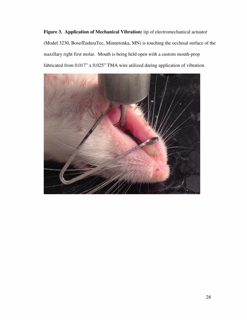
Figure 3. Application of Mechanical Vibration: tip of electromechanical actuator
(Model 3230, Bose/EnduraTec, Minnetonka, MN) is touching the occlusal surface of the
maxillary right first molar. Mouth is being held open with a custom mouth-prop
fabricated from 0.017” x 0.025” TMA wire utilized during application of vibration.
Page 34
29
Figure 4. µ-CT image: sample image from the Base Line control group that received no
orthodontic force and no application of vibration.
Figure 5. µ-CT image: sample image from the 5Hz Vibration Only control group (no
orthodontic force). This image shows that the application of vibration without an
orthodontic force will not result in any tooth movement.
Page 35
30
Figure 6. µ-CT image: sample image from the Orthodontic Force & 5Hz Vibration
Experimental group. This image shows that the application of vibration along with an
orthodontic force results in tooth movement.
Page 36
31
TABLES
Table 1. Experimental Group – Spring + 5Hz Vibration: µ-CT data showing the
amount of tooth movement and properties of surrounding bone.
Sample Name BVF
(BV/TV) Tissue Density
1st Molar Movement
Bone Volume
Spring-5Hz - 1 60.8% 1090 0.228 0.096
Spring-5Hz - 2 66.2% 1134 0.200 0.085
Spring-5Hz - 3 55.7% 1155 0.240 0.070
Spring-5Hz - 4 70.2% 1159 0.444 0.072
Spring-5Hz - 5 67.3% 1125 0.206 0.119
Spring-5Hz - 6 66.5% 1121 0.236 0.108
Average 64.4% 1130.718 0.259 0.091
SD 5.3% 25.159 0.092 0.020
Table 2. Experimental Group – Spring + 10Hz Vibration: µ-CT data showing the
amount of tooth movement and properties of surrounding bone.
Sample Name BVF
(BV/TV) Tissue Density
1st Molar Movement
Bone Volume
Spring-10Hz - 1 60.8% 1090 0.232 0.096
Spring-10Hz - 2 66.2% 1134 0.423 0.085
Spring-10Hz - 3 65.7% 1155 0.122 0.070
Spring-10Hz - 4 70.2% 1059 0.244 0.072
Spring-10Hz - 5 68.5% 1071 0.266 0.078
Average 66.3% 1101.864 0.257 0.080
SD 3.6% 41.348 0.108 0.010
Page 37
32
Table 3. Experimental Group – Spring + 20Hz Vibration: µ-CT data showing the
amount of tooth movement and properties of surrounding bone.
Sample Name BVF
(BV/TV) Tissue Density
1st Molar Movement
Bone Volume
Spring-20Hz - 1 57.6% 1122 0.130 0.061
Spring-20Hz - 2 49.3% 1131 0.120 0.048
Spring-20Hz - 3 70.9% 1136 0.370 0.121
Spring-20Hz - 4 62.6% 1112 0.170 0.091
Spring-20Hz - 5 51.3% 1129 0.331 0.081
Spring-20Hz - 6 39.7% 1074 0.151 0.060
Spring-20Hz - 7 57.0% 1122 0.166 0.075
Average 55.5% 1118.188 0.205 0.077
SD 10.0% 21.089 0.101 0.024
Table 4. Control Group – Spring Only (no vibration): µ-CT data showing the amount
of tooth movement and properties of surrounding bone.
Sample Name BVF
(BV/TV) Tissue Density
1st Molar Movement
Bone Volume
Spring-NoVib - 1 57.6% 1122 0.330 0.061
Spring-NoVib - 2 49.3% 1131 0.170 0.048
Spring-NoVib - 3 70.9% 1136 0.192 0.121
Spring-NoVib - 4 62.6% 1112 0.144 0.091
Spring-NoVib - 5 51.3% 1129 0.105 0.081
Spring-NoVib - 6 39.7% 1074 0.201 0.060
Spring-NoVib - 7 57.0% 1122 0.193 0.075
Spring-NoVib - 8 47.7% 1051 0.336 0.036
Average 54.5% 1109.822 0.209 0.072
SD 9.7% 30.678 0.083 0.027
Page 38
33
Table 5. Control Group – No Spring + No Vibration (Base Line): µ-CT data showing
the properties of surrounding bone.
Sample Name BVF
(BV/TV) Tissue Density
Bone Volume
NoSpring-NoVib - 1 84.8% 1174 0.097
NoSpring-NoVib - 2 82.5% 1136 0.109
NoSpring-NoVib - 3 75.9% 1138 0.102
NoSpring-NoVib - 4 77.8% 1130 0.100
Average 80.3% 1144.515 0.102
SD 4.1% 19.685 0.005
Table 6. Control Group – No Spring + 5Hz Vibration (no orthodontic force): µ-CT
data showing the properties of surrounding bone.
Sample Name BVF
(BV/TV) Tissue Density
Bone Volume
NoSpring-5Hz - 1 67.4% 1099 0.081
NoSpring-5Hz - 2 84.6% 1161 0.101
NoSpring-5Hz - 3 84.8% 1188 0.087
NoSpring-5Hz - 4 80.2% 1163 0.088
NoSpring-5Hz - 5 78.8% 1144 0.067
Average 79.2% 1150.964 0.085
SD 7.1% 33.168 0.012
Page 39
34
Table 7. Control Group – No Spring + 10Hz Vibration (no orthodontic force): µ-CT
data showing the properties of surrounding bone.
Sample Name BVF
(BV/TV) Tissue Density
Bone Volume
NoSpring-10Hz - 1 75.9% 1131 0.076
NoSpring-10Hz - 2 78.4% 1133 0.101
NoSpring-10Hz - 3 78.6% 1154 0.103
NoSpring-10Hz - 4 77.2% 1149 0.075
Average 77.5% 1141.961 0.089
SD 1.3% 11.427 0.015
Table 8. Control Group – No Spring + 20Hz Vibration (no orthodontic force): µ-CT
data showing the properties of surrounding bone.
Sample Name BVF
(BV/TV) Tissue Density
Bone Volume
NoSpring-20Hz - 1 76.2% 1126 0.076
NoSpring-20Hz - 2 72.2% 1146 0.068
NoSpring-20Hz - 3 76.4% 1135 0.075
NoSpring-20Hz - 4 74.0% 1107 0.072
NoSpring-20Hz - 5 79.0% 1148 0.070
Average 75.6% 1132.493 0.072
SD 2.6% 16.611 0.003
Page 40
35
REFERENCES
1. Norevall, L.I., S. Forsgren, and L. Matsson, Expression of neuropeptides
(CGRP, substance P) during and after orthodontic tooth movement in the
rat. Eur J Orthod, 1995. 17(4): p. 311-25.
2. Gameiro, G.H., et al., The influence of drugs and systemic factors on
orthodontic tooth movement. J Clin Orthod, 2007. 41(2): p. 73-8; quiz 71.
3. Tyrovola, J.B. and M.N. Spyropoulos, Effects of drugs and systemic
factors on orthodontic treatment. Quintessence Int, 2001. 32(5): p. 365-71.
4. Lee, W.C., Experimental study of the effect of prostaglandin administration
on tooth movement--with particular emphasis on the relationship to the
method of PGE1 administration. Am J Orthod Dentofacial Orthop, 1990.
98(3): p. 231-41.
5. Verna, C., M. Dalstra, and B. Melsen, The rate and the type of orthodontic
tooth movement is influenced by bone turnover in a rat model. Eur J
Orthod, 2000. 22(4): p. 343-52.
6. Collins, M.K. and P.M. Sinclair, The local use of vitamin D to increase the
rate of orthodontic tooth movement. Am J Orthod Dentofacial Orthop,
1988. 94(4): p. 278-84.
7. Yamasaki, K., Y. Shibata, and T. Fukuhara, The effect of prostaglandins
on experimental tooth movement in monkeys (Macaca fuscata). J Dent
Res, 1982. 61(12): p. 1444-6.
Page 41
36
8. Sekhavat, A.R., et al., Effect of misoprostol, a prostaglandin E1 analog, on
orthodontic tooth movement in rats. Am J Orthod Dentofacial Orthop,
2002. 122(5): p. 542-7.
9. Brudvik, P. and P. Rygh, Root resorption after local injection of
prostaglandin E2 during experimental tooth movement. Eur J Orthod,
1991. 13(4): p. 255-63.
10. Davidovitch, Z., et al., Electric currents, bone remodeling, and orthodontic
tooth movement. II. Increase in rate of tooth movement and periodontal
cyclic nucleotide levels by combined force and electric current. Am J
Orthod, 1980. 77(1): p. 33-47.
11. Davidovitch, Z., et al., Electric currents, bone remodeling, and orthodontic
tooth movement. I. The effect of electric currents on periodontal cyclic
nucleotides. Am J Orthod, 1980. 77(1): p. 14-32.
12. Darendeliler, M.A., P.M. Sinclair, and R.P. Kusy, The effects of samarium-
cobalt magnets and pulsed electromagnetic fields on tooth movement. Am
J Orthod Dentofacial Orthop, 1995. 107(6): p. 578-88.
13. Kawasaki, K. and N. Shimizu, Effects of low-energy laser irradiation on
bone remodeling during experimental tooth movement in rats. Lasers Surg
Med, 2000. 26(3): p. 282-91.
14. Darendeliler, M.A., et al., Effects of pulsed electromagnetic field vibration
on tooth movement induced by magnetic and mechanical forces: a
preliminary study. Aust Dent J, 2007. 52(4): p. 282-7.
Page 42
37
15. Nishimura, M., et al., Periodontal tissue activation by vibration: intermittent
stimulation by resonance vibration accelerates experimental tooth
movement in rats. Am J Orthod Dentofacial Orthop, 2008. 133(4): p. 572-
83.
16. Stark, T.M. and P.M. Sinclair, Effect of pulsed electromagnetic fields on
orthodontic tooth movement. Am J Orthod Dentofacial Orthop, 1987.
91(2): p. 91-104.
17. Christiansen, B.A. and M.J. Silva, The effect of varying magnitudes of
whole-body vibration on several skeletal sites in mice. Ann Biomed Eng,
2006. 34(7): p. 1149-56.
18. Rubin, C., et al., Prevention of postmenopausal bone loss by a low-
magnitude, high-frequency mechanical stimuli: a clinical trial assessing
compliance, efficacy, and safety. J Bone Miner Res, 2004. 19(3): p. 343-
51.
19. Ohmae M, S.S., Morohashi T, Qu H, Seki K Kurabayashi H,
Biomechanical acceleration of experimental tooth movement by ultrasonic
vibration in vivo: Part 1. Homo-directional application of ultrasonication to
orthodontic force. Orthod Waves, 2001. 60: p. 201 - 212.
20. Kau HC, N.J., English JD, The Clinical Evaluation of a Novel Cyclical
Force Generating device in Orthodontics Orthodontic Practice, 2011. 1(1).
21. Miles, P., et al., The effects of a vibrational appliance on tooth movement
and patient discomfort: a prospective randomised clinical trial. Aust Orthod
J, 2012. 28(2): p. 213-8.
Page 43
38
22. Kalajzic Z, P.E., Utreja A, Dyment N, Nihara J, Xu M, Chen J, Uribe F,
Wadhwa S., Effect of cyclical forces on the periodontal ligament and
alveolar bone
remodeling during orthodontic tooth movement. Angle Orthod, 2013. 2013 Aug
12. [Epub ahead of print].
23. Rygh, P., Ultrastructural changes in tension zones of rat molar
periodontium incident to orthodontic tooth movement. Am J Orthod, 1976.
70(3): p. 269-81.
24. Storey, E., The nature of tooth movement. Am J Orthod, 1973. 63(3): p.
292-314.
25. Roberts, W.E. and J.G. Chamberlain, Scanning electron microscopy of the
cellular elements of rat periodontal ligament. Arch Oral Biol, 1978. 23(7):
p. 587-9.
26. Waldo, C.M. and J.M. Rothblatt, Histologic response to tooth movement in
the laboratory rat; procedure and preliminary observations. J Dent Res,
1954. 33(4): p. 481-6.
27. Ren, Y., J.C. Maltha, and A.M. Kuijpers-Jagtman, The rat as a model for
orthodontic tooth movement--a critical review and a proposed solution. Eur
J Orthod, 2004. 26(5): p. 483-90.
28. Igarashi, K., et al., Anchorage and retentive effects of a bisphosphonate
(AHBuBP) on tooth movements in rats. Am J Orthod Dentofacial Orthop,
1994. 106(3): p. 279-89.
Page 44
39
29. Mohammed, A.H., D.N. Tatakis, and R. Dziak, Leukotrienes in orthodontic
tooth movement. Am J Orthod Dentofacial Orthop, 1989. 95(3): p. 231-7.
30. Pavlin, D., et al., Orthodontically stressed periodontium of transgenic
mouse as a model for studying mechanical response in bone: The effect
on the number of osteoblasts. Clin Orthod Res, 2000. 3(3): p. 55-66.
31. Pavlin, D., et al., Orthodontically stressed periodontium of transgenic
mouse as a model for studying mechanical response in bone: The effect
on the number of osteoblasts. Clin Orthod Res, 2000. 3(2): p. 55-66.
32. King, G.J., et al., Measuring dental drift and orthodontic tooth movement in
response to various initial forces in adult rats. Am J Orthod Dentofacial
Orthop, 1991. 99(5): p. 456-65.
33. Keeling, S.D., et al., Serum and alveolar bone phosphatase changes
reflect bone turnover during orthodontic tooth movement. Am J Orthod
Dentofacial Orthop, 1993. 103(4): p. 320-6.
34. Nixon, C.E., et al., Histomorphometric study of dental pulp during
orthodontic tooth movement. J Endod, 1993. 19(1): p. 13-6.
35. Yoshimatsu, M., et al., Experimental model of tooth movement by
orthodontic force in mice and its application to tumor necrosis factor
receptor-deficient mice. J Bone Miner Metab, 2006. 24(1): p. 20-7.
36. Alvarez, L.L. and H.G. Pardo, Guide for the care and use of laboratory
animals - Natl-Res-Council. Psicothema, 1997. 9(1): p. 232-234.
37. Lee, T.C. and D. Taylor, Bone remodelling: should we cry Wolff? Irish
Journal of Medical Science, 1999. 168(2): p. 102-5.
Page 45
40
38. Umemura, Y., et al., Five jumps per day increase bone mass and breaking
force in rats. J Bone Miner Res, 1997. 12(9): p. 1480-5.
39. Lanyon, L.E. and C.T. Rubin, Static vs dynamic loads as an influence on
bone remodelling. Journal of Biomechanics, 1984. 17(12): p. 897-905.
40. Rubin, C., et al., Quantity and quality of trabecular bone in the femur are
enhanced by a strongly anabolic, noninvasive mechanical intervention. J
Bone Miner Res, 2002. 17(2): p. 349-57.
41. Lau, E., et al., Effect of low-magnitude, high-frequency vibration on
osteocytes in the regulation of osteoclasts. Bone, 2010. 46(6): p. 1508-15.
42. Lynch, M.A., M.D. Brodt, and M.J. Silva, Skeletal effects of whole-body
vibration in adult and aged mice. J Orthop Res, 2010. 28(2): p. 241-7.