THE EFFECTS OF CULTURE MEDIA pHON FLAGELLAR MORPHOLOGY AND MOTILITY OF A Thesis Presented to The Graduate Divisi.on Drake University In Partial Fulfillment of the Requirements for the Degree Master of Arts in Biology by Sister Mary Ann Polasek August 1968
Transcript
THE EFFECTS OF CULTURE MEDIA pHON FLAGELLAR
MORPHOLOGY AND MOTILITY
OF BACILLU~ MEGATERI~
A Thesis
Presented to
The Graduate Divisi.on
Drake University
In Partial Fulfillment
of the Requirements for the Degree
Master of Arts in Biology
by
Sister Mary Ann Polasek
August 1968
J9t}/ -:-.p 70-7
I
THE EFFECTS OF
MORPHOLOGY
by
CUL/£UF,E MEDIA1!H ONW:tAG~tA!
AND 1\~Or.cILr~ OF
BACILLUS !4l!iGA,~aIUM
the Graduate DivisionDean of
TABLE OF CONTENTS
CHAPTER PAGE
I. INTRODUCTION . . . . . . . . . . . . . . . . 1
II .. SURVEY OF THE LITERATURE . • • • • " • • 10 $ 3
I. Number of Seconds Required for One Bacillus megateriwn to Traverse the 0.25 mm. Radius of a 43X Field of a Light Microscope as Observed in Hanging Drop Preparations • • • • 29
II. Mean Time and Rate of One B. me~aterium Organism in Traversing tEe O.c5 Radius of a 43X Field of a Light Microscope as Observed in Hanging Drop Preparations • • .• 32
FIGURE
1.
2.
4.
5.
6.
7.
8.
9.
10.
11.
12.
LIST OF FIGURES
PAGE
B. megaterium Cultured in Tryptic Soy Broth, pH 7.3, Stained with Leifson Flagella Stain 17
B. megaterium Cultured in Tryptic Soy Broth, pH 7.3, stained with Leifson Flagella Stain 18
B. megaterium Cultured in Tryptic Soy Broth, pH 6.0, Stained with Leifson Flagella Stain 19
lie megaterium Cultured in Tryptic Soy Broth, pH 6.0, Stained with Leifson Flagella Stain 20
~. megaterium Cultured in Tryptic Soy Broth, pH 8.5, Stained with Leifson Flagella Stain 21
B. megaterium Cultured in Tryptic Soy Broth, pH 9.0, Stained with Leifson Flagella Stain 22
B. megaterium Cultured in Tryptic soy Broth, -pH 9.5, Stained with Lefison Flagella Stain • 23
~. megaterium Cultured on Semi-Solid Motility Agar, pH 6.5, for Forty-Eight Hours 24· · · · ·
B. megateriuffi Cultured on Semi-Solid Motility -Agar, pH 7.3, for Forty-Eight Hours 25· · · · · B. ffieRaterium Cultured on Semi-Solid Motility -Agar7 pH S.5, for Forty-Eight Hours 26· · · · · B. megaterium Cultured on Semi-Solid Motility -Agar, pH g.O, for Forty-Eight Hours • 27· · Mean Time Required for One ~. megaterium Cell to Traverse the Radius of a High Power Objective Field as Observed in Hanging Drop Preparations at Varying pH Levels •• ~ . .• 30
Mean Rate of One B. megaterium in Traversing the Radius of a High Power Field as Observed in Ranging Drop Preparations at Varying pH Leve 1 s . . 0' • 0 • It- • • •• Q • • • , • • 31
CHAPTER I
INTRODUCTION
It was almost two centuries after Leeuwenhoek's
discovery of bacteria that Cohn, in 1872, first recognized
the flagella of the bacterium, Spirillum volutans as true
organelles. Since that time, using a variety of organisms
and aided by advances in technology, extensive investigations
have been carried on relative to the nature and significance
of these organelles. The size, shape and distribution of
the flagella on bacterial organisms were ascertained by
careful staining preparations, and these characteristics
have since become an important basis for bacterial identi
fication. Since flagella were found on only free-swimming
bacteria, these structures were aSbumed to be locomotory,
however, the actual physiology of the motion is still not
completely understood.
Determination of the flaBellar composition has
been made by growth of the organisms in nitrogen-free media,
subjection to heat following antibidy adsorption, paper
chromatography, X-ray diffraction studies and ultra-violet
light absorption.
These studies seem to indicate the protein nature
of the flagellar material, and hence, its susceptibility
2
to change, in varying environmental conditions such as
temperature, substrate concentration and pH.
It was the purpose of the present study to determine
the effects of hydrogen ion concentration on the morphology
of the flagella of Bacillus megaterium and to relate in
duced changes to the rate of motility.
CHAPTER II
SURVEY OF THE LITERATURE
The earliest studies of flagella were, for the
most part, concerned with general appearance and staining
reactions of the organellese Classification of bacteria
according to the position of the flagella on the soma,
their relative abundance and time of appearance from
a germinating bacterial spore were typical of the work
done at this time (1872-1930).1
Staining techniques employinB the 'use of mordants
to increase the diameter of the flagella, thus making them
more visible were developed, notably by Bailey, Gray,
Leifson and casares-Gil. 2 These methods led to obvious
distortions because of the precipitated mordant on the
flagella, although they did not necessarily affect the
wave shape characteristic of the organelle. In an attempt
to estimate the width of a flagellum, Meyer assumed that
the ratio between the stained flagellum and the stained
IGeorges Knaysi, Elements of Bacterial CltOlO~~ (Ithaca: Comstock Publishing Company, Incorporated,J51), p. 260.
r, cR. J. Conn, Mary A. Darrow, Victor M. Emmel,
Staining Procedures Used ~ The Biological Stain Commission {second edition; Baltimore: Williams and Wilkins Company, 1962), pp. 156-158
soma would be the same as for the untreated bacterium.
Subsequent studies showed that flagellar width varied
with the medium and the species and variety of the organ
ism, but that the flagella were uniform in diameter through
out their lengths. l ,2
The helicoidal nature of the flagella has been known
for some time. Recent negative contrast electron micro
scopic studies of Lowy and Hanson in 1964 have determined
that the organelle is a solid structure composed of tightly
packed globules forming subfibrils spiralling along an.
inner shaft.' Similar structural details have been re
ported by Starr and Williams,4 and by Labaw and Mosley.5
This would seem to disprove the assertion of Pijper that
~naysi, 2£. cit., p. 26,.
2Adrianus Pijper, "The Flagella of Spirillum Volutans," Journal of Bacteriology, 57:111-117, January, 1949.
'J. Lowy and Jean Hanson, "structure of Bacterial Flagella," Nature, 202:538-540, May, 1964.
4Mortimer P. starr and Robley C. Williams, "Helical Fine Structure of Flagella of a Motile Diphtheroid," Journal of Bacteriology, 63: 701-706, June. 1952.
5Claeb Weibull, "Movement" in structure (Vol. I of The Bacteria: A Treatise on structure and Function, ed. !7C. Gunsalus and Roger Stanier. New York: Academic Press, 1960) , p. 154 ..
5
the flagella are composed of fluid slime shredding from
the surface of the slime capsule, and not really entities
in themselves. l ,2
Abram, Vatter and Koffler used ghost cells and
cell membrane fragments to demonstrate that the 1'lag,ella
are attached to the cytoplasmic membrane,3 and that each
flagellum is connected to a basal granule with a hook
like crook at its distal end. 4 These more recent electron
microscopic findings settled the controversial issue of
flagellar origin that had been raging since the first
bacterial cytologists propounded the theory that these
organelles were simply cytoplasmic protrusions through
5cell wall pores.
Ip' . . t 117lJper, 2£. ~., p. •
2 , "Evidence That Amputa.tion of . Bacterial Flagella Does Not Affect Motility," Science, 109: 379-380, April, 1949
3Dinah Abram, A. E. Vatter, and Henry Koffler, "Attachment and structural Features of Flagella of Certain Bacilli," Journal £f. Bacteriology, 91:2045-2068, May, 1966.
4 , "Basal structures and Attachment of Flagella--'-i-n---""C-u-=-l~t-u-r-e-s of Proteus vulgaris, fI Journal of Bacteriology, 90: 1357-1354, November, 19b5.
5Knaysi, £E. cit., p. 265·
6
One of the most important tools in bacterial cytol
ogy in recent years has been the electron microscope.
Pijper's concept of the formation of fla.gella as a result
of motion was shown to be erroneous by Hillier, Knaysi
and Baker, whose colloidal film technique proved that the
flagella were present on the cell aesoon as the inner
spore coat split. l The electron microscope has been used
to determine the nature of some of the ultrastructures of
the bacterial cell. One of the most frequently used tech
niques involves the lysing of the cell by use of lysozyme
or some other enzyme, negative staining with potassium
phosphotungstate or uranyl acetate, and shadow cast prep
arations of ghost cells. 2 Ordinary light microscopy is
usually unsatisfactory for flagellar work with unstained
organisms, but moderate success has been achieved by use
of the dark field in motility studies. The phase contrast
microscope helped to resolve some of the refringence dif
ficulties encountered with the light microscope.
IJames Hillier, Georges Knaysi, and Richard F. Baker, "New Preparation Techniques for the Electron Microscopy of Bacteria,rt Journal of Bacterioloq;y, 56: 569-576, November, 1948.
2Claes Weibull, "Isolation of Protoplasts From Bacillus megaterium by Controlled Treatment With Lysozyme," JournaI 2! Bacteriology, 66:688-695, December, 1953.
7 Antibody absorption by flagella 'results in a
thickening and stiffening of the structures with an ag
glutination effect. The flagellar antigens, specific for
particular bacteria, are referred to as H-antigens. These
are an important menas of classification and give a hint
to the protein nature of the flagella. Pijper observed
the grouping phenomenon in 1938, confirmed by Mudd and
Anderson in 1941. Tomcsik has been one of the leading
workers in this 'area. He found that flagellar antigen
icity is lost in boiled cell preparations~ but retained
in flagells detached from the cell by mechanical agitation.
Even highly dilute antisera are capable of immobilization
of flagella attached to the cell. l
Kerridge showed that bacteria mechanically defla
gellated by centrifugation were able to regenerate flagella
if incubated in optimum conditiqns. Bacteria held at an
acid pH spontaneously deflagellated, the flagella subse
quently disintegrated and lost their characteristic
appearance as determined by electron microscopy, and could
lvleibull, "Movement" in Structure (Vol. I of The Bacteria: A Treatise on StructurEl and Function) 2l?- oit., p. 156.
8
no longer be precipitated by centrifugation forces
used for normal flagella. l
Bacterial flagella consist of homogeneous aggre
ga.tes of protein molecules termed Ilflagellin. 1l2 X-ray
diffraction studies of chemical components of flagella of
some species show that the flagellin of certain strains of
Bacillus subtilis and Proteus vulgaris belongs to the
keratin-myosin-epidermin-fibrinogen group of fibrous pro
teins.' Qualitative and quantitative analyses of flagellar
constituents showed variation from species to species with
protein content estimates to 99%. Investigations of Wei
bull and Koffler and his ~sHociates showed a much higher
percentage of nitrogen (15-l6.?~) than phosphorus (O.O~~).4
Other flagellar components found were: carbohydrates ~ O.~~),
lipids (0.8%), nucleic acids «0.1%) and ash (0.0005%).
lEo F. Gale, Synthesi,s and Organization in ~ , Bacterial Cell, (New York: JOlill Wiley and Sons, Incorporatea,1959), pp.-,g=4l.
2Terrence M. Joys and Ruth W. Frankel, "Genetic Control of Flagellation in Bacillus subtilis," Journal of Bacteriology, 94: 32-37, July, 1967.
'Weibull, "Movement" in Structure (Vol. I of The Bacteria: A Treatise on structure and Function), £2. cit., p. 1$6. - -
4H. Koffler, T. Kobayaski, E. Mallet, "CysteineCystine Content and the Free Amino A.cid Groups of Flagellin," Archives of Biochemistry and Biophysics, 64:509, October, 'Dj56.
9
Most of the amino acids except the heterocyclic
compounds, tryptophan, histidine, proline and hydroxy
proline were found. The molecular weight of the fl_
gellar subunits was estimated at about 20,000. 1
The work of Hoeniger in 1965 demonstrated that
there was a direct relation between the general morpho
logy of the flagella. of Proteus mirabilis and the pH at
which the organisms were cultured. Her research also
indicated some differences with respect to motility as
a result of this morphogenesis. 2
In view of their proteinaceous composition, it is
reasonable to hypothesize that the flagellar material
would react to environmental alterations much as a pure
protein would, and so be directly affected by the pH of
the culture medium, either by denaturation of the con
stituent proteins, or in some other more subtle way.
Although much has already been done with the fla
gella, much still remains. The physics of locomotion is
still unknown, and the influence of nuclear elements on
flagellar production remains obscure.
lR. Koffler, T. Kobayaski, G. E. Mallet, 2£. cit., p. 509.
2Judith Hoenig,er, "Influence of pH on Proteus Flagella," Journal of BacterioloBY, 90: 275-277, July, 1965.
CHAPTER III
MATERIALS AND METHODS
The organism used in this investigation was a
strain of Bacillus megaterium maintained in the micro
biology laboratory at Drake University. This bacteria
culture did not differ appreciably from the standard
description as given in Bergey's Manual of Determinative
Bacteriology, 7th Edition. For staining and hanging
drop preparations used in this study, the bacteria were
cultured in 16 mm. tubes of tryptic soy broth (Difco)
at 350 C for 24-48 hours. The pH of the culture medium
was adjusted by dropwise addition of 0.1 N NaOH or dilute
Hel within a range of 6.0 to 9.5.
Slides to be used for staining bacterial smears
were allowed to soak in a 5:1 mixture of concentrated
sulfuric acid and distilled water for a period exceeding
twenty-four hours. They were then drained, rinsed for
approximately one minute in distilled water, 95% ethanol
and flamed. When the slides were cool, they were streaked
by applying a loopful of the bacterial inoculum to their
tilted surfaces, alloWing the media to run the length of
the slides. The slides were then air dried for a minimum
of two hours and stained, using a modified Swatek adaptation
of the Leifson flagella stain.
11
Laifson Flagella Stainl
Swatek Adaptation
Solution I
KA1( S04!2· 12 H20 (sat. aqueous sol'n.) 20 ml.
Tannic Acid (20% aqueous) 10 ml.
Distilled Water 10 mI.
Solution II
Ethyl Alcohol (9~~) 15 ml.
Basic Fuchsin (sat. in 95% ethanol) 3 ml.
Solutions I and II were stored separately in a refrigerator
at approximately 50 C, mixed and triple-filtered just
prior to use. Approximately 0.5 mI. of stain was flooded
over the surface of the dried smear and allowed to remain
undisturbed for approximately yo seconds, rinsed in a
gentle stream of tap water, air dried, and observed using
the 97X oil immersion objective and a lOX ocular (Bausch
and Lomb Model STA.).
Semi-solid agar (O.~ Difco Bacto-Agar in tryptic
soy) was used for cultivation of the organism to determine
its relative motility under differing pH conditions. The
Eklund and Lankford adaptation of the motility medium was
adjusted for pH by the addition of NaOH or dilute HCI as
1Frank E. Swatek, Textbook of Microbiolo~v, (Saint Louis, Missouri: C. V. MosbY-Company, 19o~), p.?l.
12
above, and incubated for forty-eight hours at 300 C. Pyrex
brand 10 mID. petri dishes were used for this culture. The
path of the bacilli through the medium was marked by a pink
halo due to the reduction of 2,3,5-triphenyltetrazolium by
the growing bacteria.
Semi-solid Motility Mediuml
Eklund-Lankford Adaptation
Nutrient broth (Difeo tryptic soy) 100 mI.
Agar (DifcD Bacto-Agar) 0.5 %
2,3,5-triphenyltetrazolium chloride 0.001 %
Photographs of both the stained bacilli and the
petri plate cultures were made by Mr. Anthony M. Kuzma,
professional medical photographer at Marquette University
School of Medicine in Milwaukee, Wisconsin, using a Leitz
Panphot with carbon arc light source. A 90X oil immersion
objective with Apochromat lens for color correction and a
lOX ocular were used, giVing a total magnification of
1500X for the organisms, while the macro-setup for the
petri plates required a Microtessar lens.
The relative motility of the living bacteria was
stUdied by preparing standard hanging drop preparations
and observed using the 43X high power objective and a lOX
ocular. The approximate time observed for a single bacterium
Eklund and Lankford, Laboratorl Manual for General Microbiologx (Englewood Cliffs, New Jersey: Prentice HaIl, Incorporated, 1967), p. 278.
13 to traverse the radius of the high power field (0.25 mm.)
was determined by actual st'opwatch-clocking. Results were
tabulated and plotted on a graph. In all cases except
one, fifty such clockings were made. Due to the paucity
of organisms at pH 9.5, and the fact that they were barely
motile t it was possible to make only a limited number of
readings. All cultures studied were twenty-four populations.
CHAPTER IV
RESULTS AND INTERPRETATION OF DATA
This study was designed to show that the pH of the
culture medium influences the motility rate of Bacillus
megaterium and also to show possible morphological alter
ations in the flagellar pattern typical of this organism
when cultured under ideal conditions.
In order to study the change in flagellar structure,
Swatekes adaptation of the Leifson flagella stain was used
on bacterial smears made from cultures grown at differing
pH levels. Photomicrographs of these organisms comprise
the data presented for comparison.
Motility studies were conducted in two ways. One
method involved growth of ~. megaterium through semi-solid
motility medium adjusted to the desired pH by the addition
of NaOH or Hel. Photographs showing the relative growth
distributions on these plated media were then taken.
A second method utilized direct observations of
individual bacteria traversing the 0.25 mm. radius of the
high power field of a light microscope (43X) with a lOX
ocular, and the tabulation of approximate excursion times.
The relative rates of the organisms cultured at different pH
levels were compared.
15
In each case, ~. megaterium Brown at pH 7.3 were
considered the norm against which similar organisms raised
at differing pH's were compareQ.
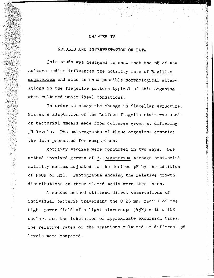
Figures 1 and 2 show~. megaterium cultured in
tryptic soy broth (Difco), at pH 7.3. The flagella of
these organisms were long and kinky, averaging five to
six waves each.
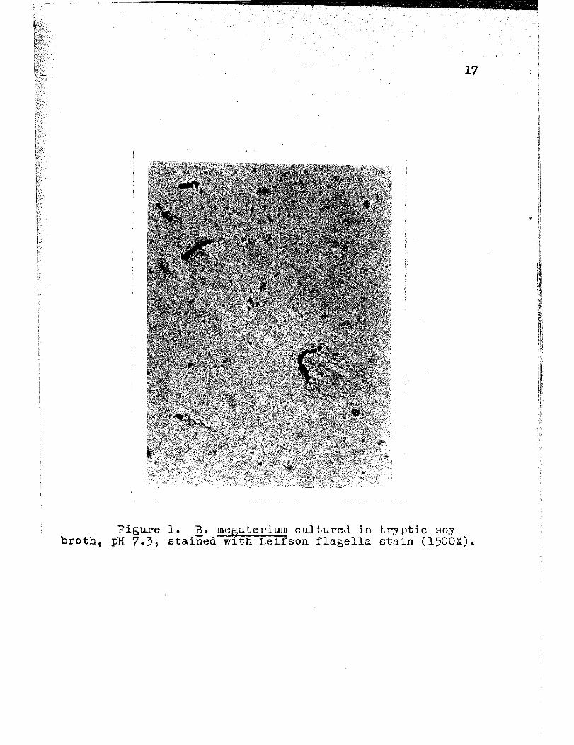
Bacteria cultured at pH 6.0 bore flagella that in
most cases were shorter than those of the organisms raised
in, the reference medium (pH 7.3), and irregularities in
the amplitude and length of the waves were shown. Instead
of the regular flection amplitude and wave length patterns
characteristic of the flagella of the bacilli cultured a~
pH 7.3, the flagella of the organisms grown in the more
acid medium deviated considerably from them, having both
deeper and more shallow waves, as well as a greater vari
ation in distallce from one wave crest to the next.
At pH 8.5, the flagella of the organisms were sli~htly
reduced in length with the kinks less abberent than those of
the bacilli grown at pH 6.0. Figure 5 is a photomicro
graph of a stained smear of ~. megaterium on which a
sllBht tendency toward straiehteninB of the flagella can
be noted when compared with these organelles on bacteria
cultured at pH 7.3 (Figures 1 and 2).
16
The flagella of the bacteria cultured in the
medium of pH 9.0 showed an increased tendency toward
straightening over that noted in the medium of pH 8.5.
At pH 9.0 there is a conspicuous lack of the otherwise
characteristically kinky normal flagella, and they were,
in fact, almost straight. Figure 6 i.s a microphotograph
of !!. megaterium cultured at pH 9.0 and prepared with
Leifson flagella stain.
Figure 7 represents bacilli grown at pH 9.5
and shows se'(eral well-defined bacteria, but there is
no evidence of flagella.
Assuming that bacterial flagella are locomotory
organelles, and that the morphology of these structures
is altered by a change in the pH of the culture medium,
it was deemed reasonable to relate the pH of the medium
with the relative rate of motility of the organism. One
seirn-quantitative estimate of the relative motility of
B. megaterium was the use of semi-solid motility medium
(o.~ Difco Bacto-Agar in tryptic soy broth) through which
the bacilli are able to move. Figures 8 through 11
show the relative growth of the organism in the O.5Pk
agar medium adjusted to varying pH levels and incubated
for forty-eight hours. The bacteria Brew well at all levels
of pH within the range tested.
17
Figure 1. B. megaterium cultured in tryptic soy broth t pH 7.3, stained with Leifson flagella stain (l500X).
18
Figure 2. B. megaterium cultured in tryptic soy broth, pH 7.3, stained with Leifson flagella stain (1500X)v
19
Figure 3. B. megaterilm cultured in tryptic soy broth, pH 6.0, stained with Leifson Flagella stain (1500X).
____------~-.....,.....-------.u••_ 20
,.
Figure 4. B. megaterium cultured in tryptic soy broth t
pH 6.0, stained with Leifson flagella stain (1500X).
21
Figure 5. ~. megaterium cultured in tryptic soy broth, pH 8.5, stained with Leifson flagella stain (1500X).
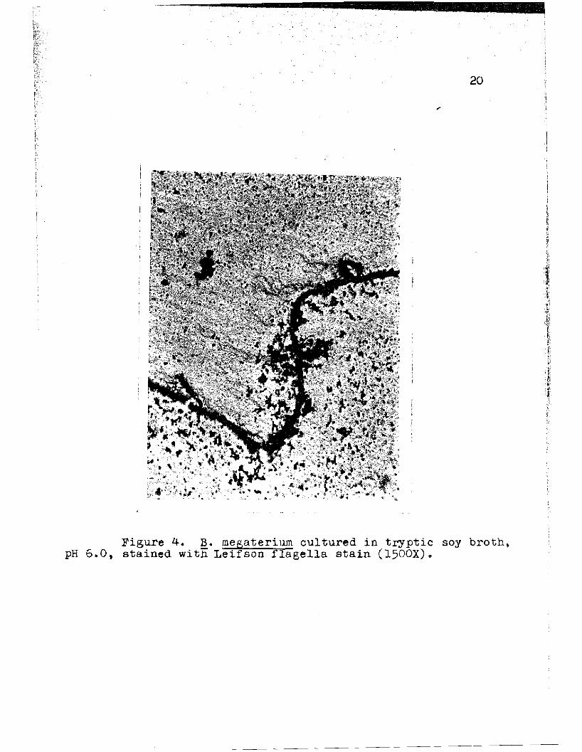
Figure 12. Mean time for one 12.. megaterium cell to traverse the radius of a high power objective as observed in hanging drop preparations at varying pH levels.
-------------.~
31
4
t I3
RATE
(10-2mm/sec.) 2
1
r)7.5 8.0 o. 5 9.5
pH LEVELS
Figure 13. Mean rate of one ~. megateriuID cell in traversing the radius of a high power field as observed in hanging drop preparations at varying pH levels.
·~---:----------·il
32
Table II presents a summary of the dat . 1 d'a, lnc u lng
a t-score to test the significance of th d'ffe 1 erences in
rates.
TABLE II
Mean Time ~nd Rate of One~. megaterium Organism' Traverslnl? the ? 25 mm. Radius of a 4-3X Field :Ln
of a L1Bht Mlcroscope as Observed in ~ Hanging Drop Preparation ~
Mean Mean pH time
( sec. ) rate 2
(x 10t-score
mrn/sec)
6.5 12.29 3.93 2.03 15·17
7.3 8.33 2.74 3.00 8.68
8.5 6·57 1.55 3.78 12.25
9.0 9.17 2.52 2.71 2736.60
9.5 146.0 18.14 0.17
Graphic representation of the datta summary is
shown on Figures 12 and 13. As the pH of the medium in
creases from pH 6.5 to pH 8.5, the point corresponding to
the lowest mean time, there is almost a two-fold decrease
in the mean time observed for the radial excursion of a
bacterium. Fom pH 8.5 to pH 9.0 there is a moderate in
crease in the observed time, and by pH 9.5, the lowest mean
time is increased by a factor of 20. Graphically represented,
the least mean time observed at pH 8.5, is the lowest point
on Figure 12, and the greatest corresponding mean rate as
shown on Figure 13.
~~----------·#I
33
Because of their protein nature, it is reasonable
to assume that the flagella of Bacillus megaterium would,
in some way, be affected by a change in the hydrogen ion
concentration of the culture medium. This study has Shown
that although the organism itself can tolerate a wide pH
apectrum, the locomotory organelles exhibit morphological
variation through the pH ran~e of this study. It will be
noted from the photographs (Figures 5 and 6), that Bacillus
megateriuffi cultured at pH 8.5 possess flasella shorter than
those of bacteria raised at pH 9.0. The organisms cultured
at pH 8.5, however, are more actively motile. This would
indicate that tnere is an optimum flagellar length-speed
ratio. At the alkaline levels where the flagella are
long and tend to mat, as indicated by the formation of a
thick pellicle (pH 9.0) in broth, it is possible that this
decrease in motility is due to a simple mechanical entangle
ment of the elongate flagella. Bacteria cultured at pH
6.0 possessed flagella comparable in length to those
raised at pH 8.5, but the organisms raised in the acid me
dium were markedly less motile. It could be hypothesized
that some chemical interference is involved.
~---------·iI
More significant than length, is the variability
of degree of flection exhibited by the flagella. There is
a trend toward decreased kinkiness as the medium becomes
less acid. At the point where this straightening is most
noticeable, pH 9.0, motility is appreciably diminished,
indicating that there is an optimum flagellar wave ampli
tude-speed ratio as well. Increasing the pH to 9.5 re
suIts in a near total immobilization of the bacteria and
an apparent loss of flagella. Electron microscope studies
by Hoeniger with Proteus mirabilis showed similar results. l
Those b~cilli cultured at pH 9.5 appear to have
lost their flagella completely (Figure 7). Two factors
could have been operdtive to explain this phenomenon:
(1) the alkaline medium may have so weakened the flagella
that they fractured upon contact with the slide, however
there are no free flagella apparent to give visual evi
dence of this possibility; (2) the basic dye (basic
fuchsin) used in the Leifson staining method would have
less affinity for the bacteria at an alkaline level, al
though the bacteria that did take up the stain were suf
ficiently dark.
lJudith Roeniger, Influence of DR on Proteus Flagella," Journal of Bac teriology, 90: 275-:n7, July, 1965.
·~---------·iI
35
Growth patterns in broth varied with the change
in hydrogen ion concentration of the media. Both extremes
of the pH spectrum used in this stUdy supported only sparse
populations which, in both cases t were restricted to a
slight turbidity at the bottom of the tUbes. Organisms
grown in the range between pH 6.5 and 7.5 were uniformly
dispersed throughout the media, and produced a moderate
turbidity in each case. At pH 9.0, however, there was a
sharp decline in the optical density of the culture, and
a heavy pellicle formed within twenty-four hours. In all
~ase8, the turbidimetric estimates were done without instru
montation, but were con~1dered definitive enough that more
refined measures were deemed unnecessary.
Motility studies that were conducted in two ways,
growth of ~. megateriuffi through semi-solid motility agar
and direct microscopic observation of living bacilli in
hanging drop preparations, showed that motility was great
est at pH 8.5 and least at both extremes of the pH range
considered.
A statistical comparison of the rates of motility
swrunarized in Table II and depicted graphically on Figures
12 and 13, indicate that these rates vary significantly
when comparing hydrogen ion concentrations studied and are
within the one per cent level of significance as determined
by the t-score.
36
From the foregoing studies, it is quite apparent
that there is a.direct relation between the motility of
Bacillus megateriuID and the hydrogen ion concentration of
the medium in which the organisms are cultured. It is also
quite clear that there is some morphological change effected
by such pH variation. This study is, by no means conclu
sive, but only suggestive of a number of avenues of investi
gation that could be followed employin~ different tech
niques and methods with other organisms.
CH~PTER V
SUMMARY
1. This study was designed to show that the pH
of the culture medium influences the motility rate of
Bacillus megaterium and also to show possible morpho
logical alteration in the flagellar pattern typical
of the organism cultured under ideal conditions.
2. The organism was cultured in tryptic soy
broth and in semi-solid motility agar medium adjusted
to varying pH levels by the addition of NaOH or HGI.
3. Leifson flagella stain preparations were made
from smears of the broth cultures and examined for any
chanBe in flagellar morphology.
4. Observation showed that there is an increased
straightening of the flagella at pH 9.0.
5. Motility rates can be compared by cultivation
of B. megaterium in semi-solid agar at varying pH's and
by hanging drop observations.
6. The bacteria having greatest motility will
travel the farthest from the line of inoculation in a
forty-eight hour period when raised in semi-solid motility
agar. Observation showed that, by this criterion, bacteria
cultured at pH 8.5 were the most motile.
38
7· stopwatch timing of bacteria in hanging drop
preparations in culture media of various pH values as
they traversed the radius of a high power field were
made. Organisms grown at pH 8.5 showed the greatest
rate of mpvement.
8. The pH of the growth medium affects both the
morphology and the motility of Bacillus megateriuffi by
altering the wave amplitude of their locomotory organ
elles, the flagella.
BIBLIOGRf>.PHY·
BIBLIOGRAPHY
A. BOOKS
conn, H. J., Mary A. Darrow,. Victor M. Emmel. Stainin proc~dure8 Uls~d & the Blo~ogical Stain CommisBion~ Baltlffiore: Wlillams and Wllkins Company, 1962.
DeRobertis, E., W. W. Nowinski, F. Saez. General C~toIOgy. Philadelphia: w. B. Saunders Company1 63. '
Eklund and Lankford. Laboratorl Manual For General Microbiology. Englewood Cliffs, New Jersey: Prentice Hall, Incorporated, 1967.
Engstrom, Arne and J. B. Fincan. Biological Ultrastructure. New York: Academic Press, 1958.
Gale, E. F. Synthesis and Organization in the Bacterial Cell. New York: John Wiley and Sons, Incorporated,1959.
Gebhardt, L. P. and D. A. Anderson. Microbiology. Saint Louis: C. V. Mosby Company, 1965.
Knaysi, Georges. Elements of Bacterial Cytology. Ithaca, New York: Com~tock Publishing Company, Incorporated, 1951,
Leifson, Einar. Atlas of Bacterial Flagellation. . New York: Acaa:emicPress; 1960.
MCE;Lroy: William D. Cellular Physiologr and Biochemistry. Englewood Cliffs, New Jersey: Prentice Hall Incorpora ted, 1961.
Swatek, Frank E. Textbook of Microbiology. Saint Louis: C. V. Mosby Company, 1967.
Weibull, Claes. "Movement" in structure. Vol. I of The Bacteria: A Treatise on Structure ~ Function. Edited by I. c7 Gunsalus and Roger Stanier. 5 vols. New York: Academic Press, 1960.
~------iII
41
B. PERIODICALS
Abram, Dinah aJ?-d He~ry Koffler. "In-Vitro Formation of Flagella-Llke Fl1aments and Other Structure F
"J .~ 1 8 rom1 11 .F age In, ournd. £f Molecular Biol0&.Y. 9'168-85July, 1964.. ,. ,
---""'1'"' A. E. Vatter, and Henry Koffler-. "Basal Structure and At~achment of Flagella in Cultures of Proteus vulga rls," Journal of Bacterio1oBl, 90: 1337-54 November, 1965. '
"A ttachment and Structural Features of Flagella of Certain Bacilli," Journal of Bacteriology, 91:2045-68, May, 1966.
Ambler, R. P. and M. W. Rees. "e-N-Methyl-Lysine in Bacterial Flagellar Protein," Nature, 184:56-7, July, 1964.
Hillier, James, Georges Knaysi and Richard F. Baker. "New Preparation Technique for the Electron Microscopy of Bac teria," . Journal of Bacteriology, 56: 569-76, November, 1948.
Roeniger, Judith. "Influence of pH on Proteus Flagella," Journal of Bacteriology, 90:275-7, July, 1965.
JoyS, Terence M. and Ruth W. Frankel. "Genetic Control of FlaGellation in Bacillus subtilis," Journal of Bacteriology, 94:32-7, July, 1967.
Koffler, H., T. Kobayaski, E. Mallet. "Cysteine-Cystine Content and the Free Amino Acid Groups of Flagellin," Archives of Biochemistry and BiorIl.ysics, 64:509, October, 19"56.
Leifson, Einar. nStaining, Shape arid Arrangement of Bacterial FlaGella," Journal of Bacteriologl, 62: 377-e9, October, 19~i.
Lowy, J. and Jean Hanson. ~IStructure of Bacterial Flage lla, Nature, 202: 538-40, May, 1964.II
, 42
Martinez, R. J. and E. Z. Gardee. "Formation of Bacterial Flagella," Journal of Bacteriolog~, . 91:870-875, February, 1966:
pijper, Adrianus. "T~e Flagella of Spirillum volutans," Journal of Bacterlology, 57:111-117, January, 1949.
• "Evidence That Amputation of Bacterial Flagella--D=--o-es Not Affect Motility," Science, 109:379-80,
April, 1949.
starr, Mortimer P. and Robley C. Williams. "Helical Fine structure of Flagella ofa Motile Diphtheroid," Journal of Bacteriology~ 63:701-6, June, 1952.
Weibull, Claes. "Isolation of Protoplasts From Bacillus megaterium Wi th Lysozyme," Journal of Bacteriology,bb:688-95, December, 1953.
C. UNPUBLISHED 1~TERIAL
Imogene Sister Mary. "The Leifson Flagella Stain in the' Hospi tal Laboratory." Unpublished Doctoral Dissertation, Fordham University, New York, 1958.


![Torque Generated by Flagellar Motorof Escherichia - DAMTP · TorqueGenerated bythe Flagellar Motorof Escherichiacoil ... TES,N-tris[hydroxymethyl]methyl-2-aminoethanesulfonic acid.](https://static.documents.pub/doc/80x56/5c90c4f509d3f2c8148bd888/torque-generated-by-flagellar-motorof-escherichia-torquegenerated-bythe-flagellar.jpg)


