The effects of surgical treatment withchondroblastoma in children andadolescents in open epiphyseal plate oflong bonesYan Xiong†, Yun Lang†, Zeping Yu, Hongyuan Liu, Xiang Fang, Chongqi Tu and Hong Duan*
Abstract
Background: Chondroblastoma is a rare benign cartilaginous tumor, which primarily occurs in children and adolescents.Chondroblastoma commonly originates in the epiphyseal plate of long bones. An aggressive curettage treatmentis recommended to manage lesion, which may jeopardize an open epiphyseal plate and result in limb shorteningand deformity as the limb grows and develops. The purpose is to observe surgical effects of chondroblastoma onopen epiphyseal plate of long bones in children and adolescents and explore influences on limb growthand development.
Methods: We retrospectively reviewed 18 cases of long bone chondroblastoma with open epiphyseal growth plateduring March 2004 to October 2010 in our center. Seven females and 11 males with mean age of 11.6 ± 2.0 years old(8–15 years) were included. Patients, who suffered from trauma and pathological fracture of the epiphyseal plate orcongenital diseases such as poliomyelitis, congenital dementia, and cartilage malnutrition, were excluded. All patientswere treated with meticulous intralesional curettage and inactivity with alcohol followed by bone grafts. All cases werefollowed up 8.2 ± 1.7 years (5–11.5 years).
Results: All had no local recurrence and distance metastasis. The length of the affected limb was short, 18.47 ± 7.22 mm(1.5–30 mm). There was no obvious relativity with tumor activity (P = 0.061). Meanwhile, there were obvious relativity withthe greatest dimension of the lesion (TGD) (P = 0.003), the vertical dimension between edge of lesion and epiphyseal line(TVD) (P = 0.010), and area ratio of lesion to local epiphysis (lesion/growth plate) (P = 0.015). The MSTS93 (RevisedMusculoskeletal Tumor Society Rating Scale 93) and SF-36 (Medical Outcomes Study 36-Item Short-Form HealthSurvey) had been significantly improved (P < 0.01).
Conclusion: Managing of chondroblastoma located in open epiphyseal plate of a long bone with meticulouscurettage, inactivity, and bone grafts can control tumor progression and recurrence effectively. Meanwhile, earlydetection and prompt surgical treatment intervention, which reduced significantly the tumor to influence limbgrowth and development, get encouraging limb function.
Trial registration: This is a retrospective study, which was not registered in any trial registry.
Keywords: Chondroblastoma, Epiphysis, Open epiphyseal plate, Limb-length, Children and adolescents
* Correspondence: [email protected]†Equal contributorsDepartment of Orthopedics, West China Hospital, Sichuan University, No 37Guo Xue Lane, Wuhou District, 610041 Chengdu, Sichuan, People’s Republicof China
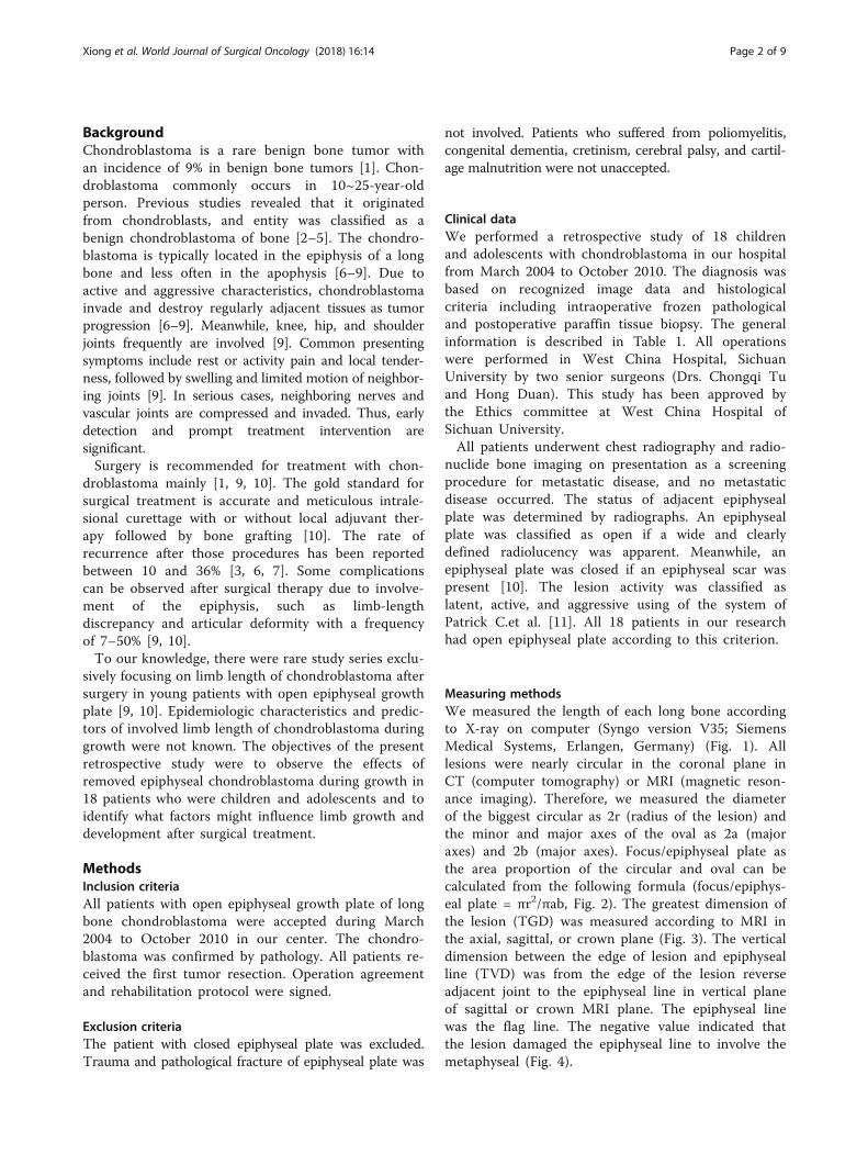
BackgroundChondroblastoma is a rare benign bone tumor withan incidence of 9% in benign bone tumors [1]. Chon-droblastoma commonly occurs in 10~25-year-oldperson. Previous studies revealed that it originatedfrom chondroblasts, and entity was classified as abenign chondroblastoma of bone [2–5]. The chondro-blastoma is typically located in the epiphysis of a longbone and less often in the apophysis [6–9]. Due toactive and aggressive characteristics, chondroblastomainvade and destroy regularly adjacent tissues as tumorprogression [6–9]. Meanwhile, knee, hip, and shoulderjoints frequently are involved [9]. Common presentingsymptoms include rest or activity pain and local tender-ness, followed by swelling and limited motion of neighbor-ing joints [9]. In serious cases, neighboring nerves andvascular joints are compressed and invaded. Thus, earlydetection and prompt treatment intervention aresignificant.Surgery is recommended for treatment with chon-
droblastoma mainly [1, 9, 10]. The gold standard forsurgical treatment is accurate and meticulous intrale-sional curettage with or without local adjuvant ther-apy followed by bone grafting [10]. The rate ofrecurrence after those procedures has been reportedbetween 10 and 36% [3, 6, 7]. Some complicationscan be observed after surgical therapy due to involve-ment of the epiphysis, such as limb-lengthdiscrepancy and articular deformity with a frequencyof 7–50% [9, 10].To our knowledge, there were rare study series exclu-
sively focusing on limb length of chondroblastoma aftersurgery in young patients with open epiphyseal growthplate [9, 10]. Epidemiologic characteristics and predic-tors of involved limb length of chondroblastoma duringgrowth were not known. The objectives of the presentretrospective study were to observe the effects ofremoved epiphyseal chondroblastoma during growth in18 patients who were children and adolescents and toidentify what factors might influence limb growth anddevelopment after surgical treatment.
MethodsInclusion criteriaAll patients with open epiphyseal growth plate of longbone chondroblastoma were accepted during March2004 to October 2010 in our center. The chondro-blastoma was confirmed by pathology. All patients re-ceived the first tumor resection. Operation agreementand rehabilitation protocol were signed.
Exclusion criteriaThe patient with closed epiphyseal plate was excluded.Trauma and pathological fracture of epiphyseal plate was
not involved. Patients who suffered from poliomyelitis,congenital dementia, cretinism, cerebral palsy, and cartil-age malnutrition were not unaccepted.
Clinical dataWe performed a retrospective study of 18 childrenand adolescents with chondroblastoma in our hospitalfrom March 2004 to October 2010. The diagnosis wasbased on recognized image data and histologicalcriteria including intraoperative frozen pathologicaland postoperative paraffin tissue biopsy. The generalinformation is described in Table 1. All operationswere performed in West China Hospital, SichuanUniversity by two senior surgeons (Drs. Chongqi Tuand Hong Duan). This study has been approved bythe Ethics committee at West China Hospital ofSichuan University.All patients underwent chest radiography and radio-
nuclide bone imaging on presentation as a screeningprocedure for metastatic disease, and no metastaticdisease occurred. The status of adjacent epiphysealplate was determined by radiographs. An epiphysealplate was classified as open if a wide and clearlydefined radiolucency was apparent. Meanwhile, anepiphyseal plate was closed if an epiphyseal scar waspresent [10]. The lesion activity was classified aslatent, active, and aggressive using of the system ofPatrick C.et al. [11]. All 18 patients in our researchhad open epiphyseal plate according to this criterion.
Measuring methodsWe measured the length of each long bone accordingto X-ray on computer (Syngo version V35; SiemensMedical Systems, Erlangen, Germany) (Fig. 1). Alllesions were nearly circular in the coronal plane inCT (computer tomography) or MRI (magnetic reson-ance imaging). Therefore, we measured the diameterof the biggest circular as 2r (radius of the lesion) andthe minor and major axes of the oval as 2a (majoraxes) and 2b (major axes). Focus/epiphyseal plate asthe area proportion of the circular and oval can becalculated from the following formula (focus/epiphys-eal plate = πr2/πab, Fig. 2). The greatest dimension ofthe lesion (TGD) was measured according to MRI inthe axial, sagittal, or crown plane (Fig. 3). The verticaldimension between the edge of lesion and epiphysealline (TVD) was from the edge of the lesion reverseadjacent joint to the epiphyseal line in vertical planeof sagittal or crown MRI plane. The epiphyseal linewas the flag line. The negative value indicated thatthe lesion damaged the epiphyseal line to involve themetaphyseal (Fig. 4).
Xiong et al. World Journal of Surgical Oncology (2018) 16:14 Page 2 of 9
Fig. 1 a–c The method of measuring length of the bone (the arrow straight line shows), humerus, from the humeral head midpoint to themidpoint of the medial and lateral condyle. The femur, from the midpoint of the femoral head to the midpoint of the medial and lateral condyle.The tibia, from the tibial plateau midpoint to within the lateral midpoint of the distal tibia
Table 1 Details of 18 patients
No Age (years) Sex Location Tumor activity Tumor size (mm3) TGD (mm) TVD (mm) Focus/growth plate Follow-up (years) Shorten (mm)
1 11 F Proximal tibia Active 35 × 50 × 50 50 − 20 25/50(50%) 9 30
The negative value indicates that the lesion involves epiphyseal plate; focus/growth plate (%), the largest area of the focus/lesion epiphyseal plate areaAbbreviations: TGD the greatest dimension of lesion, TVD the vertical dimension between edge of lesion and epiphyseal line
Xiong et al. World Journal of Surgical Oncology (2018) 16:14 Page 3 of 9
Surgical therapyFirstly, all patients have undergone meticulous intrale-sional curettage from the local cortical window andpolished tumor cavity around the lesion edge,2–5 mm by bone drill (2 mm of latent lesion, 3 mmof active, 5 mm of aggressive, Table 1). Secondly,tumor cavity was inactivated by 95% alcohol for15 min. Then, we used an electrotome to burn cavityin difficult-to-reach areas and saline solution to washcavity repeatedly. Bone grafting was performed asfollows. Allograft implantation was used in 13patients, autologous iliac bone in 2 patients, and arti-ficial bone (Medtronic, Inc., USA) in 5 patients. Withthe final processing, allogeneic bone block coveredthe cortical window, fixed with absorbable screw inseven patients and steel plate in a boy. There was noen bloc resection in this group. Patients were encour-aged to perform a rehabilitation exercise with noweight bearing on the second day following surgery,
such as joint mobilization and muscle strength train-ing. In the sixth week, patients started partial weightbearing and full-weight bearing at the third monthfollowing surgery. At 6 month after surgery, allpatients qualified for social work and sports activity.
Follow-up observationsThe follow-up occurred at 1, 2, 3, 6, 9, 12, and every6 months thereafter. Imaging studies were focused ontumor recurrence and lesion limb growth and devel-opment. Local recurrence of the tumor was suspectedif patient had persistent pain after surgery. We wouldcarry out examination or MRI to exclude the painfrom meniscus, cartilage, or soft tissue. Enlargementof the tumor on imaging studies, or bone marrowedema and cortical destruction on MRI, were thoughtto be signs of recurrence. Every 6 months after
Fig. 3 TGD as the greatest dimension of the lesion was measured according to MRI in the crown, axial, or sagittal plane
Fig. 4 TVD, the vertical dimension between the edge of lesion andepiphyseal line, was from the lower edge of the lesion to theepiphyseal line in the vertical plane of the sagittal or crown MRIplane. TVD is a negative data when the lesion is crossing theepiphyseal line
Fig. 2 The method of measuring the lesion epiphyseal plate. r is theradius of the lesion, and a and b are minor and major axes ofepiphyseal plate. Focus/epiphyseal plate = πr2/πab
Xiong et al. World Journal of Surgical Oncology (2018) 16:14 Page 4 of 9
surgery, we scheduled a CT scan to master the statusof bone grafting. The VAS scores, ISOLS grade,MSTS scores, and SF-36 scores were used to evaluatesurgery effects. A comprehensive psychological inter-vention or treatment was performed in each follow-up. To explore the effect of surgical treatment onlimb growth and development, we counted theshorten length of the lesion limb by X-ray examin-ation and analyzed the relation between the shortenlength with tumor activity, focus/epiphyseal plate,TGD, and TVD.
Statistical analysisSPSS 19.0 (IBM Corporation, Armonk, NY, USA)software was used. The values are presented as mean± standard deviation (SD). Rank correlation was usedto determine the relationship between the shortenlength with tumor activity, focus/epiphyseal plate,TGD, and TVD. rs was Spearman’s rank correlationcoefficient. Paired samples t test was used to deter-mine differences of VAS, MSTS93, and SF-36 betweenpreoperation and the last follow-up. A p value of lessthan 0.05 was considered significant.
Abbreviations: VAS visual analog scale, MSTS93 Revised Musculoskeletal Tumor Society Rating Scale, SF-36 Medical Outcomes Study 36-Item Short-Form HealthSurvey, ISOLS International Society of Limb Salvage, GH general health, PF physical function, VT vitality, RE role emotional, Soc, social function*Compared with preoperative, the differences were significant (P < 0.01)
Fig. 5 a Preoperative radiographs of a 11-year-old girl. The radiolucent lesion (arrows) and bone cortical erosion in CT and MRI were visible in theproximal tibia. b and c were the X-rays of 3 and 5 years in the course of follow-up. We can see that the density of the tumor cavity become moreand more high in the X-ray postoperative. Eight years later, the troubled limb was shorten by 14 mm compared with the healthy limb and mildvarus deformity but had a satisfied function of the knee (d)
Xiong et al. World Journal of Surgical Oncology (2018) 16:14 Page 5 of 9
ResultsAll patients were received follow-up with the medianof 8.2 ± 1.7 years (5–11.5 years). The outcomes weresummarized in Table 1. All wounds healed to gradeA. No postoperative infections, delayed deep infection,nonspecific inflammation, rejection, allergies, hyper-sensitivity, and fractures were encountered. There wasno evidence of local recurrence and distance metasta-sis in all cases. The patients’ pain was completelyresolved following surgery. There was no traumaticarthritis, joint collapse, or chronic joint pain in long-term follow-up. The ISOLS (International Society ofLimb Salvage) functional grade was 28.67 ± 1.24 onaverage at the last follow-up. The function ofMSTS93 and SF-36 have been significantly improved(P < 0.01) (Table 2, Figs. 5 and 6). Eighteen patientshave obtained excellent range of motion. Just an11-year-old girl was observed with 5° valgus deformityin the left tibia at 1-year anniversary. There wasradiographic evidence of bone grafts completely incor-porated postoperative 12 to 18 months.The length of lesion limbs were shortened 18.47 ±
7.22 mm (1.5–30 mm) compared with non-surgery limb(Table 1). The shorten length have no obvious relativitywith tumor activity (P = 0.061), but obvious relativityfocus/epiphyseal plate (P = 0.015), TGD (P = 0.003), andTVD (P = 0.010) (Table 3). In relation to the shortenlength and TVD, rs = − 0.591 was a negative correlation,which exposed that the bigger the value was, the lessereffective to the length shortening. In other words, thelesion crossing the epiphyseal line more led to moresevere limb shortening (Fig. 7).
DiscussionChondroblastoma is a benign tumor and mostlyoriginates in an epiphyseal plate of long bone, whichprimarily occurs in children and adolescents. Previousstudies reported first-line treatment should be lesioncurettage [1, 9, 12, 13]. Local recurrence rate oflesion curettage was 10~35%. Risk factors ofrecurrence include location, young age, inadequatesurgery, and biologic aggressiveness of tumor [12, 13].Schreuder et al. reported that surgical techniquemight play the most important role in chondrobla-soma recurrence [12]. Some reports showed thatsimple curettage was associated with higherrecurrence rate because of too much worry aboutdamaging an open epiphyseal plate [12, 13]. In thisstudy, we preferred to use a bone drill to polishtumor cavity. With our experience, 2 mm of latentlesion, 3 mm of active, and 3–5 mm of aggressivewere appropriate to remove residual tumor cells. Wealso took chemical (95% alcohol) and thermal (elec-trotome) methods to inactivate tumor cavity.However, in areas near or crossing the epiphysealline, we would open a small cortical window toremove tumor, which could minimize the epiphysealline being injured. Besides, using a bone drill wasrelatively conservative, while chemical and thermalmethods were more radical. Thus, those pre-processing techniques could prevent tumor recurrencedrastically and reduce epiphyseal plate damage.The epiphyseal plate is located between the epiphy-
sis and metaphysis of long bones, which has complexanatomy with the following cellular layers: reserve
Fig. 6 A 13-year-old boy’s X-ray, CT, and MRI (a) showed the lesion located at the proximal tibia. X-ray at 5.5 years postoperative told us that thegrafting bone had been incorporated with the host bone and that the troubled limb shorten by 1.5 mm (b) compared with the healthy limb
Xiong et al. World Journal of Surgical Oncology (2018) 16:14 Page 6 of 9
zone, proliferative zone, and hypertrophic zone. Itregulates endochondral maturation, degeneration, andcalcification [14, 15]. The reserve zone is called thegerminal or stem cell zone, which contains restingchondrocytes. The combination of chondrocyte prolif-eration, the enlargement of maturing chondrocytes inthe hypertrophic zone, and the production of ECM(extracellular matrix) are the major contributors tolongitudinal bone growth [16]. Tumor curettage mayhurt the epiphyseal plate. However, the epiphysealplate has limited ability to repair. Furthermore,
vascular of metaphysis invaded into the broken epi-physeal plate and formed a fiber vascular bridge. Fi-nally, a bone bridge was formed with a large numberof calcium salt depositions, which is lacking of longi-tudinal growth ability, leading to limb shortening andangular deformity [17].Previous studies have found that resistance of long
bones to growth and development is related to therange of injured epiphyseal plate [17, 18]. When theinjured percentage of the central area is more than7%, the bone bridge formed and stretched the wholeepiphyseal plate. Meanwhile, the eccentric lesionpercentage could be more than 9%, which causedobvious resistance [19, 20]. In our study, all intrale-sion curettage led to epiphyseal plate injury, andmany lesion areas were more than 10%, one was upto 50%. By follow-up, it resulted in limb shorteningand deformity differentially as the body grows. Ouroutcome showed that focus/epiphyseal plate, TGD,and TVD were the key factors in a limb-growingcapability disorder. In relation to shortened lengthand TVD, the lesion crossing the epiphyseal linemore led to more severe limb shortening and
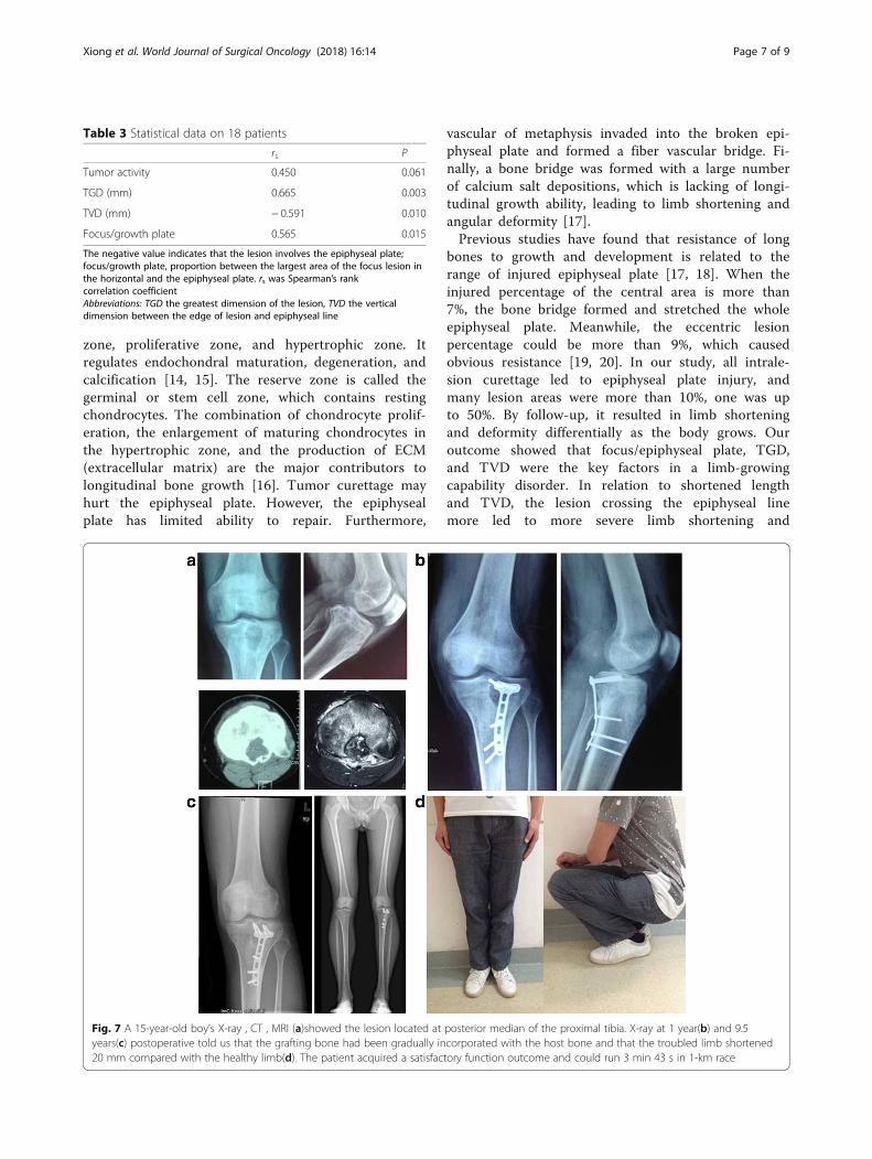
Table 3 Statistical data on 18 patients
rs P
Tumor activity 0.450 0.061
TGD (mm) 0.665 0.003
TVD (mm) − 0.591 0.010
Focus/growth plate 0.565 0.015
The negative value indicates that the lesion involves the epiphyseal plate;focus/growth plate, proportion between the largest area of the focus lesion inthe horizontal and the epiphyseal plate. rs was Spearman’s rankcorrelation coefficientAbbreviations: TGD the greatest dimension of the lesion, TVD the verticaldimension between the edge of lesion and epiphyseal line
Fig. 7 A 15-year-old boy’s X-ray , CT , MRI (a)showed the lesion located at posterior median of the proximal tibia. X-ray at 1 year(b) and 9.5years(c) postoperative told us that the grafting bone had been gradually incorporated with the host bone and that the troubled limb shortened20 mm compared with the healthy limb(d). The patient acquired a satisfactory function outcome and could run 3 min 43 s in 1-km race
Xiong et al. World Journal of Surgical Oncology (2018) 16:14 Page 7 of 9
deformity. In our series, the length of troubled limbwere shorter 18.47 ± 7.22 mm (1.5–30 mm) comparedwith the healthy limb on 5 upper limbs and 13 lowerlimbs. Thirteen patients whose lesions were located inthe lower limbs had no symptoms on walking becauseof a pelvic compensator, and the other five patients’shoulder function was satisfied at the last follow-up.Limb length discrepancy and angular deformity may
not necessarily lead to clinical problems during child-hood and puberty, but psychosocial problems mayoccur. Social withdrawal, practical problems relatingto clothing and shoes, fearing about future compatiblepartners, and career planning would suffer [21–23].Lower limb length discrepancy would lead to posturedeformation, gait asymmetry, low back pain, anddiscopathy [21, 22]. Thus, in addition to clinicalproblem improvement, it was necessary to make acomprehensive psychological intervening or treatmentto guide patient’s healthy and joyful growth. As wehave known, leg length discrepancy < 2 cm is a staticdisorder. Leg length discrepancy > 3 cm causesdistinct gait and posture disorders—the bigger the dif-ference the greater and more distinct the disordersare [22]. In our study, all the patients underwentpositive psychological intervention and clinical symp-toms treatment after surgery. All patients’ function ofVAS, MSTS93, and SF-36 have been significantly im-proved (P < 0.01). They all joined in social activities inadulthood as farmer, construction worker, college stu-dents, civil servants, and teachers, and so on.
ConclusionManaging of chondroblastoma located in an openepiphyseal plate of a long bone with meticulous curettage,inactivity, and bone grafts can control tumor progressionand recurrence effectively. Meanwhile, early detection andprompt surgical treatment intervention, which reducedsignificantly the tumor to influence limb growth anddevelopment, get encouraging limb function.
LimitationFirst, as a retrospective study, we had no controlgroup to assess the superiority of our study comparedwith different tumor management. This was a retro-spective review and is, therefore, limited by theheterogeneity of the available data and follow-up.Second, the sample size was few. We needed morecases and time to observe local recurrence, distantmetastasis, limb discrepancy, and function. Further-more, because of few cases in our study, we couldnot observe more factors, which might influence limbgrowth and development after surgical treatment,such as age and gender.
FundingNone.
Availability of data and materialsThe authors declare that all data supporting the findings of this study areavailable within the article.
Authors’ contributionsYX, YL, ZY, HL, and XF participated in the collection of the clinical data andperformed patients’ follow-up. YX and YL drafted this manuscript. HD designedthis research and reviewed the manuscript for important intellectual content.CT and HD were responsible for these patients’ operation and participated inthe project coordination and assisted with the manuscript. Each author hasparticipated sufficiently in this work to take public responsibility for theappropriate portions of the manuscript. All authors read and approve of thefinal manuscript.
Ethics approval and consent to participateThis retrospective study has been approved by the ethics committee at WestChina Hospital of Sichuan University. Meanwhile, every patient had signed aconsent about the treatment and study.
Consent for publicationWritten consent for the publication of images used in the manuscript wasobtained from the patients.
Competing interestsThe authors declare that they have no competing interests.
Publisher’s NoteSpringer Nature remains neutral with regard to jurisdictional claims inpublished maps and institutional affiliations.
Received: 6 August 2017 Accepted: 10 January 2018
References1. Lin PP, Thenappan A, Deavers MT, Lewis VO, Yasko AW. Treatment and
prognosis of chondroblastoma. Clin Orthop. 2005;438:103–9.2. Jaffe HL, Lichenstein L. Benign chondroblastoma of bone. Am J Pathol.
1942;18(6):969–91.3. Xu H, Nugent D, Monforte HL, Binitie TO, Ding Y, Letson GD, Cheong D, Niu
X. Chondroblastoma of bone in the extremities: a multicenter retrospectivestudy. JBJS. 2015;97(11):925–31.
4. Xie C, Jeys L, James SLJ. Radiofrequency ablation of chondroblastoma: long-term clinical and imaging outcomes. Eur Radiol. 2015;25(4):1127–34.
5. Ramappa AJ, Lee FY, Tang P, Carlson JR, Gebhardt MC, Mankin HJ.Chondroblastoma of bone. J Bone Joint Surg Am. 2000;82(2):1140–5.
6. Springfield DS, Capanna R, Gherlinzoni F, Picci P, Campanacci M.Chondroblastoma: a review of seventy cases. J Bone Joint Surg Am. 1985;67(5):748–55.
7. Farfalli GL, Slullitel PAI, Muscolo DL, Ayerza MA, Aponte-Tinao L. Whathappens to the articular surface after curettage for epiphysealchondroblastoma? A report on functional results, arthritis, and arthroplasty.Clin Orthop Relat Res. 2017;475(3):760–6.
8. Mashhour MA, Rahman MA. Lower recurrence rate in chondroblastomausing extended curettage and cryosurgery. Int Orthop. 2014;38(5):1019–24.
9. Van Der Geest IC, Van Noort MP, Schreuder HW, Pruszczynski M, De RooyJW, Veth RP. The cryosurgical treatment of chondroblastoma of bone: long-term oncologic and functional results. J Surg Oncol. 2007;96(3):230–4.
10. Lehner B, Witte D, Weiss S. Clinical and radiological long-term results afteroperative treatment of chondroblastoma. Arch orthop traum su. 2011;131(1):45–52.
11. Patrick CT, Robert KHJ. General principles of tumors, Enneking system forstaging benign and malignant musculoskeletal tumors. Campbell’soperative orthopaedics, vol. 12; 2013. p. 791–2.
12. Schreuder HW, Pruszczynski M, Veth RP, Lemmens JA. Treatment of benignand low-grade malignant intramedullary chondroid tumours with curettageand cryosurgery. Eur J Surg Onco. 1998;24(2):120–6.
13. Tiefenboeck TM, Stockhammer V, Panotopoulos J, Lang S, Sulzbacher I,Windhager R, Funovics PT. Complete local tumor control after curettage of
Xiong et al. World Journal of Surgical Oncology (2018) 16:14 Page 8 of 9
14. Chung R, Xian CJ. Recent research on the growth plate: mechanisms forgrowth plate injury repair and potential cell-based therapies forregeneration. J Mol Endocrinol. 2014;53(1):T45–61.
15. Romain S, Frederick KW, Dietrich P. Surgical-experimental principles ofanterior cruciate ligament (ACL) reconstruction with open growth plates. JExper Orthop. 2015;2(1):1–12.
16. Hamdy RC. Growth plate injuries: an introduction. Limb lengthening andreconstruction surgery case atlas: pediatric deformity; 2015. p. 83–6.
17. Xian CJ. Recent research on the growth plate: regulation, bone growthdefects, and potential treatments. J Mol Endocrinol. 2014;53(1):E1–2.
18. San-julian M, JD Aquerreta AB, Caadell JL. Indications for epiphysealpreservation in metaphyseal malignant bone tumors of childrenrelationship between image methods and histological findings. J PediatrOrthop. 1999;19(4):543–8.
19. Janarv PM, Wikstrom B, Hirsch G. The influence of transphyseal drilling andtendon grafting on bone growth: an experimental study in the rabbit. JPediatr Orthop. 1998;18(2):149–54.
20. Lee MA, Nissen TP, Otsuka NY. Utilization of a murine model to investigatethe molecular process of transphyseal bone formation. J Pediatr Orthop.2000;20(6):802–6.
21. Lecointre C, Toublanc JE. Psychological indications for treatment of tallstature in adolescent girls. J Pediatr Endocrinol Metab. 1997;10(5):529–31.
22. Raczkowski WJ, Daniszewska B, Zolynski K. Functional scoliosis caused byleg length discrepancy. Arch Med Sci. 2010;6(3):393–8.
23. D’Ambrosi R, Ragone V, Caldarini C, Serra N, Usuelli FG, Facchini RM. Theimpact of hereditary multiple exostoses on quality of life, satisfaction, globalhealth status, and pain. Arch Orthop Trauma Surg. 2017;137(2):209–15.
• We accept pre-submission inquiries
• Our selector tool helps you to find the most relevant journal
• We provide round the clock customer support
• Convenient online submission
• Thorough peer review
• Inclusion in PubMed and all major indexing services
• Maximum visibility for your research
Submit your manuscript atwww.biomedcentral.com/submit
Submit your next manuscript to BioMed Central and we will help you at every step:
Xiong et al. World Journal of Surgical Oncology (2018) 16:14 Page 9 of 9