THE EFFECTS OF VERTEPORFIN ON NON-SMALL CELL LUNG CANCER Todd R. Ackerman Jr Submitted to the faculty of the University Graduate School in partial fulfillment of the requirements for the degree Master of Science in the Department of Biochemistry and Molecular Biology, Indiana University August 2016
Transcript
THE EFFECTS OF VERTEPORFIN
ON NON-SMALL CELL
LUNG CANCER
Todd R. Ackerman Jr
Submitted to the faculty of the University Graduate School in partial fulfillment of the requirements
for the degree Master of Science
in the Department of Biochemistry and Molecular Biology, Indiana University
August 2016
ii
Accepted by the Graduate Faculty, Indiana University, in partial fulfillment of the requirements for the degree of Master of Science.
Master’s Thesis Committee ______________________________ Lawrence A. Quilliam, Ph.D., Chair ______________________________ Clark D. Wells, Ph.D.
______________________________ Mark G. Goebl, Ph.D.
iii
DEDICATION
I would like to dedicate this to my parents. Without their love and support, I
would never have been able to generate this work.
iv
ACKNOWLEDGEMENTS
Without the help of my mentor, Dr. Lawrence Quilliam, this project would have
never come to fruition. His dedication and endless help have helped me develop to better
analyze and go about answering biochemical questions. I would also like to mention the
other members of my committee, Dr. Mark Goebl and Dr. Clark Wells. Their advice and
support helped me to write this thesis and better articulate my thinking on my
experiments. Members of other labs that I would like to mention are Brandon Lane and
Lauren Bringman, They helped me troubleshoot and think of other methods to go about
answering my questions. Lastly, I would like to mention my co-workers and scientists
that I work with at Covance. Their input and opinions helped me to think critically about
my experiments and extract the most amount of information from my data.
v
Todd R. Ackerman Jr
THE EFFECTS OF VERTEPORFIN ON
NON-SMALL CELL LUNG CANCER
Non-small cell lung cancer (NSCLC) accounts for 85% of lung cancers and is the
leading cause of cancer death in the Unites States. Better treatments must be devised in
order to improve the prognosis of this disease. Verteporfin, an FDA approved drug, has
recently been reported to downregulate a potential core pathway of NSCLC, the Hippo
pathway. The pathway consists of a kinase cascade to control the transcriptional
coactivators YAP and TAZ. When these transcriptional coactivators lack phosphorylation
of key residues, they are able to translocate into the nucleus and bind to the TEAD
member of transcription factors. This augments transcription for genes responsible for
proliferation, survival, and stem maintenance. In this study, we report that verteporfin
limits proliferation and survival of NSCLC and may potentially be a viable treatment
option. Inhibition of cell survival dose-dependently correlated with inhibition of YAP-
TEAD transcription target CTGF. We also report the covalent homo-oligomerization of
p62, a prominent protein involved with autophagy, with the introduction of verteporfin
into NSCLC cells.
Lawrence A. Quilliam, Ph.D., Chair
vi
TABLE OF CONTENTS
LIST OF TABLES ........................................................................................................... viii
LIST OF FIGURES ........................................................................................................... ix
Table 1. List of antibodies and dilutions. ...........................................................................13
ix
LIST OF FIGURES
Figure 1. Depiction of the core cascade of the Hippo pathway in its active and inactive
states .....................................................................................................................................3
Figure 2. The two regioisomers of verteporfin. ...................................................................4
Figure 3. Verteporfin dose-dependently inhibits NCI-H460 cell viability. .......................18 Figure 4-A. Verteporfin reduces survival and proliferation. .............................................20 Figure 4-B. ........................................................................................................................ 21 Figure 5. CTGF expression is markedly reduced with the addition of verteporfin to media. .................................................................................................................................23 Figure 6-A. Dose-dependent induction of H460 cell p62 oligomerization by verteporfin under low light conditions. ..............................................................................26 Figure 6-B. Impact of histidine on verteporfin-induced p62 oligomerization. ..................26 Figure 7. H460 cell line has a relatively low level of folate receptors. .............................29 Figure 8-A. H460 NSCLC cultured have a relatively high percentage of stem cells. .......32 Figure 8-B. H460 NSCLC spheroids have a 17.7% stem cell population. .......................33 Figure 9. CTGF decreases with the addition of verteporfin, even with singlet oxygens squelched with histidine. .....................................................................................38
x
ABBREVIATIONS
ALDH Aldehyde Dehydrogenase CTGF Connective Tissue Growth Factor dPBS Dolbesco’s Phosphate Buffered Solution MTT 3-(4,5-dimethylthiazol -2-yl)-2,5-diphenyltetrazolium bromide NSCLC Non- Small Cell Lung Cancer RT-PCR Reverse Transcriptase Polymerase Chain Reaction TAZ Transcriptional Co-activator with PDZ binding motif YAP Yes-associated Protein
1
INTRODUCTION
Non-small cell lung cancer (NSCLC), accounts for 85-90% of lung cancers.
According to the American Cancer Society “Lung cancer accounts for about 27% of all
cancer deaths and is by far the leading cause of cancer death among both men and
women. Each year, more people die of lung cancer than of colon, breast, and prostate
cancers combined.”(1) Finding a new way to treat this cancer and improve prognosis is of
great need. A signaling pathway that may prove effective in targeted drug therapy for
NSCLC is the Hippo pathway. The Hippo pathway consist of a series of kinases that
regulate the transcriptional co-activators Yes-associated protein (YAP) and WW domain-
containing transcription regulator protein 1/transcriptional coactivator with PDZ binding
motif (TAZ). These transcriptional co-activators can go on to activate members of the
TEAD transcription factor family, and potentially other transcription factors, to control
proliferation, survival, and stem cell maintenance (2-4).Targeting this pathway may
prove to be very promising in the treatment of lung cancer.
Visudyne (Verteporfin), an FDA approved drug for the treatment of macular
degeneration, has been shown to have a negative effect on TEAD downstream
transcription. The current understanding of how verteporfin accomplishes this is through
preventing the YAP-TEAD complex from forming [5]. Hence, treatment with verteporfin
could effectively treat cancers with aberrant Hippo activity. NSCLC cell growth has been
shown to be inhibited by overexpressing MST1, preventing YAP from acting as a
transcriptional coactivator (5). From this it can be hypothesized that verteporfin would be
effective in the treatment of lung cancer.
2
The Hippo Pathway
The Hippo pathway is a fairly recently identified pathway, with the orthologs
being first discovered in 2002 in Drosophila melanogaster. The pathway was found to be
highly conserved in mammals and to be a major controller of organ size, which was
unprecedented in other pathways (4). The elicited response to mutation in any one of the
upstream kinases in the Hippo pathway generates an overgrowth of the targeted organ.
These mutations diminish the kinases function to phosphorylate, ultimately inactivating
the Hippo pathway. This inactivated form allow for increased proliferation and cell
growth akin to that of a tumor (2).
The mammalian Hippo pathway begins with the protein complex of MST1,
MST2, and SAV1. When the pathway is active, the phosphorylated MST1/2 complex
phosphorylates the complex of large tumor suppressors 1 and 2 (LATS1/2) and MOB1.
This in turn activates the LATS1/2 kinases to phosphorylate YAP/TAZ. This event keeps
YAP and TAZ sequestered in the cytosol, preventing them from acting upon transcription
factors in the nucleus. Depending on the phosphorylation site on YAP and TAZ, it will
either be retained in the cytoplasm or marked for degradation. There are five known sites
for LATS1/2 to phosphorylate YAP, 4 of these being conserved in TAZ. A
phosphorylation event of YAP on serine 127 creates a 14-3-3 binding site, which will
keep it retained in the cytoplasm. Subsequent Phosphorylation of serine 381 triggers
ubiquitination and proteasomal degradation of YAP (3,6). However, if the pathway is
never initiated, YAP and TAZ will be in their active unphosphorylated form and be able
to cross the nuclear membrane to act as transcriptional co-activators.
3
Figure 1. Depiction of the core cascade of the Hippo pathway in its active and inactive states. In active Hippo signaling, the phosphorylated MST1/2-SAV1 complex go on to phosphorylate LATS1/2, which in turn phosphorylate YAP/TAZ. This event keeps YAP from translocating into the nucleus and act as a transcriptional co-activator of TEAD1-4. In inactive Hippo signaling, the phosphorylation cascade never occurs, allowing YAP/TAZ to act on TEAD1-4 to modulate transcription.
Upstream signaling of the Hippo pathway is poorly understood. The core kinase
cascade of the Hippo pathway is controlled by a multitude of factors that all go into
sensing the physical state of the cell. A very prominent feature of the Hippo pathway is
its interconnection with other cellular pathways (7). This interconnectedness allows the
Hippo pathway to sense and respond to energy levels, cellular contact, growth factors,
and polarity of the cell. In tumor conditions, these interconnections may funnel some of
the classical phenotypes seen in tumors through the Hippo pathway; making it a great
potential therapeutic target.
4
Verteporfin
Verteporfin, trademarked as Visudyne, is currently used in the photodynamic
treatment of age related macular degeneration. It is composed of two regioisomers (same
chemical formula, but a functional group changes position on the parent molecule), which
are listed below. The drug is activated at a 690nm wavelength, at which point singlet
oxygens are produced while in the presence of oxygen (8). After its FDA approval, it was
also found to suppress the oncogenic activity of YAP without the need for light activation
(9). To demonstrate whether verteporfin binds to YAP or TEAD, Liu-Chittenden et al
mixed verteporfin with purfied versions of the proteins. The author found that verteporfin
coeluted with YAP, signifying verteporfins binding partner is in fact YAP.
Figure 2. The two regioisomers of verteporfin.
5
Verteporfin is known to produce singlet oxygens with exposure to light, but these
reactive species appear to be produced at modest levels even without light exposure
(10,11). Singlet oxygen is a short-lived electrophilic molecule that reacts rapidly with
unsaturated carbons, neutral nucleophiles and anions (12,13). Verteporfins basal
production of singlet oxygens causes damage to proteins near the site of where it was
formed. Consequently, some proteins may become non-functional or aggregate protein
crosslinks may arise. This is problematic for potential therapeutic value, as proteotoxicity
may be unavoidable. On the other hand, this could be a very useful secondary function of
verteporfin. Some studies have reported normal cells are able to clear out the proteotoxic
effects of verteporfin while tumor cells cannot (11,14). If this is the case, it adds more
credence to verteporfin being an effective cancer treatment.
Another interesting phenomenon cells exhibit when exposed to verteporfin is the
lack of autophagy (10). This phenotypic change can be attributed to the polymerization of
p62 with itself. The protein p62, also known as sequestosome 1 (SQSTM1), is a
multifunctional adaptor protein that recognizes and loads ubiquinated cargo into an
autophagosome (10,15). The singlet oxygens produced basally by verteporfin cause this
multimerization of p62. Which is also a downward slope, as more p62 polymerize, less
autophagic degradation occurs, allowing more damaged proteins to build up. This may
also be a major proponent of cell death with verteporfin treatment.
6
METHODS
Cell Passaging
Stock plates of cell lines were passaged every 3 days for the H460 cells and 3 to 4
days for H23 cells in RPMI 1640 medium supplemented with 10% fetal bovine serum
and penicillin/streptomycin. Medium was aspirated off the stock plates and replaced with
10 mLs of dPBS. dPBS was then aspirated and replaced by 1 mL of trypsin/EDTA
solution, followed by being placed into a cell culture incubator for 5 minutes. Afterwards,
9 mLs of media was dispensed onto the cell suspension to inactivate the trypsin. The
suspension was then agitated by gently pipetting up and down several times to ensure all
cells are detached from the plate and each other. Cells were then quantitated by pipetting
10 uLs onto both sides of a hemocytometer and counting five squares on each side. Stock
plates were set at 200,000 – 300,000 cells per 10 cm dish.
7
Clonogenic Assay
To assay for survival and proliferation, clonogenic assays were conducted. Cells
were diluted to 167 cells per mL of media. Three mLs of the diluted cell suspension was
dispensed onto a 60 mm plate for approximately 500 cells per plate. Plates were then
incubated in a cell culture incubator for 24 hours. Media was aspirated and replaced with
fresh media infused with verteporfin concentrations listed below. Cells were left to
incubate for 72 hours. The media was then aspirated off and the dish was washed twice
with 3 mLs of dPBS. Following aspiration of the dPBS, fresh media was placed into the
dish and placed back into the cell culture incubator. At this point the cells were left to
recover from the exposure to verteporfin. The recovery process takes 7-9 days. During
this period, the media was changed out by day 4. Once the colonies on the control plate
were visibly noticeable, the plates were processed for staining. Media was aspirated and
plates were washed with 4 mLs of dPBS. After aspirating the wash, 3 mLs of 10% acetic
acid / 10% methanol was pipetted onto the plate as a fixative. The plates were then
incubated for 20 minutes followed by removing the fixative. Crystal violet was then used
to stain the cells for visualization and potential colony counting. One mL of crystal violet
was pipetted onto each plate and let sit for 20 minutes. The residual crystal violet is then
aspirated off of the plates and washed under a low-pressure stream of water. The wash
was performed 4-5 times or until all of the residual crystal violet is washed away. The
stained colonies are then visualized and interpreted for changes in survival and
proliferative capacity.
8
MTT Assay
Measurements of cell viability and proliferation were also acquired utilizing MTT
assays. This assay assesses cells metabolic activity via oxidoreductase enzymes reducing
the tetrazolium dye MTT to formazan. Cells were set on a 96 well plate at 2000 cells per
well. The plates were incubated for 24 hours at 37° C. Media was then aspirated and
replaced with media infused with drug concentration of choice, in triplicates. Plates were
then incubated for ~72 hours in the cell culture incubator. 10 uLs of MTT Reagent was
added to each well and incubated for ~4 hours. Media was aspirated and 100 microliters
of alcohol/detergent reagent was added to each well to dissolve the precipitate. After 3
hours of incubation in the dark, the plate was then quantified on a spectrophotometer,
recording at an absorbance of 570 nm.
9
Matrigel Assay
Matrigel assays were conducted to view cell morphology and phenotype changes
with the addition of verteporfin in three-dimensional culture. Matrigel was thawed to 4°
C, 200 uLs was pipetted onto a 40 mm dish and spread evenly across the center of the
plate with a pipette tip. Plates were then incubated in a cell culture incubator for 20
minutes to allow the Matrigel to set. During this time cells were trypsinized and counted.
200,000 cells were dispensed on top of the Matrigel and let set for 24 hours. Media was
aspirated and replaced with verteporfin infused media. At periodic intervals, microscopic
images were collected of each set of plates at 4X or 10X magnification to discern any
phenotypic changes with the cells.
10
ALDEFLUOR Assay
To assess the percentage of stem like cells in culture, an ALDEFLUOR assay was
ran on H460 cells and H460 spheroids. The ALDEFLUOR measures aldehyde
dehydrogenase (ALDH) activity within the cell by generating a fluorescent product,
effectively enabling measurement via a flow cytometer. ALDH expressing cells are
associated with a stem like nature. For the H460 cells, the plates were trypsized for 5
minutes and inactivated with media. After quantitation with a hemocytometer, 200,000
cells were aliquoted from the cell suspension and placed into a 15 milliliter conical tube
and centrifuged at 800 RPM for 5 minutes. Supernatant was aspirated away without
disturbing the cell pellet. The cells were washed with 10 mL of dPBS and centrifuged at
800 RPM for 5 minutes. The supernatant was then aspirated off and the pellet was
resuspended into 1 mL of the ALDEFLUOR assay buffer. Two 4 mL test tubes were
labeled as experimental and control. The 1 mL of cell suspension resuspended in the
assay buffer was then transferred into the experimental tube. Five uL of the DEAB
(ALDH inhibitor) solution was pipetted into the control tube. Five uL of the activated
ALDEFLUOR substrate was pipetted into the test tube and immediately afterwards 500
uL from the test tube was transferred into the control tube. Samples were incubated for 40
minutes at 37° C. The tubes were then centrifuged at 250 x g for 5 minutes and the
supernatant was aspirateded. Lastly, 500 uL of ALDEFLUOR assay buffer was added to
both tubes and stored on ice. The samples were ran on a cytometer as soon as possible, as
the fluorescence drops in a time dependent manner.
11
Folate-FITC Assay
To quantitate the amount of available folate receptors on the H460 cells FACS
analysis was done using folate conjugated to FITC. H460 cells were grown in folate-free
media for 2 weeks prior to testing. This was done to permit folate receptors to reach a
level more akin to what it would be in vivo. Controls for this experiment were acquired
by using KB cells, which are known to have high cell surface folate receptor levels. To
avoid degradation of any surface receptors, cells were trypsinized for no longer than 5
minutes. Trypsin was then inactivated using folate-free media and cells counted. After
quantitation, 100,000 cells were aliquoted out of suspension and centrifuged at 800 RPM
for 5 minutes. Supernatant was aspirated and 500 uL of minus folate and minus serum
media was dispensed into the positive control and the experimental tube. For the negative
control, dispense 500 uL of plus folate minus serum media, which will compete with the
folate-FITC conjugate. Afterwards, 50 uL of the folate-FITC conjugate was pipetted into
each suspension and incubated for 30 minutes. Suspension was then washed twice with
cold dPBS and centrifuged at 800 RPM for 5 minutes. Supernatant was aspirated and
replaced with 500 uL of dPBS. Samples are ready to be ran on a cytometer at this point.
12
SDS PAGE Western Blotting
Cells were plated on 40 mm dishes at 100,000 per plate and let set for ~24 hours.
Desired verteporfin and reducing agent concentrations were made and added to dishes.
After 24 hours, media was aspirated and washed with ice cold PBS. Cells were lysed in
200-300 uLs of phospho-tyrosine lysis buffer (50 mM Hepes, pH 8.0, 50 mM NaCl, 2
mM EDTA, 1% Triton X-100, 10% glycerol, 1 mM Na3VO4, 10 mM NaF, 10 mM
NaH2PO4, 100 KIU/ml aprotinin and 1 mM PMSF). Plates were scraped and re-aliquoted
into sterile micro-centrifuge tubes. Lysates were centrifuged at 800 G for 10 minutes at
4° C. Supernatant was added to a fresh micro-centrifuge tube with the pellet being
discarded. The Bradford protein assay was conducted to acquire protein concentration per
lysate. Using the given concentrations, thirty micrograms of protein was pipetted from
lysates and added to ten microliters of 4x loading buffer. Samples were placed into a
water bath set at 90° C for five minutes to denature sample proteins and coat them with
sodium dodecyl sulfate. After denaturation, samples were placed in a micro-centrifuge to
pull down any water that was evaporating from the boiling process. The entirety of the
samples were then pipetted into individual wells in an 8 % poly-acrylamide gel. BD
bioscience all-blue protein ladders were used to delineate protein size. Gels were ran with
ice cold running buffer at 90-100 milliamps for 90 minutes. Once the run was completed,
the transfer apparatus was prepared. PVDF membranes were activated in 100% methanol
and placed on top of two filter papers soaked in transfer buffer. The gel was taken out of
the running apparatus and placed on top of the membrane, assuring no formation of air
bubbles. Another filter paper soaked with transfer buffer was placed over the gel. A roller
was used to force any air bubbles that may have formed out of the compilation. The
13
clamp was closed and submerged into the transfer apparatus. A constant voltage was
used, set at 100V, to transfer the proteins set on the gel to the PVDF membrane. The
transfer was ran for one hour with an ice pack placed inside. After completion a Ponceau
Stain was conducted to confirm proper transfer of proteins. At this point, the membrane
was blocked for 24 hours in 5% skim milk at 4° C. After blocking, the membrane was
washed twice in tris-buffered saline with 1% Tween-20 added (TBST). Membrane was
split in half, cut underneath the 50 kilodalton band on the protein ladder.
PRIMARY ANTIBODY Antibody dilution inTBST (uL)
P62 1/5000
YAP 1/2000
LC3 1/3000
β-Actin 1/10000
GAPDH 1/10000
Table 1. List of antibodies and dilutions.
Primary antibodies were diluted, as listed above, and added to the respective
membrane for one hour. Three washes with TBST for 5 minutes were conducted to wash
off any nonspecific binding. After washing, the secondary antibody was added to both
membranes for thirty minutes (Anti-Mouse HRP 1:30000 TBST). Another series of
washes was conducted, a total of five for five minutes per wash with TBST. Using
enhanced chemiluminescence, the membranes were developed in a dark room using
autoradiography.
14
Reverse Transcriptase PCR
Cells were set at 250,000/10 cm dish dish and placed into a cell culture incubator
for 24 hours. Media was aspirated and replaced with fresh media infused with verteporfin
concentration of choice for 24 hours in a cell culture incubator. After the 24 hour
exposure, media was aspirated off, and the dish was washed with ice cold PBS and
aspirated off. One milliliter of Tri-Reagent was placed into each dish. Using a sterile
scraper, the entire area of the dish was scraped and the Tri-Reagent was pooled into an
area on the dish. The entirety of the solution was then pipetted into a sterile DNAase and
RNAase free micro-centrifuge tube. At this point, tubes were either frozen at -80° C
being processed at a later time or RNA was immediately isolated out of the solution.
When isolating RNA, 200 uL of chloroform was pipetted into each micro-centrifuge tube
and inverted for at least 15 seconds. After 5 minutes, at room temperature, tubes were
centrifuged at 12,000 G for 15 minutes at 4° C. The tubes were then carefully placed into
a clean rack and the clear supernatant was pipetted into a fresh DNAase and RNAase free
micro-centrifuge tube. To precipitate out the RNA, 500 uL of isopropanol was then
added. After 5 minutes, the tubes were centrifuged at 12,000 G for 10 minutes at 4° C.
The supernatant in the tubes is discarded and then washed with one milliliter of 70%
ethanol. The tubes were centrifuged at 12,000 G for 5 minutes. Supernatant is discarded
without disturbing the pellet. The tubes are then incubated in a water bath at 37° C until
the remainder of the ethanol has evaporated from the tubes. The RNA isolated in the
pellet was then resuspended in 15 microliters of RNAse and DNAse free water. The tubes
were then incubated at 50° C for 5 minutes to fully dissolve the RNA into solution. The
concentration of RNA was then quantitated using a Nanodrop spectrophotometer. At this
15
point samples were standardized for reverse transcription, aliquoting 5 ug of RNA into
PCR grade RNAse and DNAse free micro centrifuge tubes. Samples were then QS to 18
microliters. In a separate container, random hexamers and dNTPs were mixed; using 4
microliters of random hexamers per sample and 2 microliters of 10 mM dNTP per
sample. Six microliters of the random hexamer and dNTP solution was dispensed into
each sample for a total of 24 uL per tube. To facilitate annealing, micro centrifuge tubes
were placed into a 60 degree water bath for 5 minutes. During this incubation the reverse
transcriptase master mix was prepared. The master mix was comprised of 9 uL of
nuclease free water, 1 uL of RNASE OUT, 4 uL of 10x reverse transcriptase buffer, and
2 uL of MuLV reverse transcriptase. After the 5 minute incubation, the samples were
taken out of the water bath and wiped dry with a Kim wipe. Afterwards, 16 uL of the
reverse transcriptase master mix was dispensed into each sample tube. The samples were
then incubated at 50° C for 50 minutes, and then 70° C for 5 minutes to denature the RT
enzyme. The cDNA product was measured on the Nanodrop on the single strand DNA
setting to acquire approximate cDNA values. At this point samples were standardized so
that each contained the same amount of cDNA, assuring that enough diluent is used to
perform the reactions desired. The master mix was assembled next; which consisted of 3
ul of nuclease free water, 1 ul of both the forward and reverse primers, and 10 ul of 2x
syBR green mix. With the mix completed, 15 ul was pipetted into a 96 well PCR grade
plate followed by 5 ul of the diluted cDNA sample. The plate was centrifuged and placed
onto an Eppendorf Mastercycler to begin and measure the reactions.
16
RESULTS
Preface The basis for this work is that verteporfin has been shown to block aberrant YAP
activity, and since NSCLC has been shown to be correlated with abnormal YAP activity
(5,16-18), it can be hypothesized that proliferation and survival may decrease
substantially by treatment with verteporfin. Beyond proliferation and survival, the change
in gene expression brought on by verteporfin may sensitize stem-like populations by the
change in gene expression. If true, verteporfin would be a great candidate for synergistic
drug therapies as the stem cell populations of tumors are more resistant to traditional
treatments. Another facet of verteporfin treatment that will be touched on is the
oligomerization of p62 and the disruption of autophagy. Disrupting autophagy would
have detrimental effects on tumor cells. This would put stressors on the cells that could
lead to apoptosis or impaired function. All of these qualities may make verteporfin a very
powerful tool in the fight against NSCLC.
The only problem that can be gleaned at this point is that there are no specific
delivery systems to the cancer cells. In cell culture this is not an issue, but at the scale of
an organism treatment may be difficult. A method that was looked into to aid in
specificity was the conjugation of folate onto verteporfin. Tumor cells typically have
upregulated folate receptors, as they are highly proliferative. This can be used to deliver
verteporfin more specifically to them. This will be touched on in the last section of the
results.
17
To start off, proliferation and survival are looked at in H460 cells and H23
NSCLC cells with the treatment of verteporfin.
18
Verteporfin Reduces Proliferation and Survival of NSCLC Cells
To assess how treatment with verteporfin would affect NSCLC cell viability, an
MTT assay was conducted. Several pilot experiments performed to ascertain how long
the drug should be left on to have noticeable effects. The exposure time varied from 72
hours, 96 hours, to 120 hours for these pilot experiments. The difference in effectiveness
was negligible, therefore 72 hours was chosen as the exposure time for the subsequent
experiments.
Figure 3. Verteporfin dose-dependently inhibits NCI-H460 cell viability. Viability is standardized to the control plate. cells were incubated with the indicated concentrations of verteporfin or DMSO solvent alone (all points were adjusted to 0.1% DMSO) for 72 hours prior to performing the MTT assay. Data points were collected in triplicates. The IC50 is approximately 2.75 uM.
0
0.2
0.4
0.6
0.8
1
1.2
0 0.03 0.1 0.3 1 3 10
VIAB
ILIT
Y
VERTEPORFIN (UM)
MTT H460 3 days
19
Results from the MTT assay show that a negative correlation between the
concentration of verteporfin and NSCLC cell survival exists. To further reinforce that
viability is decreasing as well as to get a more accurate IC50, a series of clonogenic
survival assays were conducted. Approximately 500 cells were seeded onto 60 mm dishes
a day before their exposure to verteporfin. Initial experiments established a narrower
effective concentration of verteporfin than in the MTT assay. Therefore doses between
100 and 500 nM were selected for subsequent survival assays. Dishes exposed to
verteporfin are shown below in figure 2 for both the H460 and H23 cell line for three
days.
20
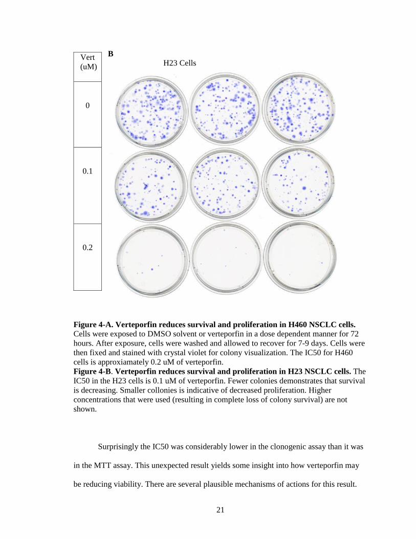
A H460 Cells
Vert (uM)
0
0.1
0.2
0.3
21
B H23 Cells
Figure 4-A. Verteporfin reduces survival and proliferation in H460 NSCLC cells. Cells were exposed to DMSO solvent or verteporfin in a dose dependent manner for 72 hours. After exposure, cells were washed and allowed to recover for 7-9 days. Cells were then fixed and stained with crystal violet for colony visualization. The IC50 for H460 cells is approxiamately 0.2 uM of verteporfin. Figure 4-B. Verteporfin reduces survival and proliferation in H23 NSCLC cells. The IC50 in the H23 cells is 0.1 uM of verteporfin. Fewer colonies demonstrates that survival is decreasing. Smaller collonies is indicative of decreased proliferation. Higher concentrations that were used (resulting in complete loss of colony survival) are not shown.
Surprisingly the IC50 was considerably lower in the clonogenic assay than it was
in the MTT assay. This unexpected result yields some insight into how verteporfin may
be reducing viability. There are several plausible mechanisms of actions for this result.
Vert (uM)
0
0.1
0.2
22
Cells are much more dispersed and in less dense colony formations in the clonogenic
assay compared to the MTT assay. This would make the cells initially more dependent on
Hippo signaling, since the cells had their junctions severed during the trypsinizing
process and weren’t able to cluster back together. In the MTT assay the surface area is
greatly reduced, allowing the NSCLC cells to cluster and perhaps be less dependent on
the Hippo pathway. Increased density may be one reason why verteporfin is much more
effective in the clonogenic assay as cells that are more dependent on the Hippo pathway
may be more sensitive to verteporfin treatment. Another mechanism may just be a matter
of time. In the clonogenic assay, after 3 days of exposure to verteporfin the media is
aspirated, the plate is washed and replaced with fresh media; the cells are left to recover
for several days at this point. This recovery period may facilitate the cells transition to an
apoptotic state (significant cell death observed at the higher drug doses was not typically
observed until after the cells had been in culture for 4-5 days). The last mechanism that
was thought of is the potential for verteporfin to remain in the cytosol of the NSCLC cells
after the plates were washed. Since verteporfin is hydrophobic, it should flux in and out
of a cell with relative ease. However, if for some reason it gets sequestered in the cytosol
the recovery period may just be elongating exposure time.
With these two assays it can be said with a degree of certainty that verteporfin
does indeed reduce the proliferative capacity as well as the surviability of NSCLC cells.
The next step was to figure out how this might be accomplished.
23
Blocking the YAP-TEAD Interaction
The basis for verteporfin to be effective in reducing the viability of NSCLC was
believed to be its blocking of YAP from binding to the TEAD family of transcription
factors. To confirm that verteporfin is attenuating YAP activity in NSCLC cells, a series
of RT-PCR assays were performed. The gene CTGF was chosen to measure the efficacy
of verteporfin to downregulate TEAD responsive genes, as CTGF is a prominent gene
associated with YAP-TEAD activity and the most frequently monitored (19,20).
Figure 5. CTGF expression is markedly reduced with the addition of verteporfin to media. H460 NSCLC cells were exposed to verteporfin for 24 hours at the concentrations above. Calculations were done using the comparative Ct method. This figure was generated by a member of the Quilliam lab. As expected, verteporfin decreases the expression of CTGF mRNA in a dose
dependent manner. The IC50 was just under 200 nM and by 300 nM CTGF expression is
0.95
1.011.15
0.85
0.59
0.27 0.22
0.00
0.20
0.40
0.60
0.80
1.00
1.20
1.40
-200 0 200 400 600 800 1000 1200
CTG
F EX
PRES
SIO
N
VERTEPORFIN (NM)
24
nearly a quarter what the control plate expressed, this shows how potent verteporfin is in
inhibiting downstream TEAD transcription. This is a massive decrease that could
potentially be extended to say that genes associated with TEADS are decreased perhaps
to the same extent; phenotypically this means proliferation, survival, and a stem like
nature should decrease as well. To confirm the above statement, other downstream TEAD
genes would have to be monitored with verteporfin treatment. The results of the PCR
correlate nearly perfectly with the dose response curve for the clonogenic assay. Which
suggests that the mechanism of death for these cells may be through the Hippo pathway.
A contending hypothesis against the above role of verteporfin acting through
inhibition of YAP-TEAD signaling was described shortly after my project began;
verteporfin was shown to promote the oligomerization of p62, which blocks autophagy
and may lead to cytotoxic apoptosis. The next section of the results will touch on this
aspect of NSCLC cell response to verteporfin treatment.
25
Low Concentrations of Verteporfin Covalently Homo-Oligomerize p62 Donohue et al showed that verteporfin covalently homo-oligomerizes p62 and
inhibits autophagy at 10 uM without light activation (10). However, they do not show
that that this crosslinking happens with the low concentrations used in the figures 4 and 5
herein. An earlier study by this group also suggested that verteporfin impacted
autophagy, but again, the doses of drug used were quite high compared to the nM levels
used in figures 4 and 5. Initially, it was assumed that these oligomeric p62 constructs
would not with the low concentrations of verteporfin being used in my studies. However,
results proved these assumptions wrong. Numerous western blots were conducted,
probing for p62, at different time points and concentrations demonstrating this finding. In
subsequent experiments, antioxidants were used to try and prevent p62 oligomers from
forming. Preventing p62 from oligomerizing would help establish if verteporfin is acting
through the Hippo pathway or by other means. The antioxidants used were glutathione, α-
tocopherol, n-acetyl cysteine, and histidine. Histidine was shown to be the most effective
at preventing p62 oligomers from forming and was used hereafter as a singlet oxygen
squelcher.
26
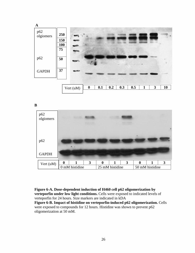
p62 olgiomers p62 GAPDH
A
0 0.1 0.2 0.3 0.5 1 3 10
B
0 1 3 0 1 3 0 1 3 0 mM histidine 25 mM histidine 50 mM histidine
Figure 6-A. Dose-dependent induction of H460 cell p62 oligomerization by verteporfin under low light conditions. Cells were exposed to indicated levels of verteporfin for 24 hours. Size markers are indicated in kDA Figure 6-B. Impact of histidine on verteporfin-induced p62 oligomerization. Cells were exposed to compounds for 12 hours. Histidine was shown to prevent p62 oligomerization at 50 mM.
250 150 100 75
50
37
p62 olgiomers p62 GAPDH
Vert (uM)
Vert (uM)
27
As shown above in figure 4-A, verteporfin causes high molecular weight p62
bands to appear as the dosage increases. This was expected, but not for the 100-200 nM
range. Also, the exposure time is one-third that of the MTT and clonogenic assays. This
is problematic for the initial hypothesis, as this puts a hole into the prior thinking.
However, it is important to note that the samples used in figure 4-A were exposed to light
during the processing of the samples, which may have had an effect on oligomerization.
To discern whether oligomerization can be prevented or reduced, histidine was used as a
singlet oxygen squelcher. As shown in figure 6-B, 50 mM of histidine blocks the
oligomerization of p62 for 1 and 3 uM of verteporfin exposed to the cells for 12 hours.
The MTT, clonogenic, and RT-PCR need to be redone using histidine in conjunction with
verteporfin, to test if the previous results are attributed to the blocking TEAD
downstream genes or cytotoxicity. Due to some unfortunate circumstances (several stock
preparations of verteporfin lost effectiveness), it has been problematic to follow through
with these experiments.
28
Folate-Verteporfin Conjugate The conjugation of folate to verteporfin could potentially make it more specific
for proliferating cells. Previous studies have shown the efficacy of conjugating folate to
molecules to enhance delivery (21). This would be a major benefit in making verteporfin
into a viable systemic treatment option. Another added benefit of folate conjugation
might be increased solubility, which would further improve drug delivery in vivo. To test
this, a preliminary study was conducted to quantify the level of folate receptors present
on the NSCLC cell line H460. Quantifying the number of folate receptors is key, as
they’re anticipated to be the major conduit of the conjugate into the cells. Once inside the
cell, the folate should be cleaved off, which leaves verteporfin’s activities unchanged.
However, if there are not enough folate receptors present, then the driving force will be
diminished. NSCLC is known to have high levels of folate receptors (22), but conditions
may change in cell culture. Cells have been passaged and grown in RPMI, which has a
very high level of folate which would promote endocytosis and down-regulation of
recpetors. Before testing the amount of folate receptors, the cells were passaged in folate
free media for two weeks. This was done in hopes of restoring the folate receptors back
to in vivo conditions.
29
Figure 7. H460 cell line has a relatively low level of folate receptors. This experiment shows the relative amount of folate receptors present on the cell membrane. The first two plots are control runs with KB cells, which are known to be highly positive for folate receptors. This facilitates a negative and a positive control, with and without folate competition, respectively. The third chart depicts the relative amount of folate receptors on the H460 cell line after being cultured in the absence of folate for ~10 days.
30
The data from the folate uptake assay shows that there are very few folate
receptors on the surface of the H460 cell line compared to the positive control, KB cells.
This is unfortunate, as this cell line wouldn’t apply for testing the verteporfin-folate
conjugate. This conjugate may prove to be a very effective delivery model in vivo, but in
cell culture, it may not apply very well, unless the appropriate cell line was selected. The
KB cell line could be suitable for pilot experiments until an appropriate NSCLC cell line
is identified.
31
H460 NSCLC Cultures Have a High Percentage of Stem Cells
Typically, stem cells are resistant to traditional chemotherapeutics, which are
thought to transport the drugs outside of the cell before they have any effect. Verteporfin
being hydrophobic, can flux back into the cell even after being transported out. More
importantly, TEAD-induced signaling supports stem cell maintenance. So, verteporfin
could potentially thwart a stem cell phenotype, possibly making them less resilient. To
measure the percentage of stem cells and to test if verteporfin may be more selective for
stem cells, an ALDEFLUOR assay was piloted, since stem cells express high levels of
aldehyde dehydrogenase isoform 1A1(ALDH1A1) (23) . The rationale behind this being
that stem cells are more reliant on the Hippo pathway for survival (24). If this is the case,
verteporfin could be quite effective at limiting their numbers. The ALDEFLUOR assay,
combined with Flow Cytometry, measures ALDH activity within the individual cells,
which is a reliable marker for stemness (25). The assay was conducted on H460 cells and
H460 spheroids, the latter being enriched with stem cells since they are resistant to
anoikis.
The results of the assays showed that after subtracting the isotype from the
experiment, the H460 cells have a 5.7% ALDH-positive stem population while the H460
spheroids have 17.7%. This is a considerable stem population, which could yield good
results for the concluding experiments. Unfortunately, my work does not include the next
step in the process, which would be to add verteporfin at varying concentrations and see
if the stem populations lessen with higher concentrations. The results are shown below.
32
A
33
B
Figure 8-A. H460 NSCLC cultured have a relatively high percentage of stem cells. The first run is a control that allows an accurate placement of the gate, which transfers over onto the experimental run. H460 cells were shown to have a 5.7% stem population. Figure 8-B. H460 NSCLC spheroids have a 17.7% stem cell population.
34
DISCUSSION
Preface With the conclusion of the results section, I am going to talk about the future
avenues that this work could pursue. In my work, it is established that verteporfin affects
NSCLC cell viability. However, there are many areas left blank that this work can be a
preamble to. For instance, developing and/or acquiring a mouse model for NSCLC would
help fortify if verteporfin would be affective in vivo or not. This among other possible
experiments will be discussed in the future experiments subsection.
Prior to going over future experiments, ideas and theories will be deliberated
about verteporfin and the phenomenon that was seen in my experiments with NSCLC.
There have been many studies with verteporfin being used as a therapeutic agent to treat
a variety of mouse models of cancers before and after my work began. Some of these
studies will be discussed to attempt to form a more complete picture.
35
Ideas and Interpretations
In my work with NSCLC it is seen that the TEAD-responsive gene CTGF is
downregulated and p62 is covalently homo-oligomerized with the treatment of
verteporfin in vitro. Therefore, verteporfin is likely to have at least 2 potential
mechanisms of action as supported by the literature: (i) inhibition of YAP/TEAD-induced
gene expression that is associated with survival and proliferation and (ii) protein
aggregation resulting in disruption of the processes such as autophagy. One or both of
these mechanisms is responsible for the affects seen in viability, discerning what
mechanism is responsible for the decrease in viability was an unfinished aim for my
study. This unfinished aim is important, however, the key point is that verteporfin
effectively reduces/kills tumors and is not toxic to normal tissues. Studies by others have
been conducted showing verteporfin’s efficacy in reducing tumor size while not being
cytotoxic to normal tissue in vivo (9,11). The main modality for cell death differs for the
two studies referenced above. Liu-Chittenden et al reports that verteporfin suppresses
liver overgrowth and the formation of hepatocellular carcinoma though the perturbation
of the YAP-TEAD interaction. While Zhang et al reports that YAP-TEAD has nothing to
do with reducing colon cancer size or proliferation and that the effects are strictly through
proteotoxic means. This difference in mechanism is very interesting. It may mean that
verteporfin might promote apoptosis through different means, depending on what the
cancers weak points are. As long as there are no deleterious effects on normal tissue;
which the two studies above show very little toxicity in normal tissue, verteporfin could
be a great drug for metastatic cancers. The duality of blocking the YAP-TEAD
interaction and promoting protein crosslinking through its singlet oxygen production are
36
very powerful modes of limiting proliferation, particularly if the crosslinking inhibits
autophagy.
Blocking the YAP-TEAD interaction leads to a decrease in transcription in genes
associated with proliferation, survival, and stemness. In other words, blocking the YAP-
TEAD interaction decreases proliferation and sensitizes cells to apoptosis while
decreasing a stem-like nature. These qualities likely make verteporfin synergistic with
other compounds. Appropriate drugs to combine with verteporfin may be cisplatin or
erlotinib. Clonogenic and MTT pilot assays were conducted with cisplatin as a candidate
for synergism, used during, before and after verteporfin treatment. There were no
discernable differences between the effectiveness of the combination. However, these
experiments were done while verteporfin was beginning to lose its potency in my assays,
so the results were not conclusive and not included herein. However, studies by others in
the lab indicated that verteporfin cooperates with erlotinib in a mutant EGF receptor
NSCLC cell line, HCC4006, to promote cell death. In addition, after all my experiments
were concluded, a study reported that ablating YAP1 improves sensitivities to other
modes of treatment in NSCLC (26). This fortifies the thinking that verteporfin would be
effective at synergistic treatments and should be further studied. Since the maximal
effects of verteporfin on inhibiting cell survival and CTGF expression occurred at lower
doses than the induction of p62 oligomerization, it is possible that cell death in my hands
was more dependent on YAP-TEAD signaling.
Many other proteins other than p62 are more likely than not crosslinked during
treatment with verteporfin; ones that have been confirmed are STAT3 and lamins (11).
Also, PCNA maybe crosslinked during treatment, since a study has reported that it is
37
crosslinked with the introduction of singlet oxygen causing agents (27). If the cells have
normal autophagic and/or proteosomal activity these may be able to be dealt with. Cells
under stressors, such as tumor cells, may be unable to deal with the added burden of
compromised proteins building up in the cytosol. Of the proteins that are known to
crosslink, it is known that the functioning of autophagy and mitosis may be compromised
(10,27). This is what I believe to be the most likely cause of the decrease in viability.
While YAP-TEAD genes are down-regulated during verteporfin treatment, creating a
higher propensity to become apoptotic, non-functional protein oligomers build up
ultimately leading to the cell’s failure. Zhang et al came to this conclusion in his studies
with the use of verteporfin in the treatment of pancreatic cancers (11). I believe the same
to be true for NSCLC. Further study is needed in different tumor types to get a more
definitive answer.
The following is an experiment that I did not include within the results sections
because of the inconsistency of the results. The inconsistencies of the results were
assumed to be from verteporfin having degraded in the stock solution. However, after
using fresh verteporfin the results were still inconsistent, which leads me to believe there
were issues elsewhere. It was noted that FBS lots were changed around the month results
became erratic, but unfortunately, the problem was never resolved.
Several RT-PCR’s were ran with the H460 cells being exposed to histidine and
verteporfin at the same time. In the experiment shown below, in figure 9, verteporfin is
proficient in down regulating CTGF in the presence of 25 mM of histidine. This suggests
that verteporfin is not acting through singlet oxygen as far as down regulating TEAD-
38
responsive genes. This experiment would have to be repeated several times to get a
conclusive answer though.
Figure 9. CTGF decreases with the addition of verteporfin, even with singlet oxygens squelched with histidine.
0 250vSeries1 1.00 0.36
0.000.200.400.600.801.001.20
CTGF Expression
0+his 250v+his
+50 mM HIS 1.00 0.16
0.000.200.400.600.801.001.20
CTGF Expression
39
Future Experiments
There are several hypotheses to investigate in order to build upon and validate the
results that were generated in the course of my studies. The first hypothesis that should be
looked into is testing if antioxidant treatment negates verteporfins deleterious effects on
NSCLC viability. To accomplish this, an MTT and clonogenic assay should be repeated
using histidine in conjunction with verteporfin. The results would help distinguish if
verteporfin is acting on viability through the hippo pathway or from protein crosslinkage.
Secondly, verteporfin may be more effective in limiting stem cell populations than other
therapies. To see if this would be a viable pursuit, NSCLC stem cells should be exposed
to verteporfin followed by conducting the ALDEFLUOR assay. In doing so, it can be
seen if verteporfin affects the population of cells expressing ALDH1A1 (stem-like cells).
Also, going back into the folate-conjugate can be looked into in the H460 cells. By
competing the folate-verteporfin conjugates intake with folate, deciphering whether the
folate-verteporfin conjugate is effective might be plausible. Lastly, verteporfin may be
more effective in vivo than in vitro. To study this, mouse models would need to be
developed. It would be costly and time consuming, but the results should yield definitive
data on whether verteporfin would be effective or not in vivo for NSCLC.
The MTT and the clonogenic assay should be repeated with histidine present, as a
singlet oxygen squelcher, during verteporfin exposure. In doing so, it could be
determined which modality of verteporfin is responsible for the decrease seen in viability.
If the cells are more sensitive to verteporfin treatment without histidine, then protein
crosslinking would be suggested to play a bigger role in cell death. On the other hand, if
there were no changes in sensitivity, it would be suggested that viability is effected
40
strictly from the Hippo pathway. While further study would be needed to say
convincingly which is the actual culprit, this experiment would be a good indicator at
which is responsible in NSCLC.
Another avenue to be looked at is how stem cells are affected with treatment of
verteporfin. To accomplish this, the ALDEFLUOR assay would be conducted after
verteporfin treatment for two days. Based on the relative percentage of stem cells
between the untreated versus the treated cells, it could be deduced how much of an
impact verteporfin is having on NSCLC stem cells. If verteporfin is effective at limiting
stem cell survival, it could potentially be used in conjunction with other therapies against
particularly resistant cancers. Stem cells are typically resistant to traditional
chemotherapeutics, so having a drug that could target stem cells could be very beneficial.
After finding out that the relative amount of folate receptors on the H460 cell line
was very low, furthering study wasn’t conducted. However, it may be possible to use the
H460 cell line even though there aren’t many receptors. Three separate conditions can be
set in cell culture; Having cells exposed to the folate-verteporfin conjugate, the other
being exposed to the folate-verteporfin conjugate + folate to compete, and a control plate.
These conditions would show if the conjugate is effective at delivering specifically
through the folate receptor. The results of this experiment would help show if the folate-
verteporfin conjugate should be further looked in to.
Lastly, developing a mouse model to test if verteporfin will be effective in vivo
for NSCLC is paramount if this drug is to go further into clinical trials. SCID mice would
be used in this experiment, being injected with human lung adenocarcinoma cells
gathered from patient biopsies. Treatment regimens would begin two weeks after
41
injection. The mice would be treated in sets of three, the regimen being solvent only,
verteporfin, and the folate-verteporfin conjugate since the latter may be more soluble if
not more effectively taken up by NSCLC cells. If tumors shrink while having no
discernable effect on normal tissues for the verteporfin treatments, verteporfin would
hold much more weight as being a viable treatment option for NSCLC.
42
REFERENCES
1. ACS. (2015) Cancer Facts & Figures 2015. American Cancer Society 2. Yu, F. X., Zhao, B., and Guan, K. L. (2015) Hippo Pathway in Organ Size
Control, Tissue Homeostasis, and Cancer. Cell 163, 811-828 3. Nguyen, H. B., Babcock, J. T., Wells, C. D., and Quilliam, L. A. (2013) LKB1
tumor suppressor regulates AMP kinase/mTOR-independent cell growth and proliferation via the phosphorylation of Yap. Oncogene 32, 4100-4109
4. Sebio, A., and Lenz, H. J. (2015) Molecular Pathways: Hippo Signaling, a Critical Tumor Suppressor. Clin Cancer Res 21, 5002-5007
5. Xu, C. M., Liu, W. W., Liu, C. J., Wen, C., Lu, H. F., and Wan, F. S. (2013) Mst1 overexpression inhibited the growth of human non-small cell lung cancer in vitro and in vivo. Cancer Gene Ther 20, 453-460
6. Yu, F. X., and Guan, K. L. (2013) The Hippo pathway: regulators and regulations. Genes Dev 27, 355-371
7. Irvine, K. D. (2012) Integration of intercellular signaling through the Hippo pathway. Semin Cell Dev Biol 23, 812-817
8. Visudyne(R) [package insert]. Valeant Ophthalmics, a division of Valeant Pharmaceuticals North America LLC. Bridgewater, NJ 08807 USA 2013.
9. Liu-Chittenden, Y., Huang, B., Shim, J. S., Chen, Q., Lee, S. J., Anders, R. A., Liu, J. O., and Pan, D. (2012) Genetic and pharmacological disruption of the TEAD-YAP complex suppresses the oncogenic activity of YAP. Genes Dev 26, 1300-1305
10. Donohue, E., Balgi, A. D., Komatsu, M., and Roberge, M. (2014) Induction of Covalently Crosslinked p62 Oligomers with Reduced Binding to Polyubiquitinated Proteins by the Autophagy Inhibitor Verteporfin. PLoS One 9, e114964
11. Zhang, H., Ramakrishnan, S. K., Triner, D., Centofanti, B., Maitra, D., Gyorffy, B., Sebolt-Leopold, J. S., Dame, M. K., Varani, J., Brenner, D. E., Fearon, E. R., Omary, M. B., and Shah, Y. M. (2015) Tumor-selective proteotoxicity of verteporfin inhibits colon cancer progression independently of YAP1. Sci Signal 8, ra98
12. Davies, K. J. (1995) Oxidative stress: the paradox of aerobic life. Biochem Soc Symp 61, 1-31
13. Maria C. Derosa, R. J. C. (2002) Photosensitized singlet oxygen and its applications. Coordination Chemistry Reviews, 351-371
14. Chen, W. S., Cao, Z., Krishnan, C., and Panjwani, N. (2015) Verteporfin without light stimulation inhibits YAP activation in trabecular meshwork cells: Implications for glaucoma treatment. Biochem Biophys Res Commun 466, 221-225
15. Chen, S., Zhou, L., Zhang, Y., Leng, Y., Pei, X. Y., Lin, H., Jones, R., Orlowski, R. Z., Dai, Y., and Grant, S. (2014) Targeting SQSTM1/p62 induces cargo loading failure and converts autophagy to apoptosis via NBK/Bik. Mol Cell Biol 34, 3435-3449
43
16. Steinhardt, A. A., Gayyed, M. F., Klein, A. P., Dong, J., Maitra, A., Pan, D., Montgomery, E. A., and Anders, R. A. (2008) Expression of Yes-associated protein in common solid tumors. Hum Pathol 39, 1582-1589
17. Su, L. L., Ma, W. X., Yuan, J. F., Shao, Y., Xiao, W., and Jiang, S. J. (2012) Expression of Yes-associated protein in non-small cell lung cancer and its relationship with clinical pathological factors. Chin Med J (Engl) 125, 4003-4008
18. Wang, Y., Dong, Q., Zhang, Q., Li, Z., Wang, E., and Qiu, X. (2010) Overexpression of yes-associated protein contributes to progression and poor prognosis of non-small-cell lung cancer. Cancer Sci 101, 1279-1285
19. Haskins, J. W., Nguyen, D. X., and Stern, D. F. (2014) Neuregulin 1-activated ERBB4 interacts with YAP to induce Hippo pathway target genes and promote cell migration. Sci Signal 7, ra116
20. Li, H., Wolfe, A., Septer, S., Edwards, G., Zhong, X., Abdulkarim, A. B., Ranganathan, S., and Apte, U. (2012) Deregulation of Hippo kinase signalling in human hepatic malignancies. Liver Int 32, 38-47
21. Lee, R. J., and Low, P. S. (1994) Delivery of liposomes into cultured KB cells via folate receptor-mediated endocytosis. J Biol Chem 269, 3198-3204
22. Nunez, M. I., Behrens, C., Woods, D. M., Lin, H., Suraokar, M., Kadara, H., Hofstetter, W., Kalhor, N., Lee, J. J., Franklin, W., Stewart, D. J., and Wistuba, II. (2012) High expression of folate receptor alpha in lung cancer correlates with adenocarcinoma histology and EGFR [corrected] mutation. J Thorac Oncol 7, 833-840
23. Condello, S., Morgan, C. A., Nagdas, S., Cao, L., Turek, J., Hurley, T. D., and Matei, D. (2015) beta-Catenin-regulated ALDH1A1 is a target in ovarian cancer spheroids. Oncogene 34, 2297-2308
24. Ramos, A., and Camargo, F. D. (2012) The Hippo signaling pathway and stem cell biology. Trends Cell Biol 22, 339-346
25. Marcato, P., Dean, C. A., Giacomantonio, C. A., and Lee, P. W. (2011) Aldehyde dehydrogenase: its role as a cancer stem cell marker comes down to the specific isoform. Cell Cycle 10, 1378-1384
26. Cheng, H., Zhang, Z., Rodriguez-Barrueco, R., Borczuk, A., Liu, H., Yu, J., Silva, J. M., Cheng, S. K., Perez-Soler, R., and Halmos, B. (2015) Functional genomics screen identifies YAP1 as a key determinant to enhance treatment sensitivity in lung cancer cells. Oncotarget
27. Bae, S. I., Zhao, R., and Snapka, R. M. (2008) PCNA damage caused by antineoplastic drugs. Biochem Pharmacol 76, 1653-1668
CURRICULUM VITAE
Todd R. Ackerman Jr
Education
2014-2016 Indiana University, Indianapolis, IN MS. in Biochemistry and Molecular Biology Thesis: The Effects of Verteporfin on Non-Small Cell Lung Cancer 2012-2014 Excelsior College, Albany, NY B.S. Health Sciences 2010-2011 The George Washington University A.S. Clinical Laboratory Science Research Experience 01/2015 – 04/2016 Master’s Research, Department of Biochemistry and Molecular
Biology, Indiana University School of Medicine, Laboratory of Dr. Lawrence A. Quilliam
Researched NSCLC cell cultures response to verteporfin. Assays conducted included, clonogenic expansion, MTT, RT-PCR, SDS-PAGE, RNA agarose blots, and various flow cytometry assays.
Professional Experience 2014-2016 Medical Technologist, Covance Central Laboratory, Indianapolis,
IN 2010-2016 Medical Technologist, United States Army, Various locations


![IUPUI [YOU]L](https://static.documents.pub/doc/80x56/568bd9361a28ab2034a630c6/iupui-youl.jpg)


