The efficiency of mandibular mini-implantsin reducing adverse effects of class IIelastics in adolescent female patients: asingle blinded, randomized controlled trialMostafa M. El-Dawlatly1*, Mohamed A. Mabrouk2, Amr ElDakroury1 and Yehya A. Mostafa2
Abstract
Background: Excessive proclination of lower incisors and other undesirable consequences usually result from the useof class II elastics during orthodontic treatment. The purpose of this study was to attempt to limit the adverse effects ofclass II elastics by the use of mini implants placed in the mandibular arch in adolescent class II female patients.
Methods: The sample comprised 28 patients, (a mean age of 15.66 ± 2 years for intervention group and 15.1 ± 2.2years for conventional group) with one-fourth or one-half unit class II canine relationship. The sample was divided intotwo equal groups. Randomization was carried out by a computer sequence generator with a 1:1 allocation ratio. In theintervention group, the mini implants were inserted between the lower second premolar and first molar, while theconventional group underwent regular class II elastics therapy. The active elastics treatment time was 8 months forboth groups. Results were assessed by measurements from pre- and post-elastics lateral cephalometric radiographs.
Results: The change in L1 inclination (0.97 ± 0.92°) and L1 AP position (0.31 ± 0.63 mm) did not show a statisticallysignificant difference between the two groups, but a statistically significant difference was found in the U1retroclination (5.23 ± 1.92°) and U1 distal movement (4.05 ± 1.4 mm) [P ˂ 0.001] and [P ˂ 0.05] respectively in favor ofthe intervention group.
Conclusion: Mini-implants in conjunction with class II elastics had no skeletal effect, mainly dentoalveolar and it didnot prevent the proclination of lower incisors. There was more distal movement in the upper incisors in the skeletalanchorage group which helped in enhancing the camouflaging of class II malocclusion.
Trial registration: Trial registered “FUE.REC (10)/10-2018” at the FUE registration council for clinical trials/IOPOrthodontic Program October 2018.
Keywords: Class II treatment, Class II elastics, Mini-implants
* Correspondence: [email protected] of Orthodontics Faculty of Oral and Dental Medicine, CairoUniversity, Cairo, EgyptFull list of author information is available at the end of the article
El-Dawlatly et al. Progress in Orthodontics (2021) 22:27 https://doi.org/10.1186/s40510-021-00368-2
IntroductionClass II malocclusion is characterized by an incorrect re-lationship between the maxillary and mandibular archesdue to skeletal or dental discrepancies or a combinationof both. The overall global prevalence of this malocclu-sion was found to be 19.56% among adolescents indifferent populations, besides, class II division 1 was themost prevalent occlusal pattern [1, 2].In growing patients having class II mandibular
retrusion, functional appliances are commonly used formandibular advancement based on the concept ofgrowth modification [3]. In adult patients with class IImalocclusion (full unit), camouflage protocols areusually used involving the extraction of maxillary firstpremolars. In patients having quarter or half unit molarand canine class 2 relation, non-extraction protocolsusually apply by using class II elastics [4]. However,several problems appeared to compromise the desiredoutcomes of such treatment, such as the flaring of lowerincisors, the over eruption of mandibular first molars,and the retroclination of upper incisors. These adverseeffects can lead to an incomplete correction of the classII dental relationship [5].Several attempts were proposed to counteract the
unwanted dento-alveolar side effects of class II elastics.The use of cinch backs to prevent lower incisor procli-nation is the most widely used method. Some studiesused skeletal anchorage in an attempt to limit the un-wanted dental effects of fixed functional appliances.These studies found that anchorage using mini implantsreduced the lower incisors proclination but they in turnincreased the upper incisors retroclination and werenot able to achieve significant skeletal mandibulargrowth [6, 7].The use of mini implants was established to be well
accepted by patients and providers, safe, and effectiveadjunct for complex orthodontic cases [8]. However, theuse of mini-implants, for the purpose of reducing theproclination of the lower incisors, was restricted in theliterature to the fixed functional appliances therapy. Thecurrent study attempted to use mini implants anchorageto reduce some of the unwanted dental effects accom-panied by class II elastics therapy in a group of adoles-cent females. The null hypothesis of the current researchwas that there is no difference in the proclination of thelower incisors after the use of class II elastics whethermini-implants were used or not.
MethodsTwenty-eight adolescent female patients (mean age,15.66 ± 2 years for mini-implant group and 15.1 ± 2.2years for conventional group) were randomized in a 1:1ratio in 2 groups (14 patients in each group). Patientswho showed up for orthodontic treatment with fixed
appliances were recruited at the outpatient clinic of theOrthodontic Department in the University. The inclu-sion criteria were mild to moderate class II malocclusion(¼ to ½ unit canine relationship), no caries, missingteeth, periodontal disease, and adequate oral hygiene.Subjects were excluded if they were unwilling to beassigned to any of the approaches or had any abnormaloral or medical condition contraindicating orthodontictreatment. Consent was obtained from the patient’s par-ents, as they were adolescents, before their recruitment.Approval of the college ethics committee “FUE.ESTHE-CIS (10)/10-2018” was obtained before embarking onthe treatment.
InterventionsThe sample was divided randomly into two equalgroups. The conventional group underwent regulartreatment using class II elastics. While the mini-implantgroup comprised the use of class II elastics combinedwith mini implants (Figs. 1 and 2). The 1st molars werebanded with molar bands. The rest of the teeth werebonded with Roth 0.022 slot sized brackets. Leveling andalignment was done by following the normal sequenceof wires: 0.014 NiTi, 0.016 NiTi, 0.016 × 0.022 NiTiwires, 0.017 × 0.025NiTi wires, and 0.017 × 0.025 stain-less steel (SS) wires, ligating the upper and lower archesfrom the first molar on one side to the first molar onthe other side. Mini-implants were then inserted buc-cally in the inter-radicular bone between the lower 2ndpremolar and 1st molar in both sides of the mandible.Short crimpable hooks were added on rectangular 0.017× 0.025 stainless steel (SS) wire distal to both lower lat-eral incisors. 0.010 ligature wire was used for fixation ofthe mini-implants to the hooks. Maximum tightening ofthe ligature wire was done aiming to hinder the “mesialkick” produced by the class II elastics in the lower arch.The crimpable hooks and ligature were fixed in placeusing flowable composite (Figs. 1 and 2).
Fig. 1 Intra-oral lateral view with mini-implants inserted betweenthe lower second premolar and first molar, ligated to crimpablehook on the arch wire (mini- implant group)
El-Dawlatly et al. Progress in Orthodontics (2021) 22:27 Page 2 of 13
The force of the elastics was adjusted at 150 g per side.This was accomplished using a force gauge and adjustedby using the appropriate size of elastics tailored to everycase individually. Patients were educated on how to usethe class II elastics and were instructed to start using itdaily (24 h per day). A timetable was given for each pa-tient during the use of class II elastics in order to recordthe number of hours of wear per day.
Patients were asked to attend for follow-up visits every3 weeks for checking the progress and evaluation of thestability of mini-implants. Both groups placed the classII elastics for 8 months. Results were assessed by mea-surements from the lateral cephalometric radiograph(Table 1) (Figs. 3 and 4). Data collection of the outcomewas done by importing pre and post class II elasticslateral cephalometric radiographs, into medical imagingsoftware (Oris Ceph, Elite Computer, Vimodrone,Milano, Italy) (Table 1), (Figs.3 and 4).
Sample size calculationSample size calculation was done to detect a minimumclinically relevant difference in the lower incisors’ inclin-ation scores (primary outcome), between the mini-implant and conventional groups based on a previousstudy by ELkordy et al. [8]. The difference in the meansof the scores of the 2 groups was set at 3.5° with a stand-ard deviation of 2.9°. The calculation indicated that for astudy with a power of 80% and an alpha error of 0.05, it
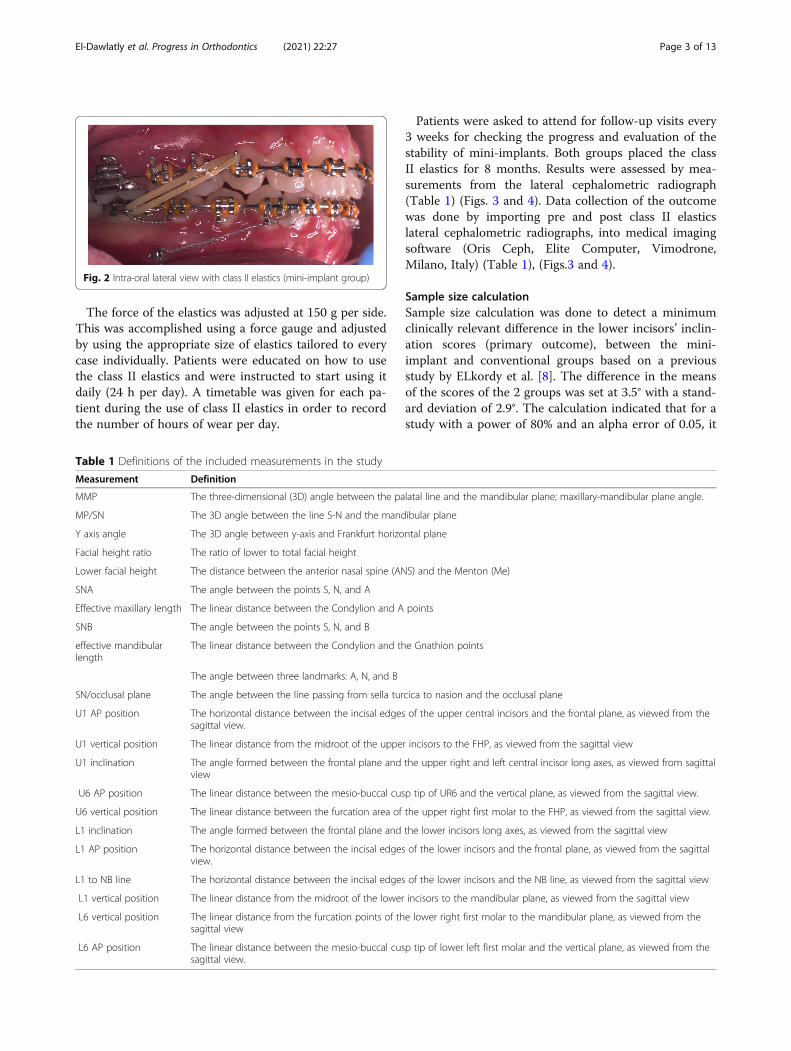
Fig. 2 Intra-oral lateral view with class II elastics (mini-implant group)
Table 1 Definitions of the included measurements in the study
Measurement Definition
MMP The three-dimensional (3D) angle between the palatal line and the mandibular plane; maxillary-mandibular plane angle.
MP/SN The 3D angle between the line S-N and the mandibular plane
Y axis angle The 3D angle between y-axis and Frankfurt horizontal plane
Facial height ratio The ratio of lower to total facial height
Lower facial height The distance between the anterior nasal spine (ANS) and the Menton (Me)
SNA The angle between the points S, N, and A
Effective maxillary length The linear distance between the Condylion and A points
SNB The angle between the points S, N, and B
effective mandibularlength
The linear distance between the Condylion and the Gnathion points
The angle between three landmarks: A, N, and B
SN/occlusal plane The angle between the line passing from sella turcica to nasion and the occlusal plane
U1 AP position The horizontal distance between the incisal edges of the upper central incisors and the frontal plane, as viewed from thesagittal view.
U1 vertical position The linear distance from the midroot of the upper incisors to the FHP, as viewed from the sagittal view
U1 inclination The angle formed between the frontal plane and the upper right and left central incisor long axes, as viewed from sagittalview
U6 AP position The linear distance between the mesio-buccal cusp tip of UR6 and the vertical plane, as viewed from the sagittal view.
U6 vertical position The linear distance between the furcation area of the upper right first molar to the FHP, as viewed from the sagittal view.
L1 inclination The angle formed between the frontal plane and the lower incisors long axes, as viewed from the sagittal view
L1 AP position The horizontal distance between the incisal edges of the lower incisors and the frontal plane, as viewed from the sagittalview.
L1 to NB line The horizontal distance between the incisal edges of the lower incisors and the NB line, as viewed from the sagittal view
L1 vertical position The linear distance from the midroot of the lower incisors to the mandibular plane, as viewed from the sagittal view
L6 vertical position The linear distance from the furcation points of the lower right first molar to the mandibular plane, as viewed from thesagittal view
L6 AP position The linear distance between the mesio-buccal cusp tip of lower left first molar and the vertical plane, as viewed from thesagittal view.
El-Dawlatly et al. Progress in Orthodontics (2021) 22:27 Page 3 of 13
is required to have 12 participants per group. The num-ber was raised to 14 to compensate for any expecteddrop-outs. The calculation was carried out using PS:Power and Sample Size calculator software Version 3.1.2(Vanderbilt University, Nashville, TN, USA)
RandomizationPatients were randomly allocated to group A (mini-implant) or B (conventional) using random sequencegenerated at random.org. Simple randomization wasdone with allocation ratio 1:1. The randomization wasdone before the start of treatment. Each number fromthe generated sequence (from 1 to 28) was put in anopaque sealed envelope and all envelopes were placedin a closed box. After completion of the leveling andalignment, each patient was requested to choose oneenvelope. According to the number in the envelope,the patient was then allocated into one of the twogroups. The randomization and allocation steps werecarried out by the clinic instructor who was not apart of the study.
BlindingBlinding was not applicable for the operators during theinsertion of the appliance and the follow-up visits. Theclinic instructor was responsible for assigning subjects tomini-implant or conventional groups according to theconcealed allocation. Appliance activation at follow-upvisits, dental impressions, and acquisition of dental castswere the responsibility of the principal investigator. Itwas a single-blinded study; only the outcome assessorswere blind. The patient names were removed from pre-and post-treatment lateral cephalometric radiographsand study models. Then, two assessors carried on,blindly and independently, the measurements and ana-lysis of the study.
Statistical analysisStatistical analysis was divided in 3 sections:
1. Comparing the skeletal and dental changes betweenthe conventional and mini-implant groups (primaryoutcome).
2. Analyzing pre and post, skeletal, and dental changesin conventional group (secondary outcome).
3. Analyzing pre and post, skeletal, and dental changesin mini-implant group (secondary outcome).
All data were collected, tabulated, and subjected tostatistical analysis. Statistical analysis was performedby SPSS in general (version 16), while Microsoftoffice Excel was used for data handling and graphicalpresentation.
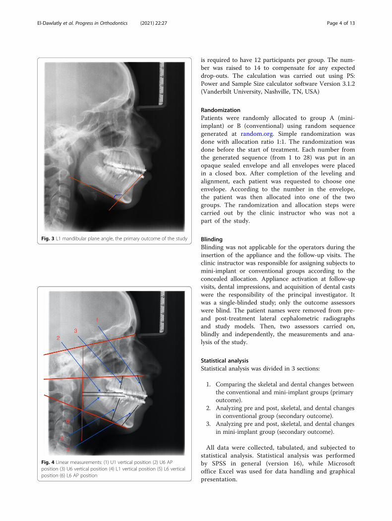
Fig. 3 L1 mandibular plane angle, the primary outcome of the study
Fig. 4 Linear measurements: (1) U1 vertical position (2) U6 APposition (3) U6 vertical position (4) L1 vertical position (5) L6 verticalposition (6) L6 AP position
El-Dawlatly et al. Progress in Orthodontics (2021) 22:27 Page 4 of 13
Quantitative variables were described by the mean,standard deviation (SD), the range (minimum–maximum),standard error (SE), and 95% confidence interval of themean.Qualitative categorical variables were described by
proportions and Percentages.Shapiro-Wilk test of normality was used to test nor-
mality hypothesis of all quantitative variables for furtherchoice of appropriate parametric and non-parametrictests. Mainly, the variables were found to be normallydistributed allowing the use of parametric tests. Paired ttest was applied for comparing the changes pre- andpost-treatment within each group and Independentsamples t test for comparing the differences (post–pre)between the two groups.For reliability analysis of inter- and intra-observer of
all measured variables, Dahlberg error (DE) and relativeDahlberg error (RDE) were used together with concord-ance correlation coefficients (CCC) including its 95%confidence limits.Significance level was considered at P < 0.05 (S); while
for P < 0.01 was considered highly significant (HS).Two-tailed tests were assumed throughout the analysisfor all statistical tests.
ResultsThe total sample comprised 28 adolescent female pa-tients (divided into 2 groups (14 per each group). Themean ages were 15.66 ± 2 years for the mini-implantgroup and 15.1 ± 2.2 years for the conventional group.There were no drop-outs, where all the patients com-pleted the full length of treatment.The treatment time (including leveling and alignment
and elastic wear period excluding the finishing stage)was about 14.75 ± 1.8 months for the mini-implantgroup and 15.12 ± 1.67 for the conventional group. Thedifference in the time interval of the use of the elasticswas insignificant between the two groups (P > 0.05)(mini-implant group 8 ± 0.93 months and control group7.47 ± 0.52 months)There was no significant difference in the degree of
compliance and adherence to elastics wear between thetwo groups. (P > 0.05). Where the mean hours the elas-tics were worn in the mini-implant group was 19.13 ±1.68 h/day, while it was 18.7 ± 1.22 for the controlgroup.Baseline analysis was done before the trial by compar-
ing conventional group and mini-implant group pre-treatment measurements SNB, ANB, and MMP, Effect-ive mandibular length and L1 inclination. There was nostatistically significant difference between the twogroups. Excellent inter-observer and intra-observer reli-ability were detected, since RDE did not exceed 10% andCCC values were recorded between 0.991 and 1.
Statistical results were divided as follows:
Analyzing pre and post, skeletal and dentalchanges in group: (Table 2)The skeletal measurements that showed statisticallysignificant change:
� Increase in the MP/SN, Y-axis angle, lower facialheight and in the clock-wise rotation of the occlusalplane (SN/occlusal plane).
The dental measurements that showed statisticallysignificant change:
� Increase in the proclination of L1, L1 AP position(anterior movement), L6 vertical position(extrusion), and the retroclination of U1.
1. Analyzing pre and post, skeletal, and dental changesin mini-implant group: (Table 3).
The skeletal measurements that showed statisticallysignificant change:
� Increase in the Y-axis angle, lower facial height,and the clock-wise rotation of occlusal plane (SN/occlusal plane).
The dental measurements that showed statistically sig-nificant change:
� Decrease in the U1 AP position, U1 inclination(retroclination)
� Increase in the proclination of L1, L1 AP position(anterior movement), L6 vertical position(extrusion), L6 AP position (mesial movement).
2. Comparing the skeletal and dental changes betweenthe conventional and mini-implant groups: (Table 4).
Only two dental measurements showed statisticallysignificant difference. Those were the lingual movementof the upper incisors U1 AP position and the retroclina-tion of the upper incisors U1 inclination (Table 4). Allthe other skeletal and dental measurements showed nostatistical significance.The overjet was reduced significantly in both mini-
implant and control groups (P < 0.001); with a meanof − 4.2 ± 0.71 in the mini-implant group and −3.81 ± 0.8 in the conventional group, while thedifference between them was not significant 0.39 ±0.31 (P > 0.05). Harms: no serious adverse effectswere observed other than gingivitis associated withplaque accumulation.
El-Dawlatly et al. Progress in Orthodontics (2021) 22:27 Page 5 of 13
Table 2 Paired t test for comparing the dental and skeletal changes after class II elastics phase for the conventional group
Variable Paired differences t P value
Mean SD Mean SD SEM
SNA Pre 82.08 2.97 − 0.33 2.23 0.64 − 0.52 0.61474 P > 0.05 NS
Post 81.75 3.19
SNB Pre 76.25 3.22 0.17 2.79 0.81 0.21 0.83988 P > 0.05 NS
Post 76.42 3.68
ANB Pre 5.83 1.11 − 0.50 1.57 0.45 − 1.11 0.29252 P > 0.05 NS
Post 5.33 1.72
MMP Pre 28.08 3.45 0.42 2.02 0.58 0.71 0.48993 P > 0.05 NS
Post 28.50 3.18
MP/SN Pre 36.08 4.58 1.38 2.23 0.64 2.14 0.05573 P ≈ 0.05 Almost S
Post 37.46 5.27
Y axis angle Pre 57.20 3.70 2.30 4.67 1.35 5.62 0.00015 P < 0.001 HS
Post 59.50 3.75
Lower facial height Pre 63.70 5.47 2.30 4.91 1.42 3.36 0.00637 P < 0.01 HS
Post 66.00 4.47
Effective maxillary length Pre 83.17 4.20 0.75 5.28 1.52 0.49 0.63212 P > 0.05 NS
Post 83.92 4.68
Effective mandibular length Pre 108.50 5.52 2.00 8.34 2.41 0.83 0.42406 P > 0.05 NS
Post 110.50 6.08
SN/occlusal plane Pre 16.66 3.44 4.30 2.65 0.77 5.62 0.00016 P < 0.001 HS
Post 20.96 3.69
U1 vertical position Pre 20.92 3.17 1.67 3.39 0.98 1.70 0.11628 P > 0.05 NS
Post 22.58 2.81
U1 AP position Pre 7.30 3.06 − 0.30 3.82 1.10 − 0.27 0.79047 P > 0.05 NS
Post 7.00 1.76
U1 inclination Pre 113.00 4.13 − 3.33 1.61 0.47 3.33 0.00002 P < 0.001 HS
Post 109.67 4.42
U6 AP position Pre 42.17 2.92 − 2.04 5.91 1.71 2.04 0.25659 P > 0.05 NS
Post 40.13 4.89
U6 vertical position Pre 16.33 2.53 0.08 2.16 0.62 0.13 0.89619 P > 0.05 NS
Post 16.42 1.94
L1 inclination Pre 100.00 6.09 5.42 2.81 0.81 6.68 0.00003 P < 0.001 HS
Post 105.42 5.12
L1 AP position Pre 5.08 1.62 1.33 1.44 0.41 3.22 0.00819 P < 0.01 HS
Post 6.42 1.62
L1 vertical position Pre 30.75 4.77 2.42 4.70 1.36 1.78 0.10243 P > 0.05 NS
Post 33.17 3.93
L6 vertical Pre 23.54 2.68 1.21 0.99 0.29 − 1.21 0.00139 P < 0.001 HS
Post 24.75 2.83
L6 AP position Pre 42.08 3.23 0.92 6.35 1.83 0.50 0.62664 P > 0.05 NS
Post 43.00 5.77
El-Dawlatly et al. Progress in Orthodontics (2021) 22:27 Page 6 of 13
A consort flow diagram (Fig. 5) together with a con-sort check list (Fig. 6) was made to describe the steps ofthe current randomized controlled trial.
DiscussionClass II elastics could be used to camouflage mild skel-etal class II or to treat mild to moderate dental class II
Table 3 Paired t test for comparing the dental and skeletal changes after class II elastics phase for the mini-implant group
Variable Paired differences t P value
Mean SD Mean SD SEM
SNA Pre 82.78 3.00 − 1.12 2.50 0.72 − 1.55 0.14986 P > 0.05 NS
Post 81.67 3.42
SNB Pre 76.54 2.79 − 0.79 2.39 0.69 − 1.15 0.27516 P > 0.05 NS
Post 75.75 3.02
ANB Pre 6.20 1.47 − 0.37 0.79 0.23 − 1.62 0.13415 P > 0.05 NS
Post 5.83 1.11
MMP Pre 29.85 3.74 0.32 2.83 0.82 0.39 0.70541 P > 0.05 NS
Post 30.17 3.49
MP/SN Pre 38.68 4.67 0.37 2.13 0.62 0.60 0.56311 P > 0.05 NS
Post 39.04 4.50
Y axis angle Pre 56.85 3.35 2.15 1.32 0.38 5.62 0.00015 P < 0.001 HS
Post 59.00 2.98
Lower facial height Pre 62.53 3.60 2.13 2.20 0.64 3.36 0.00637 P < 0.01 HS
Post 64.67 4.64
Effective maxillary length Pre 84.13 4.57 0.70 3.00 0.87 0.81 0.43606 P > 0.05 NS
Post 84.83 4.32
Effective mandibular length Pre 106.37 5.26 1.97 4.31 1.24 1.58 0.14184 P > 0.05 NS
Post 108.33 5.65
SN/occlusal plane Pre 16.54 3.52 4.12 2.18 0.63 6.55 0.00004 P < 0.001 HS
Post 20.66 4.11
U1 vertical position Pre 20.73 2.89 1.06 2.31 0.67 1.59 0.14071 P > 0.05 NS
Post 21.79 1.95
U1 AP position Pre 13.18 3.00 − 4.35 2.99 0.86 − 5.04 0.00038 P < 0.001 HS
Post 8.83 2.48
U1 inclination Pre 115.94 2.04 − 8.57 6.45 1.86 − 4.60 0.00076 P < 0.001 HS
Post 107.38 5.48
U6 AP position Pre 44.17 6.10 − 3.29 6.38 1.84 − 1.79 0.10161 P > 0.05 NS
Post 40.88 4.86
U6 vertical position Pre 15.13 2.11 0.58 0.91 0.26 2.22 0.04819 P < 0.05 S
Post 15.71 2.08
L1 inclination Pre 99.88 4.80 4.45 1.47 0.42 10.52 0.00000 P < 0.001 HS
Post 104.33 4.12
L1 AP position Pre 4.78 1.19 1.03 1.63 0.47 2.18 0.05173 P ≈ 0.05 Almost S
Post 5.81 1.59
L1 vertical position Pre 31.68 2.36 0.87 2.02 0.58 1.49 0.16480 P > 0.05 NS
Post 32.54 3.20
L6 vertical Pre 22.14 2.17 1.19 0.84 0.24 4.93 0.00045 P < 0.001 HS
Post 23.33 1.83
L6 AP position Pre 42.18 4.47 2.98 3.82 1.10 2.71 0.02043 P < 0.05 S
Post 45.17 3.69
El-Dawlatly et al. Progress in Orthodontics (2021) 22:27 Page 7 of 13
Table 4 Independent samples T test for comparing the differences in the dental and skeletal changes after class II elastics phase forbetween the 2 groups
Variable Group Differences 95% Confidence interval of the difference t P value
Mean SD Mean SD Lower Upper
SNA C − 0.33 2.23 0.78 0.97 − 1.22 2.79 0.81 0.42642 P > 0.05 NS
I − 1.12 2.50
SNB C 0.17 2.79 0.96 1.06 − 1.24 3.16 0.90 0.37587 P > 0.05 NS
I − 0.79 2.39
ANB C − 0.50 1.57 − 0.13 0.51 − 1.18 0.92 − 0.26 0.79458 P > 0.05 NS
I − 0.37 0.79
MMP C 0.42 2.02 0.10 1.00 − 1.98 2.18 0.10 0.92150 P > 0.05 NS
I 0.32 2.83
MP/SN C 1.38 2.23 1.01 0.89 − 0.84 2.85 1.13 0.26928 P > 0.05 NS
I 0.37 2.13
Y axis angle C 2.30 4.67 0.15 1.40 − 2.76 3.06 0.11 0.91572 P > 0.05 NS
I 2.15 1.32 0.15
Lower facial height C 2.30 4.91 0.17 1.55 − 3.05 3.39 0.11 0.91545 P > 0.05 NS
I 2.13 2.20 0.17 1.55 − 3.14 3.47 0.11
Effective maxillary length C 0.75 5.28 0.05 1.75 − 3.58 3.68 0.03 0.97749 P > 0.05 NS
I 0.70 3.00
Effective mandibular length C 2.00 8.34 0.03 2.71 − 5.59 5.65 0.01 0.99030 P > 0.05 NS
I 1.97 4.31
SN/occlusal plane C 4.30 2.65 0.18 0.99 − 1.87 2.24 0.19 0.85482 P > 0.05 NS
I 4.12 2.18
U1 vertical position C 1.67 3.39 0.61 1.18 − 1.85 3.06 0.51 0.61232 P > 0.05 NS
I 1.06 2.31
U1 AP position C − 0.30 3.82 4.05 1.40 1.15 6.95 2.89 0.00842 P < 0.01 HS
I − 4.35 2.99
U1 inclination C − 3.33 1.61 5.23 1.92 1.26 9.21 2.73 0.01227 P < 0.05 S
I − 8.57 6.45
U6 AP position C − 2.04 5.91 1.25 2.51 − 3.96 6.46 0.50 0.62360 P > 0.05 NS
I − 3.29 6.38
U6 vertical position C 0.08 2.16 − 0.50 0.68 − 1.90 0.90 − 0.74 0.46804 P > 0.05 NS
I 0.58 0.91
L1 inclination C 5.42 2.81 0.97 0.92 − 0.93 2.86 1.06 0.30225 P > 0.05 NS
I 4.45 1.47 − 0.97 2.90 1.06
L1 AP position C 1.33 1.44 0.31 0.63 − 0.99 1.61 0.49 0.62748 P > 0.05 NS
I 1.03 1.63
L1 vertical position C 2.42 4.70 1.55 1.48 − 1.51 4.61 1.05 0.30515 P > 0.05 NS
I 0.87 2.02
L6 vertical C 1.21 0.99 0.02 0.37 − 0.76 0.79 0.04 0.96483 P > 0.05 NS
I 1.19 0.84
L6 AP position C 0.92 6.35 − 2.07 2.14 − 6.50 2.37 − 0.97 0.34422 P > 0.05 NS
I 2.98 3.82
C conventional group, I mini-implant group
El-Dawlatly et al. Progress in Orthodontics (2021) 22:27 Page 8 of 13
malocclusions. The use of class II elastics could beextended to anchorage reinforcement, bite opening (incases having class II division2) [9], midline deviationcorrection. The adverse effects of using class II elasticscomprise both vertical and horizontal force vectors [9].This vertical force extrudes the maxillary incisors andmandibular molars and can lead to clock-wise rotationof the occlusal plane. The horizontal vector of force hasbeen shown to cause the mandibular first molars to ro-tate or tip mesially, procline the mandibular anterior
teeth, and displace the entire lower dental arch anteri-orly [10].Many studies investigated the effect of class II elastics
and compared its effects versus those of fixed functionalappliances, but none of them used skeletal anchorage toprevent or counteract the drawbacks of class II elastics[11]. Accordingly, we attempted to use mini implantsanchorage aiming to reduce the lower incisors proclina-tion and decrease the extrusion of lower first molars ac-companied by class II elastics in non-extraction cases.
Fig. 5 Consort flow diagram
El-Dawlatly et al. Progress in Orthodontics (2021) 22:27 Page 9 of 13
Fig. 6 consort check list
El-Dawlatly et al. Progress in Orthodontics (2021) 22:27 Page 10 of 13
The adolescent female patients included in thesample were in their early post-pubertal age. Onlyfemale patients were recruited, aiming to eliminatethe potential residual growth differences betweenmales and females in this age group. Representing alimitation, this unfortunately restricted the generalizationof the results of the current study and limited it to thefemale gender.Moreover, the results should be interpreted with cau-
tion due to the limited sample. In addition, the currentstudy tested a new genuine technique; accordingly, thesample size calculation was based on a study that usedthe same skeletal anchorage protocol but on a fixedfunctional appliance [8] instead of class II elastics.Mini-implants were placed between the lower second
premolar and first molar as it was stated by Fayed et al.[12] that this site is considered one of the optimal sitesfor placing mini-implants in the lower arch. Eight-millimeter-long mini-implant was chosen as it wasreported that shorter mini-implant will not be stableenough and longer mini-implants risks injury to the in-ferior alveolar nerve [13].In the current study, lateral cephalograms were taken
two times; before the start of class II elastics therapy andjust after the end of the elastics phase for each patient.However, the cephalometric radiograph has the limita-tion of reduced validity and/or reproducibility of itslandmarks; it remains the radiographic tool of choice forthe sake of avoiding the high dose of the 3D cone beamCT. The acquisition of cephalometry at the start andend of treatment was a must in order to distinguish theresults of elastics therapy excluding the incisors procli-nation effect of the leveling and alignment phase. Thiswas compensated by avoiding the acquisition of a post-treatment cephalometry.In the conventional group, the results showed that
class II elastics were effective in correcting class II mal-occlusions, and their effects were mainly dentoalveolar,including lingual tipping, retrusion, and extrusion of themaxillary incisors; labial tipping of the mandibular inci-sors and mesialization and extrusion of the mandibularmolars. Also, there was a significant clockwise rotationin the mandibular plane angle and occlusal plane. Theseresults were in agreement with Janson et al. [11] whenthey evaluated the effects of class II elastics in class IImalocclusion treatment in their systematic review.In the skeletal anchorage group, the lower molars
moved mesially significantly, and this revealed totalmovement of the whole mandibular dentition to a moreforward position. These results were in agreement withthose of Janson et al. [11] as they reported that class IIelastics enhanced the forward movement of the man-dibular first molars, moving 1.2 mm mesial, and lead toproclination of the mandibular incisors.
In the skeletal anchorage group, the use of mini-implants did not prevent the proclination of the lowerincisors as was shown in the lower incisors proclination(4.45 ± 1.47°) and lower incisors anteroposterior position(1.03 ± 1.63 mm). The upper incisors were retroclined(− 8.57 ± 6.45°) and moved distally significantly (− 4.35± 2.99 mm). There was a significant increase in theretroclination (5.23 ± 1.92°) and distal movement (4.05 ±1.4 mm) of the upper incisors. The distal movement inthe upper incisors helped in a more pronounced camou-flaging effect of the class II malocclusion in the mini-implant group [4]. The significant retroclination of theupper incisors lead to the increase of the incisal displaywhich is beneficial to cases having decreased incisalshow at rest and on smiling; this was confirmed byArmando Saga et al. [14] who found that retroclinedmaxillary incisors that occur after orthodontic anteriorretraction without torque control tend to increase theincisor exposition and worsen an existing gummy smile.On the other hand, this will lead to further deepening ofthe bite [10, 1].The significant proclination of lower incisors in both
groups may be due to the degree of play between thewire and the brackets despite using 0.017 × 0.025 stain-less steel arch wires which were heavier than the wiresused in almost all articles investigating class II elasticwear. Janson et al. [11] stated in their systematic reviewthat the current literature suggests using light forces (2.6oz) obtained with 3/16 diameter elastics and a rectangu-lar 0.016 × 0.022 stainless steel arch-wires. The use ofligature wires as a way of attachment between the mini-implant and the arch wire could have shared in the in-capability of the current technique to reduce the flaringof the lower incisors. The use of other active methods ofattaching the mini-implant, like elastic chains, in furtherstudies might have a better impact on reducing the inci-sors proclination. Also, the use of 0.019 × 0.025 stainlesssteel arch wires could be tested in further studies andcompared to other lower dimensional arch wires toreach a conclusive evidence in terms of the effect of thewire dimensions on class II elastics outcomes.Other studies used the mini-implant to limit the pro-
clination of lower incisors in conjunction with fixedfunctional appliances. They found reduction in theirproclination in the mini-implant group which was notfound in the present study [15, 16]. This could be attrib-uted to the use of a different type of appliance or to thenature of force applied on the lower incisors, where theforce delivered by the class II elastics has a more jigglingnature than that delivered by the fixed functionalappliances.Extrusion of lower first molar and upper anterior teeth
with clockwise rotation of the mandible and occlusalplane were noticed in both groups. This proved that the
El-Dawlatly et al. Progress in Orthodontics (2021) 22:27 Page 11 of 13
use of skeletal anchorage in the mandibular arch couldnot prevent the adverse effects produced by the verticalforce component of elastics attachment points. Thesefindings agree with Janson et al. [4] study, reportinginsignificant positional changes of teeth at the end ofcomprehensive class II elastic treatment but there was asignificant increase in the vertical measurements.There were no statistically significant differences in
the skeletal variables in the skeletal anchorage grouplike SNA (− 1.12 ± 2.5°), SNB (0.79 ± 2.39°), andANB (− 0.37 ± 0.79°); this agreed with Janson et al.[11] as they stated that class II elastics effect wasabout 18.9% skeletal and 71.1% dentoalveolar and thesignificant retroclination of the maxillary teethworked against the mandibular advancement. More-over, the mean age of the patients in the mini-implant group was 15.66 ± 2 years and 15.1 ± 2.2years for the conventional group. Accordingly, mostof the craniofacial growth had ended at the start oftreatment, thus, limiting the potential for mandibulargrowth enhancement.Very good adherence to the elastics was found in this
study with an average of 19 h per day wearing time. Nopatients reported any severe pain related to elastics wearor mini-implants. The results of this study concludedthat, mini-implants placed in the mandibular inter-radicular bone in conjunction with class II elastics hadno skeletal effect. The dentoalveolar effect predominatedand the current technique did not prevent the excessiveproclination of lower incisors. It had some little positiveeffects, as it enhanced more mesial movement of man-dibular dentition.
Conclusion
� Mini-implants placed in the mandibularinterradicular bone in conjunction with Class IIelastics had no significant skeletal effects, onlydentoalveolar movements resulted.
� Skeletal anchorage didn’t prevent the proclination oflower incisors, the extrusion of the lower buccalsegment, or the clockwise rotation of themandibular and occlusal planes.
� Skeletal anchorage increased the retroclination ofthe upper incisors and the distal movement of theupper incisors in the skeletal anchorage group whichhelped in enhancing the camouflaging effect of ClassII malocclusion.
Authors’ contributionsMohamed A. Mabrouk developed the research idea, shared in designing andapplying the treatment mechanics, and shared in writing the manuscript.Mostafa el Dawlatly developed the research idea, shared in designing andapplying the treatment mechanics, and shared in writing the manuscript.Amr El Dakroury shared in interpreting the results and took part in revisingthe manuscript. Yehya A Mostafa shared in developing the research idea,shared in designing treatment mechanics, and shared in writing themanuscript. All authors read and approved the final manuscript.
FundingThe study was self-funded by the authors.
Availability of data and materialsThe datasets used and analyzed during the current study are available fromthe corresponding author on reasonable request.
Declarations
Ethics approval and consent to participateEthical approval was obtained from the Future University before the start oftreatment. The number of the approval was FUE. REC. (10)/10-2018.
Consent for publicationConsent was obtained from the patient’s parents, as they were adolescents,before their recruitment.
Competing interestsThe authors declare that they have no competing interests.
Author details1Department of Orthodontics Faculty of Oral and Dental Medicine, CairoUniversity, Cairo, Egypt. 2Department of Orthodontics, Future University inEgypt (FUE), Cairo, Egypt.
Received: 22 November 2020 Accepted: 3 June 2021
References1. Alhammadi MS, Halboub E, Fayed MS, Labib A, El-Saaidi C. Global
distribution of malocclusion traits: a systematic review. Dental Press JOrthod. 2018;23(6):40.e1–40.e10.
2. Bilgic F, Gelgor IE, Celebi AA. Malocclusion prevalence and orthodontictreatment need in central Anatolian adolescents compared to Europeanand other nations' adolescents. Dental Press J Orthod. 2015;20(6):75–81.https://doi.org/10.1590/2177-6709.20.6.075-081.oar.
3. Moyers RE, Riolo ML, Guire KE, Wainright RL, Bookstein FL. Differentialdiagnosis of Class II malocclusions: Part 1. Facial types associated with ClassII malocclusions. Am J Orthod. 1980;78(5):477–94. https://doi.org/10.1016/0002-9416(80)90299-7.
4. Jones G, Buschang PH, Kim KB, Oliver DR. Class II non-extraction patientstreated with the forsus fatigue resistant device versus intermaxillary elastics.Angle Orthod. 2008;78(2):332–8. https://doi.org/10.2319/030607-115.1.
5. Aras I, Pasaoglu A. Class II subdivision treatment with the forsus fatigueresistant device vs intermaxillary elastics. Angle Orthod. 2017;87(3):371–6.https://doi.org/10.2319/070216-518.1.
6. Manni A, Pasini M, Mauro C. Comparison between Herbst applianceswith or without miniscrew anchorage. Dent Res J (Isfahan). 2012;9(Suppl 2):S216–21.
7. Luzi C, Valeriano L, Melsen B. The miniscrew-anchored herbst. 2012; Journalof clinical orthodontics: JCO, (July 2016).
8. Elkordy SA, Abouelezz AM, Fayed MMS, Attia KH, Ishaq RAR, Mostafa YA.Three-dimensional effects of the mini-implant-anchored Forsus fatigueresistant device: a randomized controlled trial. Angle Orthod. 2016;86(2):292–305. https://doi.org/10.2319/012515-55.1.
9. Langlade M. Optimization of orthodontic elastics. New York: GACInternational Ed; 2000.
10. Bratu CD, Fleser C, Glavan F. The effect on intermaxillary elastics inorthodontic therapy. 2004;54(4):406–9.
11. Janson G, Sathler R, Fernandes T, Castello Branco N, Freitas M. Correction ofclass ii malocclusion with class ii elastics: a systematic review. Am. J. Orthod.
El-Dawlatly et al. Progress in Orthodontics (2021) 22:27 Page 12 of 13
12. Fayed MM, Pazera P, Katsaros C. Optimal sites for orthodontic mini-implantplacement assessed by cone beam computed tomography. Angle Orthod.2010;80(5):939–51. https://doi.org/10.2319/121009-709.1.
13. Leo M, Cerroni L, Pasquantonio G. saverio giovanni condò, Condo R.Temporary anchorage devices (TADs) in orthodontics: review of the factorsthat influences the clinical success rate of the mini-implants. Clin. Ter. 2016;167:70–7.
14. Saga A, Araújo E, Antelo O, Meira T, Tanaka O. Nonsurgical treatment ofskeletal maxillary protrusion with gummy smile using headgear for growthcontrol, mini-implants as anchorage for maxillary incisor intrusion, andpremolar extractions for incisor retraction. Am. J. Orthod. Dentofac. Orthop.2020;157(2):245–58. https://doi.org/10.1016/j.ajodo.2018.09.021.
15. Manni A, Migliorati M, Calzolari C, Silvestrini-Biavati A. Herbst applianceanchored to miniscrews in the upper and lower arches vs standard Herbst:A pilot study. Am J Orthod Dentofacial Orthop. 2019;156(5):617–25. https://doi.org/10.1016/j.ajodo.2018.11.015.
16. Elkordy SA, Aboelnaga AA, Fayed MM, AboulFotouh MH, Abouelezz AM.Can the use of skeletal anchors in conjunction with fixed functionalappliances promote skeletal changes? A systematic review and meta-analysis. Eur J Orthod. 2016;38(5):532–45. https://doi.org/10.1093/ejo/cjv081.
Publisher’s NoteSpringer Nature remains neutral with regard to jurisdictional claims inpublished maps and institutional affiliations.
El-Dawlatly et al. Progress in Orthodontics (2021) 22:27 Page 13 of 13