Vol. 64 TRANSFRUCTOSYLATION BY LEVANSUCRASE 361 We are deeply indebted to Dr D. Gross for electrophoretic measurements and valuable suggestions. A generous gift of dextransucrase by Dr H. M. Tsuchiya is acknowledged. REFERENCES Albon, N., Bell, D. J., Blanchard, P. H., Gross, D. & Rundell, J.T. (1953). J. chem. Soc. p. 24. Baar, S. & Bull, J. P. (1953). Nature, Lond., 172,414. Bacon, J. S. D. (1954a). Annual Rep. Chem. Soc. 50, 281. Bacon, J. S. D. (1954b). Biochem. J. 57, 320. Bacon, J. S. D. & Bell, D. J. (1953). J. chem. Soc. p. 2528. Bacon, J. S. D. & Edelman, J. (1950). Arch. Biochem. 28, 467. Bacon, J. S. D. & Edelman, J. (1951). Biochem. J. 48, 114. Barker, S. A., Bourne, E. J. & Carrington, T. R. (1954). J. chem. Soc. p. 2125. Barker, S. A., Bourne, E. J., Wilkinson, I. A. & Peat, S. (1950). J. chem. Soc. p. 93. Bealing, F. J. & Bacon, J. S. D. (1953). Biochem. J. 53,277. Bell, D. J. & Dedonder, R. (1954). J. chem. Soc. p. 2866. Bell, D. J. & Edelman, J. (1954). J. chem. Soc. p. 4652. Consden, R. & Stanier, W. N. (1952). Nature, Lond., 169, 783. Decker, P. (1951). NaturwiMsenschaften, 38, 287. Dedonder, R. & Noblesse, M. (1953). Ann. Inst. Pasteur, 85, 356. Edelman, J. (1954). Biochem. J. 57, 22. Feingold, D., Avigad, G. & Hestrin, S. (1955). Bull. Re8. Council, Israel, 5A, 98. Foster, A. B. (1953). J. chem. Soc. p. 982. French, D. & Wild, G. (1953). J. Amer. chem. Soc. 75,2612. Gross, D. (1953). Nature, Lond., 172, 908. Gross, D. (1954). Nature, Lond., 173, 487. Hehre, E. J. (1951). Advanc. Enzymol. 11, 297. Hehre, E. J. & Hamilton, D. M. (1951). J. biol. Chem. 192, 161. Hestrin, S. (1953). Symp. Microbiol. Metabolism, lstituto Superiore di Sanita, Rome, pp. 63-70. Hestrin, S., Feingold, D. S. & Avigad, G. (1956). Biochem. J. 64, 340. Hestrin, S. & Lindegren, C. C. (1952). Arch. Biochem. Biophys. 38, 317. Horrocks, R. H. & Manning, G. B. (1949). Lancet, 256, 1042. Jackson, E. L. (1944). Organic Reactions, vol. 2, p. 345. New York: John Wiley and Sons Inc. Jeanes, A., Wise, C. S. & Dimler, R. J. (1951). Analyt. Chem. 23, 415. Keilin, D. & Hartree, E. F. (1948). Biochem. J. 42, 230. Kohanyi, G. & Dedonder, R. (1951). C.R. Acad. Sci., Paris, 233, 1142. Macleod, M. & Robison, R. (1929). Biochem. J. 23, 517. Mitchell, D. T., Shildneck, P. & Dustin, J. (1944). Industr. Engng Chem. (Anal.), 16, 754. Partridge, S. M. (1948). Biochem. J. 42, 238. Perlin, A. S. (1954). J. Amer. chem. Soc. 76, 4101. Raybin, H. W. (1933). J. Amer. chem. Soc. 55, 2603. Roe, J. H., Epstein, J. H. & Goldstein, N. P. (1949). J. biol. Chem. 178, 839. Seegmiller, T. E. & Horecker, B. L. (1951). J. biol. Chem. 192, 175. Somogyi, M. (1945). J. biol. Chem. 160, 69. Wallenfels, K. (1950). Naturwissenschaften, 37, 491. Whelan, W. J., Bailey, J. M. & Roberts, P. J. P. (1953). J. chem. Soc. p. 1293. White, L. M. & Secor, G. E. (1952). Arch. Biochem. Biophys. 36, 490. Williams, K. T. & Bevenue, A. (1951). Science, 113, 582. Wise, C. S., Dimler, R. J., Davis, H. A. & Rist, C. E. (1955). Analyt. Chem. 27, 33. The Enzymic Properties of Rat-liver Nuclei 1. ESTIMATION OF THE EXTENT TO WHICH CONTAMINANT MATERIAL CONTRIBUTES TO THE ACTIVITY OBSERVED IN THE NUCLEAR FRACTION BY D. B. ROODYN* Department of Biochemi8try, Univer8ity of Cambridge (Received 1 February 1956) The isolation of nuclei from tissue homogenates by differential centrifuging has not given very clear information about the enzymxic properties of the nucleus. One of the main difficulties is that often only a small percentage of the total activity of the homogenate is found in the nuclear fraction and, as many of the fractionation methods most commonly used for enzyme studies do not prevent the con- tamination of the nuclei with larger or smaller amounts of cytoplasmic particles, unbroken cells, fibrous material and erythrocytes, it is very difficult to decide whether the observed activity is to be ascribed to the nuclei themselves or to the con- taminants. It was therefore decided to investigate the position for two enzymes, aldolase and succin- oxidase, in an attempt to establish whether the low activities reported by other workers to be present in the nuclear fractions are due to the nuclei or not. Dounce (1948) has reported that liver nuclei prepared in dilute citric acid do not contain succinic dehydrogenase, and this enzyme was also reported to be absent from rat-liver and pig-kidney nuclei prepared by Lang & Siebert (1950). However, none * Present address: Medical Research Council Radio- biological Research Unit, Atomic Energy Research Estab- lishment, Harwell, Didcot, Berks.
Transcript
Vol. 64 TRANSFRUCTOSYLATION BY LEVANSUCRASE 361We are deeply indebted to Dr D. Gross for electrophoretic
measurements and valuable suggestions. A generous gift ofdextransucrase by Dr H. M. Tsuchiya is acknowledged.
REFERENCES
Albon, N., Bell, D. J., Blanchard, P. H., Gross, D. & Rundell,J.T. (1953). J. chem. Soc. p. 24.
Baar, S. & Bull, J. P. (1953). Nature, Lond., 172,414.Bacon, J. S. D. (1954a). Annual Rep. Chem. Soc. 50,
281.Bacon, J. S. D. (1954b). Biochem. J. 57, 320.Bacon, J. S. D. & Bell, D. J. (1953). J. chem. Soc. p. 2528.Bacon, J. S. D. & Edelman, J. (1950). Arch. Biochem. 28,
467.Bacon, J. S. D. & Edelman, J. (1951). Biochem. J. 48,
114.Barker, S. A., Bourne, E. J. & Carrington, T. R. (1954).
J. chem. Soc. p. 2125.Barker, S. A., Bourne, E. J., Wilkinson, I. A. & Peat, S.
(1950). J. chem. Soc. p. 93.Bealing, F. J. & Bacon, J. S. D. (1953). Biochem. J. 53,277.Bell, D. J. & Dedonder, R. (1954). J. chem. Soc. p. 2866.Bell, D. J. & Edelman, J. (1954). J. chem. Soc. p. 4652.Consden, R. & Stanier, W. N. (1952). Nature, Lond., 169,
783.Decker, P. (1951). NaturwiMsenschaften, 38, 287.Dedonder, R. & Noblesse, M. (1953). Ann. Inst. Pasteur, 85,
356.Edelman, J. (1954). Biochem. J. 57, 22.Feingold, D., Avigad, G. & Hestrin, S. (1955). Bull. Re8.
Council, Israel, 5A, 98.Foster, A. B. (1953). J. chem. Soc. p. 982.French, D. & Wild, G. (1953). J. Amer. chem. Soc. 75,2612.Gross, D. (1953). Nature, Lond., 172, 908.Gross, D. (1954). Nature, Lond., 173, 487.
Hehre, E. J. (1951). Advanc. Enzymol. 11, 297.Hehre, E. J. & Hamilton, D. M. (1951). J. biol. Chem. 192,
161.Hestrin, S. (1953). Symp. Microbiol. Metabolism, lstituto
Superiore di Sanita, Rome, pp. 63-70.Hestrin, S., Feingold, D. S. & Avigad, G. (1956). Biochem. J.
64, 340.Hestrin, S. & Lindegren, C. C. (1952). Arch. Biochem.
Biophys. 38, 317.Horrocks, R. H. & Manning, G. B. (1949). Lancet, 256, 1042.Jackson, E. L. (1944). Organic Reactions, vol. 2, p. 345.New York: John Wiley and Sons Inc.
Jeanes, A., Wise, C. S. & Dimler, R. J. (1951). Analyt. Chem.23, 415.
Keilin, D. & Hartree, E. F. (1948). Biochem. J. 42, 230.Kohanyi, G. & Dedonder, R. (1951). C.R. Acad. Sci., Paris,
233, 1142.Macleod, M. & Robison, R. (1929). Biochem. J. 23, 517.Mitchell, D. T., Shildneck, P. & Dustin, J. (1944). Industr.Engng Chem. (Anal.), 16, 754.
Partridge, S. M. (1948). Biochem. J. 42, 238.Perlin, A. S. (1954). J. Amer. chem. Soc. 76, 4101.Raybin, H. W. (1933). J. Amer. chem. Soc. 55, 2603.Roe, J. H., Epstein, J. H. & Goldstein, N. P. (1949). J. biol.
Chem. 178, 839.Seegmiller, T. E. & Horecker, B. L. (1951). J. biol. Chem.
192, 175.Somogyi, M. (1945). J. biol. Chem. 160, 69.Wallenfels, K. (1950). Naturwissenschaften, 37, 491.Whelan, W. J., Bailey, J. M. & Roberts, P. J. P. (1953).
J. chem. Soc. p. 1293.White, L. M. & Secor, G. E. (1952). Arch. Biochem. Biophys.
36, 490.Williams, K. T. & Bevenue, A. (1951). Science, 113, 582.Wise, C. S., Dimler, R. J., Davis, H. A. & Rist, C. E. (1955).
Analyt. Chem. 27, 33.
The Enzymic Properties of Rat-liver Nuclei1. ESTIMATION OF THE EXTENT TO WHICH CONTAMINANT MATERIAL CONTRIBUTES
TO THE ACTIVITY OBSERVED IN THE NUCLEAR FRACTION
BY D. B. ROODYN*Department of Biochemi8try, Univer8ity of Cambridge
(Received 1 February 1956)
The isolation of nuclei from tissue homogenates bydifferential centrifuging has not given very clearinformation about the enzymxic properties of thenucleus. One of the main difficulties is that oftenonly a small percentage of the total activity of thehomogenate is found in the nuclear fraction and, asmany of the fractionation methods most commonlyused for enzyme studies do not prevent the con-tamination of the nuclei with larger or smalleramounts of cytoplasmic particles, unbroken cells,
fibrous material and erythrocytes, it is verydifficult to decide whether the observed activity is tobe ascribed to the nuclei themselves or to the con-taminants. It was therefore decided to investigatethe position for two enzymes, aldolase and succin-oxidase, in an attempt to establish whether the lowactivities reported by other workers to be present inthe nuclear fractions are due to the nuclei or not.Dounce (1948) has reported that liver nuclei
prepared in dilute citric acid do not contain succinicdehydrogenase, and this enzyme was also reportedto be absent from rat-liver and pig-kidney nucleiprepared by Lang & Siebert (1950). However, none
* Present address: Medical Research Council Radio-biological Research Unit, Atomic Energy Research Estab-lishment, Harwell, Didcot, Berks.
D. B. ROODYN
of these workers gives recovery of enzyme afterfractionation, so that it is not clear whether theabsence could have been due to inactivation duringthe isolation procedure. It has been established bymany other workers that rat-liver nuclear fractionscontain succinoxidase but in lower concentrationthan in the whole tissue. It has also been con-sistently found that the bulk of the liver succin-oxidase is in the mitochondrial fraction (see, forexample, Schneider, 1946; Schneider & Hogeboom,1950; Schein, Podber & Novikoff, 1951; Kensler &Langermann, 1951; Shepherd & Kalnitsky, 1951;Novikoff, Podber, Ryan & Noe, 1953).Dounce & Beyer (1948) reported that rat-liver
nuclei isolated in dilute citric acid containedaldolase at a concentration of about 40% of that ofthe whole tissue. Kennedy & Lehninger (1949), byusing rat liver fractionated in 0588M sucrose, foundthat 96 % of the total activity was in the solublefraction, 1 % in the cytoplasmic particles and 3 % inthe nuclear fraction.The work to be presented is an attempt to
establish whether the small activities observed inboth cases are due to nuclei or to contaminantmaterial.
EXPERIMENTAL
Materials
Cytochrome c. This was prepared by the method ofKeilin &Hartree (1945). Recently, a sample that had been furtherpurified by the method of Margoliash (1954) was used. Nodifference in the results with the succinoxidase assay couldbe observed with the two samples.
Succinic acid. This was recrystallized twice from thecommercial sample, with ethanol and water.
Hexose diphosphate. This was prepared as the dibariumsalt by an unpublished method used in this laboratory. Theconcn. was measured by total P or by fructose analyses, bythe method of Roe (1934). For the latter it was assumedthat one mole of hexose diphosphate gave 0-525 of thecolour ofone mole offructose in the method ofRoe (Sibley &Lehninger, 1949). The barium hexose diphosphate wasdecomposed in acid solution by the addition of Na2SO4 andstored at - 150 at pH 6-0 as a 0 1 M solution. Samples weretaken for aldolase assay after adjusting to pH 8-6 and0-05M. The sodium salt had a yellow colour which waspartially removed by shaking the solution at pH 6-0 withNorit decolorizing charcoal. All aldolase assays were doneon the same sample of barium hexose diphosphate, kept drvat 00. The ratio (mg. of total P - mg. of inorganic P)/mg. offructose was 0-293 (0.291 for 100% purity) and 1-7% of thetotal P was inorganic P. The sodium salt was analysedchromatographically by the system of Hanes & Isherwood(1949), and it ran as a single spot except for the smallamount of inorganic phosphate. Thus, apart from the pale-yellow colour, the materialappeared to be of a satisfactorilyhigh standard of purity for aldolase assays.
All other compounds used were of AnalaR gradepurity, all reagents being made up in glass-distilledwater.
Methods
Phosphate determinations. These were carried out by themethod of Fiske & Subbarow (1925). The inorganic Pdeterminations were adapted so that the final colour wasdetermined in a volume of 4 0 ml. Fructose and phosphatecolours were read in a Beckman DU spectrophotometer.
Total nitrogen. This was determined by a micro-Kjeldahlprocedure.
Deoxyribonucleic acid. This was determined by themethod of Schmidt & Thannhauser (1945) as modified byDavidson, Frazer & Hutchison (1951). The nucleic acid wasestimated by its total P content.Enzyme assays. All assays were carried out within the
range in which the rate of reaction was proportional totissue concentration. All fractions from the same liver wereestimated simultaneously, as soon as the last fraction wasobtained. Also, all fractions were estimated under the sameconditions of pH, and salt and sucrose concentration. Toobtain this it was usually sufficient to suspend all fractionsfinally in the same medium and use the same volume forenzyme assay. In some cases this was not possible and it wasthen necessary to make up compensating solutions, theaddition of which brought the conditions of assay to thesame in all manometer flasks or assay tubes.
(a) Succinoxidase. The manometric method of Schneider& Potter (1943) was used, with conventional Warburgapparatus of approximately 20 ml. gas space, calibrated bythe method of Dickens (1951). The results are given in pl. of02/hr. at pH 7-4 and 37°.
(b) Aldolase. The splitting of hexose diphosphate wasfollowed by the appearance of alkali-labile phosphate(Meyerhof & Lohmann, 1934a, b). The reaction conditionswere the same as those used by Sibley & Lehninger (1949),the reaction mixture having the following final composition:hydrazine sulphate 0-056M, aminotrishydroxymethyl-methane-HCl mixture 0-04M, and sodium hexose diphos-phate 0-005m. All reagents were at pH 8-60. The reactionwas carried out at 37° for 15 min. and was started by theaddition of enzyme. It was stopped by adding 10% (w/v)trichloroacetic acid to give a final concentration of 4-5 %.The tubes were put in ice for 10 min., and then centrifugedat 2000g for 10 min. Samples of the supernatant wereestimated for inorganic phosphate and for alkali-labileplus inorganic phosphate after incubation in N-NaOH atroom temp. for 20 min. followed by neutralization withu-HC1. Blanks were set up by adding trichloroacetic acidbefore the enzyme, and it was invariably found that thelatter contained a negligible amount of phosphate that waslabile under these conditions of alkaline hydrolysis. Thusthe alkali-labile phosphate blank from 50 mg. (wet weight)of rat liver was less than 0 35,g. of P, whereas the sameamount of tissue released 700jug. of alkali-labile P in theenzyme assay. Similarly, it was found that the enzyme didnot have appreciable hexose diphosphatase activity underthe conditions used, the amount of inorganic phosphatefinally present being accounted for by that in the tissue andsubstrate. It was assumedthatthe release of 62 pg. of alkali-labile P corresponded to the splitting of 1 Lmole of hexosediphosphate. Aldolase activity is therefore expressed in,emoles of hexose diphosphate split/hr. at pH 8-60and 37°. One unit of aldolase is defined as the amountthat splits 1 jmole of hexose diphosphate/hr. under theseconditions.
362 I956
CONTAMINATION OF NUCLEAR FRACTIONSCellfractionation. All work was done on male adult black
hooded rats (body wt., 150-250 g.) and fed on a mixed stockdiet. Suitable precautions were taken at all stages to keepthe temperature of the fractions between 00 and 4°.
Method 1. The method used differed little from that ofHogeboom, Schneider & Striebich (1952) in which the nucleiare separated from the homogenate by three sedimentationsfor 10 min. each at 600 g. The liver was perfused via a poly-thene cannula through the portal vein and then through theinferior vena cava. The liver was forced through a smallLatapie mincer and then homogenized in a Potter-Elvehjemapparatus with a Perspex head fitted into Viridia precision-bore tubing. The homogenate was filtered through one layerof plain, sterile surgical lint. Supernatants were removedwith a hypodermic syringe fitted with a tapered polythenetube of such a length that when the base of the syringe isdropped over the mouth of the centrifuge tube, the end ofthe polythene tube lies just above the pellet. A clean removalofthe supernatant is obtained with minimum disturbance ofthe underlying sediment. In all other respects the methodwas carried out as described by Hogeboom et al. (1952). Theterm homogenate is defined as the liver suspension fromwhich some fibre, whole cells and nuclei have been removedby filtration; supernatants refers to the combination of allfractions derived from the homogenate, apart from thenuclear fraction. Nuclear fraction is the final sedimentobtained by Method 1 or modifications of it. All nuclearfractions were finally suspended in 0-25M sucrose-0-00018M-CaCl2 .
Method 2. A medium of graded specific gravity was used(cf. Holter, Ottesen & Weber, 1953; Kuff& Schneider, 1954;Anderson, 1955). A volume (40 ml.) of a 20% (w/v)homogenate of liver was fractionated by Method 1 and thefinal pellet made up to 5 0 ml. in 0-25M sucrose-00018M-CaC12; 1 ml. was kept for analysis and the remainder layeredover a medium of graded sucrose concentration. Allsolutions were 000018M with respect to CaCl2. A bottomlayer of 5-0 ml. of 40% (w/v) sucrose was used and over thiseleven layers, each of 3 0 ml., were carefully introduced witha bent-tipped pipette. The solutions added extended overthe range 36-11% (w/v) ofsucrose. A short centrifuging wasused and the maximum care was taken to prevent dis-turbance ofthe layers. The layers were removed with a bent-tipped pipette. (In many preparations serious clumping ofthe tissue occurred during the centrifuging and the fraction-ation was spoiled. Such clumping was kept to a minimum bypreparing the nuclear fraction by Method 1 as quickly aspossible, and by working as near to 0° as possible.) In thefractionation described in Fig. 4 the conditions were alteredsomewhat in order to obtain a better definition ofthe nuclearpeak.
RESULTS
Succinoxida8e activity of nuclearfraction8
The results of several fractionations by Method 1are given in Table 1. It can be seen that only about1.5% of the total succinoxidase was found in thenuclear fraction. The term relative concentrationrefers to the activity/mg. of N of the nuclearfraction divided by the activity/mg. of N of thehomogenate from which it was derived. Thus100 x relative concentration is equivalent to themore conmmonly used term percentage specificactivity. The relative concentration for succinoxi-dase was low, and on some occasions nuclearfractions were obtained with much lower values(0.04-0 01 or less). It was not possible to obtainsuch low values consistently, however. A roughcorrelation was observed between the whole cell andsuccinoxidase content of nuclear fractions, by thefollowing method. The number of whole cellsoriginally present in the liver used, before cellrupture, was calculated from counts of nuclei andwhole cells in the homogenate, assuming (a) that nonuclei were completely destroyed by homogeniza-tion and (b) that 30% of liver cells are binucleate(Harrison, 1953). The number of whole cells in thenuclear fraction was counted and it was found, byaveraging five fractionations, that this was 1-3% ofthe cells originally present in the liver. This wasonly slightly lower than the percentage of totalsuccinoxidase found in the nuclear fraction (Table 1)and it was likely that the difference was due tomitochondrial contamination.
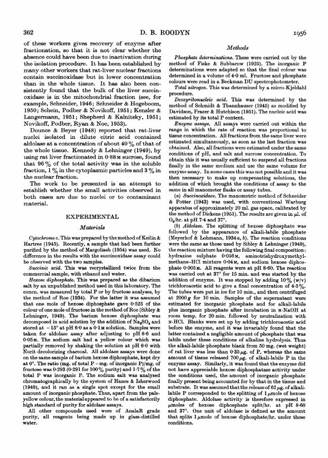
Fractionation by Method 2 confirms these indi-cations. It can be seen in Figs. 1 and 2 that there isno correlation at all between the distribution ofnuclei and of the enzyme. In the first experiment(Fig. 1) compensating solutions were not used in theassay, which probably explains the poor recovery ofenzyme. In the second experiment (Fig. 2) all theobserved activity in the original nuclear fraction wasdue to two types of contamination, one sedimentingabove the nuclei (presumably mitochondria, sincethe upper part of the centrifuge tube contained thebulk of the particulate material in the original
Table 1. Summary of results offractionations by Method 1
Average values given with standard error of mean. The number of fractionations is given in brackets. HDP, Hexosediphosphate. Al IAchas
Percentage of amount in homogenate foundin nuclear fraction
Percentage recovery after fractionationActivity/mg. of N of homogenateActivity/mg. of N of nuclear fractionRelative conen.
nuclear fraction), and one sedimenting below thenuclei (presumably unbroken liver cells, as inFig. 1). In Fig. 2 there were not enough whole cellspresent in the original nuclear fraction to obtainaccurate counts on all fractions, but it was observedqualitatively that most of the cells were found5L-6 cm. from the meniscus.
It was thought that sufficient evidence had beenobtained to conclude that nuclei do not contributeto the succinoxidase activity of nuclear fractionsobtained by Method 1.
Aldolase activity of nuclearfractionsThe results of thirty-seven fractionations by
Method 1 are given in Table 1. It can be seen thatmuch more aldolase than succinoxidase was foundin the nuclear fraction (12.5 %) and that the enzymewas 1-44 times more concentrated in the nuclearfraction than in the homogenate. In several of thefractionations parallel assays with succinoxidasewere carried out (Table 2) and on every occasion
c
u
1-
L._bOvv
c
co
Eb4!
u-co
70
i 2 3 4 5Distance from meniscus (cn.)
6 7
Fig. 1. Distribution of succinoxidase, erythrocytes, nucleiand whole cells along the length of the tube after centri-fuging in a medium of graded specific gravity. *,Succinoxidase; 0, erythrocytes; 0, nuclei; E, wholecells. The concentrations per ml. of original nuclearfraction of the components were: succinoxidase, 504pJ.of 0,/hr.; erythrocytes, 14-8 x 10⁶; nuclei, 88 x 106;whole cells, 1-38 x 106; the recoveries after fractiona-tion were 67, 100, 114 and 109%, respectively. Nuclearfractions were layered over graded medium (11-40% ofsucrose by 2-5% intervals) and centrifuged for 4 min. at300g.
1 2 3 4 5 6 7Distance from meniscus (cm.)
Fig. 2. Distribution of succinoxidase and nuclei alonglength of tube after centrifuging in a medium of gradedspecific gravity. 0, Succinoxidase; *, nuclei. Theoriginal nuclear fraction had 2-91 mg. of nitrogen, asuccinoxidase activity of 425,l. of 02/hr. and 127 x 106nuclei/ml. The recoveries after fractionation were 102,100 and 118%, respectively. The nuclear fraction alsohad 0-2 x 10⁶ whole cells/ml., but these were not sufficientto obtain accurate counts in the various fractions aftercentrifuging. Original nuclear fraction layered overgraded medium [11-40% (w/v) by 2.5% intervals] andcentrifuged for 4 min. at 300g.
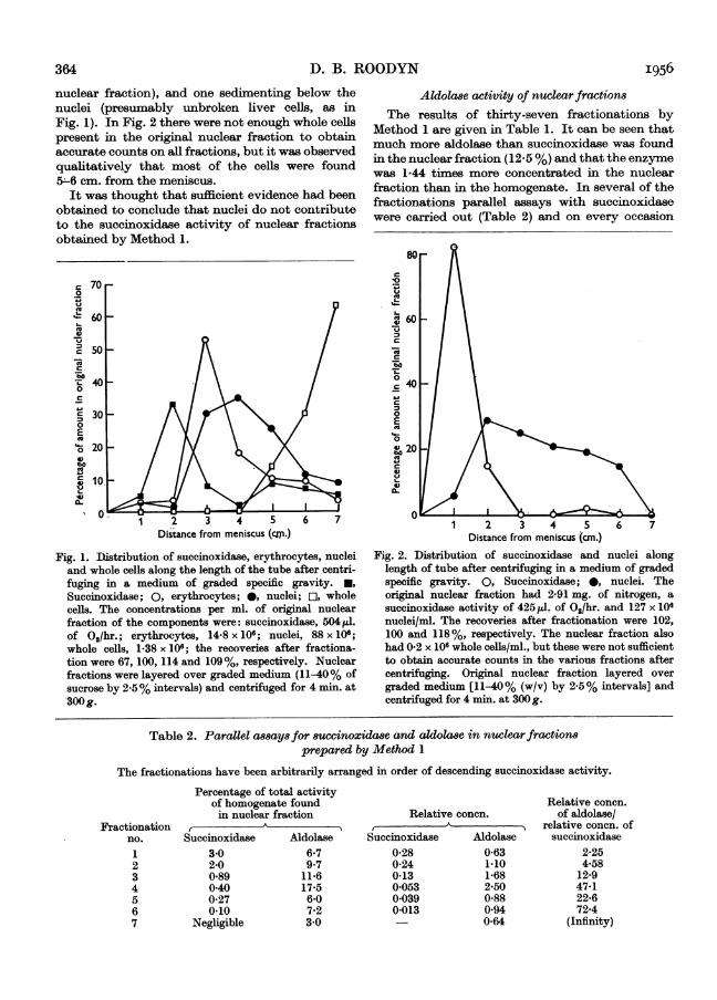
Table 2. Parallel assays for succinoxidase and aldolase in nuclear fractionsprepared by Method 1
The fractionations have been arbitrarily arranged in order of descending succinoxidase activity.
Percentage of total activityof homogenate foundin nuclear fraction
both the percentage of the total enzyme and therelative concentration were higher than for succin-oxidase. Also the values for aldolase bore no con-stant relationship to those for succinoxidase. Thedifference in behaviour ofthe two enzymes is furthershown by fractionations by Method 2. In the firstcase (Fig. 3), a sharp correlation between nuclei (asestimated by direct count) and aldolase was
600
o-
0
Eg 20
vs4)
0
.<X O.
-o I-
C0IlI. -uV- !2
4)4)
u oc 3:o0.0IV
E E
1 2 3 4 5 6 7Distance from meniscus (cm.)
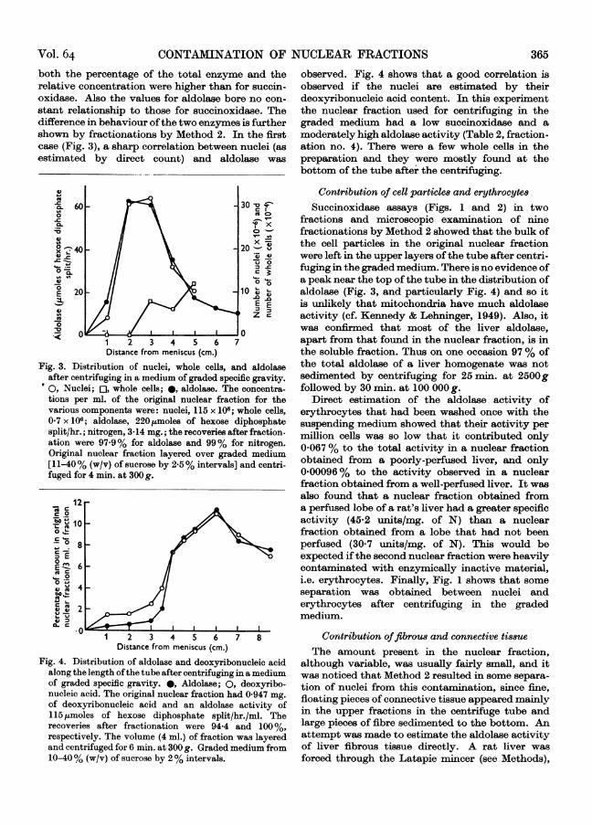
Fig. 3. Distribution of nuclei, whole cells, and aldolaseafter centrifuging in a medium of graded specific gravity.0, Nuclei; 0, whole cells; 0, aldolase. The concentra-tions per ml. of the original nuclear fraction for thevarious components were: nuclei, 115 x l0f; whole cells,0-7x 10⁶; aldolase, 220jumoles of hexose diphosphatesplit/hr.; nitrogen, 3*14 mg.; the recoveries after fraction-ation were 97-9 % for aldolase and 99% for nitrogen.Original nuclear fraction layered over graded medium[11-40% (w/v) of sucrose by 2.5% intervals] and centri-fuged for 4 min. at 300 g.
c o.
t° Q 10o
L-c 0
oLV0u3 E0E 6
1 2 3 4 5 6Distance from meniscus (cm.)
Fig. 4. Distribution of aldolase and deoxyribonucleic acidalong the length ofthe tube after centrifuging in a mediumof graded specific gravity. *, Aldolase; 0, deoxyribo-nucleic acid. The original nuclear fraction had 0 947 mg.
of deoxyribonucleic acid and an aldolase activity of115 ,moles of hexose diphosphate split/hr./ml. Therecoveries after fractionation were 94-4 and 100%,respectively. The volume (4 ml.) of fraction was layeredand centrifuged for 6 min. at 300 g. Graded medium from10-40% (w/v) of sucrose by 2% intervals.
observed. Fig. 4 shows that a good correlation isobserved if the nuclei are estimated by theirdeoxyribonucleic acid content. In this experimentthe nuclear fraction used for centrifuging in thegraded medium had a low succinoxidase and amoderately high aldolase activity (Table 2, fraction-ation no. 4). There were a few whole cells in thepreparation and they were mostly found at thebottom of the tube after the centrifuging.
Contribution of cell particles and erythrocytesSuccinoxidase assays (Figs. 1 and 2) in two
fractions and microscopic examination of ninefractionations by Method 2 showed that the bulk ofthe cell particles in the original nuclear fractionwere left in the upper layers of the tube after centri-fuging in the graded medium. There is no evidence ofa peak near the top of the tube in the distribution ofaldolase (Fig. 3, and particularly Fig. 4) and so itis unlikely that mitochondria have much aldolaseactivity (cf. Kennedy & Lehninger, 1949). Also, itwas confirmed that most of the liver aldolase,apart from that found in the nuclear fraction, is inthe soluble fraction. Thus on one occasion 97 % ofthe total aldolase of a liver homogenate was notsedimented by centrifuging for 25 min. at 2500gfollowed by 30 min. at 100 000g.
Direct estimation of the aldolase activity oferythrocytes that had been washed once with thesuspending medium showed that their activity permillion cells was so low that it contributed only0-067% to the total activity in a nuclear fractionobtained from a poorly-perfused liver, and only0-00096% to the activity observed in a nuclearfraction obtained from a well-perfused liver. It wasalso found that a nuclear fraction obtained froma perfused lobe of a rat's liver had a greater specificactivity (45.2 units/mg. of N) than a nuclearfraction obtained from a lobe that had not beenperfused (30.7 units/mg. of N). This would beexpected ifthe second nuclear fraction were heavilycontaminated with enzymically inactive material,i.e. erythrocytes. Finally, Fig. 1 shows that someseparation was obtained between nuclei anderythrocytes after centrifuging in the gradedmedium.
Contribution offibrous and connective tissueThe amount present in the nuclear fraction,
although variable, was usually fairly small, and itwas noticed that Method 2 resulted in some separa-tion of nuclei from this contamination, since fine,floating pieces of connective tissue appeared mainlyin the upper fractions in the centrifuge tube andlarge pieces of fibre sedimented to the bottom. Anattempt was made to estimate the aldolase activityof liver fibrous tissue directly. A rat liver wasforced through the Latapie mincer (see Methods),
Vol. 64 365
D. B. ROODYN
fitted with a plate pierced by 1 mm. diam. holes.The aldolase activity of the fibrous and connectivetissue that remained attached to the plate was16*8 units/mg. of N, whereas that of the whole liverwas 24*9 units/mg. of N. Under the microscope thepreparation of fibre was found to be grossly con-
tamninated with adhering liver cells, and so itslower aldolase activity, compared with the originalliver, even in the presence ofsomuch contamination,indicates that the aldolase activity of pure fibrousmaterial cannot be very high.
Effect of washing the nucleiRepeated resuspension and sedimentation of the
nuclear pellet showed that after three sedimenta-tions no more enzyme appears in the supernatant(Fig. 5). To avoid the danger of sucking up nucleiwhile removing the supernatants, 0-8 ml. of super-
natant was left overlying the nuclear pellet at eachstage, so that removal of supernatant was not as
efficient as in the normal Method 1. (Five, instead
124
8
04
t
4
EU
° 96
w'a-
o
0
" 92uV
841 2 3 4 5 6
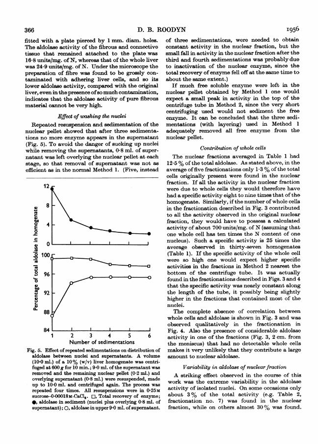
Number of sedimentationsFig. 5. Effect ofrepeated sedimentations on distribution of
aldolase between nuclei and supernatants. A volume(10 0 ml.) of a 10% (w/v) liver homogenate was centri-fuged at 600g for 10 min.; 9 0 ml. ofthe supernatant wasremoved and the remaining nuclear pellet (0.2 ml.) andoverlying supernatant (0.8 ml.) were resuspended, madeup to 10.0 ml. and centrifuged again. The process was
repeated four times. All resuspensions were in 0-25Msucrose-0 00018M-CaCl2. o, Total recovery of enzyme;
*, aldolase in sediment (nuclei plus overlying 0-8 ml. ofsupernatant); 0, aldolase in upper 9'0 ml. of supernatant.
of three sedimentations, were needed to obtainconstant activity in the nuclear fraction, but thesmall fall in activity in the nuclear fraction after thethird and fourth sedimentations was probably dueto inactivation of the nuclear enzyme, since thetotal recovery of enzyme fell off at the same time toabout the same extent.)
If much free soluble enzyme were left in thenuclear pellet obtained by Method 1 one wouldexpect a small peak in activity in the top of thecentrifuge tube in Method 2, since the very shortcentrifuging used would not sediment the freeenzyme. It can be concluded that the three sedi-mentations (with layering) used in Method 1
adequately removed all free enzyme from thenuclear pellet.
Contribution of whole cells
The nuclear fractions averaged in Table 1 had12 5 % of the total aldolase. As stated above, in theaverage of five fractionations only 1-3 % of the totalcells originally present were found in the nuclearfraction. If all the activity in the nuclear fractionwere due to whole cells they would therefore havehad a specific activity eight to nine times that of thehomogenate. Similarly, if the number ofwhole cellsin the fractionation described in Fig. 3 contributedto all the activity observed in the original nuclearfraction, they would have to possess a calculatedactivity of about 700 units/mg. of N (assuming thatone whole cell has ten times the N content of one
nucleus). Such a specific activity is 25 times theaverage observed in thirty-seven homogenates(Table 1). If the specific activity of the whole cellwere so high one would expect higher specificactivities in the fractions in Method 2 nearest thebottom of the centrifuge tube. It was actuallyfound in the fractionations described in Figs. 3 and 4that the specific activity was nearly constant alongthe length of the tube, it possibly being slightlyhigher in the fractions that contained most of thenuclei.The complete absence of correlation between
whole cells and aldolase is shown in Fig. 3 and was
observed qualitatively in the fractionation inFig. 4. Also the presence of considerable aldolaseactivity in one of the fractions (Fig. 3, 2 cm. fromthe meniscus) that had no detectable whole cellsmakes it very unlikely that they contribute a largeamount to nuclear aldolase.
Variability in aldola8e of nuclear fractionA striking effect observed in the course of this
work was the extreme variability in the aldolaseactivity of isolated nuclei. On some occasions onlyabout 3 % of the total activity (e.g. Table 2,fractionation no. 7) was found in the nuclearfraction, while on others almost 30 % was found.
366 I956
88
CONTAMINATION OF NUCLEAR FRACTIONS
Table 3. Aldolase distribution in two apparently similarfractwnations by Method 1
FractionSupernatantsNuclear fraction
Recovery
SupernatantsNuclear fraction
Recovery
Percentage oftotal N inhomogenate
89*09.5
98-5
88*010698-6
Percentage oftotal aldolase inhomogenate
49*027-676-6
9057-2
97.7
Table 3 shows two fractionations by method 1,apparently carried out in exactly the same way,where one nuclear fraction had about four times theactivity of the other. In addition, as the recoverywas poor in the first fractionation, it is possible thatthis nuclear fraction actually had much more than27 6% of the total activity. While such variationsmade a moderate difference to the total balancesheet of recovered enzyme, they made a verysignificant difference to the absolute nuclearactivities. An investigation into the origin of thisvariability is presented in the next paper (Roodyn,1956).
DISCUSSION
Absence of succinoxidase
The absence ofsuccinoxidase from rat-liver nuclei isnot surprising and is probably not an originalobservation. Indeed the fact that liver nuclei aredeficient in cytochrome oxidase (Hogeboom et al.1952) makes it difficult to imagine how nuclei couldoxidize succinate with oxygen. The questionswhether the nuclear membrane is permeable to thesubstrates used in the assay or whether the testconditions are suitable to demonstrate any oxi-dative ability of nuclei have not been investigated.The enzyme has been used in this paper entirely as
a guide to methods for revealing the extent of thecontribution of contaminants to activity in thenuclear fraction.
Association between aldolase and nuclei
The striking correlation between the distributionof aldolase and nuclei after centrifuging in thegraded medium and the observations that the mostlikely forms of contamination do not contributegreatly to aldolase in the nuclear fraction make itpossible to state that aldolase assays carried out onnuclear fractions prepared by the method ofHogeboom et al. (1952) mainly measure the activityof the nuclei present. A more detailed study of thesignificance of this association between nuclei andaldolase is presented in the next paper (Roodyn,1956).
SUMMARY
1. An attempt has been made to determinewhether the succinoxidase and aldolase activities ofrat-liver nuclear fractions are due to the nucleithemselves or to contaminant material.
2. The succinoxidase activity ofnuclear fractionsprepared by the method of Hogeboom et al. (1952)was only 1-5% of the total in the homogenate andwas only slightly higher than the percentage ofunbroken liver cells in the nuclear fraction. Centri-fuging the nuclear fraction in a medium of gradedsucrose concentration revealed a complete absenceofcorrelation between the distribution of succinoxi-dase and of nuclei along the length of the centrifugetube.
3. In thirty-seven fractionations by themethod ofHogeboom et al. (1952) the nuclear fraction carriedon the average 12-5% of the total aldolase activityof the liver homogenate, the enzyme being 1-44times as concentrated in the nuclear fraction as inthe original whole tissue.
4. The aldolase activity of seven differentnuclear fractions was always greater than thesuccinoxidase and bore no constant relation to it.
5. Evidence is presented that it is unlikely thatfibrous material, erythrocytes, cytoplasmic particlesand insufficient washing of the nuclei contributesignificant amounts to the aldolase observed in thenuclear fraction.
6. When the nuclear fraction was centrifuged ina medium of graded sucrose concentration a closecorrelation was observed between the distribution ofnuclei and of aldolase along the length ofthe centri-fuge tube. No correlation was observed between thedistribution of unbroken liver cells and aldolase.
7. It is concluded that the succinoxidaseactivity observed in the nuclear fractions was due tocontaminant material, but that the aldolase was
associated with the nuclei.
I would like to thank Dr M. Dixon, F.R.S., for suggestingthe problem and for his kind interest and guidance duringthe work. I would also like to thank Dr E. C. Webbfor useful guidance and criticism. I am grateful toMiss J. Cawkwell for running a chromatogram of hexose
Fractionationno.1
2
Relativeconcn.
2-90
0-68
Vol. 64 367
368 D. B. ROODYN I956
diphosphate, to MrD. S. Bendall for a sample of cytochromec, and to Mr D. Gardiner for preparing a sample of hexosediphosphate. I would also like to express my gratitude tothe Medical Research Council for a grant held during thelast three years.
REFERENCESAnderson, N. G. (1955). Science, 121, 775.Davidson, J. N., Frazer, S. C. & Hutchison, W. C. (1951).
Biochem. J. 49, 311.Dickens, F. (1951). Biochem. J. 48, 385.Dounce, A. L. (1948). Ann. N.Y. Acad. Sci. 50, 982.Dounce, A. L. & Beyer, G. T. (1948). J. biol. Chem. 173, 159.Fiske, C. T. & Subbarow, Y. (1925). J. biol. Chem. 66, 375.Hanes, C. S. & Isherwood, F. A. (1949). Nature, Lond., 164,
1107.Harrison, M. F. (1953). Nature, Lond., 171, 611.Hogeboom, G. H., Schneider, W. C. & Striebich, M. J.
(1952). J. biol. Chem. 196, 111.Holter, H., Ottesen, M. & Weber, R. (1953). Experientia, 9,
346.Keilin, D. & Hartree, E. F. (1945). Biochem. J. 39, 289.Kennedy, E. P. & Lehninger, A. L. (1949). J. biol. Chem.
179, 957.
Kensler, C. J. & Langermann, H. (1951). J. biol. Chem. 192,551.
Kuff, E. L. & Schneider, W. C. (1954). J. biol. Chem. 206,677.
Lang, K. & Siebert, G. (1950). Biochem. Z. 320, 402.Margoliash, E. (1954). Biochem. J. 56, 529.Meyerhof, 0. & Lohmann, K. (1934a). Biochem. Z. 271, 89.Meyerhof, 0. & Lohmann, K. (1934b). Biochem. Z. 273,413.Novikoff, A. B., Podber, E., Ryan, J. & Noe, E. (1953).
J. Histochem. Cytochem. 1, 27.Roe, J. H. (1934). J. biol. Chem. 107, 15.Roodyn, D. B. (1956). Biochem. J. 64, 368.Schein, A. H., Podber, E. & Novikoff, A. B. (1951). J. biol.Chem. 190, 331.
Schmidt, G. & Thannhauser, S. J. (1945). J. biol. Chem. 161,83.
Schneider, W. C. (1946). J. biol. Chem. 165, 585.Schneider, W. C. & Hogeboom, G. H. (1950). J. nat. Cancer
Inst. 10, 969.Schneider, W. C. & Potter, V. R. (1943). J. biol. Chem. 149,
217.Shepherd, J. A. & Kalnitsky, G. (1951). J. biol. Chem. 192,1.Sibley, J. A. & Lehninger, A. L. (1949). J. biol. Chem. 177,
859.
The Enzymic Properties of Rat-liver Nuclei2. FACTORS AFFECTING THE ALDOLASE ACTIVITY OF RAT-LIVER NUCLEI
BY D. B. ROODYN*Department of Biochemistry, University of Cambridge
(Received 1 February 1956)
It was established in the previous paper (Roodyn,1956) that rat-liver nuclei prepared in sucrose-calcium chloride media possess aldolase activity, butthat the amount recovered in the nuclear fraction indifferent experiments was very variable, althoughapparently the same method of isolation of thenuclei had been used. During the course of aninvestigation into the origins of this variabilitycertain effects were found to produce changes in thealdolase activity of the nuclei.
METHODS
Maleate buffers. These were prepared by the neutralizationwith N-NaOH of boiled solutions of maleic anhydride. Thebuffers were brought to the required ionic strength byadding NaCl.
Preparation of nuclei. Unless indicated otherwise, nuclearfractions were prepared by the method of Hogeboom,Schneider & Striebich (1952) carried out as described
previously (Method 1 of Roodyn, 1956). All other methods,materials and terms used were as described previously(Roodyn, 1956).
RESULTS
Effect of method of cell breakagePretreatment of the liver by forcing it through aLatapie mincer caused some fall in activity.(Unmincedliver gave anuclear fraction with aldolaseactivity of 52 1 units/mg. of N while minced liverfrom the same rat gave a nuclear fraction with anactivity of 46 2 units/mg. ofN.) A similar effect wasobserved by Hogeboom & Schneider (1952) withthe diphosphopyridine nucleotide-synthesizingenzyme.The time of homogenization in the Potter-
Elvehjem apparatus was not very critical. Treat-ment for 6 sec. gave a nuclear fraction with arelative concentration [see Roodyn (1956) for adefinition of this term] of 0 75; a further 24 sec.caused this to fall to 0-63 and the same activity wasobtained with up to 4 min. homogenization of theliver.
* Present address: Medical Research Council Radio-biological Research Unit, Atomic Energy ResearchEstablishment, Harwell, Didcot, Berks.