THE EPIDEMIOLOGY OF POST-TRAUMATIC SEIZURES FOLLOWING MODERATE TO SEVERE TRAUMATIC BRAIN INJURY by Anne Connelly Ritter BA, Gettysburg College, 2009 MPH, University of Pittsburgh, 2010 Submitted to the Graduate Faculty of the Graduate School of Public Health in partial fulfillment of the requirements for the degree of Doctor of Public Health University of Pittsburgh 2016
Transcript
THE EPIDEMIOLOGY OF POST-TRAUMATIC SEIZURES FOLLOWING
MODERATE TO SEVERE TRAUMATIC BRAIN INJURY
by
Anne Connelly Ritter
BA, Gettysburg College, 2009
MPH, University of Pittsburgh, 2010
Submitted to the Graduate Faculty of
the Graduate School of Public Health in partial fulfillment
of the requirements for the degree of
Doctor of Public Health
University of Pittsburgh
2016
UNIVERSITY OF PITTSBURGH
GRADUATE SCHOOL OF PUBLIC HEALTH
This dissertation was presented
by
Anne Connelly Ritter
It was defended on
February 8, 2016
and approved by
Dissertation Advisor: Caterina Rosano, MD, MPH
Professor Department of Epidemiology
Graduate School of Public Health University of Pittsburgh
Committee Members: Maria M Brooks, PhD
Professor and Vice Chair of Education Department of Epidemiology
Graduate School of Public Health University of Pittsburgh
Anthony Fabio, PhD, MPH
Assistant Professor Department of Epidemiology
Graduate School of Public Health University of Pittsburgh
Amy K Wagner, MD
Associate Professor and Endowed Research Chair Department of Physical Medicine & Rehabilitation
Figure 4. Traumatic Brain Injury Model System Prognostic Model CONSORT Figure ............. 89
Figure 5. Linkage Disequilibrium Maps for SLC1A1 and SLC1A6 .......................................... 115
Figure 6. Seizure Curves for rs10974620 During Full Follow-up .............................................. 116
Figure 7. Seizure Curves for rs7858819, Follow-up Beginning Day 2 ...................................... 117
viii
PREFACE
I would like to express my deep appreciation for those who have supported and guided me
through the process of completing this dissertation. It has been an interesting journey and one
that has been full of new experiences for many. First, I would like to thank my primary mentors,
Dr. Rosano and Dr. Wagner. Dr. Rosano, even before returning to school to pursue my doctorate,
your confidence in me and your encouragement were critical to the completion of this work. Dr.
Wagner, thank you for taking a chance on an epidemiology student. You have given me the
opportunity to learn about traumatic brain injury and presented me with challenging questions.
Your support, even of my tendencies to “rabbit hole”, has allowed us to produce a truly
impactful body of work together. Dr. Brooks and Dr. Fabio, your guidance and expertise were
paramount to shaping this work. In addition, you both provided a listening ear and words of
wisdom when I stumbled or was discouraged and for that I am grateful. Dr. Songer, your
guidance in navigating the process of completing this degree was essential. Thank you for your
support of my academic and professional pursuits since I first arrived in Pittsburgh.
In addition, I would like to thank my colleagues and friends for both supporting me and
keeping me sane throughout this process. E-Brain and Wagner labs folks, you are all so
wonderful and intelligent and I am very happy I have had the opportunity to work with and get to
know you all.
ix
And finally, to my family, there is no way I would be sitting here writing the preface to
my accepted dissertation without your continual support and love. I have shared every
breakthrough and every frustration with you, often many times over, and you continued to listen.
When I was stressed, tired, and thought quitting was easier you reminded me of how far I have
come and gave me the confidence to continue. Particularly to my mother, words can’t describe
how much your belief in my ability to do this mattered. You are my biggest advocate, strongest
supporter, and best friend, thank you is not enough.
1
1.0 INTRODUCTION
Traumatic brain injury (TBI) is a significant public health concern and is a major cause of
morbidity and mortality, especially to those under age 45, in the United States 1; 2. TBI is
extremely heterogeneous regarding mechanism of injury, injury severity, and possible outcome.
Individuals may experience many different complications and comorbidities associated with TBI
that may continue chronically, persisting for many years or even throughout the lifetime
following injury3-5.
Individuals with more severe injury are particularly affected by chronic conditions
associated with TBI. Among individuals who survive a severe TBI, disability rates are estimated
as high as 77% 6. Chronic complications include, but are not limited to, poor functional outcome,
decreased cognitive function, psychosocial and/or behavioral problems, decreased general health,
and other neurological sequelae. These complications contribute substantially to the cost of care
associated with TBI, which is estimated at more than $60 billion annually in the United States 7.
Lifetime total cost of care is estimated to exceed $1.8 million for an individual 7. In addition to
medical cost incurred, TBI may significantly increase years of potential life lost and decrease
quality of life.
Post-traumatic seizures (PTS) are a well-recognized sequela of traumatic brain. PTS have
been documented as a common complication of TBI for decades. Incidence of PTS varies
drastically throughout the literature and is dependent on many factors including study design and
2
characteristics of the study population. As incidence of TBI increases and death due to TBI
decreases, more individuals will be at risk of developing and living with chronic complications.
It can also be expected that PTS incidence will increase. Previously, seizure prophylaxis has
been shown to be effective in reducing the incidence of PTS in the first week after injury, but has
no long-term benefit (Temkin, 1990). Current recommendations from the American Academy of
Neurology and the Brain Trauma Foundation include delivery of phenytoin for seizure
prophylaxis during the first seven days post-TBI 8; 9. Yet, despite decades of research, there are
no effective pharmacological interventions to prevent post-traumatic seizures that develop after
seven days post-injury and, it does not appear that rates of PTS are decreasing. Increasingly,
novel risk factors for PTS and mechanisms of epileptogenesis following TBI are being
investigated to identify potential new targets for therapeutic treatment.
1.1 TRAUMATIC BRAIN INJURY
1.1.1 Definitions
Traumatic brain injury is caused by an impact to the head from an external force that disrupts
physiological function. External forces can include direct mechanical impact (i.e. blunt trauma),
acceleration or deceleration associated injury (i.e. whiplash), blast injury caused by a pressure
wave (i.e. explosion), or penetrating injury (i.e. gunshot).
Severity of TBI can be classified multiple ways. Originally developed to classify levels of
consciousness 10, the Glasgow Coma Scale (GCS) has become the most widely used tool to
describe TBI severity. The GCS score ranges from 3 to 15: 3-8 indicating severe, 9-12 indicating
3
moderate, and 13-15 indicating mild TBI 11. Alternative criteria based on loss of consciousness
and post-traumatic amnesia are also used 12. Many organizations, including the Department of
Defense (DoD), Veterans Affairs (VA), and Centers for Disease Control and Prevention (CDC),
continue to utilize a combination of clinical variables to assess TBI severity (Table 1).
Table 1. Classification of Traumatic Brain Injury
Criteria Mild Moderate Severe Structural Imaging1 Normal Normal or abnormal Normal or abnormal
Loss of Consciousness 0 - 30 min > 30 min but < 24 hours > 24 hours
Alteration of Consciousness2 0 - 24 hours > 24 hours
Post-Traumatic Amnesia ≤ 24 hours 24 hours – 7 days > 7 days
Glasgow Coma Scale3 13 – 15 9 – 12 3 - 8 1Abnormalities not related to trauma may be present with mild injury; 2Classification of severe injury not made based on alteration of consciousness alone; 3Typically based on best GCS score is first 24 hours post-injury *adapted from Silver, McAllister, & Yudofsky. Textbook of Traumatic Brain Injury, Second Edition
In addition to severity, TBI is often classified as closed-head or penetrating brain injury.
Many studies within the existing literature categorize TBI into one of four classifications: mild,
moderate, severe, or penetrating. Despite the fact that it is possible to obtain a GCS score on an
individual with a penetrating TBI (pTBI), researchers and clinicians often refer to pTBI as a
distinct category.
As medical care has evolved, and computed topography (CT) became a part of standard
care, neuroradiological findings have increasingly been used to differentiate and define TBI.
Abnormalities detected via CT are categorized based on location and type of injury [i.e. which
hemisphere, brain region, and pathology present (Table 2)]. Advances in neuroradiological
imaging through tools such as magnetic resonance imaging (MRI), positron emission topography
4
(PET), and magnetic resonance spectroscopy (MRS), may provide additional ways to classify
and differentiate brain injury. Some definitions regarding severity of TBI include positive
evidence of abnormal pathology via CT imaging as a distinguishing characteristic.
Table 2. Abnormal Pathologies Identified via Computed Tomography
Variable Definition Intracranial Hemorrhage (ICH)
Bleeding within the skull (cranium)
Contusion Area(s) of bleeding on the surface of the brain, most commonly along the undersurface and poles of the frontal and temporal lobes
Subdural Hematoma (SDH)
Presence of extra-axial blood clot or collection within sub-dural space, between surface of the brain and dura matter
Subarachnoid Hemorrhage (SAH)
Bleeding within the subarachnoid space, between the surface of the brain and arachnoid layer. May include blood in ambient, basal, interpenduncular cisterns or cisterna magna, or along falx or tentorium
Intra-ventricular Hemorrhage (IVH)
Blood documented within intra-ventricular space
Epidural Hematoma (EDH)
Presence of extra-axial collection within epidural space, between skull and dura matter
Penetrating TBI (pTBI)
Typically defined by penetration of the dura matter. May include bone, metal, or other foreign bodies present within the parenchyma, skull fractures displaced or depressed > 2mm, or “through and through” injuries penetrating the dura
1.1.2 Epidemiology
The incidence of traumatic brain injury and its sequelae documented in the existing literature is
heavily influenced by many factors, most importantly injury severity and type. In the United
States, approximately 2.5 million traumatic brain injuries occur annually3. Of these,
approximately 284,000 (11%) result in hospitalization, and 53,000 (2%) in death3. However,
these figures are limited by their reliance on national surveillance data and cannot account for
individuals with TBI that do not seek care, or for those who seek care from a primary care
5
physician only. Therefore, it is likely that these data are an underestimate of the incidence of
TBI, particularly mild TBI, in the United States.
Despite this limitation, national surveillance data show trends of increasing overall TBI
incidence and emergency department visits related to TBI from 2001 to 2010 (Figure 1). The
observed increase may be influenced by recent public health campaigns to increase awareness of
mild TBI (mTBI), leading to an increase in health care utilization associated with mTBI.
Figure 1. Rates of TBI Related Emergency Department Visits, Hospitalizations, and Deaths
- United States, 2001-2010
6
This theory is supported by the relatively constant incidence of TBI hospitalizations
throughout the same timeframe, suggesting the frequency of moderate and severe TBI requiring
hospitalization is not increasing and the resulting overall increase is likely attributable to more
mild injuries. Also within the last decade, rates of TBI-related death have decreased in the
United States 13.
Across all injury severities, as classified by the GCS, males have consistently higher rates
of TBI compared to females 14; 15. There are also significant differences in risk of TBI across the
lifespan. Individuals ages 0 to 4, 15 to 19, and greater than 65 years old are at a significantly
increased risk of TBI 14; 16; 17. Of these age groups, individuals greater than 75 years old have the
highest rates of hospitalization and death 13; 15; 17.
Differences in TBI rates across race and ethnicity are also documented. Annual TBI rates,
including emergency department utilization and hospitalizations, are higher for black individuals
than white and those of other racial backgrounds. However, annual average mortality rates are
lower for black individuals compared to whites 17.
1.1.3 Primary and Secondary Injury
The highly heterogeneous nature of traumatic brain injury makes it difficult to establish
standards of care across the spectrum of injury. There are currently no therapeutic interventions
proven to be effective across a broad range of clinical presentations of TBI. Similarly, outcomes
vary greatly and are difficult to predict based solely on injury severity. Differences in
neurobiological factors, such as those involved in secondary injury, likely contribute
substantially to differences in outcome.
7
Mechanistically, traumatic brain injury can be thought of as two events occurring
successively. The first event, the primary injury, refers to the moment of impact of external
forces causing the TBI. The primary injury is a discrete event.
Unlike the primary injury, the secondary injury is often thought of as a cascade, initiated
by the primary injury and evolving over time. The secondary injury is not discrete; it is
composed of multiple pathophysiological processes that may occur simultaneously or
sequentially and are not consistent across individuals. Different primary injury types (i.e. diffuse
axonal injury, intra-cerebral hemorrhage, or penetrating injury) may initiate different secondary
injury cascades 18. However, this too may differ across individuals. It is necessary to understand
the pathological processes that encompass secondary injuries and their impact on TBI
complications, especially development of PTS.
Briefly, the secondary injury cascade has been described as a two-stage process, with
multiple pathological processes occurring 19. Among the first pathological processes to take
place are direct tissue damage, abnormalities in cerebral blood flow, and deregulation and
functional impairment of cerebral metabolism 19. Damage to brain tissue including disruption
and/or destruction of cerebral blood vessels can result in cerebral ischemia. Multiple studies have
documented both focal and global episodes of cerebral ischemia following TBI 20-22.
Adding to the cerebral ischemic state, damage from the primary injury to neuronal, glial,
and endothelial cells can disrupt the brain’s autoregulatory pathways. Vasoconstriction and
vasodilation are well-documented examples of autoregulatory pathways that may be impaired, or
completely decimated, following TBI 23-25. Autoregulatory dysfunction can also cause
hypotension and hypoperfusion, progressively leading to metabolic dysfunction and inability to
8
meet glucose demands 19; 26; 27. Despite documentation of disrupted pathways, there are no
consistent findings regarding the timing of impairment or loss of autoregulatory.
Precise control of cerebral metabolism is vital to maintain proper neurological function
22. Failure to maintain cerebral metabolism and energy demands leads to mitochondrial
dysfunction, decreased ATP production, and chemical and ionic imbalances within cells 19; 28.
Each of these events can cause cell death, contributing to poor outcome following TBI.
Traumatic brain injury can also induce depolarization of the neuron and excessive release
of excitatory neurotransmitters, such as glutamate 6; 29. The excitotoxicity pathway is further
enhanced by impairment of glutamate uptake and glutamate receptors following injury 30.
Inability to compensate for excessive glutamate release can cause subsequent breakdown of the
blood brain barrier, further disrupting ionic balance 31; 32. Trying to restore proper
neurotransmitter and ion levels increases metabolic demands on tissues that may already be
suffering from metabolic dysfunction due to pathological processes described above 33. Inability
to break this cycle and restore balance can lead to cell death.
Oxidative stress, the production and release of reactive oxygen species, often occurs in
response to excitoxicity following primary TBI 34-36. Oxidative stress can then induce additional
pathological processes leading to immediate cell death 36, as well as activating inflammatory
processes 34.
Inflammatory processes can occur immediately in response to primary injury and tissue
damage, or in response to secondary injury cascades. The inflammatory process following TBI is
extremely complex and can persist well into the chronic phase 37. As part of the inflammatory
response, cytokines are released, activating subsequent proteins such as chemokines and
adhesion molecules that are responsible for activating glia, importantly microglia, and other
9
immune cells 38. Activated microglia and immune cells adhere to damaged and potentially non-
damaged cells surrounding the damaged tissue, ultimately leading to cell destruction 19; 39. This
process can spread across tissues, continue for sustained periods of time post-injury, and activate
astrocytes to produce glial scarring in the effected regions 40.
Many of the same pathological processes taking place in response to TBI can be found
during epileptogenesis. Thus, TBI primes the brain for ictogenic activity, a condition that can
persist decades following the primary injury.
1.2 POST TRAUMATIC SEIZURES
1.2.1 Definitions and Classification
Simplistically, post-traumatic seizures (PTS) refers to an incident seizure following head trauma.
The definitions and classification systems for PTS vary throughout the literature and have
changed across time. These differences make it difficult to compare findings across multiple
studies and to aggregate data for use in meta-analyses.
Post-traumatic seizures are classified based on time of seizure: immediate (<24 hours),
early (1 to 7 days), and late (>7 days) post-injury41 (Table 3). Immediate and early seizures are
considered provoked and decrease seizure threshold only temporarily after TBI 42. Recently, the
term acute symptomatic has been used to describe provoked seizures43; 44. Cut-points for PTS
classification are based on hypothesized differences in causal pathology and epileptogenic
potential 18; 31; 45; 46.
10
Late PTS and post-traumatic epilepsy (PTE) are often used interchangeably 47; 48.
However, some studies make clear distinctions between PTS, late PTS, and PTE. The distinction
typically lies in the intricacies involved with the definition of epilepsy, which has changed over
time, and in the timeframe post-injury in which the first seizure occurs. Some studies use time of
first seizure to delineate PTS from PTE. In these instances, PTS is used to refer to seizures
occurring early, up to seven days post-injury; PTE is then used to refer to seizures that occur
after seven days 49; 50. Other previous research has previously differentiated PTS from PTE based
on the number of seizures that occurred. Earlier definitions of epilepsy required the occurrence
of two or more unprovoked seizures >24 hours apart. Using this information, many researchers
defined PTE as more than one seizure occurring after 7 days post-injury; some studies then used
late PTS to describe a single seizure occurring after 7 days post-injury 18; 51-54.
Recently, the International League Against Epilepsy (ILEA) made the recommendation
to revise and operationalize the definition of epilepsy. The ILEA concluded that requiring two
unprovoked seizures to diagnose epilepsy was no longer adequate to accurately capture the
clinical variability across epilepsy disorders 55. Therefore, the recommendation was made to
revise the definition of epilepsy and include conditions where an individual has a single
unprovoked seizure and their risk of a recurrent seizure is similar to, or greater than, the risk of
seizure recurrence after two unprovoked seizures occurring ≥24hrs apart (≥60%)42. These
recommendations were adopted as the official position by the ILAE in December 2013, thus
changing the definition of epilepsy 42.
11
Table 3. Overview of Current and Past Definitions for Classification of Seizures Occurring
after Traumatic Brain Injury
Classification Definition Current Definition Immediate
PTS1 Seizure occurring <24 hours post-injury
Early PTS1 Seizure occurring 1 – 7 days post-injury Late PTS2 Seizure occurring >7 days post-injury PTE Seizure occurring >7 days post-injury;
synonymous with late PTS (not used in current work)
Past Definitions PTS Single seizure occurring post-injury PTE Two or more seizures occurring >7 days
post-injury PTS Seizures occurring ≤7 days post-injury PTE Seizure occurring >7 days post-injury Late PTS Single seizure occurring >7 days post-
injury 1Also referred to as acute symptomatic 2Individuals with immediate and early PTS remain at risk of developing late PTS
Previous research on seizure recurrence following a single, unprovoked seizure >7d post-
TBI documents risk of seizure recurrence is high enough to consider late PTS as an epileptic
condition42; 56. Haltiner and colleagues determined, of individuals with a single late post-
traumatic seizure, 86% will have a second seizure within two years 56. Following with the most
current clinical definitions of epilepsy, PTE would be defined as one or more seizures occurring
after 7 days post-injury. Therefore, late PTS and PTE are equivalent. For the purposes of this
work, PTS including immediate, early, or late classification will be used to refer to post-
traumatic seizure activity. Importantly, individuals with immediate or early PTS who have a
subsequent late seizure can be classified as having late PTS (i.e. PTE).
12
1.2.2 Mechanisms
Post-traumatic seizures may arise from multiple pathological mechanisms initiated by traumatic
brain injury. While it is hypothesized that acute symptomatic and late seizures result from
different pathological mechanisms 18, multiple epileptogenic processes can occur within one
individual.
Epileptogenesis refers to the process through which a healthy, normally functioning brain
transforms into a brain characterized by a predisposition toward seizure activity 31. Inherent to
epileptogenesis is a latent time period before the initial epileptic seizure occurs during which
cellular and molecular changes are taking place. Following TBI, these cellular and molecular
changes can occur as a part of secondary injury cascades. Because the primary injury is an acute
event, TBI allows the prospective investigation of epileptogenic processes, which is not feasible
with epilepsies of non-traumatic etiology.
Excitotoxicity is one such mechanism that may predispose the brain to epileptic seizure
activity and can occur as a result of TBI via secondary injury cascades as discussed above
(Section 1.1.3). Immediately following injury, there can be a substantially large release of
excitatory neurotransmitters, particularly glutamate57; 58; this release may cause excitotoxicity,
triggering seizures and other excitotoxic injury 29; 31; 59. These immediate seizures, in direct
response to release of excitatory neurotransmitters would be considered provoked, and therefore
would not meet the definition of epileptic seizure activity 31; 55. However, excitotoxicity can lead
to neuronal and astrocytic swelling, mitochondrial damage, cell death, and immediate/early
PTS60. Seizures can cause over-activation of excitatory amino acid receptors, inducing calcium
dependent production of nitric oxide and reactive oxygen species and free radical damage to
DNA and cellular membranes 18. These observations suggest decreased glutamate clearance, and
13
low-level excitoxicity, is an ongoing mechanism of TBI pathology and contributor to
epileptogenesis. Antecedent immediate/early seizure activity may, along with altered glutamate
transporter expression, perpetuate excitoxicity and cell death and contribute to epileptogenesis 6;
61.
Glutamate levels must be carefully regulated via release and reuptake to prevent
excitotoxic injury. There are five glutamate transporters with distinct cellular, synaptic, and
regional distributions within the human brain, each encoded by a different gene 62. It is possible
that dysfunction of glutamate transporters, from injury or genetic predisposition toward a
reduced function, could potentially increase seizure susceptibility through excitoxicity, decreased
antioxidant reserves, or decreased inhibitory neurotransmission.
Additional epileptogenic processes related to inflammation and glial activation have been
identified. As previously described (Section 1.1.3), the inflammatory response can begin
immediately following TBI and persist chronically37. Activated glia adhere to damaged cells and
initiate a feedback loop with immune cells and pro-inflammatory factors38. This feedback loop
can cause neuronal injury, promote glial scar formation, and decrease glutamate re-uptake, all of
which contribute to epileptogenesis 40; 62. The occurrence of epileptic seizure activity can also
maintain this cycle of glutamate release, inflammation, neuronal injury, and glial activation 62.
Ultimately, there are numerous mechanisms that can contribute to epileptogenesis, many
of which are initiated by secondary injury cascades. Increasing research regarding biomarkers for
PTS may help to delineate pathological processes taking place within sub-groups of individuals.
If possible, knowledge of specific epileptogenic processes occurring within individuals could
also provide new targets for prevention of PTS, particularly late PTS.
14
1.2.3 Epidemiology
The incidence of PTS varies widely within the literature and is dependent upon many factors
including study design, population characteristics, and how PTS is defined. To date, few large
epidemiological studies of PTS have been conducted. The exact percentage of individuals with
TBI who will develop PTS, including late PTS (PTE), remains unknown 54.
The incidence of acute symptomatic seizures has not been well described. Of studies that
document early PTS, most do not differentiate immediate from early seizures, nor do they
specify if immediate and early seizures are considered concurrently. In a population based study
from Rochester County Minnesota, Annegers and colleagues reported 2.1% of individuals with
TBI (all severities) developed early PTS63, and of these, approximately 76% of individuals
seized within the first 24 hours post-injury. This study included all ages and TBI severity ranges,
but when individuals with severe TBI only were considered, the documented incidence of early
PTS increased to 10.3%63. Smaller studies that do not differentiate immediate from early seizures
document early PTS incidence to range from approximately 2% to 17% 12; 45; 47; 64; 65. Although,
incidence rates as high as 25% 66 and as low as 0.9%67 have been reported in a cohort of brain
injury rehabilitation patients in Finland and a cohort of hospitalized Chinese patients,
respectively. Within a military cohort, incidence of early PTS falls within the reported range
(5%) 68. The wide range of early PTS incidence may be due to differences in seizure
classification and ascertainment methods (i.e. continuous EEG monitoring) or population
characteristics (i.e. greater proportion of children, pTBI).
Using the same population from Rochester County Minnesota, Annegers and colleagues
conducted one of the first and largest population-based studies of late PTS, investigating
individuals injured between 1935 and 1989. They documented an overall standardized incidence
15
ratio (SIR) of 3.1 for late PTS over the course of full follow-up compared to a demographically
similar, non-injured population 12. Among those with severe TBI, the five-year cumulative
probability of late PTS was 10.0%, and increased to 16.7% at 30 years post-injury12. Importantly,
Annegers and colleagues determined that individuals with severe TBI remained at a significantly
greater risk of unprovoked seizure throughout the entire study duration compared to expected
epilepsy rates in the general population. However, this study population was racially and
socioeconomically homogeneous and results may not generalize to a more diverse population.
A second, more recent, population-based study in the United States examining a more
representative sample concluded the incidence of late PTS following TBI of all severities,
presenting to a participating emergency department, was 2.2 per 100 persons in the first year
post-injury 69. Incidence increased to 4.1 and 3.1 cases per 100 persons in year two and year
three, respectively 69. Among individuals with severe TBI, the cumulative incidence of late PTS
was 13.6 per 100 persons over the first three years post-injury69.
In a study of individuals with moderate to severe TBI admitted to hospital and requiring
inpatient rehabilitation, the cumulative probability of late PTS was 13.2% at two years post-
injury 47. Of individuals with late PTS, 80.3% developed late PTS in the first year, and 92.4% of
cases occurred within the first 18 months following injury 47. Additional smaller studies, in
primarily adult populations, have reported the prevalence of late PTS to range from
approximately 5% to 19%66; 67; 70; 71.
Reported prevalence of late PTS is even higher in military populations, surpassing 53%72;
73. Compared to studies in primarily civilian populations, military populations have greater
incidence and prevalence of late PTS. In the longest follow-up study of TBI in a United States
military cohort, 23% of individuals had new onset seizures in the first year post-injury.
16
Prevalence of late PTS increased to 29% in year two and 53% in year 15; at 35 years post-injury,
prevalence was estimated at 43.7%68; 72; 73. Eleven (5.5% of study cohort) of the individuals
followed up to 35 years post-injury reported new onset seizures between the 15 and 35 year
follow-up interviews 72.
In an independent cohort of Service Members and Veterans from the Korean War, 7.1%
of individuals developed PTS in the first week post-injury and 22.7% developed late PTS 45.
However, this is likely an underestimate of late PTS since it is unclear if individuals with early
PTS developed seizures after one week post-injury, qualifying them for late PTS (i.e. PTE). The
majority of late PTS cases (54.2%) within the study cohort developed within one year post-injury
and 18.1% of cases developed during the second year 45. However, new onset cases continued to
be ascertained out to 11 years post-injury.
One very important limitation of the majority of late PTS studies thus far is inherent to all
epilepsy research: misclassification bias. Seizure activity can present in many different ways.
Any physiological brain function can manifest during a seizure and it may not be evident to the
individual that what they are experiencing is in fact, seizure activity. Similarly, not all seizures
are clinically evident. Subclinical seizure incidence is reported to be higher than incidence of
clinically recognized seizures and has been reported to be even higher for individuals with
penetrating brain injury 74. In one study of moderate and severe TBI utilizing continuous EEG
(cEEG) monitoring up to 14 days post-injury, 22% of individuals were found to have early PTS;
of individuals with early PTS, 57% of seizures were non-convulsive and only detected as a result
of continuous monitoring 75; 76. Therefore, incidence rates of immediate and early PTS reported
in the literature, which are largely based on medical record and billing review, are likely
underestimates of PTS. For studies of PTS following moderate to severe TBI, where individuals
17
are in a hospital setting and cEEG monitoring is possible, rates may be less biased. But, the risk
for bias remains, especially if cEEG monitoring is only used on subsets of individuals for clinical
care, such as those with suspected seizure activity.
1.2.4 Risk Factors of PTS
1.2.4.1 PTS at all Time Points
Few risk factors have been identified for PTS across all time-points (immediate, early, and late).
Injury severity is the most commonly examined risk factor for PTS. Various algorithms have
been used to define injury severity (see Table 1, Section 1.1.1). Within the literature, there is
general agreement that greater severity is associated with increased risk of PTS in all timeframes
12; 50; 63; 66; 67; 69-71; 77-79.
Where there are slight differences in results regarding severity as a risk factor, the
method used to categorize severity may be responsible. Englander and colleagues found the
highest cumulative probability of late PTS among individuals with moderate TBI, classified by
GCS (GCS 9-12), over the first two years post-injury 47. However, this finding highlights the
construct of survival bias, particularly for late PTS. Individuals with severe TBI are more likely
to expire within the first week post-injury, excluding them from being at risk for development of
late PTS.
Further complicating the effect of injury severity on PTS, specifically immediate and
early PTS, when continuous EEG monitoring is used to ascertain cases, there is no significant
difference in injury severity, as measured by the GCS, between individuals who seize and those
who do not 76. As such, increasing injury severity may correlate with increasing risk of late PTS,
18
limited to clinically apparent seizures. Using consistent metrics to classify injury and to ascertain
PTS status would increase comparability of findings across studies.
Injury characteristics have been extensively studied as risk factors for PTS. As CT
imaging became common practice following head trauma, specific pathology types were also
examined. Presence of intra-cerebral blood, including intracerebral hemorrhage and subdural
hematoma, has been identified as a risk factor for early and late PTS, increasing risk up to 30%
12; 47; 48; 50; 63; 71; 78; 80; 81. While pTBI is often associated with late PTS, depressed skull fracture has
been specifically documented as a risk factor for both early and late PTS 12; 50; 63; 66; 78; 82.
In addition to injury severity, age at injury is consistently cited as a risk factor for PTS.
Children are at an increased risk of PTS at all time-points post-injury, but are particularly prone
to immediate and early seizures 12; 63; 66; 77; 83. The effect of age within adult populations is less
clear. The Rochester Epidemiology Project found age greater than or equal to 65 was associated
with a late PTS rate ratio of 2.5, which remained significant even after correction for other risk
factors including early PTS and depressed skull fracture 12. Asikainen et al concluded increasing
age among adults is correlated with increasing risk of late PTS 66. Yet, other studies found no
association between age and risk of late PTS 69. Additional research is needed to more
thoroughly investigate the effect of age on late PTS among adults.
1.2.4.2 Early PTS
Characteristics of individuals who develop immediate and early PTS are more highly variable. In
addition to risk factors for PTS during all timeframes discussed above, few risk factors are
specific to immediate and/or early PTS. This is likely attributable to the fact that immediate and
early are considered acute symptomatic seizures, direct responses to the head trauma. Therefore,
research has not extensively examined risk factors for early PTS outside of injury severity. As
19
more research is conducted, novel risk factors specific to immediate and/or early seizures may be
identified.
1.2.4.3 Late PTS
Penetrating TBI is often defined by dura penetration and may include the presence of bone
and/or foreign fragments (e.g. shrapnel). In both civilian and military cohorts, pTBI is one of the
most prominent risk factors for late PTS 46; 47; 66; 68; 72; 73; 78; 84; 85. Salazar and Grafman reviewed
pTBI in both military and civilian cohorts and documented approximately 34 to 63% of
individuals with pTBI develop late PTS 86. Similarly, the highest probability of developing PTE
in a civilian population, 62.5% in two years post-injury, was associated with bone or metal
fragments and a relative risk of 3.94 compared to those with no dura penetration47. Penetrating
TBI is much more common in military cohorts compared to civilian cohorts 56; 72 and may
explain the reported differences in rates of late PTS.
Among individuals with late PTS, Weiss and Caveness found no statistically significant
differences in seizure frequency for those with penetrating compared to non-penetrating TBI 45.
Interestingly, a penetrating wound greater than 3 cm deep was highly associated with increased
seizure frequency 45, suggesting deep brain penetration may initiate different pathophysiological
pathways leading to increased seizure.
As CT technology became increasingly available and used as part of the standard of care
to diagnose TBI, abnormal neuroradiological findings were identified as risk factors for late PTS.
Englander and colleagues found cisternal compression and midline shift, often associated with
presence of intracranial bleeding and elevated intracranial pressure (ICP), were significantly
associated with late PTS in a cohort of individuals hospitalized for moderate to severe TBI 47. In
addition to intracerebral blood collection (i.e. SDH, ICH), multiple studies have documented
20
contusion as a significant risk factor for late PTS 12; 47; 64; 67; 78; 87; 88. In a large population-based
study, brain contusion and subdural hematoma remained significantly associated with late PTS
after adjusting for additional risk factors including linear skull fracture, depressed skull fracture,
and early seizure [rate ratios (95% CI); SDH: 6.3 (2.2-18.0), contusion: 5.0 (2.5-10.0)] 12. Risk of
late PTS has also been shown to vary based on specific location of contusion (e.g. temporal) 47;
70, likely related to certain brain regions/structures being more susceptible to seizure activity
independent of TBI.
While few previous studies have specifically examined risk factors for early PTS, many
studies document seizures occurring in the first week post-injury as a significant risk factor for
late PTS. In univariate models, early seizures were shown to approximately double the
probability of late PTS 47; 78. Angeleri et al determined the relative risk of late PTS was
approximately 8.6 (95%CI: 2.9-25.6) for individuals with at least one early seizure compared to
those with no early seizure activity 70. Early seizures were also found to significantly increase
risk for late PTS in multivariable models 66; 69. Contrary to these findings, early seizures were not
determined to be significantly associated with late PTS in the Rochester Epidemiology Project
cohort after controlling for other known risk factors 12. Despite the majority of evidence
indicating early seizures increase risk of late PTS, it remains unknown if immediate and early
seizures have different effects on risk of lat PTS.
With advances in medicine and neuro-critical care, neurosurgical procedures have
become common interventions following severe TBI. Neurosurgical procedures may include
ventriculostomy, craniotomy, and craniectomy. Previous research indicates that neurosurgical
interventions increase risk of late PTS 47; 78. However, craniotomy and craniectomy are
implicated as risk factors for seizure, even when used to address non-traumatic CNS pathologies
21
89. Thus, its possible post-operative seizures within a certain timeframe may be considered acute
symptomatic and not late PTS (i.e. PTE). Further research is needed to more comprehensively
describe temporal trends of seizure activity post-surgery.
In addition to injury related characteristics, recent studies have investigated the effect of
pre-morbid personal and medical history on late PTS. In a population-based sample of
individuals hospitalized for TBI, pre-morbid history of depression [adjusted risk ratio (95% CI):
1.85 (1.16-2.94)] was significantly associated with increased risk for late PTS in multivariable
analysis 69. In earlier studies, pre-morbid chronic alcoholism was also documented to be
associated with development of late PTS 71; 80. Although, seizures may also be caused by alcohol
withdrawal and the duration of time between alcohol cessation and seizure development should
be carefully inspected to ensure acute symptomatic seizures secondary to withdrawal44 are not
indicated as late PTS. Moreover, there is a bidirectional relationship between alcohol use/misuse
and epilepsy 88; 90 and further research is needed to more thoroughly examine the effect of
alcoholism on late PTS.
1.2.4.4 Genetic Variance
Few studies have examined genetic variance and possible associations with PTS. Of those that
have, a candidate gene approach, as opposed to a genome wide approach, has been adopted and
relatively few genes have been examined. Potential candidate genes have been identified from
neurobiological pathways associated with secondary injury cascades as well as non-traumatic
epileptogenic mechanisms. Candidate gene studies are preferred within TBI research due to the
large sample sizes required to power genome wide association studies (GWAS). No current
studies of TBI that collect biological samples are sufficiently large enough to support the use of a
GWAS approach.
22
Apolipoprotein E (apoE) ε4 has been previously associated with poor outcome following
TBI and is associated with other neurodegenerative diseases such as Alzheimer’s Disease 91-93.
Therefore, apoE ε4 has been investigated as a potential risk factor for late PTS. Initial findings in
a small cohort (n=106) of moderate to severe TBI indicate individuals with the ε4 allele are at an
increased risk for late PTS (RR: 2.41, 95% CI: 1.15-5.07) compared to individuals without the ε4
allele 94. Unfortunately, this result was not replicated in a second civilian cohort of moderate to
severe TBI or in a military cohort 72; 95.
A single study has identified a significant association between a known functional variant
in the methylenetetrahydrofolate reductase (MTHFR) gene, C677T, and late PTS. The MTHFR
gene is essential for metabolism of methionine and has previously been found to be associated
with neurodegenerative diseases and migraine 96-98. In a recent case-control study of Service
Members, odds of late PTS were significantly greater for individuals with the TT genotype (OR:
1.92, 95% CI: 1.01-3.64) compared to CC individuals 99. The association was made stronger
when the classification of late PTS was revised to include only those with two or more seizures,
and remained significant in multivariable analysis (AOR: 2.55, 95% CI: 1.12-5.80) 99.
Interleukin 1-beta (IL-1β) is a pro-inflammatory cytokine produced by activated glia in
the CNS. The inflammatory response associated with secondary injury cascades following TBI
can increase IL-1β expression, and increased IL-1β levels can be observed chronically post-
injury 100. IL-1β may also contribute to excitotoxicity and epileptogenic mechanisms 101. To date,
one study has examined the effect of genetic variation within the gene encoding IL-1β on risk of
PTS. Investigation revealed SNP rs1143634 was associated with differences in IL-1β
cerebrospinal fluid (CSF)/serum ratios 102. Additionally, heterozygous individuals had
significantly greater risk of late PTS (hazard ratio: 2.85, 95% CI: 1.37-5.90) after adjusting for
23
injury severity, SDH, and depressed skull fracture; the relative effect of rs1143634 genotype was
greatest of all variables in the model 102.
In addition to single candidate gene studies, previous research has examined multiple
genes within pathways related to epileptogenic mechanisms. Various genes related to the
adenosine regulatory cycle were examined in a single study of late PTS within a cohort of
individuals with moderate to severe TBI. After investigating genes encoding adenosine kinase
(ADK), ecto-5’-nucleotidase (NT5E), and equilibrative nucleoside transporter type-1 (ENT-1),
Diamond and colleagues found rs11001109 (ADK) minor allele homozygous and rs9444348
(NT5E) heterozygous individuals were at increased risk of late PTS 102. These findings remained
significant after controlling for injury severity and SDH. An additional study in a similar sample
examined the potential effect of adenosine A1 receptor (A1AR) genetic variation on PTS. SNP
rs3766553 minor allele homozygous individuals were at greatest risk of early PTS 103.
Conversely, rs3766553 major allele homozygous individuals had significantly greater risk of late
PTS. SNP rs10920573 heterozygosity was also associated with increased risk of late PTS 103.
Variation in glutamatergic and gamma-amino butyric acid (GABA) related pathways,
important for maintenance of the excitatory/inhibitory balance, has also been investigated. Two
studies specifically looked at genetic variation within glutamic acid decarboxylase (GAD) genes
and possible associations with PTS. In a civilian cohort of moderate to severe TBI, tagging SNP
rs3828275 (GAD1) was significantly associated with early PTS; two additional SNPs, tagging
SNP rs769391 and functional SNP rs3791878, were associated with risk of PTS from one week
to 6 months post-injury 104. A second study of GAD genetic variation in a military cohort also
identified an additional tagging SNP significantly associated with late PTS assessed at 15 years
post-injury 72. In the same cohort, SNP rs11074504 within GRIN, a gene encoding a glutamate
24
receptor subunit of the N-methyl-D-aspartate (NMDA) receptor, was significantly associated
with late PTS 72. However, no associations identified within the military cohort remained
significant after correcting for multiple comparisons.
Although each of these initial candidate gene studies must be further explored to
determine if results can be replicated in additional populations, these findings provide
preliminary support for the role of genetic variation in the development of PTS. It is likely that,
in the future, genetic information may facilitate a more personalized medicine approach to PTS
risk assessment, prophylaxis, and treatment.
1.3 SUMMARY
Traumatic brain injury and post-traumatic seizures are a significant public health problem.
Chronic complications of TBI incur tremendous costs in healthcare utilization, time from family
and caregivers, and increased morbidity and mortality. PTS is a well-recognized complication of
TBI that contributes greatly to the cost associated with chronic complications of TBI. As
incidence of TBI increases and death rates decrease, more individuals will be living with the
chronic complications of TBI, including PTS.
There has been a large effort within previous research to examine the epidemiology of
PTS, particularly late PTS, to identify risk factors and propose interventions. Despite these
efforts, variability in who will develop PTS remains high. Few studies have examined additional
characteristics such as premorbid conditions, acute care complications, or genetic variation that
may increase the risk of PTS. Additionally, while prophylaxis is effective for suppressing early
25
seizures, there remains no effective pharmacotherapy or targeted intervention to prevent late
PTS.
Therefore, it is vitally important to continue examining PTS to determine if and how the
epidemiology is changing in large, heterogeneous populations, and to identify additional risk
factors that may help predict PTS and time of onset. Identifying genetic variation associated with
PTS may help provide insight as to why some individuals develop PTS and others with similar
injuries do not. Together, these additional data may improve PTS prognostication, expound upon
epileptogenic mechanisms following TBI, and identify new targets for intervention.
26
2.0 SPECIFIC AIMS
2.1 CHARACTERIZATION OF PTS
Multiple studies examining the epidemiology of PTS have been conducted. However, there are
limitations regarding generalizability of previous findings. Furthermore, immediate and early
PTS are often excluded from large epidemiological studies that focus primarily on late PTS (i.e.
PTE). In order to fully understand the potential public health significance of PTS, more detailed
information on the epidemiology of all PTS classifications in a large, representative population is
required.
2.1.1 Specific Aim 1
Characterize the frequency of post-traumatic seizure at various time points post-injury, within a
cohort of individuals with moderate to severe TBI
We expect incidence and prevalence of PTS will be similar to previous studies using
similar populations. Stratified analyses may confirm established risk factors for late PTS and
provide insight into novel risk factors for immediate and early PTS.
27
2.2 IDENTIFICATION OF PREDICTORS AND RISK FACTORS FOR PTS
Although many potential risk factors for PTS have been identified, there remains a high degree
of variability in who will develop PTS. Previous research has attempted to develop prognostic
models to aid clinicians in determining an individual’s PTS risk. These attempts were made
decades ago, were never adopted for clinical use, and are not representative of current trends in
TBI severity, diagnosis, or treatment. To more definitively assess the potential usefulness of
prognostic models for PTS in research and clinical care, revised models must be developed.
Similarly, technology to assess potential risk factors related to personal biology is now
more accessible in research and clinical practice. Novel genetic risk factors for PTS have
recently been identified. Further research regarding the effect of genetic variation on PTS risk is
essential to identify potential risk factors and neurobiological mechanisms that may represents
points of intervention for PTS prophylaxis and treatment.
2.2.1 Specific Aim 2
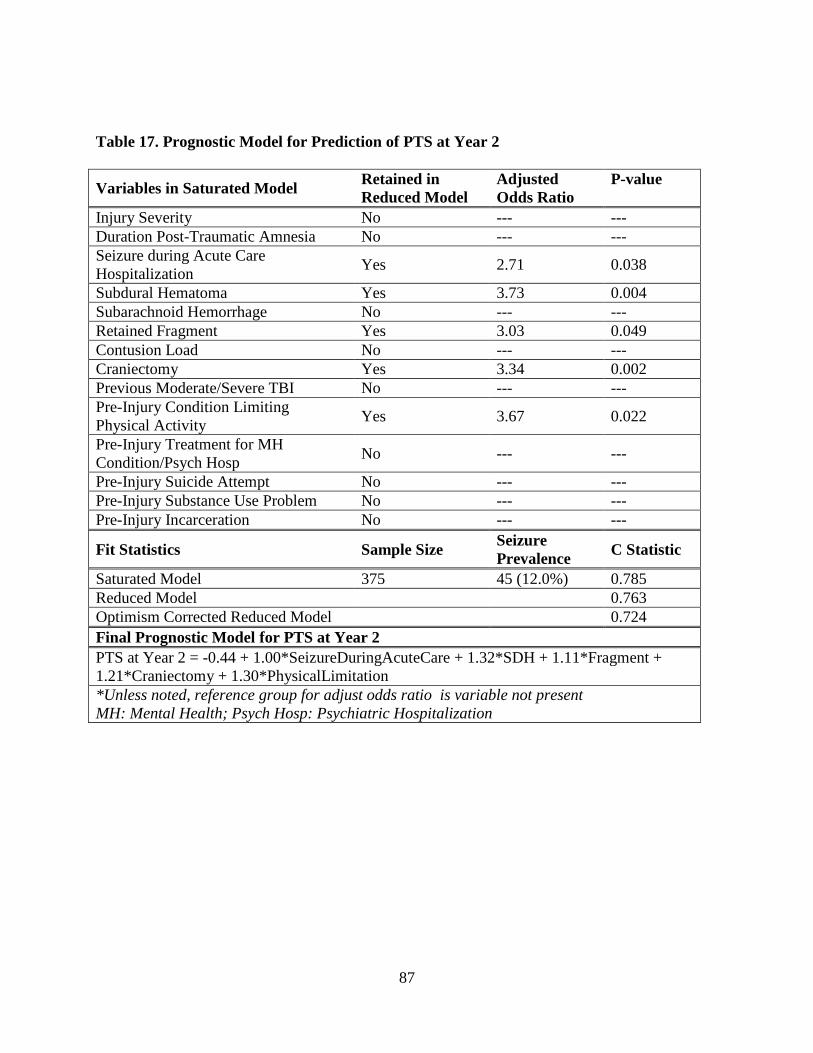
Develop prognostic models to predict PTS during acute care hospitalization, at Year 1, and Year
2 following traumatic brain injury.
Hypothesis 2.2.1. A: Personal, medical, and injury characteristics will be identified as
significant predictors of PTS
Hypothesis 2.2.1. B: Significant predictors of PTS will vary based on time PTS is
assessed post-injury
Hypothesis 2.2.1. C: Prognostic models will be internally validated
28
2.2.2 Specific Aim 3
Examine the effect of genetic variation within neuronal glutamate transporter genes, SLC1A1
and SLC1A6, on epileptogenesis following severe traumatic brain injury.
Hypothesis 2.2.2. A: Genetic variation in the neuronal glutamate transporter genes,
SLC1A1 and SLC1A6, will be significantly associated with epileptogenesis and
PTS
Hypothesis 2.2.2. B: Different genetic variants will be associated with epileptogenesis
during different subcomponents of the three-year post-injury timeframe
29
3.0 BACKGROUND
3.1 STUDY POPULATIONS
3.1.1 Traumatic Brain Injury Model Systems
The Traumatic Brain Injury Model Systems (TBIMS) study is an ongoing multi-center,
prospective, observational cohort study. Established in 1987, there are 16 currently funded
centers including the University of Pittsburgh and four previously funded centers that continue to
collect follow-up information 105. The main objective of the TBIMS program is to study recovery
and outcomes after moderate to severe TBI. Individuals participating in the TBIMS study have
the potential to be followed from inpatient rehabilitation for TBI throughout the duration of their
lifespan. Currently, the longest follow-up time-point is 25 years post-injury. All data collected
through the TBIMS program is deposited to the TBIMS National Data and Statistical Center,
where it is formatted into the TBIMS National Database (NDB), the central resource for all
TBIMS research.
TBIMS Centers are established and funded through center-specific grants, typically
awarded for five-year periods 106. To be eligible, a Center must provide a “multidisciplinary
system of rehabilitation care specifically designed to meet the needs of individuals with TBI” 107.
To fulfill this requirement, a participating Center must include emergency medical services
30
(most commonly at least one Level-1 trauma center), acute care including neurosurgical
capabilities, comprehensive inpatient rehabilitation, and long-term interdisciplinary follow-up
and rehabilitation services. The number of acute care hospitals within a single TBIMS Center
may vary. The TBIMS Center must be able to access emergency and acute care records,
however, it is not required that acute medical and rehabilitation facilities be within the same
hospital system. TBIMS Centers must include at least one Center specific study, participate in at
least one multicenter study, and collect and submit longitudinal data to the TBIMS-NDB.
The TBIMS program specifies inclusion criteria for all individuals enrolled at any
participating Center. Throughout its history, the TBIMS program has revised participant
inclusion criteria. The current inclusion criteria are defined below. Firstly, an individual must
meet the TBIMS case definition of TBI.
Damage to brain tissue caused by an external mechanical force as
evidenced by medically documented loss of consciousness or post-
traumatic amnesia (PTA) due to brain trauma or by objective neurological
findings that can be reasonably attributed to TBI on physical examination
or mental status examination 108
All participants must have a moderate to severe TBI defined by at least one of the
following classifications: post-traumatic amnesia >24 hours, trauma related neuroimaging
abnormality, loss of consciousness >30 minutes, or emergency department GCS<13 (not
influenced by intubation, sedation, or intoxication). Additionally, all participants must be 16
years or older, present to a TBIMS Center affiliated emergency department within 72 hours of
injury (previously 24 hours), receive acute and comprehensive inpatient rehabilitation within the
Center’s designated facilities, and provide written informed consent.
31
Data from emergency and acute care, referred to as Form 1, is collected retrospectively.
Data from inpatient rehabilitation and at all follow-up interviews, referred to as Form 2, are
collected prospectively. Follow-up interviews are currently conducted at 1, 2, 5, and every five
years thereafter, post-injury. Over the course of the TBIMS program, follow-up intervals and
variables collected have been revised. At the time of analysis, there were 285 variables collected
via Form 1 and 243 variables collected at each follow-up using Form 2. Data in the TBIMS-
NDB collected through the end of the first fiscal quarter of 2015 (October 2014) were used for
the current analyses. This included 13,241 cases with Form 1 data and 41,733 follow-up
interviews.
The TBIMS-NDB has previously been extensively studied to determine its
generalizability to the United States population 105; 109; 110. TBIMS investigators have recently
compared the TBIMS study population to the Uniform Data System for Medical Rehabilitation
(UDS) and eRehabData. The UDS and eRehabData were combined to form a national dataset
consisting of individuals 16 years or older with a primary diagnosis of TBI who received
inpatient rehabilitation services. Demographic, socioeconomic, and rehabilitation outcomes were
then compared between the combined dataset and the TBIMS-NDB. These studies confirm the
TBIMS-NDB is largely representative of individuals receiving inpatient rehabilitation services
for TBI in the United States 105; 109; 110. However, the TBIMS-NDB was determined to include a
larger proportion of individuals under age 65 and a greater proportion of individuals employed
prior to injury 110. Additionally, individuals in the TBIMS-NDB had a significantly shorter
length of stay in inpatient rehabilitation compared to the US TBI rehabilitation population 110. To
address these differences, methods have been developed to weight the TBIMS-NDB to represent
the general US population of individuals receiving inpatient rehabilitation
32
Additional limitations of the TBIMS study include those inherent to other multi-site
longitudinal studies such as loss to follow-up. Total loss to follow-up is approximately 24% with
varying estimates at each time point. Individuals lost to follow-up may differ across time-points.
Similarly, Centers may lose funding during certain cycles resulting in loss to follow-up of the
individual Center’s cohort during that funding cycle. Attrition due to loss of Center funding is
approximately 4% overall and does not surpass 6% for any one follow-up time-point (Table 4).
Table 4. Sample Size and Attrition Rates of the TBIMS-NDB as of March 31, 2015
Time Point Number Included % Attrition % Additional Attrition* Form 1 13,667 NA NA Form 2 45,499 19 4 Year 1 12,973 15 3 Year 2 11,518 16 5 Year 5 8,952 18 5 Year 10 4,684 19 6 Year 15 1,588 14 6 Year 20 449 14 0 *additional attrition due to loss of Center funding
3.1.2 University of Pittsburgh Local Project
Multiple smaller studies of moderate to severe TBI have been, or are currently being conducted
at the University of Pittsburgh. Under the oversight of Dr. Amy Wagner, an overarching study
protocol has been developed to collect biological samples in order to analyze various potential
biomarkers and their associations with TBI outcomes. The study objectives include investigating
the association between genetic variation and development of PTS after moderate to severe TBI.
For the current analyses, individuals 18 years of age or older with a severe TBI
(determined by a GCS score ≤8 on admission to the UPMC Level 1 trauma center) were
included. Individuals were excluded if they had less than three years of follow-up from the time
33
of their index TBI. Information on outcomes of interest, including seizure activity, was extracted
from the UPMC electronic medical record using a standardized protocol.
3.2 PROGNOSTIC MODELING
Prognostic models are statistical tools that estimate an individual’s risk for developing an
outcome of interest based on specific characteristics 111. Prognostication is common in medical
research and practice; prognostic models have previously been developed for use in multiple
fields such as oncology, cardiology, and neonatology. Within the field of research surrounding
TBI, prognostic models have been investigated to predict multiple outcomes such as survival,
disability, and global outcome 112-115. Importantly, especially because the etiology of secondary
injury mechanisms in TBI and epileptogenesis are extremely heterogeneous, the aim of
prognostic modeling is not to explain causality of the outcome 111. Prognostic models may
include predictors that are not themselves causal, but may be measuring latent variables that have
not, or cannot, be measured.
The development of reliable, validated prognostic models is essential for models to be
clinically useful. The gold standard for validating a prognostic model is through the use of a
second, independent study cohort (i.e. external validation). However, this may be prohibitive in
some specialties and recent advances in statistical methods for validation have been made.
Resampling methods are reported to be extremely proficient as a means of internally
validating a statistical model 116; 117. As computing capabilities increase, resampling methods
such as bootstrapping, are more readily available and widely used. Bootstrapping is a procedure
that involves selecting a sample, with replacement, from an original dataset. Researchers can
34
indicate the number of samples selected and specify parameters of interest (e.g. sample must
always include a designated ratio of males to females). A prognostic model of interest can be
identified a priori and tested for fit using each bootstrapped sample 118. Information from
bootstrapped samples is aggregated to determine overall fit statistics such as discrimination and
calibration of the pre-specified model 116; 117; 119.
Currently, automated programs are available using statistical software, such as R, to
streamline these processes 118; 120. In addition, computer modeling can be used to develop
reduced models (i.e. model development using stepwise elimination) while decreasing
subjectivity and the potential for investigator bias. Each bootstrap sample may therefore
indentify a different set of prognostic variables. These data are simultaneously aggregated to
determine the best-fit model allowing for user specification (i.e. AIC or alpha thresholds for
variable inclusion/exclusion) 118. After automated development, the model can be tested for fit
using the original sample set and the bootstrapped samples, allowing for computation of fit
statistics as above 116; 119.
35
4.0 MANUSCRIPT ONE
INCIDENCE AND RISK FACTORS OF POST-TRAUMATIC SEIZURES FOLLOWING
TRAUMATIC BRAIN INJURY: A TRAUMATIC BRAIN INJURY MODEL SYSTEM
STUDY
Anne C Ritter1, 2, Amy K Wagner2,3,4,5, William C Walker6, Anthony Fabio1, Jerzy P Szaflarski7, Mary Jo Pugh8,9, Allen W Brown10, Ross D Zafonte11, Flora M Hammond12,13, Tamara Bushnik14, Doug Johnson-Greene15, Timothy Shea16, Jason W Krellman17, Joseph A Rosenthal18, Laura E Dreer7
1Department of Epidemiology, University of Pittsburgh, Pittsburgh, PA 2Physical Medicine & Rehabilitation, University of Pittsburgh, Pittsburgh, PA 3Safar Center for Resuscitation Research, University of Pittsburgh, Pittsburgh, PA 4Department of Neuroscience, University of Pittsburgh, Pittsburgh, PA 5Center for Neuroscience at University of Pittsburgh, Pittsburgh, PA 6Dept of Physical Medicine & Rehabilitation, Virginia Commonwealth University 7University of Alabama at Birmingham Epilepsy Center, Department of Neurology, University of Alabama, Birmingham, AL 8South Texas Veterans Health Care System Polytrauma Rehabilitation Center, San Antionio, TX 9Department of Epidemiology and Biostatistics, University of TX Health Science Center San Antionio, San Antonio, TX
10Department of Physical Medicine and Rehabilitation, Mayo Clinic, Rochester, MN 11Spaulding Rehabilitation Hospital, Harvard Medical School, Boston, MA 12Carolinas Rehabilitation, Charlotte, NC 13Indiana University School of Medicine, Indianapolis, IN 14Rusk Rehabilitation, New York University School of Medicine, New York, NY 15Miller School of Medicine, University of Miami, Miami, FL 16Department of Physical Medicine and Rehabilitation, Ohio State University, Columbus, OH 17Department of Rehabilitation Medicine, Icahn School of Medicine at Mount Sinai, New York, NY 18Departments of Physical Medicine and Rehabilitation and Ophthalmology
Submitted for publication to Epilepsia
36
4.1 ABSTRACT
Objective: Determine the incidence of post-traumatic seizures (PTS) following traumatic brain
injury (TBI) among individuals with moderate-to-severe TBI requiring rehabilitation and
surviving at least 5 years.
Methods: Using the prospective TBI Model Systems National Database, we calculated the
incidence of PTS during acute hospitalization, and at Years-1, 2, and 5 post-injury in a
continuously followed cohort enrolled between 1989 and 2000 (n=795). Incidence rates were
stratified by risk factors of interest, and relative risk (RR) was calculated. The RR of late PTS
following immediate (<24hr), early (24hr-7d), or late seizures (>7d) versus no seizure activity
prior to discharge from acute hospitalization was also examined.
Results: PTS incidence during acute hospitalization was highest immediately (<24hrs) after
injury (8.9%). New onset PTS incidence was greatest between discharge from inpatient
rehabilitation and Year-1 (9.2%). Late PTS prevalence from injury to Year-1 was 11.9% and
reached 20.5% by Year-5. The RR of late PTS was significantly greater for individuals self-
identifying as a race other than black or white at Year-1 (RR=1.96), and for black individuals at
Year-5 (RR=2.86) versus white individuals. Late PTS was greater for individuals with certain
intracranial pathologies (i.e. subarachnoid hemorrhage). Penetrating TBI had even higher RR but
did not reach significance, likely due to small group size. Individuals with immediate and late
seizures during acute hospitalization were at a significantly greater late PTS risk (RR: 2.61 and
3.36, respectively).
Significance: In this prospective, longitudinal, observational study, incidence rates were similar
to those in previously published studies. Individuals with immediate and late seizures during
acute hospitalization are at an increased late PTS risk after hospitalization. Race and intracranial
37
pathologies also influenced RR for late PTS. Further studies are needed to examine the impact
of seizure prophylaxis in high-risk subgroups and to delineate possible contributors to race
associations on long-term seizure outcomes.
38
4.2 INTRODUCTION
Traumatic brain injury (TBI) is a prevalent public health problem with an annual incidence over
2.5 million in the United States (US), of which approximately 12% result in hospitalization or
death 3. TBI also has many related secondary chronic conditions 121 including shorter life-
expectancy after severe TBI versus demographically similar, non-TBI populations 2. Recent
work using the TBI Model Systems (TBIMS) National Database (NDB) found seizure to be an
important contributor to premature death among individuals who were hospitalized and received
inpatient rehabilitation for TBI, who had a 50-fold risk for subsequent seizure related death
compared to an uninjured similarly matched sample 2.
Post-traumatic seizures (PTS) can occur any time post-TBI. Classification is based on the
time of seizure post-injury: immediate (<24hrs), early (24hrs-7d), or late (>7d post-TBI) 41.
These cut-offs reflect proposed differences in causal mechanisms and subsequent seizure risk 42;
66. Seizures occurring within the first week following TBI, also termed acute symptomatic
seizures 43, are considered transient, decreasing seizure threshold only temporarily 42. Late PTS,
often used interchangeably with post-traumatic epilepsy (PTE), is characterized by persistent
neurobiological changes, attributed to secondary injury biochemical cascades and epileptogenic
mechanisms that eventually present as clinical seizures 18; 31. Individuals with acute symptomatic
seizures who have a subsequent late seizure are considered to have late PTS or PTE. The clinical
definition of epilepsy, revised in 2014 by the International League Against Epilepsy (ILAE),
includes conditions where an individual has a single unprovoked seizure and their risk of a
recurrent seizure is similar to, or greater than, the risk of seizure recurrence after two unprovoked
39
seizures occurring ≥24hrs apart (≥60%) 42. The recurrent seizure risk following a single,
unprovoked seizure >7d post-TBI is high enough to consider late PTS as an epileptic condition
42; 56. For the current study, late PTS is used, but is equivalent to the current definition of PTE.
Reported PTS incidence varies widely and depends on study design and population
characteristics. Few large epidemiological PTS investigations have been conducted in
heterogeneous populations. The seminal population-based study in the US examined late PTS in
a predominantly white population from 1935 to 1984 12. Among individuals with severe TBI, the
cumulative probability of late PTS was 10.0% five years after TBI; early PTS occurred in 2.6%
of individuals 12. This study included all ranges of TBI severity, was racially homogenous, and
included both adults and children. Inclusion of adults and children may confound risk
relationships and complicate accurate risk factor determination since neurological
injury/recovery mechanisms post-TBI may vary over the course of neural development or aging.
These authors also reported 10.3% of adults with severe TBI developed early PTS 63. Other,
smaller studies report early PTS incidence to range from 2.4 to 8.4% 66; 122. However, these
studies included children and adults with a range of TBI severities. Late PTS cumulative
probability rates have been more often reported and findings vary widely 12; 47; with prevalence
ranging from 4%-19% 12; 66; 67; 70; 123. While these studies provide important information
regarding PTS frequency after TBI, many are retrospective, not racially diverse, from single
medical centers, and cannot be generalized to large heterogeneous populations. Additionally,
they provide little information on immediate and/or early PTS. To address these limitations, the
purpose of the current study was to prospectively determine the incidence of PTS following TBI
among individuals with moderate-to-severe TBI requiring rehabilitation and surviving at least 5
years using a large-scale, multi-center database.
40
We used the TBIMS-NDB to calculate multiple PTS frequency measures in a cohort
followed out to five years post-injury. Additionally, we stratified PTS incidence at various time-
points by demographic and injury characteristics of interest to compute relative risk (RR) for
each factor.
4.3 METHODS
4.3.1 Study Design and Population
Data were obtained from the prospective TBIMS-NDB. The TBIMS-NDB is a multi-
center, prospective, observational study to investigate recovery and outcomes following acute
neurotrauma and inpatient rehabilitation in a heterogeneous population of individuals with
moderate-to-severe TBI, across the US. All participating sites have an affiliated trauma center
with acute neurosurgical capabilities and associated comprehensive inpatient rehabilitation.
Eligibility criteria are: moderate-to-severe TBI (PTA>24hrs, or LOC>30minutes, or emergency
department GCS<13, or positive neuroimaging findings), age ≥16yrs, admitted to a participating
hospital emergency department within 24hrs of injury, and received both acute care and inpatient
rehabilitation within a TBIMS designated hospital system. All enrolled individuals, or legal
proxy, provided written informed consent; Institutional Review Board approval exists at all sites.
An additional inclusion criterion for this study was completion of Year-5 post-injury
follow-up interview. Further, individuals were then excluded if data regarding seizure activity
during acute care hospitalization, or Year-1 and Year-2 post-injury, were not available. The
acute hospitalization seizure variable was dropped from TBIMS data collection procedures, and
41
follow-up seizure definitions changed, in 2003 and 2005, respectively. Therefore, all individuals
included in analyses were enrolled between 1989 and 2000; follow-up assessments were
completed by 2006.
4.3.2 Data Collection
Data were collected at enrollment, Year-1, Year-2, and Year-5 post-injury. Enrollment
data included demographic, social, and injury characteristics as well as personal and medical
history (pre-injury), and acute outcomes. A proxy interview was completed, for both enrollment
and follow-up when participants with TBI were unable to answer questions accurately.
Throughout the study duration, data collection protocols changed over time. Therefore, missing
data may exist even for individuals who completed assessments at all study time-points.
Outcome Variable
The main outcome variable was PTS status, determined during the course of acute care
hospitalization and at each follow-up time-point (Year-1, Year-2, and Year-5).
4.3.3 PTS During Acute Care Hospitalization
The presence/absence of a physician-confirmed clinical seizure during acute
hospitalization was identified via medical record review using a standardized form and classified
based on time from injury (immediate: <24hrs, early: 1-7days, late: >7days). Only time of first
seizure was recorded. Multiple seizures were not captured, therefore, an individual seizing
immediately or early after injury may have also seized in a subsequent time category prior to
acute discharge.
42
4.3.4 PTS at Follow-up Interviews
At each follow-up, individuals were asked “Have you been told by a physician that you
have had a seizure since your last follow-up?”. Yes/No answers were recorded; individuals self-
reporting seizure activity were documented as having PTS at the specified time-point.
4.3.5 PTS Risk Factors
Risk factors of interest included demographic and injury characteristics. Demographic
variables included age, sex, and race. Injury characteristics included admission Glasgow Coma
Scale (GCS) score, pathology on computed tomography (CT) scan obtained within 7 days of
injury, and penetrating TBI (pTBI; Supplemental Table 1). Injury severity was also classified
using alternate criteria for moderate-to-severe injury based on duration of post-traumatic amnesia
(PTA), loss of consciousness (LOC), and positive neuroimaging findings124. CT findings were
included as separate variables for specific pathology type [e.g., subdural hematoma (SDH),
subarachnoid hemorrhage (SAH)] coded as present/absent, and were not mutually exclusive.
pTBI was computed via a coding algorithm previously validated in a subsample of the TBIMS85.
A contusion load score was calculated by summing the number of regions with contusion on CT
reports, then collapsing the sum into 0, 1, 2, 3, and 4 or more regions. Seizures during acute
hospitalization were examined as PTS risk factors at follow-up (Supplemental Table 1). No
data were collected on premorbid seizure activity or history of epilepsy. ICD-9 codes indicating
neurosurgical procedures or complications were not collected, nor were medication data.
43
4.3.6 Statistical Analysis
All statistical analyses were completed using SAS version 9.4 (SAS Institute, Carry NC)
and R version 3.0.2 125. Seizure incidence was calculated during acute care hospitalization and at
follow-up. At Year-1 and Year-2, if data on seizure activity since last follow-up were missing
(n=101 and n=99, respectively), seizure status was considered not present at that time-point. If
individuals had no prior seizure activity, they were considered “at-risk” for PTS and were
included in the denominator of incidence calculations. If individuals had evidence of PTS at a
previous time-point, they were not considered “at-risk” for incident PTS. Sensitivity analyses,
including Chi-Square and Mann-Whitney, or Fisher’s Exact test and Kruskal-Wallis, were
conducted to determine whether individuals with missing data at Year-1 or Year-2 differed from
those without missing data. Late PTS incidence since last follow-up was calculated at Years-1, 2,
and 5. Late PTS prevalence from time-of-injury was also calculated at each follow-up time-point
(i.e. >7d to Year-1, >7d to Year-2, etc.). Additionally, the percent of individuals reporting
seizure activity at multiple time-points was calculated, stratified by time of onset.
Following primary incidence calculations, immediate/early PTS (injury to 7 days), late
PTS from injury to Year-1, and late PTS from Year-1 to Year-5 incidence rates were stratified by
risk factors of interest. For demographic variables identified as having significantly different RR
of PTS, sensitivity analyses were conducted to determine if there were differences in injury
characteristics.
Seizures during acute care (immediate, early, late) were then examined specifically as
risk factors. Prior to this analysis, late PTS was recalculated to remove late seizures during acute
care (>7days to discharge) from the case definition in order to evaluate RR for late PTS at Year-
1 and Year-5 follow-up.
44
4.4 RESULTS
4.4.1 Population
Data were available for 2,418 individuals injured and enrolled prior to 2001. Of these,
796 were assessed at 5-years post-injury and met inclusion criteria for this analysis (Figure 1).
Individuals were predominantly male (74.8%), self-identified as white (58.3%), and tended to be
in their mid-thirties at time of injury. The predominant cause of injury was motor vehicle
collision (51.6%) (Table 1). Individuals lost to follow-up at Year-1 or Year-2 who were
recaptured at Year-5 tended to have less severe injuries and fewer high-risk pathologies (data not
shown).
4.4.2 Frequency Measures
By the Year-5 follow-up, 219 (27.5%) individuals had reported or documented seizure
activity at some point post-injury and 163 (20.5%) developed late PTS (Table 2). During acute
care hospitalization (mean=22.8 days), 98 (12.3%) individuals seized, with highest acute
hospitalization PTS incidence immediately (<24hrs) after TBI (Table 2). 1.8% of individuals
developed incident PTS after 7d post-injury but prior to acute discharge. Incidence of new onset
and late PTS since last follow-up peaked at Year-1 (Table 2). Of all individuals developing late
PTS by Year-5, 58.3% did so by Year-1, and 82.2% did so by Year-2. Further, at Years-1, 2, and
5, more than 50% of individuals reporting seizure activity endorsed multiple seizures since last
queried.
45
Of individuals first seizing prior to Year-5, 38.0% reported seizure activity at one time
interval only. Among individuals with incident PTS at Year-1 or Year-2, 64% and 40.1%
respectively, reported interval seizure activity at subsequent follow-up(s) (Supplemental Table
2).
4.4.3 Stratified Incidence and Relative Risks
Stratified analyses showed PTS incidence was nominally lower among women than men
at each time-point, but RR did not reach significance. Individuals self-identifying as white also
tended to have lower RR of PTS versus individuals who did not identify as white with significant
differences evident at Year-1 and Year-5. Although individuals age 35-44 at injury had greater
risk of PTS at Year-5, no consistent pattern was seen (Table 3a). Sensitivity analyses showed
differences in pTBI frequencies across race [2.4% white, 7.7% black, 6.3% other with pTBI
(p=0.01)].
Immediate/early PTS incidence stratification by pathology showed no patterns. When late
PTS incidence was stratified by pathology, RR was nominally greater for individuals with each
pathology examined versus those without the given pathology, except for intra-ventricular
hemorrhage at Year-1. Highest nominal incidence was observed for individuals with pTBI at
both Year-1 and Year-5. Despite these patterns, only the RR for SAH pathology (Table 3b) and
contusion load at Year-1 reached significance (Table 3c).
PTS incidence was stratified by two measures of injury severity: GCS, and moderate
versus severe injury as determined by duration of LOC, PTA, and neuroimaging findings.
Although RR was not significantly greater for individuals with more severe TBI as classified by
these measures (Table 3c), late PTS incidence at Year-5 was nominally greater for individuals
46
with more severe. When late PTS incidence at Year-1 and Year-5 were stratified by time of
seizure during acute hospitalization, significant RR’s were found (Table 4). Individuals with
immediate/late seizures occurring during acute care were more than twice as likely (RR: 2.61
and 3.36, respectively) to develop late PTS between acute discharge and Year-1. Those with
immediate seizures also had higher risk (RR=2.06) at Year-5.
4.5 DISCUSSION
We examined multiple PTS frequency measures and associations with demographic and injury
characteristics during and following acute care hospitalization and inpatient rehabilitation for
moderate-to-severe TBI from a prospectively followed, large, nation-wide sample. Total PTS
incidence during acute hospitalization was slightly higher than previously reported, but incidence
of early seizure was consistent with previous reports 12; 48; 50; 65; 80; 122; 126. However, not all prior
PTS studies during acute hospitalization clarify if immediate and early seizures are considered
simultaneously. Few previous reports specifically delineate immediate PTS incidence. Annegers
and colleagues determined 2.1% of a Rochester county Minnesota study cohort, including
children and all injury severities injured from 1935-1974, developed early PTS 63. Of these,
75.9% first seized during the initial 24hrs post-TBI, similar to our finding that 72.4% of
individuals who developed PTS during acute hospitalization first seized within 24hrs of injury.
These rates are higher than those reported earlier by Jennett, where only 5% of the cohort
exhibited early seizures with 60% occurring within 24hrs 50. Disparate study findings might be
due to differences in participants (i.e., injury severity/age) or methods for capturing
47
immediate/early PTS. When considering only adults with severe TBI in Annegers’ study, 10.3%
developed early seizures63, similar to 10.7% in our study.
Interval new onset seizure (9.2%) and late PTS (10.8%) incidence were highest at Year-1,
similar to previously published findings in comparable study samples 66. Annual late PTS
incidence since last follow-up at Year-1 and Year-2 (10.8 and 5.5%, respectively) were higher
than annual incidence rates previously reported by Ferguson et al 69. In our study, late PTS
prevalence from injury to Year-1 (11.9%) and Year-5 (20.5%) were ≥2 times higher than for
those with severe TBI in Annegers’ study 12. In two studies by Englander and colleagues, using a
sample very similar to ours, late PTS incidence at 2 years was 16.4 and 14% 47; 127, similar to the
16.8% in our study. Differences between the percent affected in our analyses may be attributed
to the inclusion of individuals with mild TBI or children. Possible differences between our
analysis and Annegers’ work could also be attributed to differences in study design (e.g.
population-based study with medical chart review vs. prospective longitudinal cohort study and
self-report). Additionally, due to changes in clinical care over time, differences could also be
attributed to increased EEG monitoring of individuals post-TBI, and/or higher survival of the
more severely injured in the current study. Future research validating these findings, using a
more recent cohort from TBIMS-NDB could be considered with reintroduction of seizure data
collection. However, due to changes in TBIMS seizure data collection occurring after 2000, we
currently cannot validate the distinct effects of acute symptomatic seizures or late seizures during
acute hospitalization in a more recent, independent TBIMS population.
Consistent with other studies, 82.2% of individuals developing late PTS did so by Year-2
post-injury 56. Yet, it is important to recognize new onset seizures, and subsequently new cases
of late PTS, continued to be detected at Year-5. Previous PTE studies show new cases may
48
develop as many as 30 years post-injury, and epilepsy risk remains significantly elevated versus
the general population up to 10 years post-injury among those with severe TBI 12. Our data
provide a more complete picture of the extended risk of late PTS, past the first 1-3 years post-
TBI, when other modern observational cohort studies truncate their follow-up 47; 67; 69. Future
work should continue to use the TBIMS-NDB to extend the current follow-up time (earliest
participants now at Year-25 post-injury) and determine if current standardized incidence ratios
remain similar to or are greater than those found by Annegers.
Late PTS prevalence reached 20.5% by Year-5. This figure is greater than reported rates
of 13.7% by 5 years post-injury 66 but remains lower than rates in combat veteran populations 72.
Although both studies’ cohorts were recruited from rehabilitation facilities, differences in study
design and population (e.g., higher pTBI among military populations) likely contributed to
differences.
When PTS incidence was stratified by risk variables, RR patterns were observed for race,
but not sex. The lack of significantly greater RR for men could be confounded by survival bias,
if men were more severely injured and did not survive to Year-5. However, in a more recently
injured TBIMS-NDB cohort, men were at higher risk for seizures during acute care
hospitalization, even in multivariable models including markers of injury severity (Manuscript
Two). Individuals self-identifying as white had a lower late PTS risk at both Year-1 and Year-5
versus non-white individuals. No other known large epidemiological PTS studies have reported
significant differences in risk associated with race. One study demonstrated increased RR of late
PTS at 2 years for non-white groups versus white individuals, but results were not statistically
significant 47. Differences in PTS frequency by race may be partially explained by differences in
injury type (i.e. pTBI). Although, previous work developing prognostic models of PTS using a
49
more recent cohort from the TBIMS-NDB did not find race to be even nominally associated with
PTS and subsequently did not investigate race as a PTS predictor in multivariable modeling
(Manuscript Two). In addition to more frequent pTBI, differences in late PTS risk by race may
reflect differences in environmental exposure (e.g. repeat TBI, substance use) 128 as well as
known racial differences in allelic frequencies for genes thought to influence PTS risk 94; 102; 103;
129. Future work might investigate racial/ethnic differences in epilepsy related outcomes and
comorbid burden 130.
Examining incidence stratified by pathology revealed somewhat surprising result. While
patterns of nominal rates were as expected, late PTS risk was only significantly elevated for SAH
and higher contusional loads at Year-1. These data are inconsistent with most studies, which find
that SDH is a significant PTS risk factor. Our findings may be due to variability within SDH and
SAH definitions applied in the TBIMS-NDB over time. Due to other limitations in data capture,
we cannot examine incidence stratified by other potential risk factors of interest like
neurosurgical procedures.
Of special note, at Years-1 and 5, pTBI was associated with the highest nominal late PTS
RR of any characteristic studied (18.5 and 18.2%, respectively). Differences in RR were not
statistically significant, but the low numbers with pTBI suggests these analyses were
underpowered. Additionally, we did not observe consistent increases in PTS incidence with
greater injury severity based on GCS or alternative criteria.
When late PTS incidence at Year-1 and Year-5 were recalculated without individuals
seizing late during acute hospitalization included in the case definition, there were significant
differences in RR based on timing of first acute seizure. Individuals with immediate seizure
(<24hrs) had a higher late PTS incidence and greater risk at both time-points versus those not
50
seizing during acute hospitalization. Those with late seizures (>7d) during acute hospitalization
also had a higher late PTS incidence at Year-1 versus individuals not seizing acutely (Table 4).
Surprisingly, having acute seizure(s) restricted between 1-7 days did not increase late PTS risk.
Multiple reports document that early seizure increases late PTS risk 12; 47; 66; 69; 70. However, these
studies do not differentiate between immediate and early seizures. Previous work by Temkin
demonstrated increased early seizure risk associated with immediate seizures, but early seizures
(1-7 days) were associated with increased late PTS risk 78. Unlike Temkin’s work, we did not
observe increased late PTS risk associations with early seizures. It is unclear in Temkin’s work
whether individuals with early seizures seized immediately or not, and how selecting individuals
at high risk of PTS for inclusion impacted late PTS findings. In our analyses, immediate/early
seizures were mutually exclusive; therefore, we could not examine the impact of seizing both
immediately and early on late PTS risk.
The high percentage of individuals in our study seizing immediately, along with the
common use of early PTS prophylaxis after phenytoin was shown in clinical trials to reduce
early seizures 131, may complicate observed relationships between immediate/early seizures and
late PTS reported in our study. Even if seizure prophylaxis was administered immediately upon
admission, it is likely that individuals could have already experienced their first immediate
seizure 50. Our data demonstrate lower incidence of early versus immediate seizure and show
immediate seizure (but not early seizure) significantly increases late PTS risk out to Year-5.
However, seizure prophylaxis during the first week post-injury is shown to only have beneficial
effects in reducing early PTS, not late PTS 131. While not directly measured in the TBIMS-NDB,
data from a single TBIMS center from 2000-2007 show 96% received some form of AED
prophylaxis during their acute care 102. Taken together, we hypothesize that early PTS
51
prophylaxis reduces early seizures but does not reduce the epileptogenic processes initiated by
immediate seizures that may (often) occur prior to prophylaxis administration. Therefore, we
speculate that individuals in our study who may have seized prior to appropriate prophylaxis use
may remain at-risk for late PTS. Future work needs to examine late PTS risk specifically among
individuals receiving prophylaxis but who seized prior to or during AED loading, as these data
may reshape PTS prophylaxis guidelines. We suggest that antecedent, immediate/early seizure
excitotoxicity and contributing to epileptogenesis, regardless of early PTS prophylaxis 6.
While these data provide PTS characterization in a large cohort of individuals from the
US, limitations must be considered. Our cohort is restricted to individuals having received
inpatient rehabilitation and surviving to Year-5 post-TBI. Additionally, PTS and/or PTE
definitions and data collection methods used can bias descriptive analyses. We cannot specify the
data source used as evidence of seizure during acute hospitalization other than to cite
“documentation from the medical record”. It is unclear if evidence was collected using
electroencephelogram (EEG). Currently, continuous EEG (cEEG) is common in many trauma
centers, and its use identifying subclinical seizures may result in higher incidence of acute
symptomatic seizures. How cEEG might have factored into acute symptomatic seizure
identification for individuals injured prior to 2001 is less certain. In critically ill patients with
TBI, up to 50% of seizures are reported to be subclinical 76. Additionally, only the first seizure
during acute care hospitalization was documented. Therefore, individuals seizing immediately or
early, and having later seizures during acute hospitalization, were not accounted for in late PTS
calculations if they did not experience a seizure after discharge from acute hospitalization.
52
Similarly, subsequent seizures during acute care hospitalization if occurring, were not
documented.
Another potential limitation is PTS misclassification at follow-up because, while acute
symptomatic seizure information was obtained via medical record, all follow-up data were based
on participant or proxy self-report of whether a physician told them they had a seizure.
Individuals could over-report if they experienced psychogenic non-epileptic seizures or transient
neurological symptoms not due to seizure activity. Conversely, individuals experiencing true
epileptic seizure activity between follow-ups, who were not aware or did not seek care, may
contribute to underestimation of the true number of individuals with late PTS. Nevertheless, in
large epidemiological studies of seizure and epilepsy, self-report remains common and necessary
132. Adding to possible misclassification bias, individuals with missing data at Year-1 or Year-2
were included as at-risk individuals for incidence calculations. By doing so, we may have
included individuals as “at-risk” who had seized, underestimating the true incidence. However,
we examined individuals with missing data and determined they typically had less severe injury
and less intracranial pathology identified via CT than those returning for all follow-ups (data not
shown). Within the larger TBIMS-NDB, individuals with less severe TBI who presumably
recover better, are more often lost to follow-up than individuals with more complex injuries and
presumably worse outcomes 133. Therefore, excluding individuals with missing data at Year-1 or
Year-2, who were subsequently followed at Year-5, would have likely produced a greater degree
of bias and overestimated incidence of PTS. Due to limitations in data collection, we were
unable to identify, and subsequently exclude, individuals with pre-existing epilepsy disorders.
Despite these limitations, the current analyses suggest race as a possible PTS risk factor. The
53
work also provides insight into temporal PTS risk factors, including immediate/early seizure, and
examines the effect of each on late PTS incidence out to 5 years post-injury.
4.6 TABLES
Table 5. Demographic and Injury Characteristics at Baseline Visit N(%)
Sample Size = 796 Age at Injury* 35.4 (15.7) Sex Male 595 (74.8)
Female 201 (25.2) Race White 464 (58.3)
Black 246 (30.9) Other 86 (10.8)
Cause of Injury
MVA 411 (51.6) Fall 109 (13.7) Any
Violence 164 (20.6)
Any Sport 9 (1.1) Other 103 (12.9)
Injury Severity Moderate 79 (9.9) Severe 717 (90.1)
PTA (days)* 31.6 (26.3) LOC (days)* 10.7 (18.9) Admission DRS* 13.3 (5.4) Length of Acute Stay (days)* 22.8 (19.4) *mean(SD); PTA – Post-Traumatic Amnesia; LOC – Loss of Consciousness; DRS – Disability Rating Scale
54
Table 6. Frequency Measures of PTS at Follow-up Time Points after TBI
Incidence of New Onset
Seizure
Incidence of Late PTS since last
follow-up*
Prevalence of Late PTS since
injury
Time Point N (%) N (%) N (%)
Initial Population 796 796 796
Acute Seizure Status
Immediate (<24hrs) 71 (8.9) --- ---
Early (1 < 7 days) 14 (1.9) --- ---
Late (>7 days) 13 (1.8) --- ---
Year 1 64 (9.2) 86 (10.8) 95 (11.9)
Year 2 32 (5.0) 39 (5.5) 134 (16.8)
Year 5 25 (4.2) 29 (4.3) 163 (20.5)
*Year-1 represents late PTS incidence since discharge from rehab
55
Table 7. Incidence and Relative Risk of PTS Stratified by Variables of Interest
Immediate and Early PTS Late PTS Year 1 Late PTS Year 5 N(%) RR (CI) N(%) RR (CI) N(%) RR (CI) No
Seizure Seizure No Seizure Seizure No
Seizure Seizure
N (%) 711 (89.3)
85 (10.7) 701
(88.1) 95
(11.9) 633 (90.3)
68 (9.7)
Sex
Male 531 (89.6)
64 (10.8) 1.0 519
(87.2) 76
(12.8) 1.0 466 (89.8)
53 (10.2) 1.0
Female 180 (89.2)
21 (10.4)
0.97 (0.61-1.55)
182 (90.6)
19 (9.4)
0.74 (0.46-1.19)
167 (91.8)
15 (8.2)
0.81 (0.47-1.40)
Rac
e White 416 (89.7)
48 (10.3) 1.0 420
(90.5) 44
(9.5) 1.0 395 (94.1)
25 (5.9) 1.0
Black 218 (88.6)
28 (11.4)
1.10 (0.71-1.71)
211 (85.8)
35 (14.2)
1.50 (0.99-2.27)
175 (82.9)
36 (17.1)
2.86 (1.77-4.64)
Other 77 (89.5)
9 (10.5)
1.0 (0.52-1.98)
70 (81.4)
16 (18.6)
1.96 (1.16-3.31)
63 (90.0)
7 (10.0)
1.68 (0.76-3.74)
Age
15-24 224 (88.5)
29 (11.5) 1.0 219
(86.6) 34
(13.4) 1.0 204 (93.2)
15 (6.8) 1.0
25-34 165 (90.7)
17 (9.3)
0.81 (0.46-1.44)
162 (89.0)
20 (11.0)
0.82 (0.49-1.37)
145 (89.5)
17 (10.5)
1.53 (0.79-2.98)
35-44 150 (89.3)
18 (10.7)
0.93 (0.54-1.63)
146 (86.9)
22 (13.1)
0.97 (0.59-1.61)
122 (83.6)
24 (16.4)
2.40 (1.30-4.42)
45-54 87 (91.6)
8 (8.4)
0.73 (0.35-1.55)
83 (87.4)
12 (12.6)
0.94 (0.51-1.74)
75 (90.4)
8 (9.6)
1.40 (0.62-3.20)
55-64 37 (80.4)
9 (19.6)
1.71 (0.87-3.36)
40 (87.0)
6 (13.0)
0.97 (0.43-2.18)
37 (82.5)
3 (7.5)
1.10 (0.33-3.61)
65+ 48 (92.3)
4 (7.7)
0.67 (0.25-1.83)
51 (98.1)
1 (1.9)
0.14 (0.02-1.02)
50 (98.0)
1 (2.0)
0.29 (0.04-2.12)
56
Table 8. Incidence and Relative Risk of PTS Stratified by Variables of Interest
Immediate/Early PTS Late PTS Year 1 Late PTS Year 5 N(%) RR (CI) N(%) RR (CI) N(%) RR (CI)
No Seizure Seizure No
Seizure Seizure No Seizure Seizure
SDH
No 350 (88.4)
46 (11.6) 1.0 357
(90.2) 39
(9.8) 1.0 332 (93.0)
25 (7.0) 1.0
Yes
224 (92.2)
19 (7.8)
0.67 (0.40-1.12)
209 (86.0)
34 (14.0)
1.42 (0.92-2.19)
188 (90.0)
21 (10.0)
1.43 (0.82-2.50)
SAH
No 323 (89.0)
40 (11.0) 1.0 333
(91.7) 30
(8.3) 1.0 306 (91.9)
27 (8.1) 1.0
Yes
251 (90.9)
25 (9.1)
0.82 (0.51-1.32)
233 (84.4)
43 (15.6)
1.89 (1.22-2.92)
214 (91.9)
19 (8.2)
1.00 (0.57-1.77)
IVH
No 452 (89.2)
55 (10.8) 1.0 448
(88.4) 59
(11.6) 1.0 413 (92.2)
35 (7.8) 1.0
Yes
122 (92.4)
10 (7.6)
0.70 (0.37-1.33)
118 (89.4)
14 (10.6)
0.91 (0.53-1.58)
107 (90.7)
11 (9.3)
1.19 (0.63-2.28)
EDH
No 503 (90.5)
53 (9.5) 1.0 497
(89.4) 59
(10.6) 1.0 457 (92.0)
40 (8.1) 1.0
Yes
71 (85.5)
12 (14.5)
1.52 (0.85-2.72)
69 (83.1)
14 (16.9)
1.59 (0.93-2.71)
63 (91.3)
6 (8.7)
1.08 (0.48-2.45)
pTB
I
No 531 (89.7)
61 (10.3) 1.0 525
(88.7) 67
(11.3) 1.0 485 (92.4)
40 (7.6) 1.0
Yes
25 (92.6)
2 (7.4)
0.72 (0.19-2.79)
22 (81.5)
5 (18.5)
1.64 (0.72-3.73)
18 (81.8)
4 (18.2)
2.39 (0.94-6.08)
57
Table 9. Incidence and Relative Risk of PTS Stratified by Variables of Interest
Immediate/Early PTS Late PTS Year 1 Late PTS Year 5 N(%) RR (CI) N(%) RR (CI) No Seizure RR (CI)
No
Seizure Seizure
No Seizure
Seizure No
Seizure Seizure
Con
tusi
on L
oad
0 230 (88.5)
30 (11.5) 1.0 238
(91.5) 22
(8.5) 1.0 222 (93.3)
16 (6.7) 1.0
1 99 (90.0)
11 (10.0)
0.87 (0.45-1.67)
101 (91.8)
9 (8.2)
0.97 (0.46-2.03)
96 (95.1)
5 (4.9)
0.74 (0.28-1.96)
2 126 (92.0) 11 (8.0) 0.70
(0.36-1.35) 117
(85.4) 20
(14.6) 1.73
(0.98-3.05) 104
(88.9) 13
(11.1) 1.65
(0.82-3.32)
3 57 (86.4) 9 (13.6) 1.18
(0.59-2.37) 56
(84.9) 10
(15.1) 1.79
(0.89-3.60) 50
(89.3) 6
(10.7) 1.59
(0.65-3.89)
>=4 62 (93.9)
4 (6.1)
0.53 (0.19-1.44)
54 (81.8)
12 (18.2)
2.15 (1.12-4.11)
48 (88.9)
6 (11.1)
1.65 (0.68-4.03)
Inju
ry
Seve
rity
*
Mod 69 (87.3)
10 (12.7) 1.0 69
(87.3) 10
(12.7) 1.0 63 (91.3)
6 (8.7) 1.0
Severe
642 (89.5)
75 (10.5)
0.83 (0.45-1.53)
632 (88.2)
85 (11.8)
0.94 (0.51-1.73)
570 (90.2)
62 (9.8)
1.13 (0.51-2.51)
GC
S
>13 169 (88.5)
22 (11.5) 1.0 172
(90.1) 19
(9.9) 1.0 158 (91.9)
14 (8.1) 1.0
9-12 113 (86.9)
17 (13.1)
1.14 (0.63-2.05)
113 (86.9)
17 (13.1)
1.31 (0.71-2.43)
105 (92.9)
8 (7.1)
0.87 (0.38-2.01)
8 347 (90.4)
37 (9.6)
0.84 (0.51-1.38)
334 (87.0)
50 (13.0)
1.31 (0.79-2.16)
296 (88.6)
38 (11.4)
1.40 (0.78-2.51)
*Injury severity as determined by GCS, duration of loss of consciousness and post-traumatic amnesia, and neuroimaging findings
58
Table 10. Incidence and Relative Risk of Late PTS* Stratified by Variables of Interest
Acute Seizure Status
Late PTS Year 1 Late PTS Year 5 N(%) RR (CI) N(%) RR (CI)
No Seizure Seizure No
Seizure Seizure
710 (89.2)
86 (10.8) 641
(90.3) 69
(9.7)
None 634 (90.8)
64 (9.2) 1.0 577
(91.0) 57
(9.0) 1.0
Immediate 54 (76.1)
17 (23.9)
2.61 (1.62-4.20)
44 (81.5)
10 (18.5)
2.06 (1.12-3.78)
Early 13 (92.9)
1 (7.1)
0.78 (0.12-5.22)
12 (92.3)
1 (7.7)
0.86 (0.13-5.72)
Late 9 (69.2)
4 (30.8)
3.36 (1.44-7.84) 8 (88.9) 1
(11.1) 1.24
(0.19-7.97) *No seizures during acute hospitalization (including late) contribute to definition of late PTS
59
Table 11. Supplemental Table: Definitions of Risk Factors for Stratified Incidence
Variable Definition Method
Demographics
Sex Biological sex Race Self-identified race Self-report Age Age categorized into 10 year
increments, beginning at age 15 and ending with those 65 and older
Medical Record Review
Injury Characteristics
Injury Severity Moderate: normal or abnormal imaging with 30min <LOC< 24 hours, or 1day<PTA<7days, or GCS 9-12 Severe: normal or abnormal imaging with LOC >24 hours, or PTA>7days, or GCS 3-8
Medical Record Review
Subdural Hematoma (SDH)
Presence of extra-axial collection within sub-dural space including hematoma and hygroma
Medical Record Review
Subarachnoid Hemorrhage (SAH)
Blood in ambient, basal, interpenduncular cisterns or cisterna magna, or along falx or tentorium
Medical Record Review
Intra-ventricular Hemorrhage (IVH)
Blood documented within intra-ventricular space
Medical Record Review
Epidural Hematoma (EDH)
Presence of extra-axial collection within epidural space
Medical Record Review
Contusion Load Based on medical record review. Calculated by summing the number of regions with parenchymal contusions documented in medical record. Regions were specified by cortical area or non-cortical focal contusion. A maximum of 6 regions were documented (frontal, temporal, parietal, occipital, focal non-cortical, not specified)
Calculated Variable
Penetrating TBI Calculated using validated algorithm using imaging reports of retained fragment and mechanism of injury from medical record review.
Calculated Variable
Seizure during Acute Care Hospitalization
Documents time of first seizure during acute care hospitalization (no seizure, immediate, early, late). Seizure must be confirmed by a physician.
Medical Record Review
60
Table 12. Supplemental Table: Individuals Reporting Seizures at Multiple Time Points
Year 1 (64) 23 (35.9) 26 (40.6) 15 (23.4) NA Year 2 (32) 19 (59.9) 13 (40.1) NA NA Year 5 (25) 25 (100) NA NA NA
61
Figure 2. Traumatic Brain Injury Model System Five Year Follow-up CONSORT Figure
62
5.0 MANUSCRIPT TWO
PROGNOSTIC MODELS FOR PREDICTING POST-TRAUMATIC SEIZURES
DURING ACUTE HOSPITALIZATION, AND AT 1 AND 2 YEARS FOLLOWING
TRAUMATIC BRAIN INJURY
Anne C Ritter1, 2, Amy K Wagner2,3,4,5, Maria M Brooks1, Ross D Zafonte6, Mary Jo Pugh7,8, Anthony Fabio1, Flora M Hammond9,10, Jerzy P Szaflarski11, Laura E Dreer12, Tamara Bushnik13, William C Walker14, Allen W Brown15, Doug Johnson-Greene16, Timothy Shea17, Jason W Krellman18, Joseph A Rosenthal17
1Department of Epidemiology, University of Pittsburgh, Pittsburgh, PA 2Physical Medicine & Rehabilitation, University of Pittsburgh, Pittsburgh, PA 3Safar Center for Resuscitation Research, University of Pittsburgh, Pittsburgh, PA 4Department of Neuroscience, University of Pittsburgh, Pittsburgh, PA 5Center for Neuroscience at University of Pittsburgh, Pittsburgh, PA 6Spaulding Rehabilitation Hospital, Harvard Medical School, Boston, MA 7South Texas Veterans Health Care System Polytrauma Rehabilitation Center, San Antionio, TX 8Department of Epidemiology and Biostatistics, University of TX Health Science Center San Antionio, San Antonio, TX
9Carolinas Rehabilitation, Charlotte, NC 10Indiana University School of Medicine, Indianapolis, IN 11University of Alabama at Birmingham Epilepsy Center, Department of Neurology, University of Alabama, Birmingham, AL 12Departments of Physical Medicine and Rehabilitation and Ophthalmology 13Rusk Rehabilitation, New York University School of Medicine, New York, NY 14Dept of Physical Medicine & Rehabilitation, Virginia Commonwealth University 15Department of Physical Medicine and Rehabilitation, Mayo Clinic, Rochester, MN 16Miller School of Medicine, University of Miami, Miami, FL 17Department of Physical Medicine and Rehabilitation, Ohio State University, Columbus, OH 18Department of Rehabilitation Medicine, Icahn School of Medicine at Mount Sinai, New York, NY
Submitted for publication to Epilepsia
63
5.1 ABSTRACT
Objective: Post-traumatic seizures (PTS) are well-recognized acute and chronic complications
of traumatic brain injury (TBI). Risk factors have been identified, but considerable variability in
who develops PTS remains. Existing PTS prognostic models are not widely adopted for clinical
use and do not reflect current trends in injury, diagnosis, or care. We aimed to develop and
internally validate preliminary prognostic regression models to predict PTS during acute care
hospitalization, and at Year-1 and Year-2 post-injury.
Methods: Prognostic models predicting PTS during acute care hospitalization and Year-1 and
Year-2 post-injury were developed using a recent (TBI 2011-2014) cohort from the TBI Model
Systems National Database. Potential PTS predictors were selected based on previous literature
and biological plausibility. Bivariate logistic regression identified variables with a p-value<0.20
that were used to fit initial prognostic models. Backward-stepwise elimination was used to
determine reduced prognostic models and to internally validate using 1000 bootstrap samples. Fit
statistics were calculated, correcting for over-fitting (optimism).
Results: The prognostic models identified sex, craniotomy, pre-injury mental health
treatment/psychiatric hospitalization, and pre-injury limitation in
learning/remembering/concentrating as significant PTS predictors during acute hospitalization.
A condition that substantially limits one or more basic physical activities such as walking, climbingstairs, reaching, lifting, or carrying prior to injury SR
Limitation in Learning, Remembering, Concentrating
Difficulty in learning, remembering, or concentrating due to a physical, mental, or emotional condition that has been present for at least 6 months prior to injury
SR
Treatment for Mental Health Condition
Received previous treatment for any mental health problems prior to injury (e.g. depression, anxiety, schizophrenia, and alcohol & drug abuse)
SR
Psychiatric Hospitalization
Any psychiatric hospitalizations prior to injury SR
Suicide Attempt Suicide attempt prior to injury SR
Substance Use Problem
Based on self-reported alcohol (drinks per week, binge drinker, alcohol use) and drug use prior to injury
CV
Incarceration Any penal incarcerations with conviction for felony prior to injury SR
Military Service Any military service prior to injury SR
Inju
ry C
hara
cter
istic
s
Injury Severity
Moderate: normal or abnormal imaging with 30min < LOC < 24 hours, or 1day<PTA<7days, or GCS 9-12 Severe: normal or abnormal imaging with -->LOC >24 hours, or PTA>7days, or GCS 3-8
MRR
Post Traumatic Amnesia (PTA)
Days of post-traumatic amnesia MRR
Subdural Hematoma (SDH)
Presence of extra-axial collection within subdural space including hematoma and hygroma MRR
Subarachnoid Hemorrhage (SAH)
Blood in ambient, basal, interpeduncular cisterns or cisterna magna, or along falx or tentorium MRR
Intra-ventricular Hemorrhage (IVH)
Blood documented within intra-ventricular space MRR
Epidural Hematoma (EDH)
Presence of extra-axial collection within epidural space MRR
83
Table 13 Continued.
Contusion Load
Calculated by summing the number of regions with parenchymal contusions documented in medical record. Regions were specified by cortical area or non-cortical focal contusion. A maximum of 6 regions were documented (frontal, temporal, parietal, occipital, focal non-cortical, not specified)
CV
Retained Fragment Intraparenchymal fragment including fractures displaced >2mm, excluding existing surgical clips or coils
MRR
Penetrating TBI Calculated via validated algorithm using imaging reports of retained fragment and mechanism of injury from medical record review.
CV
Associated Spinal Cord Injury
Injury to neural elements of spinal cord present or absent MRR
Seizure during Acute Care Hospitalization
Inclusion of ICD-9 codes 780.39, 780.33, and 345.0x 345.9x within first 20 ICD-9 codes reported during acute care hospitalization
MRR
Surg
ical
Pr
oced
ures
Craniotomy Surgical procedure, defined as “cranium opened, something removed, cranium closed” MRR
Craniectomy Surgical procedure, define as “cranium opened and left open” MRR
Method abbreviations: MRR: medical record review; CV: calculated value; SR: self-report
84
Table 14. Demographic and Injury Characteristics at Baseline Visit
Acute Year 1 Year 2 Sample Size 2160 1164 410 Age at Injury* 45.5
(20.3) 44.3 (20.2) 41.8 (19.9)
Sex
Male 1574 (72.9)
868 (74.6) 310 (75.6)
Female 586 (27.1)
296 (25.4) 100 (24.4)
Race
White 1455 (67.6)
791 (68.0) 295 (72.0)
Black 329 (15.3)
177 (15.2) 59 (14.4)
Other 369 (17.1)
196 (16.8) 56 (13.6)
Admission Glasgow Coma Scale
Mild 773 (37.4)
401 (35.5) 126 (32.0)
Moderate 227 (11.0)
123 (10.9) 48 (12.2)
Severe 687 (33.2)
407 (36.1) 143 (36.3)
Intubated 381 (18.4)
197 (17.5) 77 (19.5)
Unknown 92 (4.3) 36 (3.0) 16 (3.9) Post Traumatic Amnesia*
22.3 (22.8)
1208 (24.6)
24.2 (27.2)
Length of Acute Stay (Days)*
20.6 (19.5)
21.7 (21.2) 22.2 (24.6)
*mean(SD)
85
Table 15. Prognostic Model for Prediction of PTS during Acute Care Hospitalization
Variables in Saturated Model
Retained in Reduced Model
Adjusted Odds Ratio
P-value
Sex (ref=female) Yes 1.51 0.039 Age No --- --- Injury Severity (ref=moderate)
No --- ---
Subdural Hematoma No --- --- Contusion Load No --- --- Craniotomy Yes 1.72 0.005 Concurrent Spinal Cord Injury No --- --- Pre-Injury Condition Limiting Physical Activity
No --- ---
Pre-Injury Limitation in Learning/Remembering/ Concentrating
Yes 1.68 0.033
Pre-Injury Treatment for MH Condition/Psych Hosp (ref=Neither)
Yes --- ---
Treatment for MH Condition 1.34 0.187 Both 1.66 0.095 Psychiatric Hospitalization 5.01 0.004 Pre-Injury Substance Use Problem
No --- ---
Fit Statistics Sample Size Seizure Prevalence
C Statistic
Saturated Model 1941 171 (8.8%) 0.595 Reduced Model 0.593 Optimism Corrected Reduced Model
0.549
Final Prognostic Model for PTS during Acute Care Hospitalization Seizure During Acute Care Hospitalization = 0.02 + 0.41*Sex + 0.54*Craniotomy + 0.29*TreatMentalHealth + 0.51*TreatMentalHealth&PsychHosp + 1.61*PsychHosp + 0.52*PreInjuryLimitationLearning *Unless noted, reference group for adjust odds ratio is variable not presentMH: Mental Health; Psych Hosp: Psychiatric Hospitalization
86
Table 16. Prognostic Model for Prediction of PTS at Year 1
Variables in Saturated Model Retained in Reduced Model
Adjusted Odds Ratio
P-value
Post-Traumatic Amnesia No --- --- Injury Severity (ref=moderate) Yes 2.23 0.030 Seizure during Acute Care Hospitalization
Pre-Injury Treatment for MH Condition/Psych Hosp (ref=Neither)
Yes
Treatment for MH Condition 1.70 0.060 Both 2.87 0.009 Psychiatric Hospitalization 4.87 0.061 Pre-Injury Suicide Attempt No --- --- Pre-Injury Substance Use Problem No --- --- Pre-Injury Incarceration Yes 2.27 0.012 Fit Statistics Sample Size Seizure
Prevalence C Statistic
Saturated Model 1039 107 (10.3%) 0.797 Reduced Model 0.770 Optimism Corrected Reduced Model
0.756
Final Prognostic Model for PTS at Year 1 PTS at Year 1 = -0.35 + 0.80*InjurySeverity + 0.57*SDH + 0.94*ContusionLoad1 + 1.21*ContusionLoad2 + 1.11*ContusionLoad3 + 0.37*ContusionLoad4 + 1.13*ContusionLoad5 + 0.95*Craniotomy + 1.50*Craniectomy + 1.13*PhysicalLimitation + 0.53*TreatMentalHealth+ 1.05*TreatMentalHealth&PsychHosp + 1.58*PsychHosp + 0.82*Incarceration*Unless noted, reference group for adjust odds ratio is variable not presentMH: Mental Health; Psych Hosp: Psychiatric Hospitalization
87
Table 17. Prognostic Model for Prediction of PTS at Year 2
Variables in Saturated Model Retained in Reduced Model
Adjusted Odds Ratio
P-value
Injury Severity No --- --- Duration Post-Traumatic Amnesia No --- --- Seizure during Acute Care Hospitalization Yes 2.71 0.038
Subdural Hematoma Yes 3.73 0.004 Subarachnoid Hemorrhage No --- --- Retained Fragment Yes 3.03 0.049 Contusion Load No --- --- Craniectomy Yes 3.34 0.002 Previous Moderate/Severe TBI No --- --- Pre-Injury Condition Limiting Physical Activity Yes 3.67 0.022
Pre-Injury Treatment for MH Condition/Psych Hosp No --- ---
Pre-Injury Suicide Attempt No --- --- Pre-Injury Substance Use Problem No --- --- Pre-Injury Incarceration No --- ---
Fit Statistics Sample Size Seizure Prevalence C Statistic
Saturated Model 375 45 (12.0%) 0.785 Reduced Model 0.763 Optimism Corrected Reduced Model 0.724 Final Prognostic Model for PTS at Year 2 PTS at Year 2 = -0.44 + 1.00*SeizureDuringAcuteCare + 1.32*SDH + 1.11*Fragment + 1.21*Craniectomy + 1.30*PhysicalLimitation *Unless noted, reference group for adjust odds ratio is variable not presentMH: Mental Health; Psych Hosp: Psychiatric Hospitalization
88
Figure 3. Histogram Depicting Variables Included in Bootstrap Samples for Prognostic
Models
89
Figure 4. Traumatic Brain Injury Model System Prognostic Model CONSORT Figure
90
6.0 MANUSCRIPT THREE
GENETIC VARIATION IN NEURONAL GLUTAMATE TRANSPORT GENES AND
ASSOCIATIONS WITH POST-TRAUMATIC SEIZURE
Anne C Ritter1,2, Candace M Kammerer3, Maria M Brooks1, Yvette P Conley4,5, Amy K Wagner2,5,6,7
1Department of Epidemiology, University of Pittsburgh, Pittsburgh, PA 2Physical Medicine & Rehabilitation, University of Pittsburgh, Pittsburgh, PA 3Human Genetics, University of Pittsburgh, Pittsburgh, PA 4Department of Health Promotion and Human Genetics, University of Pittsburgh, Pittsburgh, PA 5Safar Center for Resuscitation Research, University of Pittsburgh, Pittsburgh, PA 6Department of Neuroscience, University of Pittsburgh, Pittsburgh, PA 7Center for Neuroscience at University of Pittsburgh, Pittsburgh, PA
Submitted for publication to Epilepsia
91
6.1 ABSTRACT
Objective: Post-traumatic seizures (PTS) commonly occur following severe traumatic brain
injury (sTBI). Risk factors for PTS have been identified, but variability in who develops PTS
remains. Excitotoxicity may influence epileptogenesis following sTBI. Glutamate transporters
manage glutamate levels and excitatory neurotransmission and have been associated with both
epilepsy and TBI. Therefore, we aimed to determine if genetic variation in neuronal glutamate
transporter genes is associated with accelerated epileptogenesis and increased PTS risk after
sTBI.
Methods: Individuals (N=253), 18-75yrs with sTBI, were assessed for genetic relationships with
PTS. SNPs within SLC1A1 and SLC1A6 were assayed. Kaplan-Meier estimates and log-rank
statistics were used to compare seizure rates from injury to 3yrs post-injury for SNPs by
genotype. Hazard ratios were estimated using Cox proportional hazards regression for SNPs
significant in Kaplan-Meier analyses adjusting for known PTS risk factors.
CT 0.56 0.24 – 1.32 TT 3.39 1.10 – 10.46 rs10974620 from full follow-up model; rs7858819 from model beginning day 2 post-injury *Adjusted for subdural hematoma, stratified by depressed skull fracture
114
Table 21. Supplemental Table: Allele Frequency and Location Information for SNPs
Figure 5. Linkage Disequilibrium Maps for SLC1A1 and SLC1A6
116
Figure 6. Seizure Curves for rs10974620 During Full Follow-up
117
Figure 7. Seizure Curves for rs7858819, Follow-up Beginning Day 2
118
7.0 GENERAL DISCUSSION
7.1 SUMMARY
In the current body of work, we present relatively recent epidemiological trends and risk factors
of PTS and developed prognostic models for PTS at different time-points post-TBI.
Immediate seizures are usually considered a direct response to the primary head injury,
non-epileptic in nature, and do not influence treatment decisions in an acute setting, with the
exception of standard seizure prophylaxis44. Yet, Manuscript One demonstrates individuals who
developed immediate seizures have a significantly greater risk of late PTS at 1 and 5 years post-
TBI compared to individuals with no seizure activity during their acute hospitalization.
Conversely, those with early seizure were not at greater risk. Any seizure during acute
hospitalization was also identified as a predictor of late PTS. Although an earlier seizure is
consistently cited as a risk factor for late seizure 12; 47; 69; 77; 78, the current analysis is one of the
first reports to specifically delineate the impact of immediate and early seizure separately on late
PTS. These findings support the concept that immediate seizures may themselves initiate
pathological mechanisms that facilitate/accelerate epileptogenesis.
Prognostic models developed in Manuscript Two identified significant predictors, such
as personal history and neurosurgical procedures, which could impact screening of patients with
TBI for potential risk of late PTS and could justify research re-evaluating seizure prophylaxis
119
guidelines, particularly for those deemed at high risk. We also identified relationships between
seizure during acute hospitalization and late PTS risk, with additional implications for the
potential efficacy of current seizure prophylaxis guidelines.
Manuscript Three identified SNPs within neuronal glutamate transporter genes that are
significantly associated with seizure risk and epileptogenesis following moderate to severe TBI.
These findings add to existing research regarding personal biology in PTS risk and have multiple
implications regarding future directions for PTS genetics research, stratified clinical trials, and
novel drug discovery or repurposing for PTS that may impact practice and policy.
7.2 IMPLICATIONS AND RECOMMENDATIONS
Findings form the current body of work highlight existing and introduce new concepts that may
affect future research and programmatic decisions impacting those at risk for PTS. The finding
that immediate seizure increases late PTS risk must be further examined to determine if this
result can be replicated. If validated in external populations, individuals with immediate seizures
following TBI may represent a subpopulation that would benefit from increased and/or
prolonged seizure monitoring. Individuals seizing immediately post-injury who receive
prophylaxis and develop late PTS may also represent a high-risk population with genetic
variation within pathways relevant to excitotoxic injury that could provide insight into
epileptogenesis and biological pathways outside of the effects of AEDs. Future work should
examine the effect of immediate seizures on late PTS risk, controlling for other known risk
factors. Additionally, the effects of prophylactic and anti-epileptic medications on late PTS risk
120
and pathology must be examined in more detail, including temporal relationships regarding AED
administration and attaining effective serum levels.
Additionally, novel prognostic markers of acute symptomatic and late PTS were
identified. Future research to validate the models in external populations is necessary. If
validated, the prognostic models could be used to develop clinical decision algorithms and for
providing a much-needed tool to assess the probability of developing PTS at an individual level,
such as readily accessible risk assessment calculators similar to the Breast Cancer Risk
Assessment Tool based on validated prognostic models174. These tools could enable physicians
to more accurately discuss the risk of PTS with individual patients, encourage more frequent
monitoring, inform and extend PTS prophylaxis periods when needed, and provide greater
education regarding late PTS and signs and symptoms of seizure activity.
Future research should also continue to examine personal biology, including genetic
variation, as risk factors for PTS. Incorporating genetic risk into our current prognostic models
may account for (at least some) risk variation attributable to personal biology and may help
improve the model’s reliability and provide even more accurate probability estimates.
Ultimately, accurately screening individuals with moderate to severe TBI, and stratifying these
individuals by PTS risk, could facilitate future research and improve care. Identifying individuals
with immediate seizures who received prophylaxis yet developed late PTS, and genetic risk
factors could provide information regarding potential mechanisms of epileptogenesis. This
information may then lead to reverse-translational (i.e. from bedside to bench) research for the
development of novel pharmaceutical treatments. Future clinical trials to investigate the
effectiveness of tiered prophylaxis regimens could also benefit from the ability to identify
subpopulations at high risk of PTS. Based on the body of work, and the potential impact on
121
future research, the recommendation to re-investigate the current PTS prophylaxis guidelines
should be made. Updated guidelines may be particularly beneficial for individuals with
immediate seizures and those undergoing neurosurgical procedures, particularly craniectomy.
7.2.1 Traumatic Brain Injury Model Systems (TBIMS)
The findings from Manuscripts One and Two, examining incidence and prognostic models for
PTS, directly address future implications for the TBIMS and its potential impact on TBI
survivors with PTE. As one of the longest running, federally funded prospective observational
studies of recovery and outcomes following moderate to severe TBI, TBIMS research efforts are
vital to our understanding of the natural history of TBI, and its complications and recovery
course, including PTS. However, many lessons, including the importance of assessing the impact
of changes to data collection, can be learned and recommendations made from the current work.
Primarily, there are major limitations for longitudinal research when variables are
dropped, added, or changed throughout the course of the study. For example, due to changes in
variables collecting seizure information at baseline and during follow-up interviews, we were
unable to leverage for our analysis the full number (over 13,000) of individuals enrolled and
followed in the TBIMS-NDB. Instead, changes to seizure variables resulted in two mutually
exclusive cohorts, one with data differentiating time of seizure during acute hospitalization and
one without. Therefore, we could not examine immediate, early, and late seizure during acute
care as separate risk factors for the majority of individuals in the dataset, limiting statistical
power. It is understood that as research outside the TBIMS progresses, novel risk factors or
outcomes may be identified and variables added. Dropping or changing variable definitions is
detrimental to ongoing research efforts. However, adding a substantial number of variables to a
122
longitudinal study could strain available resources and add to participant burden. Therefore,
researchers must assess planned changes and dropping or revising variables to determine the
future effect of research efforts involving the variables in question. Prospective longitudinal
studies should clearly define variables of interest prior to study initiation and continue to collect
variables important to the study’s specific aims and objectives throughout study duration. For
studies such as the TBIMS, which has been continuously funded for 27 years and may continue
to be funded well into the future, there are likely additional stakeholders to consider when
revising data collection. Such stakeholders may potentially include the funding agency and
associated Knowledge Translation Centers, as well as the larger TBI research community.
The TBIMS research efforts focus primarily on psychosocial and behavioral outcomes.
However, the TBIMS provides an excellent infrastructure for collecting and analyzing data
regarding clinical complications and long-term sequelae of TBI, including PTS. Additional
variables regarding clinical endpoints, especially those pertinent to PTS (e.g. AED use), should
be considered for inclusion in data collection procedures. Similarly, the TBIMS research efforts
would likely benefit from the inclusion of additional clinical variables pertaining to acute care as
well as premorbid condition variables, particularly premorbid history of epilepsy. Including
previous history of epilepsy would allow investigation of the effects of TBI on seizure frequency
and severity.
In the future, Model Systems investigators, as well as outside investigators, should
capitalize on the existing infrastructure, as well as researcher experience and expertise, for
collecting medication and biological data (e.g. blood draw for biomarker and genetics).
Medication data during acute hospitalization and throughout study participation would allow
researchers to examine the effect of medication treatments on outcomes. Information pertaining
123
to AED use (acute and chronically) would allow comparison of late PTS incidence, seizure
frequency, comorbidities, and adverse effects between different AEDs. Including collection of
biological specimens would allow researchers to examine individual variability in genetic
pathways relevant to epileptogenic mechanisms to identify novel risk factors, prognostic factors,
and possible points of intervention to prevent PTS.
7.2.2 Federally Funded Research Efforts
Despite multiple federal institutes and agencies funding and attempting to streamline TBI and
epilepsy research, the capability to examine PTS and examine many of the questions raised by
the current body of work remains low. The NINDS Common Data Elements (CDE) includes two
case report forms (CRFs) to collect information regarding seizure activity. However, none of the
suggested variables can delineate immediate from early seizures, nor do they capture information
on seizure frequency. One of the CRFs focused on post-traumatic epilepsy (i.e. late PTS)
screening does differentiate seizures occurring before and after 7 days post-injury and documents
if AEDs are prescribed. Yet, no information regarding seizure prophylaxis, specific AED
medications, or late seizure frequency is captured.
Even if future PTS research collected data specified by existing CRFs, researchers would
still not be able to examine important questions related to PTS risk, prophylaxis, or treatment.
Therefore, more detailed information including specific time of first seizure, frequency of
seizures, and prophylaxis and AED use must be collected. Including immediate and early
classification categories and details on prophylaxis within the NINDS CDEs, and providing a
standardized CRF, may encourage researchers to collect this information. PTS CDEs could then
be incorporated into the Federal Interagency Traumatic Brain Injury Research (FITBIR;
124
https://fitbir.nih.gov/) database and pooled across multiple studies. This infrastructure would
enable researchers to examine immediate and early seizures, as well as the effect of prophylaxis,
in a larger more heterogeneous sample, providing greater statistical power to examine risk
factors for immediate verse early seizure, and immediate and early PTS as risk factors for late
PTS.
Future efforts from the NIH and other federal funding sources should explore the
possibility of establishing a biorepository in partnership with the TBIMS network.
Biorepositories are a critical tool in translational, clinical, and epidemiological research175. Prior
to establishment of a biorepository, it would be necessary to examine the associated costs and
potential added value associated with a large repository. In many highly prevalent diseases,
heavily influenced by environmental factors and less so by inherited traits, genetic information
does not substantially add to the prediction of disease on an individual basis 176; 177. PTS,
particularly late PTS (i.e. PTE), is likely a condition not heavily influenced by external
environmental factors. As such, genetic predisposition may play a large part in risk estimation.
Few studies have examined genetic risk factors for late PTS controlling for known
environmental risk factors like repetitive head injury or post-injury alcohol and substance sue.
Yet, studies do control for injury associated risk factors (e.g. subdural hematoma, depressed skull
fracture, and/or injury severity) and demonstrate significant associations, between genetic
markers and late PTS 102; 103; 129. Analyses should be completed to investigate potential
improvement to a C-statistic, or area under the receiver operating characteristic curve, when
genetic risk factors are added to a predictive model. The reasonably large relative effect sizes for
genetic risk factors (hazard ratios estimated to range from 2.9 to 4.5 for various SNPs) 102; 129 in
125
prior studies increases optimism that genetic risk factors would provide sufficient added benefit
to justify the cost of a future repository.
Due to the large number of sites across different regions involved in the TBIMS, and with
different laboratory capabilities and experience, establishment of a centralized biorepository
would be essential. Many organizations, including NINDS, have established “best practices” for
the successful development and execution of biorepositories178. One, central biorepository for the
TBIMS would help to eliminate multiple challenges when trying to combine information from
biological specimens processed at multiple sites and overcome issues inherent to small,
fragmented studies of biomarkers, particularly genetics. A centralized biorepository for the
TBIMS, and collection of biospecimens for genetics research, would allow investigation of
research questions critical to our understanding of PTS.
In addition to a TBIMS biorepository, a TBI specific biorepository at an NIH institute
would also greatly enhance the capability of researchers to investigate genetic questions related
to PTS. The NIH has already established biorepositories within the National Cancer Institute and
the National Heart Lung and Blood Institute. Additionally, the National Institute on Neurological
Disorders and Stroke funds multiple biorepositories, primarily for banking of post-mortem tissue
in neurodegenerative research. The establishment of a central TBI biorepository overseen by a
specific NIH institute would enable the organization to sponsor, oversee, and report on
biospecimen collection protocols179. A central body could also oversee the use of banked
biospecimens, which is critical to prevent misuse of samples and unnecessarily redundant
research, and for scientific review of proposals to use banked samples to preserve quantities.
In addition to a central biorepository and governing body to facilitate sharing of banked
biospecimens, a review of current data sharing policies and data repositories could facilitate
126
future PTS research. The FITBIR was recently established by the NIH and Department of
Defense (DoD) to share TBI related data and aid collaboration between investigators. While in
theory, a centralized data repository could enhance data sharing, there are many reservations
from federally funded investigators regarding FITBIR’s feasibility and effective stewardship
with managing the equitable and ethical use of the large datasets mandated for inclusion under its
governance. FITBIR includes a committee to approve requests to use deposited data. However, it
is not clear if requests must include research proposals previously reviewed for scientific merit,
potentially by a federal funding agency, or by an IRB. Additionally, there is no notification
system to inform investigators that the data they contributed is being requested for release,
utilized or published. Therefore, as it stands, FITBIR does not foster a sense of collaboration
among investigator or an effective roadmap for increasing the impact of TBI research datasets on
clinical research and practice.
7.3 PUBLIC HEALTH SIGNIFICANCE
PTS, including PTE, pose a significant public health burden. Using TBI data from the CDC, and
the incidence of late PTS in our cohort, of the 275,000 TBIs in the US that result in
hospitalization annually, approximately 32,000 individuals will develop late PTS within the first
year post-injury. An additional 24,000 individuals hospitalized for TBI will develop late PTS by
5 years post-injury. Similarly, using this estimate in conjunction with data regarding the annual
incidence of epilepsy180, we can calculate that incident cases of late PTS within the first year
post-injury account for approximately 20% of the annual incidence of all epilepsy cases.
127
The Institute of Medicine’s Committee on the Public Health Dimensions of the Epilepsies
recognizes the role of continued prevention efforts for established epilepsy risk factors, including
TBI, to decrease the public health burden of epilepsy180. Yet, CDC statistics indicate no decrease
in the incidence of moderate and severe TBI, but it does document decreases in TBI resulting in
death over the past decade. Similarly, data presented in Manuscript One demonstrate incidence
of acute symptomatic and late PTS is slightly higher in the TBIMS cohort examined compared to
previous PTS studies. Reasons for these modest differences are multifactorial and likely include
more individuals surviving moderate/severe TBI and being at risk of PTS, more common use of
EEG to monitor for seizure activity resulting in increased detection, as well as differences in
study design. As research and modern medicine continue to advance, it is likely that survival
after moderate to severe TBI will continue to increase. Subsequently, the number of individuals
at risk of developing chronic complications, including PTS, will also increase.
Although antiepileptic drugs (AEDs) are recommended during the first seven days
following TBI to suppress seizure activity during this time period, there is no effective
prophylactic treatment for late PTS (i.e. PTE). Compounding this problem, individuals who
develop seizures immediately after injury, likely prior to administration of early seizure
prophylaxis, are at an increased risk for late PTS.
Prophylactic antiepileptic drug (AED) use to prevent late PTS is not recommended due to
common multifaceted adverse effects, potential for interaction with other medications, and the
need for recurrent healthcare visits to monitor for therapeutic levels. Many common adverse
effects are related to AED mechanism of action and manifest as CNS signs and symptoms
including drowsiness, dizziness, cognitive impairment, and psychiatric effects, among others181.
Individual variability, such as premorbid and family history and genetic variance, may contribute
128
to who will develop adverse symptoms following AED administration. The impact of individual
variability on adverse effects is likely magnified following TBI due to highly heterogeneous
secondary injury cascades, which may affect multiple neuro-chemical processes. Future research
is needed to examine differences in adverse drug effects among individuals with late PTS
compared to individuals with epilepsy of non-traumatic etiology.
The heterogeneous nature of TBI may also be a complicating factor in treating late PTS
once a diagnosis is made. There are no specific medications for the treatment of late PTS
compared to epilepsies of different etiology. Late PTS is often refractory to clinical
management182, possibly resulting in numerous dose and/or medication changes as well as
polytherapy. Even if effective medication and dosing levels are identified, individuals with
cognitive or behavioral deficits secondary to TBI may have difficulty adhering to complicated
medication regimens.
In addition to adverse effects of AED treatment, psychiatric comorbidities, particularly
anxiety and depression, are highly prevalent among individuals with epilepsy (including those
with late PTS). Despite their already high prevalence in individuals with epilepsy, research
shows these psychiatric disorders are underdiagnosed183. Furthermore, presence of multiple
psychiatric comorbidities that may go untreated in individuals with epilepsy can significantly
decrease health-related quality of life184.
Multiple psychiatric comorbidities are also prevalent following TBI and are associated
with decreased quality of life measures140; 185; 186. Therefore, individuals with late PTS are likely
at an even greater risk of psychiatric and quality of life comorbidities compared to those with
TBI or epilepsy alone. Further research is required to more closely examine the risk of
psychiatric comorbidities and poor quality of life outcomes for individuals with late PTS, paying
129
particular attention to the effect of AEDs and psychiatric medications. However, allocating
additional resources now toward services for those at high-risk and already diagnosed with late
PTS could greatly benefit affected individuals by improving social support and potentially
reducing negative psychosocial effects associated with epilepsy.
Late PTS, its associated comorbidities, and need for frequent and specialized healthcare
utilization pose a significant quality of life, socioeconomic, and health care burden on
individuals with this condition. The work presented adds to the growing body of PTS literature
by characterizing incidence, developing prognostic models, and identifying novel genetic
variation associated with PTS. Classifying high-risk populations could facilitate future research
on the effectiveness of tiered prophylaxis and novel pharmacological interventions. Ultimately,
recognition of an individual’s risk for PTS may help patients and caregivers by providing
education regarding their risk, signs of seizure activity, and connecting them to social support as
well as proper medical resources for monitoring, prophylaxis and treatment. These actions may
help to improve long-term outcomes in individuals at high risk for PTS.
130
BIBLIOGRAPHY
1. Bruns J, Jr., Hauser WA. The epidemiology of traumatic brain injury: a review. Epilepsia
2003;44 Suppl 10:2-10. 2. Harrison-Felix C, Pretz C, Hammond FM, et al. Life Expectancy after Inpatient Rehabilitation
for Traumatic Brain Injury in the United States. J Neurotrauma 2014. 3. Report to Congress on Traumatic Brain Injury in the United States: Epidemiology and
Rehabilitation. In Editor (Ed)^(Eds) Book Report to Congress on Traumatic Brain Injury in the United States: Epidemiology and Rehabilitation, Centers for Disease Control and Prevention: Atlanta, GA; 2014.
4. Andriessen TM, Horn J, Franschman G, et al. Epidemiology, severity classification, and outcome of moderate and severe traumatic brain injury: a prospective multicenter study. J Neurotrauma 2011;28:2019-2031.
5. Brooks JC, Strauss DJ, Shavelle RM, et al. Long-term disability and survival in traumatic brain injury: results from the National Institute on Disability and Rehabilitation Research Model Systems. Arch Phys Med Rehabil 2013;94:2203-2209.
6. Andriessen TM, Jacobs B, Vos PE. Clinical characteristics and pathophysiological mechanisms of focal and diffuse traumatic brain injury. J Cell Mol Med 2010;14:2381-2392.
7. Rockhill CM, Jaffe K, Zhou C, et al. Health care costs associated with traumatic brain injury and psychiatric illness in adults. J Neurotrauma 2012;29:1038-1046.
8. Brain Trauma F, American Association of Neurological S, Congress of Neurological S, et al. Guidelines for the management of severe traumatic brain injury. XIII. Antiseizure prophylaxis. J Neurotrauma 2007;24 Suppl 1:S83-86.
9. Chang BS, Lowenstein DH, Quality Standards Subcommittee of the American Academy of N. Practice parameter: antiepileptic drug prophylaxis in severe traumatic brain injury: report of the Quality Standards Subcommittee of the American Academy of Neurology. Neurology 2003;60:10-16.
10. Teasdale G, Jennett B. Assessment of coma and impaired consciousness. A practical scale. Lancet 1974;2:81-84.
11. Teasdale G, Jennett B. Assessment and prognosis of coma after head injury. Acta Neurochir (Wien) 1976;34:45-55.
12. Annegers JF, Hauser WA, Coan SP, et al. A population-based study of seizures after traumatic brain injuries. N Engl J Med 1998;338:20-24.
14. Kerr ZY, Harmon KJ, Marshall SW, et al. The epidemiology of traumatic brain injuries treated in emergency departments in North Carolina, 2010-2011. N C Med J 2014;75:8-14.
15. Lagbas C, Bazargan-Hejazi S, Shaheen M, et al. Traumatic brain injury related hospitalization and mortality in California. Biomed Res Int 2013;2013:143092.
16. Marin JR, Weaver MD, Yealy DM, et al. Trends in visits for traumatic brain injury to emergency departments in the United States. JAMA 2014;311:1917-1919.
17. Faul M, Xu L, Wald MM, et al. Traumatic Brai Injury in the United States: Emergency Department Visits, Hospitalizations and Deaths 2002-2006. In Editor (Ed)^(Eds) Book Traumatic Brai Injury in the United States: Emergency Department Visits, Hospitalizations and Deaths 2002-2006, Centers for Disease Control and Prevention, National Center for Injury Prevention and Control: Atlanta, GA; 2010.
18. Agrawal A, Timothy J, Pandit L, et al. Post-traumatic epilepsy: an overview. Clin Neurol Neurosurg 2006;108:433-439.
19. Werner C, Engelhard K. Pathophysiology of traumatic brain injury. Br J Anaesth 2007;99:4-9.
20. Coles JP, Fryer TD, Smielewski P, et al. Defining ischemic burden after traumatic brain injury using 15O PET imaging of cerebral physiology. J Cereb Blood Flow Metab 2004;24:191-201.
21. Inoue Y, Shiozaki T, Tasaki O, et al. Changes in cerebral blood flow from the acute to the chronic phase of severe head injury. J Neurotrauma 2005;22:1411-1418.
22. Rostami E, Engquist H, Enblad P. Imaging of cerebral blood flow in patients with severe traumatic brain injury in the neurointensive care. Front Neurol 2014;5:114.
23. Varsos GV, Budohoski KP, Kolias AG, et al. Relationship of vascular wall tension and autoregulation following traumatic brain injury. Neurocrit Care 2014;21:266-274.
24. Rangel-Castilla L, Lara LR, Gopinath S, et al. Cerebral hemodynamic effects of acute hyperoxia and hyperventilation after severe traumatic brain injury. J Neurotrauma 2010;27:1853-1863.
25. Jaeger M, Schuhmann MU, Soehle M, et al. Continuous assessment of cerebrovascular autoregulation after traumatic brain injury using brain tissue oxygen pressure reactivity. Crit Care Med 2006;34:1783-1788.
26. Bramlett HM, Dietrich WD. Pathophysiology of cerebral ischemia and brain trauma: similarities and differences. J Cereb Blood Flow Metab 2004;24:133-150.
27. Hlatky R, Furuya Y, Valadka AB, et al. Dynamic autoregulatory response after severe head injury. J Neurosurg 2002;97:1054-1061.
28. Verweij BH, Muizelaar JP, Vinas FC, et al. Impaired cerebral mitochondrial function after traumatic brain injury in humans. J Neurosurg 2000;93:815-820.
29. Belousov AB. Novel model for the mechanisms of glutamate-dependent excitotoxicity: role of neuronal gap junctions. Brain Res 2012;1487:123-130.
30. Arundine M, Tymianski M. Molecular mechanisms of glutamate-dependent neurodegeneration in ischemia and traumatic brain injury. Cell Mol Life Sci 2004;61:657-668.
31. Hunt RF, Boychuk JA, Smith BN. Neural circuit mechanisms of post-traumatic epilepsy. Front Cell Neurosci 2013;7:89.
32. Lok J, Wang XS, Xing CH, et al. Targeting the neurovascular unit in brain trauma. CNS Neurosci Ther 2015;21:304-308.
132
33. Obrenovitch TP, Urenjak J. Is high extracellular glutamate the key to excitotoxicity in traumatic brain injury? J Neurotrauma 1997;14:677-698.
34. Chong ZZ, Li F, Maiese K. Oxidative stress in the brain: novel cellular targets that govern survival during neurodegenerative disease. Prog Neurobiol 2005;75:207-246.
35. Himi T, Ikeda M, Yasuhara T, et al. Role of neuronal glutamate transporter in the cysteine uptake and intracellular glutathione levels in cultured cortical neurons. J Neural Transm (Vienna) 2003;110:1337-1348.
36. Mendes Arent A, de Souza LF, Walz R, et al. Perspectives on molecular biomarkers of oxidative stress and antioxidant strategies in traumatic brain injury. Biomed Res Int 2014;2014:723060.
37. Kumar RG, Boles JA, Wagner AK. Chronic Inflammation After Severe Traumatic Brain Injury: Characterization and Associations With Outcome at 6 and 12 Months Postinjury. J Head Trauma Rehabil 2014.
38. Lucas SM, Rothwell NJ, Gibson RM. The role of inflammation in CNS injury and disease. Br J Pharmacol 2006;147 Suppl 1:S232-240.
39. Corps KN, Roth TL, McGavern DB. Inflammation and neuroprotection in traumatic brain injury. JAMA Neurol 2015;72:355-362.
40. Sofroniew MV. Molecular dissection of reactive astrogliosis and glial scar formation. Trends Neurosci 2009;32:638-647.
41. Practice parameter: antiepileptic drug treatment of posttraumatic seizures. Brain Injury Special Interest Group of the American Academy of Physical Medicine and Rehabilitation. Arch Phys Med Rehabil 1998;79:594-597.
42. Fisher RS, Acevedo C, Arzimanoglou A, et al. ILAE official report: a practical clinical definition of epilepsy. Epilepsia 2014;55:475-482.
43. Beghi E, Carpio A, Forsgren L, et al. Recommendation for a definition of acute symptomatic seizure. Epilepsia 2010;51:671-675.
44. Beleza P. Acute symptomatic seizures: a clinically oriented review. Neurologist 2012;18:109-119.
45. Weiss GH, Caveness WF. Prognostic factors in the persistence of posttraumatic epilepsy. J Neurosurg 1972;37:164-169.
46. Weiss GH, Feeney DM, Caveness WF, et al. Prognostic factors for the occurrence of posttraumatic epilepsy. Arch Neurol 1983;40:7-10.
47. Englander J, Bushnik T, Duong TT, et al. Analyzing risk factors for late posttraumatic seizures: a prospective, multicenter investigation. Arch Phys Med Rehabil 2003;84:365-373.
48. Frey LC. Epidemiology of posttraumatic epilepsy: a critical review. Epilepsia 2003;44 Suppl 10:11-17.
49. D'Ambrosio R, Hakimian S, Stewart T, et al. Functional definition of seizure provides new insight into post-traumatic epileptogenesis. Brain 2009;132:2805-2821.
50. Jennett B. Early traumatic epilepsy. Incidence and significance after nonmissile injuries. Arch Neurol 1974;30:394-398.
51. Christensen J. Traumatic brain injury: risks of epilepsy and implications for medicolegal assessment. Epilepsia 2012;53 Suppl 4:43-47.
52. D'Ambrosio R, Perucca E. Epilepsy after head injury. Curr Opin Neurol 2004;17:731-735. 53. Pitkanen A, Kemppainen S, Ndode-Ekane XE, et al. Posttraumatic epilepsy - Disease or
comorbidity? Epilepsy Behav 2014.
133
54. Verellen RM, Cavazos JE. Post-traumatic epilepsy: an overview. Therapy 2010;7:527-531. 55. Fisher RS, van Emde Boas W, Blume W, et al. Epileptic seizures and epilepsy: definitions
proposed by the International League Against Epilepsy (ILAE) and the International Bureau for Epilepsy (IBE). Epilepsia 2005;46:470-472.
56. Haltiner AM, Temkin NR, Dikmen SS. Risk of seizure recurrence after the first late posttraumatic seizure. Arch Phys Med Rehabil 1997;78:835-840.
57. Zou H, Brayer SW, Hurwitz M, et al. Neuroprotective, neuroplastic, and neurobehavioral effects of daily treatment with levetiracetam in experimental traumatic brain injury. Neurorehabil Neural Repair 2013;27:878-888.
58. Yamamoto T, Rossi S, Stiefel M, et al. CSF and ECF glutamate concentrations in head injured patients. Acta Neurochir Suppl 1999;75:17-19.
59. Obrenovitch TP, Urenjak J, Zilkha E, et al. Excitotoxicity in neurological disorders--the glutamate paradox. Int J Dev Neurosci 2000;18:281-287.
60. Zipfel GJ, Babcock DJ, Lee JM, et al. Neuronal apoptosis after CNS injury: the roles of glutamate and calcium. J Neurotrauma 2000;17:857-869.
61. Henshall DC. Apoptosis signalling pathways in seizure-induced neuronal death and epilepsy. Biochem Soc Trans 2007;35:421-423.
62. Devinsky O, Vezzani A, Najjar S, et al. Glia and epilepsy: excitability and inflammation. Trends Neurosci 2013;36:174-184.
63. Annegers JF, Grabow JD, Groover RV, et al. Seizures after head trauma: a population study. Neurology 1980;30:683-689.
64. Temkin NR, Machamer JE, Dikmen SS. Correlates of functional status 3-5 years after traumatic brain injury with CT abnormalities. J Neurotrauma 2003;20:229-241.
65. Oluwole OS. Incidence and risk factors of early post-traumatic seizures in Nigerians. Brain Inj 2011;25:980-988.
66. Asikainen I, Kaste M, Sarna S. Early and late posttraumatic seizures in traumatic brain injury rehabilitation patients: brain injury factors causing late seizures and influence of seizures on long-term outcome. Epilepsia 1999;40:584-589.
67. Zhao Y, Wu H, Wang X, et al. Clinical epidemiology of posttraumatic epilepsy in a group of Chinese patients. Seizure 2012;21:322-326.
68. Caveness WF, Meirowsky AM, Rish BL, et al. The nature of posttraumatic epilepsy. J Neurosurg 1979;50:545-553.
69. Ferguson PL, Smith GM, Wannamaker BB, et al. A population-based study of risk of epilepsy after hospitalization for traumatic brain injury. Epilepsia 2010;51:891-898.
70. Angeleri F, Majkowski J, Cacchio G, et al. Posttraumatic epilepsy risk factors: one-year prospective study after head injury. Epilepsia 1999;40:1222-1230.
71. Jennett B. Epilepsy after non-missile head injuries. Scott Med J 1973;18:8-13. 72. Raymont V, Salazar AM, Lipsky R, et al. Correlates of posttraumatic epilepsy 35 years
following combat brain injury. Neurology 2010;75:224-229. 73. Salazar AM, Jabbari B, Vance SC, et al. Epilepsy after penetrating head injury. I. Clinical
correlates: a report of the Vietnam Head Injury Study. Neurology 1985;35:1406-1414. 74. Olson S. Review of the role of anticonvulsant prophylaxis following brain injury. J Clin
Neurosci 2004;11:1-3. 75. Vespa P. Continuous EEG monitoring for the detection of seizures in traumatic brain injury,
infarction, and intracerebral hemorrhage: "to detect and protect". J Clin Neurophysiol 2005;22:99-106.
134
76. Vespa PM, Nuwer MR, Nenov V, et al. Increased incidence and impact of nonconvulsive and convulsive seizures after traumatic brain injury as detected by continuous electroencephalographic monitoring. J Neurosurg 1999;91:750-760.
77. Christensen J, Pedersen MG, Pedersen CB, et al. Long-term risk of epilepsy after traumatic brain injury in children and young adults: a population-based cohort study. Lancet 2009;373:1105-1110.
78. Temkin NR. Risk factors for posttraumatic seizures in adults. Epilepsia 2003;44 Suppl 10:18-20.
79. Yeh CC, Chen TL, Hu CJ, et al. Risk of epilepsy after traumatic brain injury: a retrospective population-based cohort study. J Neurol Neurosurg Psychiatry 2013;84:441-445.
80. Desai BT, Whitman S, Coonley-Hoganson R, et al. Seizures in relation to head injury. Ann Emerg Med 1983;12:543-546.
81. Jennett B. Epilepsy and acute traumatic intracranial haematoma. J Neurol Neurosurg Psychiatry 1975;38:378-381.
82. Jennett B, Miller JD, Braakman R. Epilepsy after nonmissile depressed skull fracture. J Neurosurg 1974;41:208-216.
83. Kollevold T. Immediate and early cerebral seizures after head injuries. Part IV. J Oslo City Hosp 1979;29:35-47.
84. Pugh MJ, Orman JA, Jaramillo CA, et al. The Prevalence of Epilepsy and Association With Traumatic Brain Injury in Veterans of the Afghanistan and Iraq Wars. J Head Trauma Rehabil 2014.
85. Walker WC, Ketchum JS, 3rd, Marwitz JH, et al. Global Outcome and Late Seizures After Penetrating Versus Closed Traumatic Brain Injury: A NIDRR TBI Model Systems Study. J Head Trauma Rehabil 2015;30:231-240.
86. Salazar AM, Grafman J. Post-traumatic epilepsy: clinical clues to pathogenesis and paths to prevention. In Grafman J, Salazar AM (Eds) Traumatic Brain Injury, Part II: Handbook of Clinical Neurology, Elsevier Science; 2015.
87. D'Alessandro R, Tinuper P, Ferrara R, et al. CT scan prediction of late post-traumatic epilepsy. J Neurol Neurosurg Psychiatry 1982;45:1153-1155.
88. Pohlmann-Eden B, Bruckmeir J. Predictors and dynamics of posttraumatic epilepsy. Acta Neurol Scand 1997;95:257-262.
89. Weston J, Greenhalgh J, Marson AG. Antiepileptic drugs as prophylaxis for post-craniotomy seizures. Cochrane Database Syst Rev 2015;3:CD007286.
90. Costin BN, Miles MF. Molecular and neurologic responses to chronic alcohol use. Handb Clin Neurol 2014;125:157-171.
91. Maiti TK, Konar S, Bir S, et al. Role of apolipoprotein E polymorphism as a prognostic marker in traumatic brain injury and neurodegenerative disease: a critical review. Neurosurg Focus 2015;39:E3.
92. Teasdale GM, Nicoll JA, Murray G, et al. Association of apolipoprotein E polymorphism with outcome after head injury. Lancet 1997;350:1069-1071.
93. Zhou W, Xu D, Peng X, et al. Meta-analysis of APOE4 allele and outcome after traumatic brain injury. J Neurotrauma 2008;25:279-290.
94. Diaz-Arrastia R, Gong Y, Fair S, et al. Increased risk of late posttraumatic seizures associated with inheritance of APOE epsilon4 allele. Arch Neurol 2003;60:818-822.
95. Miller MA, Conley Y, Scanlon JM, et al. APOE genetic associations with seizure development after severe traumatic brain injury. Brain Inj 2010;24:1468-1477.
135
96. Cronin S, Furie KL, Kelly PJ. Dose-related association of MTHFR 677T allele with risk of ischemic stroke: evidence from a cumulative meta-analysis. Stroke 2005;36:1581-1587.
97. Rubino E, Ferrero M, Rainero I, et al. Association of the C677T polymorphism in the MTHFR gene with migraine: a meta-analysis. Cephalalgia 2009;29:818-825.
98. Schurks M, Rist PM, Kurth T. MTHFR 677C>T and ACE D/I polymorphisms in migraine: a systematic review and meta-analysis. Headache 2010;50:588-599.
99. Scher AI, Wu H, Tsao JW, et al. MTHFR C677T genotype as a risk factor for epilepsy including post-traumatic epilepsy in a representative military cohort. J Neurotrauma 2011;28:1739-1745.
100. Lu KT, Wang YW, Yang JT, et al. Effect of interleukin-1 on traumatic brain injury-induced damage to hippocampal neurons. J Neurotrauma 2005;22:885-895.
101. Zhu G, Okada M, Yoshida S, et al. Effects of interleukin-1beta on hippocampal glutamate and GABA releases associated with Ca2+-induced Ca2+ releasing systems. Epilepsy Res 2006;71:107-116.
102. Diamond ML, Ritter AC, Failla MD, et al. IL-1beta associations with posttraumatic epilepsy development: a genetics and biomarker cohort study. Epilepsia 2014;55:1109-1119.
103. Wagner AK, Miller MA, Scanlon J, et al. Adenosine A1 receptor gene variants associated with post-traumatic seizures after severe TBI. Epilepsy Res 2010;90:259-272.
104. Darrah SD, Miller MA, Ren D, et al. Genetic variability in glutamic acid decarboxylase genes: associations with post-traumatic seizures after severe TBI. Epilepsy Res 2013;103:180-194.
105. Corrigan JD, Cuthbert JP, Whiteneck GG, et al. Representativeness of the Traumatic Brain Injury Model Systems National Database. J Head Trauma Rehabil 2012;27:391-403.
106. Dijkers MP, Harrison-Felix C, Marwitz JH. The traumatic brain injury model systems: history and contributions to clinical service and research. J Head Trauma Rehabil 2010;25:81-91.
107. Components of the Traumatic Brain Injury Model System Centers. In Editor (Ed)^(Eds) Book Components of the Traumatic Brain Injury Model System Centers, Model Systems Knowledge Translation Center; 2015.
108. Traumatic Brain Injury Model Systems National Database Syllabus. In Editor (Ed)^(Eds) Book Traumatic Brain Injury Model Systems National Database Syllabus, TBI Model Systems National Data and Statistical Center; 2010.
109. Corrigan JD, Selassie AW, Lineberry LA, et al. Comparison of the Traumatic Brain Injury (TBI) Model Systems national dataset to a population-based cohort of TBI hospitalizations. Arch Phys Med Rehabil 2007;88:418-426.
110. Cuthbert JP, Corrigan JD, Whiteneck GG, et al. Extension of the representativeness of the Traumatic Brain Injury Model Systems National Database: 2001 to 2010. J Head Trauma Rehabil 2012;27:E15-27.
111. Moons KG, Royston P, Vergouwe Y, et al. Prognosis and prognostic research: what, why, and how? BMJ 2009;338:b375.
112. Lingsma HF, Yue JK, Maas AI, et al. Outcome prediction after mild and complicated mild traumatic brain injury: external validation of existing models and identification of new predictors using the TRACK-TBI pilot study. J Neurotrauma 2015;32:83-94.
113. Perel P, Edwards P, Wentz R, et al. Systematic review of prognostic models in traumatic brain injury. BMC Med Inform Decis Mak 2006;6:38.
136
114. Brooks JC, Shavelle RM, Strauss DJ, et al. Long-Term Survival After Traumatic Brain Injury Part II: Life Expectancy. Arch Phys Med Rehabil 2015;96:1000-1005.
115. Brooks JC, Shavelle RM, Strauss DJ, et al. Long-term survival after traumatic brain injury part I: external validity of prognostic models. Arch Phys Med Rehabil 2015;96:994-999 e992.
116. Steyerberg EW, Harrell FE, Jr., Borsboom GJ, et al. Internal validation of predictive models: efficiency of some procedures for logistic regression analysis. J Clin Epidemiol 2001;54:774-781.
117. Steyerberg EW, Bleeker SE, Moll HA, et al. Internal and external validation of predictive models: a simulation study of bias and precision in small samples. J Clin Epidemiol 2003;56:441-447.
118. Harrell FE, Jr. Regression Modeling Strategies. Springer: Switzerland; 2015. 119. Steyerberg EW, Vickers AJ, Cook NR, et al. Assessing the performance of prediction
models: a framework for traditional and novel measures. Epidemiology 2010;21:128-138. 120. Harrell FE, Jr. rms: Regression Modeling Strategies. In Editor (Ed)^(Eds) Book rms:
Regression Modeling Strategies; 2015:http://CRAN.R-project.org/package=rms. 121. Corrigan JD, Hammond FM. Traumatic brain injury as a chronic health condition. Arch
Phys Med Rehabil 2013;94:1199-1201. 122. Lee ST, Lui TN. Early seizures after mild closed head injury. J Neurosurg 1992;76:435-
439. 123. Jennett WB, Lewin W. Traumatic epilepsy after closed head injuries. J Neurol Neurosurg
Psychiatry 1960;23:295-301. 124. Brasure M, Lamberty GJ, Sayer NA, et al. Multidisciplinary Postacute Rehabilitation for
Moderate to Severe Traumatic Brain Injury in Adults: Rockville (MD); 2012. 125. Team RC. R: A language and environment for statistical computing. In Editor (Ed)^(Eds)
Book R: A language and environment for statistical computing., R Foundation for Statistical Computing: Vienna, Austria; 2013.
126. Najafi MR, Tabesh H, Hosseini H, et al. Early and late posttraumatic seizures following traumatic brain injury: A five-year follow-up survival study. Adv Biomed Res 2015;4:82.
127. Englander J, Bushnik T, Wright JM, et al. Mortality in late post-traumatic seizures. J Neurotrauma 2009;26:1471-1477.
128. Saunders LL, Selassie AW, Hill EG, et al. A population-based study of repetitive traumatic brain injury among persons with traumatic brain injury. Brain Inj 2009;23:866-872.
129. Diamond ML, Ritter AC, Jackson EK, et al. Genetic variation in the adenosine regulatory cycle is associated with posttraumatic epilepsy development. Epilepsia 2015.
130. Szaflarski M, Szaflarski JP, Privitera MD, et al. Racial/ethnic disparities in the treatment of epilepsy: what do we know? What do we need to know? Epilepsy Behav 2006;9:243-264.
131. Temkin NR, Dikmen SS, Wilensky AJ, et al. A randomized, double-blind study of phenytoin for the prevention of post-traumatic seizures. N Engl J Med 1990;323:497-502.
132. Fisher RS, Blum DE, DiVentura B, et al. Seizure diaries for clinical research and practice: limitations and future prospects. Epilepsy Behav 2012;24:304-310.
133. Krellman JW, Kolakowsky-Hayner SA, Spielman L, et al. Predictors of follow-up completeness in longitudinal research on traumatic brain injury: findings from the National Institute on Disability and Rehabilitation Research traumatic brain injury model systems program. Arch Phys Med Rehabil 2014;95:633-641.
137
134. Zaloshnja E, Miller T, Langlois JA, et al. Prevalence of long-term disability from traumatic brain injury in the civilian population of the United States, 2005. J Head Trauma Rehabil 2008;23:394-400.
135. Weiss GH, Salazar AM, Vance SC, et al. Predicting posttraumatic epilepsy in penetrating head injury. Arch Neurol 1986;43:771-773.
136. Feeney DM, Walker AE. The prediction of posttraumatic epilepsy. A mathematical approach. Arch Neurol 1979;36:8-12.
137. McDermott S, Moran R, Platt T, et al. Prevalence of epilepsy in adults with mental retardation and related disabilities in primary care. Am J Ment Retard 2005;110:48-56.
138. Hesdorffer DC, Ishihara L, Mynepalli L, et al. Epilepsy, suicidality, and psychiatric disorders: a bidirectional association. Ann Neurol 2012;72:184-191.
139. Kumlien E, Lundberg PO. Seizure risk associated with neuroactive drugs: data from the WHO adverse drug reactions database. Seizure 2010;19:69-73.
140. Hart T, Brenner L, Clark AN, et al. Major and minor depression after traumatic brain injury. Arch Phys Med Rehabil 2011;92:1211-1219.
141. Sellier E, Uldall P, Calado E, et al. Epilepsy and cerebral palsy: characteristics and trends in children born in 1976-1998. Eur J Paediatr Neurol 2012;16:48-55.
142. Bushnik T, Englander J, Wright J, et al. Traumatic brain injury with and without late posttraumatic seizures: what are the impacts in the post-acute phase: a NIDRR Traumatic Brain Injury Model Systems study. J Head Trauma Rehabil 2012;27:E36-44.
143. Keele NB. The role of serotonin in impulsive and aggressive behaviors associated with epilepsy-like neuronal hyperexcitability in the amygdala. Epilepsy Behav 2005;7:325-335.
145. Sills GJ. Seizures beget seizures: a lack of experimental evidence and clinical relevance fails to dampen enthusiasm. Epilepsy Curr 2007;7:103-104.
146. Scharfman HE. The neurobiology of epilepsy. Curr Neurol Neurosci Rep 2007;7:348-354. 147. Staley K. Molecular mechanisms of epilepsy. Nat Neurosci 2015;18:367-372. 148. Stiver SI. Complications of decompressive craniectomy for traumatic brain injury.
Neurosurg Focus 2009;26:E7. 149. Honeybul S, Ho KM. Decompressive craniectomy for severe traumatic brain injury: the
relationship between surgical complications and the prediction of an unfavourable outcome. Injury 2014;45:1332-1339.
150. Ramakrishnan V, Dahlin R, Hariri O, et al. Anti-epileptic prophylaxis in traumatic brain injury: A retrospective analysis of patients undergoing craniotomy versus decompressive craniectomy. Surg Neurol Int 2015;6:8.
151. Fan C, Oh DS, Wessels L, et al. Concordance among gene-expression-based predictors for breast cancer. N Engl J Med 2006;355:560-569.
152. Nieoullon A, Canolle B, Masmejean F, et al. The neuronal excitatory amino acid transporter EAAC1/EAAT3: does it represent a major actor at the brain excitatory synapse? J Neurochem 2006;98:1007-1018.
153. Sepkuty JP, Cohen AS, Eccles C, et al. A neuronal glutamate transporter contributes to neurotransmitter GABA synthesis and epilepsy. J Neurosci 2002;22:6372-6379.
154. Bianchi MG, Bardelli D, Chiu M, et al. Changes in the expression of the glutamate transporter EAAT3/EAAC1 in health and disease. Cell Mol Life Sci 2014;71:2001-2015.
138
155. Bullock R, Chesnut RM, Clifton G, et al. Guidelines for the management of severe head injury. Brain Trauma Foundation. Eur J Emerg Med 1996;3:109-127.
156. Baker SP, O'Neill B, Haddon W, Jr., et al. The injury severity score: a method for describing patients with multiple injuries and evaluating emergency care. J Trauma 1974;14:187-196.
157. Miller SA, Dykes DD, Polesky HF. A simple salting out procedure for extracting DNA from human nucleated cells. Nucleic Acids Res 1988;16:1215.
158. Barrett JC, Fry B, Maller J, et al. Haploview: analysis and visualization of LD and haplotype maps. Bioinformatics 2005;21:263-265.
159. Cheverud JM. A simple correction for multiple comparisons in interval mapping genome scans. Heredity (Edinb) 2001;87:52-58.
160. Li J, Ji L. Adjusting multiple testing in multilocus analyses using the eigenvalues of a correlation matrix. Heredity (Edinb) 2005;95:221-227.
161. Coco S, Verderio C, Trotti D, et al. Non-synaptic localization of the glutamate transporter EAAC1 in cultured hippocampal neurons. Eur J Neurosci 1997;9:1902-1910.
162. Chen Y, Swanson RA. The glutamate transporters EAAT2 and EAAT3 mediate cysteine uptake in cortical neuron cultures. J Neurochem 2003;84:1332-1339.
163. Rothstein JD, Martin L, Levey AI, et al. Localization of neuronal and glial glutamate transporters. Neuron 1994;13:713-725.
164. Rothstein JD, Dykes-Hoberg M, Pardo CA, et al. Knockout of glutamate transporters reveals a major role for astroglial transport in excitotoxicity and clearance of glutamate. Neuron 1996;16:675-686.
165. Crino PB, Jin H, Shumate MD, et al. Increased expression of the neuronal glutamate transporter (EAAT3/EAAC1) in hippocampal and neocortical epilepsy. Epilepsia 2002;43:211-218.
166. Furuta A, Noda M, Suzuki SO, et al. Translocation of glutamate transporter subtype excitatory amino acid carrier 1 protein in kainic acid-induced rat epilepsy. Am J Pathol 2003;163:779-787.
167. Doi T, Ueda Y, Nagatomo K, et al. Role of glutamate and GABA transporters in development of pentylenetetrazol-kindling. Neurochem Res 2009;34:1324-1331.
168. Proper EA, Hoogland G, Kappen SM, et al. Distribution of glutamate transporters in the hippocampus of patients with pharmaco-resistant temporal lobe epilepsy. Brain 2002;125:32-43.
169. Rakhade SN, Loeb JA. Focal reduction of neuronal glutamate transporters in human neocortical epilepsy. Epilepsia 2008;49:226-236.
170. Zhang J, Sheerin C, Mandel H, et al. Variation in SLC1A1 is related to combat-related posttraumatic stress disorder. J Anxiety Disord 2014;28:902-907.
171. Gadow KD, Roohi J, DeVincent CJ, et al. Glutamate transporter gene (SLC1A1) single nucleotide polymorphism (rs301430) and repetitive behaviors and anxiety in children with autism spectrum disorder. J Autism Dev Disord 2010;40:1139-1145.
172. Stewart SE, Fagerness JA, Platko J, et al. Association of the SLC1A1 glutamate transporter gene and obsessive-compulsive disorder. Am J Med Genet B Neuropsychiatr Genet 2007;144B:1027-1033.
173. Wendland JR, Moya PR, Timpano KR, et al. A haplotype containing quantitative trait loci for SLC1A1 gene expression and its association with obsessive-compulsive disorder. Arch Gen Psychiatry 2009;66:408-416.
139
174. Costantino JP, Gail MH, Pee D, et al. Validation studies for models projecting the risk of invasive and total breast cancer incidence. J Natl Cancer Inst 1999;91:1541-1548.
175. Henderson GE, Cadigan RJ, Edwards TP, et al. Characterizing biobank organizations in the U.S.: results from a national survey. Genome Med 2013;5:3.
176. Potenciano V, Abad-Grau MM, Alcina A, et al. A comparison of genomic profiles of complex diseases under different models. BMC Med Genomics 2016;9:3.
177. Janssens AC, van Duijn CM. Genome-based prediction of common diseases: advances and prospects. Hum Mol Genet 2008;17:R166-173.
178. Vaught J, Lockhart NC. The evolution of biobanking best practices. Clin Chim Acta 2012;413:1569-1575.
179. Moore HM. The NCI Biospecimen Research Network. Biotech Histochem 2012;87:18-23. 180. Institute of Medicine (U.S.). Committee on the Public Health Dimensions of the Epilepsies.,
England MJ. Epilepsy across the spectrum : promoting health and understanding. National Academies Press: Washington, D.C.; 2012.
182. Curia G, Eastman CL, Miller JW, et al. Modeling Post-Traumatic Epilepsy for Therapy Development. In Laskowitz D, Grant G (Eds) Translational Research in Traumatic Brain Injury, CRC Press/Taylor and Francis Group: Boca Raton, FL; 2016.
183. de Boer HM, Mula M, Sander JW. The global burden and stigma of epilepsy. Epilepsy Behav 2008;12:540-546.
184. Strine TW, Kobau R, Chapman DP, et al. Psychological distress, comorbidities, and health behaviors among U.S. adults with seizures: results from the 2002 National Health Interview Survey. Epilepsia 2005;46:1133-1139.
185. Hart T, Fann JR, Chervoneva I, et al. Prevalence, Risk Factors, and Correlates of Anxiety at One Year After Moderate to Severe Traumatic Brain Injury. Arch Phys Med Rehabil 2015.
186. Whelan-Goodinson R, Ponsford J, Schonberger M. Association between psychiatric state and outcome following traumatic brain injury. J Rehabil Med 2008;40:850-857.