The establishment of a chemically definedserum-free culture system for humandental pulp stem cellsJingyi Xiao1,2,4†, Dawei Yang1,5†, Qiwen Li1, Weidong Tian1,2,3* and Weihua Guo1,2,4*
Abstract
Background: The concept of establishing a dental stem cell (DSC) bank for oral and maxillofacial regenerationhas become of great interest but it remains at a primitive stage. The routine application of serum-containingconditions for human DSC (hDSC) culture is in great controversy considering that the animal-originated serumcan cause serious ethical concerns and lead to increasingly irrelevant variables, errors, and poorrepeatability of experiment results. Thus, this study aimed to establish a safe, stable and efficient hDSC serum-free culturing system for future DSC bank usage.
Methods: Dental pulp stem cells (DPSCs) from human permanent tooth pulp were isolated, expanded,passaged, and divided into two groups according to their culture conditions: group 1 was the serum-containing medium (SCM) group; and group 2 was the serum-free Essential 8 medium (E8) group. DPSCswere characterized first, followed by cell proliferation, pluripotency, and migration study in SCM and E8medium.
Results: Human DPSCs (hDPSCs) in E8 medium demonstrated greater proliferation, pluripotency, migration ability andless apoptosis. hDPSCs could be successfully induced to the adipogenic, osteogenic, neurogenic, and chondrogeniclineages in E8 group. Real-time polymerase chain reaction indicated that the expression of PPAR-γ, RUNX2, OCNand MAP-2 was higher in E8 group.
Conclusions: Compared with serum-containing medium, E8 medium exhitibed higher ability in maintaining the cellproliferation, pluripotency, migration, and stability. This new serum-free culture environment might be applicable forhDSC culture in the future.
BackgroundHuman pluripotent stem cells (hPSCs) are clonogeniccells capable of self-renewal and multilineage differenti-ation. Over the past decades, hPSCs have shown tre-mendous potential for regenerative medicine, assistedreproductive technologies, and cell therapeutics [1]. Re-cently, Dever et al. [2] presented a CRISPER/Cas9gene-editing system with homologous recombination atthe HBB gene in hematopoietic stem cells and achieved90% targeted integration of purified hematopoietic stem
and progenitor cells. By sorting fetal human pancreaticα, β, and δ cells by staining for certain hormones, Blodgettet al. [3] demonstrated extraordinarily high levels ofmRNA expression which might pave the way for newstrategies for type 1 and type 2 diabetes treatment. Stemcells can also be applied for chronically injured organs.Karantalis and Hare [4] summarized the biology ofmesenchymal stem cells and reviewed their wideutilization in cardiac tissue repair and regeneration ther-apy. Despite the tremendous achievements made overthese decades, challenges still exist for taking clinicalapplication of hPSCs to an industrialized scale andpharmaceutical grade production level [5], such as short-ages of human stem cells available for research use [1], a
* Correspondence: [email protected]; [email protected]†Jingyi Xiao and Dawei Yang contributed equally to this work.1State Key Laboratory of Oral Diseases & National Clinical Research Center forOral Diseases & National Engineering, Chengdu, ChinaFull list of author information is available at the end of the article
low perfect histocompatibility match rate betweenpotential recipients and transplant donors [6], and thelack of understanding of the immune response towardsthe majority of stem cells [7]. Lack of appropriate cryo-preservation techniques and recovery methods alsocontribute to the barrier to the wide utilization ofhPSCs [8]. Despite all the obstacles described above,the biggest concern is the culture condition.Over the past decade, methods for hPSC culture have
evolved rapidly to meet the urgent needs of drug dis-covery and regenerative medicine; however, many prob-lems still need to be resolved, including the lack ofspecific applications of universally agreed standardizedprotocols and impurity or heterogeneity of cells [9].Traditionally, the medium has included serum of eitheranimal or human origin that was added for cell expan-sion. However, serum of human origin has raised eth-ical issues and there is a limited amount of collectedhuman serum; therefore, culture medium with addedproducts of animal origin, such as fetal bovine serum(FBS), has become a routine protocol. However, in-creasing evidence has indicated that FBS is a controver-sial ingredient owing to its risk of transmitting prion,zoonotic, or viral infections, and the xenogeneic com-pounds can trigger host immune responses, which are apotential and significant hazard. Another importantconcern is the batch-to-batch variety of the quality andprotein concentration [10–12]. The continuous main-tenance of undifferentiated stem cells over the long pe-riods of culture is also essential [13]. Related research,including multiple culturing methods and ingredients,are emerging for the establishment of serum-freemedium (SFM). The first attempt at culturing stemcells without using animal products dates back to 1976when Hayashi and Sato [14] mixed four kinds of hor-mones (T3, TRH, transferrin, PTH) for stem cell incu-bation. Hirata et al. [15] demonstrated the sustainedproliferation and expression of selected stem cellmarkers after culturing mouse DPSCs in serum-freemedia supplemented with a variety of growth factors.Bonnamain et al. [16] also reported successful expan-sion of human stem cells in a chemically definedserum-free culture medium with added growth factors.In the meantime, discoveries of signal pathways andmultiple factor mechanisms in serum-free medium areshowing steady progress, and a number of genes con-nected with the response have been unraveled, contrib-uting to the use of hPSCs as engineering tools fortherapeutic purposes [17–21].Dental pulp stem cells (DPSCs) were firstly described as
stem cells by Mooney et al. [22] in 1996. Gronthos andcolleagues [23] reported the isolation and characterizationof a stem cell population within the dental pulp. Becauseof the positive expressions of mesenchymal stem cell
(MSC) markers such as STRO-1, CD13, CD24, Oct4,Nanog, and β2 integrin, the strong proliferation,self-renewal, and multiple differentiation ability [15, 24,25], and easy access and convenient reservation [26], it isfeasible to expect their application for dental regeneration,hard tissue engineering, and bio-root regeneration [27].As dental tissues are an easily available source and the
isolation of dental stem cells (DSCs) is a relatively easyand straightforward procedure [28], the concept of es-tablishing a DSC bank has arisen. However, comparedwith the umbilical cord blood stem cell bank that hasbeen well established for decades, the DSC bank is stillat a very primitive stage. The term “tooth bank” was firstraised in 1966 [29], but it was not until 2004 that thefirst commercial tooth bank was established at theNational Hiroshima University in Japan as a venturecompany [30].The culturing environment also plays a key role in
the continuous maintenance of undifferentiated humanDPSCs (hDPSCs) over the long term. However, most ofthe research and discoveries concerning media aimed athuman embryonic stem cells (hESCs), and suitableSFM for hESCs might not be applicable for hDPSCs.Thus, despite all the effort and research devoted to thisfield, there is still no protocol for a well-defined,serum-free culturing condition for hDPSCs to maintaintheir proliferation capacity and differentiation potential.E8 medium is a chemically defined, albumin-free
medium created by the laboratory of James Thomson[31] designed for induced pluripotent stem cell (iPSC)culturing for both clinical applications and researchuse. E8 is a chemically defined medium containing Dul-becco’s modified Eagle’s medium (DMEM)/F12, 64 mg/l L-ascorbic acid-2-phosphate magnesium, 14 μg/lsodium selenium, 100 μg/l fibroblast growth factor(FGF)2, 19.4 mg/l insulin, 543 mg/l NaHCO3, and10.7 mg/l transferrin, 2 μg/l transforming growth factor(TGF)β1 or 100 μg/l Nodal. Osmolarity of the mediumwas adjusted to 340 mOsm at pH 7.4, which simplifiedthe quality control and reduced the financial cost. Thissimplified medium also gives a cleaner background forexperiments on cell death, cell differentiation, andself-renewal, and significantly improves reprogrammingefficiency. Thus far, E8 is a relatively mature mediumand, furthermore, some components in E8 such asFGF2, TGFβ1, and insulin have been shown to beessential for the maintenance and growth of hDPSCs.This makes its use for analyzing the culture conditionsof hDPSCs very practical.In the present study, we expanded hDPSCs in vitro under
E8 medium conditions and conventional serum-containingmedium (SCM) as a control, and investigated the dif-ferences in cell characteristics, cell proliferation cap-acity, and cell pluripotency potential.
Xiao et al. Stem Cell Research & Therapy (2018) 9:191 Page 2 of 15
MethodsCell culture in E8 and SCMExtracted deciduous and wisdom teeth were obtained andchecked for the viability of their pulp. The extracted teethsurfaces were cleaned and then cut up by sterilized ham-mers and cracked open to extract the dental pulp. After-wards, the pulp was digested in a solution of 3 mg/mlcollagenase type I for 0.5 h at 37 °C, and then treated bytrypsin for 2 min. Cell suspensions were seeded in cultureplates (Thermo, USA) and cultured in DMEM with 10%FBS (Hyclone, USA). After cells reached a concentrationof 80%, they were cultured under separate conditions: inthe E8 group, the cell suspensions were incubated at a celldensity of 2 × 10⁵ cells per culture plate in albumin-freeE8 culture medium (Stemcell, USA) containing chemicallydefined and concentration-determined multiple factors; inthe SCM group, the cell suspensions were plated at thesame cell density in normal medium containing DMEMand 5% FBS as a control. Passage (P)3 cells were employedfor the following analyses.
Colony-forming unit fibroblast (CFU-F) analysisP3 hDPSCs were digested, suspended, and incubated ata density of 104 cells in 12.5-cm plates. The culturemedium was replaced every 3 days. Cells were fixed andlabeled with toluidine blue for 40 min at roomtemperature at 10 days, and a random circle with adiameter of 30 mm was chosen from each plate and theformed colonies within the circles were counted (grow-ing cells with a spindle shape, and colony with > 50cells).
Flow cytometric characteristic analysisApproximately 5 × 10⁵ cells were incubated for flow cyto-metric analysis for 1 h at 4 °C with anti-CD29 (555,443,BD, 1:1000, phycoerythrin (PE)), CD31 (303,104, Biole-gend, 1:1000, fluorescein isothiocyanate (FITC)), CD44(555,478, BD, 1:1000, FITC), CD45 (555,482, BD, 1:1000,FITC), CD73 (550,257, BD, 1:1000, PE), CD90 (555,595,BD, 1:1000, FITC), CD105 (561,443, BD, 1:1000, FITC),and CD166 (559,263, BD, 1:1000, PE). FITC-conjugated orPE-conjugated isotype-matched immunoglobulins wereused to examine nonspecific staining. Goat anti-mouseand goat anti-rat IgG-FITC (Santa Cruz) were applied asthe secondary agent. FACS Caliber (Becton-Dickinson,CA, USA) was used for the analysis.
Flow cytometric cell cycle analysisBefore flow cytometric cell cycle analysis, both E8 andSCM groups were starved in DMEM culture mediumwithout FBS for at least 24 h. Cells were fixed with etha-nol and dyed with propidium iodide (PI). Data were ob-tained through the FACS Caliber (Becton-Dickinson,CA, USA) and analyzed by the cell number percentages
at different stages on the PI fluorescence histogramusing FlowJo software.
Flow cytometric cell apoptosis analysisCells (10 ml) were obtained at a density of 5 × 10⁵ cells/mland centrifuged for 5 min and washed twice. Sedimenta-tions were incubated for 20 min at 4 °C. The results wereanalyzed with FACS Caliber (Becton-Dickinson, CA,USA) at an excitation wavelength of 488 nm; 515 nm ab-sorbance was used to detect FITC fluorescence and560 nm absorbance was used to detect PI fluorescence.
Cell proliferation analysis by CCK-8Cells were digested and plated into 96-well plates(Thermo, USA) at a density of 2 × 10³ cells/well withculture conditions applied. After 24 h, 48 h, 72 h, 96 h,and 120 h of incubation, these cells were tested accord-ing to the Cell Counting Kit-8 (CCK-8, Dojindo Labora-tories, Kumamoto, Japan) procedure. Averaged datawere used to produce CCK-8 growing curves for visualobservation.
Bromodeoxyuridine (BrdU) cell proliferation assayhDPSCs were incubated with 30 μl BrdU (Sigma,Germany) for 48 h. The solutions were then dispersed,the cells were fixed, DNA was denatured, and anti-BrdUantibody (MAB3424, Millipore, USA) was applied. Thesecondary antibody used was Alexa fluor 488 goatanti-mouse (A11001, Invitrogen, USA). Fluorescencemicroscopy was used to view the stained cells.
ImmunofluorescencehDPSCs were subcultured (2 × 10⁵ cells/well) intosix-chamber slides (Thermo, USA), fixed in 4% parafor-maldehyde for 0.5 h, perforated with 0.5% Triton X-100for 0.5 h, and then treated with 1% bovine serum albu-min (BSA) for 1 h. Anti-OCT4 (D121072, Shenggong,China), anti-SOX2 (ab97959, Abcam, UK), anti-NANOG(sc-33,760, Santa Cruz, USA), anti-DMP1 (sc-6551,Santa Cruz, USA), anti-DSP1-H (sc-73,632. Santa Cruz,USA), anti-OCN (AP2002a x, Zen, China), anti-OPN(ab8448, Abcam, UK), anti-RUNX2 (ab76956, Abcam,UK), anti-BMP2 (sc-23,299, Santa Cruz, USA), andanti-P53 (ab26, Abcam, UK), and the secondary anti-bodies donkey anti-goat immunoglobulin G (A21432,Invitrogen, USA), Alexa fluor 555-conjugated goatanti-mouse immunoglobulin G (A21422, Invitrogen,USA), and goat anti-rabbit immunoglobulin G (A21428,Invitrogen, USA) were applied. Nuclei were stained withDAPI. The stained cells were observed under an Olym-pus inverted microscope.
Xiao et al. Stem Cell Research & Therapy (2018) 9:191 Page 3 of 15
Western blotCells were digested with the Total Protein Extraction Kit(KeyGene, China) and centrifuged to collect cell superna-tants. A BCA assay was used to determine the proteinconcentration. Proteins (30 μg) were separated on 10%sodium dodecyl sulphate-polyacrylamide gel and transferredto a PVDF membrane, and then blocked with 5% nonfat drymilk dissolved in TBST (Tris-buffered saline, 0.1%Tween-20). The primary antibodies anti-DMP1, anti-DSPP,anti-OPN, anti-ALP, anti-RUNX2, and GAPDH were usedat dilutions of 1:1000 for 2 h at room temperature, and thenwashed with TBST twice on a TS rocker. Secondary anti-bodies were applied in the same way. Bands were monitoredwith an electrochemiluminescence system (GE, USA).
Multidifferentiation in hDPSCshDPSCs were seeded at a density of 1 × 10⁵ into six-wellplates with 80% concentration. The cells were separatelycultured in osteogenic-inducing medium (100 nM dexa-methasone (Sigma, USA), 50 mg/ml ascorbic acid (Sigma,USA), 10% FBS, 5 mM L-glycerophosphate (Sigma, USA))for 15 days, adipogenic medium (alpha-minimum essentialmedium (MEM) supplemented with 2 mM insulin (Sigma,USA), 10% FBS, 10 nM dexamethasone (Sigma, USA), and0.5 mM isobutylmethylxanthine (IBMX; Sigma, USA)) for15 days, chondrogenic medium (6.25 mg/ml insulin, 50 μg/ml dexamethasone, 50 mg/ml ITS + Premix, 10 ng/mlTGF-β1, 100 μg/ml pyruvate, 40 μg/ml valine, 100 μg/mlpenicillin, 100 μg/ml streptomycin, 10% FBS, and DMEMhigh-sugar culture medium) for 15 days, and neurogenicmedium (200 mM butylated hydroxyanisole (Sigma, USA),2% dimethyl sulfoxide (DMSO), 10 mM forskolin (Sigma,USA), 25 mM KCl (Kelong, China), 1 mM hydroxycorti-sone (Sigma, USA), 5 mg/ml insulin (Gibco, USA), 2 mMvalporic acid (Sigma, USA) and 2 mM L-glutamine (Sigma,USA)) for 3 h. Afterwards, neurogenic-induced cells wereobserved with immunocytofluorescence for expressionlevels of the neural cell marker bIII-tubulin (Abcam, USA)and chondrogenic-induced cells with anti-col II (ab37,412,Abcam, UK). Other cells were fixed in 4% paraformalde-hyde for 10 min and incubated in 0.1% alizarin red solution(Sigma, USA) in TriseHCl (pH 8.3) for osteogenic differen-tiation detection, or stained with 0.3% Oil Red O (Sigma,USA) solution for adipogenic differentiation detection.
RNA extraction and real-time polymerase chain reaction(PCR)Osteogenic, adipogenic, and neurogenic differentiation-induced P3 hDPSCs were chosen for the real time-PCRdetection. All groups were induced for 7 days (except forneurogenic differentiation for 3 h) and then cultured for 7days. RNAiso Plus (Takara, Japan) was used to extracttotal RNA from the cells in accordance with the manufac-turer’s instructions. The complementary DNA (cDNA)synthesis was processed by the Revert Aid First StandcDNA Synthesis Kit (Thermo Scientific, USA). Theprimer pairs for RUNX2, OCN, PPARγ, MAP-2, andGAPDH are shown in Table 1.
Cell migration capacity analysishDPSCs were digested and centrifuged at 80% confluence.Sedimentations were suspended with DMEM. Cell densitywas adjusted to 3 × 10⁵/ml and culturing medium wasadded into 6.5-mm transwell 24-well plates with a sterile8.0-μm pore polycarbonate membrane (Costar, USA) at avolume of 600 μl for each well with culture conditions ap-plied. Cells were seeded into the upper wells with 120 μlDMEM. After 12 h and 24 h of incubation, cells in theupper wells were removed and lower well cells werewashed twice in phosphate-buffered saline (PBS) andstained with crystal violet for 10 min after being fixed in4% paraformaldehyde for 15 min.Marker pens were applied to draw straight lines on the
back of the six-well plates at a distance of 0.5 cm. Cellswere seeded into the six-well plates at a density of 1 ×10⁵/ml with culture conditions applied. After 3 days ofincubation, pipette tips were used to scratch accordingto the marked line. Samples were inspected at 0 h, 24 h,and 48 h.
Statistical analysisResults are presented as the means ± standarddeviations. Statistical analysis was performed using Stu-dents’ paired t test. Statistical significance was acceptedat p < 0.05.
ResultsChanges in cell morphologyCells cultured in SCM proliferated sparsely in a singlelayer and demonstrated typical spindle and polygonal
Table 1 Polymerase chain reaction primer design
Gene Forward primer (5′–3′) Reverse primer (5′–3′)
Xiao et al. Stem Cell Research & Therapy (2018) 9:191 Page 4 of 15
shapes. On the other hand, cells cultured in E8 tendedto grow in close contact with one another and demon-strated more homogeneous shapes (Fig. 1). Cells cul-tured in E8 for 48 h and 96 h did not present differencesin cell morphology.
Identification of MSC surface markersBoth the SCM group and the E8 group expressed highlevels of CD29, CD44, CD73, CD90, and CD166, and didnot express CD31, CD45, or CD105 (Fig. 2), which
agreed with MSC surface marker expression and provedthat the majority of these cells were DPSCs.
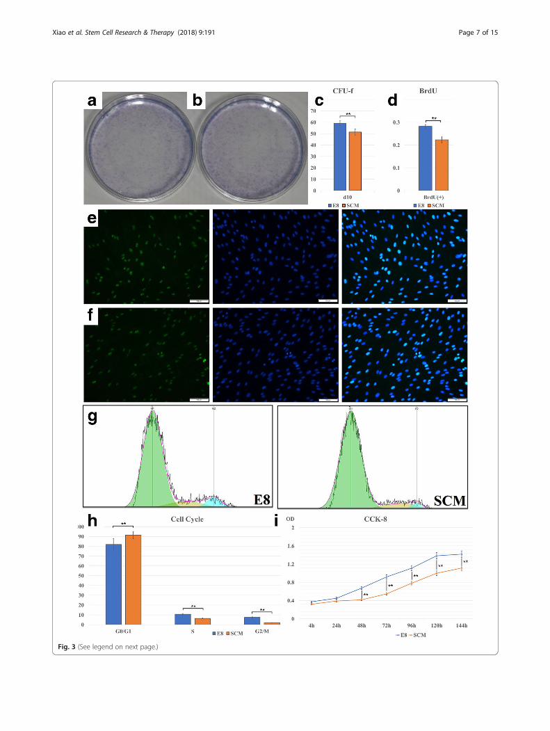
E8 can promote hDPSC proliferationCFU-F results indicated that, at 10 days, a significant dif-ference was observed between E8 and SCM (Fig. 3a–c)(p < 0.01). BrdU assay showed that, at 48 h, E8-culturedhDPSCs exhibited a stronger proliferation capacity withhigher fluorescence labeling rate than culture with SCM(Fig. 3d–f ) (p < 0.01). We used CCK-8 to analyzehDPSCs cultured for 4 h, 24 h, 48 h, 72 h, 96 h, 120 h,
Fig. 1 Cell morphology. a Images of primary culture for 14 d and 28 d. b-d Differences in cell morphology after culture in E8 (left) and serum-containing medium (right; SCM; DMEM + 5% FBS) for b 24 h, c 48 h, and d 96 h
Xiao et al. Stem Cell Research & Therapy (2018) 9:191 Page 5 of 15
and 144 h. Data were obtained as average optical density(OD) values and a CCK-8 growth curve was produced(Fig. 3i) Statistical differences were observed betweenthe E8 group and the SCM group at 24 h, 48 h, 72 h,and 96 h (p < 0.01).To study why cell proliferation rate differed between
E8 and SCM, we analyzed the cell cycle and apop-tosis. Images captured by FlowJo software are pre-sented in Fig. 3g. A significant difference was seen,and E8-cultured hDPSCs possessed fewer cell num-bers in the G0/G1 ratio (p < 0.01) and higher num-bers in the S ratio (p < 0.01) and G2/M ratio (p <0.01) (Fig. 3h). Flow cytometry was used to analyzeapoptosis, and the resultshowed difference betweenthe SCM group and the E8 group regarding early(p < 0.05), late (p < 0.01), and total apoptosis (p <0.01) (Fig. 4c). Images processed by FlowJo softwareare also presented in Fig. 4a. Western blotting andimmunofluorescence also demonstrated that the apop-tosis rate of hDPSCs in E8 group was lower than that inSCM group (Figs. 4b and 5c). Altogether, it can be de-duced that the E8 medium increased the hDPSC prolifera-tion rate through accelerating the cell splitting speed anddecreasing the cell apoptosis rate.
Expression of stem cell markers by immunofluorescenceand Western blottingImmunofluorescence was applied to analyze OCT4,SOX2, and NANOG for cell pluripotency, and a relativelylow expression was obeserved in both E8 and SCM(Fig. 5a). Odontogenesis-related markers DMP1 andDSP1-H, also showed low expression (Fig. 5b). OCN,OPN, and RUNX2, analyzed for the osteogenic tendency,demonstrated high levels of expression, but BMP2 ex-pression was lower in both groups (Fig. 5d). Westernblot results also showed low expression in the odonto-genic markers DMP1 and DSPP for both groups. For theosteogenic markers, Western blot results of OPN andRUNX2 showed expression at high levels, and ALP wasexpressed at low levels (Fig. 4d).
Induced hDPSC differentiation analysishDPSCs under E8 and SCM conditions at P3 wereincubated and induced under different cultural condi-tions. In the osteogenic-inducing group at 15 days,hDPSCs formed mineralized nodules by alizarin redstaining (Fig. 6a). After culture under adipogenic condi-tions for 15 days, hDPSCs formed lipid droplets whenstained with Oil Red O (Fig. 6b). hDPSCs also exhibited
Fig. 2 Characterization of hDPSCs surface markers by flow cytometry. The red curves are the blanks. The blue curves are the E8 or SCM.
Xiao et al. Stem Cell Research & Therapy (2018) 9:191 Page 6 of 15
Fig. 3 (See legend on next page.)
Xiao et al. Stem Cell Research & Therapy (2018) 9:191 Page 7 of 15
green fluorescence under neurogenic and chondrogenicinduction (Fig. 6c, d). Real-time PCR was used to furtherquantity the detection of the multidifferentiation capaci-ties of hDPSCs. Interestingly, significantly higher levelsof expression of OCN and RUNX2 (for the osteogenicdifferentiation test), MAP-2 (for neurogenic differenti-ation test), and PPAR- γ (for the adipogenic differenti-ation test) were detected in E8 (Fig. 6e) (p < 0.01).
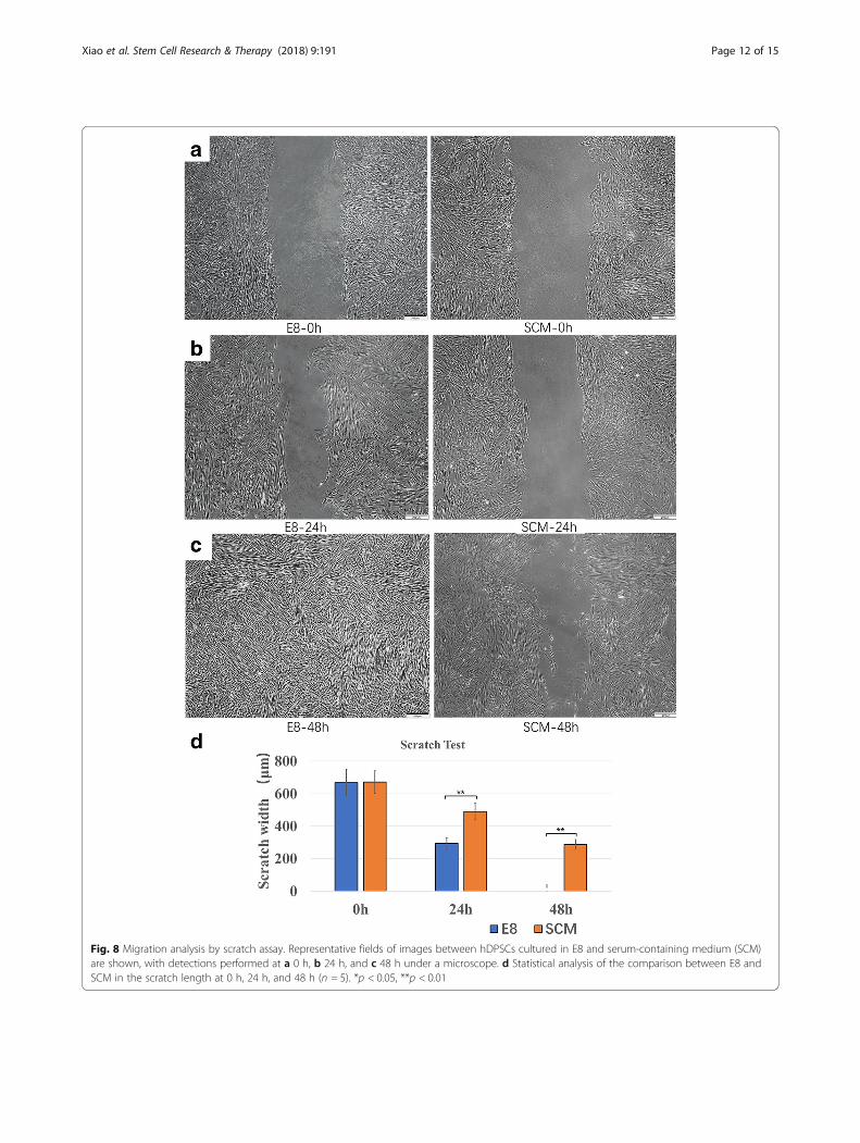
E8 promoted migration of hDPSCsUsing a transwell test, representative images of hDPSCscultured in both media at 6 h and 24 h are demonstratedin Fig. 7a, b. E8 increased cell migration numbers at 6 h(p < 0.01), 12 h (p < 0.01), and 24 h (p < 0.01) (Fig. 7c).In the scratch assay, representative images of hDPSCs
cultured in both media at 0 h, 24 h, and 48 h were cap-tured under microscopy (Fig. 8a–c). The scratch widthwas the same at the start between the two groups (p >0.05), and a clear motility difference was observed at
24 h (p < 0.01) and 48 h (p < 0.01) (Fig. 8d). A more ob-vious narrowing was observed at 24 h using E8. After48 h of incubation, the gaps in the E8 group were invis-ible, while in the SCM group they were not.
DiscussionsOver the past decade, a substantial amount of researchhas focused interest on the need for the improvement ofcell culture. In our study, hDPSCs were used tocharacterize and examine their properties. hDPSCs can beeasily accessed and extracted from human third molars ordeciduous teeth; however, as a type of highly differentiatedstem cell, hDPSCs are commonly believed to be the leastpluripotent among human DSCs. If hDPSCs could achievesatisfying proliferation and pluripotency performance inour serum-free culture environment, other human DSCs,such as periodontal ligament stem cells (PDLSCs), dentalfollicle stem cells (DFSCs) and dental epithelial stem cells
(See figure on previous page.)Fig. 3 Colony-forming unit fibroblasts (CFU-F) of a serum-containing medium (SCM) and b E8. Statistical analysis of c CFU-F comparison (n = 5)and d bromodeoxyuridine (BrdU) proliferation assay (n = 5). BrdU fluorescence of hDPSCs in e E8 and f SCM. g Cell cycles were analyzed withFlowJo software. h Statistical analysis of the cell cycle (n = 5). i Cell proliferation analysis using the CCK-8 assay. The different optical density (OD)values are presented at 4 h, 24 h, 48 h, 72 h, 96 h, 120 h, and 6 days (n = 10). *p < 0.05, **p < 0.01
Fig. 4 Cell apoptosis assay and Western blot. a Representative images of cell apoptosis from both E8 and serum-containing medium (SCM)groups. b Western blot images of cell apoptosis from both E8 and SCM groups. c Cell apoptosis comparison of the two groups (n = 5). d Westernblot of DMP1 and DSPP (for odontogenic markers), OPN, RUNX2, and ALP (osteogenic markers), and GAPDH set as control. *p < 0.05, **p < 0.01
Xiao et al. Stem Cell Research & Therapy (2018) 9:191 Page 8 of 15
(DESCs), might exhibit the same or better outcomesunder the same culture conditions.Although E8 was originally designed for iPSCs, the
composition was shown to have multiple functions instem cells, including in hDPSCs. FGF2 can regulate theWnt signal pathway and its downstream signal via thePI3-K signal channel to maintain the undifferentiatedstate of hESCs and to maintain their self-renewal cap-acity [32, 33]. Ascorbic acid is a kind of water-soluble
vitamin which plays a critical role in the formation ofcollagen that can improve the cell proliferation. Insulincan induce the absorbance of both glucose and aminoacids [34], and selenite supplementation of culturemedia is capable of restoring the antioxidative capacityof bone marrow MSCs and reducing intracellular react-ive oxygen species (ROS) production and stress-relatedgeneration of micronuclei [35]. Activin/Nodal signalingthrough Smad2/3 activation is necessary to maintain the
Fig. 5 Immunofluorescence of hDPSCs. a OCT4, SOX2, and NANOG for cell pluripotency. b DMP1 and DSP1-H for odontogenic tendency. c p53images of cell apoptosis from both the E8 and serum-containing medium (SCM) groups. d OCN, OPN, RUNX2, and BMP2 for theosteogenic tendency
Xiao et al. Stem Cell Research & Therapy (2018) 9:191 Page 9 of 15
Fig. 6 Multi-differentiation analysis of hDPSCs. E8(left); SCM(right) a hDPSCs cultured in osteogenic medium for 15 days. b hDPSCs cultured inadipogenic medium for 15 days. c hDPSCs cultured in neurogenic medium for 3 h. d hDPSCs cultured in chondrogenic medium for 15 days. eReal-time polymerase chain reaction (PCR) analysis of the multidifferentiation capacities of hDPSCs. Relative quantities were measured, andsignificantly higher levels of expressions of OCN and RUNX2 (for osteogenic differentiation), MAP-2 (for neurogenic differentiation), and PPAR-γ(for adipogenic differentiation) were detected in E8 compared with serum-containing medium (SCM) (n = 5). *p < 0.05, **p < 0.01
Xiao et al. Stem Cell Research & Therapy (2018) 9:191 Page 10 of 15
pluripotent status of hESCs [36, 37]. Other growth fac-tors including platelet-derived growth factor (PDGF),basic (b)FGF, and epidermal growth factor (EGF) alsoplay a key role in maintaining the proliferation rate andpluripotency of stem cells [38, 39]. All the informed re-search into the functions of the signal pathways ofgrowth factors gives us the foundation to use E8medium in our experimental group. Additionally, E8only contains eight essential factors [31] at a determinedconcentration, which is simple and convenient if re-searchers are willing to adjust the density of certaincompositions, or to facilitate with other methods for fur-ther studies.
To eliminate variation, we used the same batch ofhDPSCs for primary culture. In this way, although the cellswere not purified, they can be considered to contain thesame cell components and pluripotency. During CCK-8analysis we detected that the OD value between E8 andSCM (both without cell culture) were different, so we usedthe difference in OD value between the media alone andthe media cultured with cells to reduce the error caused byculture media. Hirata et al. [15] characterized and assessedcell proliferation and pluripotency under SFM conditions,but insulin-transferrin-selenium-X supplements alone werenot enough, so they added embryotrophic factor to reachthe same proliferation speed. Previous research has revealed
Fig. 7 Migration capacity analysis by transwell for hDPSCs. Representative fields of images from transwell test are shown, with visual comparisonson migrated cell numbers between E8 and serum-containing medium (SCM) at a 6 h and b 24 h. c Statistical analysis of migrated cell numbersbetween E8 and SCM at 6 h, 12 h, and 24 h (n = 5). *p < 0.05, **p < 0.01
Xiao et al. Stem Cell Research & Therapy (2018) 9:191 Page 11 of 15
Fig. 8 Migration analysis by scratch assay. Representative fields of images between hDPSCs cultured in E8 and serum-containing medium (SCM)are shown, with detections performed at a 0 h, b 24 h, and c 48 h under a microscope. d Statistical analysis of the comparison between E8 andSCM in the scratch length at 0 h, 24 h, and 48 h (n = 5). *p < 0.05, **p < 0.01
Xiao et al. Stem Cell Research & Therapy (2018) 9:191 Page 12 of 15
that ascorbic acid, a kind of water-soluble vitamin which isa part of the composition of E8, plays a critical role in theformation of collagen, and thus enhances cell attachment[34]. This might explain the reason why the E8 groupreached a high speed of expansion faster. Cell migrationanalysis showed that E8-cultured hDPSCs also had bettermobility, indicating that our serum-free culture conditionperformed better in maintaining cytoactivity. Nevertheless,further studies should be performed to exploit the specificmechanism of this condition.Previous studies demonstrated that hDPSCs present simi-
lar cell surface markers to MSCs [40] and, under certainconditions, hDPSCs can be induced to adipose tissue, osse-ous tissue, and nerve tissue [41]. The immunofluorescenceand Western blot results suggested that no significant dif-ferences in pluripotency were observed, indicating that bothgroups maintained the same pluripotency under conditionswithout stimuli. However, induced differentiation resultsshowed that hDPSCs cultured in E8 presented higher levelsof expression of OCN, RUNX2, and PPAR- γ, indicatingthat in the case of irritation E8 is more efficient for main-taining multipotentiality towards osteogenic and adipogenictissues for hDPSCs. Earlier studies displayed similar out-comes, and some also revealed the potential to be inducedto other cell lineages. Harada et al. [42] compared cell pro-liferation, cell morphology, and gene expression change be-tween SCM and SFM, but they did not detect induceddifferentiation. Ishkitiev et al. [26] successfully inducedDPSCs to the hepatic lineage under SFM conditions. How-ever, they did not induce these cells into other lineages.Okada et al. [43] also successfully induced hDPSCs to thehepatic lineage with hydrogen sulfide, and showed the sameor higher performance. Ishkitiev et al. [44] induced hDPSCsto differentiate into the pancreatic cell lineage underserum-free conditions. As stem cells extracted from dentaltissues, hDPSCs should demonstrate advantages in dentis-try and bio-root regeneration in the near future. In 2010,Huang et al. [45] showed that DPSCs were capable offorming vascularized pulp/dentin-like tissue in an emptyhuman root canal when seeded onto a polylactic-co-glycolic acid (PLGA) scaffold. This study, consistentwith previous research, supported the possibility of usinghDPSCs to establish patient-specific or industrialized-grade cell lines for stem cell therapy.Al-Saqi et al. [46] applied Mesencult-XF as their
serum-free medium to analyze its maintenance of pluripo-tency and growth towards adipose-derived MSCs. In apre-experiment, we attempted to use Mesencult-XF toculture hDPSCs. Although cells cultured in Mesencult-XFdisplayed satisfying growth, we observed cell attachmentproblems; however, this phenomenon might be due to adifference in the applied culturing process. Furthermore,Mesencult-XF is relatively costly, and clinical cultures con-sume lots of media. Our work here, for the first time,
applied E8 as a chemically defined, serum-free medium forhDPSC culture and analyzed its performance. However,most of our work was based on short-term culture compari-son, and long-term culture data are still a requirement forfurther exploration. Although serum-free medium was ap-plied during the cell culture and incubation, the use of FBSfor the isolation of a primary cell culture is still indispensableand there are no statistical data suggesting how this applica-tion will affect cell proliferation and pluripotency [30].
ConclusionsIn conclusion, our study presents a novel application ofthe E8 medium in hDPSC culture, and shows that E8 ispractical for the proliferation and differentiation ofhDPSCs to reduce the effects of the problems caused byFBS as described above. E8 possesses a simple compos-ition for the convenience of further downstream process-ing and improvement, which makes it practical to us as aprimary culture medium for the establishment of DSCbanking. Nevertheless, many challenges and obstacles stillneed to be resolved for its ultimate use in the clinic, andthe mechanisms and functions of growth factors in main-taining hDSPC potency and proliferation should be fullyanalyzed and understood to achieve the optimum cultureenvironment. More effective factors are needed, and dee-per studies are still required, for improvement of theserum-free culture conditions. Other issues, such as theapplication of FBS in cell passaging and cryopreservationshould also be resolved to establish a mature SFM culturesystem which can meet the DSC banking standards.
FundingThis study was supported by the National Key Research and DevelopmentProgram of China (2017YFA0104800 and 2016YFC1101400), the Nature ScienceFoundation of China (31470947), the Key Research and Development Programof Sichuan Province (2017SZ0031), and the College student’s Platform forInnovation and Entrepreneurship Training Program of China (2082404131053).
Availability of data and materialsAll data generated and/or analyzed during this study are available from thecorresponding author upon reasonable request.
Authors’ contributionsWG and WT designed the experiments; JX performed the experiments andwrote the paper with the assistance of DY. DY collected and analyzed thedata; QL revised and checked the language use and data. All authors readand approved the final manuscript.
Ethics approval and consent to participateNot applicable.
Xiao et al. Stem Cell Research & Therapy (2018) 9:191 Page 13 of 15
Competing interestsThe authors declare that they have no competing interests.
Publisher’s NoteSpringer Nature remains neutral with regard to jurisdictional claims in publishedmaps and institutional affiliations.
Author details1State Key Laboratory of Oral Diseases & National Clinical Research Center forOral Diseases & National Engineering, Chengdu, China. 2Laboratory for OralRegenerative Medicine, West China Hospital of Stomatology, SichuanUniversity, Chengdu 610041, China. 3Department of Oral and MaxillofacialSurgery, West China Hospital of Stomatology, Sichuan University, No.14, 3rdSection, Renmin South Road, Chengdu 610041, People’s Republic of China.4Department of Pediatric Dentistry, West China School of Stomatology,Sichuan University, Chengdu 610041, People’s Republic of China.5Department of Prosthodontics, West China Hospital of Stomatology,Sichuan University, Chengdu 610041, China.
Received: 4 March 2018 Revised: 3 June 2018Accepted: 13 June 2018
References1. Kalista T, Freeman HA, Behr B, Pera RR, Scott CT. Donation of embryos for
human development and stem cell research. Cell Stem Cell. 2011;8:360–2.2. Dever DP, Bak RO, Reinisch A, Camarena J, Washington G, Nicolas CE, et al.
CRISPR/Cas9 beta-globin gene targeting in human haematopoietic stemcells. Nature. 2016;539:384–9.
3. Blodgett DM, Nowosielska A, Afik S, Pechhold S, Cura AJ, Kennedy NJ, et al.Novel observations from next-generation RNA sequencing of highly purifiedhuman adult and fetal islet cell subsets. Diabetes. 2015;64:3172–81.
4. Karantalis V, Hare JM. Use of mesenchymal stem cells for therapy of cardiacdisease. Circ Res. 2015;116:1413–30.
6. Taylor CJ, Bolton EM, Pocock S, Sharples LD, Pedersen RA, Bradley JA.Banking on human embryonic stem cells: estimating the number of donorcell lines needed for HLA matching. Lancet. 2005;366:2019–25.
7. Onder TT, Daley GQ. New lessons learned from disease modeling withinduced pluripotent stem cells. Curr Opin Genet Dev. 2012;22:500–8.
8. Lindemann D, Werle SB, Steffens D, Garcia-Godoy F, Pranke P, Casagrande L.Effects of cryopreservation on the characteristics of dental pulp stem cellsof intact deciduous teeth. Arch Oral Biol. 2014;59:970–6.
9. Chen KG, Mallon BS, McKay RD, Robey PG. Human pluripotent stem cellculture: considerations for maintenance, expansion, and therapeutics. CellStem Cell. 2014;14:13–26.
10. Julavijitphong S, Wichitwiengrat S, Tirawanchai N, Ruangvutilert P,Vantanasiri C, Phermthai T. A xeno-free culture method that enhancesWharton's jelly mesenchymal stromal cell culture efficiency over traditionalanimal serum-supplemented cultures. Cytotherapy. 2014;16:683–91.
11. Halme DG, Kessler DA. FDA regulation of stem-cell-based therapies. N Engl JMed. 2006;355:1730–5.
13. Baker DE, Harrison NJ, Maltby E, Smith K, Moore HD, Shaw PJ, et al.Adaptation to culture of human embryonic stem cells and oncogenesis invivo. Nat Biotechnol. 2007;25:207–15.
14. Hayashi I, Sato GH. Replacement of serum by hormones permits growth ofcells in a defined medium. Nature. 1976;259:132–4.
15. Hirata TM, Ishkitiev N, Yaegaki K, Calenic B, Ishikawa H, Nakahara T, et al.Expression of multiple stem cell markers in dental pulp cells cultured inserum-free media. J Endod. 2010;36:1139–44.
16. Bonnamain V, Thinard R, Sergent-Tanguy S, Huet P, Bienvenu G, NaveilhanP, et al. Human dental pulp stem cells cultured in serum-free supplementedmedium. Front Physiol. 2013;4:1-9.
17. Chen TH, Chen WM, Hsu KH, Kuo CD, Hung SC. Sodium butyrate activatesERK to regulate differentiation of mesenchymal stem cells. Biochem BiophysRes Commun. 2007;355:913–8.
18. Oshimori N, Fuchs E. The harmonies played by TGF-beta in stem cellbiology. Cell Stem Cell. 2012;11:751–64.
19. Kratchmarova I, Blagoev B, Haack-Sorensen M. Mechanism of divergentgrowth factor effects in mesenchymal stem cell differentiation. Science.2005;308:1472–7.
20. Xiao L, Yuan X, Sharkis SJ. Activin A maintains self-renewal and regulatesfibroblast growth factor, Wnt, and bone morphogenic protein pathways inhuman embryonic stem cells. Stem Cells. 2006;24:1476–86.
21. Calne RY. Prospects for cell-based therapy. Transplant Proc. 2004;36:3183–7.
22. Mooney DJ, Powell C, Piana J, Rutherford B. Engineering dental pulp-liketissue in vitro. Biotechnol Prog. 1996;12:865–8.
23. Gronthos S et al. Stem cell properties of human dental pulp stem cells. JDental Res. 2002;81:531–5.
24. Miura M, Gronthos S, Zhao M, Lu B, Fisher LW, Robey PG, et al. SHED: stemcells from human exfoliated deciduous teeth. Proc Natl Acad Sci U S A.2003;100:5807–12.
25. Seo B-M, Miura M, Gronthos S, Mark Bartold P, Batouli S, Brahim J, et al.Investigation of multipotent postnatal stem cells from human periodontalligament. Lancet. 2004;364:149–55.
26. Ishkitiev N, Yaegaki K, Imai T, Tanaka T, Nakahara T, Ishikawa H, et al. High-purity hepatic lineage differentiated from dental pulp stem cells in serum-free medium. J Endod. 2012;38:475–80.
27. Hilkens P, Meschi N, Lambrechts P, Bronckaers A, Lambrichts I. Dental stemcells in pulp regeneration: near future or long road ahead? Stem Cells Dev.2015;24:1610–22.
28. Temmerman L, Beele H, Dermaut LR, Van Maele G, De Pauw GA. Influenceof cryopreservation on the pulpal tissue of immature third molars in vitro.Cell Tissue Bank. 2010;11:281–9.
29. Coburn RJ, Henriques BL, Francis LE. Development of an experimental toothbank using DEEP freeze and tissue culture techniques. J Oral TherPharmacol. 1966;2:445.
30. YHHe a. Dental stem cells and tooth banking for regenerative medicine. JExp Clin Med. 2010;2:111–7.
31. Chen G, Gulbranson DR, Hou Z, Bolin JM, Ruotti V, Probasco MD, et al.Chemically defined conditions for human iPSC derivation and culture. NatMethods. 2011;8:424–9.
32. Ng F, Boucher S, Koh S, Sastry KS, Chase L, Lakshmipathy U, et al.PDGF, TGF-beta, and FGF signaling is important for differentiation andgrowth of mesenchymal stem cells (MSCs): transcriptional profiling canidentify markers and signaling pathways important in differentiation ofMSCs into adipogenic, chondrogenic, and osteogenic lineages. Blood.2008;112:295–307.
33. Xu RH, Peck RM, Li DS, Feng X, Ludwig T, Thomson JA. Basic FGF andsuppression of BMP signaling sustain undifferentiated proliferation ofhuman ES cells. Nat Methods. 2005;2:185–90.
34. Harada S, Matsumoto T, Ogata E. Role of ascorbic-acid in the regulationof proliferation in osteoblast-like mc3t3-e1 cells. J Bone Miner Res.1991;6:903–8.
35. Ebert U, Zeck, et al. Selenium supplementation restores the antioxidativecapacity and prevents cell damage in bone marrow stromal cells in vitro.Stem Cells. 2006;24:1226–35.
36. Chen G, Hou Z, Gulbranson DR, Thomson JA. Actin-myosin contractility isresponsible for the reduced viability of dissociated human embryonic stemcells. Cell Stem Cell. 2010;7:240–8.
37. Vallier L, Alexander M, Pedersen RA. Activin/nodal and FGF pathwayscooperate to maintain pluripotency of human embryonic stem cells. J CellSci. 2005;118:4495–509.
38. Gronthos S, Simmons PJ. The growth-factor requirements of stro-1-positivehuman bone-marrow stromal precursors under serum-deprived conditionsin-vitro. Blood. 1995;85:929–40.
39. Wang X, Zheng F, Liu O, Zheng S, Liu Y, Wang Y, et al. Epidermalgrowth factor can optimize a serum-free culture system for bonemarrow stem cell proliferation in a miniature pig model. In Vitro CellDev Biol Anim. 2013;49:815–25.
40. Atari M, Gil-Recio C, Fabregat M, Garcia-Fernandez D, Barajas M, CarrascoMA, et al. Dental pulp of the third molar: a new source of pluripotent-likestem cells. J Cell Sci. 2012;125:3343–56.
41. Kawashima N. Characterisation of dental pulp stem cells: a new horizon fortissue regeneration? Arch Oral Biol. 2012;57:1439–58.
42. Harada K, Kawai S, Xu W-a, Xu I, Sonomoto M, Shinonaga Y, et al.Alterations in deciduous dental pulp cells cultured with serum-freemedium. J Hard Tissue Biol. 2015;24:17–22.
Xiao et al. Stem Cell Research & Therapy (2018) 9:191 Page 14 of 15
43. Okada M, Ishkitiev N, Yaegaki K, Imai T, Tanaka T, Fukuda M, et al. Hydrogensulphide increases hepatic differentiation of human tooth pulp stem cellscompared with human bone marrow stem cells. Int Endod J. 2014;47:1142–50.
44. Ishkitiev N, Yaegaki K, Kozhuharova A, Tanaka T, Okada M, Mitev V, et al.Pancreatic differentiation of human dental pulp CD117(+) stem cells. RegenMed. 2013;8:597–612.
45. Huang GTJ, Yamaza T, Shea LD, Djouad F, Kuhn NZ, Tuan RS, et al. Stem/progenitor cell-mediated de novo regeneration of dental pulp with newlydeposited continuous layer of dentin in an in vivo model. Tissue Eng A.2010;16:605–15.
46. Al-Saqi SH, Saliem M, Asikainen S, Quezada HC, Ekblad A, Hovatta O, et al.Defined serum-free media for in vitro expansion of adipose-derivedmesenchymal stem cells. Cytotherapy. 2014;16:915–26.
Xiao et al. Stem Cell Research & Therapy (2018) 9:191 Page 15 of 15