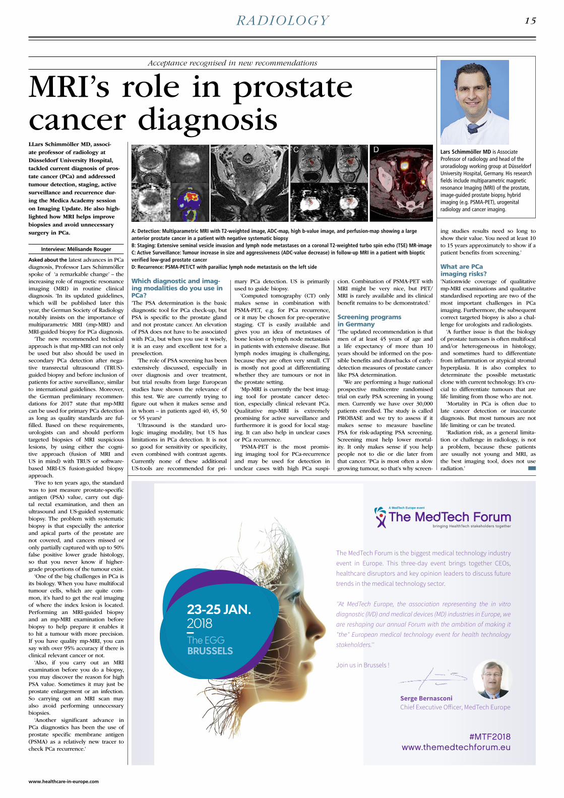
Continued on page 2 THE EUROPEAN FORUM FOR THOSE IN THE BUSINESS OF MAKING HEALTHCARE WORK VOL 26 ISSUE 6 DECEMBER/JANUARY 2017/18 CONTENTS NEWS & MANAGEMENT 1-5 SURGERY 6-9 RADIOLOGY 10-16 ULTRASOUND 17-19 IT & TELEMEDICINE 20-21 LABORATORY 22-23 DIGITAL PATHOLOGY 24 www.healthcare-in-europe.com Machine learning is playing an increasing role in computer-aided diagnosis, and Big Data is begin- ning to penetrate oncological imaging. However, some time may pass before it truly impacts on clinical practice, according to leading UK-based German researcher Professor Julia Schnabel, who spoke during the last ESMRMB annual meeting, Mélisande Rouger reports Machine learning techniques are start- ing to reach levels of human perfor- mance in challenging visual tasks. Tools such as the convolutional neural network (CNN or ConvNet), a class of deep neural networks that has been applied to analysing visual imagery, have become instrumental in segmentation tasks. Analysing such huge data is still a challenge However, a number of obstacles remain before adequate image analysis arrives, starting with the huge amount of data analysts must work with, according to Professor Schnabel, computational imaging expert at King’s College London. ‘In imaging, the challenges are that we work in 3-D or 4-D, and we have a lot of features to deal with. If we’re lucky, we deal with hundreds or thousands, but not millions of images, so we don’t have a high number of image data to work with. We have this whole sample size problem.’ The professor also identified the high associated cost and imperfec- tion of training data. Training data may be wrongly labelled, depend- ing on the expertise of the observer. Furthermore, machine learning is resource-intensive: only special- ists and consultants can perform special tasks. ‘I personally couldn’t distinguish a glass nodule from a semi-solid nodule. Only specialist consultants and expert radiologists and (max) pooling leads to loss of spatial information. In contrast , if you use very small patches, they are more susceptible to noise.’ As a solution, Schnabel points to using a multi-scale approach, i.e. having smaller patches operating on small filters and larger ones on larg- er filters, and putting them together in the end. Oncological image analysis brings challenges of its own. Machine learning-based segmentation often degrades when deployed in clini- cal scenarios. This is caused by differences between training and test data due to variations in scan- ner hardware and scanner protocols and sequences, Schnabel explained. ‘There is often an imbalance in the training or test data because of a dif- ferent ratio of healthy vs. pathologi- cal cases, individual patient variabil- ity and individual disease variability – also within the same patient. For example, lesions in the liver usually are a secondary cancer, caused by a primary cancer elsewhere, such as in the colorectum.’ Therefore, it is crucial to choose the appropriate network architec- ture. Currently three models in lit- erature are interesting: DeepMedic, FCN (in Deep Learning Toolkit) and U-Net, which owes its name to its ‘U’ shape. ‘These networks use different approaches and for all these, there is the good, the bad and the ugly,’ she pointed out. An ensemble of multiple models and architectures All three networks use CNN based approaches with good perfor- mance, but there are a lot of meta- parameters – more than input cases –, and the architecture and con- figuration influence performance and behaviour. The ugly part is that chosen models and parameters may be suboptimal of other data and applications. ‘Results and con- clusions may therefore be strongly biased,’ she said. One solution could be to use an ensemble of networks; one such example is ‘EMMA’ (ensemble of multiple models & architectures), for which performance is insensi- tive to suboptimal configuration and behaviour is unbiased by architec- ture and configuration. can do that,’ she pointed out. For a disease such as cancer, the image analysis team needs confirmation from pathology, which is often dif- ficult to obtain. For brain imaging, where different protocols exist, one sees different appearance of the same disease on different image protocols for the same patient and between patients. ‘Disease location and size of these pathologies may vary quite signifi- cantly, and the appearance of dis- ease may be very localised: it may be a very sharp “blob”, or it may be very diffused or infiltrated,’ she explained. Deep neural networks The professor shared practical advice on how to work with CNNs appropriately. She stressed the size of the receptive field of a CNN will determine the amount of informa- tion that will be obtained. ‘The size of patches used is important, since a large receptive field increases com- putation and memory requirements, Machine learning is promising For segmentation in colorectal cancer with DCE MRI, Schnabel and team extracted variability of normalised signal intensity curves from the dataset using principal component analysis. ‘It’s a very sim- ple technique. We just looked at the mean signal intensity of curves embedded within an over-segmen- tation approach, called superpixels Julia Schnabel PhD joined King’s College London, in the UK in July 2015 as Chair in Computational Imaging at the Division of Imaging Sciences & Biomedical Engineering, taking over the Directorship of the EPSRC Centre for Doctoral Training in Medical Imaging, which is jointly run by King’s College London and Imperial College London. She is also Visiting Professor in Engineering Science, at the University of Oxford. RADIOLOGY 10-16 • Psychoradiology: brain MRI-mining helps classify ADHD • Optoacoustics: the sound of cells • The DNA mismatch repair mechanism IT & TELEMEDICINE 20-21 • E-health developments in Spain • Building an organisation’s digital DNA • The key to defeating cancer is knowledge dissemination 2-D view and 3-D volumetric rendering of contiguous perfusion-supervoxels for tumour parcellation defined on a 4-D dynamic contrast-enhanced magnetic resonance imaging (DCE-MRI) scan based on signal enhancement characteristics (Courtesy: Dr Ben Irving, ISMRM 2017)
Transcript
Continued on page 2
T H E E U R O P E A N F O R U M F O R T H O S E I N T H E B U S I N E S S O F M A K I N G H E A L T H C A R E W O R K
V O L 2 6 I S S U E 6 D E C E M B E R / J A N U A R Y 2 0 1 7 / 1 8
CONTENTS
NEWS & MANAGEMENT 1-5
SURGERY 6-9
RADIOLOGY 10-16
ULTRASOUND 17-19
IT & TELEMEDICINE 20-21
LABORATORY 22-23
DIGITAL PATHOLOGY 24
www.healthcare-in-europe.com
Machine learning is playing an increasing role in computer-aided diagnosis, and Big Data is begin-ning to penetrate oncological imaging. However, some time may pass before it truly impacts on clinical practice, according to leading UK-based German researcher Professor Julia Schnabel, who spoke during the last ESMRMB annual meeting, Mélisande Rouger reports
Machine learning techniques are start-ing to reach levels of human perfor-mance in challenging visual tasks. Tools such as the convolutional neural network (CNN or ConvNet), a class of deep neural networks that has been applied to analysing visual imagery, have become instrumental in segmentation tasks.
Analysing such huge data is still a challengeHowever, a number of obstacles remain before adequate image analysis arrives, starting with the huge amount of data analysts must work with, according to Professor Schnabel, computational imaging expert at King’s College London. ‘In imaging, the challenges are that we work in 3-D or 4-D, and we have a lot of features to deal with. If we’re lucky, we deal with hundreds or thousands, but not millions of images, so we don’t have a high number of image data to work with. We have this whole sample size problem.’
The professor also identified the high associated cost and imperfec-tion of training data. Training data may be wrongly labelled, depend-ing on the expertise of the observer. Furthermore, machine learning is resource-intensive: only special-ists and consultants can perform special tasks. ‘I personally couldn’t distinguish a glass nodule from a semi-solid nodule. Only specialist consultants and expert radiologists
and (max) pooling leads to loss of spatial information. In contrast , if you use very small patches, they are more susceptible to noise.’
As a solution, Schnabel points to using a multi-scale approach, i.e. having smaller patches operating on small filters and larger ones on larg-er filters, and putting them together in the end.
Oncological image analysis brings challenges of its own. Machine learning-based segmentation often degrades when deployed in clini-cal scenarios. This is caused by differences between training and test data due to variations in scan-ner hardware and scanner protocols and sequences, Schnabel explained. ‘There is often an imbalance in the training or test data because of a dif-ferent ratio of healthy vs. pathologi-cal cases, individual patient variabil-ity and individual disease variability – also within the same patient. For example, lesions in the liver usually are a secondary cancer, caused by a primary cancer elsewhere, such as in the colorectum.’
Therefore, it is crucial to choose the appropriate network architec-ture. Currently three models in lit-erature are interesting: DeepMedic, FCN (in Deep Learning Toolkit) and U-Net, which owes its name to its ‘U’ shape. ‘These networks use different approaches and for all these, there is the good, the bad and the ugly,’ she pointed out.
An ensemble of multiple models and architectures All three networks use CNN based approaches with good perfor-mance, but there are a lot of meta-parameters – more than input cases –, and the architecture and con-figuration influence performance and behaviour. The ugly part is that chosen models and parameters may be suboptimal of other data and applications. ‘Results and con-clusions may therefore be strongly biased,’ she said.
One solution could be to use an ensemble of networks; one such example is ‘EMMA’ (ensemble of multiple models & architectures), for which performance is insensi-tive to suboptimal configuration and behaviour is unbiased by architec-ture and configuration.
can do that,’ she pointed out. For a disease such as cancer, the image analysis team needs confirmation from pathology, which is often dif-ficult to obtain.
For brain imaging, where different protocols exist, one sees different appearance of the same disease on different image protocols for the same patient and between patients. ‘Disease location and size of these pathologies may vary quite signifi-cantly, and the appearance of dis-ease may be very localised: it may be a very sharp “blob”, or it may be very diffused or infiltrated,’ she explained.
Deep neural networksThe professor shared practical advice on how to work with CNNs appropriately. She stressed the size of the receptive field of a CNN will determine the amount of informa-tion that will be obtained. ‘The size of patches used is important, since a large receptive field increases com-putation and memory requirements,
Machine learning is promising
For segmentation in colorectal cancer with DCE MRI, Schnabel and team extracted variability of normalised signal intensity curves from the dataset using principal component analysis. ‘It’s a very sim-ple technique. We just looked at the mean signal intensity of curves embedded within an over-segmen-tation approach, called superpixels
Julia Schnabel PhD joined King’s College London, in the UK in July 2015 as Chair in Computational Imaging at the Division of Imaging Sciences & Biomedical Engineering, taking over the Directorship of the EPSRC Centre for Doctoral Training in Medical Imaging, which is jointly run by King’s College London and Imperial College London. She is also Visiting Professor in Engineering Science, at the University of Oxford.
IT & TELEMEDICINE 20-21• E-health developments in Spain
• Building an organisation’s digital DNA
• The key to defeating cancer is knowledge dissemination
2-D view and 3-D volumetric rendering of contiguous perfusion-supervoxels for tumour parcellation defined on a 4-D dynamic contrast-enhanced magnetic resonance imaging (DCE-MRI) scan based on signal enhancement characteristics (Courtesy: Dr Ben Irving, ISMRM 2017)
NEWS & MANAGEMENT
Continued from page 1
Machine learning is promising
or supervoxels in 3-D. We then classified perfusion supervoxels in unseen cases using support vector machines to obtain tumour segmen-tations.’
This automated method was found to perform within the inter-rate agreement of two expert observers, but a correction step was needed when transferring the segmentation mask from the T2W to T1W DCE-MRI sequence, she noted.
Gaining a more accurate tumour segmentation ‘Tumours have considerable varia-tion in shape, so we need to bring in some anatomical context and, to do so, we have developed a graphical representation of the neighbouring anatomy. We can improve segmentation by taking into account both local and global relationships; so we know where the bladder, lumen, etc. are, and build them into a pieces-of-parts model. By classifying these pieces
of parts, we can reduce false posi-tives and obtain a more accurate final tumour segmentation,’ she explained.
Schnabel and team also performed segmentation inside the tumour using a technique called tumour parcellation, to extract locally mean-ingful, contiguous perfusion sub-regions from DCE-MRI scans. Ten female CBA mice with subcutane-ously implanted CaNT tumours were scanned over eight days to monitor tumour growth and clustering of derived perfusion supervoxels.
Imaging casculature helped to look even deeper inside the tumour. ‘We used 3-D fluorescence confocal microscopy, an imaging technique that has very anisotropic voxels of a few microns size. Endothelium and tumour cells were both fluores-cently labelled, and approximately 60 slices where acquired in the z-direction. Vasculature was visible up to 30 slices from the surface.’
In lung cancer image analysis,
most efforts concentrate on lung nodules or lymph node detection in lung CT. Deep learning is now largely replacing conventional CADe and CADx methods, which were based on texture analysis, handcraft-ed features and simple classification techniques. According to Schnabel, using deep learning, detection rate is generally very high and the main focus is now on false positives reduction.
High dimensional multi-modality datasets are not big data Machine learning is a promising tool in oncological imaging and image analysis, and the challenge is to find the right model parameters for good estimation and generalisa-tion. ‘We have high dimensional and multi-modality datasets, but it’s not really big data, it’s rather dense data. Cohort studies which collect large amounts of data ,’ she added, ‘will help a lot in that sense.’
Report: Mark Nicholls
Artificial intelligence (AI) has enormous potential to revolutionise the delivery of healthcare, being able to remove the drudgery of routine tasks, join up fragmented care records, trigger alerts when abnormal results occur, speed-up the process of identifying clusters of patients by digging deep into electronic health records, and increase efficiency of healthcare staff resources. Yet to achieve its potential, there needs to be greater cohesion between digital technology compa-nies, clinicians and hospitals if AI can enhance rather than disrupt health-care in this early phase of its estab-lishment, according to consultant car-diologist Dr Ameet Bakhai, deputy director of research at the Royal Free London NHS Foundation Trust.
Speaking with European Hospital prior to his presentation ‘AI in health-care – delivery in diagnosis’ at the UK Digital Healthcare Transfor mation Summit 2017 in London on 12-13 December, Bakhai explained that hos-pitals are at different stages of evolu-
tion in working with SMEs and large corporations in embracing digital technology and AI. ‘Some are making small incremental changes, others are some years ahead and being innova-tive, while some are still in the tradi-tional healthcare setting of the 1990s,’ he pointed out. ‘There are clinicians now willing to engage in trying out or helping to integrate a new technol-ogy; at the Royal Free Hospital we are blessed with key clinicians open to the role of digital technologies, such as remote monitoring companies, but across the NHS very few clinicians are doing that.’
Bakhai warned that the lack of a unified information share or strategy approach to how digital technologies as a group work with the NHS is an issue. Whereas the pharmaceutical sector has a strategy, with manufac-turers aware of each other’s technolo-gies, and registering and publishing in advance their on-going clinical trials and seeking peer review early, digital technology companies tend to crowd the same space without a cohesive approach to broader devel-opment of AI and innovation.
AI is currently impacting on some areas, such as diabetes, self-manage-ment of epilepsy and rare kidney diseases. Other companies are inter-rogating databases to identify patients
are relayed back to each hospital or centre, patients, GPs and social care teams.
However, the lack of joined-up working between the AI companies concerns the consultant, particularly with no generalised database of AI interventions existing, unlike pharma-ceutical clinical trials. There is also no standardised consensus and guidance on measuring the impact of a clinical trial using AI technologies.
‘Another aspect we don’t know much about is how AI will disrupt the staff economy,’ he observed. ‘Will we be able to release staff from repeti-tive, low-impact work and reduce staff shortages in the NHS, and which staff are going to be impacted on most – doctors, nurses, healthcare professionals, pharmacists, managers or administrators?’
To ensure AI can fulfil its potential in a healthcare setting, Bakhai sug-gests a Faculty of Clinicians in Digital Healthcare or AI to support a more cohesive approach of these technolo-gies in healthcare.
Touching on security, he said: ‘We think we are less in a risk area with digital technology – but look at the impact of the recent Ransomware attacks in the NHS.’ Ideally, he would like to see doctors, entrepreneurs, technologists, coders and others
who meet certain criteria for a clini-cal trial, and some deliver software to track patients and ensure timely follow-up after procedures.
‘They are removing the drudgery from some of things that we used to have to do manually, with more robustness and security,’ Bakhai added. Also in development, he said, are companies creating disease spe-cific avatars to help patients monitor and self-manage conditions – such as diabetes or heart failure – to remind them of appointments, scans, inform them about medication or their test results and to motivate patients to take exercise and their medications, for example.
While all at different stages of development and affordability, other companies use AI tools to gather data that will trigger clinical input when required, often earlier than patients recognise symptoms, thereby preventing unplanned hospitalisa-tion, he pointed out. Other innova-tions support multidisciplinary teams across different centres to co-ordinate data and decisions and ensure these
working more cohesively and for cli-nicians – while still active in clinical practice – working alongside digital technologies to conduct research and create an evidence base on the value of AI interventions.
‘The way we measure AI value in healthcare is also going to be cru-cial,’ he emphasised. ‘Often, for digi-tal technologies, we commission or introduce something with anecdotal or superficial evidence, hoping it will have some benefit, but we haven’t really put them through the rigour that we’d use with any other interven-tion in healthcare.’
He concludes that if healthcare can work cohesively with technol-ogy firms and that they look ahead together regarding AI, he believes money will be saved and duplication reduced of competing companies in the same space. It could also help companies design and better evalu-ate the technology they offer and allow clinicians and hospitals to then be proactive, rather than reactive, in changing patient pathways.
Construction has now begun of the National Centre for Healthcare Photonics in the UK to support companies developing technologies that use light for healthcare applications, Mark Nicholls reports
The Centre for Process Innovation (CPI), a UK technology innovation provider for process manufacturing, is setting up a National Centre for Healthcare Photonics (NCHP) in a bid to extend the use of healthcare photonics technologies and make them more widely available for a range of applications, including the early diagnosis and monitoring of chronic diseases such as diabetes, eye problems, cancer or brain injury.
Scheduled to open in December 2018, the NCHP will be based in northeast England and provide open access facilities and expertise to help companies develop technolo-gies and reduce barriers that com-monly prevent early research and inventions reaching the market.
‘Photonics is a key that enables technology for a range of healthcare products related to imaging, diag-nostics and therapy,’ Dr Tom Harvey, CPI’s Strategic Programme Manager for Healthcare Photonics, pointed out. ‘The new centre will provide expertise and facilities to help com-panies bring these products to mar-ket more quickly.’
The centre’s intended scope of activity, he explained, covers an innovation space from the point where the key features of a new product or process have been shown to work in principle, to a point where the product has been tested and proven in the targeted end-use so that the technology is ready to become a commercial proposition. ‘As such, the centre aims to be able to manufacture quantities required for clinical investigation and clinical validation trials but not to produce
at commercial scale,’ he added.With an initial focus on imag-
ing, diagnostics and therapy, the centre will provide a collaborative and flexible workspace for photonic technologies specialists.
Key facilities: a manufacturing area with controlled access, temper-ature and humidity control; flexible optics laboratories; a suite of life science laboratories for the prepa-ration and analysis of samples; an electronics development laboratory; a workshop with facilities for rapid prototyping; an X-ray test and devel-opment lab; and a modelling and design laboratory with access to 3-D CAD design software, optics-design related software, image analysis software.
Alongside the infrastructure, equipment and accommodation, CPI will provide clients with services such as health economic modelling, clinical trial planning, understand-ing of the regulatory approval pro-cess, advice on CE-Marking, intel-lectual property protection, supply chain analysis and access to finance.
From a healthcare perspective, photonic-enabled diagnostic meth-
Tech firms, doctors and hospitals need greater cohesion New UK centre will develop light technologies
AI could enhance or disrupt healthcare
A National Centre for Healthcare Photonics
Some innovations support multidisciplinary teams across different centres to co-ordinate data and decisions and ensure these are relayed back to each hospital or centre, plus the patients, GPs and social care teams
Consultant cardiologist Ameet Bakhai MD is deputy director of research at the British Royal Free London NHS Foundation Trust, and is himself a cardiac researcher. He undertakes the design and management of clinical trials, health economic modelling and patient pathway innovations. He is also a scientific advisor to NICE, health technology appraisal, pharmaceutical and device manufacturers and clinical trials organisations.
Sour
ce: P
anch
enko
Vla
dim
ir /
Shut
ters
tock
2
EUROPEAN HOSPITAL Vol 26 Issue 6/17
siemens.com/atellicasolution
*Product availability will vary by country. †Dependent on test mix. ‡Versus leading IVD companies.
Atellica® Solution:* Flexible, scalable, automation-ready immunoassay and chemistry analyzers engineered to deliver control and simplicity so you can drive better outcomes.
A bi-directional magnetic sample transport that is 10 times faster than conventional sample conveyors
Unprecedented flexibility with more than 300 customizable configurations including L and U shapes
An immunoassay analyzer that runs up to 440 tests per hour,† the industry’s highest productivity per square meter‡
The new standard in sample management—revolutionary technology that gives independent control over every sample
Now available!
9564_Atellica_Solution_Ad_M300_A4_EuroHosp.indd 2 12/7/17 11:36 AM
NEWS & MANAGEMENT
ods are non-contact and can be done in-vivo or in-vitro, with or without additional probes, contrast agents or other types of marker.
‘The aim is to help to improve the range and utility of photonic diagnostic methods available, with a focus on reducing the cost of manu-facture of those devices so as to be able to address established market trends, such as the move away from hospital-based and delivered services towards more local or per-sonal testing and diagnosis,’ Harvey said. ‘In general, the use of optical methods means less invasive and more accurate monitoring, diagnosis or location (imaging) of disease in the body.
‘In cancer diagnostics, differen-tiating between diseased and non-diseased tissues using non-contact optical imaging methods can be done in real time during laparo-scopic surgery by illuminating the target area with a diagnostic probe beam which measures the Raman or infra-red spectrum of the tissue, or by exciting and imaging fluorescent probes that bind to selected target tissue types. Tissue classification can
also be done by using time-resolved measurements of the spectrum.’
Other applications, Tom Harvey continued, include the opportunity to conduct skin cancer diagnosis without biopsy by using Optical Coherence Tomography and reflec-tance spectroscopy to image the suspect area on the skin; assessment of cardiovascular disease risk based on measurement of certain fluores-cent proteins in the skin; and in vitro and in-situ diagnosis of bacte-rial infections in the lung providing rapid diagnosis.
In the United Kingdom, thermal
imaging is already utilised to diag-nose problems with blood circula-tion or to detect inflammation in thyroid eye disease and to classify burns, whilst a project for in-vivo, in-situ imaging of bacterial infec-tions in the lung is at the clinical study phase.
Further innovations are expected, Harvey said, as resolution of imag-ing systems improve, the trend for miniaturisation of optical systems continues, laser power increases as costs fall and Light Emitting Diodes become more powerful and avail-able with a wider range of emission
wavelengths, further driving their adoption as a replacement to other light sources in medical use.
New UK centre will develop light technologies
A National Centre for Healthcare Photonics
Tom Harvey PhD is Strategic Programmes Manager responsible for Healthcare Photonics at the Centre for Process Innovation. His role aims to help companies with the translation of new photonic and printed electronic technologies into products and services for applications in Healthcare and Life Sciences. A Fellow of the Institute of Physics, he is an expert in these fields; before joining CPI he was employed in Industry for many years to develop displays, lenses, electronics and microfluidic components for a variety of uses.
Eye examinations using photonics technologies can help to diagnose conditions early
The United Kingdom’s future National Centre for Photonics NCHP
3
www.healthcare-in-europe.com
NEWS & MANAGEMENT
Report: Wolfgang Behrends
Taiwan presented its most exciting prod-ucts at this year’s Medica trade fair in Germany – all bearing the prestigious national stamp ‘Taiwan Excellence’. Organised by the semi-public Taiwan External Trade Development Council (TAITRA), Taiwan’s presence at this prestigious show presented the cut-ting edge of Taiwan’s medical tech-nology.
An algorithm for how good and bad differNowadays, artificial intelligence (AI) and deep learning are among the buzziest buzzwords in healthcare. Not surprisingly then that Taiwanese developers also explore the potential
After acquiring Panasonic’s Ultra-sound Imaging Division, Konica Minolta entered the ultrasound mar-ket with Sonimage HS1, a port-able system focused on point-of-care use. Randolf ten Cate, the firm’s Marketing Manager for Europe, the Middle East & Africa (EMEA), explained: ‘We developed the sys-tem for users who appreciate the added value of ultrasound imaging yet are not themselves radiologists, as is often the case, for example, in rehabilitation, anaesthesia or rheu-matology.
‘We also aim at users in intensive or trauma care who value portable, speedy solutions. The new ultra-sound system is easily accessible and can be handled intuitively. It has only eight buttons, everything else can be entered via the touchscreen.’
Despite the ease of use, the sys-tem covers a great range of func-tions, including Colour, Pulse and Continuous wave Doppler, as well as linear, convex and phased array technology, for example.
‘To keep the intuitiveness, we cre-ated a customisable interface where any user can add short-cuts to their favourite functions,’ Marco Lagustena, EMEA Product Manager for ultra-
of these new tech-nologies. For exam-ple, AmCad BioMed uses the power of algo-rithms to automatically classify thyroid tumours. ‘Our software can analyse the characteristic features, such as microcalcification and echotexture, and calculate whether a tumour is benign or malignant’, explains Peter Wu, President of AmCad. ‘Using this method, we achieve an accuracy of 90 percent – on average, 15 to 25 percent higher than the capability of the best professionals.’
Machine learning enables software to improve its precision with each and every new case it analyses. ‘Of course the doctor must make the final
sound explains. Konica also focused on integrating powerful technology. ‘Our goal was to obtain the quality of a cart-based system in a portable for-mat so, for example, we included an 18 MHz transducer and Triad Tissue Harmonic Imaging,’ ten Cate adds. The system takes almost no time to start, being ready to use in under 15 seconds. Battery-run, and operating for about one hour, when connected to a power-supply, or cart, it recharg-es yet remains operable. The sys-tem features two specific advantages Product Manager Lagustena explains. ‘We include simple needle visualisa-tion that works without any add-ons
decisions’, Wu emphasises, ‘but, our software provides all the necessary information.’ Increased precision is envisaged to reduce the number of unnecessary fine needle aspiration (FNA) biopsies by 50 percent and thy-roidectomies by 30 percent.
The technology applied in another of the company’s products works in a similar way: software automatically
or additional hardware, and also we incorporated a special rheumatology function. The Rheumatoid Arthritis Work-flow, based on the DAS28 pro-tocol, was programmed as a feature into the system, so users can follow it consecutively, experiencing a smooth workflow.’ Following their key focus on easy usage, Konica Minolta branched into a different area to develop an inno-vative system that improves wound care management. ‘With diabetes on
analyses airflow in the upper respira-tory tract to determine the risk of Obstructive Sleep Apnoea (OSA) syn-drome. ‘This procedure normally takes several hours and requires the patient to stay in a sleep monitoring facility overnight,’ Wu explains. ‘Our system can do it in just 10 minutes, while the patient is awake.’ Saving time and money by reducing length of hospital stay and the number of interventions is the professed aim of the product.Contact: http://www.amcad.com.tw/en/
Mixed Reality brings an un obstructed view without X-ray Bridging the divide between the digital and real world is the vision of Taiwan Main Orthopaedic Biotechnology’s
the rise, there are more wounds to take care of and they are tak-ing longer to heal. This obviously increases the cost of healthcare and the time patients spend in a hospital,’ explains Zhang Qiu Ying, Head of Healthcare Innovation at the Konica Minolta Business Innovation Centre in Singapore.
Capturing 3-D wound dataTo minimise the time doctors need for the tedious business of docu-mentation, Konica Minolta devel-oped a device called ‘WoundeAide’, which allows clinicians to capture non-contact 3-D wound data. ‘As of now, wound documentation is still mostly done manually and is not only rather inaccurate and time-con-suming but often invasive and there-fore painful for patients. Precious time is wasted by measuring the wound, estimating its depth, maybe photographing it and transferring the data to the hospital’s record system afterwards,’ Ying points out. ‘Our intelligent system helps with documenting the wound more con-sistently and precisely within mere seconds.’
The system uses machine algo-rithms to automatically detect wound
Smart Surgical Glasses. The product is designed to enable the surgeon to look into the patient during surgery via Mixed Reality: X-ray images or tissue images of the regions of inter-est can be viewed in real-time and in full HD resolution (1080p). ‘The rela-tive position of the projected image to the patient is determined via four infra-red sensors,’ Communications Manager Dr Min-Liang Wang explains. This provides the high degree of pre-cision required in, for example, dorsal spinal surgery.
The developers are convinced that ‘Smart surgical glasses will become a key tool for significant advances in surgery.’ The fact that the surgeon does not have to switch between patient and monitor is said to reduce the duration of the procedure by 30 percent – a fact that also means less radiation exposure for clinical staff and patient alike. At this point, there is only a prototype of the smart operat-
boundaries and it reduces the varia-bility following manual assessments. Data is then automatically trans-ferred to the hospital’s record system where it can be stored, accessed and easily shared. Since no wound con-tact is needed, the risk of infection is lowered. Furthermore, trends may be identified from the gathered data. ‘We want to enhance the system, so that in the future it will be able to make suggestions on how to treat a wound, based on the previously gathered input,’ Ying says.
The system is on trial in several hospitals and nursing homes and the feedback is promising. ‘We are work-ing to bring the solution to Europe,’ Ying reveals.
Thermal technology Being a shareholder of Mobotix, Konica Minolta is also involved in security technology develop-ment, which could be relevant in healthcare, for example, as thermal technology systems, according to Sven Lessmann, Mobotix Business Development Manager for north
3-D visualisation, augmented reality, automated tumour classification – today, the Republic of China produces cutting-edge medical technology and it’s a long time since ‘Made in Taiwan’ stood for inferior, copied products. Over recent years, this island state has successfully morphed into a productive and, above all, innovative manufacturer of medical technology available on the world market.
Konica Minolta constantly pursues new ideas and technologies for healthcare – which was clearly visible at Medica 2017, where the firm’s novel products and systems were on show. Portable ultrasound, digital wound care or secure patient monitoring – the portfolio is highly diverse. However, the successful effort to balance customisation and intuitive usage was evident in all the solutions, Lena Petzold reports
Taipei hits highs in Medica 2017
All custom-built yet easy to use
Inspired concepts increasingly impress global markets
A varied portfolio: ultrasound, wound care, patient monitoring
The software converts monoscopic images from any current 2-D system, processing them into stereoscopic 3-D images
The thermografic camera M16 Thermal detects temperatures ranging from -40 to 550°C
AmCad BioMed software analyses tumour characteristics, such as microcalcification and echotexture, as well as its margins and anechoic areas
The Wounde Aide system automatically detects a wound’s boundaries as well as the circumference and depth
Taitra’s team at Medica 2017
Smart Surgical Glasses can speed up surgical procedures because the need to switch between patient and monitor is eliminated
4
EUROPEAN HOSPITAL Vol 26 Issue 6/17
NEWS & MANAGEMENT
In clinical use since 2014, the
Xenios console combines three therapies on one platform for the
benefit of the patient and support of the user. After three years in clinical use, the Xenios console has estab-lished itself. However, those, who move around in daily clinical prac-tice will quickly realise that only everyday life defines the require-ments and poses new challenges, which can best be met by working hand in hand with users.
Stephan Schroll MD PD, senior physician at the Barmherzige Brüder hospital in Regensburg, Germany, and Christian Hoff, clinical sup-port Xenios AG, are long-standing partners, who know and trust each other. This is the starting point for expertise and safety as shown with our example.
The Barmherzige Brüder hospi-tal in Regensburg has a modern intensive care unit with 28 beds, which include 20 ventilation beds. Due to the historically grown situ-ation construction-wise, as well as the size of the intensive care unit, a central alarm registration is of great importance.
‘This also includes central mon-itoring of the most important parameters when performing an extra-corporal membrane oxygena-tion (ECMO) procedure,’ Stephan Scholl stresses. For this reason, a connection was installed between the Xenios console and the Philips
IntelliVue MX800 central monitoring system. The connection can eas-ily be established via the interface of the central patient monitoring system (Philips Intellibridge EC10 Module) to the data interface of the console (Xenios console, iLA activve system). This ensures central moni-toring of the most important param-eters, such as blood flow and speed of the pump in the extracorporeal circuit at all times.
Additionally, the most important pressure measurements are also centrally monitored in the ECMO system. By transferring the alarm settings of the Xenios console, no additional settings for central moni-toring are necessary. All settings are made automatically after connecting the interconnection cable. This a clear advantage, says Dr Stephan Scholl. ‘For us, the possibility of cen-trally monitoring important param-eters of the ECMO systems means additional safety for the patients and, at the same time, a reduced workload in everyday life of the intensive care unit.’
That is exactly one of the goals of Xenios AG, the company explains: ‘Safety in use and for the patient, bringing together innovation and support from experts for experts. From this, a continuous enhance-ment and progress results. In this concrete example, the alarm message is no longer optionally sent individually at the console in a room, but the messages are made accessible to the entire staff at
a central point, for a whole team of therapists at the monitoring centre. ‘We, in clinical support, listen to the uses and bring the therapists’
experiences to our develop-ers and the entire Xenios team,’ Christian Hoff explains. ‘Hand in hand; together into a secure future; for the benefit of patients and for their safety.’
ing room (OR) glasses, but a market launch is planned for 2018 because the necessary clinical studies have progressed well.Contact: http://www.surglasses.com/
Extra-dimension out of the boxTechnical innovations? Fair enough. However, they are of little value if hospitals do not have the neces-sary equipment to use them. This is where ‘MonoStereo’ by the ‘Taiwan Excellence’ winner MedicalTek comes in. This is a 3-D conversion box for endoscopes, as explained by Chairman Kai-Che (Jack) Liu. ‘The software we developed converts the monoscopic images from any current 2-D system and processes them in real-time to stereoscopic 3-D images.’
A strong point in favour of the product is the high degree of compat-ibility. ‘This is convenient for hospitals, because they already have all the necessary equipment,’ he points out. ‘Their endoscopy systems are simply connected to our conversion box. The depth is then perceived by the sur-geon via polarised 3-D glasses.’
3-D endoscopic images not only improve visualisation of depth, they also flatten the learning curve. ‘Surgeons need to convert the 2-D endoscopic images to 3-D movements in their mind. For some, this takes up to two years to get used to,’ Liu explains. ‘Our system takes this addi-tional challenge away by adding depth to the images. This way, a surgeon only needs half an hour to get used to the endoscopic movement. Because it is a more natural way of percep-tion, the added depth also results in
more precision and therefore, better outcomes.’ The conversion box does not impair the movement and zoom functionality of the endoscopes.Contact: http://www.medicaltek.biz/
Coming soon to a hospital near you?While some of the ‘Taiwan Excellence’ products are as yet only in the proto-type phase, others are already used in hospitals throughout Asia. Innovations such as the algorithm for tumour classification and the 3-D Conversion Box for endoscopes recently obtained FDA and CE clearance and thus will soon be admitted for use in USA and European hospitals.
Many Taiwanese companies are actively seeking sales partners – no doubt, after Medica, some contacts will be eager to knock on Taiwanese doors.
Taiwan External Trade Development CouncilChristina Lim, Tel: +886-2-2725-5200E-mail: [email protected]/index_en.htmlwww.taiwantrade.comTaiwan Trade Center, DüsseldorfE-mail: [email protected]://duesseldorf.taiwantrade.com
Germany. ‘Thermal technology is used, for example, in early warn-ing systems that are triggered when a critical temperature threshold is exceeded. Such a system, which uses cameras with integrated heat sen-sors, can detect infections in high-risk ICUs by identifying patients with fever.
‘Another potential field of applica-tion is monitoring hospital equip-ment: increased heat emission is often a sign of imminent failure, for example if there is mechanical fric-tion or when electrical components have a higher power consumption shortly before they break down. The system will detect and signal this unusual temperature increase early enough for maintenance work.’
Currently, thermal technology is being tested in a care facility in the Netherlands: a camera-controlled system warns the night watch staff of unexpected movement in patient rooms. ‘This technology makes repeated night watch rounds obso-lete’, says Lessmann, adding that this ‘increases flexibility and reduces pressure on the staff’. To ensure such a system respects patient pri-vacy, Mobotix designed a particular configuration. ‘All images recorded by the camera are pixelated. Only when the camera detects a potential emergency is the image recognisable and a signal is sent to the staff, who then check and decide whether an intervention is necessary.’
The initial feedback from a Dutch care facility is positive, above all ease of use and the practical help in daily routine are underlined. Thus the facility intends to expand the system. This seems to be another example where the approach ‘easy to use but customisable’ has borne fruit.
Central alarm management of the Xenios console via the Philips IntelliVue MX800 patient monitoring sys-tem easily and efficiently achieves its goal through ‘a combination of safety and innovation,’ the manufactur-er reports. ‘The Barmherzige Brüder hospital in Regensburg and Xenios combine both in clinical practice.’
Successful central patient monitoring
Inspired concepts increasingly impress global markets
A varied portfolio: ultrasound, wound care, patient monitoring
Sonimage HSI has only eight buttons; everything else can be entered via the touchscreen
Advertorial
The Xenios console combines three therapies on one platform
5
www.healthcare-in-europe.com
SURGERY
Report: Méisande Rouger
Resecting an entire tumour and deter-mining brain shift remain challenging for surgeons in brain cancer surgery. However, they are likelier to over-come these difficulties if they use intraoperative ultrasound, according to Dr Cristian de Quintana Schmidt, neuro oncologist at Santa Creu i Sant Pau Hospital in Barcelona. ‘Neuro navigated ultrasound provides the surgeon with confidence in the assessment of resection accuracy and in the determination of brain shift,’ he said. Last August, during the World Congress of Neurosurgery in Istanbul, Turkey, de Quintana pre-sented the results of a prospective two-year study on ultrasound use in intra-axial tumours. For surgeons, brain shift is a major problem. Even if they use pre surgical imaging to help plan surgery, the brain will change during the intervention; it will lose liquid and volume, shift shape and move, and ultimately make it harder for surgeons to perform.
Intraoperative ultrasound takes just over two minutesUnlike intraoperative magnetic reso-nance imaging (MRI), which requires 20-30 minutes time to adjust to pre-surgical images, it takes a little over two minutes (2 minutes 19 seconds) for intraoperative ultrasound to over-lap with previous images. Because ultrasound is so fast, it can be repeated as many times as neces-sary, enabling the surgeon to detect brain shift and evaluate how much tumour is left, almost instantly
Brittany’s capital Rennes is leading stereotactic radiotherapy practice as Eugène Marquis Cancer Centre gears up to welcome worldwide technicians to train on the latest CyberKnife system, Accuray’s pow-erful robotic radiosurgery system targeting small lesions.
The centre, based in the city’s University Hospital (Centre Hospitalier Universitaire), is one of the few places in France to host the new CyberKnife M6 system, which features an adapted multileaf collimator in addition to IRIS or fixed collimator, an advance that decreases treatment time dramati-cally while allowing a very high level of precision. The system has helped treat patients with benign brain tumours but also colon, breast and lung metastases as well as pri-mary tumours of the lung and liver ever since its introduction in Rennes in 2014.
Perhaps one of the most striking features of the voluminous machine, which has a 50-m2 footprint, is
its millimetric precision in tumours smaller than 1cm up to 6 cm. Sessions with the CyberKnife last longer than conventional accelera-tors, but only one to five slots are necessary per patient. The technique used is hypofractioning, which con-sists in squeezing high radiation doses in the 8-24 Grayscale in as short as possible irradiation times. After each session, patients can go home and rest, therefore reducing the hospital stay.
This comfort and precision have enabled radiotherapy physicians to access tumours that were untreat-able any other way than with chem-otherapy or many radiotherapy ses-sions and to treat patients who were long regarded as inoperable.
‘Stereotactic radiotherapy can help a lot of patients, especially those who are too fragile for sur-gery, or who have received a lot of chemotherapy. We’ve had very good results; it’s a real technological advance. For us, it means another way of thinking and working in very
targeted patients,’ said Dr Elisabeth Le Prisé, Clinical Director of Eugène Marquis Centre.
The centre is equipped with four traditional accelerators and treats
2,200 patients annually. CyberKnife has also opened “a field of possi-bilities”, especially in patients with colon cancer and resilient liver or lung metastases, according to Le Prisé. ‘We can now give them a break from their chemo treatment and increase survival in a way that
is comfortable for patients. That’s something we had not been able to offer before.’
Acquiring the system, and build-ing the site to host it, was nothing easy and the medical team shed blood and tears to convince the administrators of the tool’s value. ‘I ‘ve started discussions to acquire the system since 2008. Since we bought the CyberKnife we’ve increased our activity by 500 patients per year,’ Le Prisé noted. $6 million was poured in by Brittany’s Regional Health Agency (Agence régionale de santé de Bretagne) to build and fit in the platform. Equipment mainte-nance costs $365,000 annually.
Eugène Marquis is now a refer-ence centre for Brittany and beyond. It will soon be a training centre for the rest of the world, after a partnership with Accuray is signed. Radiotherapy physicians, medical physicists and technicians will come from India, Eastern Europe and Africa to learn how to use the plat-form. Stereotactic radiation is one of the latest developments in radio-therapy, a field that has advanced hand in hand with radiology and IT.
‘Ultrasound has changed the way we operate on patients. When I’ve finished a resection, I check if the tumour has been fully removed, or whether there is any residual there. In 14% of the cases, ultrasound helps to resect further, which significantly improves our results. Extensive resec-tion tremendously increases patient
survival and prognosis,’ de Quintana pointed out.
Intraoperative imaging enables to safely excise tumours long thought to be unresectable. At Santa Creu i Sant Pau Hospital, ultrasound has helped de Quintana to successfully carry out surgery in 10-20 patients of the 40-50 patients he operates on annually.
Another benefit of ultrasound com-pared to other intraoperative tech-niques is that it is cheap and easily moveable across the hospital, without losing too much in image quality.
After studying hundreds of cases over two years, de Quintana observed that ultrasound had achieved 78% of correlation with pre-surgery MRI. ‘That’s a totally acceptable perfor-mance for intraoperative imaging,’ he confirmed. Ultrasound is particu-larly helpful in visualising metastases, which are less infiltrative than glioma and usually easier to resect.
Last but not least, the learning curve is much faster than with MRI. ‘All you need is a bit of experience. Ultrasound is not a complicated tech-nique, but you need good equipment and good probes.’
All these benefits have convinced the researcher that ultrasound is the best imaging tool in his arsenal. ‘Based on image quality, time, ease of use and cost, ultrasound is the most efficient intraoperative imaging technique at our disposal.’
Nevertheless de Quintana stressed the role of functional MR and trac-tography in the pre surgical setting. ‘We are increasingly using these tech-niques to help prepare for surgery,’ he said. ‘They remain crucial to be able to localise tumour and deter-mine our approach, in order to resect as much tumour as possible without
damaging neurological function.’Tractography, in particular, is
instrumental in visualising subcorti-cal neural tracts and understanding how the tumour relates to surround-ing structures. The technique relies on 3-D modelling based on data col-lected by diffusion-weighted images, and uses colour to image functions such as language, vision and motion. This information is then sent to the neuro navigator to facilitate surgery.
Neurophysiological stimula-tion during surgeryDe Quintana also highlighted the role of neurophysiological stimula-tion during surgery to help distin-guish functional areas of the brain and evaluate patient response.
Brain mapping in tumours located in or close to key areas generating motion, vision and speech or linked to memory enables assessment of response while the patient is awake or asleep.
A surgeon, neurophysiologist and neuropsychologist usually perform this stage of treatment together. Once the patient is asleep, the medical team will gently wake him or her up to perform brain mapping and ask him/her to carry out tasks. Doctors then perform tumoural resection, and close up the patient when he/she is asleep or sedated.
The best intraoperative imaging technique
Stereotactic radiotherapy spreads
New navigation imaging and neurophysiological stimulation techniques enable an approach to brain tumours long considered unresectable before and during surgery, but not one does it quite as well as ultrasound, according to a leading Spanish neurosurgeon.
In Rennes, France, more than 850 patients have already been treated with a top accelerator equipped with a multileaf collimator, the first of its kind in the country, Mélisande Rouger reports
Navigated ultrasound
CyberKnife technology training in Brittany
Cristian de Quintana Schmidt MD is responsible of neurooncology in the department of surgery at Santa Creu i Sant Pau Hospital in Barcelona, Spain. He is a specialist in highly complex brain tumour resection and an expert in technological advances in this field. His publications, courses and conference presentations number more than a hundred.
Awake brain surgery – Intraoperative image. Example of a patient monitoring three languages (Catalan as well as Spanish and English)
Tactography of a German and English speaking patient
The system reacts automatically to a patient moving
www.xenios-ag.comXENIOS AG | Im Zukunftspark 1 | 74076 Heilbronn, GermanyPhone: +49 7131 2706-0 | Email: [email protected]
NOVALUNG KITS
SAFEThe integrated pressure sensors (IPS) elevate your patients’ safety to the next level.
SIMPLE1x1 Priming of the preconfigured kit requires
just one person.
Pump-driven priming in only two minutes.FAST
SURGERY
Michel van Genderen and a patient testing the glasses that could help treat Post Intensive Care Syndrome
‘I’ve been working in this field for 30 years and, in the meantime, radiotherapy has taken a gigantic step ahead thanks to advances made in imaging and IT. The future will be MRI accelerators, and we would like to purchase one within the next three to four years,’ the radiologist added.
For the time being, efforts should focus on improving software used for CyberKnife, she believes. ‘Definitions of regions of interest and treatment planning are time consuming compared with other conventional accelerators.’
Report: Madeleine van de Wouw
A patient walks slowly into the Intensive Care Unit (ICU). He sits on a hospital bed, hears unfamiliar beeps and other sounds. Doctors and nurses arrive to talk about all the surrounding machines and how things work in an ICU. Everything is calm and without stress for the patient as he listens to them. Then the virtual reality (VR) glasses he is wearing are removed, and he returns to reality. The walkabout was a scenario. Its purpose was to deal with the traumatising effects of a sudden ICU admittance by having prior experience of being there.
Dr Michel van Genderen, an intern at Erasmus MC, works at the Franciscus Gasthuis & Vlietland in Rotterdam, The Netherlands, where, in November 2017, he ini-tiated research into a method to help patients through Post Intensive Care Syndrome (PICS) by using VR. The project also includes Jolanda van der Wal, GZ-Psychologist at the same hospital, ICU specialists Evert-Jan Wils and Arjan Brouwers (Franciscus Gasthuis) and Jasper van Bommel (Erasmus MC).
Why VR-Goggles?‘Many patients are unexpectedly hospitalised and put into a coma in the ICU,’ van Genderen points out. ‘When they regain consciousness, patients speak of a “hole” in their memories. They awaken in an unfa-miliar surrounding with noises they don’t recognise; they see people they don’t know and are surrounded by equipment on which their basic existence depends. Besides that, the patient is unable to communicate, due to the use of a ventilator.
Research shows that many of them suffer from post intensive care syn-drome. They endure psychological problems, like anxieties, depression and returning nightmares. Physical complaints, such as fatigue and cog-nitive problems, like amnesia and concentration-problems also occur. These problems lead to a lower quality-of-life.
‘We think that, when a patient experiences the ICU through Virtual Reality he will be able to deal with all things that occurred in a better way, because he learns how he got into the ICU and what all the beeps mean. Virtual Reality is already being used to prepare patients for upcoming treatment, but for this project we use this technique as treatment after an unexpected event. Naturally one can’t prepare for such events.’
Post-intensive care syndrome‘This is still relatively unknown and not much research has been done. The figures regarding the preven-tion of the Post Intensive Care Syndrome are not very reliable. International research shows that approximately 30 to 60 percent of ex-ICU patients have complaints. Research at the four hospitals in Rotterdam shows that 50 to 60 per-cent have serious issues.
‘We put two-and-a-half years in preliminary research before starting with VR. Initially we needed finan-cial resources. Luckily we received a fund of €45,000 from Stichting Coolsingel, which was used to develop the software.’
Other trauma processing methods‘Sure, there are several forms like keeping a journal, reading brochures or watching information videos. This is not all very helpful to pro-cess the ICU treatment experience. A later visit to the ICU has shown to be more effective; however, that’s almost impossible to organise. With Cognitive Processing Therapy (CPT) and Eye-Movement Desensitisation
and Reprocessing (EMDR) you are completely reliant on your own memory and associations, which are not there for these patients.
‘Using Virtual Reality is less dependent on memories and this may have a positive impact on patients. Many ex-patients have remarked that they feel out of con-trol and would like to be able to experience the ICU again, with the explanation and context. VR might give them back control.’
The research project This involves 50 patients at the Franciscus Gasthuis who were admitted to the intensive care unit (ICU) with sepsis (blood infection). On the fourth day after they are moved out of the ICU into a 16-bed ward, which provides five VR glass-es, the patients can request the use of VR-glasses for one week.
They are randomly divided into two groups: 25 patients see images of the ICU. 25 are the ‘control’ group
and the images they see are of relax-ing surrounding, such as a forest, concert or other soothing scene or event. All patients receive the same personal guidance.
Research results ‘We expect to have results by mid-2018. If the results are positive, we can start to implement the meth-od in other hospitals. We want to do this regionally, in cooperation with and from the Erasmus MC. Most important is that we can help patients.
‘The fact that it is very effec-tive was shown with our first test patient, José Smit, aged 63. She developed psychological prob-lems after being committed to the ICU. After the treatment with the VR-exposure therapy she responded to have had a lot of help from it. She now sleeps and functions better. In the end, potentially it will reduce costs because patients will suffer less trauma. And there is the issue of the costs: aftercare will be less expensive.
‘Initially the hospitals must invest in this technology but, down the line, it’s also cost reductive for them. This research project might be a dif-ficult path for patients – but not as difficult as experiencing the trauma – and the potential outcome is big. If the research results show that VR can be used as a preventive means, the gain will be even larger. In a couple of years, this may become mainstream.’
A new therapy: virtual reality experiences
VR glasses could ease trauma of waking up in an ICU
CyberKnife technology training in Brittany
Dr Elisabeth Le Prisé has been head of the radiation department at Eugène Marquis Cancer Centre in Rennes, France, since 2000. She has also presided over the Eugène Marquis management team since 2011. A former hospital resident, she is an oncologist and radiation therapist specialising in cancer centre medicine.
Michel Egide van Genderen MD joined the ICU at Erasmus MC, Rotterdam, The Netherlands, in 2015, the same year in which he concluded his PhD. In 2016 he became an intern. In 2019 he will advance his training to become an intensivist. For his research, he aims to limit the impact of ICU readmittance, and later to expand this understanding for other patient groups. He also wants to improve quality of care by training doctors and medical specialists to work with the technology he is using.
7
www.healthcare-in-europe.com
SURGERY
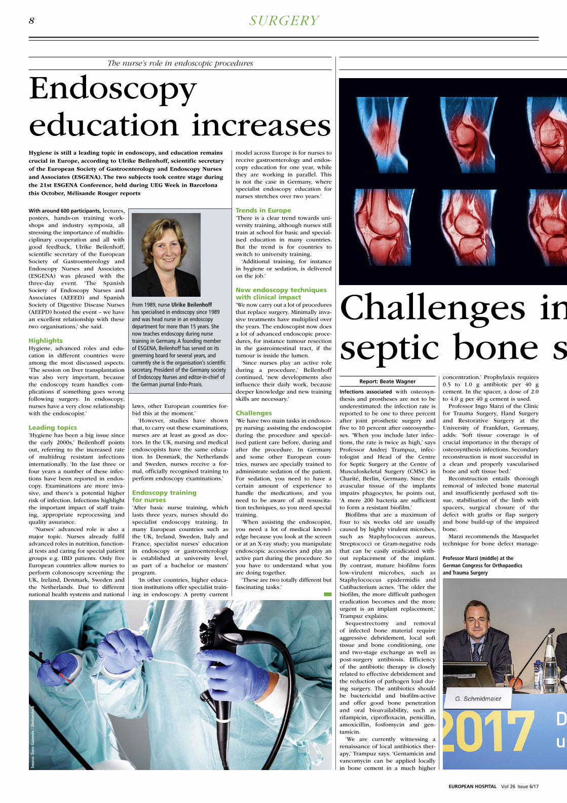
With around 600 participants, lectures, posters, hands-on training work-shops and industry symposia, all stressing the importance of multidis-ciplinary cooperation and all with good feedback, Ulrike Beilenhoff, scientific secretary of the European Society of Gastroenterology and Endoscopy Nurses and Associates (ESGENA) was pleased with the three-day event. ‘The Spanish Society of Endoscopy Nurses and Associates (AEEED) and Spanish Society of Digestive Disease Nurses (AEEPD) hosted the event – we have an excellent relationship with these two organisations,’ she said.
Highlights Hygiene, advanced roles and edu-cation in different countries were among the most discussed aspects. ‘The session on liver transplantation was also very important, because the endoscopy team handles com-plications if something goes wrong following surgery. In endoscopy, nurses have a very close relationship with the endoscopist.’
Leading topics ‘Hygiene has been a big issue since the early 2000s,’ Beilenhoff points out, referring to the increased rate of multidrug resistant infections internationally. ‘In the last three or four years a number of these infec-tions have been reported in endos-copy. Examinations are more inva-sive, and there’s a potential higher risk of infection. Infections highlight the important impact of staff train-ing, appropriate reprocessing and quality assurance.
‘Nurses’ advanced role is also a major topic. Nurses already fulfil advanced roles in nutrition, function-al tests and caring for special patient groups e.g. IBD patients. Only five European countries allow nurses to perform colonoscopy screening: the UK, Ireland, Denmark, Sweden and the Netherlands. Due to different national health systems and national
Report: Beate Wagner
Infections associated with osteosyn-thesis and prostheses are not to be underestimated: the infection rate is reported to be one to three percent after joint prosthetic surgery and five to 10 percent after osteosynthe-ses. ‘When you include later infec-tions, the rate is twice as high,’ says Professor Andrej Trampuz, infec-tologist and Head of the Centre for Septic Surgery at the Centre of Musculoskeletal Surgery (CMSC) in Charité, Berlin, Germany. Since the avascular tissue of the implants impairs phagocytes, he points out, ‘A mere 200 bacteria are sufficient to form a resistant biofilm.’
Biofilms that are a maximum of four to six weeks old are usually caused by highly virulent microbes, such as Staphylococcus aureus, Streptococci or Gram-negative rods that can be easily eradicated with-out replacement of the implant. By contrast, mature biofilms form low-virulent microbes, such as Staphylococcus epidermidis and Cutibacterium acnes. ‘The older the biofilm, the more difficult pathogen eradication becomes and the more urgent is an implant replacement,’ Trampuz explains.
Sequestrectomy and removal of infected bone material require aggressive debridement, local soft tissue and bone conditioning, one and two-stage exchange as well as post-surgery antibiosis. Efficiency of the antibiotic therapy is closely related to effective debridement and the reduction of pathogen load dur-ing surgery. The antibiotics should be bactericidal and biofilm-active and offer good bone penetration and oral bioavailability, such as rifampicin, ciprofloxacin, penicillin, amoxicillin, fosfomycin and gen-tamicin.
‘We are currently witnessing a renaissance of local antibiotics ther-apy,’ Trampuz says. ‘Gentamicin and vancomycin can be applied locally in bone cement in a much higher
concentration.’ Prophylaxis requires 0.5 to 1.0 g antibiotic per 40 g cement. In the spacer, a dose of 2.0 to 4.0 g per 40 g cement is used.
Professor Ingo Marzi of the Clinic for Trauma Surgery, Hand Surgery and Restorative Surgery at the University of Frankfurt, Germany, adds: ‘Soft tissue coverage is of crucial importance in the therapy of osteosynthesis infections. Secondary reconstruction is most successful in a clean and properly vascularised bone and soft tissue bed.’
Reconstruction entails thorough removal of infected bone material and insufficiently perfused soft tis-sue, stabilisation of the limb with spacers, surgical closure of the defect with grafts or flap surgery and bone build-up of the impaired bone.
Marzi recommends the Masquelet technique for bone defect manage-
laws, other European countries for-bid this at the moment.’
‘However, studies have shown that, to carry out these examinations, nurses are at least as good as doc-tors. In the UK, nursing and medical endoscopists have the same educa-tion. In Denmark, the Netherlands and Sweden, nurses receive a for-mal, officially recognised training to perform endoscopy examinations.’
Endoscopy training for nurses‘After basic nurse training, which lasts three years, nurses should do specialist endoscopy training. In many European countries such as the UK, Ireland, Sweden, Italy and France, specialist nurses’ education in endoscopy or gastroenterology is established at university level, as part of a bachelor or masters’ program.
‘In other countries, higher educa-tion institutions offer specialist train-ing in endoscopy. A pretty current
model across Europe is for nurses to receive gastroenterology and endos-copy education for one year, while they are working in parallel. This is not the case in Germany, where specialist endoscopy education for nurses stretches over two years.’
Trends in Europe‘There is a clear trend towards uni-versity training, although nurses still train at school for basic and special-ised education in many countries. But the trend is for countries to switch to university training.
‘Additional training, for instance in hygiene or sedation, is delivered on the job.’
New endoscopy techniques with clinical impact‘We now carry out a lot of procedures that replace surgery. Minimally inva-sive treatments have multiplied over the years. The endoscopist now does a lot of advanced endoscopic proce-dures, for instance tumour resection in the gastrointestinal tract, if the tumour is inside the lumen.
‘Since nurses play an active role during a procedure,’ Bellenhoff continued, ‘new developments also influence their daily work, because deeper knowledge and new training skills are necessary.’
Challenges ‘We have two main tasks in endosco-py nursing: assisting the endoscopist during the procedure and special-ised patient care before, during and after the procedure. In Germany and some other European coun-tries, nurses are specially trained to administrate sedation of the patient. For sedation, you need to have a certain amount of experience to handle the medications, and you need to be aware of all resuscita-tion techniques, so you need special training.
‘When assisting the endoscopist, you need a lot of medical knowl-edge because you look at the screen or at an X-ray study; you manipulate endoscopic accessories and play an active part during the procedure. So you have to understand what you are doing together.
‘These are two totally different but fascinating tasks.’
Hygiene is still a leading topic in endoscopy, and education remains crucial in Europe, according to Ulrike Beilenhoff, scientific secretary of the European Society of Gastroenterology and Endoscopy Nurses and Associates (ESGENA). The two subjects took centre stage during the 21st ESGENA Conference, held during UEG Week in Barcelona this October, Mélisande Rouger reports
Endoscopy education increases
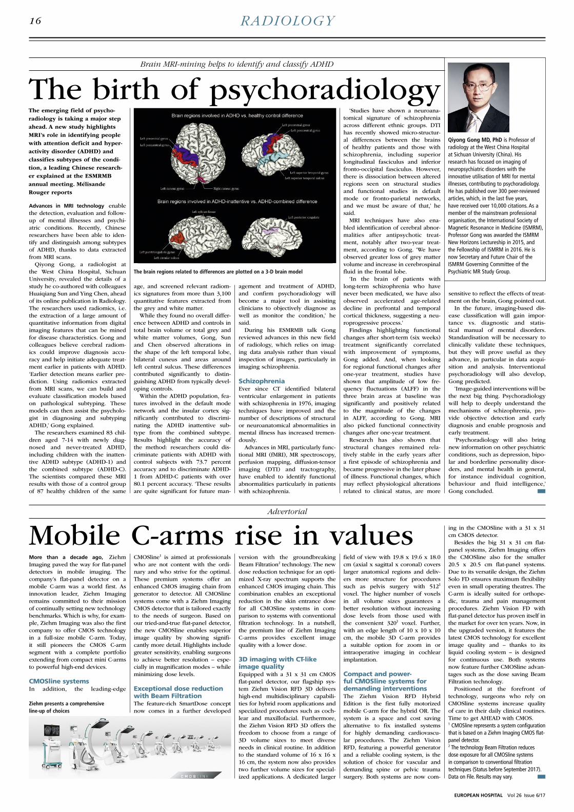
Challenges in septic bone surgery
The nurse’s role in endoscopic procedures Infection – defect – regeneration
From 1989, nurse Ulrike Beilenhoff has specialised in endoscopy since 1989 and was head nurse in an endoscopy department for more than 15 years. She now teaches endoscopy during nurse training in Germany. A founding member of ESGENA, Beilenhoff has served on its governing board for several years, and currently she is the organisation’s scientific secretary, President of the Germany society of Endoscopy Nurses and editorinchief of the German journal EndoPraxis.
Professor Marzi (middle) at the German Congress for Orthopaedics and Trauma Surgery
Sour
ce: D
oro
Guz
enda
/ Sh
utte
rsto
ck
8
EUROPEAN HOSPITAL Vol 26 Issue 6/17
RADBOOKThe Guide to Imaging Technology and Informatics in EuropeThe Guide to Imaging Technology and Informatics in Europeand Informatics in Europe
Showcase your products & solutions in our European guide to radiology equipment!
ment. In this, following thorough bone debridement and soft tissue coverage an antibiotic-loaded bone cement spacer is inserted into the bone defect. In a second interven-tion, the cement spacer is removed, without damaging the membrane, and the bone defect is filled with a mixture of BMP-7, tricalcium phosphate and endogenous bone. Bone tissue is harvested either from the iliac crest or via RIA tech-nique (Reamer-Irrigator-Aspirator). ‘Compared with other bone recon-struction methods the Masquelet technique is rather quick, even with large diaphyseal and metaphyseal femur or tibia defects,’ Marzi says.
Unlike Masquelet himself, who did not apply antibiotics in order to avoid infection masking, Professor Gerhard Schmidmaier of the German University of Heidelberg uses the procedure to deliver bone cement and high doses of gentamicin and vancomycin.
One advantage is improved per-fusion: ‘It’s amazing that today we
can improve bone perfusion with the help of the Masquelet tech-nique,’ says the interim director of Heidelberg’s Clinic of Orthopaedics and Trauma Surgery. Today, due to periosteum induction bone defects of 20-25 cm heal well. ‘In addi-tion, locally applied high doses of antibiotics ensure that all bacteria are eradicated,’ Schmidmaier points out. ‘Masquelet used his technique solely for membrane induction and
enhancement of perfusion, not to treat infections.’
Prior to the intervention cement is prepared in a bowl. ‘This makes the cement a bit more porous and the antibiotic is released better,’ Schmidmaier explains.
Nonetheless, he also routinely works with ready-to-use products such as Copa G+V, for example in infected pseudarthrosis. ‘Bone cement loaded with a mixture of gentamicin and vancomycin,’ he explains, ‘catches up to 80 percent of all microbes.’
He recommends that, when plac-ing the bone cement, it makes sense to create irregularly shaped edges on the bone, ‘for the subsequent integration of the new bone, the bone cement should overlap onto the healthy bone material’. When the bone cement is removed after six to eight weeks, the objective is to spare the membrane.
For harvesting graft material Schmidmaier favours RIA, a proce-dure that allows acquisition of large volumes (20-75 ml) of high-quality autologous bone tissue. ‘Research
indicates,’ he points out that mor-bidity decreases with harvesting using the RIA technique.’
In conclusion, he says: ‘The Masquelet technique is suited for interventions with plates and nails but also in recent trauma. It makes sense from a biological point of view and the combination of gen-tamicin and vancomycin offers ben-efits. If a tissue sample is loaded with bacteria that do not respond to the antibiotics, the spacer can be replaced or a suitable antibiotic can be applied locally.’
Challenges in septic bone surgery
Infection – defect – regeneration
Sour
ce: S
utth
a Bu
raw
onk
/ Shu
tter
stoc
k
9
www.healthcare-in-europe.com
RADIOLOGY
Report: Anja Behringer
For centuries, hands, eyes and ears were the physicians’ most important instruments when it came to detect-ing and diagnosing disease. Today, one of the traditional techniques, percussion, is being revived, sup-ported by state-of-the-art technol-ogy and dressed in a new name: optoacoustics.
In one of the most exciting vision-ary ideas in modern healthcare short laser pulses (optics) are transmitted to tissue where they generate ultra-sound signals (acoustics) that allow the identification of cells and dis-eases in the body. The advantages of this technology? No ionised radia-tion, no invasive procedure.
Pioneer of clinical optoacoustics is Professor Vasilis Ntziachristos, Chair for Biological Imaging at the Technical University Munich (TUM), Germany, and Director of the
Institute of Biological and Medical Imaging (IBMI) at Helmholtz Zentrum, Munich. His groundbreak-ing research not only sparks hope for cancer patients but also opens new diagnostic perspectives for Alzheimer’s, diabetes and dermato-logical diseases.
Multi-spectral optoacoustic tomography (MSOT)The laser pulses penetrate the body where they are absorbed differently, depending on their wavelength and on the type of target tissue. These laser pulses create a minute rise in temperature which expands the tissue. Those equally minute move-ments generate acoustic signals – with each type of tissue produc-ing unique signals. A blood cell for example “sounds” very different from a skin cell.
Ultrasound detectors on the skin surface register these different sig-
nals and a computer generates the corresponding 3-D image. Thus, single cells, for example cancer cells, can be detected. This is a major advantage compared to ultra-sound, which cannot differentiate on this level, Ntziachristos explains. Currently, multi-spectral optoacous-tic tomography shows its poten-tial particularly well in aggressive melanoma cells. But their unique sound also gives away other cell types, which might allow surgeons to check accurately during a tumour resection whether indeed all cancer cells were removed.
Following successful animal stud-ies the procedure is now being test-ed in human volunteers. Different clinical studies are currently being conducted for breast and thyroid cancer and peripheral atheroscle-rosis.
To display the images, another expert in medical imaging, Professor
Dr Daniel Razansky of Helmholtz Zentrum Munich, is developing an affordable diagnostic device for clinical use in the operating room (OR). While the device today costs around €200,000, Ntziachristos con-siders a future price tag of €50 to be realistic. Thus, in 2011 he and two partners founded the spin-off iThera Medical in Munich to fine-tune the product for a market launch. This requires capital.
Awards for visionary researchWhen it comes to raising capi-tal, the many awards Ntziachristos, a qualified electrical engineer, has collected over the past few years obviously help to give investors peace of mind. In 2013, he received the Leibniz Prize of the German Research Foundation (DFG) and last year he was awarded – for the sec-ond time – the ERC Advanced Grant of the European Research Council.
That grant of €2.49 million will be disbursed over a period of five years. The funds will be used to develop a portable device for human patients. As to market maturity the Helmholtz Zentrum did not provide any infor-mation since the product is still under development.
While working on the market-ability of the device, Ntziachristos’
research is also addressing the major limitations of optoacoustics: the laser cannot penetrate deeper
Optoacoustics: the sound of cells
Old technique + new technology diagnose disease
At the Fraunhofer Institute for Manufacturing Engineering and Automation (IPA) in Stuttgart, Germany, Andreas Rothfuss worked as an assistant while studying for his degree, specialising in manufacturing engineering at Stuttgart University. After joining the Project Group for Automation in Medicine and Bioengineering (PAMB) in Mannheim, Germany, in 2012, he began writing his diploma thesis, ‘The evaluation of shapememory actors with respect to possible applications in minimally invasive surgery’. He is now a doctoral researcher for the PAMB.
Report: Lena Petzold
‘Correct needle positioning for sur-gical interventions or biopsies is time-consuming,’ explains Engineer Andreas Rothfuss, a member of the working group for Information Systems for Medical and Bio Sciences at Fraunhofer IPA. ‘The angle and position must be accurate as mistakes can lead to the removal of the wrong tissue or even organ damage. The procedure can easily take half an hour. This represents a considerable chunk of time for doc-tors under constant time pressure, particularly as it is rarely possible to carry out these types of biopsies
in a cost-effective way.’ The project group for automation in medicine and biotechnology PAMB at the Institute has therefore set itself the task of simplifying these processes with digital technology.
Finding the largest common denominator‘The difficult part of the project was the compilation of the user profile. Finding a common language for doctors and engineers, and jointly exploring the opportunities and lim-its of such a system, was a compli-
cated undertaking – which we mas-tered step by step,’ Rothfuss says. Medics and engineers continued to keep in close contact after the devel-opment of the profile. ‘Obtaining regular input and resolving any issues arising with-out complications was extraordi-narily helpful,’ he adds, describing the Mannheim University Hospital cooperation.
Software generates a posi-tioning planThe result is a system that combines the advantages of digital support with medical ‘craftsmanship’. A soft-ware programme reads the patient’s CT or MRI images, calibrates them and generates a positioning plan. The doctor checks the plan and releases it if there are no objections.
Images and positioning can obvi-ously be precisely controlled in the system.
Once given the go-ahead, the robotic arm can automatically move into the correct position but can be
stopped manually at any time dur-ing the process. The arm reacts to touch so that safe handling of the equipment is always guaranteed.
Once the arm is in position the doctor can push the needle into the mounting bracket and guide it into the patient’s body at a precise angle. This provides human control over the process and over the tissue penetration. After the procedure the robotic arm retracts into the starting position.
Reducing radiation exposureThe system is to cover all stand-ard therapeutic procedures, such as ablation of metastases and other percutaneous techniques. The soft-ware can simulate not only the nee-dle position but also the spread of heat in the tissue.
A further advantage can be seen in the control of the placement.
As the robot holds the needle in position it is possible to take X-ray images without the doctor’s hand obscuring the image, which reduces radiation exposure for the doctor.
The robot-guided positioning also ensures that the needle cannot slip, which means not as many X-ray images are required for control, and radiation exposure for the patient is also lower.
Progress with this technique will be fastThe intuitive software and position-ing groundwork not only save time but also offer valuable support to less experienced doctors.
Rothfuss is convinced that pro-gress will be fast: ‘First clinical tests are due to be carried out from spring 2018, and we hope to be ready to launch the system in three years’ time.
The system has great potential – it may in time even facilitate fully automated placement.’
Needle placement takes six minutesCorrect placement of needles is time consuming. So much so that researchers at the Fraunhofer Institute for Manufacturing Engineering and Automation (IPA), along with experts at Kuka AG developed a robotic assistance system that enables needle placement in only six minutes. The doctor can fully concentrate on needle inser-tion, with calibration and positioning carried out by a computerised system with a robotic arm.
Software reads/calibrates CT or MRI scans to present a positioning plan
Focusing on needle insertion along with robotic assistance
When the robot holds the needle, X-rays can be obtained without the doctor’s hand in the images
10
EUROPEAN HOSPITAL Vol 26 Issue 6/17
Your Insight,Our Solutions
SonoVue® approved for detection of vesicoureteral reflux in paediatric patients
SUMMARY OF PRODUCT CHARACTERISTICS 1. NAME OF THE MEDICINAL PRODUCT SonoVue 8 microlitres/mL powder and solvent for dispersion for injection 2. QUALITATIVE AND QUANTITATIVE COMPOSITION Each mL of the dispersion contains 8 μL sulphur hexafluoride microbubbles, equivalent to 45 micrograms. For the full list of excipients, see section 6.1. 3. PHARMACEUTICAL FORM Powder and solvent for dispersion for injection. White powder. Clear, colourless solvent. 4. CLINICAL PARTICULARS 4.1 Therapeutic indications This medicinal product is for diagnostic use only. SonoVue is for use with ultrasound imaging to enhance the echogenicity of the blood, or of fluids in the urinary tract which results in an improved signal to noise ratio. SonoVue should only be used in patients where study without contrast enhancement is inconclusive. Echocardiography SonoVue is a transpulmonary echocardiographic contrast agent for use in adult patients with suspected or es-tablished cardiovascular disease to provide opacification of cardiac chambers and enhance left ventricular endocardial border delineation. Dop-pler of macrovasculature SonoVue increases the accuracy in detection or exclusion of abnormalities in cerebral arteries and extracranial carotid or peripheral arteries in adult patients by improving the Doppler signal to noise ratio. SonoVue increases the quality of the Doppler flow image and the duration of clinically-useful signal enhancement in portal vein assessment in adult patients. Doppler of microvasculature SonoVue improves display of the vascularity of liver and breast lesions during Doppler sonography in adult patients leading to more specific lesion characterisation. Ultrasonography of excretory urinary tract SonoVue is indicated for use in ultrasonography of the excretory tract in paediatric patients from newborn to 18 years to detect vesicoureteral reflux. For the limitation in the interpretation of a negative urosonography, see section 4.4 and 5.1. 4.2 Posology and method of administration This product should only be used by physicians experienced in diagnostic ultrasound imaging. Emergency equipment and personnel trained in its use must be readily available. Posology Intravenous use The recommended doses of SonoVue in adults are: B-mode imaging of cardiac chambers, at rest or with stress: 2 mL, Vascular Doppler imaging: 2.4 mL. During a single examination, a second injection of the recommended dose can be made when deemed necessary by the physician. Elderly Patients The dose recommenda-tions for intravenous administration also apply to elderly patients. Paediatric Patients The safety and efficacy of SonoVue in patients under 18 years of age has not been established for intravenous administration and use in echocardiography and vascular Doppler imaging. Intravesical use In paediatric patients the recommended dose of SonoVue is 1 mL. Method of administration For instructions on reconstitution of the me-dicinal product before administration see section 6.6. Intravenous use SonoVue should be administered immediately after drawing into the syringe by injection into a peripheral vein. Every injection should be followed by a flush with 5 mL of sodium chloride 9 mg/mL (0.9%) solution for injection. Intravesical use After introduction of a sterile 6F-8F urinary catheter into the bladder under sterile conditions, the bladder is emptied of urine and then filled with saline (normal sterile 0.9% sodium chloride solution) to approximately one third or half of its predicted total volume [(age in years + 2) x 30] mL. SonoVue is then administered through the urinary catheter. Administration of SonoVue is followed by completion of bladder filling with saline until patient has the urge to micturate or there is the first slight sign of back pressure to the infusion. Ultrasound imaging of the bladder and kidneys is performed during filling and voiding of the bladder. Immediately following the first voiding, the bladder may be refilled with saline for a second cycle of voiding and imaging, without the need of a second SonoVue administration. A low mechanical index (≤ 0.4) is recom-mended for imaging the bladder, ureters, and kidney during ultrasonography of the urinary tract with contrast. 4.3 Contraindications Hypersen-sitivity to the active substance(s) or to any of the excipients listed in section 6.1. Intravenous use of SonoVue is contraindicated in patients known to have right-to-left shunts, severe pulmonary hypertension (pulmonary artery pressure > 90 mmHg), uncontrolled systemic hypertension, and in patients with adult respiratory distress syndrome. SonoVue must not be used in combination with dobutamine in patients with conditions suggest-ing cardiovascular instability where dobutamine is contraindicated. 4.4 Special warnings and precautions for use Hypersensitivity reactions In the event of an anaphylactic reaction, beta blockers (including eye drop preparations) may aggravate the reaction. Patients may be unresponsive to the usual doses of adrenaline used to treat the allergic reactions. Intravenous use Patients with unstable cardiopulmonary status ECG monitoring should be performed in high-risk patients as clinically indicated. It is recommended to keep the patient under close medical supervision during and for at least 30 minutes following the administration of SonoVue. Use extreme caution when considering the administration of SonoVue in patients with recent acute coronary syndrome or clinically unstable ischaemic cardiac disease, including: evolving or ongoing myocardial infarc-tion, typical angina at rest within last 7 days, significant worsening of cardiac symptoms within last 7 days, recent coronary artery intervention or other factors suggesting clinical instability (for example, recent deterioration of ECG, laboratory or clinical findings), acute cardiac failure, Class III/IV cardiac failure, or severe rhythm disorders because in these patients allergy like and/or vasodilatory reactions may lead to life threatening conditions. SonoVue should only be administered to such patients after careful risk/benefit assessment and a closely monitoring of vital signs should be performed during and after administration. It should be emphasised that stress echocardiography, which can mimic an ischaemic episode, could potentially increase the risk of SonoVue utilisation. Therefore, if SonoVue is to be used in conjunction with stress echocardiography patients must have a stable condition verified by absence of chest pain or ECG modification during the two preceding days. Moreover, ECG and blood pressure monitoring should be performed during SonoVue-enhanced echocardiography with a pharmacological stress (e.g. with dobu-tamine). Chronic obstructive pulmonary disease Caution is advised when SonoVue is administered to patients with clinically significant pulmo-nary disease, including severe chronic obstructive pulmonary disease. Other concomitant diseases Caution is advisable when administering the product to patients with: acute endocarditis, prosthetic valves, acute systemic inflammation and/or sepsis, hyperactive coagulation states and/or recent thromboembolism, and end-stage renal or hepatic disease, as the numbers of patients with those conditions who were exposed to Sono-Vue in the clinical trials were limited. Patients on mechanical ventilation or with unstable neurological diseases SonoVue is not suitable for use in ventilated patients, and those with unstable neurological diseases. Interpretation of voiding urosonography with SonoVue and limitations of use False negative cases can occur with voiding ultrasonography with SonoVue and have not been clarified (see section 5.1). Technical recom-mendation In animal studies, the application of echo-contrast agents revealed biological adverse reactions (e.g. endothelial cell injury, capillary rupture) by interaction with the ultrasound beam. Although these biological side effects have not been reported in humans, the use of a low me-chanical index is recommended. Excipients This medicinal product contains less than 1 mmol sodium (23 mg) per dose, i.e. essentially ‘sodium-free’. 4.5 Interaction with other medicinal products and other forms of interaction No interaction studies have been performed. 4.6 Pregnancy, lactation, and fertility Pregnancy No clinical data on exposed pregnancies are available. Animal studies do not indicate harmful effects with respect to pregnancy, embryonal/foetal development, parturition or postnatal development (see section 5.3 Preclinical safety data). As a precautionary measure, it is preferable to avoid the use of SonoVue during pregnancy. Breastfeeding It is not known if sulphur hexafluoride is excreted in human milk . However, based on its rapid elimination from the body via the expired air, it is considered that the breastfeeding can be resumed two to three hours after administration of SonoVue. Fertility No clinical data are available. Animal studies do not indicate harmful effects on fertility. 4.7 Effects on ability to drive and use machines SonoVue has no or negligible influence on the ability to drive and use machines. 4.8 Undesirable effects Adult population-Intravenous use The safety of SonoVue after intravenous administration was evaluated in 4653 adult patients who participated in 58 clinical trials. The undesirable effects reported with SonoVue after intravenous administration were, in general, non-serious, transient and resolved spontaneously without residual effects. In clinical trials, the most commonly reported adverse reac-tions after intravenous administration are: headache, injection site reaction, and nausea. The adverse reactions are classified by System Organ Class and frequency, using the following convention: Very common (≥ 1/10), Common (≥ 1/100 to < 1/10), Uncommon (≥ 1/1,000 to < /100), Rare (≥ 1/10,000 to < 1/1,000), Very rare (< 1/10,000), not known (cannot be estimated from the available data).
System Organ Class
Adverse Drug ReactionsFrequency Category
Uncommon (≥1/1,000 to <1/100) Rare (≥1/10,000 to <1/1000) Not known Cannot be estimated from available data
Immune system disorders Hypersensitivity*Psychiatric disorders Insomnia
Musculoskeletal, connective tissue and bone disorders Back pain
General disorders and administration site conditions
Chest discomfort, injection site reaction, feeling hot Chest pain, pain, fatigue
Investigations Blood glucose increased*Cases suggestive of hypersensitivity may include: skin erythema, bradycardia, hypotension, dyspnoea, loss of consciousness, cardiac/cardio-respiratory arrest, anaphylactic reaction, anaphylactoid reaction or anaphylactic shock. **In some of the cases of hypersensitivity, in patients with underlying coronary artery disease, myocardial ischemia and/or myocardial infarctions were also reported.In very rare cases, fatal outcomes have been reported in temporal association with the use of SonoVue. In all these patients there was a high underlying risk for major cardiac complications, which could have led to the fatal outcome. Paediatric population - Intravesical use The safety of SonoVue after intravesical administration was based on evaluation of published literature involving use of SonoVue in over 6000 paediatric patients (age range 2 days to 18 years). No adverse reactions were reported. Reporting of suspected adverse reactions Reporting suspected adverse reactions after authorisation of the medicinal product is important. It allows continued monitoring of the benefit/risk balance of the medicinal product. Healthcare professionals are asked to report any suspected adverse reactions via the national reporting system listed in Appendix V. 4.9 Overdose Since there have been no cases of overdose reported to date, neither signs nor symptoms of overdose have been identified. In a Phase I study doses up to 56 mL of SonoVue were administered to normal volunteers without serious adverse events being re-ported. In the event of overdose occurring, the patient should be observed and treated symptomatically. 5. PHARMACOLOGICAL PROPER-TIES 5.1 Pharmacodynamic properties Pharmacotherapeutic group: Ultrasound contrast media. ATC code: VO8DA05. Sulphur hexafluor-ide is an inert, innocuous gas, poorly soluble in aqueous solutions. There are literature reports of the use of the gas in the study of respiratory physiology and in pneumatic retinopexy. The addition of sodium chloride 9 mg/mL (0.9%) solution for injection to the lyophilised powder followed by vigorous shaking results in the production of the microbubbles of sulphur hexafluoride. The microbubbles have a mean diameter of about 2.5 μm, with 90% having a diameter less than 6 μm and 99% having a diameter less than 11 μm. Each millilitre of SonoVue contains 8 μL of the microbubbles. The intensity of the reflected signal is dependent on concentration of the microbubbles and frequency of the ultrasound beam. The interface between the sulphur hexafluoride bubble and the aqueous medium acts as a reflector of the ultrasound beam thus enhanc-ing blood echogenicity and increasing contrast between the blood and the surrounding tissues. Intravenous use At the proposed clinical doses for intravenous administration, SonoVue has been shown to provide marked increase in signal intensity of more than 2 minutes for B-mode imaging in echocardiography and of 3 to 8 minutes for Doppler imaging of the macrovasculature and microvasculature. Intravesical use For ultrasonography of the excretory urinary tract in paediatric patients, after intravesical administration, SonoVue increases the signal intensity of fluids within the urethra, bladder, ureters, and renal pelvis, and facilitates the detection of reflux of fluid from the bladder into the ureters. The efficacy of SonoVue for detection/exclusion of vesicoureteral reflux was studied in two published open label single centre studies. The presence or absence of vesicoureteral reflux with SonoVue ultrasound was compared to the radiographic reference standard. In one study including 183 patients (366 kidney-ureter units), SonoVue ultrasound was correctly positive in 89 out 103 units with reflux and correctly negative in 226 out of 263 units without reflux. In the second study including 228 patients (463 kidney-ureter units), SonoVue ultrasound was correctly positive in 57 out of 71 units with reflux and correctly negative in 302 out of 392 units without reflux. 5.2 Pharmacokinetic properties The total amount of sulphur hexafluoride administered in a clinical dose is extremely small, (in a 2 mL dose the microbubbles contain 16 μL of gas). The sulphur hexafluoride dissolves in the blood and is subsequently exhaled. After a single intravenous injection of 0.03 or 0.3 mL of SonoVue/kg (ap-proximately 1 and 10 times the maximum clinical dose) to human volunteers, the sulphur hexafluoride was cleared rapidly. The mean terminal half-life was 12 minutes (range 2 to 33 minutes). More than 80% of the administered sulphur hexafluoride was recovered in exhaled air within 2 minutes after injection and almost 100% after 15 minutes.In patients with diffuse interstitial pulmonary fibrosis, the percent of dose recovered in expired air averaged 100% and the terminal half-life was similar to that measured in healthy volunteers. 5.3 Preclinical safety data Non-clinical data reveal no special hazard for humans based on conventional studies of safety pharmacology, genotoxicity and toxicity to reproduction. Caecal lesions observed in some repeat-dose studies with rats, but not in monkeys, are not relevant for humans under normal conditions of administration. Intravesical local tolerance for SonoVue was also assessed. A single-dose study and a repeat-dose study, both followed by a treatment-free period, were performed in female rats with local toxicity evaluated through macroscopic and histopathological examination of both kidneys, ureters, the urinary bladder and urethra. It did not reveal any test item-related lesions in any of the examined or-gans, in particular in the urinary bladder, in both the single-dose and the repeat-dose studies. It was therefore concluded that SonoVue is well tolerated in the urinary tract in the rat. 6. PHARMACEUTICAL PARTICULARS 6.1 List of excipients Powder: Macrogol 4000, Distearoylphos-phatidylcholine, Dipalmitoylphosphatidylglycerol Sodium, Palmitic acid Solvent: Sodium chloride 9 mg/mL (0.9%) solution for injection. 6.2 Incompatibilities This medicinal product must not be mixed with other medicinal products except those mentioned in section 6.6. 6.3 Shelf life 2 years. Once reconstituted, chemical and physical stability has been demonstrated for 6 hours. From a microbiological point of view, the medicinal product should be used immediately. If not used immediately, in use storage times and conditions prior to use are the responsibility of the user. 6.4 Special precautions for storage The medicinal product does not require any special storage conditions. For storage conditions after reconstitution of the medicinal product, see section 6.3. 6.5 Nature and contents of container Type I colourless glass vial containing 25 mg of dry, lyophilised powder in an atmosphere of sulphur hexafluoride closed with a grey butyl rubber stopper and sealed with an alu-minium crimp seal with a flip-off disc. A transfer system (MiniSpike). Type I clear glass pre-filled syringe containing 5 mL sodium chloride 9 mg/mL (0.9%) solution for injection. 6.6 Special precautions for disposal Before use examine the product to ensure that the container and clo-sure have not been damaged. SonoVue must be prepared before use by injecting through the septum 5 mL of sodium chloride 9 mg/mL (0.9%) solution for injection to the contents of the vial. The vial is then shaken vigorously for twenty seconds after which the desired volume of the dispersion can be drawn into a syringe as follows: 1 Connect the plunger rod by screwing it clockwise into the syringe. 2 Open the MiniSpike transfer system blister and remove syringe tip cap. 3. Open the transfer system cap and connect the syringe to the transfer system by screwing it in clockwise. 4 Remove the protective disk from the vial. Slide the vial into the transparent sleeve of the transfer system and press firmly to lock the vial in place. 5 Empty the contents of the syringe into the vial by pushing on the plunger rod. 6 Shake vigorously for 20 seconds to mix all the contents in the vial to obtain a white milky homogeneous liquid. 7 Invert the system and carefully withdraw SonoVue into the syringe. 8 Unscrew the syringe from the transfer system. Do not use if the liquid obtained is clear and/or if solid parts of the lyophilisate are seen in the suspension. SonoVue should be administered immediately by injection into a peripheral vein for use in echocardiography and in vascular Dop-pler imaging in adults or by intravesical administration for use in ultrasonography of the excretory urinary tract in paediatric patients. If SonoVue is not used immediately after reconstitution the microbubble dispersion should be shaken again before being drawn up into a syringe. Chemical and physical stability of the microbubble dispersion has been demonstrated for 6 hours. The vial is for a single use only. Any unused medicinal product or waste material should be disposed of in accordance with local requirements. 7. MARKETING AUTHORISATION HOLDER Bracco International B.V. Strawinskylaan 3051 NL - 1077 ZX Amsterdam The Netherlands 8. MARKETING AUTHORISATION NUMBERS EU/1/01/177/002 9. DATE OF FIRST AUTHORISATION/RENEWAL OF THE AUTHORISATION Date of first authorisation: 26 March 2001 Date of latest renewal: 24 April 2006 10. DATE OF REVISION OF THE TEXT 24/08/2017
AD SV VUR A4 09 2017.indd 1 11/09/17 12:41
RADIOLOGY
than four to five centimetres and the sound loses quality the deeper it moves into the tissue.
Thus, liver studies, for example, are impossible.
Ntziachristos’s colleague Razansky is particularly interested in the molecular level, the biochemical reaction in the cell. Whatever the individual research interests of the two scientists are, the overarching aim of the team is early detection of diseases and tracking of disease progression so as to save time-to-
diagnosis and money and above all to help patients by improving prognosis.
Additionally, the optoacoustic imaging pioneers think even further. They envisage new interdisciplinary fields of medical science and new areas of application, such as the diagnosis of inflammation or meta-bolic disorders and neurology. To test their ideas the research-ers successfully applied for Horizon 2020, the major European Union research and innovation programme for 2014 to 2020.
Old technique + new technology diagnose disease
Research carried out by Professor Vasilis Ntziachristos encompasses the development of new methods and devices for biological and medical imaging, focusing on innovative noninvasive approaches that
visualise previously unseen physiological and molecular processes in tissues. His research also aims to translate these methods to advance biological discovery, accelerate drug development and offer efficient methods for diagnostics and theranostics. Ntziachristos studied electrical engineering at Aristotle University, Thessaloniki, Greece, and gained Master’s and doctoral degrees from the Bioengineering Department at the University of Pennsylvania, USA. He was assistant professor and director of the Laboratory for BioOptics and Molecular Imaging at Harvard University and Massachusetts General Hospital. He now directs the Biological and Medical Imaging Institute at Helmholtz Zentrum Munich, Germany.
Multi-spectral optoacoustic tomography (MSOT) could help to detect cancer cells early
Sour
ce: M
urad
Om
ar –
Hel
mho
ltz
Zent
rum
Mün
chen
ETIM 2018After this year’s successful first congress about Emerging Technologies in Medicine (ETIM) in Essen, Germany, the organiz-ing committee sets the stage for the second edition on February, 16 and 17, 2018.
The congress will again take place at the Lehr- und Lernzentrum of the medical faculty of University Hospital of Essen, Germany. The first day is dedicated to Artificial Intelligence and the second to Robotics. Key top-ics will include the role of artificial intelligence in diagnostics and the future of robot-assisted surgery.
Advances in healthcare and medi-cal research are already strongly driven by information technology and engineering, the ETIM commit-tee points out. Technologies like individual genome sequencing or high-performance multiparamet-ric imaging generate exponentially growing datasets while contempo-rary data-mining techniques allow to extract valuable data from existing archives. These offer the opportunity for highly specific clinical decision making and personalized precision medicine. Further acceleration of medical innovation can be safely predicted. However, as complex chal-lenges often require complex solu-tions, these technologies demand an interdisciplinary approach between clinicians, computer scientists, engi-neers, researchers, healthcare provid-ers, legislators and many other disci-plines. The 2018 ETIM will therefore provide an opportunity for experts to get together.
Detailed information is available at https://etim.uk-essen.de/.
11
www.healthcare-in-europe.com
RADIOLOGY
Report: Cynthia E. Keen
Many challenges and opportunities exist for innovation in diagnostic imaging in the 21st century. One lies in biomedicine, especially in terms of personalised medicine. Just as radiology today is essential for the diagnosis and treatment of dis-ease, imaging’s contribution to bio-medicine has the potential to dra-matically alter medical treatment by focusing on proactive intervention prior to disease progression, Elias A Zerhouni MD, told attendees at the opening session of the 2017 RSNA annual meeting.
Historically, innovations in diag-nostic imaging have been interdisci-plinary, achieved through collabora-tions in medical physics, chemistry, and medicine. The biomedical infor-mation clinicians need to under-stand the body is multidimensional. Defining bio-imaging science as ‘the extraction of spatially and temporal-ly resolved, structural and functional multidimensional biological informa-tion from molecules to humans,’ Zerhouni rhetorically asked: ‘How do we get biomedical imaging infor-mation at the molecular level and transfer this into an organ?’
Report: Mélisande Rouger
Radiographers are key team players in medical imaging, nuclear medicine and radiation therapy, and their role is growing, boosted by the rising demand for imaging studies and pro-cedures and the continuous shortage of radiologists in many countries. But radiographers have many faces and names, and this compromises the rec-ognition of their skills across health-care, according to Jonathan McNulty, EFRS newly elected president.
‘The official title we use is radiogra-pher, but there are 20 or more other titles for the profession! Radiographers can also be nurses, technicians, radio manipulators (…) and may provide very distinct services depending on the country.’
Not only does the profession have multiple identities, but also education varies considerably across Europe. While some countries offer masters or doctoral qualifications specifically for radiographers, other countries only have two-year programs for entry into the profession and sometimes do not even provide a national board registration.
‘We have a very complicated jigsaw here,’ McNulty summed up.
The EFRS plans to change this by promoting a bachelor’s level as the entry level to the profession, ver-sus shorter, vocational qualifications;
meaning it will require a minimum three-year program at level 6 in its European qualification framework (EQF) benchmarking document. The organisation has also introduced an EQF level 7 (masters level) bench-marking document, in a move to raise the bar for future generations.
‘We very strongly feel that radiogra-phers across Europe should be trained to the highest possible level. That allows them to make a greater contri-bution to healthcare and to enhance radiology and radiation therapy ser-vices. The profession must be duly regulated,’ McNulty said. ‘A bachelor’s
level qualification is seen as the mini-mum standard for us; it should be the entry level to the profession, but it’s still not the case in some countries.’
To make up for the gap between seasoned radiographers, who had their qualification decades ago, and what they want the profession to look like from now on, the EFRS is also work-ing on the practicalities of launching a European Diploma in Radiography very soon, which was among topics discussed by the federation during its last annual business meeting, in November in Alcalá de Henares near Madrid. ‘This remains one of our major objectives. We hope to progress this through 2018, with a view to launching in 2019. Radiology has such a diploma, medical physics has it too, so it’s something we want to certify as a certain standard of professional radiography knowledge in Europe,’ McNulty explained.
Taking the diploma could be useful for those who only received a two-year education in their countries but have 20-year experience. ‘The diploma could help show that they have what the EFRS sees as minimum level of knowledge for the practice of radiog-raphy,’ he added.
Continued professional develop-ment (CPD) is also instrumental in securing high professional stand-ards across Europe and it should be homogenised too, according to Håkon Hjemly, immediate past president of the EFRS. ‘It’s necessary to maintain and renew your skills and CPD is an excellent system for that. This is some-thing that many countries have man-datory, but many still don’t,’ he said.
The lack of homogeneity in European radiographers’ education has become a pressing issue, as the unequal distribution of professionals leads to migration, which can prove tough when countries have different curricula.
‘About 50% of our member societies have a shortage of radiographers and the other half is producing too many. In Italy, for instance, many radiog-raphers who qualify struggle to find work and many will have to work for free to get a foot through the door. Consequently, many have come to Ireland and the United Kingdom for a job but, for others, their qualification may not be recognised because it may not be equivalent to bachelor level,’ McNulty explained.
Having the same education could also help improve the profession’s profile among medical specialties. Radiographers are simply under-utilised, yet research shows they would have the skills to take on more
responsibilities, according to McNulty. ‘I feel quite strongly how under-uti-lised and under-valued radiographers are. There is not enough recognition of our knowledge and skills in some countries, whereas in others radi-ographers are involved in reporting
He explained that the multidimen-sional complexity of medicine can be perceived as layers of interactiv-ity that incorporate the DNA/RNA/and proteins interacting with each other within a cell, and the cells interacting with tissues, organs, and the environment. There are seven or eight layers in total that need to be understood with respect to interacting with each other in vary-ing environments. This complexity is multiplied by the number of per-
mutations in genes and RNA. More than 50 years of research have been spent trying to understand these interactions.
‘The challenge facing medicine is how to combine the need to under-stand the complexity while achiev-ing precision at an individual level. This is the centre core of biomedi-cal innovation in the 21st century. Medicine should no longer wait for disease to strike a patient, but rather pre-empt disease before it strikes.’
Zerhouni explained that it is nec-essary to understand individual vari-ations in disease risks and pathways. Radiology researchers have begun to identify correlations between imag-ing data and genotype data.
Science is shifting from the ‘hardware’ to the ‘software’ of life. Zerhouni believes that imaging sci-ences will play a major role in precise reclassification of what medi-cine is all about: a network of mol-ecules interacting. These are affected by genetic, infectious or autoim-mune diseases and, because of their interaction complexity, diseases are not homogeneous. No one disease can be controlled by just one tar-get, which is why the response by patients to treatments differs. Radiologists can contribute to per-sonalised medicine by identifying the ‘tools’ to address targets. ‘If imag-ing can show the interaction of every therapeutic agent and every one of their targets, and the consequences of their interaction, this ‘gold mine’ of information would make progress very fast. Diagnostics and therapeu-tics are two faces of the same coin,’ he pointed out.
‘Understanding molecular net-works, disease pathways, and their regulation in health and disease will lead to a functional and more pre-cise reclassification of most diseases based on their specific molecular pathways, enable predictive bio-markers discovery, and offer a great-er understanding of environmental drivers,’ Zerhouni said. Identification of reliable biomarkers needs to be a
strategic goal of imaging innovation. A goal today of research in therapeu-tics is to develop ‘dream molecules’ that will do multiple things at once. The challenge to radiology is to localise and assess functionality of therapeutic molecules and their tar-gets in vivo.
Multi-modality imaging biomark-ers (PET/CT, PET/MRI) need to be identified and correlated with bio-logical markers. Zerhouni predicted that diagnostic imaging departments in academic research organisations would have their staff dedicated to this.
With respect to machine-augment-ed diagnostics and artificial intelli-gence (AI), Zerhouni predicts that in 5-10 years its use will become stand-ard in radiology. He sees the technol-ogy as being able to improve radi-ologists’ performance and to help standardise levels of performance.
He predicts that huge global refer-ence databases queried by AI will enable radiologists within seconds to compare an exam they are interpret-ing with a wealth of stored and rel-evant-to-patient data. ‘I see a future where a radiologist will say in a radi-ology report that the patient corre-sponds to RSNA Reference Database Number xxxxx. Radiologists will be able to track the evolution of disease and extract novel information.’
Zerhouni concluded by offering this advice to the RSNA meeting attendees:
• Ask not what radiology can do but what this discipline should do
Pre-empting disease before it strikes
Levelling EU qualifications
Medicine will be fundamentally reclassified
Radiographers gain a European diploma by 2019
With a master’s in clinical health and postgraduate education in digital imaging processing and Xray protection Håkon Hjemly is Manager of Policy for the Norwegian Society of Radiographers, covering health political issues and radiographers’ role development. Earlier, he worked in Norway’s private and public sector, as clinical radiographer, QAadministrator, manager, radiation protection officer, product specialist and sales rep. (CT, Mammography, Carm), and pioneered identifying pitfalls and quality assurance controls for medical imaging digitisation at Xray departments. He has chaired and delivered numerous presentations at international conferences. Håkon was elected member of the EFRS election committee in 2010, then Treasurer in the 20112014, and then EFRS Vice President in 20142015 and Presidentin 20152017.
Radiographers are increasingly central to patient care, but the het-erogeneous education and skills across Europe remain challenging. Dr Jonathan McNulty and Håkon Hjemly, of the European Federation of Radiographers Societies (EFRS), explained how they plan to improve radiographers’ visibility and work towards homogenising training across Europe, notably by launching a European Diploma in Radiography by early 2019.
The lack of homogeneity in European radiographers’ education has become a pressing issue, as the unequal distribution of professionals leads to migration
Sour
ce: w
atch
ara
/ Shu
tter
stoc
k
12
EUROPEAN HOSPITAL Vol 26 Issue 6/17
RADIOLOGY
imaging examinations and performing certain procedures traditionally done by medical doctors.
‘The impact radiographers can have on hospitals is dramatic. A lot of research shows that, when radiogra-phers take on advanced practices such as these, it can result in efficiencies, save time and money, and well trained radiographers have been shown to perform such tasks as well as radi-ologists. Ultimately radiographers may improve patient care by providing a more timely report in certain instanc-es,’ he said.
Recognising radiographers’ skills could be pertinent in countries with an acute shortage of radiologists, based on the experience of coop-eration between radiographers and radiologists in the UK or Ireland, for example. In the end, a collaborative approach can only benefit healthcare, McNulty concludes.
in the 21st century.
• Remain the key integrator that can improve healthcare for patients through imaging.
• Participate in solving core scien-tific challenges of today.
• Train and proactively mentor the next generation of radiologists, because they will represent the future of radiology in the 21st century.
The hope now is that this research could help reduce the number of unnecessary breast cancer surger-ies because it could pinpoint which lesions are cancerous more accu-rately and more efficiently.
In the study, the system was trained on information about such lesions and looked for pat-terns among a range of data points, including demographics, family his-tory, biopsies and pathology reports.
When tested on 335 high-risk lesions, it correctly diagnosed 97% as malignant.
The researchers suggest that such levels of accuracy could lead to a reduction in the number of unnec-essary surgeries by more than 30%.
While mammograms can detect cancers, there is also a risk of false positive results that can lead to unnecessary biopsies and surger-ies – often from ‘high-risk’ lesions that appear suspicious on mam-mograms and have abnormal cells when tested by needle biopsy. The researchers say patients have the lesion surgically removed but often it is benign and the operations were unnecessary.
To address this, the team devel-oped the machine learning system to predict if a high-risk lesion identi-fied on needle biopsy after a mam-mogram will upgrade to cancer at surgery.
‘Because diagnostic tools are
so inexact, there is an understand-able tendency for doctors to over-screen for breast cancer,’ Dr Regina Barzilay, MIT’s Delta Electronics Professor of Electrical Engineering and Computer Science, pointed out. ‘When there’s this much uncertainty in data, machine learning is exactly the tool that we need to improve detection and prevent over-treat-ment.
‘A model like this will work any-time you have lots of different fac-tors that correlate with a specific outcome. It hopefully will enable us to start to go beyond a one-size-fits-all approach to medical diagnosis.’
Using a method known as a ‘ran-dom-forest classifier’, the model resulted in fewer unnecessary sur-geries compared to the strategy of always doing surgery, while also being able to diagnose more cancer-ous lesions than the strategy of only doing surgery on traditional ‘high-risk lesions’.
Dr Constance Lehman, Professor at Harvard Medical School and chief of the Breast Imaging Division at MGH’s Department of Radiology added: ‘To our knowledge, this is the first study to apply machine learning to the task of distinguishing high-risk lesions that need surgery from those that don’t. We believe this could support women to make more informed decisions about their treatment, and that we could pro-
vide more targeted approaches to healthcare in general.’
It is hoped that MGH radiologists will begin incorporating the model into their clinical practice over the next year. ‘In the past, we might have recommended that all high-risk lesions be surgically excised, Lehman said. ‘But now, if the model determines that the lesion has a very low chance of being cancerous in a specific patient, we can have a more informed discussion with our patient about her options. It may be
reasonable for some patients to have their lesions followed with imaging rather than surgically excised.’
The team – which also includ-ed Manisha Bahl, director of the Massachusetts General Hospital Breast Imaging Fellowship Program - says that they are working to fur-ther evolve the model and in future hope to incorporate the actual images from the mammograms and images of the pathology slides, as well as more extensive patient infor-mation from medical records.
Artificial Intelligence helps to detect breast cancer
Medicine will be fundamentally reclassified In 335 high-risk lesions the system correctly diagnosed 97% as malignant
Radiographers gain a European diploma by 2019
Elias A Zerhouni MD is a world re nowned leader in radiology and medical research, who has helped change per ceptions of the importance of imaging science within the global medical community, He is president of Global Research and Development at Sanofi, the Parisbased global pharmaceutical company in France. He has developed innovations in both computed tomography (CT) and magnetic resonance imaging (MRI) to improve diagnosis of cancer and cardiovascular disease. As director of the USA’s National Institutes of Health (NIH) from 2002 to 2008, Zerhouni launched the NIH Roadmap for Medical Research, an initiative designed to transform the translation of research into medical practice.
Dr Jonathan McNulty is Associate Dean for Graduate Taught Studies and Head of Subject, Radiography in the School of Medicine, University College Dublin, Ireland. He oversees more than 75 postgraduate programs and continuing professional development activities across the school. He is President of the European Federation of Radiographer Societies (EFRS), and Board Member since 2014. He chaired the Education Wing, a network of 59 educational institutions within the EFRS, and VicePresident from 20162017.
Scientists are using Artificial Intelligence to support more effective breast cancer detection. The researchers at Massachusetts Institute of Technology (MIT) Computer Science and Artificial Intelligence Laboratory (CSAIL), Massachusetts General Hospital (MGH), and Harvard Medical School, are using the machine learning system to predict whether breast lesions identified from a biopsy will turn out to be cancerous, Mark Nicholls reports
From left:Manisha Bahl MD is a breast imaging radiologist and director of the Breast Imaging Fellowship Program at Massachusetts General Hospital/Harvard Medical School in Boston, USA. After graduating from the Harvard School of Public Health with an MPH in Health Policy and Management, she completed a radiology residency and breast imaging fellowship at Duke University Medical Centre and joined the faculty at Massachusetts General Hospital/Harvard Medical School in July 2016.
Regina Barzilay MD is a Delta Electronics Professor in the Department of Electrical Engineering and Computer Science and
a member of the Computer Science and Artificial Intelligence Laboratory at the Massachusetts Institute of Technology. USA. Her research focuses on natural language processing, applications of deep learning to chemistry and oncology.
Constance Lehman MD is a Professor at Harvard Medical School in Boston, USA, and chief of the Breast Imaging Division at MGH’s Department of Radiology. After graduating from Duke University and receiving medical and doctoral degrees at Yale University, she became Professor and vice chair of Radiology and division chief of Breast Imaging at the Seattle Cancer Care Alliance before her recent move to Massachusetts General Hospital.
(Pho
togr
aph:
Jas
on D
orfm
an/C
SAIL
)
13
www.healthcare-in-europe.com
RADIOLOGY
Report: Mark Nicholls
A new genetic study by UK-based sci-entists suggests that immunotherapy drugs could prove to be an effective treatment for some breast cancer patients.
Scientists from the Wellcome Trust Sanger Institute, near Cambridge – one of the world’s leading genome centres – and their collaborators, have identified particular genetic changes in a DNA repair mechanism in breast cancer.
Led by Dr Serena Nik-Zainal, the researchers suggest it could open up the possibility of another therapy option for around 1,000 UK breast cancer patients who could benefit from existing drugs.
The study found that a particular group of breast cancer patients have genetic changes, or mutations, that occur due to an abnormality of a DNA repair mechanism known as mis-match repair, which is a mechanism to recognise and remedy mistakes in the genetic code that arise during DNA replication and recombination. The mechanism also repairs some forms of DNA damage.
When cells lack the mismatch repair pathway, mutations build up, which results in cancerous tumours formation. These mutations are found in other cancers, such as colorectal cancer, but are rarely looked for in breast cancer.
In recent work in the USA, colorec-tal cancers with deficient mismatch repair have been treated with immu-notherapies, which exploit the fact that, under the influence of these so-called check point inhibitors, highly
Report: Mark Nicholls
A new genetic test to assess breast cancer risk in women who have a family history of the disease could be introduced into clinical practice in the UK within the next few months.
Devised at Manchester University NHS Foundation Trust (MFT) and the University of Manchester, researchers believe the test for high-risk groups could also help reduce the number of women needing to have surgery to remove their breasts. By narrow-ing down their risk, women will be better informed about whether to have a mastectomy, or not, explained Professor Gareth Evans, who led the work that resulted in the new test.
Scientists say the test will accurately
mutated tumour cells can be rec-ognised as ‘foreign’ by the patient’s immune system. The results of this new Sanger Institute study suggest that these immunotherapies could also be effective for some breast can-cer patients, based on the same muta-tion patterns seen in their tumours.
‘We’ve unequivocally found mis-match repair deficient breast can-cers,’ Serena Nik-Zainal said ‘As these tumours have the same mutational signatures as those of other cancers, like colorectal cancer, in theory they should respond to the same immuno-therapy drugs.
predict breast cancer risk in women who do not test positive for BRCA1/2 gene mutations and, in some cases, may also help to refine breast cancer risk in those with the BRCA1/2 muta-tions.
The most common cancer that affects women having a parent or sib-ling with breast cancer makes women twice as likely to develop breast cancer themselves. Mutations in the BRCA1/2 genes have been identified as a cause of hereditary cancer, but only account for 15-20% of the under-lying inherited genetic trigger for the condition.
‘Our results suggest expanding the cohort of cancer patients that could possibly be treated with checkpoint inhibitors to include these mismatch repair deficient breast cancer patients.’
The study researchers analysed the whole genome sequences of 640 breast cancer tumours and looked for patterns in the mutations, known as mutational signatures, which indi-cated abnormalities in the mismatch repair mechanism.
From the mutational signatures, the team identified 11 tumours that had the mismatch repair defects causing the breast cancer.
The new genetic test assesses breast cancer risk based on genetic variations - single nucleotide poly-morphisms (SNPs) - in an individual’s DNA.
Researchers found that mutations of 18 SNPs were indicative of breast cancer risk for women who did not carry BRCA1/2 mutations. These were found to have minimal effect in iso-lation, but when combined could increase or decrease breast cancer risk considerably.
The study recruited 451 women (112 with BRCA1/2 mutations) with a family history of breast cancer who had developed breast cancer. The researchers compared the diagnosis of invasive breast cancer and genetic profile in the case group against that
‘Using whole genome sequencing we can start to stratify breast can-cer patients into different catego-ries based on their mutational sig-natures,’ said one of the researchers Dr Helen Davies, from the Wellcome Trust Sanger Institute. Current clinical criteria means these tumours would not have been detected as being defi-cient in the mismatch repair pathway. We have shown that there is, in fact, another category of breast cancers – those with defective mismatch repair.’
Professor Karen Vousden, chief sci-entist at Cancer Research UK (CRUK), added: ‘Immunotherapies have shown promise for some cancer patients, but the challenge for doctors has been predicting which patients they are
likely to help. This study reveals more about the genetic patterns that could show which women with breast cancer are more likely to respond to immunotherapy treatments.’
The next step is to stage clinical tri-als to determine if immunotherapies could help selected breast cancer patients.
The Wellcome, CRUK, DanaFarber/Harvard Cancer Centre SPORE in Breast Cancer, and National Research Foundation of Korea with grants from the Korean government, supported this research project.
of a control group of 1,605 women (691 with BRCA1/2 mutations).
The analysis of DNA using par-ticipants’ blood samples was used to determine their individual genetic makeup and predict an overall risk estimate alongside other risk factors such as age at first assessment, fam-ily history of first and second-degree relatives, age at first child, first period and menopause, height and weight, and history of prior non-cancerous breast disease.
From the findings, women original-ly viewed as at high risk (lifetime risk of 30% or greater) were reclassified to a lower risk, where mastectomy is not recommended to reduce the risk.
The study suggested that the num-ber of women with BRCA1/2 muta-tions who currently choose to have a mastectomy could now fall by a third from 50% to 36%.
Professor Evans, a Consultant in Medical Genetics and Cancer Epidemiology at The University of Manchester and the city’s Saint Mary’s Hospital – where the test will first be made available – said: ‘This new test will help women at risk of familial breast cancer to make more informed decisions about their care.
‘BRCA1 and BRCA2 are just part of what we should be looking for when assessing risk and in Manchester we plan to incorporate screening for these new genetic markers in clinical practice within the next six months.
‘We are committed to improving cancer prevention through research
and, with funding from the NIHR Manchester Biomedical Research Centre, we plan to develop new screening strategies and biomarkers for other common cancers, including womb, bowel, ovarian and prostate.’
Professor Evans is hoping the test will become more widely available, and describes it as a ‘massive game changer for breast cancer’, which can accurately assess risk in the whole population from those with a family history and those with BRCA muta-tions.
Defining which breast cancer patients could gain from immunotherapy
Mastectomies due to ‘family history’ could drop by a third
The DNA mismatch repair mechanism
DNA test is ‘a massive game changer’
Serena Nik-Zainal MD is a Career Development Fellow (CDF) Group Leader in the Cancer Genome Project at the Sanger Institute in Cambridge and an Honorary Consultant in Clinical Genetics at nearby Addenbrookes Hospital, UK. Having qualified in medicine from the University of Cambridge in 2000, trained as a physician and subsequently specialised in Clinical Genetics, she undertook a PhD at the Wellcome Trust Sanger Institute in 2009. Her research focuses on pursuing biological understanding of the mutational signatures that have been identified in primary human cancers, with a specific interest in breast cancer.
Gareth Evans is Professor of Medical Genetics and Cancer Epidemiology at the University of Manchester, UK, and Consultant in Medical Genetics and Cancer Epidemiology, Central Manchester Hospitals NHS Foundation Trust and The Christie NHS Foundation Trust. He has an international reputation in clinical and research aspects of cancer genetics, particularly in neurofibromatosis and breast cancer, in which his group is particularly interested in biomarkers that indicate the risk of developing breast cancer.
Sour
ce: C
I Pho
tos
/ Shu
tter
stoc
k
The researchers analysed the whole genome sequences of 640 breast cancer tumours and looked for patterns in the mutations
Sour
ce: C
onvi
t / S
hutt
erst
ock
The test is supposed to help women at risk of familial breast cancer to make more informed decisions about their care
14
EUROPEAN HOSPITAL Vol 26 Issue 6/17
#MTF2018www.themedtechforum.eu
The MedTech Forum is the biggest medical technology industry event in Europe. This three-day event brings together CEOs, healthcare disruptors and key opinion leaders to discuss future trends in the medical technology sector.
''At MedTech Europe, the association representing the in vitro diagnostic (IVD) and medical devices (MD) industries in Europe, we are reshaping our annual Forum with the ambition of making it "the" European medical technology event for health technology stakeholders.''
Join us in Brussels !
Serge Bernasconi Chief Executive O�icer, MedTech Europe
RADIOLOGY
LLars Schimmöller MD, associ-ate professor of radiology at Düsseldorf University Hospital, tackled current diagnosis of pros-tate cancer (PCa) and addressed tumour detection, staging, active surveillance and recurrence dur-ing the Medica Academy session on Imaging Update. He also high-lighted how MRI helps improve biopsies and avoid unnecessary surgery in PCa.
Interview: Mélisande Rouger
Asked about the latest advances in PCa diagnosis, Professor Lars Schimmöller spoke of ‘a remarkable change’ – the increasing role of magnetic resonance imaging (MRI) in routine clinical diagnosis. ‘In its updated guidelines, which will be published later this year, the German Society of Radiology notably insists on the importance of multiparametric MRI (mp-MRI) and MRI-guided biopsy for PCa diagnosis.
‘The new recommended technical approach is that mp-MRI can not only be used but also should be used in secondary PCa detection after nega-tive transrectal ultrasound (TRUS)-guided biopsy and before inclusion of patients for active surveillance, similar to international guidelines. Moreover, the German preliminary recommen-dations for 2017 state that mp-MRI can be used for primary PCa detection as long as quality standards are ful-filled. Based on these requirements, urologists can and should perform targeted biopsies of MRI suspicious lesions, by using either the cogni-tive approach (fusion of MRI and US in mind) with TRUS or software-based MRI-US fusion-guided biopsy approach.
‘Five to ten years ago, the standard was to just measure prostate-specific antigen (PSA) value, carry out digi-tal rectal examination, and then an ultrasound and US-guided systematic biopsy. The problem with systematic biopsy is that especially the anterior and apical parts of the prostate are not covered, and cancers missed or only partially captured with up to 50% false positive lower grade histology, so that you never know if higher-grade proportions of the tumour exist.
‘One of the big challenges in PCa is its biology. When you have multifocal tumour cells, which are quite com-mon, it’s hard to get the real imaging of where the index lesion is located. Performing an MRI-guided biopsy and an mp-MRI examination before biopsy to help prepare it enables it to hit a tumour with more precision. If you have quality mp-MRI, you can say with over 95% accuracy if there is clinical relevant cancer or not.
‘Also, if you carry out an MRI examination before you do a biopsy, you may discover the reason for high PSA value. Sometimes it may just be prostate enlargement or an infection. So carrying out an MRI scan may also avoid performing unnecessary biopsies.
‘Another significant advance in PCa diagnostics has been the use of prostate specific membrane antigen (PSMA) as a relatively new tracer to check PCa recurrence.’
Which diagnostic and imag-ing modalities do you use in PCa? ‘The PSA determination is the basic diagnostic tool for PCa check-up, but PSA is specific to the prostate gland and not prostate cancer. An elevation of PSA does not have to be associated with PCa, but when you use it wisely, it is an easy and excellent test for a preselection.
‘The role of PSA screening has been extensively discussed, especially in over diagnosis and over treatment, but trial results from large European studies have shown the relevance of this test. We are currently trying to figure out when it makes sense and in whom – in patients aged 40, 45, 50 or 55 years?
‘Ultrasound is the standard uro-logic imaging modality, but US has limitations in PCa detection. It is not so good for sensitivity or specificity, even combined with contrast agents. Currently none of these additional US-tools are recommended for pri-
A: Detection: Multiparametric MRI with T2-weighted image, ADC-map, high b-value image, and perfusion-map showing a large anterior prostate cancer in a patient with negative systematic biopsyB: Staging: Extensive seminal vesicle invasion and lymph node metastases on a coronal T2-weighted turbo spin echo (TSE) MR-image C: Active Surveillance: Tumour increase in size and aggressiveness (ADC-value decrease) in follow-up MRI in a patient with bioptic verified low-grad prostate cancerD: Recurrence: PSMA-PET/CT with parailiac lymph node metastasis on the left side
mary PCa detection. US is primarily used to guide biopsy.
‘Computed tomography (CT) only makes sense in combination with PSMA-PET, e.g. for PCa recurrence, or it may be chosen for pre-operative staging. CT is easily available and gives you an idea of metastases of bone lesion or lymph node metastasis in patients with extensive disease. But lymph nodes imaging is challenging, because they are often very small. CT is mostly not good at differentiating whether they are tumours or not in the prostate setting.
‘Mp-MRI is currently the best imag-ing tool for prostate cancer detec-tion, especially clinical relevant PCa. Qualitative mp-MRI is extremely promising for active surveillance and furthermore it is good for local stag-ing. It can also help in unclear cases or PCa recurrence.
‘PSMA-PET is the most promis-ing imaging tool for PCa-recurrence and may be used for detection in unclear cases with high PCa suspi-
cion. Combination of PSMA-PET with MRI might be very nice, but PET/MRI is rarely available and its clinical benefit remains to be demonstrated.’
Screening programs in Germany ‘The updated recommendation is that men of at least 45 years of age and a life expectancy of more than 10 years should be informed on the pos-sible benefits and drawbacks of early-detection measures of prostate cancer like PSA determination.
‘We are performing a huge national prospective multicentre randomised trial on early PSA screening in young men. Currently we have over 30,000 patients enrolled. The study is called PROBASE and we try to assess if it makes sense to measure baseline PSA for risk-adapting PSA screening. Screening must help lower mortal-ity. It only makes sense if you help people not to die or die later from that cancer. ‘PCa is most often a slow growing tumour, so that’s why screen-
ing studies results need so long to show their value. You need at least 10 to 15 years approximately to show if a patient benefits from screening.’
What are PCa imaging risks? ‘Nationwide coverage of qualitative mp-MRI examinations and qualitative standardised reporting are two of the most important challenges in PCa imaging. Furthermore, the subsequent correct targeted biopsy is also a chal-lenge for urologists and radiologists.
‘A further issue is that the biology of prostate tumours is often multifocal and/or heterogeneous in histology, and sometimes hard to differentiate from inflammation or atypical stromal hyperplasia. It is also complex to determinate the possible metastatic clone with current technology. It’s cru-cial to differentiate tumours that are life limiting from those who are not.
‘Mortality in PCa is often due to late cancer detection or inaccurate diagnosis. But most tumours are not life limiting or can be treated.
‘Radiation risk, as a general limita-tion or challenge in radiology, is not a problem, because these patients are usually not young and MRI, as the best imaging tool, does not use radiation.’
Acceptance recognised in new recommendations
MRI’s role in prostate cancer diagnosis
Lars Schimmöller MD is Associate Professor of radiology and head of the uroradiology working group at Düsseldorf University Hospital, Germany. His research fields include multiparametric magnetic resonance Imaging (MRI) of the prostate, imageguided prostate biopsy, hybrid imaging (e.g. PSMAPET), urogenital radiology and cancer imaging.
15
www.healthcare-in-europe.com
RADIOLOGY
Advances in MRI technology enable the detection, evaluation and follow-up of mental illnesses and psychi-atric conditions. Recently, Chinese researchers have been able to iden-tify and distinguish among subtypes of ADHD, thanks to data extracted from MRI scans.
Qiyong Gong, a radiologist at the West China Hospital, Sichuan University, revealed the details of a study he co-authored with colleagues Huaiqiang Sun and Ying Chen, ahead of its online publication in Radiology. The researchers used radiomics, i.e. the extraction of a large amount of quantitative information from digital imaging features that can be mined for disease characteristics. Gong and colleagues believe cerebral radiom-ics could improve diagnosis accu-racy and help initiate adequate treat-ment earlier in patients with ADHD. ‘Earlier detection means earlier pre-diction. Using radiomics extracted from MRI scans, we can build and evaluate classification models based on pathological subtyping. These models can then assist the psycholo-gist in diagnosing and subtyping ADHD,’ Gong explained.
The researchers examined 83 chil-dren aged 7-14 with newly diag-nosed and never-treated ADHD, including children with the inatten-tive ADHD subtype (ADHD-1) and the combined subtype (ADHD-C). The scientists compared these MRI results with those of a control group of 87 healthy children of the same
age, and screened relevant radiom-ics signatures from more than 3,100 quantitative features extracted from the grey and white matter.
While they found no overall differ-ence between ADHD and controls in total brain volume or total grey and white matter volumes, Gong, Sun and Chen observed alterations in the shape of the left temporal lobe, bilateral cuneus and areas around left central sulcus. These differences contributed significantly to distin-guishing ADHD from typically devel-oping controls.
Within the ADHD population, fea-tures involved in the default mode network and the insular cortex sig-nificantly contributed to discrimi-nating the ADHD inattentive sub-type from the combined subtype. Results highlight the accuracy of the method: researchers could dis-criminate patients with ADHD with control subjects with 73.7 percent accuracy and to discriminate ADHD-1 from ADHD-C patients with over 80.1 percent accuracy. ‘These results are quite significant for future man-
agement and treatment of ADHD, and confirm psychoradiology will become a major tool in assisting clinicians to objectively diagnose as well as monitor the condition,’ he said.
During his ESMRMB talk Gong reviewed advances in this new field of radiology, which relies on imag-ing data analysis rather than visual inspection of images, particularly in imaging schizophrenia.
Schizophrenia Ever since CT identified bilateral ventricular enlargement in patients with schizophrenia in 1976, imaging techniques have improved and the number of descriptions of structural or neuroanatomical abnormalities in mental illness has increased tremen-dously.
Advances in MRI, particularly func-tional MRI (fMRI), MR spectroscopy, perfusion mapping, diffusion-tensor imaging (DTI) and tractography, have enabled to identify functional abnormalities particularly in patients with schizophrenia.
‘Studies have shown a neuroana-tomical signature of schizophrenia across different ethnic groups. DTI has recently showed micro-structur-al differences between the brains of healthy patients and those with schizophrenia, including superior longitudinal fasciculus and inferior fronto-occipital fasciculus. However, there is dissociation between altered regions seen on structural studies and functional studies in default mode or fronto-parietal networks, and we must be aware of that,’ he said.
MRI techniques have also ena-bled identification of cerebral abnor-malities after antipsychotic treat-ment, notably after two-year treat-ment, according to Gong. ‘We have observed greater loss of grey matter volume and increase in cerebrospinal fluid in the frontal lobe.
‘In the brain of patients with long-term schizophrenia who have never been medicated, we have also observed accelerated age-related decline in prefrontal and temporal cortical thickness, suggesting a neu-roprogressive process.’
Findings highlighting functional changes after short-term (six weeks) treatment significantly correlated with improvement of symptoms, Gong added. And, when looking for regional functional changes after one-year treatment, studies have shown that amplitude of low fre-quency fluctuations (ALFF) in the three brain areas at baseline was significantly and positively related to the magnitude of the changes in ALFF, according to Gong. MRI also picked functional connectivity changes after one-year treatment.
Research has also shown that structural changes remained rela-tively stable in the early years after a first episode of schizophrenia and became progressive in the later phase of illness. Functional changes, which may reflect physiological alterations related to clinical status, are more
sensitive to reflect the effects of treat-ment on the brain, Gong pointed out.
In the future, imaging-based dis-ease classification will gain impor-tance vs. diagnostic and statis-tical manual of mental disorders. Standardisation will be necessary to clinically validate these techniques, but they will prove useful as they advance, in particular in data acqui-sition and analysis. Interventional psychoradiology will also develop, Gong predicted.
‘Image-guided interventions will be the next big thing. Psychoradiology will help to deeply understand the mechanisms of schizophrenia, pro-vide objective detection and early diagnosis and enable prognosis and early treatment.
‘Psychoradiology will also bring new information on other psychiatric conditions, such as depression, bipo-lar and borderline personality disor-ders, and mental health in general, for instance individual cognition, behaviour and fluid intelligence,’ Gong concluded.
Brain MRI-mining helps to identify and classify ADHD
The birth of psychoradiologyQiyong Gong MD, PhD is Professor of radiology at the West China Hospital at Sichuan University (China). His research has focused on imaging of neuropsychiatric disorders with the innovative utilisation of MRI for mental illnesses, contributing to psychoradiology. He has published over 300 peerreviewed articles, which, in the last five years, have received over 10,000 citations. As a member of the mainstream professional organisation, the International Society of Magnetic Resonance in Medicine (ISMRM), Professor Gong was awarded the ISMRM New Horizons Lectureship in 2015, and the Fellowship of ISMRM in 2016. He is now Secretary and Future Chair of the ISMRM Governing Committee of the Psychiatric MR Study Group.
The emerging field of psycho-radiology is taking a major step ahead. A new study highlights MRI’s role in identifying people with attention deficit and hyper-activity disorder (ADHD) and classifies subtypes of the condi-tion, a leading Chinese research-er explained at the ESMRMB annual meeting. Mélisande Rouger reports
The brain regions related to differences are plotted on a 3-D brain model
More than a decade ago, Ziehm Imaging paved the way for flat-panel detectors in mobile imaging. The company’s flat-panel detector on a mobile C-arm was a world first. As innovation leader, Ziehm Imaging remains committed to their mission of continually setting new technology benchmarks. Which is why, for exam-ple, Ziehm Imaging was also the first company to offer CMOS technology in a full-size mobile C-arm. Today, it still pioneers the CMOS C-arm segment with a complete portfolio extending from compact mini C-arms to powerful high-end devices.
CMOSline systemsIn addition, the leading-edge
CMOSline1 is aimed at professionals who are not content with the ordi-nary and who strive for the optimal. These premium systems offer an enhanced CMOS imaging chain from generator to detector. All CMOSline systems come with a Ziehm Imaging CMOS detector that is tailored exactly to the needs of surgeon. Based on our tried-and-true flat-panel detector, the new CMOSline enables superior image quality by showing signifi-cantly more detail. Highlights include greater sensitivity, enabling surgeons to achieve better resolution – espe-cially in magnification modes – while minimizing dose levels.
Exceptional dose reduction with Beam FiltrationThe feature-rich SmartDose concept now comes in a further developed
version with the groundbreaking Beam Filtration2 technology. The new dose reduction technique for an opti-mized X-ray spectrum supports the enhanced CMOS imaging chain. This combination enables an exceptional reduction in the skin entrance dose for all CMOSline systems in com-parison to systems with conventional filtration technology. In a nutshell, the premium line of Ziehm Imaging C-arms provides excellent image quality with a lower dose.
3D imaging with CT-like image qualityEquipped with a 31 x 31 cm CMOS flat-panel detector, our flagship sys-tem Ziehm Vision RFD 3D delivers high-end multidisciplinary capabili-ties for hybrid room applications and specialized procedures such as coch-lear and maxillofacial. Furthermore, the Ziehm Vision RFD 3D offers the freedom to choose from a range of 3D volume sizes to meet diverse needs in clinical routine. In addition to the standard volume of 16 x 16 x 16 cm, the system now also provides two further volume sizes for special-ized applications. A dedicated larger
field of view with 19.8 x 19.6 x 18.0 cm (axial x sagittal x coronal) covers larger anatomical regions and deliv-ers more structure for procedures such as pelvis surgery with 5123 voxel. The higher number of voxels in all volume sizes guarantees a better resolution without increasing dose levels from those used with the convenient 3203 voxel. Further, with an edge length of 10 x 10 x 10 cm, the mobile 3D C-arm provides a suitable option for zoom in or intraoperative imaging in cochlear implantation.
Compact and power-ful CMOSline systems for demanding interventionsThe Ziehm Vision RFD Hybrid Edition is the first fully motorized mobile C-arm for the hybrid OR. The system is a space and cost saving alternative to fix installed systems for highly demanding cardiovascu-lar procedures. The Ziehm Vision RFD, featuring a powerful generator and a reliable cooling system, is the solution of choice for vascular and demanding spine or pelvic trauma surgery. Both systems are now com-
ing in the CMOSline with a 31 x 31 cm CMOS detector.
Besides the big 31 x 31 cm flat-panel systems, Ziehm Imaging offers the CMOSline also for the smaller 20.5 x 20.5 cm flat-panel systems. Due to its versatile design, the Ziehm Solo FD ensures maximum flexibility even in small operating theatres. The C-arm is ideally suited for orthope-dic, trauma and pain management procedures. Ziehm Vision FD with flat-panel detector has proven itself in the market for over ten years. Now, in the upgraded version, it features the latest CMOS technology for excellent image quality and – thanks to its liquid cooling system – is designed for continuous use. Both systems now feature further CMOSline advan-tages such as the dose saving Beam Filtration technology.
Positioned at the forefront of technology, surgeons who rely on CMOSline systems increase quality of care in their daily clinical routines. Time to get AHEAD with CMOS.1 CMOSline represents a system configuration that is based on a Ziehm Imaging CMOS flatpanel detector.2 The technology Beam Filtration reduces dose exposure for all CMOSline systems in comparison to conventional filtration techniques (Status before September 2017). Data on File. Results may vary.
Advertorial
Mobile C-arms rise in values
Ziehm presents a comprehensive line-up of choices
16
EUROPEAN HOSPITAL Vol 26 Issue 6/17
ULTRASOUND
The EFSUMB world’s first guide-lines on gastrointestinal ultrasound (GIUS) contain 19 recommendations on basic technical and clinical meth-ods to perform ultrasound of the GI tract including, for example, how to follow the small intestine, or how to use Doppler.
‘Elastography is well established in the breast, prostate, and most importantly the liver, for the diag-nosis of hepatitis B and C, and degree of fibrosis and cirrhosis,’ explains senior consultant and Professor Odd Helge Gilja, from Haukeland University Hospital, in Bergen, Norway.
‘Elastography is fairly new in GI tract, however. We use it to evalu-ate stiffness of tissue, and indicate if is more fibrotic or inflamma-tory, for instance in Crohn’s disease. Elastography will help us charac-terise and decide what treatment to chose: anti-inflammatory drugs when the tissue is soft, or to recom-mend surgery when there is pre-dominant fibrosis.
‘Contrast agents are also a rather new addition to GIUS. SonoVue has been used for almost twenty years in Europe, for the liver, pancreas and now GI tract. It gives a better understanding of blood perfusion of the GI tract in various diseases and enables depiction of vasculature or indicates inflammation in the tissue. Furthermore, it’s very helpful for abscess detection in and around the intestines.
‘Contrast agents and elastography are showing good results, so much that we can now give recommen-dations, and an increasing num-ber of papers support the use of these techniques, especially contrast agents.
‘New GIUS guidelines will be on Crohn’s disease and inflamma-tory bowel disease, and we have four more in the pipeline on GI conditions. It’s a really big pro-
ject. Furthermore, EFSUMB is now updating both the CUES and general elastography guidelines.’ ‘There are many indications for ultrasound use here, but the main one would be inflammatory bowel
disease, Crohn’s disease being the most important.
US and the intestine: sparing invasive colonoscopies ‘You can spare a lot of invasive colo-
noscopies by doing an ultrasound examination instead. It’s cheaper, safer, and you can obtain a lot of clinically useful information. ‘Doing endoscopy, you only see the surface, but with ultrasound you can see beyond the surface and estimate bowel wall thickness and what is beyond –lymph nodes, for example.
‘Avoiding colonoscopy means a more comfortable examination for the patient, and you can do ultra-sound beside or on an ambulatory basis.
A first modality of choice, in some cases ‘The choice depends where you are in the world. In some African countries, doctors often use ultra-sound directly. In the USA, com-puted tomography tends to be the most widely used modality, whereas in Europe, the trend is to use ultra-sound first in a clinical setting. ‘There is a huge benefit in ultra-sound in that the clinician can do it immediately, without referring to a radiologist and subsequently hav-ing to wait for a report coming one week later. So you can gain a lot of information non-invasively directly at bedside.’
Bowel ultrasound needs training ‘Ultrasound of the GI tract is a little bit more advanced than liver and pancreas ultrasound. Air and bowel movement need specific training. So usually we recommend doing standard training on abdominal ultrasound first, and then progress to more advanced GI training.
‘EFSUMB has ultrasound learning centres (ULCs) all across Europe, which are happy to take begin-ners, who can come to stay for two to four weeks and have training on ultrasound and specific topics, including GI tract ultrasound. The courses are designed for different levels - doctors need to update their skills constantly, even the most experienced professionals.
‘At UEG Week we provided post-graduate ultrasound courses, hands-on training sessions, and offer GI ultrasound specific training under supervision of an expert every day.’
GIUS training candidates‘Anyone needs training who will use the modality, and that means radiol-ogists, radiographers, surgeons who can specialise in GI and internists – in some countries, internists also treat patients with GI disorders.’
The first ever guidelines on gastrointestinal ultrasound
The EFSUMB’s nineteen GIUS recommendations The PastPresident of EFSUMB and
acting chair in the WFUMB Education Committee, Odd Helge Gilja MD, PhD, Director and Senior Consultant at National Centre for Ultrasound in Gastroenterology at Haukeland University Hospital, Bergen, Norway, is also a Professor at the University of Bergen. His interests are ultrasound, gastrointestinal (GI) motility, biomechanics of the GI tract, contrastenhanced ultrasound, perfusion, registration of dynamic image sequences, advanced visualisation, medical imaging, functional dyspepsia, ultrasound microbubbles, targeted diagnosis and treatment. From 200107, he presided over the Norwegian Society for Diagnostic Ultrasound in Medicine. He has edited seven books and authored more than 230 scientific papers, as well as 60 popular science publications.
Ultrasound of the gastrointestinal (GI) tract is advancing with the development of elastography and con-trast agents. Odd Helge Gilja, director and senior consultant at the National Centre for Ultrasound in Gastroenterology at Haukeland University Hospital, Bergen, Norway, has worked with the technique for the past fifteen years. At UEG Week in Barcelona, Gilja presented new guidelines on GI ultrasound, now published by the European Federation of Societies for Ultrasound in Medicine and Biology (EFSUMB). Mélisande Rouger reports
A GI ultrasound examination usually starts in the right lower abdomen and often the appendix can be identified. In this image the appendix is shown behind the distal ileum and marked between the two green crosses with a diameter of 0.26 cm. If appendicitis is present, the diameter will increase above 0.6 cm.
17
www.healthcare-in-europe.com
ULTRASOUND & IT
Sour
ce: a
ngel
lode
co /
Shut
ters
tock
Interview: Mélisande Rouger
What is Savana?‘As doctors, we have been gather-ing information from our patients’ clinical records for many years. This information has great value, but until now it hasn’t really been exploited, because we write our reports in natural language, or free speech. We write in complex semantics and narratives, rather than in a struc-tured way. ‘For some years we have been using natural language and linguistics computational process-ing, so that computers can decode human language. That’s the technol-ogy used by Google, for instance. ‘Savana is the first company that has been able to subspecialise this AI technology to convert free speech contained in clinical records in a database, and to mine this data.’
What inspired this business idea initially?‘In our society, we have access to large databases all the time, whether for music, banking, etc. In healthcare, very large quantities of data are being generated, most of which are digital, however, we did not reuse it – which is possible with technology and a bit of organisa-tion. So that’s what we did.’
‘Today, it’s safe to say, the advantages of CEUS, particularly in paediatrics, are obvious, as Paul Sidhu, Professor of Imaging Sciences at King’s College London and President of the British Medical Ultrasound Society, clearly showed in his presentation. Since April 2016, when microbubbles were approved for liver diagnostics in chil-dren, the range of diagnostic options has broadened,’ Professor Clevert points out. CEUS is important in trau-ma diagnostics to detect liver injuries or liver haemorrhage; moreover it facilitates the precise description of liver lesions.
The second presentation, by Professor Stephanie Wilson, Clinical Professor of Radiology at the University of Calgary and member of the Diagnostic Imaging Department at Foothills Medical Centre, focused on benign lesions and their changes over time.
‘She was able to show in case stud-ies that contrast-enhanced imaging as an additional diagnostic tool offers reliable and fast results,’ says Clevert, who adds, ‘With CEUS waiting times can be reduced and patients can undergo further diagnostic proce-dures right after abdominal sonog-raphy.’
Deep insightsThe final presentation on CEUS advantages examined ‘the different forms of hepatocellular carcinoma (HCC), from rather rare lesions in the non-cirrhotic liver to mixed HCCs, such as cholangiocarcinoma (CCC), plus HCC. Whilst a CCC, as such, cannot yet be differentiated in ultra-sound, a biopsy will tell whether a bile duct carcinoma is present, or a CCC plus HCC,’ Professor Clevert explains.
However, CEUS does not only play an important role in HCC diagnosis; it is also extremely helpful in evaluat-ing treatment response, as Professor Clevert points out: ‘There are sev-eral intervention options for HCC – from transarterial embolisation (TAE) and transarterial chemoembolisation (TACE) to radio frequency ablation and microwave ablation. All these therapies need adequate follow-up and assessment whether a vital resid-ual tumour is present post-surgery. This is easy to ascertain with CEUS.’
A very rare variant of HCC, the fibrolamellar hepatocellular carci-noma, can occur in young women without coexistent liver disease. This is, as Professor Clevert explains, ‘a tumour originating in the liver in younger patients without liver cir-
rhosis. The tumour usually remains a singular mass in the liver but metastasises to the lymph nodes. The origins of these tumours have not yet been fully researched; genetic dispo-sition cannot be excluded.’
Above and beyond the liverAt the RSNA symposium, Professor Vito Cantisani from Policlinico Umberto I, Sapienza University in Rome, Italy, looked at CEUS fields of application beyond the liver, such as the evaluation of plaques. ‘Today,’ Clevert explains, ‘plaque evaluation is done by CT, which, however, cannot confirm vascularisation of plaque. This is where CEUS might come in.’ ‘Other presenters at the symposium shared their CEUS expe-rience in terms of vessels, kidneys, and other organs. A crucial advan-tage of CEUS is the fact that, in many cases, changes can only be made vis-ible by administering contrast agents. For example, when an aortic stent has been placed, conventional tech-niques carry a 30 percent risk that endoleaks are not detected. As far as kidneys are concerned, the diagnosis of kidney cysts is so complex that only contrast media allow precise and fast confirmation of carcinoma or cyst,’ Clevert points out.
CEUS – the future‘No doubt, we’ll see an expansion of ultrasound diagnostics and a clear focus on multiparametric imaging,’ Clevert predicts. ‘A major item on our wish list is the display of numeric values in multiparametric imaging for us to be able to detect temporary
changes early on and initiate tar-geted treatment,’
Artificial Intelligence and Machine Learning, major issues at this year’s RSNA, present particular challenges for ultrasound, as Clevert knows.
Is Savana unique?‘There are many innovative compa-nies in Spain; social entrepreneur-ship is growing steadily. Technology is a great way to improve peo-ple’s lives. Savana handles very big amounts of medical information, which very few private or public projects do.We manage tens of mil-lions of clinical episodes and this makes us very unique. ‘
How is big data developing in Spain?‘Unlike Germany, the UK and the US, Spain did not pave the way for big
Artificial intelligence (AI) took centre stage during the Medica Academy sessions. While talks focused on initiatives made in Germany, European Hospital took a look at Spain and spoke with Ignacio Hernández Medrano, a neurologist recently elected as one of the most influential people in healthcare (HC). At just 34 years old, Medrano has already founded two flagship AI projects, one of which enables extraction of valuable data from clinical reports written in free speech: Savana. With a name that echoes fertile lands, the sol-ution may prove very helpful when it comes to mining valuable clinical information.
In April 2016 CEUS received the USA’s FDA approval. This year‘s RSNA Samsung Symposium ‘Contrast-Enhanced Ultrasound (CEUS): Innovations and a Problem-Solving Tool in Clinical Practice’ pro-vided an opportunity to take stock. For European Hospital, Professor André Clevert, Director of the Interdisciplinary Centre for Ultrasound at University Hospital Munich, Germany, describes the current state of affairs and ventures a guess regarding the future of this technology.
Savana goes data mining
Experts present CEUS insights
data use. We need to get on board now and use big data in healthcare. Just as e-banking is becoming bank-ing, e-health is becoming health. The Spanish healthcare system is very strong, but things may change within 10 years if we don’t realise that health is becoming digital.’
Are doctors or healthcare people resisting this? ‘Innovation means realising that you need to get it wrong three or four times before it works. This is very hard to accept in healthcare. Mistakes are badly tolerated, so it’s harder for innovation to go further
in this sector. That’s why big data and digitisation have advanced in other areas, such as banking.
‘Nevertheless, no human produc-tion generates as much data as a hospital. So big data has an impor-tant role to play in healthcare too; and it already does, at the level of drugs and diagnostic or therapeutic algorithms, which improve human capacities.
‘It’s true that doctors tend to have a conservative attitude, especially regarding their role in society. But, when one realises that powerful algorithms that can improve diagno-sis and treatment can be obtained through managing large amounts of data, then everything will fall into place, because patient care improves. If a machine gives what’s best to the patient, doctors will fol-low. And that’s not the future: that’s right now.’
Currently, how many hospitals use Savana? ‘We provide services to around 40 hospitals, so that would be a six million population. We definitely should have more by the end of the year. The more clinical information we have, the better it will be for everyone.
‘Outside Spain, we have informa-tion from Chile, and contacts with the United Kingdom, the United States and Argentina, and we hope we will expand soon.’
Are you working on other projects? ‘Yes, I’m working with Mendelian, a company in the United Kingdom, which has developed an online rare disease search engine, built with the aim of increasing diagnostic hit rates. I met the other people behind Mendelian while studying at the Singularity University.
‘Rare diseases are complex and take a long time to be diagnosed. There’s very little knowledge around these diseases, and our tool offers to speed up the process. Rare diseases associated genes and their exist-ing gene panels are algorithmically matched to phenotypes. Recently we’ve helped a kid with a rare dis-ease to be diagnosed.’
What did you learn at the Singularity University?‘Private companies founded the uni-versity eight years ago to promote the impact of positive technology. The school has an annual number of 80 people who all want to improve
Extracting valuable information from clinical reports
Contrast enhanced ultrasound delivers diverse benefits
Increased vascularisation of the lesion (yellow arrow) is not visible in colour-coded duplex sonography
65-year-old patient after surgical resection of colorectal cancer: B-mode image shows delineated suspicious echo-poor liver lesion (yellow arrows)
After administration of contrast medium the liver lesion (white arrow) shows increased wash-out in the portal venous phase. Contrast-medium kinetics indicates liver metastasis
18
EUROPEAN HOSPITAL Vol 26 Issue 6/17
Mortality and morbidity reduction through AF ablation?
CASTLE-AF has the answer!
Learn more: www.castle-af.org
ULTRASOUND
‘Sonography generates huge data volumes since we are dealing with moving images rather than stills. Consequently, in a first step, an image documentation standard has to be defined for the system to be able to learn anything at all. Furthermore the tumour classifications “benign” and “malignant” will not be sufficient to obtain valid results. A lot of work remains to be done!’
living conditions by using high impact technologies and a positive, exponential approach.
‘Technology is changing the lives of many people. Having an expo-nential approach means that we believe things will go much faster than we expect. This way of think-ing changed me; the future is much closer than what we believe. The future will come and it will be for the good; there will be associ-ated problems, of course, but it will improve people’s lives.’
Link: https://savanamed.com
Neurologist Ignacio Hernández Medrano MD at Ramón y Cajal Hospital in Madrid, Spain, was elected one of the most influential healthcare professionals in 2016 by the specialised press for his work in HC systemic change due to information technology and big data use. He holds several masters in HC management, and he notably sits on the strategic board of Ramón y Cajal HC Research Institute. He gained a bachelor degree at the Singularity University (NASA, Silicon Valley) in 2014 and founded two emerging companies using artificial intelligence: Savana (electronic clinical records processing) and Mendelian (rare disease diagnosis and treatment).
Professor Dirk-André Clevert began his career at the MRI Diagnostics Institute Westend in the city of his birth, Berlin, Germany, and in the Department for Internal Medicine at the Waldkrankenhaus Gransee. He then worked as a specialist registrar in the Department of Radiology at Passau Hospital for three years. In 2003, he moved to Munich to join the newly founded (2004) Interdisciplinary Centre for Ultrasound at the Grosshadern Campus, University Hospital Munich. The centre coordinates all ultrasound activities in the hospital. As course director and congress president, he organises numerous national and international ultrasound courses and congresses. On the 80th founding anniversary of the Medical Faculty of Tbilisi State Medical University, as head of the Interdisciplinary Ultrasound Centre, the professor was awarded an honorary doctorate.
Extracting valuable information from clinical reports
Contrast enhanced ultrasound delivers diverse benefits Ultrasound device can now be smartphone size
Emergency ultrasound trainingTraining is at the heart of the biggest annual fair globally, thanks to the newly introduced Medica Academy sessions, i.e. full-day seminars that deal with practical questions, current techniques and advances in medicine. One of the hot topics tackled by the new format will be emergency ultrasound, with renowned experts such as Dr Wolfgang Heinz from Stuttgart giving hands-on training to use this modality.
Report: Mélisande Rouger
In 2010 the German Society for Medical Ultrasound (DEGUM) intro-duced a curriculum on emergency ultrasound, a basic course for phy-sicians and medical staff with little to none experience in the modality. Seven years later, the program has become a reference in Germany, and about 60 courses are deliv-ered annually all across the coun-try – this year including Medica. Dr Wolfgang Heinz is head of inter-nal medicine at Clinic I, Karl-Olga Hospital GmbH in Stuttgart, and is among the DEGUM course instruc-tors. He chairs the seminar on Tuesday and invites every professional who has to deal with emergency patients to sign up, as ultrasound is central to emergency pathology diagnosis. ‘Ultrasound is particularly useful in detecting and differentiating condi-tions that could easily be missed were it not performed. It is a great tool for emergency physicians. For example in patients who are short of breath, one can differenti-ate within seconds whether pleural effusions, pulmonary oedema or pneumothorax is the cause. In the
pre-hospital setting, ultrasound also helps to bring patients with free intra-abdominal fluid after trauma to the appropriate hospital,’ Heinz pointed out.
The course offers both theoreti-cal and practical knowledge, and participants can immediately put in practice what they have learned, and to ask speakers questions.
‘Participants train by performing examinations on each other and car-rying out ultrasound-guided punc-ture using models. We teach them how to position the probe and share tips and useful content,’ Heinz said.
The course adopts a practical approach and gives insight into protocols such as FAST, eFAST and FEEL, but also focus on various regions of the body – abdomen, thorax, etc.
Emergency ultrasound is a very specific field, therefore Heinz rec-ommends physicians who are used to perform ultrasound examination in their daily practice to attend addi-tional emergency training.
Ultrasound will often be key to support or question initial diagno-sis, and the general rule is to see as much as possible. ‘It’s become our motto as emergency physicians: the more you see, the more you know,’
Heinz said. You will definitely see more if you use ultrasound. It can give you core information and help you to use the right therapeutic approach because the diagnosis may be more accurate.’
According to Heinz, ultrasound is as mandatory as a laboratory test in an emergency patient – and it is the most available imaging modal-ity across all specialties. ‘You just need ultrasound when you meet the patient, and use it as an additional tool to broaden your diagnostic probability.’
Technology has prompted the widespread use of ultrasound in the emergency setting. Recent develop-ments have squeezed the size of the equipment to smartphone dimen-sions – and often found ways to insert it directly into a phone.
‘Ultrasound devices are so mobile and small that you can bring them anywhere – on a helicopter, sub-marine, cruise ship, at home, on Mount Kilimanjaro… wherever you want and for any situation. The patient does not have to come to be scanned with a big machine, but we can bring the equipment to the patient,’ he said.
Notably, the German Air Rescue (DRF Luftrettung in German) has equipped its helicopter fleet with ultrasound devices. Also, for the past three years, its teams of doctors and paramedics have received train-ing through the DEGUM curriculum.
For Heinz, it’s both challenging and motivating to teach such a wide variety of audiences. ‘It makes a big difference to teach experienced doc-tors or paramedics. The latter usu-ally don’t have deeper knowledge of human anatomy, so I have to explain things a bit differently.’
The DEGUM syllabus is a two-day training designed for everyone using emergency ultrasound – these may be orthopaedic and trauma sur-geons, anaesthesiologists who work within the ICU, internal doctors who work inside the emergency room, people who are underway as emer-gency physicians or part of a rescue team, paediatric doctors, and those in private practice.
At Medica, participants only receive the first of the two-day train-ing, but may take the second day, which is entirely dedicated to echo-cardiography, on any other occa-sion, regardless of where or when.
Wolfgang Heinz MD heads internal medicine at KarlOlgaKrankenhaus GmbH in Stuttgart, Germany, which he joined in 2014. His other interests include gastroenterology, diabetology, palliative care, infectious diseases, intensive care and emergency medicine and flight surgery. He is a DEGUM instructor for the Internal Medicine and Emergency Ultrasound level III course and represents the society’s working group on emergency ultrasound.
Dr Heinz training with ultrasound equipment
Used in a rescue helicopter after trauma to rule out pneumothorax
Typical emergency situation in Germany
Fluid in the Douglas space after trauma
19
www.healthcare-in-europe.com
IT & TELEMEDICINE
Esalud, the Spanish annual event, took place in Madrid at the end of November with 200 on-site partici-pants, a live streaming audience and over 55 million viewers on twitter. European Hospital correspondent Mélisande Rouger spoke with Carlos Mateos, Vice president of the Spanish E-health Researchers Association, who organised the conference, to assess the latest advances in e-health projects across the peninsula.
Highlights – ‘The meeting fea-tured the second consensus on internet-based health information. Healthcare professionals, journal-ists and patients worked together on ways to improve the quality of information available online, and confront issues such as rumours or erroneous information,’ explained Carlos Mateos, Vice president of the Spanish E-health Researchers Association.
‘We were happy to welcome once again a great variety of health-relat-ed institutions – scientific societies, patient organisations, professional colleges, management associations and healthcare authorities.
‘Our session tackled advances in artificial intelligence, Big Data, wearable devices, applications, robotics and virtual reality.
‘We also offered a space for entre-preneurs to show their ideas and start networking with potential investors and companies. A lot of interesting solutions are now being developed that can really improve healthcare (HC). The main problem remains that the adoption curve for every technology is stagnant.’
Healthcare gamification Gamification – using aspects of game playing, i.e. point scoring, competition with others, rules, in marketing and in this case – to edu-cate and rehabilitate, has definitely
Report: Mark Nicholls
The NHS in the UK aims to be paper-less by 2020. While some observers may regard this timescale as ambi-tious, other NHS trusts are well advanced in the transition toward a fully digital environment.
One leader in the field, Oxford University Hospitals NHS Foundation Trust, is about to go live with its first fully digital ward and also in the process of making all nursing docu-mentation digital across the entire organisation and then to make the remainder of medical documenta-tion digital, with a target to be paperless within two years.
Yet to reach this stage of ‘digi-tal maturity’, the trust’s Chief Information and Digital Officer Peter Knight acknowledges it has been a lengthy journey with a num-ber of people driving the project with leadership and determination over the past few years.
Speaking about what it takes to be a truly digital healthcare facility, he said: ‘You also need a vision to where you want to get to, but that vision will not necessarily stay static because technology develops and moves rapidly. Part of the focus is to make sure you keep up to date with technology and that is really impor-tant in the cyber security space, for example. What you have to remem-ber is that you are building the digi-
consolidated. When we organised our first conference on videogames use in healthcare in 2014, most HC professionals and a significant amount of companies had no idea what gamification was. Now it is part of healthcare training and edu-cation programs.
‘Many applications aiming at improving treatment adhesion and raising awareness of healthy habits also use gamification, and so do most wearable and rehabilitation devices.
‘A number of projects also use gamification to rehabilitate
tal DNA of the organisation.’Establishing a patient administra-
tion system is a critical first step and then building a clinically-based delivery point and collection of clini-cal data from that. Oxford University Hospitals has already seen digitisa-tion implemented in the emergency department and a neuro intensive care unit that is completely paper-less, with all documentation online with the electronic patient record (EPR). ‘We have built clinical func-tionality - being able to order medi-cation and tests are fundamental to that model of delivery,’ Knight explained, adding that an important facet is to offer positive apps that
patients who suffer from neuro-logical pathologies. I’m thinking of CicerOn, an initiative led by the research chair in accessible tech-nologies at the University Centre for Technology and Digital Art (U-Tad), Universia Foundation and IT com-pany Indra. CicerOn helps patients with Asperger’s syndrome to train for public presentations and social interactions.’
Big Data and Spanish healthcare ‘Healthcare very much lags behind industries like banking or finance, which really integrated Big Data. Healthcare insurance companies and hospitals are using large amounts of data to improve their profits
clinicians can use intuitively.Additionally, the transition to digi-
tal has population health opportuni-ties and the chance to bring clinical records together - whether from pri-mary, secondary or community care - into the core system to see the lon-gitudinal record of the patient. From that, there are population manage-ment opportunities and using the data to manage conditions – such as COPD or diabetes – more effectively in the community, with a preventive and well-being agenda and lighten-ing pressure on the hospital system.
and knowledge of patients and HC professionals’ behaviour, but much more could be done.
‘One of the big issues is data safety, which is being increasingly threatened by massive cyber attacks and the system’s many vulnerabili-ties.
‘In terms of disease prediction in Spain, we have interesting initiatives that use Big Data to locate bloodmo-biles, detect genes linked to disease and even predict disease risk by sending a daily picture, the focus of start-up Scan4Us.’
Virtual and augmented reality ‘Most initiatives involving virtual reality (VR) and augmented real-ity (AR) aim to update healthcare knowledge, but many now also focus on phobia treatment and motor rehabilitation.
‘For example, the Spanish com-pany Psious uses VR to treat fear of flying, driving or speaking in public. The Foren Centre in Madrid uses neurorehabilitation based on VR and has already achieved results in patients with disease or trauma induced motor lesions.’
Social media challenges in healthcare ‘The main challenge for profession-als is to find value in training and communication with other special-ists, and to improve communication with the patient. Many say they lack the time to do so, but the efficient use of social media enables time saving and improved work and busi-
Other benefits are in manag-ing the organisation and flow of patients and enabling patients to access the system and their records, book appointments, talk to clinical teams and record information such as a pre-operative assessment or post-operative diaries.
With clinicians able to access records and imagery, from wherever they are, patients gain a faster, bet-ter service.
However, the Chief Information and Digital Officer stressed the importance of guarding against ‘overloading’ clinicians and staff with digital data as they transition.
With the digital transition, cyber security remains a critical issue, especially after some NHS units were among organisations affect-ed in the global cyber attack last May. ‘When that attack on the NHS occurred,’ said Knight, ‘we survived because we take cyber security seri-ously. We protect our borders with the latest technology; our patch management strategy is as auto-mated as possible and we respond as soon as we are alerted; and we are good at communicating to our staff about things like not opening emails which look “dodgy”, but we can never be complacent.’
With the Oxford University Hospitals NHS Trust aiming to become paperless within two years, it is already moving towards most referrals into hospital from clini-cians, patients and primary care, being done electronically. However,
ness perspectives. ‘Patients need to make themselves
heard. Some associations receive a lot of echo on all their activities because they are very present on social media, while others remain invisible to the public eye and only once in a while publish something.’
Wearables ‘Smart materials are becoming basic to control risk, not only in cardiac patients in a non-intrusive way, but also in healthy patients who prac-tice sports and want to avoid a big scare.’
Chatbots as a solution to Google searches‘Chatbots use is taking off and their pertinence will grow, along with the number of interactions. They will be key in the future,’ Mateos pre-dicts, ‘to deliver first assistance and internet-based information.’
that goal is also dependent on the rate at which partner organisations work towards being fully digital.
As hospitals and health systems across the UK and Europe work towards fully digital, Knight has a clear message for them: ‘Make digi-tal the core of your business.
‘In essence,’ he added, ‘it will enable you to manage your patients and clinical risks more effectively, it will enable you to deliver better services and you can use real data to manage your business rather than rely on anecdote.’
AI, Big Data, wearables, apps, robotics and VR
Creating a paperless healthcare facility
E-health developments in Spain
‘We are building the digital DNA of the organisation’
With over 20 years of journalism experience specialising in healthcare management, Carlos Mateos is Vice President of the Spanish Ehealth Researchers Association and director of Com Salud, a communication agency focused on healthcare. The agency also organises the National Congresses of Health Games, Wearables and Big Data in Healthcare.
Initiatives involving virtual reality focus on updating healthcare knowledge, phobia treatment and motor rehabilitation
The Oxford Trust aims to become paperless within two years
Before joining the United Kingdom’s Department of Health in 2007, Peter Knight headed the introduction of Cerner at the Winchester and Eastleigh National Health Trust, among his other operational and board responsibilities. He became Deputy Director at the Department of Health for Research Contracting, Information Intelligence and Stakeholder Engagement, working under Professor Dame Sally Davies, the Chief Medical Officer, until 2016. He is now Chief Information and Digital Officer and an Executive Director of Oxford University Hospitals NHS Foundation Trust, and a Senior Fellow of Health Informatics at the Nuffield Department of Population Health, University of Oxford.
20
EUROPEAN HOSPITAL Vol 26 Issue 6/17
IT & TELEMEDICINE
Editor-in-Chief: Brenda Marsh
Art Director: Olaf Skrober
Editorial team: Wolfgang Behrends, Lena Petzold, Marcel Rasch
Senior Writer: John Brosky
Executive Director: Daniela Zimmermann
Founded by HeinzJürgen Witzke
ISSN 09429085
CorrespondentsAustria: Michael Kraßnitzer, Christian Pruszinsky.China: Nat Whitney France: Jane MacDougall. Germany: Anja Behringer, Annette Bus, Walter Depner, Cornelia WelsMaug, Holger Zorn. Great Britain: Brenda Marsh, Mark Nicholls. Malta: Moira Mizzi. Spain: Mélisande Rouger, Eduardo de la Sota. The Nether-lands: Madeleine van de Wouw. USA: Cynthia E. Keen, i.t. Communications, Lisa Chamoff.
USA & Canada: Hanna Politis, Media International Phone: +1 301 869 66 10, EMail: hanna@mediaintl.com
All company, brand and product names in this publication are the property of their respective holders. Users must obtain permission from those holders before copying or using the owner’s trademarks, product and company names or logos.
Half of cancers can be avoided if institutions would exchange knowledge, according to Joxel García, executive director of the University of Texas MD Anderson Cancer Center in Houston, who opened the Center’s meeting in Madrid in October 2016.
Report: Mélisande Rouger
Technology has progressed enormously and there has never been that much knowledge of cancer to prevent it and find treatment tools.
‘We can prevent 50% of cancers approximately; and if we can’t do that, we can at least detect them in stage 1 or 2 instead of 3 or 4,’ Garcia stated in his inaugural lecture. But, he added, medicine is heterogeneous and its focus not well adjusted. ‘The current clinical care model is episodic, reactionary and very expensive; it varies from country to country. We concentrate on who finds the next cure and what is going to be the next silver bullet. In 20-30 years from now I think we’re going to look back and say: you guys did it wrong.’
For instance, health professionals know the cost of disease but do not have a value for a person’s health. ‘In population healthcare we can only talk of pathology; it’s a pathology-centric process. We have to combine the knowledge of scientists to be able to identify the real diagnosis and have people like me and others find a way to prevent that cancer from happen-ing.’
One way to do so is for healthcare providers to use models based on quality evidence-based decision sup-port, he argued.
MD Anderson’s Moon Shots pro-gram is an initiative that uses a trans-disciplinary approach to speed up the development of new treatments, diag-nostic methods and prevention pro-grams from scientific discoveries. The centre, which collaborates with com-munity hospitals and health systems in the USA and has a local branch in Madrid, has 13 moon shots, each dedicated to a particular cancer area.
‘If the knowledge we have today was applied effectively, it would reduce cancer mortality within the next five to 10 years of initiation of a moon shot project,’ García said quot-ing Ronald DePinho, president of MD Anderson Cancer Center. ‘What he meant is that our goal is to do what currently takes eight to 10 years in three to five years.’
The centre’s Moon Shots program notably inspired former US President Barack Obama, who announced a
national cancer moon shot to cure cancer.
On the US level the centre has managed to influence change in some states’ legislation regarding minors’ protection, for instance by forbidding youngsters’ access to tanning beds, a known risk factor for melanoma in younger populations.
This remains a significant challenge in the US, according to García. ‘We have a trillion dollar system which is very ineffective; there’s a huge dispar-ity of knowledge, one of which is among physicians.’
One of the organisation’s aims is to help spread knowledge to non-spe-cialised centres through their network.
The centre also cooperates with the WHO on prevention and control, and provides community based services and teleconsulting in nutrition, exer-cise, smoking, prevention, UV protec-tion and vaccination at various sites
across the world. One of the main issues in cancer
research is knowledge dissemination. ‘There’s a lot of knowledge in a lot of pockets, but they do not exchange intelligence between them. Why don’t we share data and information?’ he suggested.
A step in that direction, and a cur-rently highly discussed idea in popu-lation health, is to open clinical trials not only to people who can meet the criteria, but also to people who might have other diseases.
Typically a medication approved by the FDA to go to a clinical trial comes out successfully in only two to three percent of patients who actually quali-fied for that trial, Garcia pointed out.
‘As soon as the drug goes into the market it has actually never been tested in people with asthma, diabetes or other chronic diseases. And then the drug fails and comes out of the market, and you’ve lost a billion dol-lars in research and 20 years of work.’
MD Anderson also plans to narrow the gap between providers to increase the number of available phenotypes. The centre’s US network tries to iden-tify locations that have other genomic pools to identify more mutations and see how those are affected in terms of phenotypes per se.
Additionally, the institution is work-ing to create a digital platform for second opinion pathologists, because those specialists are cruelly lacking in many areas, including Africa, Asia, parts of Latin America and Eastern Europe, and the USA. ‘Several years ago we had areas in Connecticut where the diagnosis would come from a general surgeon. Endometriosis was diagnosed as ovarian carcinoma and
a patient would have had three to four different chemotherapies, she was cured after six months… but she never had cancer. This should not happen in a community.’
Having predictive models compris-ing people’s genotypes and identify-ing which factors can amplify cancer risk, such as living area, are the future of medicine, not only in cancer but also chronic diseases, García believes.
Screening services will have to adapt to the needs of a particular person, unlike the ‘utopic idea that people should have mammograms at this age, or a PSA test at this time. A lot of biomarkers will help and change the way we practice medicine,’ he believes.
However policy makers must back up institutions so that communities can have more access to up-to-date healthcare.
Knowledge dissemination is key to defeating cancer, says renowned expert
A former fourstar admiral in the USA’s Public Health Service Commissioned Corps and US assistant secretary for health in that service, since 2013 Dr Joxel García has been Executive Director of the MD Anderson Cancer Control and Prevention Platform in Houston, Texas, USA. He is also a member of the leadership team of the MD Anderson Moon Shots program. Prior to these endeavours, he served as president and dean at the Ponce School of Medicine and Health Sciences in Puerto Rico.From left: Dr Joseph Khoury with Dr Juan Fernando García García MD, from the
Anderson Cancer Center Madrid and Joxel García, MD Anderson Cancer Center Houston.
When Taiwan-based Adlink acquired Penta, the buyer benefited from 20 year’s experience in design and hard-ware manufacturing. Today, Adlink provides state-of-the-art computing products such as advanced TCA pro-cessor blades and platforms, com-pact PCI/PlusIO, VPX blades, indus-trial SBCs, motherboards and chas-sis, plus embedded flash storage, computer-on-modules, rugged small form factor SBCs & systems, fan-less embedded computer, wireless inter-net gateways, and SEMA cloud. The firm’s IMPS product lines include industrial mobile handheld com-puters, smart panels, smart touch computers, industrial & panel PCs, medical PCs & dis-play.
All these products and components are governed by strict revision control, medical regu-lations and risk analy-sis, and the
firm emphasises that it also ensures support for customers who want to integrate the medical product solutions into their own medical systems.
‘Adlink products also support multiple operating systems and include comprehensive and easy-to-use software packages and services,’ the manufacturer confirms.
Medical panel computersThe medical panel computers and monitors have been designed
with optimum viewing capabilities and hygienic fully-sealed and eas-ily cleanable housings, suitable for patient vital sign monitoring, nurs-ing care, clinical diagnosis, PACS, anaesthesia monitoring and OR documentation.
‘The MLC 5 medical panel com-puter is designed for digitally inte-grated operating theatres to enable easy access to PACS images, EHRS and other relevant patient data. The device allows surgeons to man-age the patient’s vital parameters and other critical information dur-ing surgery’, Adlink explains. ‘The MLC 5 therefore is ideally used
to simplify surgi-cal patient data monitoring with superior graphic processing capa-bilities.’
Solid IT hard ware and communication
21
www.healthcare-in-europe.com
LABORATORY
Report: Walter Depner
Up to the early 16th century, essen-tially medical diagnostics was limit-ed to uroscopy – the observation of a urine sample in a uroscopy flask with a candle providing light. In a visual examination the doc-tor would determine the colour of the urine as well as cloudiness and precipitates, followed by a smell and taste test.
Report: Lena Petzold
Norms and directives are the back-bone of medical devices manufacture. Frequent updates keep them current, but also often create unforeseen problems, especially for smaller and medium-size companies, because the bureaucracy is rarely considered.
No exception to this, the EU IVD directive, issued in May 2017, con-tains new definitions of requirements for in-vitro diagnostics. The new ver-sion stipulates more extensive docu-mentation requirements than before. ‘This creates significant, additional expenditure for manufacturers,’ says Christian Hötzl, founder of Teco Medical Instruments, Production + Trading in Neufahrn, Germany.
He knows this from his own expe-rience with ISO 13485, a standard that governs requirements for medi-cal products manufacture – its latest version came into force in 2016.
‘We are a company with 20 employees, which offers products and solutions for blood tests,’ Hötzl explains. ‘We have customers world-wide. Prior to the introduction of the standard, it was possible for an employee appointed as a quality management representative to take on the documentation requirements, i.e. the drawing up of protocols, writing of product descriptions and defining of workflows. The extension of the obligations has made it nigh on impossible for all the necessary steps to be managed by one person alone, especially as the risk assess-ment now includes the manufacture.’
This means that a comprehensive risk analysis must be carried out
during the production process. ‘As a manufacturer we obviously always try to optimise processes and make them safe; but now we must docu-ment in writing that we have ana-lysed and, if necessary, remove the possible risks involved in each and every step of the procedure.’
Critical market surveillance obligations
One particularly critical point is the expansion of the market sur-veillance obligations. ‘Manufacturers must document that they scrutinise the market extensively and that they react to any potential risks,’ Hötzl explains. Companies are asked
to publish any notifiable errors with-out delay. These are published in central databases such as the BfArM (Federal Institute for Drugs and Medical Devices) portal.
‘We have to check these notifica-tions and, if problems are reported for products in similar areas, such as competitors’ products in the field of coagulation diagnostics, we need to investigate without delay if these problems can potentially also occur with our products. If this is the case we need to take the necessary countermeasures at once, otherwise
we are obliged to pro-vide precautions to
ensure that
the problem cannot even arise.’ In the worst case, this can affect an entire series of devices and may necessitate a product recall. As we know from the automotive industry, this can quickly assume alarming proportions. However, this is not the only reason why accurate operation is a top priority. ‘Coagulation diag-nostics is an extremely sensitive field because incorrect treatment with anticoagulants can be life-threaten-ing,’ he warns.
The manufacturers’ responsibility not only extends to accurate opera-tion. According to the standards, all manufacturers must take precautions to ensure that their products cannot
predictably be misused. ‘Exactly what this means is not clearly defined in the standard, forcing manufactur-ers to comprehensively safeguard themselves in all directions,’ Hötzl emphasises. This includes the design of devices with long-term stabil-ity in mind. ‘We carry out extensive life-time measurements and test and record the stability of the measure-ment optics, the heaters and all other parts potentially prone to wear and tear,’ Hötzl explains. ‘However, it’s even more important to maintain good working relationships with dis-tributors and customers to find out exactly how the products are being used on a day to day basis. Workflow differs from laboratory to laboratory, so it’s therefore essential to maintain communication and find out about any potential sources of error at an early stage.’
Preventing anomalous useHowever, even this is not enough
to guard against misuse. ‘We are also required to prevent so-called anomalous use. This includes, for instance, the use of cuvettes that are not licensed. ‘Our quality cuvettes, devices and other consumer and wear parts are subject to the strict-est quality standards. Products from manufacturers that are not licensed can be of lower quality and can block the devices or even falsify measurement results. Although we are not responsible for the potential use of any third-party cuvettes we can ultimately be made liable.
‘From a legal perspective, as soon as we become aware of the existence of any such problematic cuvettes
Liquid chromatography mass spectrometry
The information he gathered pro-vided the basis of his diagnosis.
One hundred years later, the first microscopes were developed to examine insects in 60x magnifica-tion.
Around 1650, the devices had become powerful enough for scien-tists in the Netherlands to discover red blood cells. From then on the development of technical devices and instruments boomed. While we
can assume this evolution has not yet reached its pinnacle, in the 18th and 19th centuries it did take very different routes in the life sciences (medicine, chemistry, biology): in chemistry and medicine, a series of distinct analysers were invented. Only in the 1960s, some clever people thought it high time to bind again what had been strictly divided by convention.
Combining chromatography instruments and mass spectrome-ters, for instance, began with the sole purpose of chemical analysis.
Only later it was recognised that this fused technology yields excellent results in medical diagnostics.
Pioneer Herbert Keller A major European trailblazer of laboratory analytics, clinical chem-istry and related disciplines was Professor Herbert Keller MD PhD, at Kantonspital St. Gallen, Switzerland. In a presentation ‘Artificial Intel-ligence’ given in 1990 at a sympo-sium in honour of his 65th birthday, Keller underlined a ‘very desirable cooperation’ between chemistry and
medicine, ‘wherever such a co-oper-ation makes sense’. In the 1950s, Keller had com-pleted his chemistry and medical studies with a double doctorate. In the 1970s and 1980s, he served as President first of the German Society for Laboratory Medicine and subsequently of the German Society for Clinical Chemistry. More than fifty years ago, he (and a few others) realised the cross-fertilisation potential of interdiscipli-nary work, and the fusion of clinical chemistry and medical diagnostics was to become one of his life- long projects.
Unfortunately, he did not live to see his endeavours come to fruition in 2003. While he did witness the
• Easy sample transfer with integrated blood collection scoop
• No accessories necessary, such as capillaries or funnels
• Leakproof caps for safe transport
• Complete version particularly for automated analysis of the sample
Every Drop Counts
MiniCollect® Capillary Blood Collection System
*System is currently not available on the U.S. market
LABORATORY
we may be accused of not having done enough against any predictable anomalous use.’
Therefore, the company has gone on the offensive and has developed a closed system for its own products. ‘Our devices only allow the use of certified, original cuvettes. After delivery to the end user these are scanned via a barcode system, or an RFID-tag. This identification prevents the use of other products.’
Risk analysis and safeguarding against any misuse may amount to a heavy workload, but Hötzl is opti-mistic. ‘We have more than twenty-five years’ experience in the market and will master this, despite all the bureaucracy.’
early days of tandem mass spec-trometry (LC-MS/MS), the combina-tion of liquid chromatography (LC) and two mass analysers in mass spectrometry (MS/MS), he passed away before this technology con-quered clinical routine.
High-performance tandem mass spectrometry The technique entails combining chromatographic separation with subsequent highly specific and sen-sitive detection. One crucial advantage of this meth-od is that, depending on the method, several values can be determined in one run. Other widely used meth-ods, such as immunoassays (ELISA, RIA), photometry or convention-al liquid chromatography (HPLC), are all highly specific and do not possess the same high degree of substrate specificity as LC-MS/MS. When used properly, the capital expense is quickly amortised, since high-performance analytical meth-ods can be established quickly with low operating costs for supplies and chemicals. Efficiency can be further increased by using fast UHPLC sepa-ration and commercially available open automatisation platforms.
Newborn screening and drug monitoring What are the current and future fields of application of tandem mass spectrometry? According to Dr Matthias Weber, LaborDiagnostik, Karlsruhe, Germany ([email protected]), new-born screening for metabolic dis-
orders is important and indeed has been mandatory since 2005 because its long-term benefit EBM is well established. Similarly, LC-MS/MS has long been considered gold standard and indispensable in therapeutic drug monitoring and drug analytics.
A more recent field of application is steroid analytics (e.g. cortisol, testosterone, 17-hydroxy-progester-one). Since cross-reactions, a well-known problem with the routinely used immunoassays, do not occur in tandem mass spectrometry, this procedure yields much better results
and unambiguous clinically relevant information. LC-MS/MS is also use-ful for proteomic, metabolomic and steroid profiling in clinical routine and will surely enable the clinician to arrive not only at a faster but a more precise diagnosis.
Increased precisionOn possible future applications in analytics, Weber said, ‘We already use conventional LC-MS/MS systems routinely for qualitative questions such as haemoglobin differentia-tion, thus closing an underreported
diagnostic gap. Moreover, a num-ber of tumour markers and panels were described that can be detected with high sensitivity and specific-ity in plasma or urine with LC-MS/MS. Synthetic peptides and metabo-lites, even in stable isotope-labelled forms, are easily available today, which is a precondition for wide-spread use of these methods in the short term. In my opinion, the increased use of mass spectrometry methods will significantly increase diagnostic precision.’
A brief history of mass spectrometry
Manufacturers vs. bureaucracy
Dr Matthias Weber is a Consultant in Laboratory Medicine and General Medicine, as well as Clinical Mass Spectrometry in Karlsruhe, Germany.
Christian Hötzl is founder and shareholder of Teco Medical Instruments, Production + Trading. Following an apprenticeship as a physics laboratory technician, he completed a degree course in medical technology at the Medical Technical Academy Esslingen, Germany. For the next six years, he worked for a medical devices manufacturer in Munich, Germany, initially as international head of service and later entered sales and development, his last role being acting production manager for coagulation analysers. In 1990, by buying the coagulation division, he founded Teco GmbH and has since successfully focused on worldwide distribution of his products.
23
www.healthcare-in-europe.com
The world’s fastest hemoglobin analyzer
DiaSpect Tm with POC Connect app
Store results, patient / user IDs, and QC data on your smartphone using the POC Connect app.
Now with connectivity
ekfdiagnostics.com
5–8 Feb. 2018, Dubai Stand #Z2.J31
DIGITAL PATH0LOGY
Colour enhancement gives the user full control over colour reproduction on the monitor
Report: Mark Nicholls
Professor Jo Martin, the newly-appointed President of the Royal College of Pathologists in the United Kingdom, believes the National Health Service (NHS) is on the brink of embracing digital pathology more widely. A number of UK laborato-ries, he explained, are adopting digi-tal pathology in histopathology – in line with some labs in Sweden and Holland, where it has become rou-tine – and the benefits to clinicians and patients in increased efficiency, quicker results, and flexibility are ever more apparent.
However, there remain investment challenges, particularly at a time when the NHS is facing severe financial pressures. ‘I think the bar-riers to wide-scale adoption are largely around capital investment and IT capacity,’ Martin pointed out. ‘However, I think there is an increasing recognition that digital pathology – with the workforce
issues we have in pathology and his-topathology in particular – will help us work in a more effective way.’
Challenges also lay in integration with the electronic health record and laboratory information manage-ment systems and having the capac-ity to implement such major change. Yet, she also said digital pathology offers ‘huge advantages’ in the way it will improve workflow, mean-ing pathologists can work remotely and share slides and information digitally – as opposed to current glass slides – as they make a diag-nosis or seek a second opinion for patients and deliver quicker results for patients.
Additionally, it offers flexibility and more efficient working, routine quality assessment, quality assur-ance and training opportunities.
A keynote speaker at the Digital Pathology Congress in London with the presentation ‘Digital pathology – making a difference’, Jo Martin added: ‘There is also the integration
of digital pathology with molecu-lar pathology and genetics, in the way that we are already doing for integrated reporting, for example in haematological oncology where histopathologists are already using genetic data, and flow cytometry data, haematological data and mor-phological data and integrating those into one report. Combining the image based potential with the other elements of the genetic data is
very important.’ Martin also pointed to the potential for the integra-tion of machine learning and artifi-cial intelligence, and how validated algorithms can cut down on routine workloads and save time, such as with the ability to count mitoses per high power field.
Digital pathology is already mak-ing a difference, she pointed out, in areas of training, education and revalidation, where pathologists can share digital slides and make diag-nosis and comparisons as part of a learning and education process, such as through the EQA (External Quality Assessment) process.
As for future trends and oppor-tunities she continued: ‘The poten-tial is huge for sharing, learning for more adaptable training pro-grammes, and for more flexible working. It will help in retaining people in the workforce longer, enabling more flexible working, and those returning to work and in creating the potential for resilience between sites.’
The Royal College of Pathologists (RCPath) is working actively to encourage and support the expan-sion of digital pathology and exper-tise in the field, along with a range of other measures to help make working pathologists lives easier at a time of great workload pressure.
‘We have issued guidelines about the use of digital pathology and will continue to look at professional standards in relation to the use of digital pathology,’ Martin added. ‘The curriculum requirements are constantly being reviewed, not just for digital pathology but also for information technology, and we are looking at training modules that will support that.’ During her com-ing three-year tenure as RCPath
President, she is keen to raise the profile of the profession, increase its influence with other national bod-ies, and further highlight the levels of expertise within the discipline of digital pathology.
The other element is to ensure that the organisation continues to be active in research and development, she said. ‘Digital pathology has come about in huge part through the activities of pathologists; we have very skilled practitioners work-ing with industry to create these ground-breaking products and this is happening across pathology.
‘I want to raise awareness of the R&D and innovation that is going on throughout the profession and show that, as pathologists, we are not just stuck in labs, we are out there preventing, monitoring and in many cases helping to treat disease through new drug development and new technology development.’
Digital pathology places particularly high demands on image quality and thus on monitors. Especially the exact colour rendering is a challenge – no other discipline needs such pre-cision for a reliable diagnosis. To that end, following ‘intensive research and development’, JVC Kenwood has launched the JD-C240, a 24.1-inch colour monitor. ‘We are draw-ing on many years of experience in the professional video sector, espe-cially regarding colour calibration and adjustment,’ Marcel Herrmann, Marketing Manager for Totoku at JVC Kenwood, pointed out.
The JD-C240 has some new, innovative technologies, including contrast enhancement, which was developed specifically for imaging in pathology. Usually, such technolo-gies only improve the contrast and the dynamics, but this leads to a less realistic image reproduction, the company points out. ‘To avoid this, we have taken a completely new approach,’ Herrmann says. ‘The con-trast enhancement recognises transi-tions like structures and improves them. The rest of the picture remains untouched.’
The colour enhancement gives the user full control over colour reproduction. So far, only red, green and blue can be adjusted. ‘With our monitor, users can select any colour from RGB or CMYK and adjust the
colour saturation, hue and bright-ness,’ Herrmann explains. ‘In addi-tion, a tolerance for the settings can be specified for each channel.’
Same colour impression on all displaysHowever, to take full advantage of the monitor’s possibilities regular calibration is important – particu-larly for pathology the display must
reproduce unadulterated colours. It is important that all monitors within a workflow display the character-istics the same. ‘All of this is sup-ported by our new calibration kit CAL016 on the JD-C240,’ Herrmann points out. ‘For the first time, we’ll be able to profile all displays within a hospital and thus ensure a uniform image impression across all depart-ments.’
The CAL016 also supports the 3-D Look Up Table of the JVC JD-C240.
Capital investment and IT capacity are hurdles
A new display and calibration kit
UK uptake increases in digital pathology
Accurate colour augments pathology diagnostics
Professor Jo Martin, who became President of the UK’s Royal College of Pathologists in November, is a practising histopathologist, specialising in renal pathology and gastrointestinal neuromuscular disease at the Royal London Hospital and is Director of Academic Health Sciences, Barts Health NHS Trust. As Professor of Pathology at Queen Mary University of London, her research interests are in gastrointestinal and neuromuscular disease of the gut as well as drug development.
Digital pathology is already making a difference in areas of training, education and revalidation