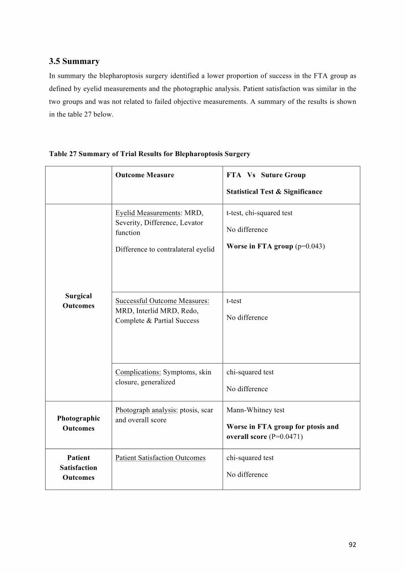
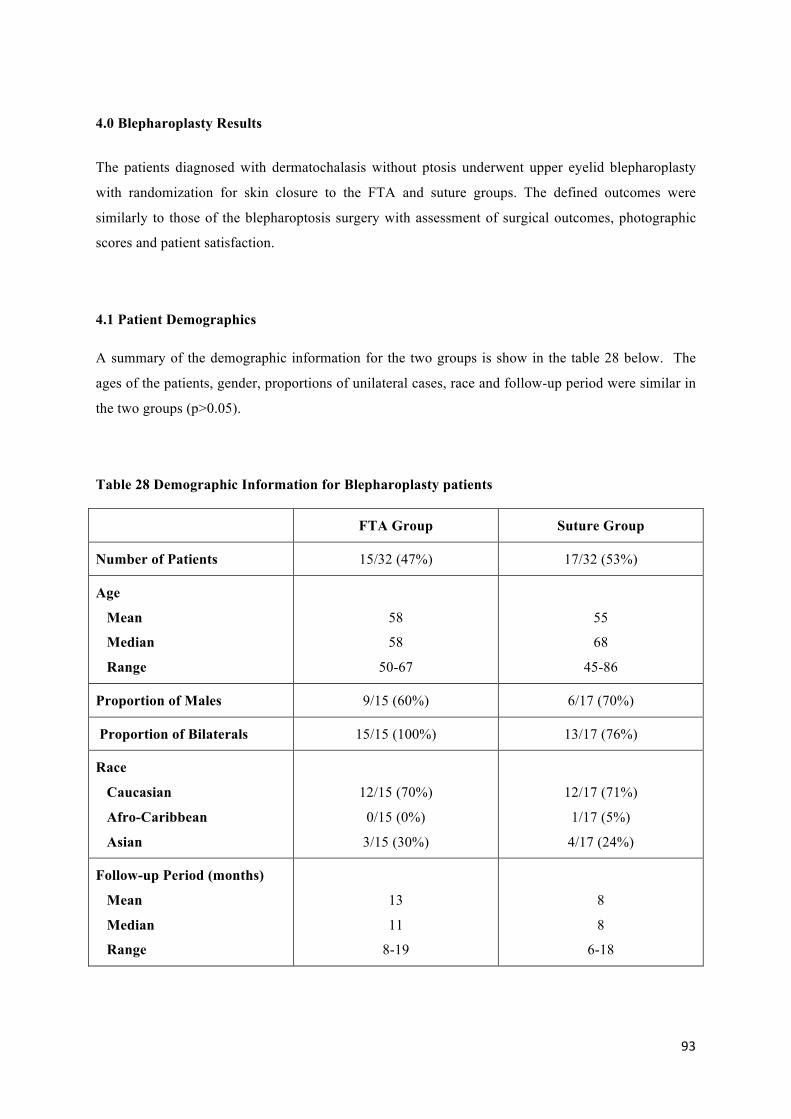
The use of fibrin tissue adhesives in clinical practice has grown over the past 10 years, and there has
been increased use of adhesives in different surgical subspecialties. Conventional suture closure of
periorbital tissues is effective however may result in complications, which has led to the search for
other techniques and innovations. Although tissue adhesives have been used in clinical practice there
is a paucity of randomized controlled studies that have evaluated their advantages and disadvantage in
surgical practice.
The goals of eyelid surgery include the restoration of tissue structure and function while
causing minimal morbidity. The use of a medical product that induces physiological clotting and
fibrin formation is appealing in both theory and clinical practice. However fibrin tissue adhesives
present disadvantages and complications of their own. This thesis set out to evaluate the use of fibrin
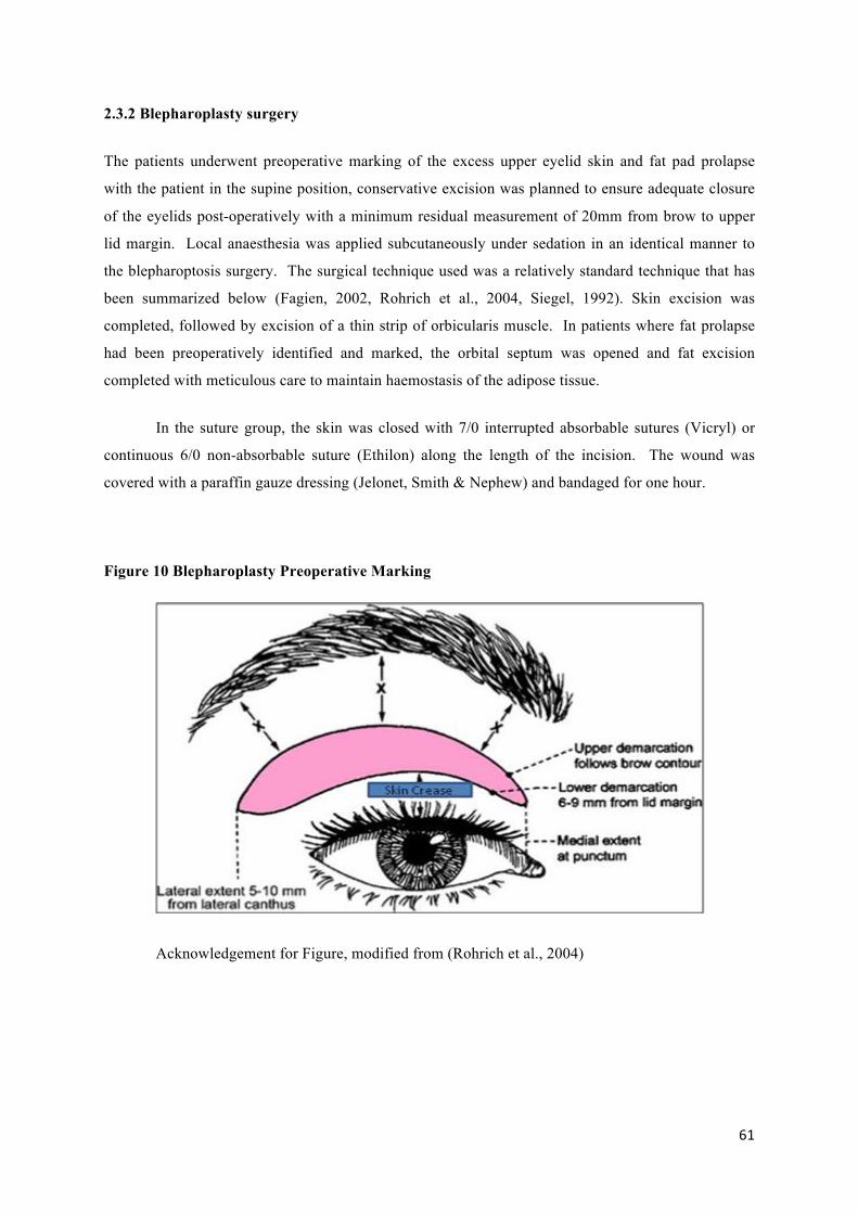
tissue adhesives in eyelid surgery and includes a 5-year randomized control study comparing fibrin
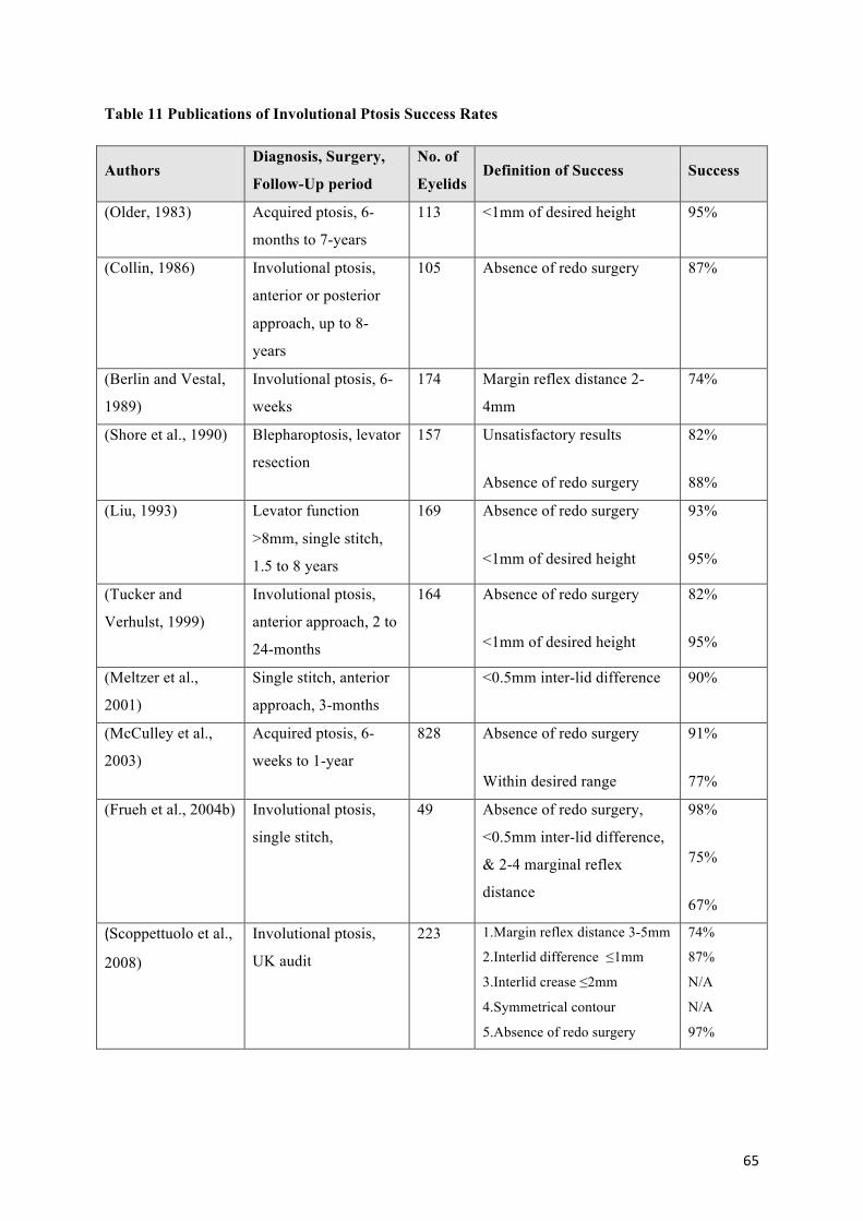
tissue adhesives to suture closure of skin.
A challenge in the evaluation of eyelid surgery is the definition of a successful outcome.
Surgical outcomes have traditionally been measured by surgical complications and the need for
further redo surgery. Other relevant aspects of surgical outcome that have rarely been evaluated
include surgical healing and scar formation, asymmetry that is present however not requiring further
surgery, patient experience and satisfaction. This research set out to further define outcomes for
eyelid surgery to enable a more comprehensive and objective evaluation of surgical outcome.
! 3!
Acknowledgements
I would like to express my gratitude to my supervisors, Dr Jelena Gavrilovic and Miss Jane Olver, for
their support and guidance in this research project.
I would like to thank and acknowledge all the contributors to this multi-centre study, including all the
hospital employees that had a role in collating data and patient care. In particular I would like to thank
Miss Maryam Zamani, Mr Michael Michaelides and Mr Sheng Lim.
I would like to thank my wife, family and friends, for their support in this study that took several
years to complete.
! 4!
Table of Contents!Abstract!............................................................................................................................!2!
1.0!Introduction!..............................................................................................................!11!1.1 Background!........................................................................................................................!11!1.2 Importance of Research!.....................................................................................................!13!1.3 Ptosis!..................................................................................................................................!14!
1.3.1 Anatomy of the Eyelid!........................................................................................................!14!1.3.2 Ptosis Definition!.................................................................................................................!23!1.3.3 Ptosis Causes!.......................................................................................................................!23!1.3.4 Histopathology of ptosis!.....................................................................................................!25!1.3.5 Assessment of Ptosis!...........................................................................................................!26!1.3.6 Clinical diagnosis of ptosis!.................................................................................................!30!1.3.7 Factors affecting upper eyelid position!...............................................................................!31!1.3.8 Treatment of Ptosis!.............................................................................................................!32!
1.4 Upper Lid Blepharoplasty!..................................................................................................!35!1.5 Fibrin Tissue Adhesives!.....................................................................................................!36!
1.5.1 Overview of FTAs!..............................................................................................................!36!1.5.2 Mechanism of Adhesion!.....................................................................................................!36!1.5.3 Adhesive Properties & Use!.................................................................................................!39!1.5.4 FTA Risks & Complications!...............................................................................................!43!1.5.5 Cyanoacrylates!....................................................................................................................!44!1.5.6 Medical use of FTAs!...........................................................................................................!44!
1.6 Surgical Outcomes!.............................................................................................................!46!1.7 Research Objectives!...........................................................................................................!47!
1.7.1 Objective I (Surgical outcomes and complications in the FTA and suture groups)!............!47!1.7.2 Objective II (Patient satisfaction in the FTA and suture groups)!........................................!47!1.7.3 Objective III (Photograph analysis in the FTA and suture groups)!.....................................!47!1.7.4 Objective IV (Comparison between three outcome measures)!...........................................!47!
2.5 Data Collection & Analysis!................................................................................................!76!2.5.1 Data Collection!...................................................................................................................!76!2.5.2 Statistical Analysis!..............................................................................................................!77!
painful suture removal and scarring (Adams and Feurstein, 1986, Lowry and Bartley, 1994).
Granulomas develop as a foreign body reaction to suture trimmings or other debris trapped in the
wound during closure and present as nodular thickenings beneath the incision site (Bennett and Matas,
1982). In addition sutures provide point fixation that provides focal adhesion and not continuous
adhesion of the wound surfaces that are optimal for vascularisation, reduced haematoma formation
and postoperative scarring (Gibran et al., 2007). FTAs were proposed as an alternative method of
skin closure that triggers a physiological clotting cascade with advantages over conventional sutures
and staples (Greene et al., 1999, Mandel, 1992, Howell et al., 1995). FTAs are available as a two
component system that contains highly concentrated fibrinogen and thrombin. The mixing of these
agents immediately prior to use, promoted fibrin cross-linking resulting in physiological haemostasis
and subsequent wound healing. The adhesive has been used in a number of surgical subspecialties
including skin transplantations following burn injuries, neurosurgical closure of dura following
central spinal fluid (CSF) leaks and the fixation of orbital implants. Studies have reported advantages
including improved wound healing, reduced surgical time, complications and reduced pain compared
to conventional sutures (Greene et al., 1999, Mandel, 1992, Howell et al., 1995). Anecdotally studies
have reported improved healing with the use of FTAs although objective evidence and high quality
research studies have been absent (Foster et al., 2006). There have been notable disadvantages that
have included additional cost, risk of dehiscence and the potential risk of infection from the FTA
product that is derived from human plasma. There have been few high quality studies evaluating the
use of FTAs and no prospective randomized studies evaluating the technique following eyelid
surgery.
!!
12!
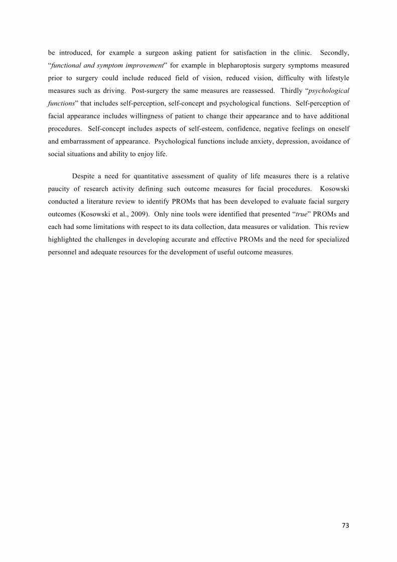
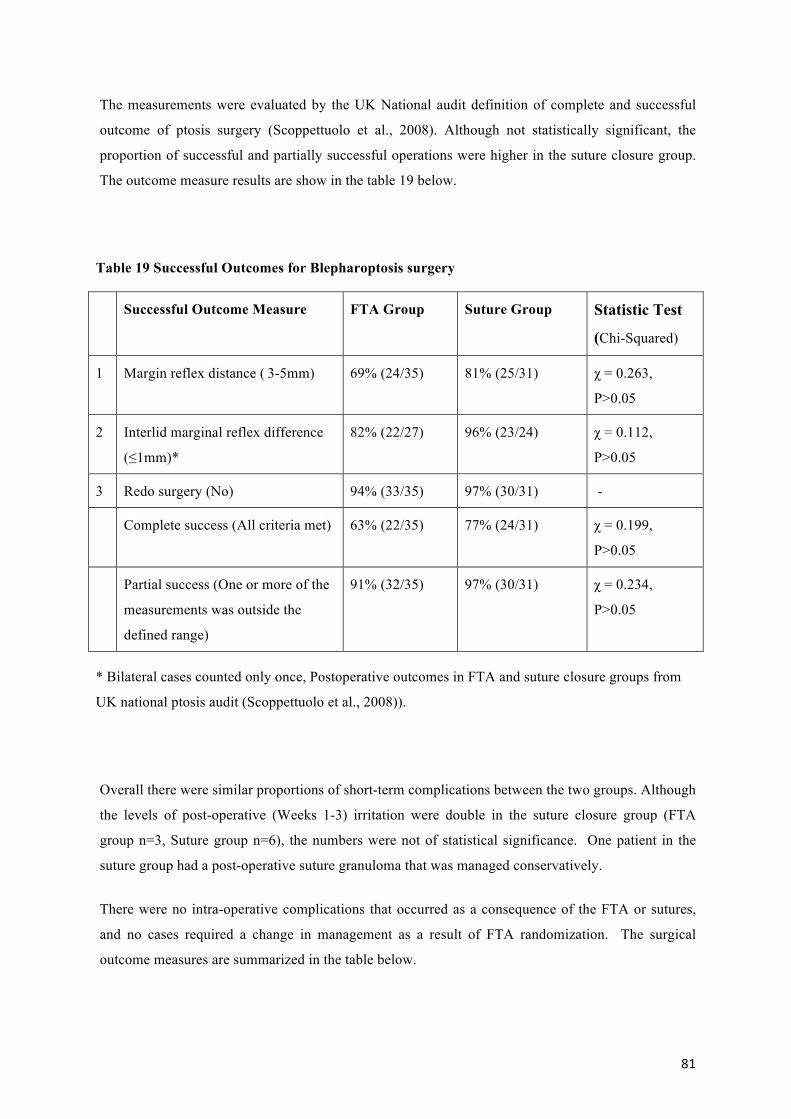
Successful outcome following eyelid surgery has been frequently defined by surgical
outcomes that highlight complications such as infection, delayed healing and the need for redo
surgery. However these outcome measures often fail to account for important factors that impact
surgical outcomes including facial symmetry, scarring and patient satisfaction. The proportion of
redo cases are not representative of surgical failure as not all patients need or are prepared to undergo
further surgery despite an unsatisfactory outcome. The importance of outcome measures has been
highlighted by Darzi’s report of “High Quality Care for All” (Darzi, 2007). The importance of Patient
Reported Outcome Measures (PROMs) have been recognized as an essential component of measuring
quality of care and for many surgical procedures including oculoplastic operations remain
undeveloped (Block, 2006, Langley, 1998, Walburg, 2006). An objective of this study was to further
define outcomes for eyelid surgery with particular focus in three specific areas; objective surgical
measurements, patient satisfaction and the evaluation of standardized postoperative photographs.
!!
13!
1.2 Importance of Research
The healing response following eyelid surgery and resulting scar formation has marked implications
to surgical outcomes and patient satisfaction. Conventional suture closure of the skin has been
associated with complications related to the non-physiological constitution of the suture material.
FTAs mechanism of action is to trigger the physiological clotting cascade and result in fibrinogen
formation. There are very few randomized studies evaluating the use of FTAs and limitations of past
studies include both confounding variables and subjective outcome measures. This study set out to
evaluate the use of FTA in a randomized control study with a series of objective measures. This is the
first randomly controlled study to evaluate the use of fibrin tissue adhesive skin closure for eyelid
surgery.
The outcomes following eyelid surgery have traditionally evaluated measurements of the
eyelids and re-operation rates, these outcomes provide an incomplete evaluation of surgical outcome.
Patient satisfaction and the masked observation of standard photographs provide a more objective
assessment of outcome. This study evaluated three patient outcomes following eyelid surgery with
the objective of developing a benchmark for future eyelid surgery procedures.
! 14!
1.3 Ptosis
Eyelid surgery was first described over 2000 years ago in the Susruta, an ancient Indian document
written in Sanskrit (Kansupada and Sassani, 1997). Arabian surgeons are known to have cauterized
excess upper eyelid skin to relieve eyelid droop as early as the tenth century (Dupuis and Rees, 1971),
and the modern-day surgical technique is attributed to Costañares (Costañares, 1951, Rohrich et al.,
2004).
1.3.1 Anatomy of the Eyelid
The upper eyelid is a mobile structure that protects the eye from injury and enables the even
distribution of the tears on blinking. The eyelid consists of three principal layers: anterior lamellar
(skin, subcutaneous tissue, orbicularis oculi muscle) middle lamellar (orbital septum) and the
posterior lamellar (tarsal plates, smooth muscle and conjunctiva).
Eyelids
The skin of the upper eyelid is thin and divided by a horizontal furrow termed the superior palpebral
sulcus or “skin crease”, and is formed by insertion of the levator palpebral aponeurosis insertion in the
skin of the upper eyelid (Fengzhi et al., 2009). In general the skin crease is approximately eight to
twelve mm in Caucasian and Afro-Caribbean patients. The upper eyelid meets the lower eyelid at the
medal and lateral canthal angles that when open form an angle of approximately sixty degrees. The
lateral canthus is approximately two millimetres (mm) higher than the medical canthus in Caucasian
and Afro-Caribbean patients (Snell and Lemp, 1998). The normal position of the upper eyelid, when
the eye is open and looking straight ahead, covers the top 2-3mm of the superior cornea. The position
of the lower eyelid is independent of the upper eyelid and usually lies at the edge of the cornea termed
the limbus.
The margin of the upper eyelid is approximately thirty mm in length and two mm in
thickness. At approximately five mm from the medial angle there is a small elevation termed the
papilla lacrimalis, this is the entry to canaliculus lacrimalis and the lacrimal drainage channel. The
eyelashes are short hairs at the margin of the eyelids that curve in an anterior direction, and are
arranged in two to three rows. A sagittal cross-section of the upper eyelids is show in Figure 1.
!!
15!
In Asian patients there is often an absence of the eyelid skin crease, although the most
common cosmetic procedure in Southeast Asia is the creation of an upper eyelid skin crease.
Anatomical studies have identified a lack of the fibrous connection between the aponeurosis and the
skin in the Asian double eyelid. Together with the lower positioned orbital septum, prominent pre-
aponeurotic fat and thick orbicularis oculi result in the distinctive eyelid characteristics that are
different to the Caucasian eyelid (Fengzhi et al., 2009).
The motor nerve supply to the orbicularis muscle in the upper eyelids is from the buccal,
zygomatic and frontal branches of the facial nerve, the branches form superior and inferior plexuses
that innervate the orbicularis muscles (Knize, 2000, Ouattara et al., 2004).
!!
16!
Figure 1 Sagittal Section of the Upper Eyelid
a. Skin. b. Orbicularis oculi muscle. c. Levator palpebrae. d. Conjunctiva. e. Tarsus. f. Tarsal gland. g. Sebaceous gland. h. Eyelashes. i. Small hairs of skin. j. Sweat glands. k. Posterior tarsal glands.
Acknowledgement for Figure: Grays Anatomy (Gray, 1918)
! 17!
Anterior Lamellar
The eyelid skin is the thinnest in the body, beneath the skin is the loose subcutaneous tissue of the
eyelids with increased elastic fibers and minimal fat. The orbicularis oculi muscle is an elliptical
muscle that surrounds the globe it is divided into two principal parts the innermost palpebral part that
is present in the eyelids and an outer orbital part. The function of the orbicularis oculi is to close the
eyelids like a purse string. The muscle is innervated by the facial nerve from temporal and zygomatic
branches on the deep surface from the parotid area and is not injured by transcutaneous eyelid
surgery. The orbicularis muscle is the antagonist to the levator aponeurosis (striated muscle) and
Horner’s muscle (smooth muscle) which opens the upper eyelids (Albert et al., 2008, Snell and Lemp,
1998, Bron et al., 1995, Wolff and Last, 1961).
Middle Lamellar
The orbital septum forms a fibrous divide between the skin and the orbital cavity, the layer is a
continuation of the periosteum at the orbital rim. The septum lies posterior to the medial palpebral
ligament and anterior to the lateral palpebral ligament, and blends with the levator aponeurosis above
the superior tarsal border (Fuchs and Duane, 1924). The orbital septum provides an important
functional barrier in the eyelid that protects the spread of infection from superficial skin tissues to the
orbital cavity. Although preseptal cellulitis is a very common infection of the skin, direct spread
through the septum to the orbital cavity is rare. In addition anatomical variation in the position of the
septum changes the appearance of the periorbital soft tissues. A low riding orbital septum with the
orbital fat advancing forward onto the eyelid results in a “full” appearance of the eyelid, conversely a
high septum may result in a “hollow” appearance of the eyelid as the orbital fat is stopped from
coming forwards (Bron et al., 1995).
Posterior Lamellar
The tarsal plates are a dense fibrous tissue that gives the eyelids a defined shape and structure. The
tarsus in the upper lid measures approximately ten mm in height and twenty mm in length and is
attached to the medially via the medial palpebral ligament to the lacrimal crest and laterally to
Whitnall’s ligament. Attached at the superior edge of the upper tarsus are the smooth muscle fibers of
the levator superior tarsal muscle (Müller’s muscle) and the aponeurosis of the levator palpebral
superioris. Within the tarsus are a series of meibomian glands (tarsal glands) that are modified
sebaceous glands, which consist of a long central canal surrounded by over ten acini. Their oily
secretions reduce the evaporation of tears and their gland orifices exit the eyelid just posterior to the
lashes. The non-keratinzed squamous epithelium of the tarsal conjunctiva is tightly adherent to the
upper eyelid and the thin mucous membrane reflects upwards as the superior fornix.
!!
18!
!!
19!
Levator Complex
The upper eyelid is elevated by two muscles, the levator palpebral superioris (striated muscle) and the
superior tarsal muscle (smooth muscle). The levator palpebral superioris originates from the lesser
wing of the sphenoid bone, and just above the optic foramen it becomes the levator aponeurosis and is
approximately fifty-five mm in length (Finsterer, 2003). The muscle transitions to an aponeurosis
tendon approximately fifteen mm from the superior tarsus, attaching to the superior transverse
ligament of Whitnall which acts as a check ligament of the levator (Anderson et al., 1990, Anderson,
1987, Anderson and Dixon, 1979c). Whitnall’s ligament extends from the trochlea medially to the
lacrimal gland capsule and frontal periosteum laterally, and the ligament changes the direction of
Aponeurotic pull. The aponeurosis inserts into the anterior aspect of the superior tarsus and sends
some fibers to the skin to form the upper eyelid crease. The aponeurosis extends in both medial and
lateral expansions termed horns which may have a pathological role in upper eyelid retraction in
conditions such as thyroid eye disease. The lateral horn indents the lacrimal gland partially dividing
the gland into a smaller palpebral and thicker orbital portion. The lateral horn is attached to the
marginal tubercle of the zygomatic bone and the medial horn fuses with the medial palpebral
ligament. The levator palpebral superioris is innervated by the superior branch of the oculomotor
nerve and elevates the eyelid by twelve to fifteen mm from pull closure to wide-eyed staring (Snell
and Lemp, 1998, Carraway and Tran, 2009).
The superior tarsal muscle (Müller’s muscle) is a vascularised smooth muscle that is
innervated by the sympathetic nervous system. The muscle originates from the inferior surface of the
levator aponeurosis and inserts into the superior tarsal plate. The muscle is approximately twenty by
twenty mm with a one mm tendon, and elevates the upper eyelid by approximately two mm (Cohen,
1972, Finsterer, 2003). Müller’s muscle is responsible for setting the upper lid level in conjunction
with the levator palpebral superioris. Increased stimulation of the sympathetic nervous system such as
stress or adrenaline elevates the eyelid, and corresponding damage to the sympathetic innervation
results in one to two mm ptosis.
!!
20!
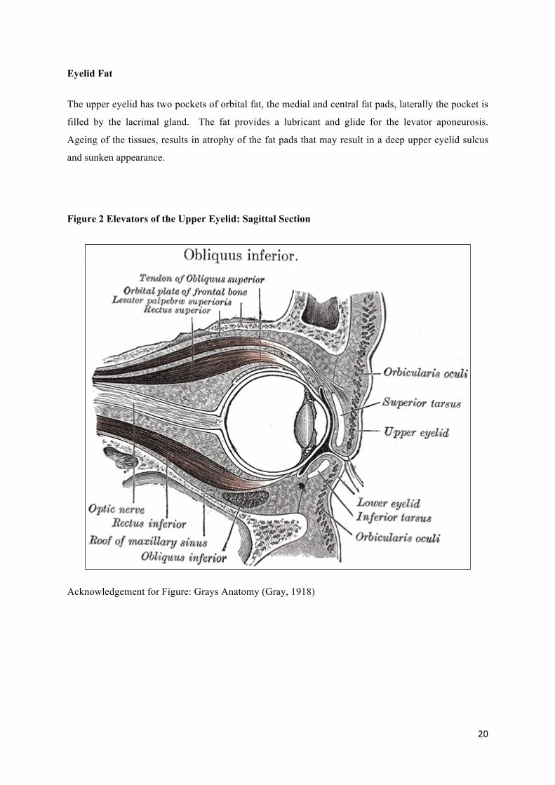
Eyelid Fat
The upper eyelid has two pockets of orbital fat, the medial and central fat pads, laterally the pocket is
filled by the lacrimal gland. The fat provides a lubricant and glide for the levator aponeurosis.
Ageing of the tissues, results in atrophy of the fat pads that may result in a deep upper eyelid sulcus
and sunken appearance.
Figure 2 Elevators of the Upper Eyelid: Sagittal Section
Acknowledgement for Figure: Grays Anatomy (Gray, 1918)
!!
21!
Figure 3 Elevators of the Upper Eyelid: Coronal Section
Acknowledgement for Figure: Grays Anatomy (Gray, 1918)
!!
22!
Blood Supply of the Upper Eyelids
The eyelids have a profuse blood supply from the lateral and medial palpebral arteries that form a marginal and peripheral arterial arch in the upper and lower eyelids. The lateral palpebral arteries are derived from the lacrimal artery and the medical palpebral arteries from the ophthalmic artery. The venous drainage of the upper medial one thirds of the eyelid is to the submandibular glands and from the lateral two thirds to the superficial parotid glands.
Figure 4 Blood Supply of the Upper Eyelids
1. supraorbital artery and vein. 2. nasal artery. 3. angular artery. 4. facial artery. 5. suborbital artery. 6. anterior branch of the superficial temporal artery. 6.’ malar branch of the transverse artery of the face. 7. lacrimal artery. 8. superior palpebral artery. 8’. external arch. 9. anastomoses of the superior palpebral with the superficial temporal and lacrimal. 10. inferior palpebral artery. 11. facial vein. 12. angular vein. 13. branch of the superficial temporal vein.
Acknowledgement for Figure: Grays Anatomy (Gray, 1918)
!!
23!
1.3.2 Ptosis Definition
Ptosis has been defined as drooping of the upper eyelid and may be classified by several different
means including; severity, aetiology, onset and levator function (Beard, 1989). The exact incidence
of ptosis is not known, however the condition is common and has equal frequency among different
races and between the sexes (Finsterer, 2003).
Elevation of the upper eyelid is the result of the levator palpebrae superioris which is a
composite muscle made of a striated muscle, a long aponeurotic tendon and a non-striated
sympathetically innervated (Muller’s) muscle. The eyelid height is affected by local ocular and
orbital tissues, as well as conditions effecting the cranial nerve and sympathetic nerve innervation and
muscles(Jones et al., 1975). Ptosis may result from a condition affecting the eyelid elevator
mechanism, in elderly patients there is attenuation of the insertion of the aponeurosis into the tarsal
plate.
As the upper eyelid descends it covers the superior aspect of the cornea, restricting the
superior visual field. With time the upper eyelid may eventually cover the visual axis resulting in a
reduced visual acuity. Patients often describe some variability of the eyelid height, with worsening at
latter parts of the day that may result from muscular fatigue, myasthenia gravis must be excluded.
1.3.3 Ptosis Causes
Ptosis may be caused by a large number of different aetiologies including age (involutional), birth
defects (congenital), reduced innervation (neurogenic), eydlid lumps (mechanical) or other (e.g.
following intraocular surgery), a comprehensive list is show in Table 1. The diagnosis of involutional
ptosis (also called aponeurotic ptosis) is dependent on the exclusion of other causes of ptosis.
The upper eyelid may have a false appearance of ptosis which is termed pseudoptosis. This
must be excluded for the condition to undergo the correct treatment. Causes of pseudoptosis include
lid abnormalities, abnormal globe position and a small or sunken globe (Table 1).
! 24!
Table 1 Ptosis- Classified by Aetiology
Aetiology of Ptosis
Disorders
Involutional Age-related dehiscence of the levator aponeurosis from the superior tarsal plate
Congenital Defined as the onset of ptosis within the first year of life. Idiopathic, localized myogenic dysgenesis. myogenic, neurogenic, mechanical and other causes
Neurogenic Apraxia of lid opening, blepharospasm (benign, essential), botulinism, botulinum toxin therapy, cerebellar vermis hypoplasia, cerebral tumour, cerebral, ocular, dental, auricular, skeletal anomalies (CODAS) syndrome, cerebral vasculitis and venovascular hypertension, chronic rhinocerebral mucormycosis, cluster headache, cortical dysplasia and maldevelopment of the basal ganglia, facial nerve palsy, hemifacial spasm, Horner’s syndrome, Marcus Gunn jaw-winking syndrome, multiple sclerosis, mycotic meningitis, migraine ophthalmique, optic glioma, orbital dermoidal cyst, oxiliplatin neuropathy, paraneoplastic syndrome, Raeder Syndrome (acquired Horner’s syndrome with ipsilateral headache), Recklinghausen’s neurofibromatosis, rheumatoid pachymeningitis, Riley-Day syndrome, Schwartz-Jampel syndrome, sleep apnea syndrome, stroke (mesencephalic, hemispheric), SUNCT syndrome, syringomyelia, third-nerve palsy (carotid aneurysm, cavernous sinus thrombosis, congenital, degenerative CNS diseases, heavy metal intoxication, increased intracerebral pressure, trauma, superior orbital fissure syndrome, tumours (dermoidal cyst, fibrous tumour, neurinoma, non-Hodgkin’s orbital lymphoma)), vascular lesions, Wernicke’s encephalopathy
Mechanical Scarring, excessive weight—dermatochalasis, eyelid mass (lid tumours: neurinoma, neurofibroma), orbital mass
Traumatic Birth trauma, forceps delivery, corneal abrasion, corneal foreign body, eyelid laceration, hard contact lens embedding, orbital fracture (apex or floor), orbital haemorrhage, post-cataract ptosis, transorbital penetrating brain injury, trauma to the levator aponeurosis
Miscellaneous Anophthalmos, atopic dermatitis, blepharochalasis, blepharophimosis-ptosis-epicanthus inversus syndrome (BPES) due to FOXL2 gene mutations, capillary haemangioma, carotid aneurysm, carotid cavernous fistula, chalazion, chromosome 14q terminal deletion syndrome, combined valproate and hydantoin embryopathy, craniofacial syndromes, de-novo duplication dup (Xq22.1–q25), distichiasis with FOXC2 truncating mutations, double partial monosomy (10p13–10pter and Xp11.4–Xpter), Down syndrome, Duane syndrome, exophthalmos, eyelid oedema, foetal alcohol syndrome, fibrosis syndrome (CFEOM1 locus on chromosome 12), floppy eyelid syndrome, giant papillary conjunctivitis, glaucoma, iris coloboma, hypertelorism, mental retardation due to deletion on chromosome 2, Joubert’s
!!
25!
syndrome, King-Denborough syndrome, lacrimal gland hemangiopericytoma, mandibulofacial dysostosis, mucolipidosis type IV, mycotic aneurysm of the internal carotid artery, oculo-facio-cardiac-dental syndrome, orbital artery obstruction, orbital or preseptal cellulitis, orbital fat prolapse, orbital fibromatosis, orbital Langerhans cell granulomatosis, orbital osteoclastoma, orbital phlegmona, Parry-Romberg syndrome, partial trisomy 1q32–qter _pure,_ poorly fitting ocular prosthesis, Rubinstein-Taybi syndrome, Smith-Magenis syndrome, Smith-Lemli-Opitz syndrome, socket contraction, Sturge-Weber syndrome, supernumery chromosome
Iatrogenic Intra-ocular surgery, contact lens wear, chronic inflammation of the conjunctiva
Pseudoptosis Eyelid abnormalities (ipsilateral excess eyelid skin termed dermatochalasis, contralateral eyelid retraction), abnormal globe position (down-turning of the eye termed hypotropia) and a small or sunken globe (anophthalmos, enophthalmos, microphthalmos, phthisis bulbi, anisometropia)
Modified from: (Finsterer, 2003, Cetinkaya and Brannan, 2008)
1.3.4 Histopathology of ptosis
Intraoperative macroscopic evaluation of the levator aponeurosis in patients with involutional ptosis
may identify a “normal” appearance of the levator with localized dehiscence or fatty degenerative
change in the anterior part of the levator aponeurosis. These fatty changes may be associated with a
relatively normal skin crease, more marked ptosis in the medial part of the lid (Collin, 1986). One
study retrospectively evaluated the patient that required reoperation and found the rate double in the
patients with fatty levator changes, the reoperation rate was 29% for the fatty-appearing levators and
14% of the normal-appearing levators (Tucker and Verhulst, 1999).
!!
26!
1.3.5 Assessment of Ptosis
The assessment of a patient with ptosis includes establishing the diagnosis, the aetiology and the
corresponding development of a management plan. The evaluation is conducted in a systematic
manner to ensure accuracy and consistency, including history taking, patient examination and where
necessary further investigations.
History Taking
Patients with involutional ptosis often complain of a droopy upper eyelid and a sleepy appearance.
With increasing severity the descent of the upper eyelid position occludes the superior visual field and
eventually the pupillary axis. Patients may tilt their head backwards and adopt a chin-up position to
improve their sight or use a finger to physically lift the eyelids. The restriction of superior visual field
may interfere with the patient’s lifestyle, as patients may have difficulty with driving, reading and
going up stairs. Over time, patients may use their forehead muscles to elevate the eyelids this results
in elevation of their eyebrow and may result in an unusual “surprised” appearance.
History taking must include family history, generalized systemic conditions (e.g. diabetes
mellitus, cardiovascular disorders) and specific neurological disorders (including symptoms of
fatigue, weakness, dysarthria and dysphasia). In addition should include the time of onset, associated
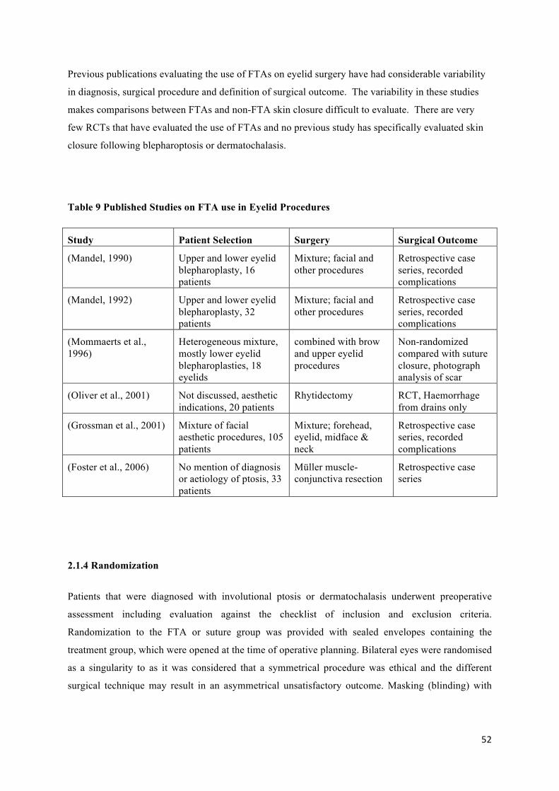
Non-randomized compared with suture closure, photograph analysis of scar
(Oliver et al., 2001) Not discussed, aesthetic indications, 20 patients
Rhytidectomy RCT, Haemorrhage from drains only
(Grossman et al., 2001) Mixture of facial aesthetic procedures, 105 patients
Mixture; forehead, eyelid, midface & neck
Retrospective case series, recorded complications
(Foster et al., 2006) No mention of diagnosis or aetiology of ptosis, 33 patients
Müller muscle-conjunctiva resection
Retrospective case series
2.1.4 Randomization
Patients that were diagnosed with involutional ptosis or dermatochalasis underwent preoperative
assessment including evaluation against the checklist of inclusion and exclusion criteria.
Randomization to the FTA or suture group was provided with sealed envelopes containing the
treatment group, which were opened at the time of operative planning. Bilateral eyes were randomised
as a singularity to as it was considered that a symmetrical procedure was ethical and the different
surgical technique may result in an asymmetrical unsatisfactory outcome. Masking (blinding) with
!!
53!
respect to treatment was not possible following surgery because the distinctive attributes of treatment
modalities.
2.1.5 Sample Size Justification
Sample size justification was completed using the most recently published FTA trial when the study was designed in 2002 (Table 9). No randomized control study has not been published that provided a measure of outcome in both FTA and no FTA groups. It was recognised that haemorrhage drained was not an ideal indicator of sample size as the level of haemorrhage in blepharoplasty surgery is generally very small, the measure provided the most objective measure available for the power calculation.
FTA No FTA
Total haemorrhage drained/ mls
Mean 10 30
Standard deviation 21.16 37.74
OLIVER et al. 2001. A prospective, randomized, double-blind trial of the use of fibrin sealant for face lifts. Plast Reconstr Surg, 108, 2101-5, discussion 2106-7
Statistical calculation of patient numbers:
Haemorrhage drained mean (sd)
With use of FTA 10 (21.2)
No FTA 30 (37.7)
Using sample size formulae m= 2x[z(1-a/2)+ z(1-b)]2/ (delta)2
Calculating values for a significance level of 5% ( z(1-a)2 = 1.96 ) and a power of 80%
m= 2x [1.96+1.2816]2/ (sd)2
m=24
!!
54!
2.2 Fibrin Tissue Adhesive Preparation
The Tisseel FTA was available as a commercial kit and was prepared in accordance with the
manufacturer’s instructions. The Tisseel kit was stored between 2 to 8°C before use. The product
was prepared approximately 20minutes before use, with the components stored in four separate vials;
2 vials of solvents and 2 vials of powders. The Tisseel protein concentrate (containing human
fibrinogen, plasminogen, plasma fibroectin, factor XIII) was reconstituted in the fibrinolysis inhibitor
solution (bovine aprotinin). The dried human thrombin was reconstituted in the calcium chloride
solution.
Table 10 FTA Constituents
Vials Components
Constituent 1
Protein concentrate
Fibrinogen, Plasminogen,
fibronectin, Factor XIII
Fibrinolysis inhibitor solution Aprotinin
Constituent 2 Dried protein Thrombin
Solution Calcium chloride
The Tisseel kit was prepared approximately twenty minutes before use. The two components were
preheated (10 minutes) before being mixed in a magnetic spinner on a heating plate (15minutes) and
then inserted into a specialized “Duploject” syringe (Hyland Division, Baxter Laboratories
Corporation) (Figure 6). The syringe mixed the components at its tip for application to the two
wound surfaces. Once mixed the sealant takes only one minute to activate and three minutes to
solidify. The adhesive is only prepared immediately prior to use, and the skin tissues fully prepared
before application of the FTA (Giampapa and Bitar, 2002). The thrombin comes in two
concentrations (500IU and 41IU/ml) these result in a fast and slow setting solution respectively. The
fast setting solution was used for skin closure and previous reports of the slow setting mixture have
suggested it is unreliable for skin approximation (Mommaerts et al., 1996).
!!
55!
Figure 6 FTA Preparation
Upper: Preheating of constituents, Centre: Reconstitution of aprotinin, Lower: Insertion of syringe into Duploject
!!
56!
2.3 Surgical Procedure
2.3.1 Blepharoptosis surgery
The blepharoptosis was performed by a senior oculoplastic fellow under the supervision of a single
consultant (JMO) by a previously reported technique (Anderson and Dixon, 1979b, Anderson and
Dixon, 1979a, Mandel, 1990, Mandel, 1992, Linberg et al., 1988). All the procedures were performed
under local anaesthesia with sedation as this enabled lid height, contour and symmetry to be adjusted
using patient cooperation during the operation. The local anaesthesia use was combination of
lidocaine 1% and bupivocaine 0.25% with 1:200 000 adrenaline. The bupivocaine has a longer half-
life and provides postoperative analgesia. The low concentration adrenaline reduced local
haemorrhage and aids in prolonging the effect of the local anaesthesia. A small volume of local
anaesthesia (<1 ml) was injected subcutaneously, care was taken to ensure the anaesthesia was not
injected deeply through the orbital septum as it may alter the levator superioris and Müller’s muscle
action. In the past, patients had surgery under general anaesthesia with a predefined “cookie-cutter”
surgical technique that had limited results. To increase the postoperative predictability and
consistency local anaesthesia was adopted as it enabled voluntary involvement of the patient to adjust
lid height intraoperatively.
The surgical technique has been previous described by Anderson and Dixon (Anderson and
Dixon, 1979b, Anderson and Dixon, 1979a), a summary is described below: Preoperative marking of
the skin crease and excess skin was conducted with the patient in the supine position. A skin crease
incision was made and the excess skin and underlying orbicularis muscle excised as a single crescent
shaped flap (Figure 6). The orbicularis was then tented upwards and a full thickness incision made
through the orbicularis to the anterior tarsal surface with the scissors perpendicular to the tissues. The
orbicularis incision was then completed with a minimal number of cuts to reduce unnecessary
haemorrhage from multiple incisions. Blunt dissection with a cotton bud was used to identify the
orbital septum and levator aponeurosis. The orbital septum was opened and to aid identification of
the levator aponeurosis the patient was asked to look up and down which corresponded to movement
of the aponeurosis. If the fat pads were found to have prolapsed on preoperative examination they
were identified, freed and excised with meticulous attention to haemostasis. The aponeurosis edge
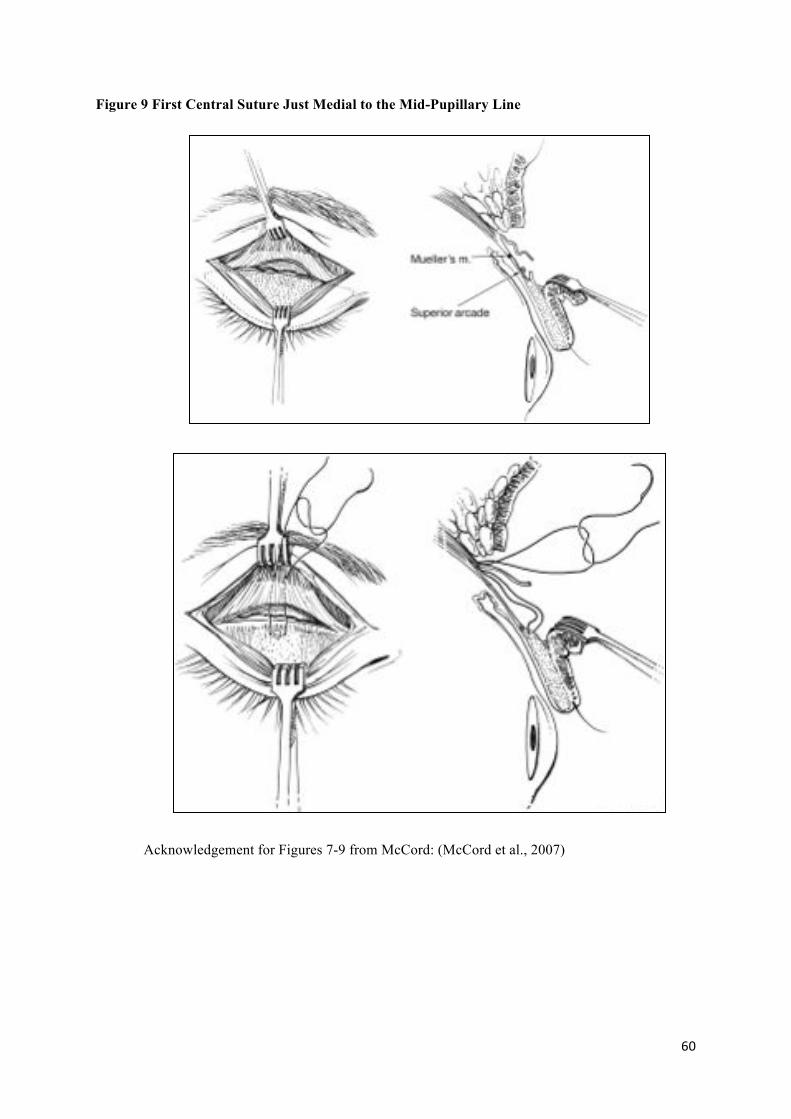
and posterior surface was released and the aponeurosis was reattached to the mid-tarsus with three 6/0
absorbable mattress sutures (Vicryl, polyglactin 910) (Figure 7). With each suture the eyelid was
everted to ensure partial thickness tarsal bites to avoid secondary corneal erosions. The first central
suture was placed just medial to the mid-pupillary line (highest point of eyelid) and two subsequent
sutures medial and lateral to the first (Figure 8). The eyelid height and contour were checked with the
patient looking in up and downgaze before the sutures were tied off, with the aim of 1mm
overcorrection as there is usually a small drop of 1mm eyelid height in the postoperative period as a
!!
57!
consequence of the adrenaline and local anaesthetic effects having worn off. If the eyelid height was
too high or too low, or the eyelid contour unsatisfactory, the suture positions on the levator or tarsus
were adjusted accordingly. The skin crease was internally reformed using three 6/0 interrupted
sutures (Vicryl) from the edge of the levator aponeurosis then through the lower orbicularis but not
through the epidermis. The skin was then closed in accordance with its preoperative randomization to
A randomised prospective study to evaluate blepharoplasty skin closure by tissue adhesive in comparison with conventional suturing techniques.
What do you regard as the most important ethical issue that necessitates review of your project by the LREC?
Approval of tissue adhesive use for skin closure in blepharoplasty.
Is the research being done at other centres? NO
If YES, where else is it being done?
Is St Marys the Lead Centre? YES
If NO, who is the lead centre?
Main research question:
Identify the advantages of blepharoplasty skin closure using tissue adhesive over conventional suturing techniques.
Brief methodology:
1. Pilot Study of 10 patients to identify number of patients required for the prospective
!!
113!
randomised control study. 2. Entry to trial. Patients will be assessed according to a clearly defined protocol
incooperating both inclusion criteria, exclusion criteria, patient information leaflet and written consent.
3. Randomization of patients to tissue adhesive and suturing skin closure groups. 4. Post operative assessment of both all patients at 1 week, 3 weeks and 2 months. 5. Post operative assessment of by standardized photographs and patient satisfaction.
Proposed start date: 2/2003
End date: 10/ 2003
Number of participants/subjects in research:
Pilot study of 10 patients.
Prospective randomized control study, patient no. determined by pilot study.
Brief outcome measure description:
1. Standardized photographs.
2. Patient satisfaction.
Name/address/tel no. of Drug Company sponsor (if applicable):
Principal Investigator(s): Miss Jane Olver and Sheng Lim
!!
114!
Name Signature Designation
All other Investigator(s): D Julian De Silva and Doris Zuercher
Name Signature Designation
Head of Dept/consultant/GP/Community Physician, etc, in overall charge if different from above: Miss Jane Olver
Name Signature Designation
Name, address, tel. No, fax No & Email of investigator to whom all correspondence will be sent:
Miss Jane Olver, Consultant Oculoplastic Surgeon
Western Eye Hospital, London, NW1 5YE
Tel. No. 0207 886 3265
3. AIMS OF PROJECT:
This study aims to compare the success rates of skin closure following blepharoplasty surgery, by tissue adhesive versus conventional suturing techniques.
4. BACKGROUND OF PROPOSED STUDY:
Blepharoplasty is one of the most commonly performed functional as well as cosmetic ophthalmic plastic surgical procedures. The operation may be used to remove skin and lid obstruction of the visual field and to remove involutional change in the eyelids secondary to ageing.
The procedure is most commonly performed under local anaesthesia as a day case procedure. The excess skin and skin crease are first marked. An incision is made through the marked skin and the skin and underlying orbicularis muscle excised as a single crescentic flap. The orbital septum is identified and opened. The fat pads visualized and excised with caution to maintain haemostasis. Sutures are then placed in skin, muscle and levator aponeurosis to form the eyelid skin crease. The skin is conventionally closed by suturing technique. Sutures are place along the length of the blepharoplasty wound. Sutures cause an individual tissue healing response and variable scar formation. Patients attend outpatients at 1 week for suture removal, which is an uncomfortable procedure. Patients are then seen for further follow up at 3 weeks and 2 months. The patients are assessed by lid measurements and photography.
!!
115!
We propose the use of a tissue adhesive to close the skin following blepharoplasty. The adhesive will be applied as in conjunction with the manufacturing guidelines to both surfaces of the wound, which are then gently opposed to allow sealing. The adhesive has been used in a number of other oculoplastic procedures such as; entropion skin closure, skin transplants, fixation of orbital implants following enucleation and reconstruction of lacerated lacrimal canaliculi. The use of tissue adhesive will reduce the need for 1 week postoperative suture removal, decrease the length of blepharoplasty operating time and indirectly may reduce waiting list time. Tissue adhesive is likely to generate less inflammatory healing response and may improve the aesthetic appearance. In addition, tissue adhesive may improve patient satisfaction as a combination of these factors.
To date, there have been no randomised prospective comparative studies comparing the use of tissue adhesive and conventional suturing in blepharoplasty skin closure. We have chosen to compare these two techniques and determine whether there is a significant difference between these two treatments using a carefully designed study. A multicenter prospective randomised comparative study design appears to be the best way to answer this question.
References.
Fibrin sealant in Ophthalmic plastic and Reconstructive surgery. FJ Steinkogler, A Kuchar; Fibrin sealing in surgical and nonsurgical fields. Springer-Verlag, Berlin-Heideelberg 1994.
5. DESIGN OF STUDY:
Give a brief description of what will be done and how it differs from normal practice.
1. Entry to trial, according to inclusion criteria.
2. Randomization to suturing and tissue adhesive groups.
3. Surgery; the patients will undergo the same blepharoplasty procedure. Only the skin closure will differ. The patients will either undergo suture skin closure or tissue adhesive closure.
4. All patients will be followed up at 1 week, 3 weeks and 2 months. The sutured skin closure groups will have skin sutures removed at 1 week.
5. All patients will be assessed in outpatients according to a standardized protocol and patient satisfaction.
6. POTENTIAL BENEFITS AND HAZARDS: If the patient is to be given a placebo or to be deprived of active treatment, or if the patient’s regular treatment of known efficacy is to be changed for the purpose of the study, describe the justification for these intentions.
!!
116!
For questionnaire studies, state what steps are to be taken to ensure reliability and to minimise anxiety or embarrassment.
No patients will be deprived of treatment or given a placebo in this study.
b. Name & address of responsible organisation if not St Mary’s NHS Trust, or ICSM (Remember you need the approval of the establishment before starting the research)
8. RECRUITMENT OF SUBJECTS:
Patients presenting to the participating hospitals requiring blepharoplasty and fulfilling the inclusion criteria will be invited to take part in the study.
a. Will they be patients, staff, students or other volunteers?
Patients.
Record inclusion and exclusion criteria.
Inclusion Criteria
1. Patients requiring blepharoplasty procedure.
2. Informed consent obtainable.
3. Patient clinical follow up 12 months after surgery.
!!
117!
Exclusion Criteria
1. Previous blepharoplasty surgery that has failed requiring redo surgery.
2. History of previous lid surgery. E.g. Tumour excision, ectropion, entropion.
3. Complicated blepharoplasty requiring multiple operations or other procedures. E.g. Reconstruction.
4. Patients with only one functional eye.
5. Patients medically unfit for surgery.
Record any ethnic or social class implications. Nil
How many will be recruited? To be calculated following pilot study.
How is recruitment to be achieved?
Patients requiring blepharoplasty procedure from the oculoplastic clinics at the participating hospitals who meet the entry criteria will be invited to take part in this study. If they agree, they will be entered into the study and randomised. A standardised pre-operative assessment form will be completed, written informed consent obtained and a patient information sheet given to the patient.
Will medical/nursing staff or students be involved as volunteers?
No
b. If recruiting patients who are not your direct clinical responsibility, has the permission of the consultant in charge or the co-ordinator of research in your patient group been obtained?
Not Applicable
Name Signature
c. Is the patient’s GP to be consulted over an individual’s recruitment?
!!
118!
YES
At what stage will the GP be informed?
Entry to trial.
Do you intend to send the GP a copy of the patient information sheet?
YES
If you don’t intend to inform the GP, state why not:
d. Will recruits be paid an honorarium? NO
If YES: how much?
e. Will travelling expenses be reimbursed: NO
If NO please give reasons
Travelling to and from hospital for both surgery and follow up appointments remains unchanged from conventional surgical management of blepharoplasty. No funding available for patient travelling expense.
9. ADMINISTRATION OF STUDY
a. Insurance / Indemnity cover
What arrangements will be in place to cover subjects/patients
!!
119!
(If you are unsure about this please contact Donna Twyman, Research & Contract Office, Medical School, W2 Ext 020 7594 3664)
b. If this is a drug study, at what stage is this in its evaluation?
c. Is this drug being supplied by a company with a clinical trial certificate in response to an investigator with a clinical trial exemption.
NO
d. If the drug is licensed but being used in a non-licensed context which is not being sponsored by the pharmaceutical company concerned, investigators must obtain a DDX from the Medicine Control Agency (020 7273 0327/8). Clinical Research must not be undertaken in patients unless a CTX or DDX is in operation.
NO
Give the Clinical Trial Certificate (CTC) or Clinical Trials Exemption (CTX) numbers if relevant.
e. If this is a company sponsored trial, are the investigators free to publish their results (subject to a reasonable period of consultation with the company)?
NO
g. If any form of radiation is to be used (eg. X rays, radioactive isotopes, heat, UV, laser, etc) this form must be signed by the Radiation Protection Advisor, or a separate letter attached.
Not applicable.
Name: Signature:
10. SUBSTANCES TO BE ADMINISTERED. The Committee must be informed immediately of any severe or unexpected adverse side effects.
!!
120!
a. Please give details of substance to be administered, route, amount, frequency, risks to subject and others, and side effects.
Tisseel adhesive.
A thin layer of Tisseel adhesive is applied proportional to the wound area.
The wound is help clamped for 3-5 minutes to allow adequate time for setting.
Side effects include thromboemoblic complications and allergic reactions. The Tisseel adhesive contains extracts of human plasma and although extensively screened , the transmission of infective agents cannot be totally excluded. No cases of such transmission have been reported.
11. WHAT WILL BE DONE TO SUBJECTS BECAUSE THEY ARE TAKING PART IN THE STUDY?
Describe briefly under headings below, what will be required of subjects; indicate if anything is additional to normal clinical management; indicate discomfort and risk to subject & others.
a. Are any treatments or procedures being withheld, which would otherwise be given?
NO
b. Samples to be taken: NIL
c. Tests to be undertaken: (Please circle appropriate test and give details)
Photographs- Standardized to enable a comparison of adhesive and sutures outcomes.
Hospital admissions for purposes of project: Day case surgery
Outpatient visits: 1 weeks, 3 weeks and 2 months.
Describe what results you expect and how they will be analysed.
The results of blepharoplasty skin closure will be compared between the tissue adhesive and the suturing closure techniques. We hypothesize:
1. Tissue adhesive to reduce operating time for blepharoplasty procedure.
2. Tissue adhesive to reduce the number of follow up appointments.
3. Tissue adhesive to improve aesthetic appearance of blepharoplasty result.
4. Tissue adhesive to improve patient satisfaction.
!!
122!
List discomfort, inconvenience, possible side effects and dangers, untoward signs or symptoms.
We expect discomfort and inconvenience to be unchanged from conventional blepharoplasty surgery, and adhesive may reduce discomfort compared to conventional suturing.
Specific side effects of Tisseel adhesive include thromboemoblic complications and allergic reactions. The Tisseel adhesive contains extracts of human plasma and although extensively screened , the transmission of infective agents cannot be totally excluded.
List precautions which are to be taken with regard to above, and what arrangements will be in place for medical cover. If relevant indicate whether patient information sheet will include name(s) and phone nos. of investigator(s) to be contacted in the event of unexpected reactions of incidents.
Patients will be monitored during and after surgery for both allergic reactions and thromboemobolic complications. Patients will be given specific advice regarding complications and indications to obtain medical help. Contact numbers will be provided.
12. OTHER RESOURCES (Contact your Directorate General Manager to discuss)
a. Will this project make use of hospital resources? (eg,. beds, X rays, NMRI, ECGs, operating time, blood tests, etc?)
NO
b. List departments / Outpatients / Inpatient involvement
Outpatient department – 3 Visits
Photography – 3 Visits
c. How much will they cost?
!!
123!
d. Is the cost being met by a research grant?
NO
e. Obtain signatures of approval from head of each department involved
Name Signature
f. If a compound/drug/device is to be used/tested as part of the study, state the source of funding for its provision.
Tisseel adhesive, provided by Baxter Healthcare Ltd.
g. Will a questionnaire be used?
YES
If YES, and less than 4 A4 sheets, attach a copy with each form copy. If questionnaire is standard, validated, and / or longer than 4 sheets send 2 copies only.
f. Will a semi-structured interview be used?
NO
!!
124!
13. HAVE YOU HAD STATISTICAL ADVICE?
YES
a. From whom did you get it?
b. …in preparing the protocol? NO
c. …in designing the analysis? NO
d. …in deciding the power of the study and number of subjects needed?
YES
14. SENIOR NURSE OUTPATIENT / WARD
The senior nurse should be supplied with a copy of patient information sheet relating to studies on patients under her supervision.
a. Do you plan to ensure this is achieved? YES
15. CONFIDENTIALITY
a. What steps will be taken to safeguard the confidentiality of patients’ records?
!!
125!
Patient records for the trial will be stored separate to conventional hospital notes in a secure location in the Western Eye and Charring Cross Hospitals. Access to the records will be limited to staff associated to the study.
b. Is data to be recorded automatically?
Data is to be recorded according to standardized protocols. These will include both outpatient assessments and photographic records.
If non coded information is being collected, provide copy of your data registration form. It is necessary to comply with the requirements of the data if in doubt contact District Data Protection Officer (020 7594 5535)
c. If the study is a company sponsored trial, will the company require access to the patients’ notes? YES
If YES provide documentation to the effect that confidentiality will be respected.
16. CONSENT AND PARTICIPANT INFORMATION SHEET
Inadequate or incomprehensible information is the most common reason for delay in projects being approved by the LREC. Information for participants must be fully comprehensible by lay individuals. Read the Guidelines carefully and make sure your sheet addresses appropriate headings, eg opt out clause, researcher’s name/tel no., invite to do research, risks and benefits, etc.
a. IS CONSENT REQUIRED? YES
If YES, will consent be: WRITTEN – Customised form.
If WRITTEN is the LREC Consent form to be used? If you are customising this form please send a copy with each application form copy.
!!
126!
If NO, explain why consent is not required, or explain any difficulty that might arise in obtaining consent.
c. IS A PATIENT INFORMATION SHEET TO BE MADE AVAILABLE?
YES / NO If YES please enclose a copy with each application form copy.
Consult the guidelines carefully for necessary headings.
* Ensure this includes statements to the effect:
* Entry to the study is entirely voluntary
* Failure to enter, and subsequent decision to withdraw from the study will not effect the patient’s medical care.
* Paragraph about indemnity cover is included: (eg. ABPI Guidelines for drug sponsored studies)
* Risks and benefits
c. What arrangements will be made for subjects for whom English is not a first language?
Patients who are unable to comprehend a basic understanding of the English language are not appropriate for local anaesthesia technique under conventional suturing or tissue adhesive technique.
d. Who will obtain consent?
Doctors directly involved in the project, who have received detailed training in the protocol.
!!
127!
e. Will participants be informed of the nature and risks of their participation?
Patients will be informed of the principles of the trial in a patient information sheet.
f. I / we confirm that the following will be placed in the patient’s records and in the case of research volunteers these will be held by the named investigator for the study:
* the signed consent form: * patient information sheet:
Name(s) of those who will be obtaining consent Signature:
17. PAYMENTS / SPONSORSHIP
a. Are any / all of the investigators in receipt of any payments / sponsorship?
NO
b. Who is funding the investigation?
Baxter Healthcare Ltd.
c. How much money may be provided for this project alone? Give details, specifying whether this funding is part of a larger sum granted for a number of projects.
18. WILL THE INVESTIGATOR(S) / DEPARTMENT RECEIVE GRANTS/PAYMENTS/SPONSORSHIP FOR THE WORK UNDERTAKEN?
!!
128!
YES
a. How is the money to be spent? (List major items of equipment, staff, etc)
Tisseel tissue adhesive kits + Surgical equipment
Digital Camera + Photography
Medical Records + Data Storage
Stationary + Secretarial Time
b. Please give details of any other related payments
19. WHAT PROBLEMS MAY HINDER A SUCCESSFUL COMPLETION OF THIS STUDY? (This may include ethical problems that may arise during the course of the project).
NIL
20. OTHER FACTORS Please indicate any other factors relevant to approval from LREC.
Please send 11 photocopies of this application form + additional information as specified, to:
Rosalind Cooke, Mailbox 121, R&D St Marys Hospital, Praed Street, London W2 1NY
Tel: 020 7886 6514 fax: 1529
!!
129!
6.2 Appendix 2- Patient consent form
Clinical study comparing two types of skin closure following blepharoplasty. (COREC number)
1. This study compares 2 different skin closure techniques; Conventional suture closure of skin and tissue adhesive closure.
2. I confirm that I have read and understood the information sheet for patients relating to the above study and have had the opportunity to ask questions.
3. I understand that my participation is voluntary and I am free to withdraw at any time, without giving reasons, without my medical care or legal rights being affected.
4. I understand that sections of my medical notes may be looked at by responsible individuals from within this hospital or from regulatory authorities where it is relevant to my participation in research. I give permission to these individuals to have access to my records.
5. I give permission for information which is collected in the above study to be stored both as paper and electronic records. I also give permission for this information to be analysed as part of this research.
6. I agree to participate in the above study.
________________________ Signature of doctor obtaining consent
________________________ Name of doctor
________________________ Date
________________________ Signature of patient
________________________ Name of patient / guardian
________________________ Date
!!
130!
!!
131!
6.3 Appendix 3- Patient Information Sheet
Patient information sheet : Blepharoplasty study
Thank you for considering participation in the blepharoplasty study.
Purpose of study
This study compares two surgical techniques in closure of the wound following blepharoplasty.
Why have I been selected ?
You have an eyelid/ s which requires surgery. Surgery is the only method by which the shape of your eyelid may be changed. The surgical method to do this is well established. We would like to compare the results of closure of the wound following blepharoplasty.
1. Traditionally the wound is closed using sutures. These sutures are then removed one week after the operation.
2. A tissue adhesive is available that can seal the wound in a few minutes avoiding the need for sutures and one week suture removal. Tissue adhesive has been used by plastic surgeons for a number of years to close skin with good cosmetic results.
The blepharoplasty surgery technique would be unchanged except for wound closure at the end of the operation. We would like to find out if there is a difference between the two methods that would justify using only one method in the future. There have not been any studies done before comparing these methods and the answer would help us determine the best way to close skin following blepharoplasty.
Do I have to take part?
You are not obliged to take part. If you decide to take part, you will be given this information sheet and be asked to sign a consent form. The operation is similar to what you would be offered if you
!!
132!
were not part of the study. You are free to withdraw at anytime and without giving a reason. If you decide not to take part, the quality of your care will not be affected in any way.
What is involved if I decide to take part?
Half of the patients taking part in this study will be randomised to have one type of surgery and the other half to the other. The surgery is normally done under local anaesthetic. Whatever surgery you have been allocated to, we will check you eyelid in the clinic at 1 weeks, 3 weeks, and 2 months after the operation. If you notice any problems with your eyelid before or between these scheduled visits then, you should contact one of your treating doctors.
Are there risks in this operation?
The risks of the blepharoplasty operation are recurrence, over or under correction, infection and inflammatory reaction. This operation is not a new procedure and is very safe, however very rarely blepharoplasty may be complicated with blindness. Tissue adhesive has been used for a range of plastic surgery techniques and rarely has been complicated by allergy or an increased risk of blood clots. The tissue adhesive is made from human blood products and is extensively screened for infectious material. No reports of infection spread by tissue adhesive have been reported.
What happens if something goes wrong?
If you wish to complain about the way you have been treated during the course of the study, the normal NHS complaints mechanisms will be available to you. If you are harmed due to someone else’s negligence, then you may have grounds for legal action but you may have to pay for it. There are no special compensation arrangements attached to this study.
Will information on me taking part in this study be kept confidential?
All information collected about you during the course of the study will be kept strictly confidential.
What will happen to the results of the study?
The results of the study will be published after 2 years of you completing your treatment. You will not be identified in any report or publication.
!!
133!
Who has reviewed the study?
The research ethics committee of the NHS Trust of your local hospital has reviewed and approved this study.
For further information, please contact your own Consultant Ophthalmologist or The Project Co-ordinator :
Miss Jane Olver, Consultant Ophthalmologist,
The Western Eye Hospital
Marylebone Road,
London NW1 5YE
Telephone : 020 7886 3264
Fax : 020 7886 3259
!!
134!
6.4 Appendix 4- Information sheet for the patient’s general practitioner
Blepharoplasty surgery : A comparison of skin closure techniques following blepharoplasty, using conventional suturing techniques compared to tissue adhesive. (COREC number)
Dear Doctor,
Your patient has agreed to participate in the above study. It is a multicentre prospective randomised study aimed at comparing the results of suturing and tissue adhesive surgical skin closure.
All patients will undergo blepharoplasty in the same manner. Skin closure will be randomized to conventional suturing and tissue adhesive. Suture closure requires additional operating time, suture removal at 1 week post-operation and may give a variable tissue healing response and resulting scar. Tissue adhesive has been utilized for a number of plastic and oculoplastic procedures with favourable aesthetic results. However, there have been no randomised controlled prospective studies comparing these two procedures.
Our study aims to determine whether there is any significant difference in the success rates of these two operations.
If you have any enquiries about this study, please contact the local Consultant Ophthalmologist in charge of your patient or the Project Co-ordinator :
Miss Jane Olver,
Consultant Ophthalmologist,
The Western Eye Hospital,
Marylebone Road,
London NW1 5YE
Name!:!
Date!of!birth!:!
Hospital!Number!:!
Male!/!Female!
!
!!
135!
Telephone : 020 7886 3264
Fax : 020 7886 3259
!!
136!
6.5 Appendix 5- Operative Record
Operative record
1. Date:
2. Type of surgery(please circle): Suture Vs Adhesive
3. Surgeon (Name and grade) :
4. Study eye (please circle) : Right / Left/ Both
5. Anaesthetic : Local
6. Operative complications (if any):
7. Comments (eg. if procedure or materials different from protocol)
Name!:!
Date!of!birth!:!
Hospital!Number!:!
Male!/!Female!
!
Hospital!:!!!!!(Please!circle)!
!
Western!Eye!Hospital,!London!
Charing!Cross!Hospital,!London!
!
!!
137!
6.6 Appendix 6- Postoperative record
.
1. Date:
2. Type of surgery(please circle): Suture / Adhesive
3. Time after surgery (Please circle) : 1 Week/ 3 Weeks/ 2 Months
4. Study eye (Please circle): Right / Left/ Both
5. Form completed by (Name and grade) :
6. Symptoms (please circle):
Irritation Discharge Pain Other:
Stickiness Foreign body sensation Epiphora
Name!:!
Date!of!birth!:!
Hospital!Number!:!
Male!/!Female!
!
Hospital!:!!!!!(Please!circle)!
!
Western!Eye!Hospital,!London!
Charing!Cross!Hospital,!London!
Raigmore!Hospital,!Inverness!
!!
138!
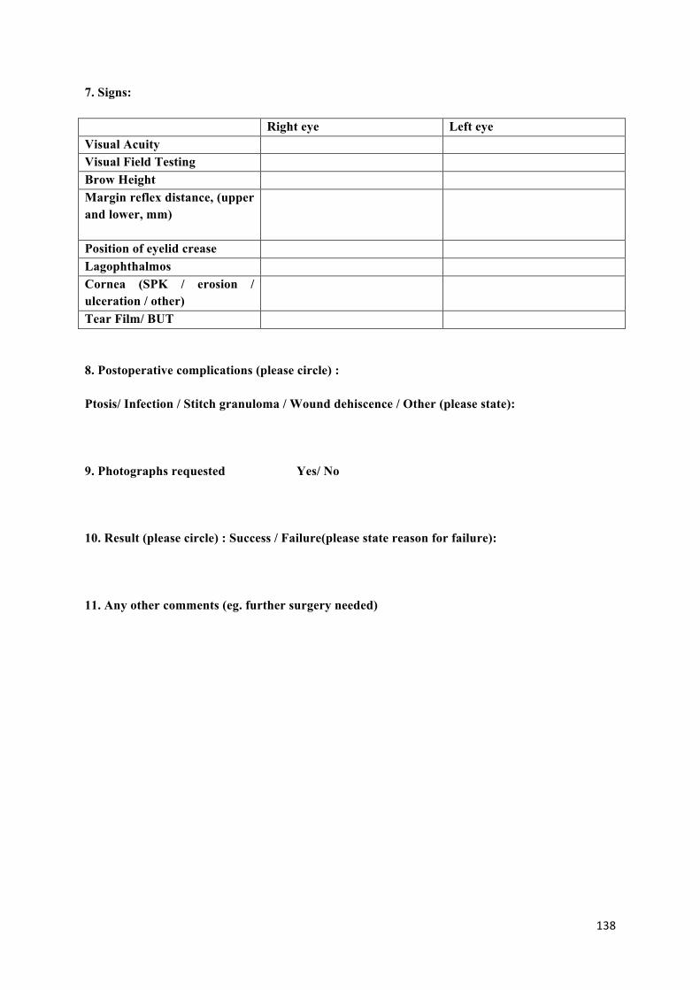
7. Signs:
Right eye Left eye Visual Acuity Visual Field Testing Brow Height Margin reflex distance, (upper and lower, mm)
Position of eyelid crease Lagophthalmos Cornea (SPK / erosion / ulceration / other)
We would appreciate you completing this form so we can assess your treatment completely and aim to improve the oculoplastic service further. Your comments are confidential
Please circle your choice:
Were you happy with your blepharoplasty surgery? Yes/ No
Is there any part of the surgery you were not happy with?
Prior to operation Yes/ No
Operation Yes/ No
After operation Yes/ No
1. How well do you like the appearance of your eyes and eyelids?
Not at all Somewhat Moderately Very much Completely
0 1 2 3 4
2. Do you feel like the appearance of your eyes makes you look tired?
Completely Very much Moderately Somewhat Not at all
0 1 2 3 4
3. How much do you feel your friends and loved ones like the appearance of your eyes?
Not at all Somewhat Moderately Very much Completely
0 1 2 3 4
4. Do you feel the current appearance of your eyes limits your social or professional activities?
Always Usually Sometimes Rarely Never
0 1 2 3 4
!!
141!
5. How confident are you that the appearance of your eyes is the best that it can be?
Not at all Somewhat Moderately Very much Completely
0 1 2 3 4
6. Would you like to surgically alter the appearance of your eyes?
Definitely Most likely Possibly Probably not No
0 1 2 3 4
Other Comments regarding your surgery:
Are there any other ways we could continue to improve the oculoplastic service?
!!
142!
6.9 Appendix 9- Ethics Approval Letter
!!
143!
!!
144!
7.0 ReferencesScientific requirements for Antarctic conservation : Symposium : Papers.
ADAMS, B. J. & FEURSTEIN, S. S. 1986. Complications of blepharoplasty. Ear Nose Throat J, 65, 6-18.
ALBALA, D. M. & LAWSON, J. H. 2006. Recent clinical and investigational applications of fibrin sealant in selected surgical specialties. J Am Coll Surg, 202, 685-97.
ALBERT, D. M., JAKOBIEC, F. A. & MILLER, J. W. 2008. Albert & Jakobiec's principles and practice of ophthalmology, Philadelphia, Pa. ; Edinburgh, Saunders/Elsevier.
ALSARRAF, R. 2000. Outcomes research in facial plastic surgery: a review and new directions. Aesthetic Plast Surg, 24, 192-7.
ALSARRAF, R. 2002. Outcomes instruments in facial plastic surgery. Facial Plast Surg, 18, 77-86.
ALSARRAF, R., LARRABEE, W. F., JR., ANDERSON, S., MURAKAMI, C. S. & JOHNSON, C. M., JR. 2001. Measuring cosmetic facial plastic surgery outcomes: a pilot study. Arch Facial Plast Surg, 3, 198-201.
ANDERSON, R. L. 1987. Whitnall's sling, not a "new procedure". Ophthalmic Surg, 18, 549.
ANDERSON, R. L. & DIXON, R. S. 1979a. Aponeurotic ptosis surgery. Arch Ophthalmol, 97, 1123-8.
ANDERSON, R. L. & DIXON, R. S. 1979b. Neuromyopathic ptosis: a new surgical approach. Arch Ophthalmol, 97, 1129-31.
ANDERSON, R. L. & DIXON, R. S. 1979c. The role of Whitnall's ligament in ptosis surgery. Arch Ophthalmol, 97, 705-7.
ANDERSON, R. L., JORDAN, D. R. & DUTTON, J. J. 1990. Whitnall's sling for poor function ptosis. Arch Ophthalmol, 108, 1628-32.
ASRANI, S. G. & WILENSKY, J. T. 1996. Management of bleb leaks after glaucoma filtering surgery. Use of autologous fibrin tissue glue as an alternative. Ophthalmology, 103, 294-8.
BAHAR, I., WEINBERGER, D., GATON, D. D. & AVISAR, R. 2007. Fibrin glue versus vicryl sutures for primary conjunctival closure in pterygium surgery: long-term results. Curr Eye Res, 32, 399-405.
BATMAN, C., OZDAMAR, Y., ASLAN, O., SONMEZ, K., MUTEVELLI, S. & ZILELIOGLU, G. 2008. Tissue glue in sutureless vitreoretinal surgery for the treatment of wound leakage. Ophthalmic Surg Lasers Imaging, 39, 100-6.
BATMAN, C., OZDAMAR, Y., MUTEVELLI, S., SONMEZ, K., ZILELIOGLU, G. & KARAKAYA, J. 2009. A comparative study of tissue glue and vicryl suture for
!!
145!
conjunctival and scleral closure in conventional 20-gauge vitrectomy. Eye (Lond), 23, 1382-7.
BEARD, C. 1972. Complications of ptosis surgery. Ann Ophthalmol, 4, 671-5.
BEARD, C. 1989. A new classification of blepharoptosis. Int Ophthalmol Clin, 29, 214-6.
BEN SIMON, G. J., LEE, S., SCHWARCZ, R. M., MCCANN, J. D. & GOLDBERG, R. A. 2005. External levator advancement vs Muller's muscle-conjunctival resection for correction of upper eyelid involutional ptosis. Am J Ophthalmol, 140, 426-32.
BENNETT, J. E. & MATAS, J. A. 1982. A minor complication of blepharoplasty. Plast Reconstr Surg, 69, 856-8.
BERKE, R. N. 1959. Results of resection of the levator muscle through a skin incision in congenital ptosis. AMA Arch Ophthalmol, 61, 177-201.
BERKE, R. N. 1971. Surgical treatment of traumatic blepharoptosis. Am J Ophthalmol, 72, 691-8.
BERLIN, A. J. & VESTAL, K. P. 1989. Levator aponeurosis surgery. A retrospective review. Ophthalmology, 96, 1033-6; discussion 1037.
BETHARIA, S. M., GROVER, A. K. & KALRA, B. R. 1983. The Fasanella-Servat operation: a modified simple technique with quantitative approach. Br J Ophthalmol, 67, 58-60.
BIEDNER, B. & ROSENTHAL, G. 1996. Conjunctival closure in strabismus surgery: Vicryl versus fibrin glue. Ophthalmic Surg Lasers, 27, 967.
BLOCK, D. J. 2006. Healthcare outcomes management : strategies for planning and evaluation, Sudbury, Mass. ; London, Jones and Bartlett.
BODIAN, M. 1982. Lip droop following contralateral ptosis repair. Arch Ophthalmol, 100, 1122-4.
BRON, A. J., TRIPATHI, R. C., TRIPATHI, B. J. & WOLFF, E. 1995. Wolff's anatomy of the eye and orbit, London, Chapman & Hall.
BROWN, D. M., BARTON, B. R., YOUNG, V. L. & PRUITT, B. A. 1992. Decreased wound contraction with fibrin glue--treated skin grafts. Arch Surg, 127, 404-6.
BRUCK, H. G. 1982. Fibrin tissue adhesion and its use in rhytidectomy: a pilot study. Aesthetic Plast Surg, 6, 197-202.
BRUCK, S. D. 1978. The effect of the physiological environment on the mechanical properties of biomaterials in cardiovascular applications. Biomater Med Devices Artif Organs, 6, 341-59.
CARLSON, A. N. & WILHELMUS, K. R. 1987. Giant papillary conjunctivitis associated with cyanoacrylate glue. Am J Ophthalmol, 104, 437-8.
!!
146!
CARRAWAY, J. H. & TRAN, P. 2009. Blepharoplasty with ptosis repair. Aesthet Surg J, 29, 54-61.
CARTER, S. R., SEIFF, S. R., CHOO, P. H. & VALLABHANATH, P. 2001. Lower eyelid CO(2) laser rejuvenation: a randomized, prospective clinical study. Ophthalmology, 108, 437-41.
CETINKAYA, A. & BRANNAN, P. A. 2008. Ptosis repair options and algorithm. Curr Opin Ophthalmol, 19, 428-34.
CHAN, S. M. & BOISJOLY, H. 2004. Advances in the use of adhesives in ophthalmology. Curr Opin Ophthalmol, 15, 305-10.
CHVAPIL, M. & KOOPMANN, C. F., JR. 1984. Scar formation: physiology and pathological states. Otolaryngol Clin North Am, 17, 265-72.
CIUCI, P. M. & OBAGI, S. 2008. Rejuvenation of the periorbital complex with autologous fat transfer: current therapy. J Oral Maxillofac Surg, 66, 1686-93.
CLARK, R. A. 1985. Cutaneous tissue repair: basic biologic considerations. I. J Am Acad Dermatol, 13, 701-25.
COHEN, H. B. 1972. Congenital ptosis. A new pedigree and classification. Arch Ophthalmol, 87, 161-3.
COLLIN, J. R. 1979. Complications of ptosis surgery and their management: a review. J R Soc Med, 72, 25-6.
COLLIN, J. R. 1986. Involutional ptosis. Aust N Z J Ophthalmol, 14, 109-12.
COLLIN, J. R. & TYERS, A. G. 1985. Senile ptosis II--posterior approach and complications. Trans Ophthalmol Soc U K, 104 ( Pt 1), 17-21.
COSTAÑARES, S. 1951. Blepharoplasty for herniated intra-orbital fat: Anatomical basis for a new approach. Plast. Reconstr. Surg, 8.
DADEYA, S. & MS, K. 2001. Strabismus surgery: fibrin glue versus vicryl for conjunctival closure. Acta Ophthalmol Scand, 79, 515-7.
DARLINGTON, J. K., LEE, W. B. & SCHWAB, I. R. 2006. Corneal perforation during laser blepharoplasty. Ophthalmic Surg Lasers Imaging, 37, 327-9.
DARZI, A. 2007. High quality care for all: NHS Next Stage Review-final report. http://www.dh.gov.uk/en/Publicationsandstatistics/Publications/PublicationsPolicyAndGuidance/DH_085825. [Accessed 3rd October 2009].
DAWN, A. G. & LEE, P. P. 2004. Patient expectations for medical and surgical care: a review of the literature and applications to ophthalmology. Surv Ophthalmol, 49, 513-24.
!!
147!
DEMERE, M., WOOD, T. & AUSTIN, W. 1974. Eye complications with blepharoplasty or other eyelid surgery. A national survey. Plast Reconstr Surg, 53, 634-7.
DRESNER, S. C. 1991. Further modifications of the Muller's muscle-conjunctival resection procedure for blepharoptosis. Ophthal Plast Reconstr Surg, 7, 114-22.
DUCHESNE, B., TAHI, H. & GALAND, A. 2001. Use of human fibrin glue and amniotic membrane transplant in corneal perforation. Cornea, 20, 230-2.
DUPUIS, C. & REES, T. D. 1971. Historical notes on blepharoplasty. Plast Reconstr Surg, 47, 246-51.
DVORAK, H. F., HARVEY, V. S., ESTRELLA, P., BROWN, L. F., MCDONAGH, J. & DVORAK, A. M. 1987. Fibrin containing gels induce angiogenesis. Implications for tumor stroma generation and wound healing. Lab Invest, 57, 673-86.
EBY, J. B., NAVARRO, R. A., DUNKELMAN, A., LICHTMAN, J., FISHBEIN, M. C., ARONOWITZ, J. A. & KULBER, D. A. 2001. The effect of fibrin sealant on the healing of laser-resurfaced skin. Aesthet Surg J, 21, 509-17.
ELLIS, D. A. & PELAUSA, E. O. 1988. Fibrin glue in facial plastic and reconstructive surgery. J Otolaryngol, 17, 74-7.
ERB, M. H., KERSTEN, R. C., YIP, C. C., HUDAK, D., KULWIN, D. R. & MCCULLEY, T. J. 2004. Effect of unilateral blepharoptosis repair on contralateral eyelid position. Ophthal Plast Reconstr Surg, 20, 418-22.
ERBAGCI, I. & BEKIR, N. 2007. Sutureless closure of the conjunctiva with a commercial fibrin sealant in extraocular muscle surgery for strabismus. Strabismus, 15, 89-94.
ERBIL, H., SINAV, S., SULLU, Y. & KANDEMIR, B. 1991. An experimental study on the use of fibrin sealants in strabismus surgery. Turk J Pediatr, 33, 111-6.
EVERSBUSCH, O. 1883. Zur Operation der congenitalen Blepharoptosis. Klin Monatsbl Augenheilkd, 21, 100-7.
FAGIEN, S. 2002. Advanced rejuvenative upper blepharoplasty: enhancing aesthetics of the upper periorbita. Plast Reconstr Surg, 110, 278-91; discussion 292.
FASANELLA, R. M. & SERVAT, J. 1961. Levator resection for minimal ptosis: another simplified operation. Arch Ophthalmol, 65, 493-6.
FEIBEL, R. M., CUSTER, P. L. & GORDON, M. O. 1993. Postcataract ptosis. A randomized, double-masked comparison of peribulbar and retrobulbar anesthesia. Ophthalmology, 100, 660-5.
FENGZHI, X., WEI, Z., GUO-KANG, F., JIANG, C. & HUA, L. 2009. Double eyelid operation recreating the anatomic microstructure. Ann Plast Surg, 63, 242-8.
!!
148!
FINSTERER, J. 2003. Ptosis: causes, presentation, and management. Aesthetic Plast Surg, 27, 193-204.
FORD, R. C., BACH, S. A. & FOTTLER, M. D. 1997. Methods of measuring patient satisfaction in health care organizations. Health Care Manage Rev, 22, 74-89.
FORSETH, M., O'GRADY, K. & TORIUMI, D. M. 1992. The current status of cyanoacrylate and fibrin tissue adhesives. J Long Term Eff Med Implants, 2, 221-33.
FOSTER, J. A., HOLCK, D. E., PERRY, J. D., WULC, A. E., BURNS, J. A., CAHILL, K. V. & MORGENSTERN, K. E. 2006. Fibrin sealant for Muller muscle-conjunctiva resection ptosis repair. Ophthal Plast Reconstr Surg, 22, 184-7.
FREEMAN, P. D., KAHOOK, M. Y. & CURTIS, T. H. Glaucoma drainage device implantation in children using fibrin glue. J AAPOS, 14, 169-71.
FRUEH, B. R. 1980. The mechanistic classification of ptosis. Ophthalmology, 87, 1019-21.
FRUEH, B. R., MUSCH, D. C. & MCDONALD, H. 2004a. Efficacy and efficiency of a new involutional ptosis correction procedure compared to a traditional aponeurotic approach. Trans Am Ophthalmol Soc, 102, 199-206; discussion 206-7.
FRUEH, B. R., MUSCH, D. C. & MCDONALD, H. M. 2004b. Efficacy and efficiency of a small-incision, minimal dissection procedure versus a traditional approach for correcting aponeurotic ptosis. Ophthalmology, 111, 2158-63.
FUCHS, E. D. & DUANE, A. 1924. Text-Book of Ophthalmology ... Authorized translation ... by Alexander Duane ... Eighth edition, revised, pp. xv. 997. J. B. Lippincott Co.: Philadelphia & London.
GAUSAS, R. E. 1999. Complications of blepharoplasty. Facial Plast Surg, 15, 243-53.
GIAMPAPA, V. C. & BITAR, G. J. 2002. Use of fibrin sealant in neck contouring. Aesthet Surg J, 22, 519-25.
GIBRAN, N., LUTERMAN, A., HERNDON, D., LOZANO, D., GREENHALGH, D. G., GRUBBS, L., SCHOFIELD, N., HANTAK, E., CALLAHAN, J. D., SCHIESTL, N., RIINA, L. H. & GROUP, F. I. C. S. 2007. Comparison of fibrin sealant and staples for attaching split-thickness autologous sheet grafts in patients with deep partial- or full-thickness burn wounds: a phase 1/2 clinical study. J Burn Care Res, 28, 401-8.
GOLDBERG, R. A. 2000. Transconjunctival orbital fat repositioning: transposition of orbital fat pedicles into a subperiosteal pocket. Plast Reconstr Surg, 105, 743-8; discussion 749-51.
GRABOSCH, A., BOGUSCH, K., PLOGMEIER, K. & OLLINGER, R. 1994. Fibrin Sealing in Surgical and Nonsurgical Fields, Berlin, Springer-Verlag.
GRAY, H. 1918. Gray's Anatomy, Descriptive and Applied, London, Longmans, Green and Co.
!!
149!
GREENE, D., KOCH, R. J. & GOODE, R. L. 1999. Efficacy of octyl-2-cyanoacrylate tissue glue in blepharoplasty. A prospective controlled study of wound-healing characteristics. Arch Facial Plast Surg, 1, 292-6.
GREWING, R. & MESTER, U. 1997. Fibrin sealant in the management of complicated hypotony after trabeculectomy. Ophthalmic Surg Lasers, 28, 124-7.
GREY, E. G. 1915. Fibrin as a hemostatic in cerebral surgery. Surg. Gynaecol. Obstet., 21, 452-454.
GROSSMAN, J. A., CAPRARO, P. A. & BURNEIKIS, V. 2001. Minimizing complications in the use of fibrin sealant in aesthetic facial procedures. Aesthet Surg J, 21, 32-9.
GUNTER, J. P. & ANTROBUS, S. D. 1997. Aesthetic analysis of the eyebrows. Plast Reconstr Surg, 99, 1808-16.
HARRIS, W. A. & DORTZBACH, R. K. 1975. Levator tuck: a simplified blepharoptosis procedure. Ann Ophthalmol, 7, 873-8.
HART, C. W., HESKETT, J. L. & SASSER, W. E., JR. 1990. The profitable art of service recovery. Harv Bus Rev, 68, 148-56.
HARTMAN, A. R., GALANAKIS, D. K., HONIG, M. P., SEIFERT, F. C. & ANAGNOSTOPOULOS, C. E. 1992. Autologous whole plasma fibrin gel. Intraoperative procurement. Arch Surg, 127, 357-9.
HASS, A. N., PENNE, R. B., STEFANYSZYN, M. A. & FLANAGAN, J. C. 2004. Incidence of postblepharoplasty orbital hemorrhage and associated visual loss. Ophthal Plast Reconstr Surg, 20, 426-32.
HICKSON, G. B., FEDERSPIEL, C. F., PICHERT, J. W., MILLER, C. S., GAULD-JAEGER, J. & BOST, P. 2002. Patient complaints and malpractice risk. JAMA, 287, 2951-7.
HINO, M., ISHIKO, O., HONDA, K. I., YAMANE, T., OHTA, K., TAKUBO, T. & TATSUMI, N. 2000. Transmission of symptomatic parvovirus B19 infection by fibrin sealant used during surgery. Br J Haematol, 108, 194-5.
HOLLANDER, J. E., SINGER, A. J., VALENTINE, S. & HENRY, M. C. 1995. Wound registry: development and validation. Ann Emerg Med, 25, 675-85.
HORTON, R. 2008. The Darzi vision: quality, engagement, and professionalism. The Lancet, 372, 3-4.
HOWELL, J. M., BRESNAHAN, K. A., STAIR, T. O., DHINDSA, H. S. & EDWARDS, B. A. 1995. Comparison of effects of suture and cyanoacrylate tissue adhesive on bacterial counts in contaminated lacerations. Antimicrob Agents Chemother, 39, 559-60.
HUNT, T. K., KNIGHTON, D. R., THAKRAL, K. K., GOODSON, W. H., 3RD & ANDREWS, W. S. 1984. Studies on inflammation and wound healing: angiogenesis and collagen
!!
150!
synthesis stimulated in vivo by resident and activated wound macrophages. Surgery, 96, 48-54.
ISENBERG, S. F. & ROSENFELD, R. M. 1997. Problems and pitfalls in community-based outcomes research. Otolaryngol Head Neck Surg, 116, 662-5.
JABS, A. D., JR., WIDER, T. M., DEBELLIS, J. & HUGO, N. E. 1992. The effect of fibrin glue on skin grafts in infected sites. Plast Reconstr Surg, 89, 268-71.
JACKSON, M. R. 2001a. Fibrin sealants in surgical practice: An overview. Am J Surg, 182, 1S-7S.
JACKSON, M. R. 2001b. New and potential uses of fibrin sealants as an adjunct to surgical hemostasis. Am J Surg, 182, 36S-39S.
JAGGI, R., HART, R. & TAYLOR, S. M. 2009. Absorbable suture compared with nonabsorbable suture in upper eyelid blepharoplasty closure. Arch Facial Plast Surg, 11, 349-52.
JONES, L. T., QUICKERT, M. H. & WOBIG, J. L. 1975. The cure of ptosis by aponeurotic repair. Arch Ophthalmol, 93, 629-34.
KAHOOK, M. Y. & NOECKER, R. J. 2006. Fibrin glue-assisted glaucoma drainage device surgery. Br J Ophthalmol, 90, 1486-9.
KALAYCI, D., FUKUCHI, T., EDELMAN, P. G., SAWHNEY, A. S., MEHTA, M. C. & HIROSE, T. 2003. Hydrogel tissue adhesive for sealing corneal incisions. Ophthalmic Res, 35, 173-6.
KAMER, F. M. & NGUYEN, D. B. 2007. Experience with fibrin glue in rhytidectomy. Plast Reconstr Surg, 120, 1045-51; discussion 1052.
KANSUPADA, K. B. & SASSANI, J. W. 1997. Sushruta: the father of Indian surgery and ophthalmology. Doc Ophthalmol, 93, 159-67.
KARALEZLI, A., KUCUKERDONMEZ, C., AKOVA, Y. A., ALTAN-YAYCIOGLU, R. & BORAZAN, M. 2008. Fibrin glue versus sutures for conjunctival autografting in pterygium surgery: a prospective comparative study. Br J Ophthalmol, 92, 1206-10.
KAVANAGH, M. C., OHR, M. P., CZYZ, C. N., CAHILL, K. V., PERRY, J. D., HOLCK, D. E. & FOSTER, J. A. 2009. Comparison of fibrin sealant versus suture for wound closure in Muller muscle-conjunctiva resection ptosis repair. Ophthal Plast Reconstr Surg, 25, 99-102.
KAWAMURA, M., SAWAFUJI, M., WATANABE, M., HORINOUCHI, H. & KOBAYASHI, K. 2002. Frequency of transmission of human parvovirus B19 infection by fibrin sealant used during thoracic surgery. Ann Thorac Surg, 73, 1098-100.
KHEIRKHAH, A., CASAS, V., SHEHA, H., RAJU, V. K. & TSENG, S. C. 2008. Role of conjunctival inflammation in surgical outcome after amniotic membrane transplantation with or without fibrin glue for pterygium. Cornea, 27, 56-63.
!!
151!
KNIZE, D. M. 2000. Muscles that act on glabellar skin: a closer look. Plast Reconstr Surg, 105, 350-61.
KORANYI, G., SEREGARD, S. & KOPP, E. D. 2004. Cut and paste: a no suture, small incision approach to pterygium surgery. Br J Ophthalmol, 88, 911-4.
KORDIC, H., FLAMMER, J., MIRONOW, A. & KILLER, H. E. 2005. Perioperative posterior ischemic optic neuropathy as a rare complication of blepharoplasty. Ophthalmologica, 219, 185-8.
KOSOWSKI, T. R., MCCARTHY, C., REAVEY, P. L., SCOTT, A. M., WILKINS, E. G., CANO, S. J., KLASSEN, A. F., CARR, N., CORDEIRO, P. G. & PUSIC, A. L. 2009. A systematic review of patient-reported outcome measures after facial cosmetic surgery and/or nonsurgical facial rejuvenation. Plast Reconstr Surg, 123, 1819-27.
KRAM, H. B., BANSAL, M., TIMBERLAKE, O. & SHOEMAKER, W. C. 1991a. Antibacterial effects of fibrin glue-antibiotic mixtures. J Surg Res, 50, 175-8.
KRAM, H. B., CLARK, S. R., OCAMPO, H. P., YAMAGUCHI, M. A. & SHOEMAKER, W. C. 1991b. Fibrin glue sealing of pancreatic injuries, resections, and anastomoses. Am J Surg, 161, 479-81; discussion 482.
LAGOUTTE, F. M., GAUTHIER, L. & COMTE, P. R. 1989. A fibrin sealant for perforated and preperforated corneal ulcers. Br J Ophthalmol, 73, 757-61.
LAINE, C. & DAVIDOFF, F. 1996. Patient-centered medicine. A professional evolution. JAMA, 275, 152-6.
LANGLEY, P. C. 1998. Disease management outcomes, London, FT Healthcare.
LEE, M. G. & JONES, D. 2005. Applications of fibrin sealant in surgery. Surg Innov, 12, 203-13.
LEE, S., PHAM, A. M., PRYOR, S. G., TOLLEFSON, T. & SYKES, J. M. 2009. Efficacy of Crosseal fibrin sealant (human) in rhytidectomy. Arch Facial Plast Surg, 11, 29-33.
LELLI, G. J., JR. & LISMAN, R. D. 2010. Blepharoplasty complications. Plast Reconstr Surg, 125, 1007-17.
LEVINSON, W., ROTER, D. L., MULLOOLY, J. P., DULL, V. T. & FRANKEL, R. M. 1997. Physician-patient communication. The relationship with malpractice claims among primary care physicians and surgeons. JAMA, 277, 553-9.
LINBERG, J. V., VASQUEZ, R. J. & CHAO, G. M. 1988. Aponeurotic ptosis repair under local anesthesia. Prediction of results from operative lid height. Ophthalmology, 95, 1046-52.
LINDER-PELZ, S. U. 1982. Toward a theory of patient satisfaction. Soc Sci Med, 16, 577-82.
!!
152!
LIU, D. 1993. Ptosis repair by single suture aponeurotic tuck. Surgical technique and long-term results. Ophthalmology, 100, 251-9.
LOWRY, J. C. & BARTLEY, G. B. 1994. Complications of blepharoplasty. Surv Ophthalmol, 38, 327-50.
LUCARELLI, M. J. & LEMKE, B. N. 1999. Small incision external levator repair: technique and early results. Am J Ophthalmol, 127, 637-44.
MANDEL, M. A. 1990. Closure of blepharoplasty incisions with autologous fibrin glue. Arch Ophthalmol, 108, 842-4.
MANDEL, M. A. 1992. Minimal suture blepharoplasty: closure of incisions with autologous fibrin glue. Aesthetic Plast Surg, 16, 269-72.
MARONE, P., MONZILLO, V., SEGU, C. & ANTONIAZZI, E. 1999. Antibiotic-impregnated fibrin glue in ocular surgery: in vitro antibacterial activity. Ophthalmologica, 213, 12-5.
MCCORD, C. D., SEIFY, H. & CODNER, M. A. 2007. Transblepharoplasty ptosis repair: three-step technique. Plast Reconstr Surg, 120, 1037-44.
MCCULLEY, T. J., KERSTEN, R. C., KULWIN, D. R. & FEUER, W. J. 2003. Outcome and influencing factors of external levator palpebrae superioris aponeurosis advancement for blepharoptosis. Ophthal Plast Reconstr Surg, 19, 388-93.
MCGILL, V., KOWAL-VERN, A., LEE, M., GREENHALGH, D., GOMPERTS, E., BRAY, G. & GAMELLI, R. 1997. Use of fibrin sealant in thermal injury. J Burn Care Rehabil, 18, 429-34.
MELTZER, M. A., ELAHI, E., TAUPEKA, P. & FLORES, E. 2001. A simplified technique of ptosis repair using a single adjustable suture. Ophthalmology, 108, 1889-92.
MENTENS, R. & STALMANS, P. 2007. Comparison of fibrin glue and sutures for conjunctival closure in pars plana vitrectomy. Am J Ophthalmol, 144, 128-31.
MILANOV, N. O., ADAMIAN, R. T., SHEKHTER, A. B., ISTRANOV, A. L. & EIUBOV, I. 2004. [Use of fibrin glue for covering microsurgical autotransplants with free split skin]. Khirurgiia (Mosk), 4-9.
MOGFORD, J. E., TAWIL, B., JIA, S. & MUSTOE, T. A. 2009. Fibrin sealant combined with fibroblasts and platelet-derived growth factor enhance wound healing in excisional wounds. Wound Repair Regen, 17, 405-10.
MOHAN, K., MALHI, R. K., SHARMA, A. & KUMAR, S. 2003. Fibrin glue for conjunctival closure in strabismus surgery. J Pediatr Ophthalmol Strabismus, 40, 158-60.
MOMMAERTS, M. Y., BEIRNE, J. C., JACOBS, W. I., ABELOOS, J. S., DE CLERCQ, C. A. & NEYT, L. F. 1996. Use of fibrin glue in lower blepharoplasties. J Craniomaxillofac Surg, 24, 78-82.
!!
153!
MORAX, S. & TOUITOU, V. 2006. Complications of blepharoplasty. Orbit, 25, 303-18.
MORLEY, A. M., TABAN, M., MALHOTRA, R. & GOLDBERG, R. A. 2009. Use of hyaluronic Acid gel for upper eyelid filling and contouring. Ophthal Plast Reconstr Surg, 25, 440-4.
MOSER, M. H., DIPIRRO, E. & MCCOY, F. J. 1973. Sudden blindness following belpharoplasty. Report of seven cases. Plast Reconstr Surg, 51, 364-70.
MOST, S. P., ALSARRAF, R. & LARRABEE, W. F., JR. 2002. Outcomes of facial cosmetic procedures. Facial Plast Surg, 18, 119-24.
MURAKAMI, C. S. & PLANT, R. L. 1994. Complications of blepharoplasty surgery. Facial Plast Surg, 10, 214-24.
NESI, F. A., KATZEN, L. B. & LIVECCHI, J. T. 1987. Complications of Blephaoplasty in Ophthalmic Plastic and Reconstructive Surgery, St.Louis, Mosby.
NEUBERGER, J. 2000. The educated patient: new challenges for the medical profession. J Intern Med, 247, 6-10.
NIAMTU, J., 3RD 2008. Radiowave surgery versus CO laser for upper blepharoplasty incision: which modality produces the most aesthetic incision? Dermatol Surg, 34, 912-21.
NIESSEN, F. B., SPAUWEN, P. H., SCHALKWIJK, J. & KON, M. 1999. On the nature of hypertrophic scars and keloids: a review. Plast Reconstr Surg, 104, 1435-58.
O'BRIEN, M. K., PETRIE, K. & RAEBURN, J. 1992. Adherence to medication regimens: updating a complex medical issue. Med Care Rev, 49, 435-54.
OLDER, J. J. 1978a. Levator aponeurosis disinsertion in the young adult. A cause of ptosis. Arch Ophthalmol, 96, 1857-8.
OLDER, J. J. 1978b. Levator aponeurosis tuck: a treatment for ptosis. Ophthalmic Surg, 9, 102-10.
OLDER, J. J. 1983. Levator aponeurosis surgery for the correction of acquired ptosis. Analysis of 113 procedures. Ophthalmology, 90, 1056-9.
OLIVER, D. W., HAMILTON, S. A., FIGLE, A. A., WOOD, S. H. & LAMBERTY, B. G. 2001. A prospective, randomized, double-blind trial of the use of fibrin sealant for face lifts. Plast Reconstr Surg, 108, 2101-5, discussion 2106-7.
OSWALD, A. M., JOLY, L. M., GURY, C., DISDET, M., LEDUC, V. & KANNY, G. 2003. Fatal intraoperative anaphylaxis related to aprotinin after local application of fibrin glue. Anesthesiology, 99, 762-3.
OUATTARA, D., VACHER, C., DE VASCONCELLOS, J. J., KASSANYOU, S., GNANAZAN, G. & N'GUESSAN, B. 2004. Anatomical study of the variations in innervation of the orbicularis oculi by the facial nerve. Surg Radiol Anat, 26, 51-3.
!!
154!
PANDA, A., KUMAR, S., KUMAR, A., BANSAL, R. & BHARTIYA, S. 2009. Fibrin glue in ophthalmology. Indian J Ophthalmol, 57, 371-9.
PARK, D. H. 2007. Anthropometric analysis of the slant of palpebral fissures. Plast Reconstr Surg, 119, 1624-6.
PARSA, F. D., MIYASHIRO, M. J., ELAHI, E. & MIRZAI, T. M. 1998. Lower eyelid hernia repair for palpebral bags: a comparative study. Plast Reconstr Surg, 102, 2459-65.
PARSA, F. D., WOLFF, D. R., PARSA, N. N. & ELAHI A, E. E. 2001. Upper eyelid ptosis repair after cataract extraction and the importance of Hering's test. Plast Reconstr Surg, 108, 1527-36; discussion 1537-8.
PATEL, B. C., FLAHARTY, P. & ANDERSON, R. L. 1993. The use of fibrin glue in mucous membrane grafting of the fornix. Ophthalmic Surg, 24, 501.
PERRY, J. D., KADAKIA, A. & FOSTER, J. A. 2002. A new algorithm for ptosis repair using conjunctival Mullerectomy with or without tarsectomy. Ophthal Plast Reconstr Surg, 18, 426-9.
PIECHOTTA, F. U. & FLEMMING, I. 1983. The maximization of wound healing with fibrin glue. Aesthetic Plast Surg, 7, 81-2.
PRICE, K. M., GUPTA, P. K., WOODWARD, J. A., STINNETT, S. S. & MURCHISON, A. P. 2009. Eyebrow and eyelid dimensions: an anthropometric analysis of African Americans and Caucasians. Plast Reconstr Surg, 124, 615-23.
PRYOR, S. G., SYKES, J. & TOLLEFSON, T. T. 2008. Efficacy of fibrin sealant (human) (Evicel) in rhinoplasty: a prospective, randomized, single-blind trial of the use of fibrin sealant in lateral osteotomy. Arch Facial Plast Surg, 10, 339-44.
PUTTERMAN, A. M. 1983. Simultaneous treatment of lower eyelid dermatochalasis and abnormal-appearing skin. Am J Ophthalmol, 96, 6-9.
PUTTERMAN, A. M. & URIST, M. J. 1975. Muller muscle-conjunctiva resection. Technique for treatment of blepharoptosis. Arch Ophthalmol, 93, 619-23.
PUTTERMAN, A. M. & URIST, M. J. 1976. Reconstruction of the upper eyelid crease and fold. Arch Ophthalmol, 94, 1941-54.
QUICK, A. J. 1967. Hemostasis and thrombosis: a new look. Minn Med, 50, 1333-7.
RADOSEVICH, M., GOUBRAN, H. I. & BURNOUF, T. 1997. Fibrin sealant: scientific rationale, production methods, properties, and current clinical use. Vox Sang, 72, 133-43.
RITLAND, J. S., TORKZAD, K., JUUL, R. & LYDERSEN, S. 2004. Radiosurgery versus conventional surgery for dermatochalasis. Ophthal Plast Reconstr Surg, 20, 423-5.
!!
155!
ROHRICH, R. J., COBERLY, D. M., FAGIEN, S. & STUZIN, J. M. 2004. Current concepts in aesthetic upper blepharoplasty. Plast Reconstr Surg, 113, 32e-42e.
RYCROFT, P. V. 1967. The complications and results of ptosis surgery. Trans Ophthalmol Soc U K, 87, 291-303.
SALTZ, R. 1992. Fibrin glue. Plast Reconstr Surg, 90, 726-7.
SCARDINO, M. S., SWAIM, S. F., MORSE, B. S., SARTIN, E. A., WRIGHT, J. C. & HOFFMAN, C. E. 1999. Evaluation of fibrin sealants in cutaneous wound closure. J Biomed Mater Res, 48, 315-21.
SCHECHTER, R. J. 1985. Management of inadvertent intraocular injections. Ann Ophthalmol, 17, 771-5.
SCHIEVINK, W. I., GEORGANOS, S. A., MAYA, M. M., MOSER, F. G. & BLADYKA, M. 2008. Anaphylactic reactions to fibrin sealant injection for spontaneous spinal CSF leaks. Neurology, 70, 885-7.
SCOPPETTUOLO, E., CHADHA, V., BUNCE, C., OLVER, J. M., WRIGHT, M. & BOPSS 2008. British Oculoplastic Surgery Society (BOPSS) National Ptosis Survey. Br J Ophthalmol, 92, 1134-8.
SEIFF, S. R. 2002. The fat pearl graft in ophthalmic plastic surgery: everyone wants to be a donor! Orbit, 21, 105-9.
SELIGSOHN, A., MOSTER, M. R., STEINMANN, W. & FONTANAROSA, J. 2004. Use of Tisseel fibrin sealant to manage bleb leaks and hypotony: case series. J Glaucoma, 13, 227.
SHARMA, A., KAUR, R., KUMAR, S., GUPTA, P., PANDAV, S., PATNAIK, B. & GUPTA, A. 2003. Fibrin glue versus N-butyl-2-cyanoacrylate in corneal perforations. Ophthalmology, 110, 291-8.
SHERBOURNE, C. D., HAYS, R. D., ORDWAY, L., DIMATTEO, M. R. & KRAVITZ, R. L. 1992. Antecedents of adherence to medical recommendations: results from the Medical Outcomes Study. J Behav Med, 15, 447-68.
SHIELDS, M. & PUTTERMAN, A. 2003. Blepharoptosis correction. Curr Opin Otolaryngol Head Neck Surg, 11, 261-6.
SHIRAI, T., SHIMOTA, H., CHIDA, K., SANO, S., TAKEUCHI, Y. & YASUEDA, H. 2005. Anaphylaxis to aprotinin in fibrin sealant. Intern Med, 44, 1088-9.
SHORE, J. W., BERGIN, D. J. & GARRETT, S. N. 1990. Results of blepharoptosis surgery with early postoperative adjustment. Ophthalmology, 97, 1502-11.
SIEGEL, R. J. 1992. Advanced upper lid blepharoplasty. Clin Plast Surg, 19, 319-28.
!!
156!
SMALL, R. G., SABATES, N. R. & BURROWS, D. 1989. The measurement and definition of ptosis. Ophthal Plast Reconstr Surg, 5, 171-5.
SMITH, B. & LISMAN, R. D. 1983. Dacryoadenopexy as a recognized factor in upper lid blepharoplasty. Plast Reconstr Surg, 71, 629-32.
SMITH, B. C. & NESI, F. A. 1979. Upper lid loss due to formalin injection: surgical reconstruction. Ophthalmology, 86, 1951-5.
SNELL, R. S. & LEMP, M. A. 1998. Clinical Anatomy of the Eye, Malden, Blackwell Science.
STEINKOGLER, F. J. 1986a. Treatment of upper eyelid entropion. Lid split surgery and fibrin sealing of free skin transplants. Ophthal Plast Reconstr Surg, 2, 183-7.
STEINKOGLER, F. J. 1986b. The use of fibrin sealant in lid surgery, Berlin, Springer.
STEINKOGLER, F. J. & KUCHAR, A. 1994. Fibrin Sealant in Ophthalmic Plastic and Reconstructive Surgery in Fibrin Sealing in Surgical and Non surgical Fields, Berlin, Springer-Verlag.
SUMICH, P. M., COOK, S. D. & TOLE, D. M. 2003. Fibrin tissue-filler glue for persistent epithelial defects. Clin Experiment Ophthalmol, 31, 267-9.
SYNIUTA, L. A., GOLDBERG, R. A., THACKER, N. M. & ROSENBAUM, A. L. 2003. Acquired strabismus following cosmetic blepharoplasty. Plast Reconstr Surg, 111, 2053-9.
THOMPSON, D. F., LETASSY, N. A. & THOMPSON, G. D. 1988. Fibrin glue: a review of its preparation, efficacy, and adverse effects as a topical hemostat. Drug Intell Clin Pharm, 22, 946-52.
TIELSCH, J. M., STEINBERG, E. P., CASSARD, S. D., SCHEIN, O. D., JAVITT, J. C., LEGRO, M. W., BASS, E. B. & SHARKEY, P. 1995. Preoperative functional expectations and postoperative outcomes among patients undergoing first eye cataract surgery. Arch Ophthalmol, 113, 1312-8.
TORIUMI, D. M., O'GRADY, K., DESAI, D. & BAGAL, A. 1998. Use of octyl-2-cyanoacrylate for skin closure in facial plastic surgery. Plast Reconstr Surg, 102, 2209-19.
TSENG, Y. C., HYON, S. H., IKADA, Y., SHIMIZU, Y., TAMURA, K. & HITOMI, S. 1990. In vivo evaluation of 2-cyanoacrylates as surgical adhesives. J Appl Biomater, 1, 111-9.
TUCKER, S. M. & CABRAL, H. 2000. Incidence of lagophthalmos after aponeurotic ptosis repair. Orbit, 19, 61-66.
TUCKER, S. M. & VERHULST, S. J. 1999. Stabilization of eyelid height after aponeurotic ptosis repair. Ophthalmology, 106, 517-22.
!!
157!
UY, H. S., REYES, J. M., FLORES, J. D. & LIM-BON-SIONG, R. 2005. Comparison of fibrin glue and sutures for attaching conjunctival autografts after pterygium excision. Ophthalmology, 112, 667-71.
VALIMAKI, J. 2006. Fibrin glue for preventing immediate postoperative hypotony following glaucoma drainage implant surgery. Acta Ophthalmol Scand, 84, 372-4.
VRABEC, M. P. & JORDAN, J. J. 1994. A surgical technique for the treatment of central corneal perforations. J Refract Corneal Surg, 10, 365-7.
WALBURG, J. 2006. Performance management in healthcare : improving patient outcomes : an integrated approach, London, Routledge.
WATTS, M. T. & COLLIN, R. 1992. The use of fibrin glue in mucous membrane grafting of the fornix. Ophthalmic Surg, 23, 689-90.
WOLFF, E. & LAST, R. J. 1961. Eugene Wolff's Anatomy of the Eye and Orbit, including the central connections, development and comparative anatomy of the visual apparatus. Fifth edition. Revised by R. J. Last, etc. [With plates.], pp. vii. 500. H. K. Lewis & Co.: London.
WRIDE, N. K. & SANDERS, R. 2004. Blindness from acute angle-closure glaucoma after blepharoplasty. Ophthal Plast Reconstr Surg, 20, 476-8.
YAMADA, Y., BOO, J. S., OZAWA, R., NAGASAKA, T., OKAZAKI, Y., HATA, K. & UEDA, M. 2003. Bone regeneration following injection of mesenchymal stem cells and fibrin glue with a biodegradable scaffold. J Craniomaxillofac Surg, 31, 27-33.