The evolution and maintenance of Hox gen in vertebrates and the teleost-specific genome duplication SHIGEHIRO KURAKU and AXEL MEYER* Lehrstuhl fur Zoologie und Evolutionsbiotogie, Department of Biology, University of Konstanz, Konstanz, Germany ABSTRACT Hox genes are known to specify spatial identities along the anterior-posterior axis during embryogenesis. In vertebrates and most other deuterostomes, they are arranged in sets of uninterrupted clusters on chromosomes, and are in most cases expressed in a "colinear" fashion, in which genes closer to the 3' -end of the Hox clusters are expressed earlier and more anteriorly and genes close to the 5' -end of the clusters later and more posteriorly. In this review, we summarize the current understanding of how Hoxgene clusters have been modified from basal lineages of deuterostomes to diverse taxa of vertebrates. Our parsimony reconstruction of Hox cluster architecture at various stages of vertebrate evolution highlights that the variation in Hox cluster structures among jawed vertebrates is mostly due to secondary lineage-specific gene losses and an additional genome duplication that occurred in the actinopterygian stem lineage, the teleost-specific genome duplication (TSGO). KEY WORDS: co/ineanty, two-round geno7lU! duplication, secondary gene loss Introduction Hox genes are transcription factors that serve crucial roles during development in particular in embryonic anterior-posterior (A-P) patterning. In vertebrates and most other deuterostomes, Hox genes are arranged in sets of uninterrupted clusters on chromosomes. They specify the positional identities along the A- P axis and are in most cases expressed in a "coli near" fashion. i.e., genes closer to the 3'-end ofthe Hoxclusters are expressed earlier and more anteriorly and genes close to the 5'-end of the clusters later and more posteriorly (Duboule, 1994; Kessel and Gruss, 1991; Lewis, 1978; McGinnis and Krumlauf, 1992). By now, it is understood that the multiple Hox gene clusters in the genomes of vertebrates are the remnants of an ancestral single homeobox gene cluster that was generated by successive rounds of tandem duplications early during metazoan evolution (re- viewed in Garcia-Femandez, 2005a). At least one (more or less complete and uninterrupted) Hoxgene cluster is present in the genomes of almost all extant animal phyla, except for poriferans (Garcia-Femandez, 2005b; Kamm, et aI., 2006). The so-called colinear relationship between their genomic arrangement and their temporal and spatial expression remains one of the most interesting aspects of Hox clusters. It has been suggested that there is a link between this special genomic architecture and the origin of morphological novelties, such as modifications of axial segmental elements seen in the carapace of turtles (Ohya et aI., 2005), loss of limbs in snakes (Cohn and Tickle, 1999), and the acquisition of jaws in gnathostomes (Cohn, 2002; Takio et aI., 2004). In this review, we aim to summarize briefly the standing variation in the structures of Hox gene cluster architectures among vertebrates and attempt to reconstruct their evolutionary history. In light of known phylogenetic relationships we discuss alternative evolutionary processes that might have led to the clustered chromosomal arrangement of Hox genes. We also briefly survey the potential evolutionary forces that kept Hox genes clustered. Early deuterostome orgins of the Hox cluster Invertebrates typically, but not always, possess a single unin- terrupted cluster of Hoxgenes while vertebrates have at least four such clusters (Fig. 1). There are no reports so far of invertebrates with more than one Hoxcluster. In some invertebrate lineages, Abbreviations used in this paper: TSGD, teleost-.;pecilic genome duplication. -Address correspondence to: Or. Axel Meyer. Lehrstuhl fOr Zoologie und Evolutionsbiologie, Department of Biology, University of Konstanz, 78457 Konstanz, Germany. Fax: +49·7531·88·4163 or 3018. e·mail: axel.meyer@uni·konstanz.de
Transcript
The evolution and maintenance of Hox gen in vertebrates and the teleost-specific genome duplication
SHIGEHIRO KURAKU and AXEL MEYER* Lehrstuhl fur Zoologie und Evolutionsbiotogie, Department of Biology,
University of Konstanz, Konstanz, Germany
ABSTRACT Hox genes are known to specify spatial identities along the anterior-posterior axis during embryogenesis. In vertebrates and most other deuterostomes, they are arranged in sets of uninterrupted clusters on chromosomes, and are in most cases expressed in a "colinear" fashion, in which genes closer to the 3' -end of the Hox clusters are expressed earlier and more anteriorly and genes close to the 5' -end of the clusters later and more posteriorly. In this review, we summarize the current understanding of how Hoxgene clusters have been modified from basal lineages of deuterostomes to diverse taxa of vertebrates. Our parsimony reconstruction of Hox cluster architecture at various stages of vertebrate evolution highlights that the variation in Hox cluster structures among jawed vertebrates is mostly due to secondary lineage-specific gene losses and an additional genome duplication that occurred in the actinopterygian stem lineage, the teleost-specific genome duplication (TSGO).
KEY WORDS: co/ineanty, two-round geno7lU! duplication, secondary gene loss
Introduction
Hox genes are transcription factors that serve crucial roles during development in particular in embryonic anterior-posterior (A-P) patterning. In vertebrates and most other deuterostomes, Hox genes are arranged in sets of uninterrupted clusters on chromosomes. They specify the positional identities along the AP axis and are in most cases expressed in a "coli near" fashion. i.e., genes closer to the 3'-end ofthe Hoxclusters are expressed earlier and more anteriorly and genes close to the 5'-end of the clusters later and more posteriorly (Duboule, 1994; Kessel and Gruss, 1991; Lewis, 1978; McGinnis and Krumlauf, 1992). By now, it is understood that the multiple Hox gene clusters in the genomes of vertebrates are the remnants of an ancestral single homeobox gene cluster that was generated by successive rounds of tandem duplications early during metazoan evolution (reviewed in Garcia-Femandez, 2005a). At least one (more or less complete and uninterrupted) Hoxgene cluster is present in the genomes of almost all extant animal phyla, except for poriferans (Garcia-Femandez, 2005b; Kamm, et aI., 2006). The so-called colinear relationship between their genomic arrangement and their temporal and spatial expression remains one of the most interesting aspects of Hox clusters. It has been suggested that
there is a link between this special genomic architecture and the origin of morphological novelties, such as modifications of axial segmental elements seen in the carapace of turtles (Ohya et aI., 2005), loss of limbs in snakes (Cohn and Tickle, 1999), and the acquisition of jaws in gnathostomes (Cohn, 2002; Takio et aI., 2004). In this review, we aim to summarize briefly the standing variation in the structures of Hox gene cluster architectures among vertebrates and attempt to reconstruct their evolutionary history. In light of known phylogenetic relationships we discuss alternative evolutionary processes that might have led to the clustered chromosomal arrangement of Hox genes. We also briefly survey the potential evolutionary forces that kept Hox genes clustered.
Early deuterostome orgins of the Hox cluster
Invertebrates typically, but not always, possess a single uninterrupted cluster of Hoxgenes while vertebrates have at least four such clusters (Fig. 1). There are no reports so far of invertebrates with more than one Hoxcluster. In some invertebrate lineages,
Abbreviations used in this paper: TSGD, teleost-.;pecilic genome duplication.
-Address correspondence to: Or. Axel Meyer. Lehrstuhl fOr Zoologie und Evolutionsbiologie, Department of Biology, University of Konstanz, 78457 Konstanz, Germany. Fax: +49·7531·88·4163 or 3018. e·mail: axel.meyer@uni·konstanz.de
their Hoxclusterwas secondarily broken - as seen, for example, in the fly (Drosophila melanogaster, Von Allmen et aI., 1996) and the nematode (Caenomabditis elegans; Van Auken et aI., 2000). This Hox cluster breakage is thought to have been caused by lineage-specific events that interrupted and dislocated an ancestrally intact Hoxcluster (Akam, 1989; Aboobaker and Blaxter, 2003; Negre et aI., 2003). Future sequencing efforts will determine in how many animal phyla the Hoxcluster is intact and how often during evolution it disassembled and partly relocated onto different chromosomes.
During the evolution of chordates from deuterostome ancestors, the genome was duplicated most likely twice consecutively in its entirety (Ohno, 1970; Lundin, 1993; Sidow, 1996; Dehal and Boore, 2005; McLysaght et aI., 2002; Kasahara, 2007). The evolution of Hoxgene repertoires and their genomic structures in deuterostomes need to be reconstructed based on correctly inferred phylogenetic relationships between deuterostome phyla and major lineages within them (Fig. 1). Formerty, the hemichordata
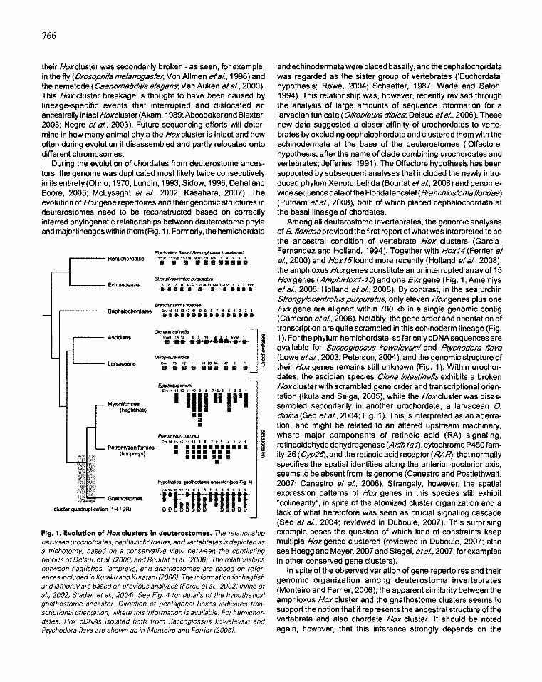
Fig. 1. Evolution of HOKclusters in deuterostomes. The relationship between urochordates. cephalochordates. and vertebrates is depicted as a trichotomy. based on a conservative view between the conflicting reports of Delsuc et al. (2006) and Bourlat et al. (2006). The relationships between hag fishes, lampreys, and gnathostomes are based on references included in Kuraku and Kuratani (2006). The information for hagfish and lamprey are based on previous analyses (Force et al., 2002; Irvine et al., 2002; Stadler et al., 2004). See Fig. 4 for details of the hypothetical gnathostome ancestor. Direction of pentagonal boxes indicates tran· scriptionalorientation, where this information is available. For hemichordates, Hox cDNAs isolated both from Saccoglossus kowalevski and Ptychodera flava are shown as in Monteiro and Ferrier (2006!.
and echinodermata were placed basally, and the cephalochordata was regarded as the sister group of vertebrates ('Euchordata' hypothesis; Rowe, 2004; Schaeffer, 1987; Wad a and Satoh, 1994). This relationship was, however, recently revised through the analysis of large amounts of sequence information for a larvacian tunicate (Oikopleura dioica; Delsuc et aI., 2006). These new data suggested a closer affinity of urochordates to vertebrates by excluding cephalochordata and clustered them with the echinodermata at the base of the deuterostomes (,Olfactore' hypothesis, after the name of clade combining urochordates and vertebrates; Jefferies. 1991). The Olfactore hypothesis has been supported by subsequent analyses that included the newly introduced phylum Xenoturbellida (Bourlat el al.. 2006) and genomewide sequence data ofthe Florida lancelet (Branchlosloma florldae) (Putnam el al.. 2008). both of which placed cephalochordata at the basal lineage of chordates.
Among all deuterostome invertebrates. the genomic analyses of B. floridae provided the first report of what was interpreted to be the ancestral condition of vertebrate Hox clusters (GarciaFernandez and Holland, 1994). Together with Hox14 (Ferrier et al.. 2000) and Hox15found more recently (Holland etal., 2008). the amphioxus Hoxgenes constitute an uninterrupted array of 15 Hoxgenes (AmphIHox1-15) and one Evxgene (Fig. 1; Amemiya et al.. 2008; Holland et al.. 2008). By contrast. in the sea urchin Strongylocentrotus purpuratus. only eleven Hoxgenes plus one Evx gene are aligned within 700 kb in a single genomic contig (Cameron et al.. 2006). Notably. the gene order and orientation of transcription are quite scrambled in this echinoderm lineage (Fig. 1). Forthe phylum hemichordata, so far onlycDNAsequences are available for Saccoglossus kowalevskii and Plychodera flava (Lowe et aI., 2003; Peters on. 2004), and the genomic structure of their Hoxgenes remains still unknown (Fig. 1). Within urochordates. the ascidian species Ciona intestinalis exhibits a broken Hoxcluster with scrambled gene order and transcriptional orientation (Ikuta and Saiga, 2005), while the Hox cluster was disassembled secondarily in another urochordate, a larvacean 0. moica (Seo et al.. 2004; Fig. 1). This is interpreted as an aberration, and might be related to an altered upstream machinery • where major components of retinoic acid (RA) signaling. retinoaldehyde dehydrogenase (Aldh 1 a 1). cytochrome P450 family-26 (Cyp26). and the retinoic acid receptor (RAJ\? that normally specifies the spatial identities along the anterior-posterior axis. seems to be absent from its genome (Canestro and Postlethwait, 2007; Canestro et aI., 2006). Strangely. however, the spatial expression patterns of Hox genes in this species still exhibit "colinearity". in spite of the atomized cluster organization and a lack of what heretofore was seen as crucial signaling cascade (Seo et al.. 2004; reviewed in Duboule, 2007). This surprising example poses the question of which kind of constraints keep multiple Hox genes clustered (reviewed in Duboule. 2007; also see Hoegg and Meyer. 2007 and Siegel. et al.. 2007, for examples in other conserved gene clusters).
In spite of the observed variation of gene repertoires and their genomic organization among deuterostome invertebrates (Monteiro and Ferrier, 2006), the apparent similarity between the amphioxus Hox cluster and the gnathostome clusters seems to support the notion that it represents the ancestral structure ofthe vertebrate and also chordate Hox cluster. It should be noted again, however. that this inference strongly depends on the
phylogenetic position of cephalochordates. Forthe time being, we still favor the traditional hypothesis that the last common ancestor of cephalochordates and vertebrates is expected to have already possessed a single"Hoxcluster containing up to 14 Hoxgenes plus one Evxgene that were all transcribed in the same direction (Fig. 1).
How many Hoxgenes made up the ancestral vertebrate Hox cluster?
A related question is how many Hox genes made up the ancestral vertebrate Hox cluster. The answer to this question hinges on some difficult issues that are specific to Hox genes. Molecular phylogenetic analyses of Hox genes usually can only provide phylogenetic trees with limited confidence (Malaga-Trillo and Meyer, 2001; Meyer, 1998). This is mainly due to the conserved nature of the homedomain that is only 60 amino acids in length. Regions outside of the homeobox cannot be aligned reliably across large evolutionary distances and therefore are not available for phylogenetic inferences. This is particularly problematic for the posterior abdomina/genes (Hox9-1.1), since it is often difficult to determine paralogy group relationships, (posterior flexibility; Ferrier et aI., 2000). This is also reflected in the uncertain nomenclature for posterior Hox genes, for example in hemichordate and echinoderms (Fig. 1).
The situation is further complicated by the recent discovery of the Hox14 genes located between the Hox13 and Evx gene in vertebrate Hox clusters. This paralogy group was first reported from rather basal vertebrate lineage - from horn shark and the coelacanth (Powers and Amemiya, 2004a,). This has been followed by the discovery in the elephant fish (Venkatesh et aI., 2007) and lamprey (Kuraku et aI., 2008). Interestingly, the molecular phylogenetic tree including available posterior genes did not suggest orthology of the Hox14 genes between amphioxus and vertebrates, whereas the position of the intron ('split homeobox') and gene location (between Hox13 and Evx) supported a single origin of Hox14 in the common ancestor of cephalochordates and vertebrates (Powers and Amemiya, 2004a; also see Ferrier, 2004; Garcia-Fernandez, 2005b, for reviews).
A
-cc Urcchordates
Cephalochordates
t. L-. Vertebrates
~ A single origin of Hox14
+ 'Euchordata' phylogeny
B C '''MI,'·'' ~
Cephalochordates
~. t Urochordates
Vertebrates Gain cif HoK14
A single origin of Hox14 + 'Olfactore' phylogeny
767
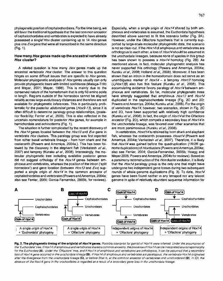
Especially, when a single origin of Hox14 shared by both amphioxus and vertebrates is assumed, the Euchordata hypothesis described above seemed to fit this scenario better (Fig. 2A). However, under the Olfactore hypothesis that is currently supported by large-scale molecular phylogenetic data, the scenario is not so clear-cut. Ifthe Hox140f amphioxus and vertebrates are orthologous to each other, a loss of Hox14should be assumed in the urochordate lineages, because none of species in this group has been shown to possess a Hox14 homo log (Fig. 2B). As mentioned above, in fact, molecular phylogenetic analysis has never supported this orthology (Powers and Amemiya, 2004a; Kuraku et aI., 2008; Holland et aI., 2008). Moreover, it has been shown that an intron in the homeodomain does not serve as an unambiguous marker of Hox14 - a lamprey Hox13 homolog (LjHox13{J) also has this feature (Kuraku et aI., 2008). This accumulating evidence favors paralogy of Hox14 between amphioxus and vertebrates. So far, molecular phylogenetic trees have strongly suggested that amphioxus Hox13 and Hox14 duplicated in the cephalochordate lineage (Fig. 2C and 20; Powers and Amemiya, 2004a; Kuraku et aI., 2008). For the origin of vertebrate Hox14, however, two scenarios, shown in Fig. 2C and 20, have been supported with relatively high confidence (Kuraku et aI., 2008). In fact, the origin of Hox14 at the Olfactore ancestor (Fig. 20), which compels a secondary loss of Hox14in the urochordate lineage, was favored over other scenarios that are more parsimonious (Kuraku et aI., 2008).
In vertebrates, Hoxd14is retained by horn shark and elephant fish, whereas the coelacanth possesses Hoxa14 (Powers and Amemiya, 2004a; Venkatesh et aI., 2007). Therefore, it is likely that Hox14 was gained before the quadruplication (1 Rl2R genome duplications) of Hoxclusters (Powers and Amemiya, 2004a; also see Ferrier, 2004; Garcia-Fernandez, 2005b; Hoegg and Meyer 2005; Powers and Amemiya, 2004b for reviews). Based on a parsimony reconstruction of the Hoxcluster evolution, it is likely that the Hox14 paralogy group is the only one that might have undergone two independent gene losses during two subsequent rounds of whole genome duplications (Fig. 3). To date, Hox14 genes have been found neither in any tetrapod nor any teleost genome in spite of relatively abundant sequence information for
D
-cc Cephalochordates
Urochordates t Urochordates
Vertebrates tL I Vertebrates
(Giln$HdX14] ~ II·HiI'.' Independent origins of Hox14
+ 'Olfactore' phylogeny Independent origins of Hox14
+ 'Olfactore' phylogeny
Fig. 2. The phylogenetic timing of the originlsl of Hox14 genes. Possible scenarios for gain(s) of Hox14 were inferred. Under the assumption of the 'Euchordate' tree, if Hox14 of amphioxus and vertebrates shared a common ancestry, the presence of Hox 14 can be interpreted as a synapomorphy for the Euchordata IAI. Under the 'O/factore' tree, and if Hox14 of amphioxus and vertebrates are orthologous, it can be assumed that a secondary loss of Hox14 gene occurred in the urochordate lineage 181. If Hox14 of amphioxus and vertebrates are paralogous. the vertebrate Hox14 originated after the divergence from the urochordate lineage (C)' or before (that is, at the common ancestor of vertebrates and urochordates) 101. In (D), the absence of the Hox 14 gene in the urochordates is regarded as a result of a secondary gene loss in the urochordate lineage.
768
these lineages. It is suggested that less functional constraint, suggested by loss of expression in axial elements where the Hoxcode is normally functioning, permitted the secondary losses of Hox14 genes in these lineages (Kuraku et aI., 2008). Data from other taxa, such as hagfishes, non-teleost actinopterygians, nontetrapod sarcopterygians will provide further information about the history of gains and losses of Hox14.
The example of Hox14 potentially cautions that gene order in Hox clusters does not necessarily imply orthology. Early in the evolution of bilateral body plans , the Hoxcluster was generated as the result of successive tandem duplications (reviewed in Garcia-Fernandez, 2005a). Moreover, secondary shutning of gene order in a Hox gene cluster is frequently observed in invertebrate deuterostomes (Fig . 1; Monteiro and Ferrior, 2006). Thus, relative positions of genes within a cluster itself, which have been the basis of gene annotation especially in studies of Hox genes, cannot alone serve as sole and unequivocal criterion for postulating 'homology relationships among Hox genes and their assignment to paralogy groups. This ci rcumstance combined with the difficulty of reliable phylogeny reconstruction renders orthology statements tenuous. This makes it difficult to reconstruct unequivocally the evolutionary history of posterior Hox genes - at least between some deuterostome lineages (Fig. 1).
Agnathans: how many Hox clusters?
Agnathans are jawless fishes that branched off the chordate stem lineage early during vertebrate evolution. Extant agnathans are grouped into hagfishes (Myxiniformes) and lampreys (Petromyzoniformes), and the phylogenetic relationshi ps of these two lineages with gnathostomes Uawed vertebrates) remained a controversial issue for over a century. However, recent molecular phylogenetic analysis using a I'arge number of genes suggested that hagfishes and lampreys form a monophyletic group, the Cyclostomata (Fig . 1; Blair and Hedges, 2005; Delarbre et aI., 2002; Delsuc et aI. , 2006; Furlong and Holland, 2002; Kuraku et ai , 1999; Mallatt and Sullivan, 1998; Stock and Whitt, 1992; Takezaki et aI., 2003; also see Kuraku and Kuratani , 2006; reviewed in Meyer and Zardoya, 2003).
Although several attempts have been made to determine the Hox repertoires and their genomic organization of lamprey by cDNA isolation (Pendleton et aI. , 1993; Sharman and Holland , 1998; Takio eta/. , 2007; Takio et a/. , 2004) and genomicsequencing (Force et ai , 2002; Irvine et a/. , 2002), not all Hoxgenes have been discovered so far and their genomic organization remains uncertain. In addition to a high GC-content in protein coding regions (Kuraku and Kuratani, 2006) , available cDNA sequences are usually incomplete and often derived from multiple closely related species. These factors prevented a precise categorization of available sequences into paralogy groups . Targeted genomic sequencing of Hoxclusters in a sea lamprey Petromyzon man'nus succeeded in the identification of regions containing multiple Hox genes, but they did not encompass entire Hox clusters (Force et a/., 2002; Irvine et ai , 2002). A phylogenetic analysis ·using available sequences suggested that lamprey has at least three or four Hox clusters (Force et aI. , 2002; Irvine et a/., 2002; Fig. 1), which might, at least partly, be the result of cluster duplications specific to the cyclostome or lamprey lineage (Fried et ai , 2003). In contrast, a PCR survey of genomic sequences in the Pacific
hagfish Eptatretus stoullidetected at most nine genes in a single paralogy group (Fig. 1), suggesting again the ,possibility of independent duplication(s) of Hox clusters in the cyclostome or hagfish lineage (Stadler et ai , 2004). Since hagfishes and lampreys diverged apparently relatively early (more than 400 million years ago )dunng cyclostome evolution-(Blair and 'Hedges, 2005;
Just before 1 R I 2R Evx 14 13 12 11 10 9 B 7 6 5 4 3 2 1
B •• -. •••••••••• _ c-a. __ •••••• o ••••••• • •••••
Losses of HoxA8, HoxB11, HoxB14, HoxC2, HoxC 14. HoxD6
Post-1 R I 2R state one o( Evxs El/)( 14 13 12 11 10 9 B 7 8 5 4 3 2 1
A • ••••• X ••••••• B *. - x, ........... -c -X~ • ••••• - •••• *. D • ••••••• X •••••
1 Hypothetical gnathostome ancestor
EVlC 1'1 13 12 11 10 9 8 7 6 5 • 3 2 1
A ......... .. B~.--......... . c - • D
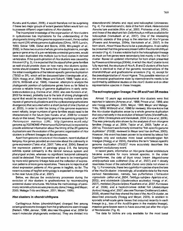
Fig. 3. Processes of Hoxcluster quadruplication based on currently available data. A possible scenario of quadrupllcation of Hox clusters was reconstructed based on parsimony criteria. For the gene repertoires of the hypothecial gna thostome ancestor (bo ttom), see Fig. 4. The clus ter structure at the pre- 7 R/2R state (top) represents that of a hypothetical ancestor jus t before the cluster quadruplication that had already gained Hox 74. It is notable that Hox14 is the only paralogy group of Hox genes that might have undergone two gene losses in the course of cluster quadruphcat ion. The pa ttern of Hox cluster rela tionships we assume here is that the A and B and the C and 0 clusters are "sister clusters ", that IS, I(A,BUC, o )1 (see Meyer, 1998). This assumption is confirmed b y our parsimony reconstruction of patterns of cluster duplications usmg the hypothetical gnathostome ancestor: each of three possible scenarios of cluster duplications, that is, I{A. B), (C, 0)], I{A, C), (B, 0)], and {(A,o), (B,C)I, requires nine, eleven, and ten steps of gene losses, respectively
Kuraku and Kuratani. 2006), it would therefore not be surprising if these two major groups of extant jawless fishes would tum out to possess different organizations of Hoxclusters.
The incomplete knowledge of the organization of Hoxclusters in cyclostomes has implications for the understanding of the phylogenetic timing of the genome expansion event, the so-called two-round (2R) whole genome duplications (Ohno, 1970; lundin, 1993; Sidow 1996, Dehal and Boore, 2005; Mclysaght et aI., 2002). In these two rounds of whole genome duplications, a single gene or gene array of a pre-vertebrate ancestral genomes, would have been duplicated into multiple (up to four) copies in derived vertebrates. If the quadruplication of Hoxclusters was caused by this event (Fig. 3), it is expected thatthe size of other gene famities as well should provide clues on how many Hox clusters cyclostomes should possess. The same line of reasoning was used in the initial investigations ofthe teleost-specific genome duplication (TSGD or 3R), which will be discussed later (Vandepoele et aI., 2004; Hoegg etal., 2004; Meyerand Schartl, 1999; Taylor etal., 2001 b, Wittbrodt et aI., 1998). However. attempts to analyze the phylogenetic position of cyclostome genes have so far failed to provide a reliable timing of genome duplications in early vertebrate evolution (e.g, Escriva et aI., 2002; also see Kuratani et aI., 2002 for review), probably due to the lack of strong phylogenetic signal that would be required for resolving multiple events (two rounds of genome duplications and the cyclostome/gnathostome divergence) that occurred within a short period of time (Horton et aI., 2003). In order to infer the timing of this genome expansion event reliably. larger numbers of gene families would need to be characterized in the future (see Kuraku et aI., 2009 for a recent study on this issue). The ongoing genome sequencing project for Petromyzon marinlJs (URl: http://genome.wustl.edu/ genome.cgi?GENOME=Petromyzon%20marinus) is expected to provide additional information about the timing of the 2R genome duplications and the evolution of the genomic organization of Hox dusters in different lineages of deuterostomes.
Apart from genomic structure of Hoxclusters, recent studies of lamprey Hoxgenes provided an overview about the colinearity in gene expression (Takio eta/., 2007; Takio etal.. 2004). Based on the expression patterns of paralogy group 2-8, the lamprey exhibits spatial colinearity in the central nervous system and pharyngeal arches, whereas no significant temporal colinearity could be detected. This observation will have to be investigated by more solid genomic linkage data and the collection of expression patterns of more gene repertOires. There are no reports at all about expression pattems of Hox genes in hagfish, so far. The recent success of hagfish embryology is expected to change this in the near future (Ota et al.. 2007).
Below, we discuss the evolutionary processes during the evolution of Hox clusters in gnathostomes by reconstructing ancestral cluster structures. We follow the methodology of parsimony reconstructions as was previously done (Hoegg and Meyer, 2005; Malaga-Trillo and Meyer, 2001; Meyer, 1998).
Hox clusters In chondrichthyans
Cartitaginous fishes (chondrichthyes) diverged first among extant gnathostome lineages from the gnathostome stem lineage (Nelson, 1994; Janvier, 1996; also see Kikugawa etal., 2004 for recent molecular phylogenetic evidence). They are divided into
769
elasmobranchii (sharks and rays) and holocephali (chimaeras; Fig. 4). For elasmobranchii, data ofthe hom shark HeteradontlJs francisciare available (Kim et aI., 2000, Prohaska et aI., 2004), and those of the elephant fish CallorhinchlJs milii are available for holocephali (Venkatesh et aI., 2007). One of the interesting genomic aspects of this group is the retention of Hox14 genes (Powers and Amemiya, 2004a; Venkatesh et a/., 2007). In the horn shark, Hoxa 14was found to be a pseudogene. It can safely be presumed that this gene was present still in the chond richthyan ancestor (Fig. 4). It is also notable that in the holocephalan lineage as much as five genes were secondarily lost mainly in the HoxD cluster. Based on updated information for hom shark presented by Powers and Amemiya (2004b), in which the HoxC cluster is not fully reported, the structure of HoxA, Band D clusters ofthe horn shark is identical to that of the genomic reconstructions of the chondrichthyan and gnathostome ancestors (Fig. 4), except for the pseudogenization of Hoxa14gene. This possible retention of the ancestral gnathostome state by elasmobranchs needs to be confirmed by additional studies on the HoxC cluster and on other representative species in these lineages.
The actinopterygian lineage: Pre-3R and Post-3R modes
Almost 10 years ago extranumeral Hox clusters were first reported in teleosts (Amores et aI., 1998; Prince et aI., 1998; also see Hoegg and Meyer, 2005; Meyer, 1998; Meyer and MalagaTrillo, 1999; Wittbrodt et aI., 1998 for reviews). We now know that the Hoxcluster duplicates arose by a whole genome duplication that occurred early in the evolution of teleost fishes (VandePoule, eta1.2004; ChristophelsandVenkatesh, 2004; Crowetal., 2006). This was previously also shown by the analyses of non-Hoxgene families (Hoegg et aI., 2004; Taylor et aI., 2003; Taylor et aI., 2001 a). This event was formerly termed the "fish-specific genome duplication" (FSGD; reviewed in Meyer and Van de Peer, 2005). However, this event has been proven to be shared by teleost fish lineages only and excludes more basal actinopterygian fish lineages (Hoegg et al. 2004). therefore the term "teleost-specific genome duplication (TSGD)" more accurately describes this important evolutionary event.
In recent years. information on Hoxgene cluster architecture became available for more teleost species. In the order Cypriniformes, the data of blunt snout bream Megalobrama amblycephala was published (Zou et al.. 2007) and it closely resembles those of the zebrafish Danio rerio (data not shown in Fig. 1). For example, these two cypriniform species share the loss of the HoxDb cluster. Interestingly, all available data for the more derived Neoteleostei. namely. two pufferfishes (Tetraodon nigraviridis- Jaillon et a/., 2004; TakiflJglJ rlJbripes- Aparicio et aI., 2002). and stickleback (GasterastelJs aClJleatlJs-, Hoegg et aI., 2007). medaka (Oryzias latipes-, Kasahara et al.. 2007; Kurosawa et al.. 2006). and a haplochromine cichlid fish (Astatot/lapla blJtton;, Hoegg etal., 2007; also see Thomas-Chollier and ledent. 2008). showed thatthey shared the loss ofthe HoxCb cluster, as described previously (Hoegg and Meyer. 2005). In addition to sporadic small-scale gene losses that occurred recently in each lineage (e.g .• loss of the hoxBla gene in the medaka lineage). massive gene losses seem to have occurred just after the origin of Neoteleostei (Fig. 4).
The data for bichirs are only available for the most basal
770
AM .,. ....... I •••• f. .. .. - . .. . ......... . .. -Co a.--I"_
Fig. 4. Reconstructed evolutionary history of Hoxcluster evolution within the jawed vertebrates. Closed squares indicate genes that have been previouslV described. Open squares are reported pseudogenes. Shaded squares are genes that have not been sequenced vet, but probablv are presen t III the c/llster(s). Hvpothetical organizations of Hox clus ters. shown in grev boxes, in the hvpothetica l ancestors of various evolu tionarv lineages were reconstructed based on parslmonv principles. SecondarV losses of Hox gene and entire clusters are shown in vellow and blue boxes, respectivelv. See text for the original literature that reported the organization of Hox cllls ters for particular species. Possible genome duplication events are indicated with red bars. Genomic organiza tion of Hox clusters in the horn shark' and the coelacanth were based on unpublished data reported bV Powers and Amemwa (2004b!. Possession and pseudogenization of teleost hoxC la are based on a recent report bV Thomas-Cho/lier and Ledent (2008). Abbreviations. TSGO, teleost-specific genome duplica tIOn, 1 R/2R, first- and second-ro und genome duplications.
actinopterygian lineage that represents the pre-3R (TSGD) state (Chiu et aI. , 2004; Ledje et aI. , 2002). For Polypterus pa/mas, only cDNA isolation was performed (Ledje et aI., 2002), while the genomic sequences of the entire Hox A cluster is determined for Polyptems senega/us (Chiu et aI. , 2004). Only very few gene losses seem to have occurred around the actinopterygiil sarcopterygii split, though this might be partly due to insufficient information for non-teleost actinopterygians. Although the structure of the Hox B, C, and D clusters remain to be determined for this lineage, it seems probable that bichirs have retained a similar set of Hoxgene repertoires to that of the hypothetical ancestor of the Actinopterygii, Euteleostomi, and Sarcopterygii. Currently, there is no information available yet about Hox genes in Acipenseriformes (sturgeons). Amiiformes (bow fins). and Semionotiformes (gars). Knowledge about the genomic situation of Hoxgenes in these basal fish lineages would provide valuable
information about the transition from the pre-3R state (before the TSGD and the evolutionary diversification of teleosts) to the post-3R genomic architecture.
Structural stabHity in the sarcopterygian lineage
For the sarcopterygian lineage including tetra pods, there are some reports for early-branching taxa. In the Australian lungfish Neoceratodus forsteri and the Indonesian coelacanth Latimeria menadoensis, only fragmental data have been reported so far based on cDNA isolation (Koh et aI., 2003; Longhurst and Joss, 1999). Recently, an overview of coelacanth Hox clusters was reported (Powers and Amemiya, 2004b), and its genome was found to contain highly similar Hox gene repertoires to those of tetra pods (Fig. 4). In the tetrapod lineage, four Hoxgenes including HoxA 14, that is still present in the coelacanth, have been lost
secondarily (Fig. 4). Especially, it is curious to see whether the lungfishes possess Hox14 genes in their genomes. In the lineages leading to western clawed frog (Xenopus tropica/is), two genes (Hox813and HoxD12) are thought to have been lost (Fig. 4). based on an in si/ico survey by Hoegg and Meyer (2005). For the chicken (Gal/us gal/us), a recent report on genomic annotation of Hoxclusters describes that the assembly is not complete, and thus entire or partial coding regions of some Hoxgenes (Hox82, HoxC4, HoxCS, HoxC13, HoxD1, HoxDB, HoxD9 and HoxD10) are still missing (Richardson et a/., 2007). Among these genes, entire coding regions of two genes (HoxC4and HoxC5) have not been found by our survey in available genomic and cDNA sequences (Fig. 4). These potential losses have to be confirmed with more complete genomic assembly in the future.
It remains an open question if phenotypic evolution and genomic evolution march to the beat of the same drummer. Interestingly. the hypothetical ancestral organization of tetra pods reconstructed from currently available information is identical to that still found today in the human genome (Fig. 4). In general it would appear that during almost 400 million years of tetrapod evolution only very few gene losses seem to have occurred. It appears as if the diversification of body plans in land vertebrates and a possible macroevolutionary trend towards increased complexity during the evolution of vertebrates was not accompanied by any (at least obvious major) changes in their Hoxcluster architecture. Based on our preliminary analysis, however. the grey short-tailed opossum Monodelphis domestica might be an exception. In an in si/ico survey of its still somewhat preliminary genomic sequences (version MonDom5; URL. http://www.broad.mit.edu/mammals/ opossum/; Mikkelsen el a/., 2007), only a single Hox gene (HoxC6) has been found so far in the HoxC cluster. But, the Hox A. B. and D clusters are almost identical to the situation in the human genome. We also investigated the genomic sequences of the duck-billed platypus Omithorhynchus anatinus (Ensembl database: U RL, http://www.ensembl.org/Ornithorhynchus_anatinus/ ), but it would be premature to derive any conclusions based on this incomplete data set.
It is possible, however. that the TSGD is not only temporally correlated but even causally linked to the significantly accelerated rates of diversification/speciation and increased levels of phenotypic complexity in teleost fish (e.g. Wittbrodt et aI., 1998; Meyer and Schartl. 1999; for more detailed diSCUSSion). Future work on a possible relationship of Hoxcluster architecture and phenotypic diversification and increased complexity should also attempt to quantify changes in regulatory regions as well as protein coding regions and numbers of Hoxgene repertOires.
Conclusions
In contrast to invertebrates, all vertebrates have four or more Hoxgene clusters. Vertebrate Hoxclusters are peculiar in that the linkages of genes in clusters are never broken and that all genes in a cluster are transcribed in the same direction (Duboule. 2007). A possible exception is the opossum HoxC cluster, but more data are needed to confirm this hypotheSis. Also for agnathans, more reliable information is eagerly awaited since it might shed light on the phylogenetic timing of the 1 Rand 2R genome duplication events during chordate evolution. For the gnathostomes our reconstruction of the genomic organizations of the Hox clusters
771
during vertebrate evolution suggests that there are two major types of Hoxcluster architectures: (1) the four-cluster type and (2) the post-3R teleost type. The former is further divided into three themes: (1 A) an ancestral gnathostome type (still seen in the horn shark A, B, and D clusters); (1B) non-teleost bony fish type (as found in the coelacanth and probably in the bichir); and (1 C) the tetrapod type (e.g. human). To date, there is no solid genomic data about the organization of Hoxgene clusters in some crucial lineages, such as hagfishes, lampreys and reptiles, and marsupials. These data are needed urgently for a more complete understanding of the patterns and processes of Hoxcluster evolution in deuterostomes.
Knowledge of expression patterns of Hoxgenes might help in the understanding of the evolutionary history, mechanisms and constraints that shaped Hoxcluster evolution. Expression information would also aid in the identification of potentially homologous morphological structures among species that belong to phenotypically extremely diverged lineages of vertebrates.
Acknowledgements This study was financially supported by the Deutsche
FOfSchungsgemeinschaft (DFG). We thank Michael Richardson, Shigeru Kuratani, Joost WoHering, 8yrappa Venkatesh, Simone Hoegg, and Jenny Graves for helpful comments and discussion.
References
ABOOBAKER, A. and BLAXTER, M. (2003). Hox gene evolution in nematodes: novelty conserved. CUff. Opin. Genet Oev. 13: 593-598.
AKAM, M. (1989). Hox and HOM: homologous gene clusters in insects and vertebrates. Cell 57: 347-349.
AMEMIYA, C.T., PROHASKA, S.J., HILL·FORCE,A., COOK, A., WASSERSCHEID, J., FERRIER, D.E., PASCUAL-ANAYA, J., GARCIA-FERNANDEZ, J., DEWAR, K. and STADLER, P.F. (2008). The amphioxus Hoxcluster: characterization, comparative genomics, and evolution. J Exp Zoolog B Mol Dev Eval31 0: 465-477.
APARICIO, S" CHAPMAN, J., STUPKA, E., PUTNAM, N., CHIA, J.M., DEHAL. P., CHRISTOFFELS, A., RASH, S., HOON, S., SMIT, A. et a/. (2002). Wholegenome shotgun assembly and analySiS of the genome of Fugu rubdpes. Science 297: 1301-1310.
BLAIR, J.E. and HEDGES, S.B. (2005). Molecular phylogeny and divergence times of deuterostome animals. Mol. Bioi. Evol. 22: 2275-2284,
BOURLAT, S.J., JULlUSDOTTIR, T., LOWE, C.J., FREEMAN, R .. ARONOWICZ, J" KIRSCHNER, M., LANDER, E.S., THORN DYKE, M .. NAKANO, H., KOHN, A.B. et al. (2006). Deuterostome phylogeny reveals monophyletic chordates and the new phylum Xenoturbellida. Natute444: 85-88.
CAMERON, R.A., ROWEN, L., NESBITT, R, BLOOM, S., RAST, JP .. BERNEY, K .. ARENAS-MENA, C., MARTINEZ, P., LUCAS, S., RICHARDSON, P.M. etal. (2006). Unusual gene order and organization of the sea urchin hox cluster. J. Exp. Zoo/ag. B (Mol. Oev. Evol.) 306: 45-58.
CANESTRO, C. and POSTLETHWAIT, J.H. (2007). Development of a chordate anterior'posterior axis without classical retinoic acid signaling. Dev. BioI. 305: 522-38.
CANESTRO, C., POSTLETHWAIT, J,H., GONZALEZ-DUARTE, R.andALBALAT, R (2006). Is retinoic acid genetic machinery a chordate innovation? Evel. Dev. 8: 394-406.
CHIU, C.H., DEWAR, K., WAGNER, G.p .. TAKAHASHI, K., RUDDLE, F., LEDJE, C., BARTSCH, P., SCEMAMA, J.L., STELLWAG, E., FRIED, C. etal. (2004). Bichir HoxA cluster sequence reveals surprising trends in ray-finned fish genomic evolution. Genome Res. 14: 11-17.
772
CHRISTOFFELS, A., KOH, E.G., CHIA, J.M., BRENNER, S., APARICIO, S. and VENKA TESH, B. (2004). Fugllgenome analysis provides evidence for a wholegenome duplication early during the evolution of ray-finned fishes. Mol. Bioi. Evol21: 1146-1151.
COHN, M.J. (2002). Evolutionary biology: lamprey Hox genes and the origin of jaws. Natlllli> 416: 386-387.
COHN, M.J. and TICKLE, C. (1999). Developmental basis of limblessnessand axial patternlng In snakes. Natllre399: 474-479.
CROW, K.D., STADLER, P.F., LYNCH, V.J., AMEMIYA, C. and WAGNER, G.P. (2006). The «fish-specific. Hox cluster duplication Is coincident with the origin ofteleosts. Mol. Bioi. EvoI.23: 121-136.
DEHAL, P. and BOORE, J.L. (2005). Two rounds of whote genome duplication in the ancestral vertebrate. PLoS BioI. 3: e314.
DELARBRE, C., GALL UT, C" BARRIEL, V., JANVIER, P. and GACHELlN, G. (2002). Complete mitochondrial DNA of the hagfish, Eptatretlls bllrgeri. the comparative analysis of mitochondrial DNA sequences strongly supports the cyclostome monophyly. Mol. Phylogenet Evo/. 22: 184-192.
DELSUC, F., BRINKMANN, H., CHOURROUT, D. and PHILIPPE, H. (2006). Tunicates and not cephalochordates are the closest living relatives of vertebrates. Natllre 439: 965-968.
DUBOULE, D. (1994). Temporal colinearity and the phylotypic progression: a basis for the stability of a vertebrate Bauplan and the evolution of morphologles through heterochrony. Dev. SlIppl135-142.
DUBOULE, D. (2007). The rise end fall of Hox gene clusters. Oevelopmentl34: 2549-2560.
ESCRIVA, H., MANZON, L., YOUSON, J. and LAUDET, V. (2002). Analysis of lamprey and hagflsh genes reveals a complex history of gene duplications during early vertebrate evolution. Mol. Bioi. Evol 19: 1440-1450.
FERRIER, D.E. (2004). Hox genes: Did the vertebrate ancestor have a Hoxl4? Cliff. BioI. 14: R210-R211.
FERRIER, D.E., MINGUILLON, C., HOLLAND, P.w. and GARCIA-FERNANDEZ, J. (2000). The amphioxus Hox cluster: deuterostome posterior flexibility and Hox14. Evo/. Dev. 2: 284-293.
FORCE, A., AMORES, A. and POSTLETHWAIT, J.H. (2002). Hox cluster organization in the jawless vertebrate Petromyzon mannlls. J. EXp. Zoo/. B (Mo/, Oev. Evo/.) 294: 30-46.
FRIED, C" PROHASKA, S.J. and STADLER, P.F. (2003). Independent Hox-cluster duplications in lampreys. J. Exp. Zoo/ag. B (Mol. Dev. Evo/.) 299: 18-25.
FURLONG, R. F. and HOLLAND, P.w.H. (2002) Bayesian phylogenetlc analysis supports monophyly of ambulacrarla and of cyclostomes. Zoo/ag. Se/. 19: 593-599.
GARCIA-FERNANDEZ, J. (2005a). The genesis and evolution of homeobox gene clusters. Nat. Rev. Genet 6: 881-892.
GARCIA-FERNANDEZ, J. and HOLLAND, P. W. (1994). Archetypal organization of the amphioxus Hox gene cluster. Natllre 370: 563-566.
HOEGG, S" BRINKMANN, H., TAYLOR, J.S. and MEYER, A. (2004). Phylogenetic timing of the fish-specific genome duplication correlates with the diversification of teleost fish. J. Mol. Evo/. 59: 190-203.
HOEGG, S. and MEYER, A. (2005). Hox clusters as models for vertebrate genome evolution. Tnmds GeJflet 21: 421-424.
HOEGG, S. and MEYER, A, (2007). Phylogenomicanalyses of KCNA gene clusters in vertebrates: why do gene clusters stay intact? BMC Evol. BioI. 7: 139.
HOEGG, S. BOORE, J., KUEHL, J,V. and MEYER, A. (2007). Comparative phylogenomlc analyses of teleost fish Hox gene clusters: lessons from the cichlid fish Astatotllapia bllttonl. BMC Geflomics. 8: 317.
HOLLAND, L.Z., ALBALAT, R., AlUMI, K., BENITO-GUTIERREZ, E., BLOW, M.J., BRONNER-FRASER, M., BRUNET, F., BUTTS, T., CANDIANI, 5., DISHAW, L.J. et sI. (2008). The amphioxus genome illuminates vertebrate origins and cephalochordatebiology. Genome Res. 18: 1100-1111.
HORTON, A.C., MAHADEVAN, N.R., RUVINSKY, I. and GIBSON-BROWN, J.J. (2003). Phylogenetic analyses alone are insufficient to determine whether genome dupllcation(s) occurred during early vertebrate evolution. J. Exp. Zoolog. B (Mol. Dev, Evol.) 299: 41-53.
IKUTA, T. and SAIGA, H. (2005). Organization of Hox genes in ascidians: present, past, and future. Dev. Dyn. 233: 382-389.
IRVINE, S.O., CARR, J.L., BAILEY, W.J., KAWASAKI, K., SHIMIZU, N., AMEMIYA, CT. and RUDDLE, F,H. (2002). Genomic analysis of Hox clusters in the sea lamprey Petromyzon man'nlls. J. Exp. Zoo/. B (Mol. Dev. Evol.) 294: 47·62.
JAILLON, 0., AURY, J.M., BRUNET, F., PETIT, J.L., STANGE-THOMANN, N., MAUCELI, E" BOUNEAU, L., FISCHER, C., OZOUF-COSTAl, C., BERNOT, A. et al (2004). Genome duplication in the teleost fish Tetraodon nlgrovltidls reveals the early vertebrate proto-karyotype. Natlllli> 431 : 946-957.
JANVIER, p, (1996) Early Vertebrates. Oxford Univ. Press, Oxford,
JEFFERIES, RP.S. (1991). In Blolag/caIAsymmetlJlandHandedfless(ed, Bock, G. R. & Marsh, J.). Wiley, Chichester, pp 94--127.
KAMM, K., SCHIERWATER, B" JAKOB, W., DELLAPORTA, S.L. and MILLER, D.J. (2006). Axial palternlng and diversification In the cnidaria predate the Hox system. ClIrT. BioI. 16: 920-926.
KASAHARA, M. (2007). The 2R hypothesis: an update. ClIlT. Opin.lmmllnol. 19: 547-552.
KASAHARA, M., NARUSE, K., SASAKI, S., NAKATANI, Y., OU, W., AHSAN, B., YAMADA, T., NAGAYASU, Y., 001, K., KASAI, Y. etal. (2007). The medaka draft genome and insights into vertebrate genome evolution. Natlllli> 447: 714-719.
KESSEL, M and GRUSS, P. (1991). Homeotic transformations of murine vertebrae and concomitant alteration of Hoxcodes Induced by retinoic acid. Ce1167: 89-104.
KIKUGAWA, K., KATOH, K" KURAKU, S., SAKURAI, H., ISHIDA. 0., IWABE, N. and MIYATA, T. (2004). Basal jawed vertebrate phylogeny Inferred from multiple nuclear DNA-coded genes. BMC BioI. 2: 3,
KIM, C.B., AMEMIYA, C., BAILEY, W., KAWASAKI, K., MEZEY, J., MILLER, W., MINOSHIMA, 5., SHIMIZU, N., WAGNER, G. and RUDDLE, F. (2000). Hox cluster genomics In the horn shark, Heterodontus francisci. Proc, Natl. Aead. Set: USA 97: 1655-1660.
KOH, E,G., LAM, K., CHRISTOFFELS, A., ERDMANN, M.V., BRENNER, S. and VENKATESH, B. (2003). Hox gene clusters in the Indonesian coelacanth, Latimeria menadoensis. Proe. Natl. Aead. Se!. USA 100: 1084-1088.
KURAKU, S., HOSHIYAMA, D., KATOH, K., SUGA, H. and MIYATA, T. (1999). Monophyly of lampreys and hagfishes supported by nuclear DNA-coded genes. J. Mol. Evol. 49: 729-735.
KURAKU, S., TAKIO, Y., TAMURA' K., AONO, H" MEYER, A. and KURATANI, S. (2008). Noncanonical role of Hox14 revealed by its expression patterns in lamprey and shark. Proc, Natl. Aead. Se!. USA 105: 6679-6683.
KURAKU, S., MEYER, A., and KURA TANI, S. (2009) Timing of genome duplications relative to the Origin ofthe vertebrates: did cyclostomes diverge before, or after? Mol. Bioi. Evol. 26: 47-59.
KURAKU, S. and KURATANI, S. (2006). Time scale for cyclostome evolution inferred with a phylogenetic diagnosis of hagfish and lampreycDNA sequences. Zoolag. Sel. 23: 1053-1064.
KURATANI, S., KURAKU, S. and MURAKAMI, Y. (2002). Lamprey as an evo-devo model: lessons from comparative embryology and molecular phylogenetics. Geflesis34: 175-183.
KUROSAWA, G., TAKAMATSU, N., TAKAHASHI, M., SUMITOMO, M., SANAKA, E., YAMADA, K., NISHII, K., MATSUDA, M., ASAKAWA, 5., ISHIGURO, H. et sI. (2006). Organization and structure of hox gene loci in medaka genome and comparison with those of pufferfish and zebrafish genomes. Gene370: 75-82.
LEDJE, C., KIM, C.B. and RUDDLE, F.H. (2002). Characterization of Hox genes in the blchir, Po/yptertJspalmas. J. Exp. Zool. B (Mol. Dev. Evol.) 294: 107-111,
LEWIS, E.B. (1978). A gene complex controlling segmentation in Drosophila. Nalllre 276: 565-70.
LONGHURST, T.J. and JOSS, J.M. (1999). Homeobox genes In the Australian lungfish, Neoceratodlls forsteri. J. Exp. Zoo/. B (Mol. Dev. Evol.) 285: 140-145.
LOWE, C.J., WU, M., SALlC, A., EVANS, L., LANDER, E., STANGE-THOMANN, N., GRUBER, C.E., GERHART, J. and KIRSCHNER, M. (2003). Anteroposterior patterning In hemlchordates and the origins of the chordate nervous system. Ce11113: 853-865.
LUNDIN, L.G. (1993). Evolution ofthevertebrate genome as reflected in paralogous chromosomal regions in man and the house mouse. Genomlcs16: 1-19.
MALAGA-TRILLO, E. and MEYER, A. (2001). Genome duplications and accelerated evolution of Hox genes and cluster architecture in teleost fishes. Amer. Zoo141: 676-686.
MALLA TT, J. and SULLlVAN, J. (1998). 28S and 18S rDNA sequences support the monophyly of lampreys and hagflshes. Mol BioI. EvoI. 15: 1706-1718.
MCGINNIS, W. and KRUMLAUF, R (1992). Homeobox genes and axial patternlng. Cel168: 283-302.
MCL YSAGHT, A., HOKAMP, K and WOLFE, KH. (2002). Extensive genomic duplication during early chordate evolution. Nal. Genet. 31: 200-204.
MEYER, A. (1998). Hox gene variation and evolution. NatlJre 391: 225: 227-228.
MEYER, A. and MAlAGA-TRILLO, E. (1999). Vertebrate genomlcs: More fishy tales about Hox genes. CIJIT. BioI. 9: R210-R213.
MEYER, A. and SCHARTL, M. (1999). Gene and genome duplications in vertebrates: the one-to-four (-to-eight in fish) rule and the evolution of novel gene functions. CIJIT. Opin. Cal/Biol. 11: 699-704.
MEYER, A and VAN DE PEER, Y. (2005). From 2R to 3R: evidence for a fishspeCific genome duplication (FSGD). Bioessays27: 937-945.
MEYER, A. and ZARDOYA, R (2003). Recent advances in the (molecular) phytogeny of vertebrates. AnnlJ. Rev. Eeol. Evol. Syst. 34: 311-338.
MIKKELSEN, T.SWAKEFIELD, M.J.AKEN, B.AMEMIYA, C.T.CHANG, J.L.DUKE, S.GARBER, M,GENTLES, A.J.GOODSTADT, L.HEGER, A al al. (2007). Genome of the marsupial Monodelphls domestica reveals innovation in noncoding sequences. NatlJre 447: 167-177.
MONTEIRO, AS. and FERRIER, D.E. (2006). Hox genes are not always Colinear. 1nl, J BioI. Sei 2: 95-103.
NEGRE, B., RANZ, J.M .• CASALS, F., CACERES, M. and RUIZ, A. (2003). A new spilt of the Hox gene complex In Drosophila. relocation and evolution ofthe gene labial. Mol. BioI. Evol. 20: 2042-2054.
NELSON, J.S., (1994). Fishes ofthe World. Wiley, New York.
OHYA, YK, KURAKU, S. and KURATANI, S. (2005). Hox code In embryos of Chinese soft-shelled turtle Pelodiseus sinansiscorrelates with the evolutionary Innovation in the turtle. J. Exp. Zoolog. B (Mol Dev. EvoI.) 304; 107-118.
OTA, KG., KURAKU, S. and KURATANI, S. (2007). Hagflsh embryology with reference to the evolution of the neural crest. NatlJre 446: 672-675.
PENDLETON, J.W., NAGAI, B.K., MURTHA, M.T. and RUDDLE, F.H. (1993). Expansion of the Hox gene family and the evolution of chordates. Proc. Nad. Aead. Scl IJSA 90: 6300-6304.
PETERSON, K.J. (2004). Isolation of Hox and Parahox genes in the hemichordate Ptychodel7iJ flava and the evolution ofdeuterostome Hox genes. Mol. Phylogenet, Evol. 31: 1208-1215,
POWERS, T.P. and AMEMIYA, C.T. (2004a). Evidence for a Hox14 paralog group in vertebrates. CUlT. BioI. 14: R183-Rl84.
POWERS, T.P. and AMEMIYA, C.T. (2004b). Evolutionary plasticity of vertebrate Hox genes. CUlT. Genomics5: 459-472.
PRINCE, V.E" JOLY, L., EKKER, M. and HO, RK. (1998). Zebrafish hox genes: genomic organization and modified colinear expression patterns in the trunk. Deve/opment125: 407-420.
PROHASKA, S.J., FRIED, C., AMEMIYA, C.T., RUDDLE, F.H., WAGNER, G.P. and STADLER, P.F. (2004). The shark HoxN cluster is homologous to the human HoxD cluster. J. Mol. Evo!. 58; 212-217.
PUTNAM, N.H., BUTTS, T., FERRIER, D.E., FURLONG, RF., HELLSTEN, U., KAWASHIMA, T., ROBINSON-RECHAVI, M., SHOGUCHI, E., TERRY, A., YU, J.K. at el. (2008). The amphioxus genome and the evolution of the chordate karyotype. NallJre453: 1084-1071.
RICHARDSON, M.K., CROOIJMANS, RP. and GROENEN, MA (2007). Sequenclng and genomic annotation of the chicken (Gal/lIS gal/lJs) Hox dusters, and mapping of evolutionarily conserved regions. CyfGg9net. Genome Res. 117: 110-119.
ROWE, T. (2004). Chordate phylogeny and development. In Assembling Ihe lree of life, (ed. Cracraft, J. and Donoghue, M. J.). Oxford University Press, Oxford, New York, pp. 384-409.
SCHAEFFER, B. (1987). Deuterostome monophyly and phylogeny. Evol. Bioi. 21: 179-235.
SEO, H.C .. EDVARDSEN, RB., MAELAND, A.D., BJORDAL, M., JENSEN, M.F.,
773
HANSEN, A .. FLMT, M., WEISSENBACH, J., LEHRACH, H., WINCKER, P. et al (2004). Hox cluster disintegration with persistent anteroposterior order of expression in Oikop/elJl71dioica. NallJre431: 67-71.
SHARMAN, A.C. and HOLLAND, P.w. (1998). Estimation of Hox gene cluster number in lampreys. /nl. J. Dav. Bioi 42; 617-620.
SIDOW, A. (1996). Gen(om)e duplications in the evolution of early vertebrates. CIJIT. Opin. Gene!. Dev. 6: 715-722.
SIEGEL, N., HOEGG, S., SALZBURGER, W., BRMSCH, I., MEYER, A. (2007) Comparative genomics of ParaHox clusters of teleost fishes: gene cluster breakup and the retention of gene sets following whole genome duplications. BMCGenomies 8: 312.
STADLER, P.F., FRIED. C., PROHASKA, S.J., BAILEY, W.J., MISOF, B.Y., RUDDLE, F.H. and WAGNER, G.P. (2004). Evidence for independent Hox gene duplications in the hagfish lineage: a PCR-based gene inventory of EptalrellJs slolJlii. Mol PhylGg9net Evo132: 686-94.
STEINKE, D., HOEGG, S., BRINKMANN, H. and MEYER, A. (2006). Three rounds (1 R/2R/3R) of genome duplications and the evolution ofthe glycoly1ic pathway In vertebrates. BMC BioI. 4: 16.
STOCK, D.W. and WHITT, G.S. (1992). Evidence from 18S ribosomal RNA sequences that lampreys and hagfishes form a natural group. Seianee257: 787-789.
TAKEZAKI, N., FIGUEROA, F., ZALESKA-RUTCZYNSKA, Z. and KLEIN, J. (2003). Molecular phylogeny of early vertebrates: monophyly of the agnathans as revealed by sequences of 35 genes. Mol. BioI. Evol. 20: 287-292.
TAKIO, Y., KURAKU, S .• MURAKAMI, Y., PASQUALETTI, M .. RIJLI, F.M .. NARITA, Y., KURATANI, S. and KUSAKABE, R. (2007). Hoxgene expression patterns in Lethentaronjaponicumembryos-Insights Into the evolution of the vertebrate Hox code. Dev. BioI. 308: 606-620.
TAKIO, Y., PASQUALETTI, M .. KURAKU, S., HIRANO, S .. RIJLI, F.M. and KURATANI, S. (2004). Evolutionary biology: lamprey Hox genes and the evolution of jaws. Natu171429: 1 p following 262.
TAYLOR, J., BRMSCH, I., MEYER, A. and VAN DE PEER, Y. (2003). Genome duplication, a traitshared by 22,000 species of ray-finned fish. GenomeRes. 13: 382-390.
TAYLOR, J.S., VAN DE PEER, Y., BRAASCH, I. and MEYER, A (2001a). Comparative genomics provides evidence for an ancient genome duplication event in fish. Phi/os. Trams R. Soc. Lond. B BioI. Sd 356: 1661-79.
TAYLOR, J.S., VAN DE PEER, Y. and MEYER, A (2001 b). Revisiting recent challenges to the ancient fish-specific genome duplication hypothesiS. CUlT. BioI. 11: Rl005-R1008.
THOMAS-CHOLLlER, M. and LEDENT, V. (2008). Comparative phylogenomic analyses of teleost fish Hox gene dusters: lessons from the clchlid fish Aslatotilapia burloni. comment. BMC Genomics 9; 35.
VAN AUKEN, K., WEAVER, D.C., EDGAR, L.G. and WOOD, W.B. (2000). Caenomabditis e/agans embryonic axial patterning requires two recently discovered posterior-group Hox genes. Proe. Nad. Aead. Se!. IJSA 97; 4499-4503.
VANDEPOELE, K .. DE VOS, W., TAYLOR, J.S., MEYER, A. and VAN DE PEER, Y. (2004). Major events in the genome evolution of vertebrateS: paranome age and size differ considerably between ray-finned fishes and land vertebrates. Proc. Natl. Aead. Sei IJSA 101: 1638-1643.
VENKATESH, B., KIRKNESS, E.F., LOH, Y.H .. HALPERN, AL., LEE, AP., JOHNSON, J., DANDONA, N., VISWANATHAN, L.D., TAY, A., VENTER, J.C. et al. (2007). Survey Sequencing and Comparative Analysis of the Elephant Shark (Cal/ominehlJs milil) Genome. PLoS BioI. 5: el01.
VON ALLMEN, G., HOGGA, I., SPIERER, A., KARCH, F., BENDER, W., GYURKOVICS, H. and LEWIS, E. (1996). Splits in fruitfly Hox gene complexes. NatlJre 380: 116.
WADA, H. and SATOH, N. (1994). Details of the evolutionary history from Invertebrates to vertebrates, as deduced from the sequences of 18S rDNA Proc. Natl. Aesd. Se!. IJSA 91: 1801-1804.
WITTBRODT, J., MEYER, A. and SCHARTL, M. (1998). More genes in fish? Bioessays20: 511-515.
ZOU, S.M., JIANG, X,V, HE, Z.Z., YUAN, J., YUAN, X.N. and LI, S.F. (2007). Hox gene clusters in blunt snout bream, Mega/obl7iJma amblycephala and comparison with those of zebrafish, fugu and medaka genomes. Gene. 400: 60-70.