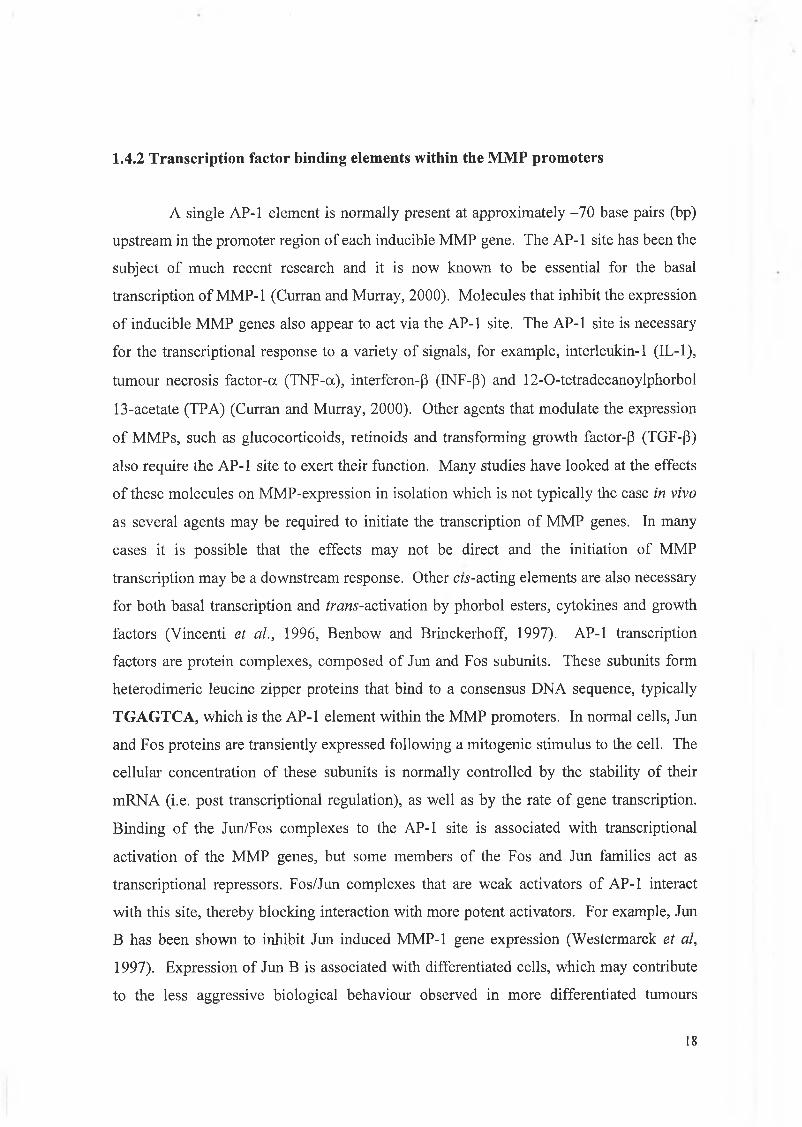
The expression and regulation of matrilysin (MMP-7) in human colon cancer and leukaemia cell lines A dissertation submitted for the degree of Ph.D by Conor C. Lynch B.Sc. Under the supervision of Dr. Susan McDonnell June 2001 School of Biotechnology, Dublin City University, Dublin 9, Ireland.
Transcript
The expression and regulation of matrilysin (MMP-7) in human colon cancer and leukaemia
cell lines
A dissertation submitted for the degree of Ph.D
by
Conor C. Lynch B.Sc.
Under the supervision of Dr. Susan McDonnell
June 2001
School of Biotechnology, Dublin City University, Dublin 9, Ireland.
Declaration
I hereby certify that this material, which I now submit for assessment on the programme of
study leading to the award o f Doctor o f Philosophy is entirely my own work and has not
been taken from the work of others save and to the extent that such work has been cited
and acknowledged within the text of my work.
Signed:
Date:
I.D.No. 97970115
i
Acknowledgments
Firstly I would like to thank my supervisor Dr. Susan McDonnell for her constant
support, patience and advice during my Ph.D. studies and for making a scientist out of me.
I would also like to thank other graduate students in the McDonnell lab, both past and
present, Maria for showing me the ropes and Dave and Aine for putting up with the
constant whinging and persecution and providing mental support. I would like to thank the
many friends I have made during my time at DCU and my friends from home who always
assisted me in my alcoholic endeavours.
A big thank you goes to Howard Crawford, Barbara Fingleton and Lynn Matrisian
for giving me the opportunity to work in their laboratory and showing me various new
molecular techniques which assisted me in the completion of my Ph.D.
I would like to thank my family Jason, Liam Valerie, Neil and Amy for their love
and support.
Finally, this thesis is dedicated to my mother and father for their love,
understanding and constant support both mental and financial. I could not have achieved
any of it without you.
"Remembering you how you used to be
Slow drowned, you were angels
So much more than everything
Hold for the last time then slip away quietly
Open my eyes, but I never see anything"
Robert Smith, 1989.
II
Abstract
M atrilysin (M M P-7, EC 3.4.24.23) is the smallest m em ber o f the matrix m etalloproteinase
(M M P) family and has been shown to be overexpressed in various tumours including breast and
colon cancers. M atrilysin has also been shown to play an im portant role in several aspects o f
tum our biology including growth, progression, invasion and metastasis. W ith respect to colon
cancer, m atrilysin is unique in that it is the only M M P expressed exclusively by the malignant
epithelia o f colonic adenocarcinomas. These facts com bine to make matrilysin a promising
therapeutic target. However, in order to develop drugs which specifically inhibit matrilysin it is
important to understand how m atrilysin gene expression is controlled, something which to date
remains poorly understood.
W e have examined a panel o f human colon tum our cell lines and have shown that
m atrilysin expression can be upregulated by a num ber o f cytokines including EGF, IL-6 and bFGF.
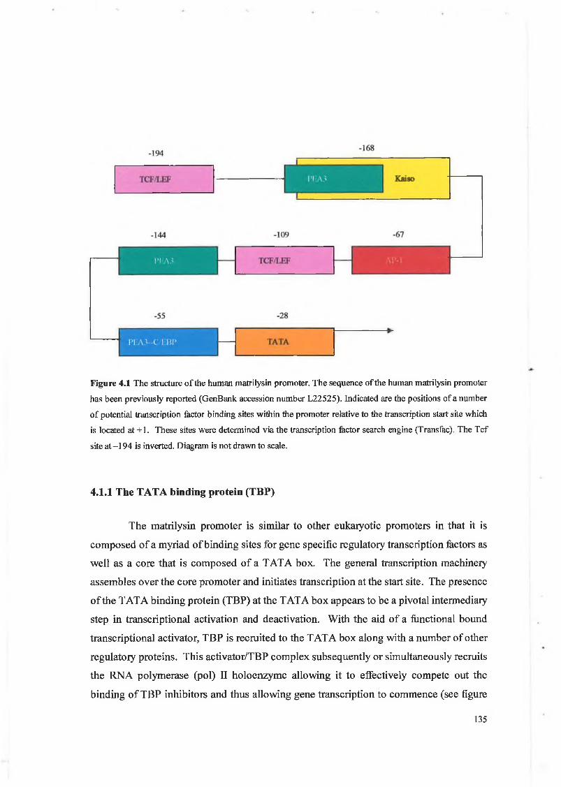
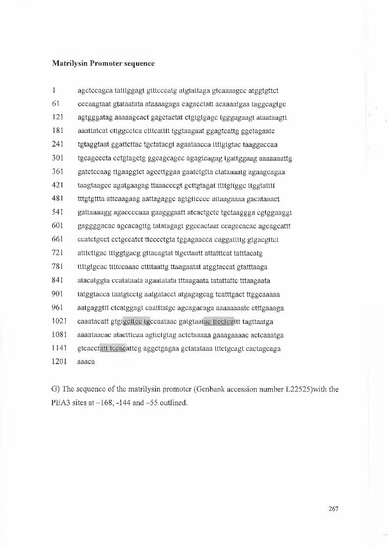
Analysis o f the m atrilysin prom oter revealed the presence o f a number o f potential transcription
factor binding sites including three ETS sites. We have shown that EGF treatm ent increased
matrilysin gene expression by activation o f PEA3 transcription factors using artificial promoter,
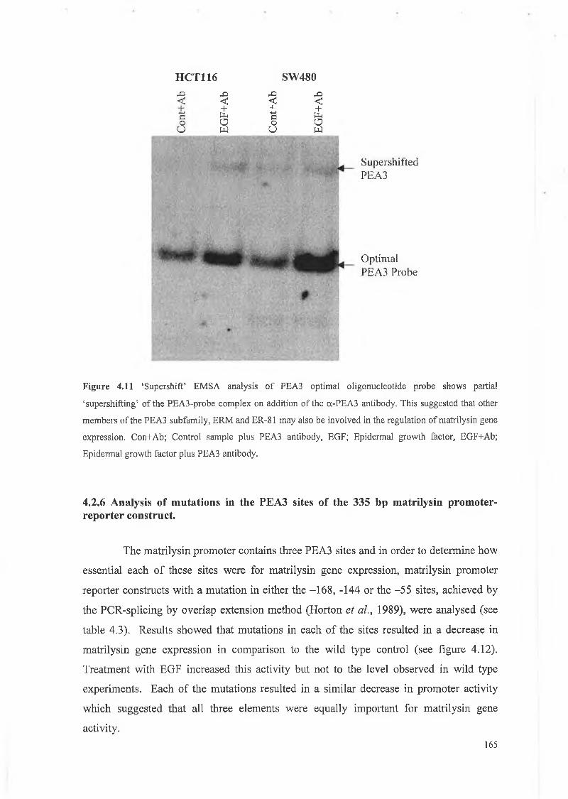
western blot and EM SA analysis. ‘Supershift’ EM SA analysis showed that other PEA3 subfamily
members such as ERM and ER81 may also be involved which is in agreem ent with other studies.
In addition, we have found that EGF increased cellular levels o f [3-catenin through destabilisation
o f the E-cadherin/catenin com plex which resulted in increased binding to the T cf site within the
m atrilysin promoter.
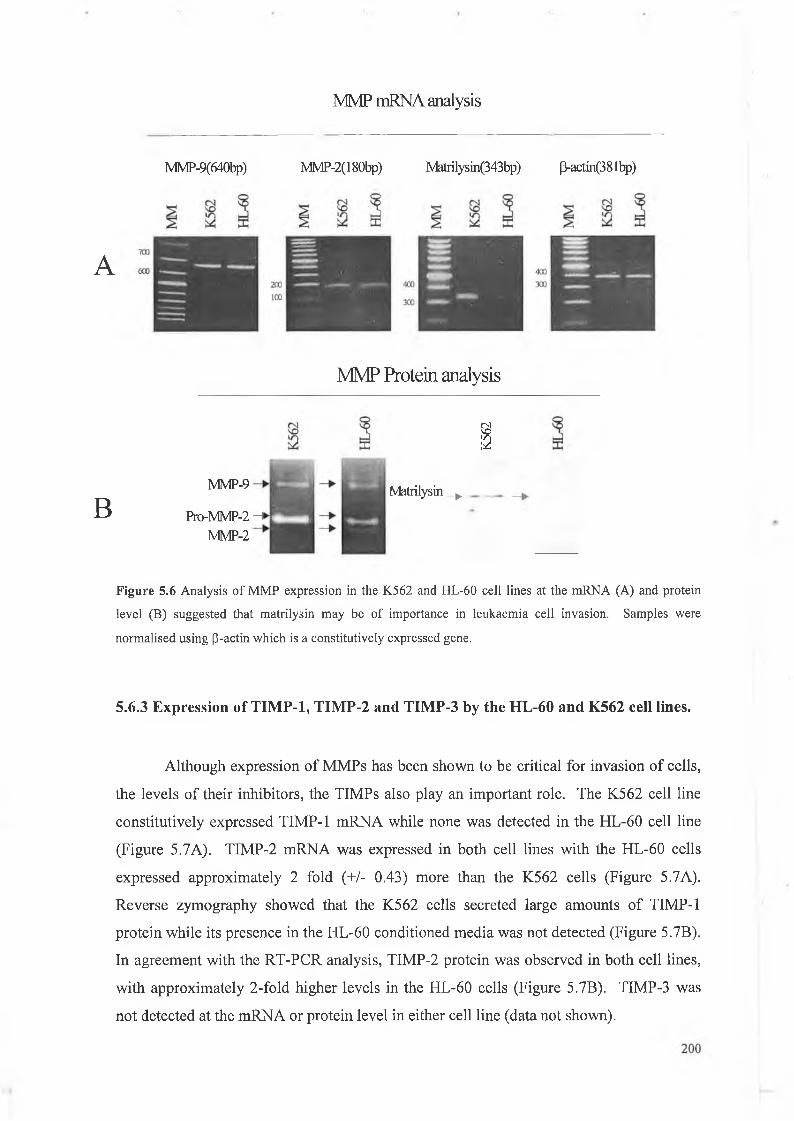
W e also exam ined the expression and regulation o f matrilysin in the K562 and HL-60
myeloid leukaemia cell lines. Results showed that only the K562 cell line expressed m atrilysin and
in vitro invasion assays showed that the K562 cells were up to 4 times more invasive than the HL-
60 cell line. M atrilysin antibody blocking experiments showed a significant decrease in invasion in
the K562 cell line suggesting a role for matrilysin in leukaemia invasion. The MMP and TIMP
profiles o f these cell lines were also examined.
Our data suggests that EGF plays an important role in the regulation of matrilysin gene expression
via a number of new mechanisms. Furthermore, we have shown that matrilysin plays an important role in
leukaemia cell line invasion. These findings have identified possible new drug targets that will inhibit
matrilysin expression which in turn should lead to decreased tumourigenesis and invasion and metastasis.
Ill
Abbreviations
(3-gal P-galactosidase
yP32-ATP Gamma phosphorous 32 labelled ATP
Abs Absorbance
ACF Aberrant crypt foci
ADAM A disintegrin and metalloproteinase
ADAMTS ADAM thrombospondin
aFGF acidic fibroblast growth factor
ALL Acute lymphoblastic leukaemia
AML Acute myelocytic leukaemia
AOM Azoxymethane
AP Activator protein
APC Adenomatous polyposis coli
APL Acute premyelocytic leukaemia
AR Amphiregulin
ATF Activating transcription factor
ATP Adenosine triphosphate
BAD Bel associated death promoter
BCA Bicinchoninic acid assay
BCR Breakpoint cluster region
bFGF basic FGF
BHK Baby hamster kidney fibroblasts
BM-MNC Bone marrow mononuclear cells
BP-1/6C3/APA BP-l/6C3/aminopeptidase A
BSA Bovine serum albumin
BTB/POZ-ZF Broad complex Tramtack Brie a brae, Pox virus and
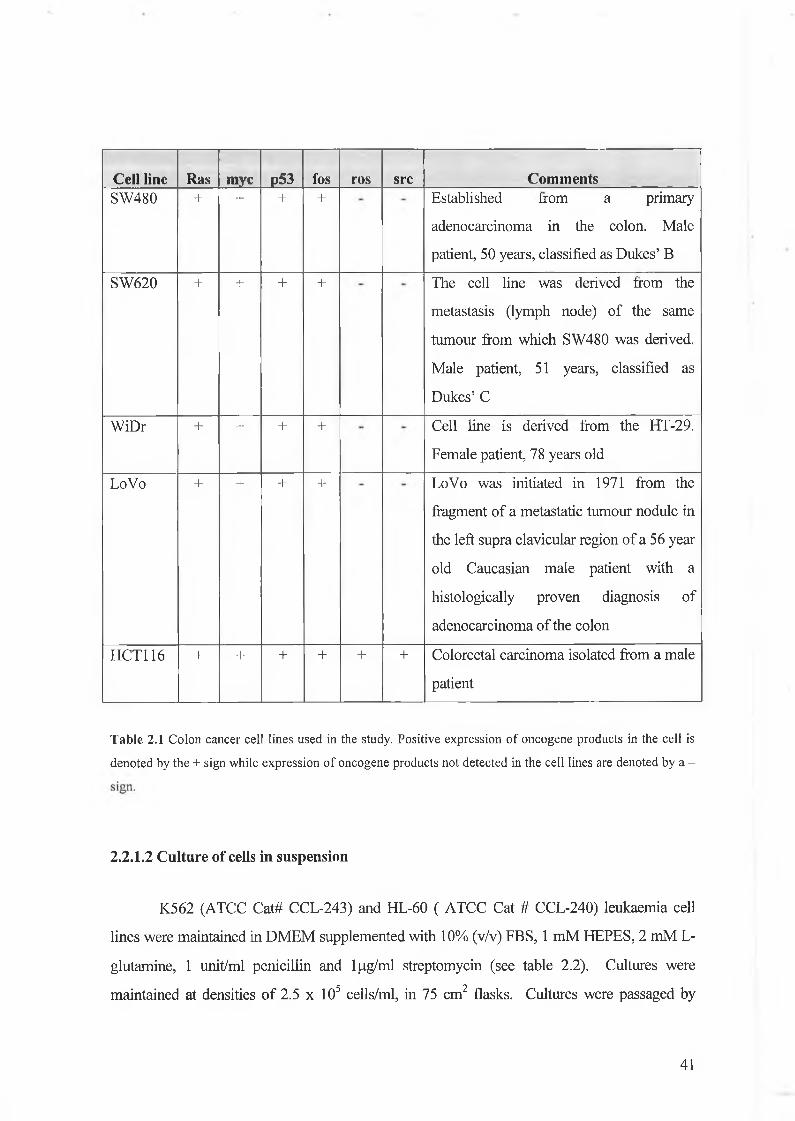
(ATCC Cat# CCL-121 ), HCA7 and BHK92 cell lines were maintained in Dublecco’s
modification of Eagles medium (DMEM) supplemented with 5% (v/v) [DMEM S5] foetal
bovine serum (FBS), 2 mM L-glutamine, 1 mM N-[2-Hydroxyethl]piperazine-N’-[2-
ethanesulfonic acid] (HEPES) and 1 unit/ml penicillin and l|jg/ml streptomycin (see table 2.1
for colon cell line characteristics). All cells were cultured in 25cm2 or 75 cm2 tissue culture
flasks. As these were all strongly adherent cell lines, trypsinisation was required for harvesting
cells prior to subculturing. For trypsinisation, the growth medium was aspirated and the flask
rinsed with 3 ml of phosphate buffered saline (PBS) to remove any residual FBS which
contains a trypsin-inhibitor (a 2-macroglobulin). 2 ml of fresh trypsin ethylenediamine
tetracetic acid (EDTA) (0.025% (w/v) trypsin with 0.02% (w/v) EDTA in 0.15 M PBS, pH
7.4) was then placed in each flask and the flask incubated at 37°C for 5-10 min or until all the
cells had detached from the surface. The cell suspension was removed to a sterile universal
container containing 5 ml growth medium and centrifuged at 2000 rpm for 5 min. Cells were5 6 ' • 2resuspended in culture medium at 2 x 1 0 - 1 x 1 0 cells/ml, using 20 ml of medium per 75cm
culture flask and 10 ml per 25cm2 flask. All cell lines were incubated in a humid, 5% (v/v)
CO2 atmosphere at 37°C in a Heraeus cell culture incubator.
40
Cell line Ras myc p53 fos ros src CommentsSW480 + + + + Established from a primary
adenocarcinoma in the colon. Male
patient, 50 years, classified as Dukes’ B
SW620 + + + + The cell line was derived from the
metastasis (lymph node) of the same
tumour from which SW480 was derived.
Male patient, 51 years, classified as
Dukes’ C
WiDr + + + + Cell line is derived from the HT-29.
Female patient, 78 years old
LoVo + + + + LoVo was initiated in 1971 from the
fragment of a metastatic tumour nodule in
the left supra clavicular region of a 56 year
old Caucasian male patient with a
histologically proven diagnosis of
adenocarcinoma of the colon
HCT116 + + + + + + Colorectal carcinoma isolated from a male
patient
Table 2.1 Colon cancer cell lines used in the study. Positive expression of oncogene products in the cell is
denoted by the + sign while expression of oncogene products not detected in the cell lines are denoted by a -
Buffers used in this protocol were kept at 4°C and prior to use the following was added
(final concentration of each in brackets): DTT (0.5mM), PMSF (0.2mM), Leupeptin (lpg/pl),
Aprotinin (lpg/pl), and Pepstatin (0.25 pM). Cells were grown to approximately 70%
confluency in 100mm cell culture dishes under normal conditions (DMEM S5 at 37°C). Cells
were then serum starved overnight. The following day the cells were washed with PBS and
treated with various cytokines (see 2.2.1.5 for dilutions) for 8 hr at 37°C in serum free media.
After incubation the supernatant was removed and the cells were washed three times in PBS
solution. 1 ml of PBS was added to each plate and using a cell scraper the cells were collected.
The cells were spun out (pulse spin in microfuge ~ 13,000 rpm for 30 s), the supernatant was
removed and the cells were resuspended in hypotonic buffer (lOmM HEPES, pH 7.9 at 4°C
61
1,5mM MgCl2, lOmM KC1, 50mM NaF, and ImM NaV0 4 ). The cells were pulsed briefly in a
microfuge and the resultant pellet was resuspended in two packed cell volumes (approx. 1 0 0 -
200 pi) of hypotonic buffer. The cells were then incubated on ice for 10 min. In order to
promote cell lysis 1 pi of 5% NP-40 was added to the cells and the suspension was mixed by
pipetting 3-4 times with a pasteur pipette. The cells were then analysed for lysis by taking lpl
of the cell suspension and adding trypan blue exclusion reagent. When approximately 60% of
the cells were blue in colour, thus indicating that the cell walls had been disrupted, the nuclei
were immediately pelletted and washed once in hypotonic buffer. The supernatant was
removed and the cells were gently resuspended in a Vi packed nuclear volume (approx. 50-100
pi) of low salt buffer (20mM HEPES, pH 7.9 @ 4°C, 1.5mM MgCl2, 0.02M KC1, 0.2mM
EDTA and 25% glycerol). A Vi packed nuclear volume (approx. 50-100 pi) of high salt buffer
(20mM HEPES, pH 7.9 at 4°C, 1.5mM MgCl2, 1.2M KC1, 0.2mM EDTA and 25% glycerol)
was added drop by drop with careful mixing after each drop. The suspensions were then
placed on a rocker/rotator for 30 min at 4°C. One volume of dialysis buffer (20mM HEPES,
pH 7.9 at 4°C, lOOmM KC1, 0.2mM EDTA and 20% glycerol) was then added to the
suspension which was then pulsed in order to remove debris. The supernatant was then placed
in visking tubing and dialysed overnight at 4°C in dialysis buffer using an agitated vessel.
Once dialysed the contents of the dialysis tubing were removed into separate tubes, flash frozen
in liquid nitrogen and stored at -80°C. One tube from each sample was retained and a BCA
protein assay (See section 2.2.4) was performed in order to determine the concentration of
protein. Samples were also run on SDS-PAGE gels (see section 2.2.5.1) in order to confirm
that the isolated nuclear proteins were intact.
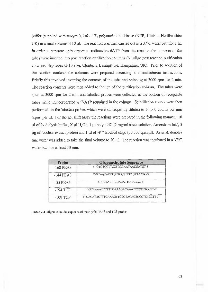
2.2.11.2 Preparation of radioactively labelled probes
Oligonucleotide primers for PEA3 (optimal, proximal, upstream and downstream)
and TCF were generated (MWG Biotech, UK) and N labelled using yP32-ATP (see table2.4).
Radioactive probes were made of each using the following protocol: 50 ng of the• . . . • 32oligonucleotide of interest was aliquoted into sterile 0.5ml tube. To this was added lp l of y-P
labelled dATP (3000 Ci/mmol, NEN Life science products, Zaventen, Belgium), lpl lOx
62
buffer (supplied with enzyme), 1 pi of T4 polynucleotide kinase (NEB, Hitchin, Hertfordshire
UK) in a final volume of 10 pi. The reaction was then carried out in a 37°C water bath for 1 hr.
In order to separate unincorporated radioactive dATP from the reaction the contents of the
tubes were inserted into post reaction purification columns (N’ oligo post reaction purification
columns, Sephadex G-10 size, Clontech, Basingstoke, Hampshire, UK). Prior to addition of
the reaction contents the columns were prepared according to manufacturers instructions.
Briefly this involved inverting the contents of the tube and spinning at 3000 rpm for 2 min.
The reaction contents were then added to the top of the purification column. The tubes were
spun at 3000 rpm for 2 min and labelled probes were collected at the bottom of receptacle
tubes while unincorporated yP32-ATP remained in the column. Scintillation counts were then
performed on the labelled probes which were subsequently diluted to 50,000 counts per min
(cpm) per pi. For the gel shift assay the reactions were prepared in the following manner. 10
pi of 2x dialysis buffer, X pi H20*, 1 pi poly dldC (2 mg/ml stock solution, Amersham Int.), 5
pg of Nuclear extract protein and 1 pi of yP32 labelled oligo (50,000 cpm/pl). Asterisk denotes
that water was added to take the final volume to 20 pi. The reaction was incubated in a 37°C
In order to identify nuclear proteins binding to sequences of interest, antibodies
generated against a particular protein were added to the nuclear extracts. The antibody-protein
complexes were allowed to form by incubating at 4°C for 30 min. The other reactants (see
section 2.2.11.3) were then added to the tubes and incubated at 37°C for 30 min prior to
running on the gel. ‘Supershift’ bands migrate more slowly through the gel due to a higher
molecular weight complex formed by the antibody and protein thus resulting in a higher band
when compared to the control.
64
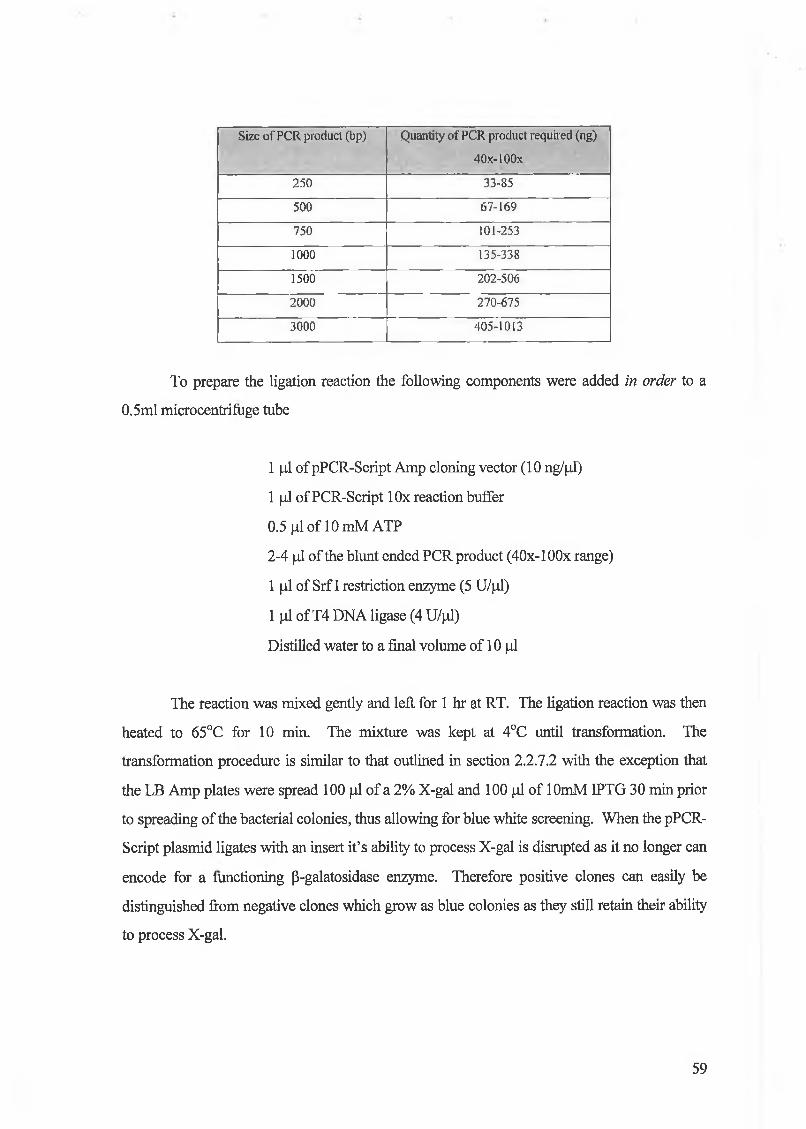
Biocoat Matrigel invasion chambers provided a system that allowed assessment of
cells invasive activity in vitro. The cell culture inserts contained an 8 jj.m pore size membrane
that was coated with a layer of matrigel basement membrane matrix. The layer of matrigel
serves as a reconstituted basement membrane in vitro. This layer occludes the pores of the
membrane blocking non-invasive cells from migrating through the membrane. In contrast,
invasive cells were able to migrate through the Matrigel coated membrane. The 6 -well plate
invasion chambers were removed from 4°C storage and allowed to come to room temperature
and warm (37°C) serum-free culture medium was added to the interior of the inserts and
allowed to rehydrate for 2 hr at RT. After rehydration the media was removed from the inserts
and replaced with 2 ml cell suspension in serum-free medium prepared at 3.5xl05 cells/ml.
Chemoattractant 2.5 ml (media containing 20% FBS) was added to the wells of the plate. The
plates were then incubated for 48 hr at 37°C in 5% C 0 2 incubator. After incubation, the non
invading cells are removed from the upper surface of the membrane by scrubbing with a cotton
tipped swab. The cells on the lower surface of the membrane were then fixed in methanol for 2
min, stained for 1 min in Mayers Haematoxylin solution and rinsed in tap water several times.
The membrane was then counterstained for 1 min using Eosin stain. The cells and membrane
were then dehydrated by incubating for 2 min each in a series of ethanol solutions (30, 50, 70,
90, 100% ethanol). The membranes were then removed from the insert housing and mounted
on slides using DPX mounting medium. Invading cells were then viewed under the
microscope at 40 X magnification and counted. The percentage invasion for each cell line was
calculated by counting all of the stained cells on the filter underside and dividing by the total
number of cells plated. For experiments involving the effect of cytokines on invasion,
cytokines were added to the media used to rehydrate the inserts, the cell suspension, and to the
media containing the chemoattractant at the concentrations given in section 2 .2 .1 .5.
2.2.12 In vitro invasion assays
65
2.2.13 Zymography
Zymography was used to localise enzyme activity by molecular weight. The gel was
prepared by incorporating the protein substate of interest (gelatin) within the polymerized
acrylamide matrix. 1 0 % or 15% acrylamide gels were used and the amounts for one gel are
given below, volumes for 15% gels are in brackets.
Resolving gel: 2.5 ml [2.5 ml] Buffer A (1.5 M Tris-HCl, pH 8 .8 ; 0.4% (w/v) SDS)
2.5 ml [2.5 ml] 3 mg/ml gelatin stock
3.3 ml [5 ml] 30% acrylamide stock
1.7 ml [0 ml] distilled water
33 pi [3 3 pi] 10% ammonium persulphate (freshly prepared)
5 pi [5 pi] TEMED
Stacking gel: 0.8 ml Buffer B (0.5 M Tris-HCl, pH 6 .8 ; 0.4% SDS)
0.5 ml 30% acrylamide stock
2 ml distilled water
3 3 pi 10% ammonium persulphate (freshly prepared)
5 pi TEMED
Samples were mixed 3:1 with 4X sample buffer (10% sucrose; 0.25 M Tris-HCl, pH
6 .8 ; 0.1% (w/v) bromophenol blue) and loaded. The gels were run at 20 mA per gel in running
buffer (0.025 M Tris; 0.19M glycine; 0.1% SDS) until the dye front reached the bottom of the
gel. Following electrophoresis the gel was soaked in 2.5% Triton-X-100 with gentle shaking
for 30 min at RT with one change. The gel was then rinsed in substrate buffer (50 mM Tris-
HCl, pH 8.0; 5 mM CaCl2) and incubated for 24 hr in substrate buffer at 37°C. The gel was
then stained with Coomassie blue for 2 hr with shaking and destained in water until clear bands
were visible.
To confirm bands as metalloproteinases, identical gels were run as described above
except the substrate buffer contained one of the following protease inhibitors: lOmM EDTA
66
(MMP inhibitor), 0.3 mM 1,10-phenanthroline (MMP inhibitor), 1 mM PMSF (serine
proteinase inhibitor), or 1 mM pepstatin A (aspartic proteinase inhibitor).
2.2.14 Reverse Zymography
Reverse zymography was used to detect the presence of TIMPs, SDS-PAGE gels
were prepared with the incorporation of matrix metalloproteinases and gelatin into the
acrylamide matrix o f the gel. 15% gels (10 ml volume) were prepared as in section 2.2.13
except that the water component of the gel is reduced by 1 ml to allow for the addition of 0.5
ml of MMP-rich conditioned medium from both the BHK92 cells and the HT-1080 cells. Gels
were loaded and ran as above, T1MP standard, containing TIMP-1, TIMP-2 and TIMP-3
(Chemicon Ltd.), was loaded as a positive control. After electrophoresis, gels were incubated
in a solution of 2.5 % Triton-X-100; 50 mM Tris-HCl, pH 7.5; 5 mM CaCb once for 15 min,
then again overnight at RT with gentle shaking. Next day gels, were rinsed three times with
water, then incubated in 50 mM Tris-HCl pH 7.5 and 5 mM CaCl2 for 24 hr at 37°C to allow
digestion of the gelatin substrate. Gels were then stained in Coomassie blue for 2 hr and
destained until the desired contrast was achieved. The majority of the gel does not stain as the
gelatin has been degraded. Dark bands represent inhibition of gelatin degradation by TIMPs
within the sample.
2.2.15 Densitometry and statistics.
Densitometry of protein and RT-PCR gels was performed using a Pharmacia
Amersham Densitometer with Imagemaster software. Two tailed T tests were performed using
Microsoft Excel™.
67
Chapter 3
The effect of cytokines on matrilysin mRNA and protein
expression in human colon cancer cell lines
3. Introduction
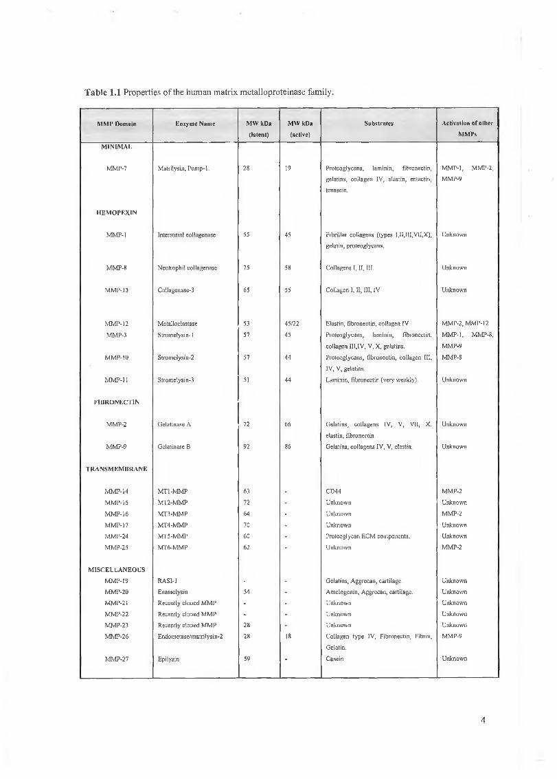
Matrilysin is one o f the smallest members of the matrix metalloproteinase family
and is capable o f degrading many components o f the basement membrane and interstitial
ECM. Matrilysin has been implicated in many disease processes, such as, cancer invasion
and metastasis, and rheumatoid arthritis (Adachi et al., 1999 and Cawston, 1998).
Recently, several reports have shown that matrilysin is involved, not just in ECM
breakdown but in a wide range of normal biological processes including proliferation,
wound healing, angiogenesis and the activation of other MMP members involved in the
proteolytic cascade resulting in total ECM degradation (Sang et al., 2000, Nagashima el
al., 1997, Lu et al., 1999 and, Barille et al., 1999)
3.1 Structure and function
Matrilysin is similar to the stromelysins in its substrate specificity and to
interstitial collagenase in the crystal structure o f its catalytic domain but the enzyme is
unique in that it lacks the carboxyl terminal domains present in other MMPs (Wilson and
Matrisian, 1996). Matrilysin has three domains consisting of a pre, pro and catalytic
domain which are essential for cellular processing of the protein and enzymatic activity.
Matrilysin is secreted as an inactive protein with a molecular weight of 28 kDa which is
subsequently proteolytically cleaved to produce the active 19 kDa enzyme. The cDNA
encoding human matrilysin was first isolated by Muller et al. (1988) from a mixed tumour
library in an effort to clone stromelysin related genes involved in tumour invasion and
metastasis. The cDNA, at 1078 bp, was shown to be derived from a previously
undescribed gene by comparison to stromelysin and collagenase sequence to which it was
49% and 44% homologous, respectively. The gene was initially termed pump-1 to
indicate that it encoded a putative metalloproteinase, with similarity to other MMPs but
lacking sequence encoding the caboxyl-terminal hemopexin-like domain. Subsequent
purification of the matrilysin protein and analysis of the enzymes substrate specificity
revealed that it was capable of degrading a wide spectrum of high molecular weight
proteins including fibronectin, gelatins, collagen, laminin, entactin, and elastin (Quantin et
al., 1989)
68
3.2 Matrilysin in disease processes
The MMPs are primarily involved in ECM remodelling and their activity is
tightly regulated at the transcriptional and protein level. When the regulation of MMPs
becomes aberrant in diseases such as cancer, then there is excessive ECM destruction
which may ultimately lead to tumour invasion and metastasis to a secondary site.
Matrilysin, which plays an important role in ECM degradation, has been reported to be
overexpressed in a number of tumour types, in particular, breast, colon and prostate
cancers (Rudolph-Owen et al., 1998a, Wilson et al., 1997 and Udayakumar et al., 2001).
Moreover, recent studies have shown that matrilysin is also involved in other aspects of
tumourigenesis including, growth, migration and angiogenesis (Chambers and Matrisian
1997, Wilson et al., 1997 and Patterson and Sang, 1997).
3.2.1 The genetics of human colon cancer.
Approximately 22,000 new cases of cancer arise in Ireland each year. Non
melanoma skin cancer is the commonest diagnosis but these are easily detected, treated
and rarely cause death. O f the more serious cancers, colon cancer, is the most common
accounting for approximately 9% or just under 2000 of total new cases. Males are at a
slightly higher risk of developing the disease. The 5 year survival rates for colon cancer
patients are less that 55% and colon cancer attributes to 13.5% of the total of cancer
related deaths (National Cancer Registry of Ireland, 1997). Colon cancer can be sporadic
or inherited. Inherited forms of colon cancer include familial adenomatous polyposis
(FAP) and hereditary nonpolyposis colon cancer (HNPCC) which account for up to 20%
of total colon cancer cases. The mean age for patients with sporadic colon cancer tends to
be over 60 while FAP and HNPCC patients tend to present with colon cancer much earlier
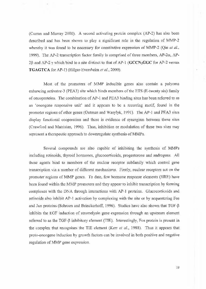
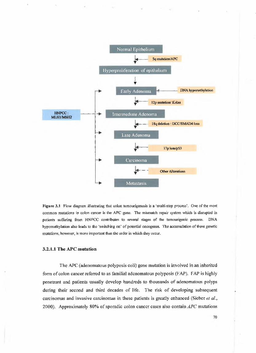
with the ages being approximately 20-30 and 40 years old respectively. In 1990, Fearon
and Vogelstein put forward a hypothesis for colon cancer progression whereby a number
o f genetic mutations or ‘hits’ were required for the progression o f colon cancer (see figure
3.1), (Fearon and Vogelstein, 1990).
69
N orm al E pithelium
I-4 5q mutation/APC
HNPCC : MLH1/MSII2
Hyperproliferation of epithelium
IEarly Adenoma DNA hypomethylation
* 12p mutation/ K-Ras
Intermediate Adenoma
-4--------- 18q deletion : DCC/SMAD41 oss
Late Adenoma
Carcinoma
17p loss/p53
Other Alterations
Metastasis
Figure 3.1 Flow diagram illustrating that colon tumourigenesis is a ‘multi-step process’. One of the most
common mutations in colon cancer is the APC gene. The mismatch repair system which is disrupted in
patients suffering from HNPCC contributes to several stages of the tumourigenic process. DNA
hypomethylation also leads to the ‘switching on’ of potential oncogenes. The accumulation of these genetic
mutations, however, is more important than the order in which they occur.
3.2.1.1 The APC m utation
The APC (adenomatous polyposis coli) gene mutation is involved in an inherited
form o f colon cancer referred to as familial adenomatous polyposis (FAP). FAP is highly
penetrant and patients usually develop hundreds to thousands o f adenomatous polyps
during their second and third decades o f life. The risk o f developing subsequent
carcinomas and invasive carcinomas in these patients is greatly enhanced (Sieber et al.,
2000). Approximately 80% of sporadic colon cancer cases also contain APC mutations
which implies that the APC protein plays an important role in the development o f both
sporadic and familial colon cancer. A major function o f the APC protein involves
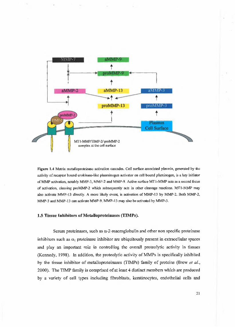
targetting o f a cytoskeletal protein known as P-catenin for degradation. Mutated APC proteins do not have the ability to assist in the degradation o f P-catenin, therefore excess
P-catenin accumulates within the cytoplasm. The excess p-catenin is efficiently
translocated to the nucleus where it effects the transcription o f several target genes
including cyclin D, c-myc (He et ah, 1998) and, as will be discussed later, the MMP
matrilysin (see figure 3.2). APC also plays an important role in cytoskeletal organization
as it has been shown to bind to the microtubule cytoskeleton via its basic domain. In
normal epithelial cells this association occurs at actively migrating regions o f the cell
membrane. For example if cell migration is induced by wounding a cell monolayer or
treating with a human hepatocyte growth/scatter factor APC clusters intensify at the edges
o f migrating cell membranes (Smith et ah, 1994 and Nathke et ah, 1996). APC has also
been shown to indirectly modulate the organisation o f the actin filament network and
consequently affects cell morphology polarity and migration. This modulation occurs via
the binding o f Asef to APC which in turn enhances the interaction o f Asef with Rac, a
molecule which is involved in the regulation o f the actin cytoskeleton ( Kawasaki et ah,
2000).
Ubiquitination of P -catenin in the
0 presence of wild1 type APC
Translocation of p -catenin in the
presence of truncated APC
Matrilysin, c-myc and cyclin D expression
Lef-1/ p -catenin
Figure 3.2 The role of APC in the regulation of P-catenin
71
3.2.1.2 The ras mutation
Another important early somatic alteration identified in colon tumours is the ras
gene mutation. When transfected into suitable recipient host cells it was found that
mutated ras genes conferred neoplastic properties. The importance of the ras gene
mutation in early colorectal tumourigenesis was further bolstered by experiments
examining a number of patient samples. It was found that 50% of colorectal carcinomas
and a similar percentage of adenomas greater than 1cm in size had a ras gene mutation. In
contrast such mutations have been identified in fewer than 1 0 % of adenomas less than
lcm in size regardless of whether the tumours arose sporadically or in patients with an
inherited predisposition for their formation (Bos et ah, 1987 and Vogelstein et al., 1989).
The ras gene product is a small membrane bound GTPase protein which plays an
important role in the transduction of cell signals. It is involved in several signalling
cascades which include the mitogen activated protein kinase (MAPK) pathway and
phosphoinositol (PI) pathway which will be dealt with in more detail later. In its inactive
state the Ras protein is GDP bound. Stimulation of receptor tyrosine kinases such as the
EGF receptor leads to the activation o f Ras, hence the activation of the subsequent
signalling cascades associated with Ras. This is achieved via the tyrosine kinase activity
o f the receptor which subsequently activates SOS (son of sevenless, a GTP exchange
molecule) via an intermediate docking protein known as GRB. SOS facilitates the
activation o f Ras via the exchange of GDP for GTP. Activated Ras is then involved in the
activation of various signalling cascades. Once the required effects have been achieved
by the cell, Ras reverts back to its inactive state via NF1-GTPase activating protein (see
figure 3.3).
72
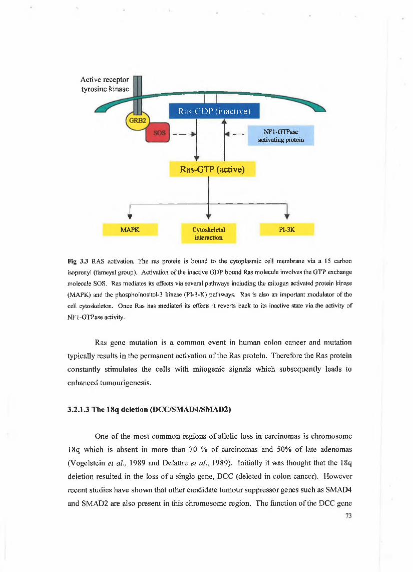
Active receptor tyrosine kinase
iMAPK
R as-G D P (inactive)
NFl-GTPase activating protein
Ras-GTP (active)
Cytoskeletal PI-3Kinteraction
Fig 3.3 RAS activation. The ras protein is bound to the cytoplasmic cell membrane via a 15 carbon
isoprenyl (fameysl group). Activation of the inactive GDP bound Ras molecule involves the GTP exchange
molecule SOS. Ras mediates its effects via several pathways including the mitogen activated protein kinase
(MAPK) and the phosphoinositol-3 kinase (PI-3-K) pathways. Ras is also an important modulator of the
cell cytoskeleton. Once Ras has mediated its effects it reverts back to its inactive state via the activity of
NFl-GTPase activity.
Ras gene mutation is a common event in human colon cancer and mutation
typically results in the permanent activation o f the Ras protein. Therefore the Ras protein
constantly stimulates the cells with mitogenic signals which subsequently leads to
enhanced tumourigenesis.
3.2.1.3 The 18q deletion (DCC/SMAD4/SMAD2)
One of the most common regions o f allelic loss in carcinomas is chromosome
18q which is absent in more than 70 % of carcinomas and 50% of late adenomas
(Vogelstein et al., 1989 and Delattre et al., 1989). Initially it was thought that the 18q
deletion resulted in the loss o f a single gene, DCC (deleted in colon cancer). However
recent studies have shown that other candidate tumour suppressor genes such as SMAD4
and SMAD2 are also present in this chromosome region. The function o f the DCC gene
protein product was initially thought to be involved in cell adhesion but it was later
identified as a receptor (semaphorin) for extracellular chemotactic proteins (netrins) which
are involved in cell migration. It is now thought, however, that the SMAD deletion is
more important in colon tumourigenesis as SMAD4 knockout mice were shown to
develop colon cancers whereas DCC knockout mice remained cancer free. The SMAD
proteins are involved in TGF-P (transforming growth factor P) signalling. TGF-P is a
multifunctional cytokine and has a wide variety o f functions including the inhibition of
cell proliferation and induction o f ECM proteins. TGF-p mediates its effects via a Type I
and Type II receptor which in turn utilise the SMAD molecules. Loss of the SMAD
molecules would therefore result in the cell ignoring TGF-P mediated cell proliferation or
inhibition. SMAD4 and SMAD2 function is commonly lost in colon cancer. The net
result o f the loss of SMAD4 and SMAD2 is a loss of differentiation, increased
proliferation and altered cell adhesion (Tafara et al., 2000).
3.2.1.4 The 17p loss (p53)
The loss of a large portion of chromosome 17p through chromosomal loss or
mitotic combination has been detected in 75% of colorectal carcinomas but this loss is not
apparent in early adenomas. Moreover, in several patients this loss was found to correlate
with the progression of adenoma to carcinoma (Fearon et al., 1987). Other cancers such
as lung, breast and bladder have also shown a loss in this chromosomal region (Yokota et
al., 1987, MacKay et al., 1987 and Hartmann et al., 2000). The region lost on
chromosome 17p has been identified and contains the p53 gene, which is essential in
maintaining and regulating normality during the cell cycle. p53 is known as the ‘guardian
o f the genome’ and plays an important role in coordinately blocking cell proliferation,
stimulating DNA repair and promoting apoptosis. p53 can mediate its effects directly by
acting as a transcription factor and binding to specific sites within the promoter of target
genes such as BAX, p21 and insulin like growth factor binding protein 3 (IGFB3). p53
can also indirectly inhibit the expression of proto-oncogenes such as jun and fos through
its interaction with the TATA binding protein (TBP). By inhibiting the interaction of TBP
with the TATA site within the promoter of these genes p53 can prevent gene transcription.
Thus a point mutation and/or loss of the p53 gene is commonly associated with
carcinomas from several tissues including colon tumours. p53 activity can also be74
regulated by MDM2 (murine double minute clone 2) which directly controls the rate of
p53 degradation.
Studies examining many colon cancer samples have shown that the genetic
mutations outlined above take place during certain stages of colon tumourigenesis leading
to the hypothesis that colorectal carcinoma development is a step wise procedure, however
accumulation of these mutations is more important than the order in which they occur.
3.2.1.5 Hereditary non-polyposis colon cancer (HNPCC)
Other forms of inherited colon cancer include Lynch syndrome or Hereditary
Non Polyposis Colorectal Cancer (HNPCC). HNPCC is a common cancer predisposition
syndrome with a familial component that has been recognised for over a century (Bradley
and Evers, 1997). Historically HNPCC, which is inherited in an autosomal dominant
fashion, has been difficult to define but three minimum criteria, known as the Amsterdam
criteria, must be met for a diagnosis of HNPCC: i) at least three relatives, one of which is
first degree, should have histologically verified colorectal cancer, persons with FAP
should be excluded, ii) at least two successive generations should be affected and iii) one
o f the three relatives must have a cancer that was diagnosed prior to the age of 50 (Vasen
et a l , 1991). HNPCC is thought to account for approximately l%-5% of all colorectal
carcinomas but estimates of up to 15% have been reported (Cunningham and Dunlop,
1994 and Bellacosa et al., 1997). Strict adherence to the criteria set out above should
rectify these figures. By studying family histories via RFLP (restriction length fragment
polymorphisms), scientists discovered the location o f the genes involved in HNPCC on
chromosome 2pl6-15 and 3p and the role of the genes located at these positions and their
role in the development o f colorectal cancer are now being elucidated. The current theory
for function of the mutated genes in HNPCC implicates a defective DNA mismatch repair
system producing microsatellite instability in HNPCC tumours. The mismatch repair
system is responsible for correcting deletions or expansions that occur as a result of
physical damage to the DNA, misincorporation of nucleotides during DNA replication, or
mismatched nucleotides that occur during genetic recombination (Fishel et al., 1993).
Thus the major function of the mismatch repair system is to correct the errors that escape
proofreading (Fishel et al., 1993 and Branch et al., 1995). Reports have shown that75
HNPCC tumours have at least a 100 fold increase rate in their mutation rate when
compared to sporadic tumours (Parsons et al., 1993) which may go some way to
explaining why HNPCC affected individuals are diagnosed with colorectal cancer at such
an early age (Parsons et al., 1993). The mismatch repair system was first identified in S.
Cerevisiae and E. Coli and there are a number o f genes involved, /zMSH2 and GTBP
which are involved in mismatch binding while /zMLHl, /zPMSl and /?PMS2 are involved
in complex formation. In the majority o f HNPCC patients /zMSH2 and /zMLHl mismatch
repair enzymes have been found to be defective (Bradley and Evers for review, 1997).
3.2.2 The role of matrilysin in colon cancer
When the proto-oncogenes and tumour suppressor genes involved in hereditary
and sporadic colorectal cancer development are mutated/deleted the regulation of many
other genes within the cell are also affected. For example mutations in the ras gene leads
to a protein product which constantly stimulates the cells to proliferate via signalling
cascades such as the MAPK pathway. This in turn leads to an increase in transcription
factors such as jun and fos which bind to the AP-1 sites within the promoters o f target
genes. MMP promoters contain several transcription factor binding sites such as AP-1
and therefore mutations in proteins such as ras may directly lead to an increase of MMPs
including matrilysin. Matrilysin has been shown to be overexpressed in several colon
tumour studies and the regulation of the matrilysin gene at the transcriptional level by
oncogenes/tumour suppressor genes will be dealt with in detail later. Matrilysin has also
been shown to correlate well with colon tumour aggressiveness and its value as a
prognostic marker has been well documented (Ichikawa et al., 1998 and Adachi et al.,
1999), however, matrilysin has recently been shown to play an important role in early
colorectal tumouri genesis.
Initial studies focussed on the expression of matrilysin in gastrointestinal cancers
and using various techniques such as immunohistochemistry and northern blot analysis it
was established that matrilysin was produced by tumour epithelial cells, unlike most
MMPs which are secreted by stromal cells surrounding the tumour, and that this
expression was upregulated in tumour tissue in comparison to that of normal adjacent
tissue (McDonnell et al., 1991 and Newell et al., 1994). Overall matrilysin mRNA was
76
detected in 80% of gastrointestinal tumours via northern blot analysis and in 90% of
colorectal tumours examined by in situ hybridisation and northern blot analysis. More
interestingly, studies examining matrilysin levels in a panel of gastrointestinal samples,
found that matrilysin expression was often upregulated in lesions which were
histologically classified as pre-invasive, i.e. adenomas and carcinomas in situ, which
suggested that matrilysin could play a role in gastrointestinal cancer other than that of
ECM degradation (Newell et al., 1994).
Experimental evidence which supported the role o f matrilysin in intestinal
tumourigenesis was obtained via the genetically matched human colon adenocarcinoma
derived cell lines SW480 and SW620 (Witty et al., 1994). The SW620 cell line, derived
from a lymph node metastasis, expressed matrilysin while the SW480 primary tumour cell
line, derived from the same patient expressed no matrilysin. Both cell lines were analysed
for their expression of other MMPs and with the exception of SW480 producing low
amounts of MMP-9, neither cell line was found to express other MMPs. Transfection of
the SW480 cell line with either a wild type or an activated form of matrilysin did not
result in an increase in the in vitro invasive ability using invasion assays. Nor, did it result
in liver metastasis following orthotopic injection into nude mice, however, an increase in
tumour load was observed. It was found that when the SW480 parental cell line was
injected into the cecum of nude mice that no tumours were detected while in the mice
injected with the matrilysin transfected SW480 cell line, 50 % had detectable tumours.
The complementary experiment also proved that matrilysin was involved in early
intestinal tumourigenesis. This involved the transfection of the SW620 cell line with a
full length anti-sense matrilysin cDNA. Once again no detectable difference between the
parental and the transfected cell lines was observed with respect to in vitro invasive ability
however, a significant reduction in tumour incidence was observed when both cell lines
were injected into nude mice. In this case the incidence o f primary tumours was halved
from 80% to 40% which suggested that matrilysin, although important in tumour invasion
and metastasis, plays a significant role in early tumourigenesis (Witty et al., 1994).
Animal models have also been employed to demonstrate the relationship between
matrilysin and early stage intestinal tumourigenesis. Colorectal tumour progression
proceeds in a well defined series of steps from precursor lesions known as aberrant crypt77
foci (ACF) to adenoma, to carcinoma, to invasive carcinoma (Muto and Bussey, 1975 and
Smith et al., 1994). The genetic lesions associated with this process, as outlined above,
involve early mutations of the APC tumour suppressor gene and ras oncogene. The
matrilysin promoter, as will be discussed later in chapter 4, contains an AP-1 transcription
factor binding site and it has been shown previously that ras induces AP-1 transcription
factor binding proteins which are essential for the basal transcription of matrilysin. The
APC gene encodes a 300 kDa protein and as discussed earlier is involved in familial
adenomatous polyposis (FAP). In FAP and the majority o f sporadic colon cancers the
APC gene is mutated. The mutation results in a truncated form of the protein which in
turn is associated with the formation of multiple polyps in the colon, (Powell et al., 1992
and Miyaki et al., 1994). A mouse model of FAP known as Min (multiple intestinal
neoplasia) was generated by treating pedigree mice with the mutagen ethylnitrosourea.
Min mice carry an autosomal dominant germline mutation in the Ape gene which results
in the development of intestinal polyps (Moser et al., 1990). This and other similar
models have proved to be invaluable tools in analysing the early development of colon
tumours and the role that matrilysin plays in their development.
MMP expression was examined in a number of adenomas excised from Min
mice. The majority of the tumours examined via immunohistochemistry were positive for
matrilysin expression (22/25 = 88%) and this expression was confined to the tumour
epithelial cells which was consistent with observations made in human studies (Wilson et
al., 1997). Similarly, other MMP family members, such as MMP-9 and MMP-3, were
detected in the stromal cells surrounding the tumour which was again consistent with
human studies. No matrilysin was detected in normal adjacent tissue which indicated that
matrilysin was associated with benign tumour formation in the Min mice. In order to
prove that matrilysin was involved in early tumourigenesis a set o f experiments involving
the crossing of Min mice with matrilysin null mice (mmp7 -/-) were performed. The
matrilysin null mice were created by homologous recombination and displayed no overt
phenotype. The matrilysin null mice were then back crossed into a C57/BL6 background
and mated with Min mice creating an animal model whereby Min mice in which the gene
for matrilysin was ablated, could be compared to wild type Min mice. The results showed
that the average number of tumours in the Min+/+/rnmpT1' decreased by approximately
78
58% when compared to the average number identified in the ‘wild type’. A significant
reduction in tumour size was also observed (Wilson et al., 1997).
To further elucidate matrilysin’s role in early tumourigenesis, the AOM
(azoxymethane) animal model was employed as it provides a particularly useful system
that mimics the adenoma-carcinoma sequence observed in humans. AOM treatment
induces colon cancer that progresses in a step-wise fashion (Vivona et al., 1993). The
first lesions associated with carcinogen treatment are known as ACF and are regarded as
pre-neoplastic lesions (McLellan and Bird, 1988 and Vivona et al., 1993). Similar lesions
are also detected in human colonic mucosa (Pretlow et al., 1991). Both the matrilysin null
mice (mmp7 -/-) and wild type mice were treated with AOM and after six weeks the
number of foci per animal were examined. A significant difference was observed
whereby the number of foci formed in the matrilysin null mice was approximately 50%
less than those which formed in the wild type mice mice. These results implicated that
matrilysin expression was important in the initial stages of tumour development
(Fingleton et al., 1999).
Together these in vitro and in vivo studies demonstrate that matrilysin plays an
important but as yet undefined role in early colon tumour development.
3.2.3 The role of matrilysin in other cancers
Matrilysin is also involved in the progression of other tumours such as breast,
prostate, lung, esophageal, myeloma and head and neck squamous carcinomas (Chambers
and Matrisian, 1996, Barille et al, 1999, Ocharoenrat et al., 1999, Yamashita et al., 2000
and Udayakumar et al., 2001). Matrilysin is also involved in other diseases such as
rheumatoid arthritis and osteoarthritis as overexpression of matrilysin leads to excessive
cartilage destruction (Cawston, 1998).
3.2.3.1 Matrilysin’s role in breast cancer tumourigenesis
After skin cancer, breast cancer is the most common cancer among women with
approximately 1,800 new cases being diagnosed in Ireland each year and is responsible79
for approximately 18% of cancer related deaths (National Cancer Registry Ireland, 1997).
The 5 year survival rates are less than 55%. Breast cancer is the leading cause of
mortality due to cancer in non-smoking women in the US (Parker et al., 1997). Lethality
is usually the result of local invasion and metastasis of neoplastic cells from the primary
tumour into the underlying stroma, entry into the circulation and growth of the cancer
cells at distant sites in the body (Liotta, 1986). By the time a patient presents with breast
cancer, detectable métastasés may be present. Breast cancer can arise sporadically due to
a series of genetic mutations but approximately 10% of breast cancers occur as a result of
highly penetrative germline mutations in cancer predisposition genes (Claus et al., 1991).
These genes include BRCA1, BRCA2 and BRCA3. The BRCA1 and BRCA2 protein
products have been shown to play an important role in DNA damage repair through their
interaction with Rad51 (Gowen et al., 1998, Abbott et al., 1999). Rad51 has been shown
to play a central role in mediating homologous recombination events and can promote
strand exchange alone in vitro (Bhattacharyya el al., 2000). It has also been suggested
from studies examining BRCA1 knockout mice that BRCA1 and p53 may operate via a
common functional pathway (Hakem et al., 1997 and Ludwig et al., 1997).
neu/ErbB-2 is a relative o f the epidermal growth factor receptor and the gene
protein product (Her2/Neu) is known to mediate cell mitogenesis via signalling pathways
upon dimérisation. Hence overexpression of this mutated gene leads to increased
mitogenic signaling in affected cells. Several studies have observed that a high degree of
neu/ErbB-2 amplification is associated with poor clinical outcome (Slamon et al., 1987
and Varley et al., 1987). MMPs have been described as being involved in proliferation
and migration (Sang et al., 2000 and Nagashima et al., 2000) and therefore mutations in
genes which regulate cell growth such as BRCA1, BRCA2 and neu/ErbB-2 may have a
direct or indirect effect on MMPs and their deletion or amplification may play a role in the
regulation o f many MMPs including matrilysin.
The role of MMPs in breast cancer has traditionally focussed on their expression
with respect to invasion and metastasis and several groups have reported that MMP
expression increases with advancing tumour stage (Rudolph-Owen and Matrisian, 1998a).
In particular, MMP-2 and matrilysin mRNA transcripts have been shown to be expressed
at higher levels in malignant than in normal breast tissues (Pacheco et al., 1998). O f all80
the MMPs involved in mammary cancer, matrilysin is important in that it is the only
MMP almost exclusively expressed in the epithelial component of the tumour, compared
to the predominantly stromal expression of other MMPs (Rudolph-Owen et al 1998a).
Matrilysin mRNA has been detected in the neoplastic epithelial tumour cells of 70%-91%
of breast adenocarcinomas and also in benign breast fibroadenomas which indicates that
matrilysin by logical inference is involved in not only tumour invasion and metastasis but
also in early mammary tumourigenesis (W olf et al., 1993 and Rudoph-Owen et al.,
1998b). In a recent report matrilysin has been shown to influence early stage mammary
tumourigenesis (Rudolph-Owen et al., 1998b). These studies used transgenic mice that
expressed human matrilysin under control of the mouse mammary tumour virus (MMTV)
promoter. Overexpression of matrilysin in these animals did not produce any observable
morphological changes during mammary gland development. However closer inspection
showed that 50% of the transgenic mice contained abnormal structures in the mammary
gland in comparison to control mice. These areas of hyperplasia strongly resemble
structures previously termed HANs (hyperplastic alveolar nodules), which are considered
to be premalignant precursors that are prone to develop into mammary carcinomas
(Cardiff, 1984). HANs have been reported as being susceptible to carcinogens and
exposure to agents such as chemical carcinogens or radiation increases the tumour
incidence o f the hyperplastic cells and usually decreases the tumour latency period
(Cardiff, 1984). In order to assess if the HANs type structures observed in mice
overexpressing matrilysin could be induced to form tumours, the mice were crossed with
mice which overexpressed Her2/Neu. The result revealed a striking acceleration in the
onset of mammary tumour formation by 13 weeks. It was suggested that the role of
matrilysin may be involved in the production of soluble ligands that bind to the Her2/Neu
receptor hence creating a loop whereby increased matrilysin expression led to increased
Her2/Neu signalling which in turn leads to cell proliferation and increased matrilysin
production. Matrilysin has therefore been shown to play an important role in mammary
tumour formation in conjunction with other mutations.
3.2.3.2 The role of matrilysin in prostate cancer
Matrilysin has also been shown to play an important role in the invasion and
metastasis of prostate cancer (Udayakumar et al., 2001), a disease which afflicts males81
only and the average age of diagnosis is approximately 65 (National cancer registry of
Ireland, 1997). Recent evidence has shown that factors such as FGF, EGF or their
receptors are commonly upregulated in prostate cancers and that this upregulation is
associated with increased matrilysin expression (Udayakumar et al., 2001 and Sundrashen
et al., 1999). Interestingly, a recent report by Powell et al (1999) has shown that
matrilysin plays a role in the Fas ligand/Fas receptor (FasL/Fas) mediated apoptotic
system in the normal involuting rodent prostate. Studies showed that matrilysin can
process recombinant and cell associated FasL to soluble FasL (sFasL) which was
subsequently capable of inducing apoptosis in a target epithelial cell population. Mice
deficient in matrilysin also showed a 67% reduction in apoptotic index in the involuting
prostate compared with wild type which once again implicates matrilysin in the FasL
mediated apoptotic process (Powell et al., 1999).
3.2.4 Therapeutic implications
Matrilysin has been shown to play an important role in several tumour types and
evidence suggests that it is involved in the early tumour development of colon and breast
tumours besides playing its more traditional role in tumour invasion and metastasis.
Although we cannot yet define the mechanisms by which matrilysin expression aids
tumour growth it is clear that its expression confers an important advantage with respect
to tumour formation. It may therefore be of benefit to treat tumours at early stages using
MMP inhibitors directed against matrilysin in order to prevent tumour growth, invasion
and metastasis. As we shall see matrilysin also plays roles in other processes and with
improvements in drug delivery mechanisms diseases such as breast and colon cancer
could be treated without deleterious side effects.
3.3 Matrilysin in normal biological processes.
Many studies have investigated the role of matrilysin with respect to cancer
tumourigenesis and invasion and metastasis but growing evidence suggests that
matrilysin, has important roles in various normal biological processes which may have
implications for tumour biology.
82
3.3.1 Matrilysin and angiogenesis
Angiogenesis involves the formation of new blood vessels in response to
angiogenic stimuli secreted by cells (Risau, 1997). The process involves the destruction
of the ECM by vascular endothelial cells in order to create room for the developing blood
vessels. MMPs including matrilysin have been shown to play an integral part in the
regulation of this process. Recent studies have shown that matrilysin is involved in the
hydrolysis o f plasminogen resulting in the production of angiostatin fragments (Patterson
and Sang, 1997). Angiostatin is one o f the most potent inhibitors o f angiogenesis,
however, the physiological relevance of the generation of angiostatin by matrilysin
remains to be elucidated. This would appear to be contradictory to the role of matrilysin
in tumour formation and growth as angiogenesis is a key element in tumour growth and
progression. Models in which matrilysin is not expressed contain angiostatin fragments
suggesting that other proteases are involved in the process (Patterson and Sang, 1997).
Matrilysin may have a low catalytic activity for cleavage thus preventing other more
suitable proteases producing angiostatin fragments thereby allowing the process of
angiogenesis to proceed at a quicker rate.
3.3.2 Matrilysin and apoptosis
Matrilysin has been shown to be involved in epithelial cell apoptosis via the
generation o f soluble Fas ligand. The Fas ligand/Fas receptor (FasL/Fas) system was first
described as an important mediator of immune cell apoptosis whereby the binding o f FasL
to Fas resulted in the initiation of the apoptotic cascade (Schulze-Osthoff et al., 1998).
Matrilysin has been shown to be involved in the cleavage of many of the high molecular
weight proteins within the ECM and has also been reported to cleave a variety of secreted
and cell surface proteins. These cell surface proteins include the membrane bound
precursor to TNF-a, a member o f the same protein family as FasL. Cleavage of the TNF-
a precursor by matrilysin and other proteases can result in the release o f active ligand
from the cell surface (Gearing et al., 1994). Further experiments showed that FasL is a
substrate for matrilysin with cleavage resulting in soluble Fas ligand. The soluble Fas
ligand was then shown to induce apoptosis in human epithelial cells (Powell et al., 1999).
This new biological role of matrilysin could be contrary with respect to its role in83
tumourigenesis but it is quite possible that matrilysin may be involved in the generation of
FasL resistant cells or that the cells may receive other mutations which render them
resistant to FasL induced apoptosis (Personal communication from Barbara Fingleton).
3.3.3 Matrilysin’s role in innate host defence.
Recently matrilysin has been shown to play an important role in innate host
defence. A study by Wilson et al. (1999) reported that matrilysin was capable of
activating molecules known as defensins which are antibiotic peptides secreted by
epithelial Paneth cells located in the intestinal lumen. The defensins are a family of
cationic peptides that kill bacteria by membrane disruption. Granulocytes and several
epithelial tissues, including the Paneth cells of the small intestine of most mammals
produce a-defensins as prepropeptides. These antibacterial agents are released from
secretory granules in response to bacteria or cholingeric agents. The pro region of the
peptides keeps them in an inactive state. Evidence has shown that the expression of
defensins parallels that of matrilysin in the Paneth cells and that there is an accumulation
of pro-peptide defensins in matrilysin null mice (Wilson et al., 1999). Further studies
revealed that matrilysin was capable of directly processing the active form of a-defensin
(Wilson et al., 1999). The study also showed that in comparison to the wild type mice
(MAT+/+), the matrilysin null mice (MAT7') died much more quickly upon exposure to a
virulent strain of Salmonella typhimurium with the LD50 for MAT+/+ being 1.14 x 10⁵ cells
and MAT’7' being 1.41 x 10⁴ cells. Other MMPs expressed in the mouse small intestine
(MMP-10 and MMP-13) were also examined but their presence was not detected in the
Paneth cells which again implicated matrilysin’s involvement in the processing of
defensins (Wilson et al., 1999). These results clearly show that MMPs such as matrilysin
may play an important role in the processing of a wide variety of proteins and not just
those associated with the ECM.
3.3.4 Matrilysin in other biological processes
Matrilysin is also involved in several important normal biological processes
including wound healing, menstruation and herniated disc resorption (Arumugam et al.,
1999, Koks et al., 2000 and Haro et al., 2000). Response to tissue injury begins with the84
deposition of a fibrin rich clot of the provisional matrix. The provisional matrix consists
o f plasma borne matrix molecules that serve as scaffolding for the ensuing migration of
cells. During wound repair, multiple cell types must migrate through the clot matrix
scaffolding, and the migration of these cells through this matrix is dependent on the
expression o f MMPs such as matrilysin (Arumugam et al., 1999). Matrilysin has been
shown to be involved in the menstrual cycle and its expression has been found to be at its
highest during the extensive remodeling o f the endometrium (Koks et al., 2000). In a
recent report matrilysin has also been implicated in the release o f soluble TNF-a which in
turn plays an important role in herniated disc resorption (Haro et al., 2000).
3.4 Cytokine function and mode of action
In order for cells to carry out their proper biological functions they need to be
able to ‘sense’ their external environment and this is typically achieved by the expression
o f various receptors on the cell surface, for example, cell adhesion molecules (CAMs) and
hormone and growth factor receptors (Parmiani et al., 2000). When ligands bind to these
receptors internal signaling pathways are activated which in turn activate the transcription
of target genes. Cytokines are one of the most common signalling molecules used in
biology. Cytokines are proteins or glycoproteins that mediate potent biological effects on
many cell types. Cytokines mediate their effects through various signal cascades and have
been found to play critical roles in almost all aspects of cellular functions including
development, migration, haematopoiesis, inflammatory response and the development and
maintainence of the immune system. Cytokines are also potent mediators of cell growth
and many of the genes which express either cytokines or the receptors for them are
considered to be proto-oncogenes and mutations in these genes can have catastrophic
effects on cellular function including uncontrolled cell proliferation thus increasing the
affected cells potential to become tumourigenic. Based on their mode o f action cytokines
can be divided into three groups, i) autocrine, in that they act on the cells which have
secreted them, ii) paracrine, in that they are secreted by a cell but cause a cellular reaction
in a neighboring cell, and iii) endocrine, in that they are normally secreted by specialised
glands, transported in the blood and carry out their effects on distant target cells (see
figure 3.4).
85
Paracrine Autocrine Endocrine
Epithelial Cells
Basement Membrane
Figure 3.4 Mode of action of cytokines on epithelial cells. Cytokines may also act in a juxtacrine manner
whereby they can signal a neighboring cell by using membrane bound signalling peptides and receptors.
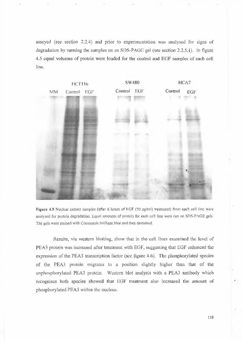
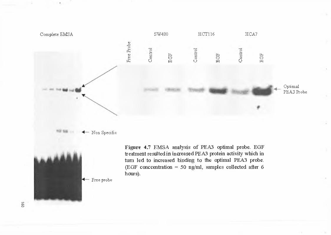
Cells use cytokines and other factors to ‘sense’ their external environment and for example when a cell receives a stimulus to proliferate a number of events occur such as entry into the cell cycle and D N A replication. In order for the cell to divide it must loosen the bonds of the E C M which anchor it and this is achieved through the regulation of cell adhesion molecules and the expression of proteases such as M M P s (Beksac el al., 2000 and Madri and Graesser, 2000). If M M P s are therefore expressed in response to external stimuli from cytokines it is possible that aberrant cytokine expression could result in the overexpression of M M P s resulting both in increased cell proliferation and increased E C M degradation.
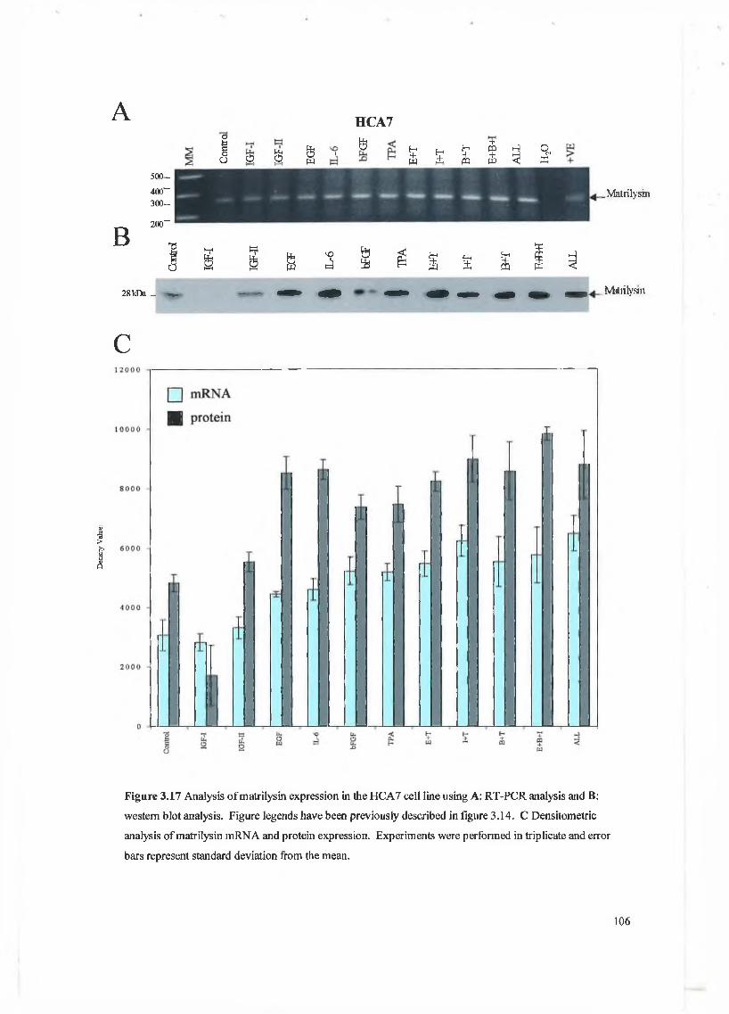
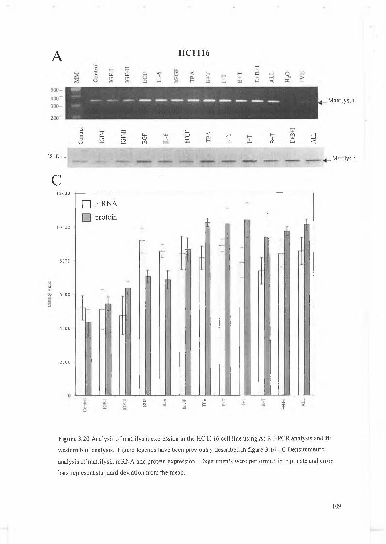
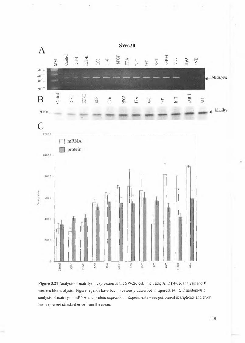
This chapter focuses on the expression and regulation of matrilysin by several cytokines in a panel of colon cell lines. We have chosen the colon system as much work to date has shown that matrilysin expression is critical in colon tumour formation, proliferation, invasion and metastasis and is unique in that it is the only M M P expressed exclusively by the tumour epithelial cells. W e have examined a panel of cytokines for their effect on matrilysin expression in these cell lines and hope to establish that cytokine regulation is an important feature in matrilysin expression during colorectal tumourigenesis. The cytokines examined here include IGF-I, IGF-II, EGF, bFGF, IL-6 and a known upregulator of M M P expression 12-O-tetradecanoyl-phorbol-l 3-acetate (TPA).
3.4.1 EGF and the EGF receptor (EGF-r)
EGF is part of a large family of cytokines which can be characterised by the fact that they contain at least one extracellular EGF structural unit (a conserved 6-cysteine
motif that forms three disulphide bonds). Family members include EGF itself, transforming growth factor-a (TGF-a), Heparin binding-epidermal growth factor (HB- EGF), amphiregulin (AR), betacellulin (BTC), sensory and motor neuron derived factor (SMDF) and the heregulins (HRGs). EGF is secreted as a 9 kDa protein which is proteolytically cleaved to yield an active 6 kDa form. It is a multifunctional cytokine most commonly associated with stimulating the growth of epithelial cells and mediates this effect by binding to the EGF-r. The EGF-r is a common receptor for EGF, TGF-a, A R and HB-EGF and the genes encoding for each of these molecules are important in growth and development. The EGF-r has tyrosine kinase properties which are used in the transduction of external signals and is a member of the src group of oncogenes. Although the EGF-r is expressed by a wide variety of adult cell types in vivo and cell lines in culture, EGF was so named because of its first recognised activity in an in vivo assay for epidermal maturation. Tissues that continue to divide in order to renew themselves generally have more EGF-r present or are more responsive to EGF than other tissues. These tissues would include the epithelial lining of the gut and epidermal layer of the skin (Wilson and Gibson, 2000).
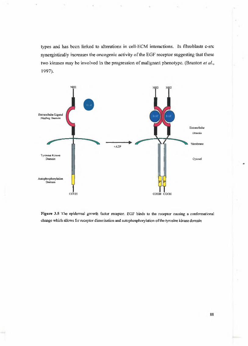
The EGF-r gene encodes a 170 kDa transmembrane receptor with an intracellular domain similar to that of the src gene product (see figure 3.5). The EGF-r is characterised by its ability to autophosphorylate and upon autophosphorylation dimerisation with another nearby EGF-r occurs. The tyrosine kinase receptor can then carry out its function via the phosphorylation of signaling molecules docked to its intracellular domain. The EGF receptor as has been stated is a trans-membrane receptor with intrinsic tyrosine kinase activity and there have been numerous reports on the expression of this receptor and its ligands, EGF and TGF-a, in colon tumours. In an immunohistochemical study, synchronous expression of EGF and EGF-r in highly invasive colon carcinomas suggested that an EGF autocrine loop may play a role in the regulation of these tumours (Karamerias et al., 1993). Such autocrine loops have been described in several colon carcinoma cell lines where cell growth was inhibited by using antisense TGF-a and blocking antibodies to the EGF receptor (Karnes et al., 1992). It has also been reported that the expression of EGF-r by colon carcinoma cells directly correlates with their metastatic potential (Radinsky et al., 1995). EGF promotes the migration and invasion of a number of cell
87
types and has been linked to alterations in cell-ECM interactions. In fibroblasts c-src synergistically increases the oncogenic activity of the EGF receptor suggesting that these two kinases may be involved in the progression of malignant phenotype. (Brunton et al.,
1997).
NH2
Extracellular Ligand Binding Domain
Tyrosine Kinase Domain
AutophosphoiylationDomain T
+ATP
Si
COOHTT
Extracellular
Domain
Membrane
Cytosol
COOH COOH
Figure 3.5 The epidermal growth factor receptor. EGF binds to the receptor causing a conformational
change which allows for receptor dimerisation and autophosphoiylation o f the tyrosine kinase domain
Membrane bound IGF
IGF receptor
ExtracellularDomain
Membrane
Cytosol
Figure 3.6 The IGF system. Several proteases including MMPs have been implicated in the production of
biologically active IGF
3.4.2 IGF-I and IGF-II
Insulin like growth factors (IGF) are so called because of their ability to bind to the insulin receptor (also known as the IGF-I receptor). The IGF family is comprised of IGF-I and IGF-II, both of which have an active molecular weight of 7.5 kDa. IGFs are multifunctional polypeptides that are capable of regulating growth, diiferentiation and survival in several cell and tissue types. The tGF system includes ligands (IGF-I and IGF- II), receptors (IGF-IR and IGF-IIR), IGF binding proteins (IGFBP-1-7) and IGFBP proteases (see figure 3.6).
The IGF system regulates pre- and postnatal growth, the establishment and maintenance of differentiated çdl function via paracrine, autocrine and endocrine signalling. ÎGFs are jpolent mitogenic and anti-apoptotic molecules involved in the regulation of cell proliferation in renewing epithelial cell populations of organs including
89
breast, prostate, colon and lung. Unlike many other regulatory peptides, IGFs have characteristics of both classic ‘endocrine’ hormones and also characteristics of tissue growth factors (Jones and Clemmons, 1995 and Rajaram et al., 1997). Circulating levels of IGFs are subject to complex physiological regulation and the vast majority of IGFs are bound to high affinity binding proteins which need to be proteolytically cleaved in order to produce the active IGF cytokine. It has been shown that >90% of circulating IGF is bound to a high molecular weight complex comprised of IGFBP-3 and a separate protein known as the acid labile sub-unit. However, IGF bioactivity in tissues is not merely a function of circulating levels, as local expression of genes encoding IGFs, IGFBPs, and proteases that digest IGFBPs are also important (Ramajaran et al., 1997). The classic source of IGF-I is the liver, where the peptide is produced in response to growth hormone. It is considered a primary growth regulator in pre and post-natal life. In contrast to IGF-I, the physiological function of IGF-II is mainly involved in stimulating undifferentiated cells and plays a key role in foetus development (Poliak, 2000). The IGF-I receptor is structurally very similar to insulin and is comprised of 2a subunits and 2(3 subunits which are linked by disulphide bonds (see figure 3.7). The receptor is comprised of an extracellular, transmembrane and intracellular domain. When a ligand binds, the IGF-I receptor undergoes rapid auto-phosphorylation and the intracellular domain once phosphorylated is a potent tyrosine kinase and mediates its effects through the phosphorylation of secondary signaling molecules. The IGF-II receptor is similar to the receptor for mannose-6-phosphate, is structurally very different to that of IGF-I, and is capable of binding both IGF-I and IGF-II. As IGFs are potent mitogenic agents it is quite possible that aberrant gene expression may result in an increased rate of epithelial cell division which in conjunction with other oncogene expression may induce malignant transformation.
90
A B
Cysteine rich region
NH2 NH2 NH2
NH2
Tyrosin kinase domain
-S-S-
-S-S- NH2
-S-S-COOH COOH
Extracellular
Domain
Membrane
Cytosol COOH
COOII COOII
Figure 3.7 A The IGF-I receptor is comprised of 2a and 2(3 subunits. Once the IGF ligand has bound the
intracellular tyrosine kinase domain is activated. B The IGF-II receptor which is similar in structure to the
mannose-6- phosphate receptor
3.4.3 Interleukin-6
Interleukin-6 (IL-6) is a member of the family of cytokines collectively termed “the interleukin-6 type cytokines”. IL-6 is variably glycosylated and the 22-27 kDa secreted glycoprotein serves as a prototype for a family of molecules that includes leukemia inhibitory factor (LIF), oncostatin M (OSM), ciliary neurotrophic factor (CNTF), cardiotrophin-1 (CT-1) and IL-11. Although all these molecules possess a similar helical structure, their association is due to their functional redundancy and receptor interactions (Hibi et al., 1996). IL-6 achieves its effects through the ligand-specific IL-6 receptor (IL-6R). Unlike most other cytokine receptors, the IL-6R is active in both membrane bound and soluble forms. Among its many functions, IL-6 plays an active role in immunology, bone metabolism, reproduction, arthritis, neoplasia, and aging. IL-6 expression is regulated by a variety of factors, including steroidal hormones, at both the transcriptional and post-transcriptional levels (Keller et al., 1996).
91
P-chaingp130
a-chain
Extracellular
Domain
Membrane
Cytosol
Ig superfamily domain
Fibronectin type III domain
Trp-Ser-X-Trp-Ser motif
Contactin/fibronectin III spacer modules
COOH
Figure 3.8 The IL-6 receptor is comprised of 2 chains, the a/gp80 chain and the p/gpl30 chain.
The human IL-6 receptor, also known as gp80 and IL-6R, is an 80 kDa protein consisting of 467 amino acids (Keller et ah, 1996). The receptor is comprised of an extracellular region, a transmembrane region and an intracellular region. The IL-6R is not capable of mediating signal transduction directly and must initially bind to gp-80 which gives rise to a low affinity receptor complex. Homodimerisation of gp-130 is subsequently required for IL-6 signal transduction (Murakami et ah, 1993) (see figure 3.8). Although it was originally considered that one unit of IL-6 and the IL-6R bound to a gp-130 homodimer, the stoichiometry and number of this reaction appears to involve a hexameric complex consisting of two molecules each of IL-6, IL-6R gp-80, and gp-130 (Hammacher et ah, 1994). This complex forms a high affinity binding site for IL-6, as opposed to the low affinity binding observed with IL-6 and IL-6R gp-80 in the absence of gp-130.
92
The IL-6 receptor is expressed in a variety of cells and the number of receptors normally range between 100-2000 sites per cell. However, in myeloma cell lines up to 29,000 sites per cell have been observed (Snyers et al., 1989). The receptor has also been shown to be upregulated in several cancers including bladder and prostate cancer (Meyers et al., 1991 and Siegsmund et al., 1994). IL-6 is a pleiotropic cytokine and is involved in initiating several cellular processes including proliferation and differentiation. IL-6 is also involved in the inflammatory response cascade and is often found at high levels in the stroma surrounding advancing tumours. Many of the promoters of M M P genes contain transcription factor binding elements to which transcription factors triggered by IL-6 such as NF-IL6 (nuclear factor interleukin-6), can bind and these will be discussed later. Therefore, a possible link with cancer proliferation and overexpression of MMPs, including matrilysin, due to increased expression of IL-6R or IL-6 by the tumour cell may exist.
3.4.4 Basic fibroblast growth factor (bFGF)
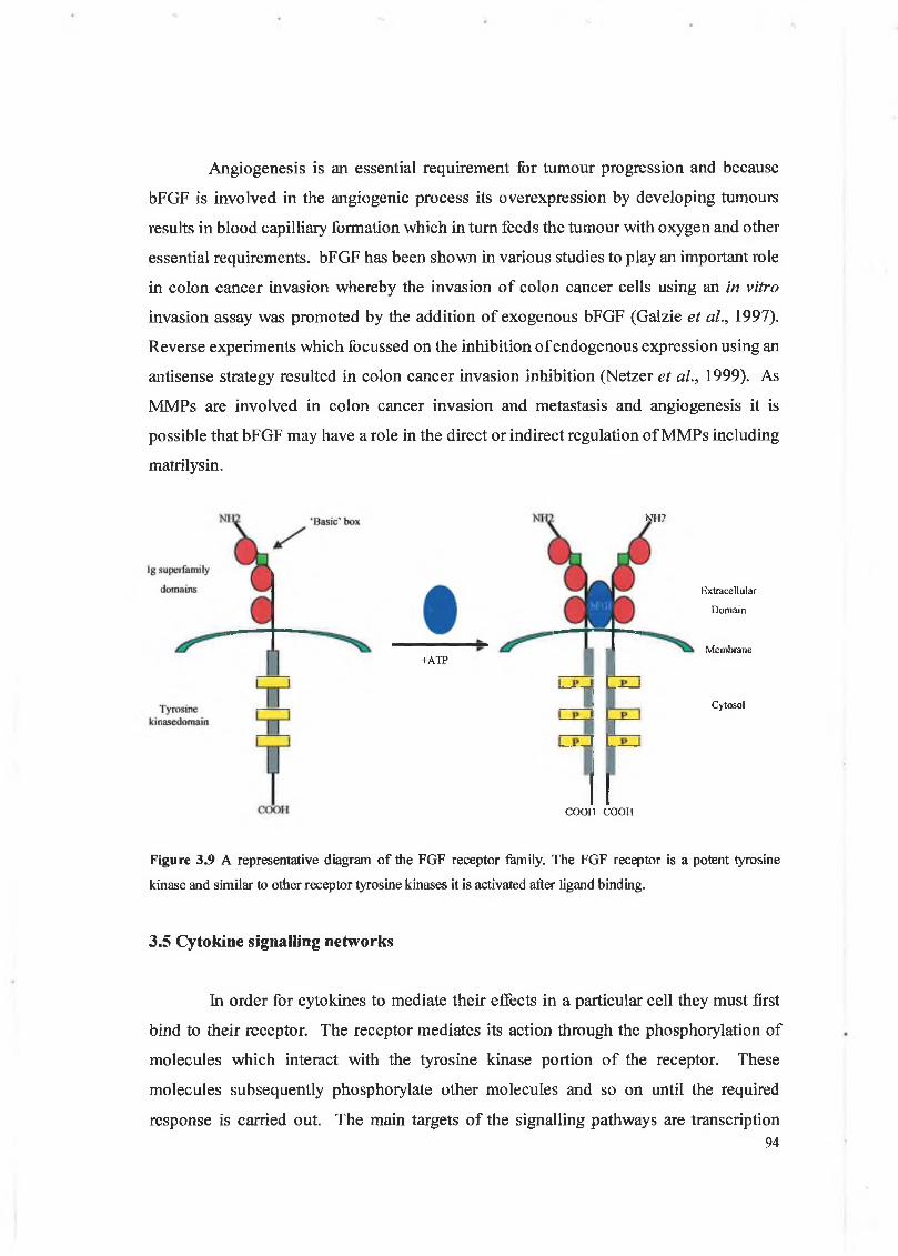
Fibroblast growth factors are a family of secreted peptide ligands containing at least 15 members. Signalling by the FGFs and their receptors, which are a family of 4 transmembrane protein tyrosine kinases in mammals, is critical for diverse aspects of embryonic, foetal and post natal development of a variety of tissues (see figure 3.9) (Matsui et al., 1999). Acidic and basic fibroblast growth factors (aFGF and bFGF) were among the first members of the FGF family to be isolated. bFGF plays a role in autocrine and paracrine regulation of cell proliferation, migration, angiogenesis and vascular development (Olson et al., 2000 and Sezer et al., 2001). bFGF is secreted as an 18 kDa protein and mediates it’s effects through binding to the FGF receptor-2 (FGFR-2). Various forms of bFGF also exist in that the gene which encodes for the bFGF protein has several transcription initiation start sites. bFGF has also been shown to be essential in the formation of monolayers of cells and has been found to be present as an insoluble proteoglycan incorporated into the E C M which indicates its role as a local regulator of cell migration, proliferation and regeneration during wound healing (Aktas and Kayton, 2001).
93
Angiogenesis is an essential requirement for tumour progression and because bFGF is involved in the angiogenic process its overexpression by developing tumours results in blood capilliary formation which in turn feeds the tumour with oxygen and other essential requirements. bFGF has been shown in various studies to play an important role in colon cancer invasion whereby the invasion of colon cancer cells using an in vitro
invasion assay was promoted by the addition of exogenous bFGF (Galzie et al., 1997). Reverse experiments which focussed on the inhibition of endogenous expression using an antisense strategy resulted in colon cancer invasion inhibition (Netzer et al., 1999). As M M P s are involved in colon cancer invasion and metastasis and angiogenesis it is possible that bFGF may have a role in the direct or indirect regulation of M M P s including matrilysin.
N112
i ATP
Extracellular
Domain
Membrane
Cytosol
COOH COOH
Figure 3.9 A representative diagram of the FGF receptor family. The FGF receptor is a potent tyrosine
kinase and similar to other receptor tyrosine kinases it is activated after ligand binding.
3.5 Cytokine signalling networks
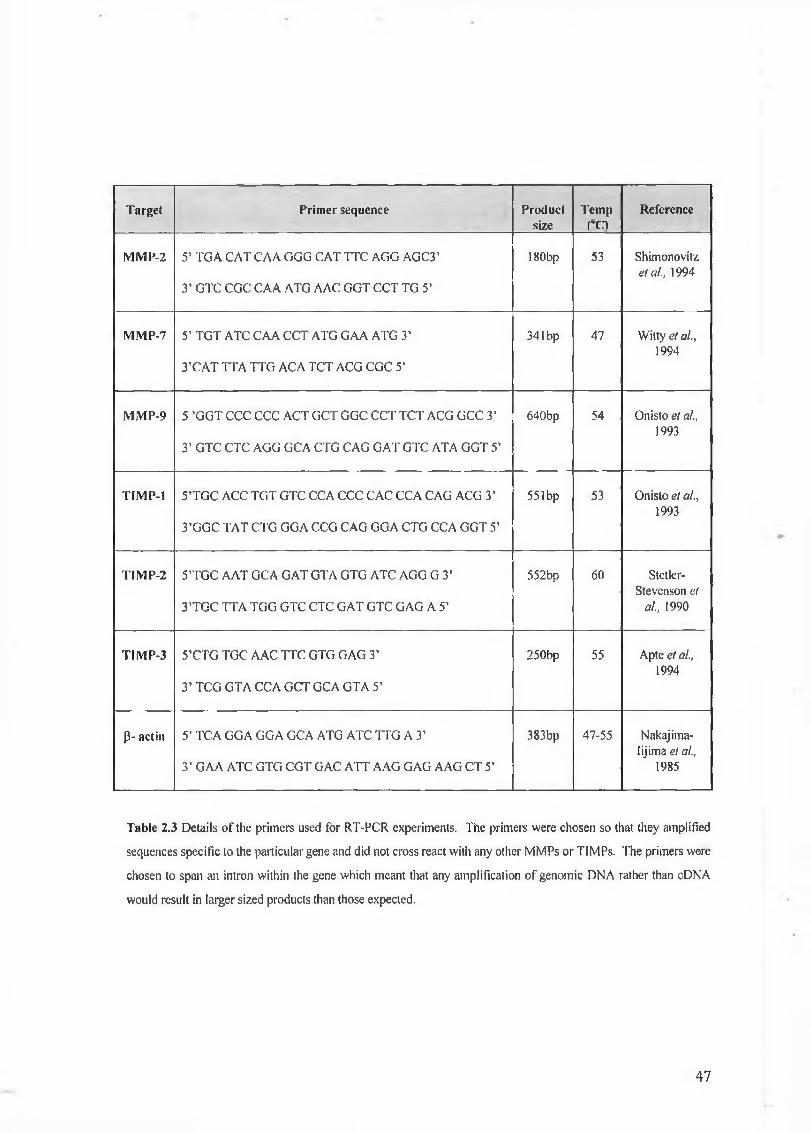
In order for cytokines to mediate their eifects in a particular cell they must firstbind to their receptor. The receptor mediates its action through the phosphorylation ofmolecules which interact with the tyrosine kinase portion of the receptor. Thesemolecules subsequently phosphorylate other molecules and so on until the requiredresponse is carried out. The main targets of the signalling pathways are transcription
94
factors which bind to the promoter region of target genes. There are many important pathways involved in cell signalling and many of these ‘cross talk’ in that the pathways overlap each other or use the same signalling molecules. Signalling pathways can also overlap to enhance or silence a particular signal. The regulation of these pathways is also important and aberrant expression of mutated receptors which continually ‘fire’ signals at the nucleus wreak havoc on the cell and normally result in excessive proliferation which when accompanied by other mutations will result in cancer development.
There are many signalling pathways used to effect cellular processes, for example, the mitogen activated protein kinase (MAPK) pathway the phosphotidyl inositol-3 pathway (PI-3) and the janus kinase/ signal transducer and activator of transcription (JAK/STAT) pathway. Several of the receptors outlined above also engage these pathways in order to mediate their desired effect. One of the first molecules engaged by many of the receptor tyrosine kinases (RTKs) is the Ras protein.
The M A P K pathways can effect a wide variety of processes but they are primarily involved in stimulating cell proliferation. Common transcription factors stimulated by the M A P K effector molecules include those of the AP-1 and Ets family. As discussed earlier many of the M M P gene promoters including that of matrilysin contain D N A binding regions for these transcription factors and therefore stimulation of the M A P K pathway may lead to increased proliferation as well as increased M M P gene transcription.
3.5.1 Ras and the M APK pathways
Ras proteins activate multiple signalling pathways (see figure 3.10). Ras binds to at least three types of effector protein : i) kinases of the raf family including M A P K and ERK, ii) phosphoinositide-3 kinases (PI-3) and iii) RalGDS (guanine nucleotide disassociation stimulator) proteins (McCormick, 1999). The molecular mechanisms that lead to the activation of Ras effectors are complicated and unclear and the downstream consequences of activation are more complicated still. Both the Raf and PI-3 pathways have been shown to be involved in proliferation, migration and angiogenesis via their signalling networks and their complex interaction or ‘cross talk’ with other networks. The
95
RalGDS is also involved in gene transcription and its mechanisms of signalling are now being elucidated.
Several receptor tyrosine kinase (RTKs) including the EGFr and the FGFr use Ras in order to signal to the cell nucleus. Tyrosine phosphorylation of specific residues on the internal domain or the receptor creates binding sites for several molecules including Src- homology 2 (SH2) and phosphotyrosine binding (PTB) domain containing proteins. Some of these proteins are enzymes that are tyrosine phosphorylated and thereby activated, such as Src, phospholipase Cy (PLCy) and PI-3 kinase, whereas She, Grb2, Grb7 and Nek are adaptor molecules that link RTKs to downstream signalling pathways (Hackel et al., 1999).
Growth factor receptor bound protein 2 (Grb2) is a small adaptor protein that is essential in the Ras signalling pathway (Lowenstein et al., 1992). Il is comprised of one SH2 domain of approximately 100 amino acids surrounded by two SH3 domains, each containing about 60 amino acids. After receptor dimérisation and tyrosine transphosphorylation Grb2 docks with the internal domain of the receptor and upon phosphorylation Grb2 then recruits SOS proteins. SOS accelerates the change of GDP to GTP in Ras, which leads to Ras activation. Once loaded with GIT, Ras is then capable of interacting with effectors such as Raf which ultimately leads to the activation of the mitogen activated protein kinase (MAPK) pathway.
96
1eI
M A PR (p42/p44 M A P k )
Cytoplasm
IGene transcription
Cyclin D1HB-EGFFosp21
MMPs ?
Nucleus
Proliferation
Figure 3.10 Once activated, membrane bound Ras proteins activate a number of signalling pathways
including the mitogen activated protein kinase pathway. An increase in Fos and other AP-1 transcription
factor family members may also lead to an increase in MMP activity.
The chain of events leading to M A P K activation take place within the cell cytoplasm. Having been recruited to the cell membrane and activated by Ras, Raf proceeds to phosphorlyate, hence activate, serine residues on the mitogen activated protein kinases 1 and 2 (MKK1, MKK2). The discovery of the first mammalian M A P K pathway was based on the identification of p42 and p44 MAPKs (Ray and Sturgill, 1987). p42/p44 MAPKs, once activated, have been shown to be translocated to the nucleus upon growth factor stimulation (Lenormand et al., 1993). This nuclear translocation allows the MAPKs to contact and phosphorylate key substrates. In vitro studies show that p42/44 MAPKs have the ability to phosphorylate a number of substrates including EGF-r,
phospholipase A2, protein kinase p90RSK, Myc and Elkl, however, in vivo the p42/p44 M A P K s have been best characterised by their interaction with the nuclear transcription factor Elkl. After phosphorylation by MAPK, Elkl is responsible for the induction of c-
fo s gene expression. The p42/p44 M A P K pathway can, therefore, integrate the signals coming from various growth factor receptors and by translocation to the nucleus and activation of transcription factors, relay this information to modulate gene expression accordingly.
3.5.2 Ras and the PI-3 kinase pathway
Oncogenic Ras suppresses apoptosis through the PI-3 kinase pathway, partly through the activation of protein kinase B (PKB) (see figure 3.11). The precise role of PKB in suppressing apoptosis is complex: PKB phosphorlyates BAD, a member of the Bcl-2 family that promotes cell death, and caspase 9, a protease that degrades cellular substrates during the process of cell death. However, PKB also regulates transcription through the phosphorylation of Forkhead proteins, which might themselves regulate other effectors of apoptosis, suggesting a much more complex and indirect route to cell survival (McCormick, 1997). Ras activated PI-3 kinase also plays a role in promoting cell growth through the inhibition of glycogen synthase kinase 3 (GSK3) by PKB. Activated GSK3 is capable of degrading, via ubiquitination, the cyclin D1 proteins which drive the cell cycle from the G1 to the S phase. Inhibition of GSK3 therefore promotes cell division through the accumulation of cyclin D1 (Garbay et al., 2000).
The PI-3 kinase pathway may also stimulate the production of M M P s indirectly via the increase of transcription factors associated with cell proliferation. As discussed earlier matrilysin has been shown to cleave FasL into its soluble form and is involved in FasL/Fas mediated apoptosis. Since the PI-3 kinase pathway can increase levels of Fas L it may also be appropriate that the pathway lead to an increase in molecules which are capable of converting FasL to its soluble form such as matrilysin.
98
l 'I - ' Kina
í I
msm
i i
MikhtóíU
CellMembrane
Cytoplasm
Nucleus
Gene transcription
Fas
MMPs ?Apoptosis or
Proliferation
Figure 3.11 Ras is also capable of activating the PI-3 kinase pathway which has numerous effects.
3.5.3 The JAK/STAT pathway
The STATs (signal transducers of and activators of transcription) constitute a multiple member family of signal transduction proteins that are activated in the cytoplasm by the binding of extracellular polypeptides to the transmembrane receptors which then regulate the transcription of immediate response genes (Horvath and Darnell, 1997), (see figure 3.12). The Janus kinase-signal transducer and activator of transcription factor (JAK-STAT) signalling pathways were originally identified in the study of interferon signalling and are now believed to be the important pathways in signal transduction for many cytokines including IL-6. These pathways have been implicated in a variety of cellular functions in the haematopoietic, immunologic, neuronal and hepatic systems (Chen et al., 1999). It is well established that binding of IL-6 to the IL-6 receptor-a chain induces homodimerisation of the signal transducing (3 chain, gpl30, which is followed by
99
the activation of the receptor associated tyrosine kinases JAK1, JAK2 and Tyk2. A process assisted by ROS (reactive oxygen species). This receptor tyrosine kinase complex interacts with and activates the Src homology 2 (SH2) containing cytoplasmic STAT3 transcription factor, which then translocates to the nucleus to activate the transcription of many target genes including JunB, C C A A T enhancer binding protein (C/EBP) and p21 WA '/C'pi (Chen et al, 1999). Although it is clear that JAKs serve to phosphorylate STATs in receptors that lack intrinsic tyrosine kinase domains, it has also been shown that RTKs have the ability to activate STATs without recruiting JAKs (Horvath and Darnell, 1997).
IL-6receptor .11 U — = = = = = = = = =f t ) - ! JAKs
CellMembrane
1STATs
tCytoplasm
Nucleus
Gene transcriptionIL-6
IL-2 m-CSF
IL-8 MMPs
INF PA
gm-CSF
Various
Responses
Figure 3.12 Activation of the JAK/STAT pathway leads to the activation o f several transcription factors and
the production of various gene products including IL-6 and MMPs
100
3.6 Summary
Matrilysin is the smallest M M P family member, has one of the largest substrate specificities and has been shown to play a role in many normal and disease processes. Much of the research into matrilysin thus far, has been associated with its role in cancer invasion and metastasis, in particular, its involvement in the spread of colon and breast tumours. More recently, matrilysin has been shown to play an important role in several aspects of colon and breast tumour progression with respect to initial tumour formation, growth and angiogenesis. Matrilysin is also unique in that it is the only M M P member expressed exclusively by the epithelial cells of advancing colon tumours. These characteristics of matrilysin have combined to make it an exciting therapeutic target. While much is known about the matrilysin protein and its substrates, relatively little is known about how matrilysin gene expression is regulated. Cells in order to function properly need to ‘sense’ their external environment and this is achieved through signalling molecules such as cytokines and signalling pathways which carry these signals to the nucleus. Overexpression of cytokines or their receptors in combination with other mutations leads to the uncontrolled proliferation of cancer cells. As matrilysin is involved in the growth and invasion and metastasis of colon cancer it is quite possible that aberrant cytokine signalling may subsequently lead to matrilysin overexpression in tumours however the mechanisms of this interaction between cytokine stimulation and matrilysin expression has yet to be elucidated. In order to determine if a link exists we have taken a panel of colon cell lines and investigated whether matrilysin expression at the transcriptional level and protein level can be affected by cytokines which employ several different signalling pathways in mediating their effects.
101
3.7 Results
A panel of colon cancer cell lines (HCT116, SW480, SW620, LoVo, WiDr and HCA7) were investigated with respect to their expression of matrilysin and if this expression could be regulated via cytokine treatment using IGF-I, IGF-II, EGF, IL-6 and bFGF. Properties of the cell lines used and a summary of the oncogenes expressed in each are illustrated in table 2.1 in section 2.2.1. The HCA7, LoVo and SW480 cell lines all represent early to late adenocarcinomas while HCT116, SW620 and W i D R represent colon carcinomas at later stages of development. The SW480 and SW620 cell line have both been isolated from the same patient and are a genetically matched pair. SW480 was established from a primary adenocarcinoma in the colon which was classified as Duke’s B while the S W620 cell line was derived from a lymph node metastasis of the same tumour from which SW480 was derived. Interestingly, SW480 has been shown not to express matrilysin while SW620 has previously been shown to express matrilysin at the basal level (Witty et al., 1994).
3.7.1 RNA isolation from colon cancer cell lines
Prior to reverse transcription polymerase chain reaction analysis the total R N A isolated from each cell line was examined for degradation by running an aliqout of each isolated R N A sample (see figure 3.13). The presence of 28s, 16s and 6s R N A ribosomal sub-units is typical of undegraded RNA.
28 s
1 6 s
6 s
Fig 3.13 Total RNA was isolated and examined for the presence of 28s, 16s and 6s ribosomal sub-units.
The samples shown here were isolated from the LoVo cell line (left to right : Control, IGF-I, IGF-II, EGF,
IL-6, bFGF, TP A) and similar results were obtained for other cell lines.
Isolated R N A was quantified by measuring the absorbance of the samples at 260nm (1 O D unit equals 40 pg RNA). The quality of R N A was also determined by measuring the absorbance of the samples at 280nm. The absorbance 260/280 ratio is typically between 1.6 and 1.8 for total R N A free of DNA, protein and organic chemical contaminants.
3.7.2 RT-PCR/W estern analysis of matrilysin expression in colon cancer cell lines
Once the total R N A had been isolated and purified, lpg of sample was used in the RT-PCR procedure outlined in section 2.2.3. The RT-PCR procedure was semi- quantitative and P-actin, a constitutively expressed cytoskeletal structural protein, was used as an internal control. Due to the similarity in product size the p-actin PCR reaction was performed in a separate tube to that of the matrilysin PCR reaction. The levels of (3- actin served to normalise the amount of matrilysin m R N A expression observed in control and cytokine treated cell lines. Figure 3.14 shows P-actin levels for control and treated samples for the LoVo cell lines. The figure illustrates that cytokine treatment of the LoVo cell line has no effect on P-actin levels. This result was consistent in all cell lines investigated and therefore the levels of P-actin in the LoVo cell line are illustrated as an example while the p-actin data for other cell lines are not shown.
Cytokines used in the study included IGF-I, IGF-II, EGF, bFGF. TPA was also used as a positive control as it has been previously shown to upregulate M M P s (Mackay et
al, 1992). Figure 3.15 demonstrates RT-PCR analysis of matrilysin m R N A in the LoVo cell line in normal and cytokine treated samples. The lower panel in the figure shows the entire gel and illustrates that non-specific binding of the matrilysin primers did not occur and the expected product size of 341 bp was obtained. This was also observed in the other cell lines investigated. In order to clearly see increases/decreases in matrilysin m R N A levels, the section which contains the matrilysin PCR products has been enlarged. Subsequent RT-PCR and western blot analysis have also been presented in this enlarged manner.
103
Figure 3.14 RT-PCR analysis of P-actin mRNA expression in the LoVo cell line. The expected PCR
product size for P-actin was 383 bp. MM; molecular marker, IGF-I; Insulin like growth factor I, IGF-II;
Insulin like growth factor II, EGF; epidermal growth factor, IL-6; interleukin 6, bFGF; basic fibroblast
growth factor, TP A; tetradecanoyl, phorbol acetate, E+T; cells treated with EGF and TPA, I+T; cells treated
with IL-6 and TPA, B+T; cells treated with bFGF and TPA, E+B+I; cells treated with EGF, bFGF and IL-6,
ALL: cells treated with EGF, IL-6, bFGF and TPA.
2 0 0 “
Fig 3.15 RT-PCR analysis of matrilysin expression in the LoVo cell line. Matrilysin PCR product size was
341 bp. Concentration of all cytokines and TPA were 50ng/ml. MM; molecular marker, IGF-I; Insulin like
growth factor I, IGF-II; Insulin like growth factor II, EGF; epidermal growth factor, IL-6; interleukin 6,
bFGF; basic fibroblast growth factor, TPA; tetradecanoyl, phorbol acetate, E+T; cells treated with EGF and
TPA, I+T; cells treated with IL-6 and TPA, B+T; cells treated with bFGF and TPA, E+B+I; cells treated
with EGF, bFGF and IL-6, ALL: cells treated with EGF, IL-6, bFGF and TPA, H20: sterile water used as a
negative control in PCR, +ve; positive PCR control using matrilysin cDNA. Illustrated molecular marker
units are in base pairs. The lower gel indicates that no non-specific binding occurred, an observation which
was consistent in all cell lines examined.
104
The effects of cytokine treatments on matrilysin m R N A expression on each of the cell lines investigated are displayed in figures 3.17 to 3.21. In order to confirm that the cytokine effects on matrilysin expression at the m R N A level were resulting in higher levels of matrilysin protein, western blot analysis was also performed on the normal and cytokine treated cell lines (for methodology see section 2.2.6). Supernatants were measured for total protein content and equal amounts of total protein were loaded onto SDS-PAGE gels, ensuring that the protein levels detected are as a direct result of cytokine treatment. These results for each individual cell line are also shown in figure 3.17 to 3.21.
Matrilysin m R N A and protein was detected in all cell lines with the exception of SW480 (see figure 3.16 and data not shown).
SW480
M K +S ^ E E o £ ?2 u ¿2 w r i Ë H w + m ü j < x +
Figure 3.16. Matrilysin was not detected at the mRNA level in the SW480 using RT-PCR. Treatment with
various cytokines and cytokine combinations failed to initiate the transcription of matrilysin. Western blot
analysis also failed to detect the presence of matrilysin protein in SW480 samples (data not shown).
Experiments on both m R N A and protein expression were performed in triplicate. Densitometry was performed on the gels and blots and the average results with standard deviation from the mean (Microsoft Excel™) are detailed in figure 3.17-3.21.
105
Dens
ity
Val
ue,'
A
B
5 0 0 -
400“3 0 0 -
HCA701O P.
t-Ha&ow i
t-tso % £H+m
+¥w $ q,m
.Matrilysin
28kDa -
200“
1 2 1 § s§ ECF
IL-6
bFGF
TPA
E+T
I+T S 1 i
- — —— — — — — — - - — .Mitrilysn
c
Figure 3.17 Analysis of matrilysin expression in the HCA7 cell line using A: RT-PCR analysis and B:
western blot analysis. Figure legends have been previously described in figure 3.14. C Densitometric
analysis of matrilysin mRNA and protein expression. Experiments were performed in triplicate and error
bars represent standard deviation from the mean.
1 0 6
Dens
ity
Val
ueWiDr
Matrilysin
JVkritysn
2 00 0
Figure 3.18 Analysis of matrilysin expression in the WiDr cell line using A: RT-PCR analysis and B:
western blot analysis. Figure legends have been previously described in figure 3.14. C Densitometric
analysis of matrilysin mRNA and protein expression. Experiments were performed in triplicate and error
bars represent standard deviation from the mean.
107
Dens
ity
Val
ueLoVo
A â â & i ipH m
Mairi lysin
28kDa
cMainly sin
2 0 0 0 0
1 6 0 0 0 1
1 4 0 0 0
I 2 0 0 0
1 0 0 0 0
2 000
□ m R N A ] protein
% ¥«
Figure 3.19 Analysis of matrilysin expression in the LoVo cell line using A: RT-PCR analysis and B:
western blot analysis. Figure legends have been previously described in figure 3.14. C Densitometric
analysis o f matrilysin mRNA and protein expression. Experiments were performed in triplicate and error
bars represent standard deviation from the mean.
108
A H C T 116
L Matri lysin
c
oJ=1o
p-<O tL.o P-Mawp-aIX, < E—*
+ PJ
+PQ
CQ-f-PJ
d<
28 kD a - .Matri lysin
1 2 0 0 0
10000
8 0 0 0
■a>& 6 0 0 0
4 0 0 0
2000
□ m R N A□ protein
J.Oui
r
oU-i
k
<& ?w
Figure 3.20 Analysis of matrilysin expression in the HCT116 cell line using A: RT-PCR analysis and B:
western blot analysis. Figure legends have been previously described in figure 3.14. C Densitometric
analysis of matrilysin mRNA and protein expression. Experiments were performed in triplicate and error
bars represent standard deviation from the mean.
109
A
5 00 —
400
300 —
2 0 0 “ I
O¿3GOO o
SW620
ai
ai-baw j
dH+W
H PQ -3+ + >“]B B <OCN
*
M a tr ily s ir
BOGOO
P<a o
voI
hJtbaU-i <cu
E— UJ i-H+ +■CQ+CQ+LlJ
28 kDa _
c_Matrilys
ow t00
Figure 3.21 Analysis of matrilysin expression in the SW620 cell line using A: RT-PCR analysis and B:
western blot analysis. Figure legends have been previously described in figure 3.14. C Densitometric
analysis of matrilysin mRNA and protein expression. Experiments were performed in triplicate and error
bars represent standard error from the mean.
110
Time and dosage experiments found that the optimal time point for endogenous matrilysin m R N A expression subsequent to cytokine treatment was 8 hours. Time points of 2, 4, 6 12 and 24 were also examined (data not shown). Cytokine concentrations of 10 ng/ml, 25 ng/ml, 50 ng/ml and 100 ng/ml were examined and 50 ng/ml of each cytokine was found to be the maximum concentration for induction (data not shown). For protein analysis, cell lines were incubated with 50 ng/ml of the appropriate cytokine for 48-72 hours.
Expression of matrilysin in the HCA7 cell line was observed at both the m R N A and protein levels (see figure 3.17). An increase in matrilysin m R N A expression was generally observed when the cells were treated with EGF, IL-6, bFGF or TPA. This increase was also observed at the protein level using western blot analysis. The effect of bFGF on matrilysin protein expression in the HCA7 cell line appears to be similar to that of the control level but subsequent studies showed that bFGF was capable of stimulating matrilysin protein expression in this cell line in a similar manner to that of EGF and IL-6 as shown by densitometric analysis. Interestingly, IGF-I appears to have little or no effect on matrilysin expression and seems to inhibit the production of matrilysin protein. However repeat experiments do not consistently show a significant inhibition in matrilysin protein expression in the H C A 7 cell line upon treatment with IGF-I suggesting that IGF-I has little or no effect on the regulation of matrilysin expression. IGF-II also appears to have little or no effect on matrilysin regulation in the HCA7 cell line. In vivo there may be many signals from different signalling molecules converging on the matrilysin promoter in order to initiate its transcription. Therefore matrilysin expression may not necessarily be initiated/enhanced by a single cytokine but in response to a number of cytokines working in concert to promote matrilysin gene expression. W e therefore investigated if treatment of the HCA7 cell line and other cell lines with a combination of cytokines would further enhance matrilysin gene transcription. Combination of cytokines (outlined in figure 3.17) showed an increase in matrilysin expression in the HCA7 cell line, in particular at the m R N A level, but these increases were not much greater than those observed when the cells were treated with individual cytokines such as EGF. Cells were also treated with IGF-I and IGF-II in combination with TPA but the observed effects were similar to individual TPA treatment which again suggested that neither IGF-I or IGF-II were involved in the regulation of matrilysin in the HCA7 cell line (data not shown).
I l l
The WiDr cell line was also found to express matrilysin at both the m R N A and protein level. However in contrast to HCA7, cytokine treatment appeared to have little or no effect on the expression of matrilysin (see figure 3.18) at either the m R N A or protein level.
The LoVo cell line expressed matrilysin at the m R N A and protein level and this expression could be modulated by a number of cytokines including EGF, IL-6 and bFGF. TPA stimulates the expression of matrilysin m R N A and protein expression to a greater extent than EGF, IL-6 and bFGF. Combination of cytokines with TPA or with each other do not appear to stimulate matrilysin m R N A expression any more than individual treatments but the effect on matrilysin protein appears to be more pronounced. IGF-I and IGF-II do not appear to have any effect on matrilysin m R N A or protein expression (see figure 3.19).
The HCT116 cell line expressed matrilysin at the m R N A and protein level and this expression could be modulated by EGF, IL-6 and bFGF. Treatment of the HCT116 cell line also increased matrilysin m R N A and protein. Combination treatments appear to have little effect on the level of matrilysin m R N A expression in comparison to individual cytokine treatment but the effect was more pronounced at the protein level in a similar manner to that observed in the LoVo cell line. This may suggest that the m R N A half life was increased by combination treatment of the cells with various cytokines (see figure 3.20).
The SW620 cell line exhibited a similar matrilysin expression pattern in comparison to the HCA7, LoVo and HCT116 cell line in that the normal levels of matrilysin expression can be enhanced by EGF, IL-6 and bFGF. IL-6 and bFGF in particular appeared to strongly enhance matrilysin at the protein level in the SW620 cell line. TPA treatment was again found to enhance matrilysin expression. Combination treatments slightly enhanced matrilysin m R N A and protein expression (see figure 3.21).
112
3.7.3 Analysis of matrilysin gene expression using a promoter-lueiferase construct
In order to elucidate the mechanisms through which cytokines such as EGF, IL-6 and bFGF were stimulating matrilysin gene expression, two matrilysin promoter constructs were obtained. 2.3 kbp (pHMATPro-2.3-Luc) and 335 bp (pHMATPro-335- Luc) of the human matrilysin promoter were inserted into a luciferase reporter gene vector (see table 3.1). Thus increases in matrilysin promoter activity, as a response to cytokine treatment, could be determined by an increase in luciferase protein product. The regions of D N A within the matrilysin promoter responsible for transcription factor binding will be the subject of chapter 4. The promoter-reporter transient transfection technique is extremely sensitive and unlike RT-PCR is a quantitative method. The methodology involves transfection, cytokine treatment and luciferase detection which are outlined in the methods and materials section 2.2.8. On average transfection efficiencies of 10-20% were obtained (see figure 3.22)
Name Size Diagnostic Cut (Expected fragments)
Description
PH M A T Pro-2 .3-Luc 7.9 kb Eco RI (5.6 kb and 2.3 kb)
A 2.3 kb M fe I fragm ent o f the hum an m atrilysin
prom oter was cloned into the E coR l site o f
pB luescript (Stratagene). The fragm ent was then
cut using Hind III and BamHI and cloned into
the Hind III and Bgl II sites in the pG L2-Basic
vector (Prom ega).
PH M A TPi'o-335-Luc 5.9 kb Eco RI (5.6 kb and 335 bp)
T his plasmid was constructed in a similar
m anner to that o f pH M A T Pro-2.3-L uc with the
exception that only 335 bp o f the human
m atrilysin prom oter was inserted into the pG L2-
B asic vector
PR L-SV 40 3.7 kb Bam H I and Pst I (1.4 kb
and 2.3 kb)
C ontains the SV40 early enhancer/prom oter
region which gives strong constitutive
expression o f R enilla luciferase.
PC H 110 7.1 kb Pvu II (3.5 kb, 2.6 kb, 700
bp and 360 bp)
C ontains a functional Lac Z gene, the product o f
w hich is P-galactosidase. This plasmid can be
transfected into cells and transfection efficiency
can be determ ined by assaying for blue cells
using an X-gal stain
Table 3.1 Characterisation of plasmids used in transfection studies (see appendix 1 for plasmid maps)113
Figure 3.22 Transfection conditions for each cell line was optimised using (3-galactosidase activity. Cells
which were transfected attained the ability to process X-gal which results in a blue colour. SW620
transfected cells can be clearly observed against non-transfected cells.
In order to normalise transfection efficiencies between each experiment an internal
control plasmid (pRL-SV40 at a concentration o f 10 ng) was also transfected with the
matrilysin promoter construct. This ensured that increases in matrilysin promoter activity
observed after cytokine treatment were in fact due to the cytokine and not due to
variations in transfection efficiencies between each sample. The internal control in these
experiments was Renilla luciferase and was a supplied component o f the Promega Dual
Luciferase™ kit. Stimulation o f matrilysin promoter activity by several cytokines was
observed in all cell lines using the promoter-reporter construct (see figure 3.23 to 3.28).
This increase was even more pronounced when combination treatments were used, e.g.
EGF with IL-6 and bFGF which may imply that a number o f elements were required to
bind to the matrilysin promoter before transcription could occur. Similar results were
observed in the experiments using the 335 bp matrilysin promoter reporter construct
which suggested that transcription factor binding sites important in effecting the
transcription ofmatrilysin were located within the first 335 bp.
HCA7
0 100 200 300 400 500 600 700 800 900
% Increase over control
Figure 3.23 2.3 kbp and 335 bp promoter-luciferase analysis in theH C A 7 cell line. Treatment with EGF,
IL-6, bFGF and combination treatments upregulate matrilysin expression. Legend definitions outlined in
figure 3.14.
115
WiDr
% Increase ova- control
Figure 3.24 2.3 kbp and 335 bp promoter-luciferase analysis in the WiDr cell line. Treatment with EGF,
IL-6, bFGF and combination treatments upregulate matrilysin expression. Legend definitions outlined in
figure 3.14.
116
ALL
E+ISiI
B+T
TPA
tKF
IL-6
ECF
ICF-II
KF-I
0 100 200 300 400 500 600 700 800 900
"/»Increase over control
Figure 3.25 2.3 kbp and 335 bp promoter-luciferase analysis in the LoVo cell line. Treatment with EGF,
IL.-6, bFGF and combination treatments upregulate matrilysin expression. Legend definitions outlined in
figure 3.14.
LoVo
■ 335 bp matrilysin promoter
CH 2.3 kbp matrilysin promoter
117
HCT116
% Increase over control
Figure 3.26 2.3 kbp and 335 bp promoter-luciferase analysis in the HCT116 cell line. Treatment with EGF,
11^6, bFGF and combination treatments upregulate matrilysin expression. Legend definitions outlined in
figure 3.14.
118
SW 620
TPA
-100 0 100 200
I 335 bp matrilysin promoter
D 2.3 kbp matrilysin promoter
300 400 500
%Inaease ovffconlni
600 TO 800 900
Figure 3.27 2.3 kbp and 335 bp promoter-luciferase analysis in the SW620 cell line. Treatment with EGF,
IL-6, bFGF and combination treatments upregulate matrilysin expression. Legend definitions outlined in
figure 3.14.
119
SW480
ALL
E-®H
Bl-T
I+T
E+T
T PA
hFCF
IL-6
ECF
IGF-II
IGF-I
100 200
■ 335 bp matrilysin promoter
CD 2.3 kbp matrilysin promoter
300 400 500
% Increase over control
600 700 800 900
Figure 3.28 2.3 kbp and 335 bp promoter-luciferase analysis in the SW480 cell line. Treatment with EGF,
IL-6, bFGF and combination treatments upregulate matrilysin expression. Legend definitions outlined in
figure 3.14.
120
For matrilysin promoter-reporter studies time points of 2, 4, 6, 8, 12 and 24 hours were examined and similarly to RT-PCR, 8 hours was found to be the optimal response time. In all the cell lines investigated there was a consistent increase in luciferase activity with both the 2.3 kb and 335 bp matrilysin promoter constructs when the cells were treated with EGF, IL-6, bFGF and TPA. Figure 3.23 demonstrates the effects of cytokine treatment on the 2.3 kb and 335 bp matrilysin promoter in the HCA7 cell line. Cytokines such as EGF, IL-6 and bFGF and the phorbol ester TPA all gave small but significant increases in promoter reporter activity. In general this was found to be the typical response in all the cell lines examined (see figure 3.24 to 28). IGF-I and IGF-II also showed stimulation of the matrilysin promoter in several of the cell lines treated, in particular for HCA7 and LoVo (see figure 3.23 and 3.25 respectively) while other cell lines such as WiDr and SW620 showed decreases or very slight increases in matrilysin promoter activity in comparison to control (see figure 3.24 and 3.28 respectively). The increases in matrilysin promoter activity that were observed after IGF treatment were low in comparison to those observed after treatment of the cells with EGF, IL-6, bFGF and TPA. These results confirmed the RT-PCR and western results whereby IGF-I and IGF-II treatment appeared to have little or no effect on matrilysin gene activity whereas EGF, bFGF and IL-6 gave a small but consistent increase in the majority of cell lines examined.
Perhaps the most interesting observation in these experiments was the fact that treatment of the cells with a combination of cytokines resulted in a consistently large increase in matrilysin promoter activity in comparison to the control. For example treatment of the majority of cell lines with EGF , bFGF and IL-6 resulted in an approximately 400-700% increase in matrilysin promoter activity in comparison to control levels (see figure 3.23 to 3.28). This suggested that the matrilysin promoter may require the effect of a number of signalling cascades before gene transcription can commence. Interestingly, similar effects with combination treatments were not observed via RT-PCR and western blot analysis. This may be due to the fact that the promoter-luciferase reporter assay system was much more sensitive than RT-PCR and western analysis or that the endogenous matrilysin gene activity within the cell lines investigated was already being stimulated and that combination treatments were only slightly able to augment endogenous matrilysin activity.
121
Surprisingly the SW480 cell line also showed similar increases in matrilysin promoter activity in response to cytokine treatment when compared to the other cell lines investigated (see figure 3.28). This result was in complete contrast to those obtained for SW480 using RT-PCR and western blot analysis whereby endogenous matrilysin was not detected nor could it be stimulated by treatment of individual or combination treatment of cytokines. W e therefore hypothesised that although the SW480 cell line contained the signalling cascades and transcription factors required to initiate the transcription of matrilysin, the matrilysin gene within the SW480 cell line was unresponsive to the effect of these transcription factors, a process which may be explained by promoter hypermethylation.
In the cell lines investigated with the matrilysin promoter reporter constructs, LoVo (figure 3.25) appeared to be the most responsive while WiDr (figure 3.24) appeared to be least responsive to cytokine treatment. Many of these cell lines contain activated signalling cascades which may result in the cell nucleus being saturated with various transcription factors that can promote matrilysin transcription. It may also be possible that cytokine treatment only augments the levels of these transcription factors and therefore the initial level of transcription factors within the cell may dictate how responsive that cell line may be to cytokine treatment. For example the W i D R cell line may have reached a saturation point of transcription factors that are responsible for the stimulation of the matrilysin promoter and would therefore be unresponsive to further cytokine treatment.
In the majority of luciferase assays the 2.3 kb matrilysin promoter reporter construct typically gave higher values than those of the 335 construct which may be an artifact of R N A polymerase stabilisation as the 335 matrilysin promoter construct also gave significant increases after cytokine treatment in comparison to control. These data would suggest that the transcription factors responsible for the transcriptional regulation of the matrilysin gene bind to the promoter within the first 335 base pairs. However, the effect of enhancers or silencers which can be many thousands of base pairs away cannot be ignored.
122
3.7.4 Silencers and enhancers within the murine matrilysin gene
Silencers and enhancers are elements which repress or enhance gene transcription. These can be located up to several kilobase pairs upstream or downstream of the target gene or may also be located in the introns within the gene itself (Kamakaka, 1997). Enhancers are cis acting elements and stimulate gene expression by binding to transcription factors bound to the promoter of the gene in question and causing a conformational change which ‘enhances’ gene transcription. Repressor proteins bind to silencer elements and slow or ‘silence’ transcription of a particular gene. Even though these elements may be thousands of base pairs away silencers and enhancers can still mediate their effects due to the helical looping of D N A which brings them into contact with target genes.
The murine matrilysin gene is 15.5 kbp in size and contains 6 exons and 5 introns. In order to assess these introns for enhancing/silencing properties, the introns within the gene were inserted into a promoter plasmid (pGI,2 Promoter. Promega), transfected into cells and analysed for luciierase activity. The promoter plasmid contains the SV40 gene upstream of the luciferase gene. D N A fragments containing putative promoter enhancer silencer elements can be inserted upstream/downstream of the promoter-luciferase transcriptional unit (see appendix 1 for plasmid map). Initial cloning of the whole murine matrilysin gene into the promoter vector was unsuccessful due to the size of the insert. Thus a different strategy involving PCR cloning of the introns was employed, see section 2.2.9 for details.
PCR primers, forward and reverse, were developed for each of the introns within the murine matrilysin gene and the expected basepair sizes were as follows; Intron 1, 3 kb; Intron 2, 250 bp; Intron 3; 650bp; Intron 4, 1.6 kbp and Intron 5, 1.7kbp. Following PCR and isolation of the products (see figure 3.29 A and B), the D N A encoding each intron was subsequently ligated into pBlueScript™ (Stratagene, see appendix 1 for plasmid map), see materials and methods section 2.2.9. Positive clones were miniprepped and the introns then ligated into pGLiPromoter. The resultant plasmids were then transfected into several cell lines and examined for their effect on the pGL2Promoter’s luciferase activity (figure 3.30).
123
A
M M Intron 1
500 b p ___
400 bp — 350 b p -
300 b p ___
200 b p ___
— 3 kb
— 2 kb
— 1.6 kb
— 1 kb
— 500 bp
— 400 bp— 350 bp
— 300 bp
— 200 bp
B
M M Intron 1 Intron 2 In tron 3 Intron 4 In tron 5 M M
Fig 3.29 A PCR products of the five introns within the matrilysin gene. The expected base pair sizes
were 3, 0.25, 0.65, 1.6 and 1.7 kbp for introns 1-5 respectively. B PCR products illustrated in 3.17 A
were ligated into pBlueScript™. Restriction of the pBS plasmids With. Xho / and SacI result in the
separation of the insert from the pBS vector. The resultant stagger ended cDNA fragments were then
ligated into pGL2Promoter.
124
pGLlntronS
p(jL Intron4
pG LIntron3
pGl.Inlron2
pGLIntronl
-80 -70 -60 -50 -40 -30 -20 -10 0 10 20
% Increase/decrease over pGI,Promoter
Figure 3.30 Transfection of the promoter intron constructs into the HCT116 cell line. A number of the
constructs show a decrease in promoter luciferase activity suggesting the possible location of silencer
elements within the introns.
125
3.8 Discussion
Previous reports have shown that M M P expression can be modulated by a variety of agents including cytokines. Matrilysin expression has been shown to be upregulated by EGF in colon, prostate, head and neck and cervical cancer (Gaire et al., 1994, Witty et al.,
1994, Sundareshan et al., 1999, O-Charoenrat et al., 1999 and Ueda et al., 1998) while bFGF and IL-6 have been shown to be involved in matrilysin expression in the prostate and mononuclear phagocytes (Klein et al., 1999, Udayakumar et al., 2001 and Busiek et
al., 1995). However, the mechanisms of how these agents mediate their effects, in particular with respect to matrilysin expression, are poorly understood. W e have investigated the regulation and expression of matrilysin in colon cancer as several studies have implicated matrilysin to be clinically relevant in colon tumour invasion and metastasis and more recently in early stages of colon tumour growth and development. An understanding of how matrilysin is regulated at a gene level is therefore important in determining i) how its expression in early tumours is controlled and ii) the link between tumour growth and matrilysin expression. Understanding these questions will play a key role in the development of therapies which will be effective in inhibiting matrilysin’s role in colon tumourigenesis.
In the panel of colon cell lines investigated in this chapter EGF, IL-6 and bFGF were found to upregulate matrilysin expression in HCT116, SW620, HCA7 and LoVo while in general WiDr matrilysin levels remained unaffected when treated with cytokines. The SW480 cell line did not express matrilysin at a basal level and could not be stimulated to do so by cytokine treatment.
Semi-quantitative RT-PCR analysis showed that in the 4 cell lines stimulated by EGF, IL-6 and bFGF, matrilysin m R N A expression increased by up to two fold on average. RT-PCR is a semi-quantitative method and gives an approximate indication of the levels of matrilysin m R N A in samples. In order to determine a more accurate portrayal of the levels of m R N A after cytokine treatments an internal positive control that is normally constitutively expressed was used. (3-actin is a cytoskeletal protein expressed in the majority of cell types and was therefore used as a control in the RT-PCR experiments.
126
Fig 3.14 shows an example of P-actin expression in the LoVo cells following treatment with the various cytokines. In general levels of P-actin did not fluctuate in response to cytokine treatment in any of the cell lines. Slight inaccuracies in the addition of m R N A may result in a higher or lower yield of PCR products but using densitometric analysis on P-actin m R N A levels, these inaccuracies can be rectified and matrilysin levels subsequently calculated, thus yielding the accurate level of matrilysin m R N A expression. Although the RT-PCR used in these experiments is semi-quantitative, figures 3.17-3.21 show consistently that in many cell lines there is a distinct increase in matrilysin m R N A levels. RT-PCR can be made quantitative, but a means to negate the tube to tube variability which is inherent in the procedure must be found. The use of an internal standard is normally used to make RT-PCR more accurate. Data presented in figures3.17-3.21 do not show the P-actin products as the matrilysin and p-actin reactions were performed in separate tubes. However earlier experiments did incorporate P-actin into the matrilysin PCR reaction and these results were consistent with those displayed in figures3.17-3.21 with respect to cytokine stimulation of matrilysin at the m R N A level. Northern blot analysis is traditionally used in analysing m R N A levels but the procedure involves radioactive probes and therefore can be quite hazardous. Competitive RT-PCR and Northern blot analysis were not used to analyse matrilysin levels as promoter-reporter constructs were used to ensure that the cytokines chosen were having a direct effect on matrilysin gene expression and that the increase in m R N A levels observed via RT-PCR were not as a result of secondary signalling or stabilisation of the mRNA.
Although matrilysin m R N A expression was increased in response to EGF, IL-6 and bFGF, this may not necessarily result in an increase in protein expression. Therefore the effects of the cytokines on matrilysin protein levels was also examined. Figure 3.17-21 shows the detection of matrilysin protein via western blot analysis. The antibody used was a monoclonal antibody which detects the latent and intermediate forms of the matrilysin protein (Shattuck-Brandt et ah, 1999). Matrilysin protein was detected in all the cell lines with the exception of S W480 which is consistent with the m R N A analysis in that no matrilysin expression was observed via RT-PCR. Levels of matrilysin in the remaining cell lines could be increased via treatment with EGF, bFGF and IL-6 in varying degrees. WiDr however showed only slight increases which is also consistent with RT-
127
PCR analysis in that only small increases were observed. Figure 3.17 (HCA7) and figure 3.20 (HCT116) show that in general the effects on matrilysin m R N A expression by EGF, IL-6 and bFGF resulted in increased levels of matrilysin protein expression. This suggests that EGF, IL-6 and bFGF stimulate matrilysin m R N A expression which is subsequently translated into increases at the protein level and therefore the overexpression of these cytokines or their receptors in cancer cells could lead to an increase in matrilysin expression which, as we have discussed, may subsequently play a role in colon cancer tumourigenesis and colon cancer invasion and metastasis.
The use of matrilysin promoter reporter constructs also verified that the increases in matrilysin expression observed via RT-PCR analysis were as a result of direct stimulation at a transcriptional level by EGF, IL-6 and bFGF. Figures 3.23 to 28 illustrate that transient transfection of both the 2.3 kbp and 335 bp matrilysin promoter constructs and treatment with EGF, IL-6 and bFGF resulted in an increase in luciferase levels in all cell lines including the SW480 and WiDr cell lines. The increase in luciferase production for EGF was up to 50% greater than that of the control cells in many of the cell lines. Interestingly, some combination treatments with various cytokines showed an increase of 700% in the production of luciferase. For example, in Figure 3.23, treatment of the HCA7 cell line with a combination of EGF, IL-6 and bFGF resulted in a 500% increase in luciferase production in comparison to control values. These results indicated that the transcription factors stimulated by these cytokines converge on the matrilysin promoter and synergistically cooperate to enhance matrilysin transcription as an additive effect would have only given an increase of approximately 150%. These increases, observed in combination treatments, suggest that a number of transcription factors need to be present on the matrilysin promoter in order to enhance transcription of the matrilysin gene. It is important when examining the effects of a single signalling agent in vitro to realise that these agents do not operate in isolation. Many of the signalling pathways used by receptors engage molecules used by other receptors and therefore there is a great deal of ‘cross talk’ between signalling molecules within the signalling cascades. The ‘cross talk’ between receptor signalling pathways can serve to inhibit gene expression or enhance the signal and increase the rate of expression of target genes. It is therefore plausible that combination treatment with factors involved in proliferation, migration, wound response and inflammation will result in the ‘cross talk’ between various receptors and their
signalling molecules. This in turn would result in an increase of transcription factors geared towards the expression of matrilysin and other target genes. The transcription factors stimulated by these receptors converge on the matrilysin promoter and through contact with each other and via conformational change of the matrilysin promoter enhance the expression of matrilysin.
TP A, a known regulator of several MMPs, was shown in the majority of the experiments illustrated in figures 3.17 to 3.21 to increase matrilysin expression at the m R N A and protein level. TPA has been shown to act as a diacyl glycerol analogue in the PKC pathway as its effects can be blocked by the addition of staurosporine (Mackay et al.,
1992). The treatment of cells with TPA also resulted in an increase of the AP-1 binding site molecules Jun and Fos. Recent evidence has also shown that TPA is involved in the activation of the TNF-a converting enzyme (TACE) which converts TNF-a and TGF-a into their biologically active form (Jolly-Tornetta and Wolf, 2000). TNF-a mediates its effects through the PKC pathway while TGF-a exerts its effects through the EGF receptor. Therefore TPA uses a number of pathways which result in the increase of transcription factors such as jun and fos which in turn can bind to the AP-1 site located in the promoter of many of the M M P s including matrilysin (Fukasawa et al., 2000). TPA was therefore used in experiments as a positive control. In matrilysin promoter reporter experiments illustrated in figures 3.23-3.28, TPA treatment resulted in an increase of approximately 40% luciferase production when compared to control in the majority of cell lines. Combination of TPA with EGF, IL-6 or bFFG enhanced matrilysin promoter activity by up to 500% over control. This suggested that although TPA in isolation can increase matrilysin via transcription factors such as jun and fos, the promoter also requires the presence of other transcription factors stimulated by EGF, IL-6 and bFGF to enhance its activity. The resultant increase in promoter activity when cells are treated with a combination of TPA and EGF, TPA and IL-6, TPA and bFGF or a combination of EGF, IL-6 and bFGF suggests a number of factors are required to stimulate matrilysin activity and that these factors act in a synergistic manner.
The large increases observed in promoter-reporter experiments after combinationtreatments were not observed to the same extent at the m R N A and protein level asevidenced in figures 3.17 to 3.21. Luciferase reporter experiments are extremely sensitive
129
and therefore differences in activity are much more pronounced. This is primarily due to the long half life of the luciferase protein and the sensitivity of the assay. It is also possible that matrilysin gene transcription is rate limiting as the gene can only be translated at a certain rate. The control levels of matrilysin were quite high in many of the cell lines investigated with the exception of SW480 and so the differences in matrilysin activity at the m R N A and protein levels were not as apparent. WiDr also showed that luciferase levels can be stimulated after cytokine treatment although these increases were not observed via RT-PCR and western blot analysis. The luciferase technique is quite sensitive and therefore increases in m R N A and protein level may be more difficult to observe via RT-PCR and western blot analysis in the WiDr cell line.
Using RT-PCR and western blot analysis, SW480 was found not to express matrilysin and could not be stimulated to do so when treated with cytokines in isolation or in combination. However matrilysin promoter reporter studies showed that the transcriptional machinery required for matrilysin transcription is present in the cells as the control experiments produced high levels of luciferase activity and these levels could be enhanced even further when the transiently transfected cells were treated with cytokines in isolation and in combination. Witty et al., (1994) have shown that the SW480 cell line can be stably transfected with the matrilysin cDNA under control of a SV-40 promoter. The transfected SW480 cells as a result become much more invasive as was evidenced when the cells were injected into nude mice. It is therefore possible that the matrilysin gene may be hypermethylated which would suggest that the gene is ‘silenced’ and therefore not expressed in SW480. Increased methylation is associated with cancer with the genes being ‘silenced’ normally being tumour suppressor genes. It is therefore possible that other genes such as matrilysin may be the target of random hypermethylation in a G C area which results in a restriction of expression in the affected gene (Kamakaka et
al., 1997).
Cytokine treatments were initially performed on the cell lines transfected with the2.3kbp human matrilysin promoter reporter construct. However, most of the potentialtranscription factor binding sites are located within the first 335 bp. Therefore in order todetermine the sites within the promoter that were responsible for the enhanced activityafter cytokine treatment a 335 bp promoter-reporter plasmid was constructed. The
130
plasmid was transfected into the same cell lines and the activity of the promoter was analysed after the various cytokine treatments. Although the luciferase levels were lower than those observed with the larger 2.3 lcbp matrilysin promoter the percentage increases over control were comparatively similar. The lower values may have been due to R N A polymerase instability due to the length of the promoter but it is also possible that additional transcription factor binding sites or enhancers may exist further upstream. The fact that the increases over control were similar to those obtained with the 2.3 kbp promoter suggested that the key transcription factor binding D N A sites required for matrilysin gene transcription are present in the first 335 bp.
Interestingly, IGF-I and IGF-II, irrespective of dose, proved not to stimulate matrilysin expression at the m R N A and protein level or to stimulate matrilysin promoter activity. Previous studies have shown that certain M M P s are capable of activating IGF-I and IGF-II by cleaving bound inhibiting proteins, the IGFBPs (Lahm et al, 1994, Collett- Solberg and Cohen, 1996 and Fowlkes et al., 1995). These studies suggest that a link between IGF activation and stimulation of M M P expression may exist. Although IGFs may be involved in the stimulation of other MMPs, experimental evidence in this chapter indicates that IGF does not play a direct role in the stimulation of matrilysin and therefore no autocrine loop between matrilysin stimulation and IGF activation exists.
Doses of IGF-I and IGF-II of up to 200ng/ml were examined but these failed toincrease matrilysin expression (data not shown). Only slight increases were observed inmatrilysin promoter studies but these increases were inconsistent, unlike other cytokinesexamined such as EGF, bFGF and IL-6. Matrilysin promoter experiments in which cellswere treated with a combination of IGF-I and IGF-II failed to increase promoter activity.Addition of TPA with IGF-I and IGF-II increased expression by approximately 40% in anumber of cell lines but this increase was due to the activity of TPA on the matrilysinpromoter as TPA treatment on its own yielded a 40 % increase in promoter activity. Inmany cancer cell lines the production of cytokines and their receptors is elevated or thereceptor may be continually firing irrespective of whether a cytokine is bound or not.Many of the cell lines examined in this study have high basal levels of matrilysin and thisis not surprising since the majority of the cell lines studied express activated Ras whichswitches on pathways involved in cell proliferation, therefore increasing transcription
131
factors associated with cell proliferation, such as the AP-1 binding complex of Jun and Fos. This increase in transcription factors such as AP-1 in turn increase the expression of many of the M M P s including matrilysin. Slight increases in promoter activity after cytokine treatment may therefore be hard to identify in these colon cancer cell line model systems. In addition to Ras activation, the cell lines as stated previously may also have upregulated or activated mutated receptors. Our studies therefore suggest that while matrilysin may play an extracellular role in the activation of IGFs (either directly or through the activation of proteolytic cascades) the active IGFs in turn do not play a direct role in upregulating matrilysin gene expression.
The presence of enhancer and silencer elements within the introns of the murine matrilysin gene was also investigated. Enhancers and silencers can be many thousands of base pairs away from their target gene but the helical and looping nature of D N A ensures that these elements come in contact with the promoter region in the gene of interest. Enhancers and silencers can be upstream and downstream of the transcription initiation site but can also be located in the intron of the gene of interest. Having cloned the murine matrilysin introns into a promoter-luciferase reporter plasmid the effects of the introns on the promoter were examined in various cell lines. Figure 3.30 gives a representative example of the effects of the introns on the promoter-luciferase construct in the HCT116 cell line. Introns 1, 2 and 5 gave a significant reduction in promoter activity when compared to the control while introns 3 and 4 gave only slight increases. This may suggest that ‘silencing’ or repressing elements may be present in introns 1, 2 and 5 of the murine matrilysin gene. The presence of enhancer elements outside the gene are also possible and this will involve cloning of several kilobases at either side of the gene and their subsequent examination.
132
3.9 Conclusions
This chapter focussed primarily on the expression and regulation of matrilysin by IGF-I, IGF-II, EGF, IL-6 and bFGF in a number of colon cancer cell lines. All cytokines, with the exception of the IGF members, were found to increase matrilysin m R N A and protein levels in the cell lines tested with the exception of the SW480 cell line. These observations were confirmed via matrilysin promoter-reporter experiments which indicated that EGF, IL-6 and bFGF are capable of enhancing matrilysin gene transcription. Increased promoter activity was observed in all cell lines including SW480. Combination treatments of cytokines with or without TP A increased both the 335 bp and 2.3kb promoter activity considerably suggesting that the transcription factors stimulated by these agents act in a synergistic manner in the activation of the matrilysin promoter although these effects were not observed via RT-PCR and western blot analysis. Matrilysin promoter experiments coupled with RT-PCR analysis suggested that EGF, IL-6 and bFGF have a direct effect on the expression and regulation of matrilysin. The increases observed using the 2.3 kb promoter experiments were also observed with the 335 bp promoter experiments and this suggested that the transcription factor elements causing the increases were binding to D N A sequences within the first 335 base pairs of the transcription initiation site of the matrilysin promoter.
IGF-I and IGF-II were shown not to affect matrilysin expression thus indicating that although matrilysin may be involved in the processing of IGFs into biologically active molecules, IGFs are not involved in the direct stimulation of matrilysin gene expression. The presence of silencer and enhancer elements within the murine matrilysin gene was also examined. Potential silencer elements may exist within introns 1, 2 and 5. The existence of silencer and enhancer elements outside the matrilysin gene may also be possible but this will require further studies.
Although previous reports have indicated that EGF, IL-6 and bFGF have a stimulatory effect on MMPs, including matrilysin, this is one of the first studies examining the effect of these cytokines on a number of colon cell lines. However the exact mechanisms by which these cytokines and their receptors mediate their effects are poorly understood and will be examined in more detail in chapter 4.
133
Chapter 4
The mechanisms controlling matrilysin gene transcription :
EGF upregulates matrilysin expression through activation of
PEA3 transcription factors
4.1 Analysis of the human matrilysin promoter
Experiments carried out in chapter 3 showed that matrilysin gene transcription could be influenced by a number of cytokines including EGF, bFGF and IL-6. EGF treatment of colon cell lines, in particular, consistently resulted in an increase in matrilysin promoter activity. Other reports have shown that EGF can upregulate matrilysin transcription in various cancer cell lines including those of a prostate, colon and head and neck squamous carcinoma origin (Sundareshan et al., 1999, Gaire et al., 1994 and O- charoenrat, 1999). However, no reports have adequately addressed the mechanisms by which EGF, or other cytokines for that matter, can upregulate the transcription of matrilysin. As discussed earlier cytokines, such as EGF, mediate their effects by binding to their specific receptors. The receptor in turn activates a number of signalling molecules which then activates other molecules involved in a signalling cascade. This cascade converges on the nucleus resulting in the activation/enhanced expression of transcription factors. These transcription factors subsequently bind to the promoter of target genes and, in combination with other transcriptional elements, prepare the gene for transcription.
Gene transcription is the fundamental cellular process that regulates growth, development, cell maintenance and many responses to external stimuli. The transcription machinery is exceptionally complicated, consisting of three multisubunit R N A polymerases, complex polymerase associated transcription factors, co-activators and hundreds (currently defined) to thousands (the projected number) of upstream transcription factors that bind to D N A and interact with the promoter bound transcription complex. The transcription factor proteins bind to sequence specific elements within the promoters of target genes. The sequence of the human matrilysin promoter has been previously reported (Gaire et al., 1994) and insertion of the matrilysin promoter into a transcription factor search engine (http://transfac.gbf.de/TRANSFAC/) reveals a number of key potential transcription factor D N A binding sequences within the first 500 base pairs (see figure 4.1). The potential transcription factor binding sites within the matrilysin promoter include TATA, AP-1, C/EBP, PEA3, Tcf/LEF and Kaiso binding elements.
Figure 4.1 The structure of the human matrilysin promoter. The sequence of the human matrilysin promoter
has been previously reported (GenBank accession number L22525). Indicated are the positions of a number
of potential transcription factor binding sites within the promoter relative to the transcription start site which
is located at +1. These sites were determined via the transcription factor search engine (Translac). The Tcf
site at-194 is inverted. Diagram is not drawn to scale.
4.1.1 The TATA binding protein (TBP)
The matrilysin promoter is similar to other eukaryotic promoters in that it is
composed o f a myriad o f binding sites for gene specific regulatory transcription factors as
well as a core that is composed o f a TATA box. The general transcription machinery
assembles over the core promoter and initiates transcription at the start site. The presence
o f the TATA binding protein (TBP) at the TATA box appears to be a pivotal intermediary
step in transcriptional activation and deactivation. With the aid o f a functional bound
transcriptional activator, TBP is recruited to the TATA box along with a number o f other
regulatory proteins. This activator/TBP complex subsequently or simultaneously recruits
the RNA polymerase (pol) II holoenzyme allowing it to effectively compete out the
binding o f TBP inhibitors and thus allowing gene transcription to commence (see figure
135
4.2). Once the transcription factor activator signal is removed, rendering the activator
non-functional, TBP inhibitors may be at a competitive advantage over the poi II
holoenzyme thus inhibiting gene expression (see Pugh, 2000 for review).
TFIIA TBP TFIID
TATA ATG
1
[TATA ATG
------- ►
1
Figure 4.2 Simplified diagram of two of the major steps in transcription complex assembly. Activators
bound to their cognate sites on promoter DNA recruit TFIIA and TBP/TBP associated factors (TAFs)
complex TFIID to the core promoter. This complex then enables the pol II holoenzyme to bind and initiate
transcription.
136
4.1.2 AP-1 transcription factor binding proteins.
The AP-1 D N A binding sequence, also known as the TRE site (TPA response element), has been shown to bind a number of AP-1 protein complexes. These are primarily Jun, JunB, Fos, FosB, Fral, Fra2 and JunD. Each of these proteins can bind to form homo or heterodimers and are structurally grouped in the basic leucine zipper class of transcription factors (Borden and Heller, 1997). The resultant AP-1 complexes can bind to AP-1 D N A elements, and the content of the complex, i.e. Jun/Fos or JunB/Fos can dictate the speed at which gene transcription can take place. Thus within the cell the levels of each of the factors which combine and bind to the AP-1 D N A sequence are important in determining whether gene transcription can commence as some complexes are weaker activators than others.
The AP-1 D N A binding site has been shown to play a pivotal role in the regulation of M M P gene expression by growth factors, cytokines and oncogenes (Lafyatis et al., 1990, McDonnell et al., 1990 and Hu et al., 1994). Many of the M M P s contain an AP-1 binding site within the first 100 bp of the transcription start site and this is also true for matrilysin (see figure 1.3). The general AP-1 sequence, found in many of the M M P and TIMP promoters is TGAGTCA. Human matrilysin has been shown to be induced by a number of factors such as EGF and TPA (Gaire et al. 1994). In these experiments matrilysin-chloroamphenicol acetyl transferase (CAT) promoter constructs were transfected into HeLa cells and treated with EGF which resulted in increased C A T activity over control. Gaire et a l , (1994) developed promoter-reporter constructs of various sizes which showed that the PEA3 and AP-1 sites were essential for increased promoter activity following EGF treatment. Several reports have shown that AP-1 is important in growth factor/TPA induction of many of the M M P genes but the AP-1 site alone is insufficient for full activity (Mattei et al., 1990, Lafyatis et al., 1990 and Matrisian et al., 1991). Increases in AP-1 transcription factors in the cell nucleus would therefore contribute to increased expression of matrilysin. In normal cells the increase in AP-1 transcription factors can be as a result of stimulation by external stimuli such as EGF but in cancer cells mutations in oncogenes such as ras can lead to constitutively high levels of AP-1 transcription factor binding proteins. Various reports have shown that AP-1 activity can be induced by neoplastic transformation and expression of transforming oncogenes such
as activated H-ras in the colon and breast (Smith et al., 2000 and Sassone-Corsi et al.,
1989). Yamamoto et al., (1995) have also shown that matrilysin can be induced by activated Ki-ras via AP-1 in SW1417 colon cancer cells. This was achieved by the stable transfection of Ki-ras into the SW1417 cell line and its subsequent activation which resulted in increased levels of AP-1 factors (Yamamoto et al., 1995). Thus, the increase in AP-1 levels resulted in increased levels of matrilysin in the SW1417 cell line. As previously discussed matrilysin is involved in early colorectal tumourigenesis and a ras mutation is one of the early genetic ‘hits’ in Fearon and Vogelstein’s colorectal cancer progression model which may therefore suggest that mutations in ras lead to increased matrilysin expression which is subsequently involved in tumour growth and progression. The members of the AP-1 transcription factor family are also capable of forming dimers with ATF (activating transcription factor) family members, comprised of ATF1, ATF2, ATF3, ATF4, ATF6, CREB, E4TF3 and E2A.E which are also involved in cell growth and differentiation (Vinson et al., 1993).
4.1.3 Potential activation matrilysin via the C/EBP element
As previously discussed in chapter 3, the IL-6 receptor is thought to mediate its signal via the JAK-STAT signalling pathways. IL-6 binding to the IL-6 receptor a-chain induces homodimerisation with the signal transducing (5-chain, gp-130, which is followed by the activation of the receptor tyrosine kinases JAK-1, JAK-2 and Tyk-2. This receptor kinase complex then interacts with and activates the SRC homology 2 (SH2)-containing cytoplasmic STAT3 transcription factor which then translocates to the nucleus and aids the transcription of many target genes including, c-jun, c-myc and C C A A T enhancer binding proteins or C/EBPs. The C/EBPs encompass a family of transcription factors with structural as well as functional homologies. Similarities between C/EBP family members suggest an evolutionary history of genetic duplications with subsequent pressure to diversify. The resulting family of proteins vary in trans-activating ability and tissue specificity. Since the cloning of the family’s original member, C/EBPa, five other C/EBPs have been identified that interact with each other and with transcription factors in other protein families to regulate m R N A transcription. The other five family members have been designated C/EBPp, C/EBPy, C/EBP8 C/EBPs and C/EBP^ (see Lekstrom-
Himes and Xanthopoulos, 1998 for review). C/EBPs in general, like many other transcription factors, are modular proteins consisting of an activation domain, a D N A binding basic region and a leucine rich dimerisation domain. The dimerisation domain or ‘leucine zipper’ is a heptad of leucine repeats that intercalate with repeats of the dimer partner, forming a coil of a-helices in parallel orientation. Electrostatic interactions between amino acids along the dimerisation interface determine the specificity of dimer formation among family members as well as with transcription factors of the NF-kB and Jun/Fos families (Vinson el al., 1993). The presence of a potential C/EBP element (CAATT) adjoining the -55 PEA3 site on the matrilysin promoter may indicate that matrilysin can be directly activated by factors such as IL-6 via C/EBP stimulation. Previous results in Chapter 3 have shown that matrilysin gene activity was enhanced after the addition of IL-6 and therefore it is possible that IL-6 mediates its effects on matrilysin gene activity via the upregulation of transcription factors such as C/EBP.
4.1.4 The Ets family of transcription factors
There are three potential PEA3 transcription factor binding elements within the matrilysin gene suggesting that members of the ETS family of transcription factors are involved in matrilysin gene transcription. The v-ets oncogene, the first member of the ets
gene family to be described, was discovered in the E26 (‘E-twenty-six’) acutely transforming retrovirus of chicken from which it derives it name (LePrince et al., 1983 and Watson et al., 1990). The main family members include Ets-1, Ets-2, GABP-a, Erg, Fli-1, D-Elg, E74A, Elf-1, Sap-1, Elk-1, PU.l and PEA3 which were cloned from a variety of tissues (see table 4.1). In most family members, the conserved D N A binding domain, the ETS domain, is located at the carboxyl terminus of the protein with the exception of Elk-1, SAP-1, and Elf-1, where it is found at the amino acid terminus (Rao and Reddy, 1992 and Thompson et al., 1992). The ETS domain, which is approximately 85 amino acids, has no structural homology to other D N A binding motifs such as the zinc finger, homeodomain, leucine zipper or helix turn motifs but has limited homology to the D N A binding domain of the c-myb oncogene product.
139
Protein MolecularWeight
Amino Acid homology to ETS domain of Ets-1
Expression/Features
Ets-1 39-52 100%Elevated expression in the thymus and endothelial cells; phosphorylated; alternatively spliced; positively autoregulates transcription
Ets-2 58/62 90%Expression induced following macrophage differentiation and T-cell activation; alternatively spliced; phosphorylated
Erg 41/52 70% Alternatively spliced; 98% homologous to Fli-1
Fli-1 51 68% 98% homologous to Erg
Elk-1 60 76%Ets like factor-1; Ets domain located in amino terminus of the protein; forms ternary complex with SRF; Shows three regions of homology with Sap-1
Sap-1 52/58 75%
SRF accessory protein-1; which like Elk-1 forms a ternary complex with SRF; contains three regions of homology with Elk-1; including the Ets domain which is located at the amino terminus of the protein; the two isoforms, Sap-la and Sap-lb, differ in their caboxy-termini
PEA3 68 68%
Expressed in brain, epididymis and in fibroblast and epithelial cells, down regulated in embryonic cell lines in response to retinoic acid induced differentiation; subfamily members include ERM and ER-81
TCFl-ot 55 55%
Very limited homology to ETS domain exists within the HMG box of this factor; expression is generally restricted to the thymus and is induced following T-cell activation; regulates activity of the TCRa enhancer
Table 4.1 Details of selected members of the ETS family of transcription factors.
The sequence conservation of the proteins and chromosome locations of the genes provide clues to gene duplication and divergence events which gave rise to this gene family. For example the ets-1 and fli-1 genes both map to the same chromosome region, llq23, while ets-2 and erg both map to 21q22. This suggests that duplication of an ancestral e/.v-related gene may have occurred, giving rise to two sister genes, which subsequently diverged over time.
140
The expression of ets genes is controlled at several levels; the initiation of transcription, alternative splicing, post translational modification and protein stability. The expression of the ets gene family also varies between tissues. Regulation of transcriptional initiation is best understood for the human ets-2 gene. Like all other ets
promoters described and many other oncogene promoters, the ets-2 promoter lacks the classical T A T A and C C A A T box sequences found in many R N A polymerase II transcribed genes. A putative Ets-binding site (EBS) has been identified downstream of the promoter which may indicate that the ets-2 gene is negatively autoregulated. Adjacent to this EBS there is a putative activator protein 1 site (AP-1) and factors bound at these sites may cooperate in trans activation. The promoter of the ets-1 gene contains putative AP-1, EBS, AP-2 and Spl binding sites. Expression of exogenous AP-2 and Ets-1 increases the level of transcription from the ets-1 promoter indicating an activating role for AP-2 in ets-1 expression and positive auto regulation for ets-1 transcription. The human ets-1 m R N A can be alternatively spliced and gives rise to two forms of ets-1
mRNA, p68 c'e,s'1 and p54 c'ets'1. Phosphorylation of Ets-1 and Ets-2 results in the loss of non-specific binding activity and phosphorylation by a number of signalling molecules, including those of the PKC pathway, are thought to be involved (McLeod et al., 1992).
Ets proteins bind to the polyoma enhancing activator 3 (PEA3) element and this D N A sequence can vary (core consensus sequence is typically G G A A ) and gives rise to varying degrees of binding affinity depending on the nucleotide content. The p68 c'els~I protein binds to the PEA3 element of the polyoma enhancer virus promoter. The D N A sequence A G C A G G A A G T is specifically recognised by p68 c~ets~I in the polyoma enhancer and binds p68 c'ets' 1 with moderate affinity as determined by electrophoretic mobility shift assays (EMSAs). Mutational analysis of the PEA3 element has identified several higher affinity EBS such as A G C C G G A A G T , in which the second A has been replaced by a C.
The identification of PEA3 elements in several genes including M M P s such asstromelysin-1 and matrilysin, suggests that these genes are regulated by Ets relatedproteins. Ets-1 and Ets-2 have been shown to activate human stromelysin and thereforemay be implicated in the activation of other M M P s (Jayaraman et al., 1999). Many of theEts proteins which bind to PEA3 elements trans-activate gene expression through
141
cooperation with AP-1 bound protein complexes and this cooperation is usually in the form of interactions between the proteins bound to the promoter. This general combination of nuclear oncoprotein Ets and AP-1 binding sites has been dubbed the ‘oncogene response unit’ and is found in the promoters and enhancers of many other genes (see Gutman and Wasylyk, 1991 for review). These interactions result in conformational changes which activate expression of the target gene.
The role of ets gene family members as oncogenes has been tested in classical transformation experiments in which overexpression of c-ets-1 and c-ets-2 oncogenes in NIH-3T3 experiments mouse fibroblasts generated transformed foci in low serum conditions (Seth and Papas, 1990). These transformed cells grew in soft agar and formed tumours in nude mice. The transforming capacity of the c-ets-1 proto-oncogene may be associated with its ability to auto-regulate its own expression. Evidence has also suggested that Raf-1 kinase is directly/indirectly involved in activating Ets driven promoters following Ras transformation of fibroblasts (Bruder et al., 1992). Another interesting aspect of Ets proteins in growth control has been the observation that Ets-1 expression correlates with the proliferation of endothelial cells in normal blood vessel development during normal processes such as wound healing but also with the vascularisation of tumours during malignant progression. This evidence combined with the ability of Ets proteins to regulate M M P s such as matrilysin suggests that these transcription factors play an important role in tumourigenesis and in tumour progression (Wernert et al., 1992 and Wasylyk et al., 1991).
There are three inverted putative PEA3 elements within the matrilysin promoterlocated at, -168 (GCTTCCTG), -144 (ACTTCCTC) and -55 (TTCCACAT).However, it is not clear which members of the Ets family bind to the promoter andmediate its activation or what mechanisms promote the increases in Ets transcriptionfactors which bind to the matrilysin promoter. It is perhaps likely that a number of Etsproteins are capable of binding to these elements but, in a similar manner to the AP-1protein complexes, the Ets members which bind to PEA3 sites in the promoter may havedifferent affinities and therefore may be weak or strong activators of matrilysin geneexpression. However, it is clear that the Ets proteins that bind to M M P promoters such ascollagenase and stromelysin, act in co-operation with AP-1 proteins which in turn activate
142
gene expression (White et al., 1997). By considering only two cis elements which are common to most of the M M P promoters including matrilysin, the AP-1 and PEA3 sites, it is possible to speculate upon the conditions for the differential transcription of M M P genes. For instance, matrilysin expression may be promoted by the presence of Fos and JunD in epithelial cells but repressed by Ets-1 in stromal fibroblasts. Collagenase-3 expression may be dependent on either Ets-1 or Ets-2 in collaboration with an activating AP-1 complex. If we consider only two members of the ets family and assume full occupation of all AP-1 and PEA3 sites in the M M P promoters, there are at least 36 possible Fos/Jun/Ets combinations, each of which could have a different effect on M M P transcription. On the other hand, if we consider all of the Ets family members presently known and all possible Fos/Jun combinations acting on the stromelysin-1 promoter and that each PEA3 and AP-1 site acts independently, there are over 4,000 possible combinations. Despite the potential combinatorial complexity of Ets and AP-1 factors, for this complexity to have any meaning there must be mechanisms whereby various combinations of factors distinguish one M M P promoter from another. Differences between the number and spacing of the oncogene responsive units have been discussed and it is possible that these differences are sufficient for differential regulation of the M M P genes. Flowever, such a mechanism of regulation remains highly idealised (Crawford and Matrisian, 1996).
4.1.5 The regulation o f Ets transcription factors by various signalling pathways
In general the ultimate target of activated receptor tyrosine kinases and their associated signalling cascades are transcription factors within the cell nucleus. Several Ets transcription factors have been shown to be the target of the M A P K pathway, a pathway which is employed by the EGF receptor. Experiments carried out in the previous chapter showed that EGF upregulated matrilysin gene expression and so the next logical questions would be: Does EGF upregulate matrilysin gene expression through the activation of the EGF-r and its associated pathways which results in increased activation of Ets factors and, do these Ets factors bind to the PEA3 elements within the matrilysin promoter and enable its transcription?
143
Several studies have shown the involvement and relevance of PEA3 transcription factors in breast cancers and how these transcription factors are effected by Her2tneu.
Her2/«ew proto-oncogene (also known as c-erbB-2) encodes a 185 kDa class I receptor tyrosine kinase that is structurally related to the EGF receptor. The EGF receptor family includes EGF-r (c-erbB-1), c-erbB-2, c-erbB-3 and c-erbB A . Overexpression of the human Uer2/neu is implicated in the genesis of a number of carcinomas affecting several different organs including the breast, ovary, stomach, colon, kidney, bladder and salivary gland (Hynes and Stern, 1994). In breast cancer, Her 2/new is associated with poor prognosis for the patient (Slamon et a l., 1987). Breast tumours expressing Her2/Neu protein have an increased propensity to metastasize and respond poorly to hormonal and chemotherapeutic agents. The Her2/Neu protein is synthesized at elevated levels in breast tumours as a consequence of both Her2tneu gene amplification and transcriptional upregulation of those genes that are amplified; transcriptional activation accounts for a 6-8 fold increased abundance of Her2/«ew m R N A molecules per gene copy number (Liu et al.,
1992 and Iglehart et al., 1990). The enhanced rate of transcription of Her2tneu in breast tumour cells has been ascribed to the increased activity of transcription activators including AP-2 and any one of several Ets proteins that regulate transcription initiation from the Her2/new promoter (Hollywood and Hurst, 1993 and Scott et a l , 1994). Under normal physiological conditions the Her2/Neu protein is activated by ligand binding which effects its homodimerisation or heterodimerisation with other EGF-r family members, and subsequent auto-phosphorylation at multiple specific tyrosine residues in the intracellular cytoplasmic portion of the protein. Effectors of the Her2/Neu receptor include GRB-2, GRB-7, SHC, phospholipase C-y, PI-3-kinase, Ras, Src and protein tyrosine phosphatase (Pawson, 1995). One of the pathways used by the Her2/Neu receptor is the Ras/ERK pathway which, as discussed earlier involves several docking and membrane bound molecules which serve to activate signalling pathways such as E R K and JNK cascades (Hynes and Stern, 1994). These activated M A P kinases directly phosphorylate and elevate the activity of a subset of nuclear transcription factors including members of the fos, jun and ets families resulting in changes in gene expression.
Previous murine studies have shown that PEA3 transcription factors areexpressed at a low level in normal mammary epithelium (Xin et al., 1992). However, this
144
expression of PEA3 is substantially elevated in mammary tumours and lung metastases that develop in transgenic mice genetically modified to express normal HER2/Neu in their mammary epithelial cells suggesting a potential role for PEA3 in Her2/Neu induced mammary tumourigenesis (Trimble et al., 1993). Her2/Neu receptors are overexpressed in 30% of breast cancers and 94% of these cases also show increased levels of PEA3 which suggests that PEA3 may also play a role in human mammary tumourigenesis (Scott et al., 1994). To account for the increased abundance of PEA3 transcripts in Her2Ineu-
induced mammary tumours, it was suggested by O ’Hagan and Hassell (1999) that the Her2/Neu tyrosine kinase enhances transcription of the PEA3 gene by stimulating the activity of transcription factors that bind to the PEA3 promoter. Recent data also suggests that the PEA3 protein itself can regulate the expression of the PEA3 gene by binding to sites in the PEA3 promoter. These findings have suggested the possibility that elevated levels of PEA3 protein may upregulate transcription of the PEA3 gene thereby leading to increased amounts of its transcript and protein product (O’Hagan and Hassell, 1999).
As we have discussed, PEA3/Ets transcription factors can be stimulated by EGF and EGF like receptors such as Her2/Neu. The PE A3 group of transcription factors belongs to the ETS family and is comprised of PEA3, E R M and ER-81 which are more than 95% identical within their respective DNA-binding domains, the ETS domain, and which demonstrate 50% amino acid similarity overall (Brown and McKnight, 1992 and Monte et a l., 1994) (see table 4.1). This group of transcription factors possess functional domains responsible for DNA-binding, DNA-binding inhibition and transactivation. New data suggests that the Ets transcription factors are also targets for signalling cascades such as the Ras dependent ones, and thus may contribute first to the nuclear response to stimulation and second, to Ras-induced cell transformation (O’Hagan and Hassell, 1999). The expression of PEA3 group members in numerous developing murine organs, and especially, in epithelial-mesenchymal interaction events, suggests that the PEA3 transcription factors play an important role in murine organogenesis. Moreover, the expression of PEA3 transcription factors in certain breast cancer cells suggests that the factors themselves or the proto-oncogenes encoding them are involved in the appearance, progression and invasion of malignant cells (de Launoit et al., 1997).
145
Various reports have shown that Ets transcription factor activity can be
modulated by the EGF-r and the MAPK pathway. Recently, Ets-1 and Ets-2 have been
shown to activate the promoters of MMP-9 and urokinase genes in response to EGF
(Watabe et al., 1998). In these experiments, the Her2/Neu overexpressing cell line SK-
BR-3 was stimulated with EGF which resulted in increased levels of Ets-1 and Ets-2
protein and in a more invasive phenotype. This increase in Ets transcription factors
correlated with the induction of MMP-9 and uPA. Ets have also been shown to be
involved in the regulation of EGF molecules such as heparin bound EGF (HB-EGF)
(McCarthy et al., 1997). HB-EGF gene transcription is rapidly activated in NIH-3T3 cells
transformed by oncogenic Ras and Raf. Co-transfection o f a 1.7 kbp HB-EGF promoter
reporter construct with activated Raf was found to enhance HB-EGF promoter activity.
This activity was dependent on a PEA3/AP-1 element located -974 and -988 bp upstream
of the transcription initiation site. Co-transfection of Ets-2 and p44 MAPK enhanced this
stimulation even further which suggested that increased levels of Ets-2, activated by p44
MAPK resulted in increased levels of HB-EGF.
A large proportion of the MMP family members have been designated as Ets
target genes and this is mainly due to the fact that many of the MMP promoters contain
PEA3 binding elements which suggests that increased levels o f PEA3 may play a role in
the upregulation of MMPs. In agreement with this, overexpression of PEA3 in the non
metastatic MCF-7 breast cancer cell line leads to an increase in its invasive and metastatic
properties in cell culture which implies that there is an increase in MMP activity. These
findings raise the prospect that elevated levels of PEA3 in Her2/Neu positive breast
tumours may account for their increased metastatic potential (Kaya et al., 1996).
4.1.6 Recent advances in our understanding of matrilysin gene expression
As discussed in the previous chapter the expression of matrilysin has been shown
to play an important role in early colon cancer development. The mechanisms of how
matrilysin is ‘switched on’ still remain unclear but several advancements have been made
in the past 5 years which clarify to some extent the reasons why matrilysin plays an active
role in cancer progression.
As mentioned earlier, matrilysin gene expression is dependent on the binding of
the AP-1 transcription factor complex to the promoter, a state which can be induced by
Ras signalling. Studies with colon cell lines have shown however that AP-1 binding is not
sufficient for matrilysin expression. For example, the SW480 cell line carries a Ki-ras
mutation which results in activated ras gene expression but nevertheless the cell line has
been shown not to secrete matrilysin (see Fingleton et al. 1999 for review). The
observation that a high proportion of Min tumours (mouse model of FAP), in which both
of the alleles of the tumour suppressor gene APC are known to be lost or mutated,
expressed matrilysin led to the speculation that the APC protein may be involved in
matrilysin gene expression (Fingleton et al., 1999). The 300 kDa APC protein has been
shown to regulate cellular levels of the p-catenin protein (Rubinfeld et al., 1997 and
Polakis, 1997). p-catenin is a component of the cadherin complex at celkcell junctions,
however, it has also been found to interact with the family of T-cell factor (Tcf) DNA
binding proteins and therefore can act as a transcriptional regulator.
The regulation of P-catenin is complex and poorly understood. A number of
mechanisms are involved in controlling the levels of the cytoplasmic pool of P-catenin. In
epithelial cells, p-catenin is normally found associated with the cytoplasmic tail domain of
the homotypic cell adhesion molecule E-cadherin (see figure 4.3). E-cadherin is crucial to
the intercellular adherens junctions which are involved in the organisation and structure of
epithelial cells and the supression of tumour invasion (Kemler, 1993). Loss of E-cadherin
expression by tumour cells has been shown to be associated with increased invasiveness
(Flao et al., 1997). E-cadherin is associated with the actin cytoskeleton via cytoplasmic
proteins including a-catenin, p-catenin and y-catenin, also known as plakoglobin, which
together form the cadherin/catenin complex, a-catenin, has sequence similarities to
vinculin and is responsible for linking E-cadherin to the actin filament network
(Nagafuchi and Takeichi, 1988). Cellular levels of p-catenin are regulated by APC which
binds excess P-catenin and directs phosphorylation by the serine/threonine kinase GSK-
3p. The levels of active GSK-3p in the cytoplasm are determined by the Wnt signalling
4.1.6.1 Matrilysin is a target of [3-catenin/Tcf transcription factors
147
pathway (see figure 4.3). Wnt binds to its receptor, frizzled which in turn activates
dishevelled (Dvl). Activated Dvl prevents GSK-3P phosphorylation of p-catenin which
inhibits p-catenin being ‘tagged’ for degradation by ubiquitin. It was poorly understood
how Dvl, GSK-3P and APC combine to phosphorylate P-catenin directly but recently it
has been shown that axin plays an important role in the preparation of P-catenin for
(Broad complex Tramtrack Bric-a-brac, Pox virus and zinc finger) protein/protein
interaction domain and three carboxy-terminal zinc fingers of the C2H2 DNA binding type
(Albagli et al., 1995). Monoclonal antibodies specific for Kaiso, co-precipitated with a
variety of p i 20 antibodies but not with antibodies to a-catenin, P-catenin, E-cadherin or
APC suggesting that Kaiso interacts with different and as yet undefined binding partners
(Daniel and Reynolds, 1999). Like other POZ-ZF proteins, Kaiso was found to localise to
the nucleus and was associated with specific nuclear dots. Kaiso homodimerises via its
POZ domain and translocates to the nucleus. Other proteins grouped in the same family,
for example, PLZF (promyelocytic leukaemia zinc finger), form macromolecular
complexes with nuclear transcriptional machinery which aid the transcription process
(Chang et al., 1996). It is therefore possible that Kaiso may be involved in mediating
p l2 0 c<" signalling. The presence of a putative Kaiso site overlapping a PEA3 site in the
matrilysin promoter suggests that Kaiso, once released from the E-cadherin/p 120ctn
complex may translocate to the nucleus and effect the transcription of several genes
including matrilysin in a similar manner to that of p-catenin. It is also possible that Kaiso
may be involved in matrilysin regulation at the various stages of colorectal tumour
progression and excess Kaiso in the free cytoplasmic pool of proteins may translocate to
the nucleus and facilitate the transcription of matrilysin.
151
Several studies have examined the levels of E-cadherin and the cadherin/catenin
complex in human colorectal tumours (Van Acken el ah, 1993 and Dorudi el ah , 1993). It
has been shown that decreased expression of E-cadherin and its associated cytoplasmic
proteins results in increased dysplasia. Therefore low levels of expression of the
cadherin/catenin complex in cancer cells would result in decreased cell-cell interaction
and allow the cells to invade more readily. Previous reports have shown that matrilysin
activity can be modulated by E-cadherin. In squamous cell carcinomas of the head and
neck the expression o f matrilysin was found to directly correlate to the level of cell-cell
interaction and E-cadherin played an important role in this observation. Therefore
increased E-cadherin levels in the cell resulted in decreased matrilysin gene expression
which makes sense in that, the levels of ‘free pool’ p-catenin are reduced due to increased
interaction with the cytoplasmic tail of E-cadherin (Borchers et ah, 1997). In a normal
cell, with wild type APC and P-catenin, the levels of cytoplasmic ‘free pool’ p-catenin can
be controlled by the release of p-catenin from the E-cadherin/catenin complex and the
activity of GSK-3P which is controlled through Wnt signalling. Therefore, if the Wnt
pathway is activated and there is a release of P-catenin from the adherens junctions, there
is an increase of free pool P-catenin in the cell cytoplasm which translocates to the
nucleus and effects the transcription of several genes including matrilysin. The question
that remains, however, with respect to the E-cadherin release of P-catenin is, under what
circumstances in the normal cell does this process occur and what ‘triggers’ the release of
P-catenin from the cadherin/catenin complex? Is it possible that the release of p-catenin
by E-cadherin could be mediated by receptor tyrosine kinases such as the EGF-r?
Previous studies have shown that overexpression of E-cadherin in human colon cell lines
results in a decrease in proliferation as a result of an increase in the level of cyclin
dependent kinase inhibitor p27/£,/j/, a molecule whose levels are decreased during EGF-r
stimulated proliferation (St. Croix et ah, 1998). Subsequent experiments showed that
inhibition of E-cadherin via antibody addition, and the addition of TGF-a to the media
resulted in a 14-fold increase in growth rate. The mechanism of interaction between the
EGF-r and E-cadherin was proposed to be via the increase in endogenous phosphatase
activity by E-cadherin (through the addition of sodium vanadate which prevents
4.1.6.3 Potential role for EGF-r in the release of P-catenin from E-cadherin
phosphatase activity). It has also been shown that several receptor protein tyrosine
phosphatases (RPTPs) are activated across the cell membrane and have been shown to
colocalise with the cadherin/catenin complex. Tight adhesion mediated by E-cadherin,
may therefore serve to strengthen interactions with adjacent RPTPs, ensuring high
continuous phosphatase activity which could result in the dephosphorylation of the EGF-r
(St. Croix et al., 1998). Interestingly, it has been shown via immunoprécipitation studies
that P-catenin is linked to several receptor tyrosine kinase receptors including EGF-r and
Her2/Neu (St. Croix et al., 1998). It has also been shown in some studies that the EGF-r
can interact with P-catenin through phosphorylation which in turn leads to the
destabilisation of the cadherin/catenin complex and results in the release of p-catenin as
phosphorylated P-catenin is not found at the cadherin/catenin complex. Earlier studies
conflicted with these observations and have shown that p-catenin phosphorylation is not
required for complex destabilisation (St Croix et al. 1998). However, other more recent
reports have confirmed that phosphorylation of P-catenin does indeed cause
cadherin/catenin complex destabilisation. In vitro experiments showed that tyrosine
phosphorylation of p-catenin by pp60c'i,c resulted in decreased binding to the E-cadherin
cytoplasmic domain (Roura et al., 1999). Other studies have also shown that in src and
ras transformed cells, tyrosine phosphorylation of adherens junction components is related
to the impairment of cell-cell adhesion, indicating that increases in phosphorylation result
in destabilisation of the cadherin/catenin complex (Lampugnani et al., 1997).
These studies suggest that the phosphorylation of the cadherin/catenin complex
leads to its destabilisation and subsequent detachment from the cadherin/complexes of
other cells, hence, the ‘rounding up’ of the cell. This detachment is necessary in normal
cell processes for migration and proliferation. It may be possible, therefore, that when a
cell receives a mitogenic signal from its external environment, for example EGF/EGF-r
interaction, the cadherin/catenin junctions destabilise through either direct or indirect
action of the tyrosine kinase activity of the EGF-r cytoplasmic domain. A number of in
vitro studies have demonstrated that EGF is a potent morphogen affecting cell shape and
motility. Indeed, a link between the EGF-r and the cytoskeleton has been recognised for
several years: activation of the EGF-r by EGF initiates a number of cellular changes
including cell rounding, membrane ruffling, cytoskeletal reorganisation and redistribution
of the EGF-r (Kadowaki et al., 1986). Furthermore, EGF-r has been demonstrated to bind
to the actin cytoskeleton (den Hartigh et al., 1992) and has been shown to co-localise with
the cadherin/catenin complex at the lateral membrane of epithelial cells (Kirkpatrick et al.,
1995). Another report has shown that the EGF-r interacts with (3-catenin and y-catenin
and catalyses their phosphorylation which, as mentioned earlier, leads to the
destabilisation of the cadherin/catenin complex (Solic and Davies, 1997). Overall EGF
has been shown to decrease cell-cell contact by affecting components o f the adherens and
desmosomal junctions and to increase cell-substratum interactions by enhancing integrin
expression. As EGF and the EGF-r have been shown to play a role in the destabilisation
of the E-cadherin/catenin complex it is plausible that EGF can regulate matrilysin through
the release of [3-catenin which translocates to the nucleus and interacts with the LEF
bound Tcf site within the matrilysin gene promoter.
154
4.1.7 Summary
Analysis o f the matrilysin promoter reveals a number o f potential transcription
factor binding sites. Previous experiments have shown that binding of AP-1 protein
complexes to the AP-1 site within the matrilysin promoter were essential for gene
transcription. Recently it has been demonstrated that P-catenin is involved in matrilysin
gene transcription via its interaction with the LEF bound Tcf sites within the matrilysin
promoter. In chapter 3, we observed a consistent increase in matrilysin gene activity
following treatment with EGF. EGF and the EGF related receptor Her2/Neu have been
shown to be involved in the upregulation of PEA3 transcription factors in breast cancer.
In addition the EGF receptor has also been shown to be involved in modulating the
cadherin/catenin complex in various colon cancer cell lines. In chapter 4 the mechanisms
by which EGF regulates matrilysin gene transcription in number o f colon cell lines
(SW480, HCA7 and HCT116) were investigated.
155
4.2 Results
The experiments detailed in chapter 4 used three cell lines; SW480 cells which did
not express matrilysin at the mRNA or protein level, HCT116 cells which expressed
intermediate levels of matrilysin at both the mRNA and protein level and HCA7 cells
which expressed high levels of matrilysin at the mRNA and protein level.
Artificial promoters containing multiple sequence motifs for AP-1, ETS and PEA3
(see table 4.2) were constructed and transfected into the SW480, HCT116 and HCA7 cell
lines. For example, the transcription factor binding site for PEA3 contains an
AGCAGGAAGT core DNA consensus sequence which PEA3 transcription factors
recognise via their ETS domain. In an artificial promoter this sequence is repeated a
number of times and is attached to a luciferase reporter-gene. Therefore increases in
PEA3 protein activity within the nucleus would result in increased expression of a PEA3
artificial promoter. As EGF has been shown to mediate its effect through the MAPK
pathway we also analysed an AP-1 and ETS artificial promoters as AP-1 and Ets
transcription factors can also be stimulated via the MAPK pathway. The results show that
following the addition of EGF, the activity of the PEA3 promoter increased by up to 70%
indicating that EGF may indeed be stimulating an increase in PEA3 transcription factors
(see figure 4.4). Increases in AP-1 promoter activity were also observed but this activity
was lower in comparison to that of the PEA3 promoter. The ETS artificial promoter
showed only a slight increase in activity after EGF treatment. These data suggested that
PEA3 activity was being stimulated in the colon cancer cell lines via EGF treatment.
After transfection with the PEA3 artificial promoter the SW480 and HCT116 cell lines
were found to be most responsive to EGF treatment. The increase in PEA3 artificial
promoter activity observed in the HCA7 cell line was much lower in comparison to the
SW480 and HCT116 cell lines which may indicate that PEA3 levels are already quite high
in the HCA7 cell line (see figure 4.6).
4.2.1 EGF increases PE A3 artificial promoter activity
i
Artificial promoter plasmid Sequence No. of repeats
PEA3 AGCAGGAAGT 3
ETS GCCGGAAGT 5
AP-1 TGAGTCA 5
Table 4.2 Details of the PEA3, ETS and AP-1 artificial promoters
120
100
80
SW 4 8 0 H C T 1 1 6 H C A 7
Q P E A 3
■ A P-1
■ E T S
Figure 4.4 PEA3, AP-1 and ETS artificial promoters were transfected individually into various cell lines
which were subsequently treated with EGF (50 ng/ml) for 6 hours. In many of the cell lines increases of up
to 80% were observed in PEA3 activity.
4.2.2 EGF increases PEA3 protein and PEA3 activity
Having shown that PEA3 transcription factors were being stimulated by EGF
using artificial promoter reporter experiments, we investigated whether nuclear levels of
PEA3 protein were upregulated in order to determine if EGF was stimulating PEA3
protein expression. Nuclear extracts from cells were prepared in order to examine the
levels o f PEA3 protein within the nucleus after EGF treatment (see methods and materials
section 2.2.11.1). The phosphorylated species o f the PEA3 transcription factors were also
examined and this was achieved through the addition of sodium vanadate which prevents
phosphatase action and protects against the dephosphorylation o f substrates. Once the
nuclear protein was extracted it was quickly aliquoted into small volumes, flash frozen
and maintained at -80°C. The concentration of protein in the samples was subsequently
assayed (see section 2.2.4) and prior to experimentation was analysed for signs of
degradation by running the samples on an SDS-PAGE gel (see section 2.2.5.1). In figure
4.5 equal volumes of protein were loaded for the control and EGF samples of each cell
line.
HCT116
MM Control EGF'■’* f l f f f V |H U
SW480
Control EGI:
■ |
HCA7
Control EGF
'
Figure 4.5 N u c le a r e x tr a c t s a m p le s (a fte r 6 h o u rs o f E G F (5 0 n g /m l) tr e a tm e n t) from e a c h c e ll l in e w ere
a n a ly s e d fo r p r o te in d e g r a d a tio n . E q u a l a m o u n ts o f p r o te in fo r e a c h c e l l l in e w e r e run o n S D S -P A G E g e ls .
T h e g e ls w e r e sta in e d w ith C o o m a s s ie b r illia n t b lu e and th e n d e sta in e d .
Results, via western blotting, show that in the cell lines examined the level of
PEA3 protein was increased after treatment with EGF, suggesting that EGF enhanced the
expression of the PEA3 transcription factor (see figure 4.6). The phosphorylated species
of the PEA3 protein migrates to a position slightly higher than that of the
unphosphorylated PEA3 protein. Western blot analysis with a PEA3 antibody which
recognises both species showed that EGF treatment also increased the amount of
phosphorylated PEA3 within the nucleus.
158
HCT116 SW480 HCA7
p)iosPEA3 ► .
pea3-— >
Figure 4.6 Western blot analysis of PEA3 protein levels in a variety of colon cell lines following EGF
treatment (50 ng/ml) for 6 hours. The antibody used also recognises the phosphorylated form of PEA3. 50
jag of total protein was loaded for each sample.
4.2.3 Increased levels of PEA3 via EGF treatment result in increased binding to optimal PEA3 probes
Initial experiments showed that EGF treatment resulted in an increase in PEA3
protein expression and in the PEA3 phosphorylated form. However, this may not
necessarily mean increased binding to the PEA3 sites within the matrilysin promoter. In
order to ascertain first of all if the increase in PEA3 protein would result in increased
binding to PEA3 DNA binding sites we used an optimal PEA3 probe. PEA3 DNA
binding sites differ slightly in terms of nucleotide content but primarily are comprised of a
GCAGGAAGTG core. The PEA3 probe used in these experiments was designed for
optimal binding o f PEA3. The effect o f EGF on the binding o f PEA3 protein to the
optimal probes was analysed via electrophoretic mobility shift assay (see section 2 .2 .1 1 ).
Briefly the technique involved mixing nuclear protein extract samples with radioactively
labelled oligonucleotide probes and retard their migration through the gel. If PE A3
transcription factors were present then they would bind to the probes. The samples were
run on an acrylamide gel which separates the transcription factor bound probe from the
free probe. Results showed clearly that in all the cell lines assayed using this technique
EGF stimulated PEA3 transcription factors which in turn led to increased binding to the
optimal PEA3 oligonucleotide probe (see figure 4.7 and 4.8).
)l EGF Control EGF Control EGF
rs -68 kEb
159
Complete EMSA
IDo
SW480 H CTI 16 HCA7
£ U pq Ö W U [il
-g pH ^ -g Mh“ o 0 o 0 o 0
*SfOptimal PEA3 Probe
Non Specific
Figure 4.7 EMSA analysis of PE A3 optimal probe. EGF treatment resulted in increased PEA3 protein activity which in turn led to increased binding to the optimal PEA3 probe. (EGF conccentration = 50 ng/ml, samples collected after 6 hours).
Free probe
OSO
120000
100000
8 0 0 0 0
60 0 0 0
4 0 0 0 0
20000
0
Figure 4.8 EMSA analysis of PEA3 optimal oligonucleotide probe. Graphical anaylsis shows that treatment of various cell lines results in a significant two fold increase in PEA3 transcription factor binding to the optimal PEA3 oligonucleotide probe.
SW48Q S W 4 8 QE G F H C T 1 1 6 H C T 1 1 Ö E G F HCA7 HCA7 EGF
4.2.4 EGF stimulated PEA3 protein binds to matrilysin promoter PEA3 sites
We examined if the PEA3 transcription factors would bind to the potential PEA3
oligonucleotide sites within the first 335 bp of the matrilysin promoter. Oligonucleotides
for each of these PEA3 sites, located at -55, -144 and -168 bp down stream from the
initiation site were radioactively labelled (see section 2.2.11 for probe sequence). The
PEA3 sites contain a core GGAA sequence which is recognised by the ETS family of
transcription factors. The flanking DNA however differs slightly at each of these sites and
can dictate how well different transcription factors can bind. Results showed that in
HCT116 nuclear extracts, PEA3 transcription factors bound to all of the PEA3 sites within
the matrilysin promoter and that stimulation of PEA3 transcription factors via EGF
treatment resulted in increased binding to these sites (see figures 4.9 and 4.10). Similar
results were also found for other cell lines (SW480 and HCA7, data not shown). It was
noted that PEA3 transcription factors bound with similar intensities to each of the sites
suggesting that all three are important in the activation of matrilysin gene expression.
Interestingly, the PEA3 sites at -168 and -55 overlap with potential Kaiso and C/EBP
sites respectively and it is possible that these transcription factors may co-operate or
compete with PEA3 transcription factors in binding to the matrilysin promoter and
therefore offer an alternative means of regulating matrilysin gene transcription.
4.2.5 ‘Supershift’ EM SA confirmation o f PEA3 transcription factors
In order to confirm that PEA3 transcription factors were binding to the PEA3
oligonucleotide probes, the samples were mixed with a PEA3 antibody (see section
2.2.11). The PEA3 antibody complexes with the PEA3 transcription factor and the
radiolabelled oligonucleotide probe which results in a ‘supershift’ as the complex has a
much higher molecular weight than the probe and transcription factor combined (see
figure 4.11). Results, using the optimal PEA3 probe show that the bands only partially
‘supershift’ which suggests that other factors belonging to the PEA3 subfamily such as
ERM and ER-81 may also be involved in the activation of matrilysin gene expression.
Previous reports have shown that other ETS family members such as Ets-1 and Ets-2 bind
to the matrilysin promoter but are very weak activators even in the presence of c-jun
(Crawford et al., 2001).
Full E MS A
Free
Pr
obe
-168 PE A3 -144 PEA3 -55 PEA3
+ o!-PEA3 + a-PEA 3 + «-PEA3
i
Figure 4.9 EMSA analysis of the PEA3 sites within the m airily sin promoter. EGF treatment (50 ngrnl) of the HCT116 cells results in increased PEA3 protein activity which in turn leads to increased binding to each of the PEA3 sites within the promoter. This may suggest that all three elements are important in the activation of matrilysin gene expression.
RLU
25000
2 0 0 0 0 -
-1 6 S P E A 3 -1 4 4 P E A 3 -5 5 P E A 3
Figure 4.10 EMSA analysis of PEA3 binding sites within the matrilysin promoter. Graphical analysis shows that treatment of the HCT116 cell line with EGF (50 ngfail) resulted in significant increases in the binding of PEA3 transcription factors to each of the PEA3 sites within the matrilysin promoter. Cant; Control samples, Con+Ah; Control sample plusPEA3 antibody, EGF; Epidermal growth factor, EGF+Ab; Epidermal growth factor plus PE A3 antibody
O',•Ft
EGF+
Ab
X) JJ X> ,D< < < <B & i Èo O o OU W U W
HCT116 SW480
SupershiftedPEA3
Optimal PEA3 Probe
Figure 4.11 ‘Supershift’ EMSA analysis o f PEA3 optimal oligonucleotide probe shows partial
‘supershifting’ of the PEA3-probe complex on addition of the oc-PEA3 antibody. This suggested that other
members of the PEA3 subfamily, ERM and ER-81 may also be involved in the regulation of matrilysin gene
expression. Con+Ab; Control sample plus PEA3 antibody, EGF; Epidermal growth factor, EGF+Ab;
Epidermal growth factor plus PEA3 antibody.
4.2.6 Analysis of mutations in the PEA3 sites of the 335 bp matrilysin promoter- reporter construct.
The matrilysin promoter contains three PEA3 sites and in order to determine how
essential each o f these sites were for matrilysin gene expression, matrilysin promoter
reporter constructs with a mutation in either the -168, -144 or the -55 sites, achieved by
the PCR-splicing by overlap extension method (Horton et ah, 1989), were analysed (see
table 4.3). Results showed that mutations in each of the sites resulted in a decrease in
matrilysin gene expression in comparison to the wild type control (see figure 4.12).
Treatment with EGF increased this activity but not to the level observed in wild type
experiments. Each o f the mutations resulted in a similar decrease in promoter activity
which suggested that all three elements were equally important for matrilysin gene