Page 1 of 4 Case study Licensee OA Publishing London 2012. Creative Commons Attribution License (CC-BY) Competing interests: none declared. Conflict of interests: none declared. All authors contributed to the conception, design, and preparation of the manuscript, as well as read and approved the final manuscript. All authors abide by the Association for Medical Ethics (AME) ethical rules of disclosure. F�� ��� ����� ��������: Alzahrani FR, Alqahtani KH. The facial nerve versus the retromandibular vein: a new anatomical relationship. Head Neck Oncol. 2012 Nov 27;4(4):82. The facial nerve versus the retromandibular vein: a new anatomical relationship FR Alzahrani*, KH Alqahtani Abstract The facial nerve is an important nerve that controls different functions. Its relationship to the retromandibular vein and parotid tumours should be pre-operatively estimated to avoid nerve injury or bleeding that may result from a different unknown nerve–vein relationship. Here, we report the case of a 65-year-old male with Warthin’s tumour of the right parotid gland and who under went superficial parotidec- tomy that showed a novel facial nerve and retromandibular vein relationship. Introduction The facial nerve (FN) is an important and sensitive nerve having different essential functions. Its location and anatomical relationship with the sur- rounding tissues in the parotid gland, especially the retromandibular vein (RMV), are of great importance. Vari- ations in the course of the FN in the parotid gland must be taken into con- sideration when performing parotid tumour excision in order to avoid nerve injury. There are several meth- ods to localize the FN. One of them involves using standard anatomical landmarks including the mastoid pro- cess, posterior belly of digastric muscle, tragal pointer and tympanomastoid fissure. Subsequently, by following the nerve from the mastoid process or tracing its distal branches in a ret- rograde manner, the main trunk can be located. In posteriorly located tumours between the parotid gland and mastoid process, there is another method for locating the FN; this method involves locating the RMV in the neck and following it upward until the infe- rior division of the FN superficially passes to the vein. By locating the inferior division, the main trunk of the nerve can be traced and located 1 . In the present study, we report a novel variation of an abnormal relationship between the FN and RMV that, if missed, may increase the risk of bleeding and nerve injury. Case Study A 65-year-old male patient presented with progressive painless periauricu- lar swelling in the right parotid region for the last three years with no facial asymmetry. There was no complaint of dysphagia or shortness of breath. No weight loss was noticed. A 1.5 × 2 cm tail of parotid mass was observed on physical examination that was firm, static and mildly tender. The colour of the skin over and around the mass was normal with no trophic changes. The FN was intact with no other swellings. Neck lymph nodes were not palpable. A computed tomogra- phy (CT) scan showed that the right parotid gland was mildly and diffusely prominent, showing two oval, relatively hyperdense and well-defined struc- tures located in the superior and facial portion of the right parotid gland. The larger structure was approximately 2.2 × 1.4 cm in diameter with a rela- tively hypodense centre (Figure 1). There was minimal enhancement after administration of intravenous contrast material. These features were sugges- tive of an enlarged right intra-parotid lymph node. The patient underwent fine needle aspiration that showed Warthin’s tumour. Surgical procedure After obtaining the patient’s consent, he was taken to an operating room where he underwent the surgery under general anaesthesia. The surgical pro- cedure was initiated using a modified Blair incision, raising the sub-superficial musculoaponeurotic system, sub- platysmal flap until we reached the masseter muscle. The aim was to identify the FN using an antegrade technique. To achieve this aim, the greater auricular nerve was identified and followed until it reached the auricle. Subsequently, the sterno- cleidomastoid muscle was separated, the digastric muscle was identified and it was followed until the digastric ridge. We dissected the region between the ear cartilage and parotid gland until we reached the FN. We identified the main trunk of the FN and followed it using an ante- grade technique until we reached all branches. The FN was observed to pass through the RMV, which is very unusual. There were two separate upper and lower rings in the vein course; the superior and inferior divi- sions of the FN passed through these rings, respectively (Figures 2 and 3). Subsequently, the tumour was removed. Haemostatic agents were applied, and the skin was closed into two layers. No immediate post-operative complications were reported, and the patient had an uneventful recovery. Discussion Knowledge of the normal anatomy of the extra-cranial FN and its relation- ship with the RMV is essential for surgeons dealing with parotid glands. Formation of the RMV by union of the superficial temporal vein and maxil- lary vein mostly occurs at a level * Corresponding author Email: [email protected]College of Medicine, King Saud University, Saudi Arabia
Transcript
Page 1 of 4
Case study
Licensee OA Publishing London 2012. Creative Commons Attribution License (CC-BY)
Com
petin
g in
tere
sts:
non
e de
clar
ed. C
onfli
ct o
f int
eres
ts: n
one
decl
ared
.A
ll au
thor
s co
ntrib
uted
to th
e co
ncep
tion,
des
ign,
and
pre
para
tion
of th
e m
anus
crip
t, a
s w
ell a
s re
ad a
nd a
ppro
ved
the
final
man
uscr
ipt.
All
auth
ors
abid
e by
the
Ass
ocia
tion
for M
edic
al E
thic
s (A
ME)
eth
ical
rule
s of
dis
clos
ure.
F�� �������� ��������: Alzahrani FR, Alqahtani KH. The facial nerve versus the retromandibular vein: a new anatomical relationship. Head Neck Oncol. 2012 Nov 27;4(4):82.
The facial nerve versus the retromandibular vein: a new anatomical relationship
FR Alzahrani*, KH Alqahtani
AbstractThe facial nerve is an important nerve that controls different functions. Its relationship to the retromandibular vein and parotid tumours should be pre-operatively estimated to avoid nerve injury or bleeding that may result from a different unknown nerve–vein relationship. Here, we report the case of a 65-year-old male with Warthin’s tumour of the right parotid gland and who under went superficial parotidec-tomy that showed a novel facial nerve and retromandibular vein relationship.
IntroductionThe facial nerve (FN) is an important and sensitive nerve having different essential functions. Its location and anatomical relationship with the sur-rounding tissues in the parotid gland, especially the retromandibular vein (RMV), are of great importance. Vari-ations in the course of the FN in the parotid gland must be taken into con-sideration when performing parotid tumour excision in order to avoid nerve injury. There are several meth-ods to localize the FN. One of them involves using standard anatomical landmarks including the mastoid pro-cess, posterior belly of digastric muscle, tragal pointer and tympanomastoid fissure. Subsequently, by following the nerve from the mastoid process or tracing its distal branches in a ret-rograde manner, the main trunk can be located. In posteriorly located tumours between the parotid gland
and mastoid process, there is another method for locating the FN; this method involves locating the RMV in the neck and following it upward until the infe-rior division of the FN superficially passes to the vein. By locating the inferior division, the main trunk of the nerve can be traced and located1. In the present study, we report a novel variation of an abnormal relationship between the FN and RMV that, if missed, may increase the risk of bleeding and nerve injury.
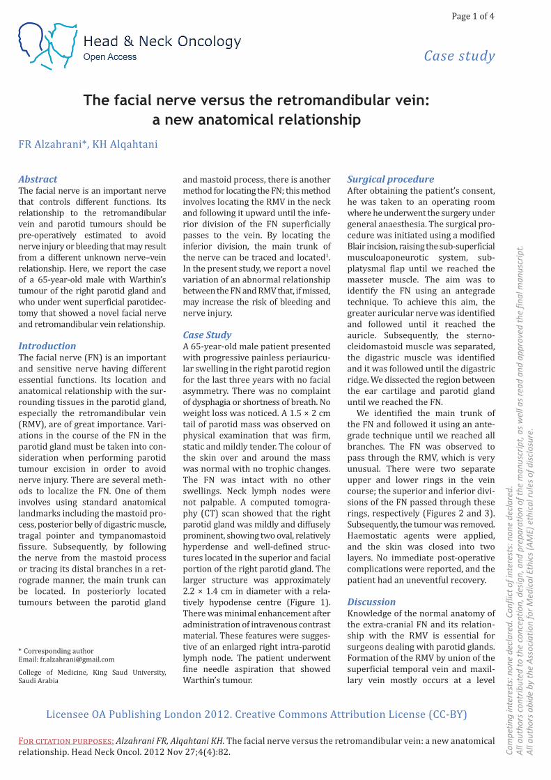
Case StudyA 65-year-old male patient presented with progressive painless periauricu-lar swelling in the right parotid region for the last three years with no facial asymmetry. There was no complaint of dysphagia or shortness of breath. No weight loss was noticed. A 1.5 × 2 cm tail of parotid mass was observed on physical examination that was firm, static and mildly tender. The colour of the skin over and around the mass was normal with no trophic changes. The FN was intact with no other swellings. Neck lymph nodes were not palpable. A computed tomogra-phy (CT) scan showed that the right parotid gland was mildly and diffusely prominent, showing two oval, relatively hyperdense and well-defined struc-tures located in the superior and facial portion of the right parotid gland. The larger structure was approximately 2.2 × 1.4 cm in diameter with a rela-tively hypodense centre (Figure 1). There was minimal enhancement after administration of intravenous contrast material. These features were sugges-tive of an enlarged right intra-parotid lymph node. The patient underwent fine needle aspiration that showed Warthin’s tumour.
Surgical procedure After obtaining the patient’s consent, he was taken to an operating room where he underwent the surgery under general anaesthesia. The surgical pro-cedure was initiated using a modified Blair incision, raising the sub-superficial musculoaponeurotic system, sub-platysmal flap until we reached the masseter muscle. The aim was to identify the FN using an antegrade technique. To achieve this aim, the greater auricular nerve was identified and followed until it reached the auricle. Subsequently, the sterno-cleidomastoid muscle was separated, the digastric muscle was identified and it was followed until the digastric ridge. We dissected the region between the ear cartilage and parotid gland until we reached the FN.
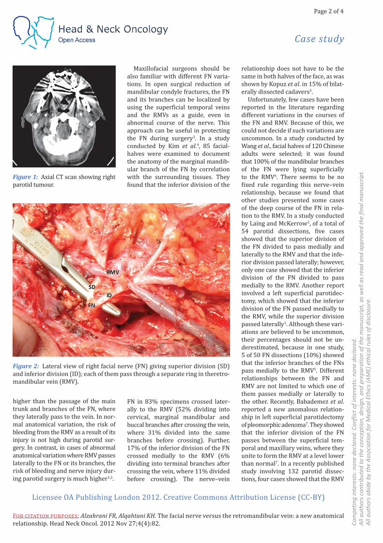
We identified the main trunk of the FN and followed it using an ante-grade technique until we reached all branches. The FN was observed to pass through the RMV, which is very unusual. There were two separate upper and lower rings in the vein course; the superior and inferior divi-sions of the FN passed through these rings, respectively (Figures 2 and 3). Subsequently, the tumour was removed. Haemostatic agents were applied, and the skin was closed into two layers. No immediate post-operative complications were reported, and the patient had an uneventful recovery.
Discussion Knowledge of the normal anatomy of the extra-cranial FN and its relation-ship with the RMV is essential for surgeons dealing with parotid glands.Formation of the RMV by union of the superficial temporal vein and maxil-lary vein mostly occurs at a level
College of Medicine, King Saud University, Saudi Arabia
Page 2 of 4
Case study
Licensee OA Publishing London 2012. Creative Commons Attribution License (CC-BY)
Com
petin
g in
tere
sts:
non
e de
clar
ed. C
onfli
ct o
f int
eres
ts: n
one
decl
ared
.A
ll au
thor
s co
ntrib
uted
to th
e co
ncep
tion,
des
ign,
and
pre
para
tion
of th
e m
anus
crip
t, a
s w
ell a
s re
ad a
nd a
ppro
ved
the
final
man
uscr
ipt.
All
auth
ors
abid
e by
the
Ass
ocia
tion
for M
edic
al E
thic
s (A
ME)
eth
ical
rule
s of
dis
clos
ure.
F�� �������� ��������: Alzahrani FR, Alqahtani KH. The facial nerve versus the retromandibular vein: a new anatomical relationship. Head Neck Oncol. 2012 Nov 27;4(4):82.
Figure 1: Axial CT scan showing right parotid tumour.
Figure 2: Lateral view of right facial nerve (FN) giving superior division (SD) and inferior division (ID); each of them pass through a separate ring in theretro-mandibular vein (RMV).
higher than the passage of the main trunk and branches of the FN, where they laterally pass to the vein. In nor-mal anatomical variation, the risk of bleeding from the RMV as a result of its injury is not high during parotid sur-gery. In contrast, in cases of abnormal anatomical variation where RMV passes laterally to the FN or its branches, the risk of bleeding and nerve injury dur-ing parotid surgery is much higher1,2.
Maxillofacial surgeons should be also familiar with different FN varia-tions. In open surgical reduction of mandibular condyle fractures, the FN and its branches can be localized by using the superficial temporal veins and the RMVs as a guide, even in abnormal course of the nerve. This approach can be useful in protecting the FN during surgery3. In a study conducted by Kim et al.4, 85 facial-halves were examined to document the anatomy of the marginal mandib-ular branch of the FN by correlation with the surrounding tissues. They found that the inferior division of the
FN in 83% specimens crossed later-ally to the RMV (52% dividing into cervical, marginal mandibular and buccal branches after crossing the vein, where 31% divided into the same branches before crossing). Further, 17% of the inferior division of the FN crossed medially to the RMV (6% dividing into terminal branches after crossing the vein, where 11% divided before crossing). The nerve–vein
relationship does not have to be the same in both halves of the face, as was shown by Kopuz et al. in 15% of bilat-erally dissected cadavers5.
Unfortunately, few cases have been reported in the literature regarding different variations in the courses of the FN and RMV. Because of this, we could not decide if such variations are uncommon. In a study conducted by Wang et al., facial halves of 120 Chinese adults were selected; it was found that 100% of the mandibular branches of the FN were lying superficially to the RMV6. There seems to be no fixed rule regarding this nerve–vein relationship, because we found that other studies presented some cases of the deep course of the FN in rela-tion to the RMV. In a study conducted by Laing and McKerrow2, of a total of 54 parotid dissections, five cases showed that the superior division of the FN divided to pass medially and laterally to the RMV and that the infe-rior division passed laterally; however, only one case showed that the inferior division of the FN divided to pass medially to the RMV. Another report involved a left superficial parotidec-tomy, which showed that the inferior division of the FN passed medially to the RMV, while the superior division passed laterally1. Although these vari-ations are believed to be uncommon, their percentages should not be un-derestimated, because in one study, 5 of 50 FN dissections (10%) showed that the inferior branches of the FNs pass medially to the RMV5. Different relationships between the FN and RMV are not limited to which one of them passes medially or laterally to the other. Recently, Babademez et al. reported a new anomalous relation-ship in left superficial parotidectomy of pleomorphic adenoma7. They showed that the inferior division of the FN passes between the superficial tem-poral and maxillary veins, where they unite to form the RMV at a level lower than normal7. In a recently published study involving 132 parotid dissec-tions, four cases showed that the RMV
Page 3 of 4
Case study
Licensee OA Publishing London 2012. Creative Commons Attribution License (CC-BY)
Com
petin
g in
tere
sts:
non
e de
clar
ed. C
onfli
ct o
f int
eres
ts: n
one
decl
ared
.A
ll au
thor
s co
ntrib
uted
to th
e co
ncep
tion,
des
ign,
and
pre
para
tion
of th
e m
anus
crip
t, a
s w
ell a
s re
ad a
nd a
ppro
ved
the
final
man
uscr
ipt.
All
auth
ors
abid
e by
the
Ass
ocia
tion
for M
edic
al E
thic
s (A
ME)
eth
ical
rule
s of
dis
clos
ure.
F�� �������� ��������: Alzahrani FR, Alqahtani KH. The facial nerve versus the retromandibular vein: a new anatomical relationship. Head Neck Oncol. 2012 Nov 27;4(4):82.
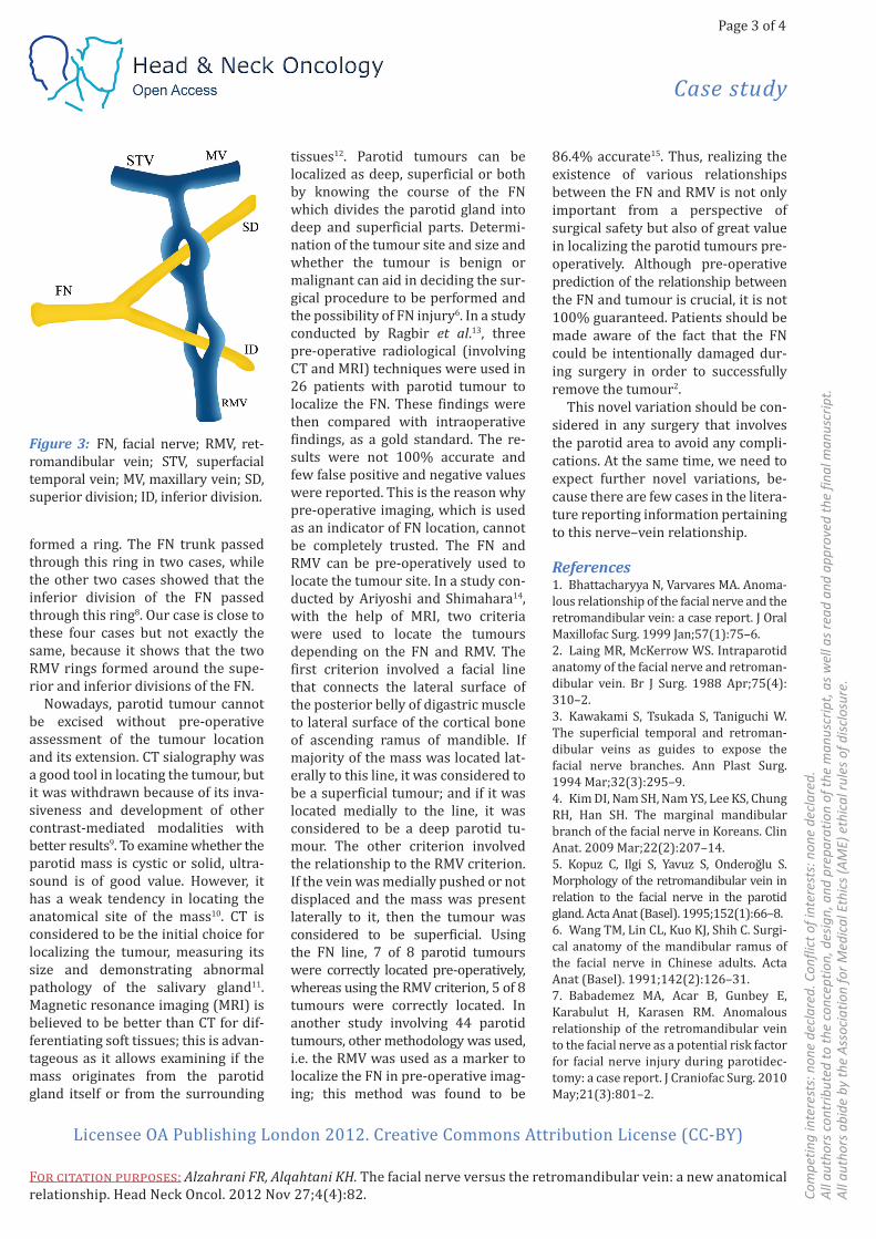
formed a ring. The FN trunk passed through this ring in two cases, while the other two cases showed that the inferior division of the FN passed through this ring8. Our case is close to these four cases but not exactly the same, because it shows that the two RMV rings formed around the supe-rior and inferior divisions of the FN.
Nowadays, parotid tumour cannot be excised without pre-operative assessment of the tumour location and its extension. CT sialography was a good tool in locating the tumour, but it was withdrawn because of its inva-siveness and development of other contrast-mediated modalities with better results9. To examine whether the parotid mass is cystic or solid, ultra-sound is of good value. However, it has a weak tendency in locating the anatomical site of the mass10. CT is considered to be the initial choice for localizing the tumour, measuring its size and demonstrating abnormal pathology of the salivary gland11. Magnetic resonance imaging (MRI) is believed to be better than CT for dif-ferentiating soft tissues; this is advan-tageous as it allows examining if the mass originates from the parotid gland itself or from the surrounding
tissues12. Parotid tumours can be localized as deep, superficial or both by knowing the course of the FN which divides the parotid gland into deep and superficial parts. Determi-nation of the tumour site and size and whether the tumour is benign or malignant can aid in deciding the sur-gical procedure to be performed and the possibility of FN injury6. In a study conducted by Ragbir et al.13, three pre-operative radiological (involving CT and MRI) techniques were used in 26 patients with parotid tumour to localize the FN. These findings were then compared with intraoperative findings, as a gold standard. The re-sults were not 100% accurate and few false positive and negative values were reported. This is the reason why pre-operative imaging, which is used as an indicator of FN location, cannot be completely trusted. The FN and RMV can be pre-operatively used to locate the tumour site. In a study con-ducted by Ariyoshi and Shimahara14, with the help of MRI, two criteria were used to locate the tumours depending on the FN and RMV. The first criterion involved a facial line that connects the lateral surface of the posterior belly of digastric muscle to lateral surface of the cortical bone of ascending ramus of mandible. If majority of the mass was located lat-erally to this line, it was considered to be a superficial tumour; and if it was located medially to the line, it was considered to be a deep parotid tu-mour. The other criterion involved the relationship to the RMV criterion. If the vein was medially pushed or not displaced and the mass was present laterally to it, then the tumour was considered to be superficial. Using the FN line, 7 of 8 parotid tumours were correctly located pre-operatively, whereas using the RMV criterion, 5 of 8 tumours were correctly located. In another study involving 44 parotid tumours, other methodology was used, i.e. the RMV was used as a marker to localize the FN in pre-operative imag-ing; this method was found to be
86.4% accurate15. Thus, realizing the existence of various relationships between the FN and RMV is not only important from a perspective of surgical safety but also of great value in localizing the parotid tumours pre-operatively. Although pre-operative prediction of the relationship between the FN and tumour is crucial, it is not 100% guaranteed. Patients should be made aware of the fact that the FN could be intentionally damaged dur-ing surgery in order to successfully remove the tumour2.
This novel variation should be con-sidered in any surgery that involves the parotid area to avoid any compli-cations. At the same time, we need to expect further novel variations, be-cause there are few cases in the litera-ture reporting information pertaining to this nerve–vein relationship.
References 1. Bhattacharyya N, Varvares MA. Anoma-lous relationship of the facial nerve and the retromandibular vein: a case report. J Oral Maxillofac Surg. 1999 Jan;57(1):75–6.2. Laing MR, McKerrow WS. Intraparotid anatomy of the facial nerve and retroman-dibular vein. Br J Surg. 1988 Apr;75(4):310–2.3. Kawakami S, Tsukada S, Taniguchi W. The superficial temporal and retroman-dibular veins as guides to expose the facial nerve branches. Ann Plast Surg. 1994 Mar;32(3):295–9.4. Kim DI, Nam SH, Nam YS, Lee KS, Chung RH, Han SH. The marginal mandibular branch of the facial nerve in Koreans. Clin Anat. 2009 Mar;22(2):207–14.5. Kopuz C, Ilgi S, Yavuz S, Onderoğlu S. Morphology of the retromandibular vein in relation to the facial nerve in the parotid gland. Acta Anat (Basel). 1995;152(1):66–8.6. Wang TM, Lin CL, Kuo KJ, Shih C. Surgi-cal anatomy of the mandibular ramus of the facial nerve in Chinese adults. Acta Anat (Basel). 1991;142(2):126–31.7. Babademez MA, Acar B, Gunbey E, Karabulut H, Karasen RM. Anomalous relationship of the retromandibular vein to the facial nerve as a potential risk factor for facial nerve injury during parotidec-tomy: a case report. J Craniofac Surg. 2010 May;21(3):801–2.
Licensee OA Publishing London 2012. Creative Commons Attribution License (CC-BY)
Com
petin
g in
tere
sts:
non
e de
clar
ed. C
onfli
ct o
f int
eres
ts: n
one
decl
ared
.A
ll au
thor
s co
ntrib
uted
to th
e co
ncep
tion,
des
ign,
and
pre
para
tion
of th
e m
anus
crip
t, a
s w
ell a
s re
ad a
nd a
ppro
ved
the
final
man
uscr
ipt.
All
auth
ors
abid
e by
the
Ass
ocia
tion
for M
edic
al E
thic
s (A
ME)
eth
ical
rule
s of
dis
clos
ure.
F�� �������� ��������: Alzahrani FR, Alqahtani KH. The facial nerve versus the retromandibular vein: a new anatomical relationship. Head Neck Oncol. 2012 Nov 27;4(4):82.
8. Touré G, Vacher C. Relations of the facial nerve with the retromandibular vein: anatomic study of 132 parotid glands. Surg Radiol Anat. 2010 Dec;32(10):957–61.9. Som PM, Biller HF. The combined CT-sialogram. Radiology. 1980 May;135(2): 387–90.10. Soler R, Bargiela A, Requejo I, Rodrfguez E, Rey JL, Sancristan F. Pictorial review: MR imaging of parotid tumours. Clin Radiol. 1997 Apr;52(4):269–75.
11. Bryan RN, Miller RH, Ferreyro RI, Sessions RB. Computed tomography of the major salivary glands. AJR Am J Roent-genol. 1982 Sep;139(3):547–54.12. Mandelblatt SM, Braun IF, Davis PC, Fry SM, Jacobs LH, Hoffman JC Jr. Parotid masses: MR imaging. Radiology. 1987 May;163(2):411–4.13. Ragbir M, Dunaway DJ, Chippindale AJ, Latimer J, Mohammed F, McLean NR. Prediction of the position of the intrapa-rotid portion of the facial nerve on MRI
and CT. Br J Plast Surg. 2002 Jul;55(5):376–9.14. Ariyoshi Y, Shimahara M. Determin-ing whether a parotid tumour is in the superficial or deep lobe using magnetic resonance imaging. J Oral Maxillofac Surg. 1998 Jan;56(1):23–6.15. Divi V, Fatt MA, Teknos TN, Mukherji SK. Use of cross-sectional imaging in predicting surgical location of parotid neoplasms. J Comput Assist Tomogr. 2005 May–Jun;29(3):315–9.


![REVIEW Cancer of the oral cavity and oropharynx...(mylohyoid, digastric, geniohyoid muscles)[3]. The retro-molar trigone is a small mucosal area on the mandibular ramus behind the](https://static.documents.pub/doc/80x56/5e85041280b1cc36ed4e1591/review-cancer-of-the-oral-cavity-and-oropharynx-mylohyoid-digastric-geniohyoid.jpg)


