Page 1
Accepted Manuscript
Title: The family Rhabdoviridae: mono- and bipartitenegative-sense RNA viruses with diverse genome organizationand common evolutionary origins
Author: Ralf G. Dietzgen Hideki Kondo Michael M. GoodinGael Kurath Nikos Vasilakis
PII: S0168-1702(16)30457-9DOI: http://dx.doi.org/doi:10.1016/j.virusres.2016.10.010Reference: VIRUS 96979
To appear in: Virus Research
Received date: 20-7-2016Revised date: 18-10-2016Accepted date: 18-10-2016
Please cite this article as: Dietzgen, Ralf G., Kondo, Hideki, Goodin, MichaelM., Kurath, Gael, Vasilakis, Nikos, The family Rhabdoviridae: mono- and bipartitenegative-sense RNA viruses with diverse genome organization and commonevolutionary origins.Virus Research http://dx.doi.org/10.1016/j.virusres.2016.10.010
This is a PDF file of an unedited manuscript that has been accepted for publication.As a service to our customers we are providing this early version of the manuscript.The manuscript will undergo copyediting, typesetting, and review of the resulting proofbefore it is published in its final form. Please note that during the production processerrors may be discovered which could affect the content, and all legal disclaimers thatapply to the journal pertain.
Page 2
1
The family Rhabdoviridae: mono- and bipartite negative-sense RNA viruses with
diverse genome organization and common evolutionary origins
Ralf G. Dietzgen a, *, Hideki Kondo b, Michael M. Goodin c, Gael Kurath d, Nikos Vasilakis e
a Queensland Alliance for Agriculture and Food Innovation, The University of Queensland, St. Lucia,
Queensland 4072, Australia
b Institute of Plant Science and Resources, Okayama University, Kurashiki 710-0046, Japan
c Department of Plant Pathology, University of Kentucky, Lexington KY 40546, USA
d U.S. Geological Survey, Western Fisheries Research Centre, Seattle WA, USA
e Institute for Human Infection and Immunity, University of Texas Medical Branch, Galveston TX, 77555,
USA
* Corresponding author:
Ralf G. Dietzgen, QAAFI, The University of Queensland, St. Lucia QLD 4072, Australia
Tel.: +61733466503, Fax: +61733466503
Email: [email protected]
Page 3
2
Graphical abstract
Highlights
Family Rhabdoviridae is comprised of thirteen genera
Rhabdoviruses are mostly enveloped and infect ecologically diverse hosts
Rhabdovirus negative-sense RNA genome has five canonical structural protein genes
Rhabdovirus genomes may contain additional ORFs encoding putative accessory
proteins
Diverse genome structure may guide taxonomic resolution at genus and species levels
Abstract
The family Rhabdoviridae consists of mostly enveloped, bullet-shaped or bacilliform viruses with a
negative-sense, single-stranded RNA genome that infect vertebrates, invertebrates or plants. This
ecological diversity is reflected by the diversity and complexity of their genomes. Five canonical
Page 4
3
structural protein genes are conserved in all rhabdoviruses, but may be overprinted, overlapped or
interspersed with several novel and diverse accessory genes. This review gives an overview of the
characteristics and diversity of rhabdoviruses, their taxonomic classification, replication mechanism,
properties of classical rhabdoviruses such as rabies virus and rhabdoviruses with complex genomes,
rhabdoviruses infecting aquatic species, and plant rhabdoviruses with both mono- and bipartite genomes.
Key words: Rhabdovirus; Negative-sense RNA virus; Genome organization; Diversity; Replication;
Taxonomy.
1. Introduction – Properties and diversity of rhabdoviruses
Rhabdoviruses are a large and ecologically diverse group of viruses, which infect terrestrial and
aquatic vertebrates, invertebrates and plants. They include many pathogens of significance to public
health, agriculture and fisheries (Dietzgen and Kuzmin, 2011). The majority of rhabdoviruses are
transmitted by arthropods to vertebrate or plant hosts, but lyssaviruses (e.g. rabies virus) and
novirhabdoviruses (e.g. infectious hematopoietic necrosis virus) have evolved to circulate among
vertebrates without a biological vector, and sigmaviruses (e.g. Drosophila melanogaster sigma virus) are
Page 5
4
congenitally transmitted in fruit flies. High throughput host genome sequencing has revealed the
integration of rhabdovirus-like elements into the genomes of some arthropods and plants (Ballinger et al.,
2012; Fort et al., 2012; Chiba et al., 2011; Katzourakis and Gifford, 2010; Li et al., 2015), suggesting an
ancient evolutionary origin and long-standing association of rhabdoviruses with their hosts.
Rhabdoviruses are taxonomically classified in thirteen genera in the family Rhabdoviridae, order
Mononegavirales, which also includes families Bornaviridae, Filoviridae, Paramyxoviridae and recently
established Nyamiviridae (Dietzgen et al., 2011; Walker et al., 2015). The name is based on their
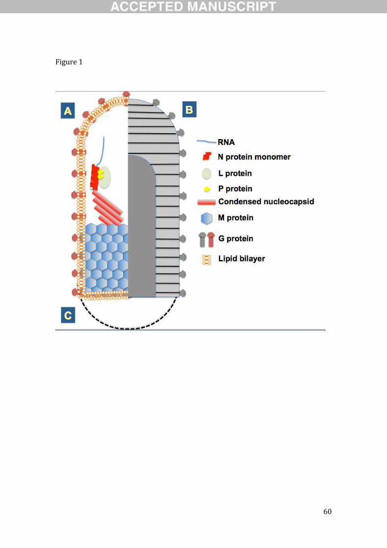
characteristic shape and derived from the Ancient Greek word rhabdos meaning “rod or wand”. Classic
rhabdoviruses typified by vesicular stomatitis virus (VSV) and sonchus yellow net virus (SYNV) form
characteristic bullet- or cone-shaped (animal host; Ge et al., 2010) or bacilliform (plant host; Jackson et
al., 2005) enveloped virions that contain non-segmented, negative-sense, single-stranded (ss) RNA
genomes of 11–16 kb in length (Figs. 1 and 2). Virions range in size from 100 to 430 × 45 to 100 nm. The
basic genome organisation shared by all rhabdoviruses includes five canonical genes that encode (from 3’
to 5’) the nucleoprotein (or nucleocapsid protein, N), phosphoprotein (P), matrix protein (M),
glycoprotein (G) and large protein (L, RNA-dependent RNA polymerase) (Fig. 2). This group of
structural protein genes is flanked by regulatory 3’ leader and 5’ trailer sequences that show terminal
Page 6
5
complementarity and contain promoter sequences to initiate replication. The individual genes are flanked
by conserved transcription stop and start signals separated by short untranscribed intergenic sequences
(Dietzgen and Kuzmin, 2012). The infectious nucleocapsid core [a ribonucleoprotein (RNP) complex],
which is active in transcription and replication, consists of the genomic RNA that is always tightly
associated with N protein, together with P and L proteins. M protein is responsible for condensation of
RNP complex during virion assembly at the host plasma membrane, and the transmembrane spike protein
G likely plays an important role in assembly, budding and host cell entry (Dietzgen and Kuzmin, 2012).
Rhabdovirus genomes are diverse and often complex because the five canonical genes may be
overprinted, overlapped or interspersed with a range of novel accessory genes, such that the number of
genes range from 5-10 or more (Walker et al., 2011; 2015; Fig. 3). Among RNA viruses, rhabdovirus
genomes have an unusual capacity for plasticity and appear to have frequently acquired and lost new
genes during evolution (Walker et al., 2015). Furthermore, two groups of plant-infecting rhabdoviruses
(genera Dichorhavirus and Varicosavirus) were recently found to have a divided genome with the L gene
located on a separate RNA segment (Dietzgen et al., 2014; Kormelink et al., 2011). For other reviews on
rhabdoviruses see Blondel et al. (2015), Walker et al. (2011), Dietzgen and Kuzmin (2011), Dietzgen et al.
(2011), Ammar et al. (2009) and Jackson et al. (2005).
Page 7
6
2. Taxonomic classification – past, present and future
Rhabdovirus taxonomy is evolving rapidly in an effort to incorporate ever-increasing numbers of new
viral sequences obtained through high throughput technologies and to harmonize classifications across
genera and families in the order Mononegavirales. In the 9th report of the International Committee on
Taxonomy of Viruses (ICTV) the family Rhabdoviridae comprised six genera (Dietzgen et al., 2011). In
the 2015 Taxonomy Release [http://www.ictvonline.org/virusTaxonomy.asp], thirteen genera were
recognised: Cytorhabdovirus, Dichorhavirus, Ephemerovirus, Lyssavirus, Novirhabdovirus,
Nucleorhabdovirus, Perhabdovirus, Sigmavirus, Sprivivirus, Tibrovirus, Tupavirus, Varicosavirus and
Vesiculovirus (Fig. 4). Among these, in the recently recognized Dichorhavirus and Varicosavirus genera
(Afonso et al., 2016; Adams et al., 2016) for the first time, virus species containing plant-infecting
negative-sense ssRNA viruses with bipartite genomes have been classified as rhabdoviruses based on
significant genome sequence identities with nucleo- and cytorhabdoviruses, respectively. Furthermore,
binomial species names that indicate which genus the species belongs to have been introduced for all
rhabdovirus species, to facilitate easier differentiation between virus names and the names of taxonomic
species these viruses are classified in (Afonso et al., 2016; Adams et al., 2016). For example, rabies virus
Page 8
7
is a virus in the species Rabies lyssavirus, and potato yellow dwarf virus is a virus in the species Potato
yellow dwarf nucleorhabdovirus. Based on the recent large-scale discovery and analysis of vertebrate and
arthropod rhabdoviruses, several new genera have been proposed (e.g. Almendravirus, Bahiavirus,
Curiovirus, Hapavirus, Ledantevirus, Sawgravirus and Sripuvirus) (Walker et al., 2015; Longdon et al.,
2015; Li et al, 2015) (Fig. 4).
3. Replication, transcription and translation
The rhabdovirus replication mechanism is almost universal across the family. Replication mechanisms of
some plant-infecting rhabdoviruses differ slightly due to the plant cell environment and the establishment
of replication factories in the nucleus (rather than the cytoplasm) for nucleorhabdoviruses and
dichorhaviruses. Otherwise, the universal pathway of the cytoplasmic replication cycle follows (i) cell
entry, facilitated by clathrin-mediated or receptor-binding endocytosis (or vector-mediated penetration of
the plant cell wall); (ii) uncoating; (iii) transcription and translation; (iv) genome replication and
encapsidation; and (v) assembly and release (budding). Fusion of endocytosed virus with endosomes and
its subsequent lysis releases the RNP complex into the cytoplasm allowing for the initiation of early
transcription and replication events. A critical step in this process is the dissociation of the M protein
Page 9
8
from the nucleocapsid (Mire et al., 2010), which is required for the initiation of viral transcription
(Clinton et al., 1978; Pal et al., 1985) also called primary transcription. The term denotes the short nature
of transcription from parental templates as opposed to subsequent prolonged transcription events from
progeny templates (secondary transcription) following genome replication. Transcription of the negative-
stranded genome is facilitated by a transcriptase complex and occurs progressively on a decreasing molar
gradient based on gene distance from the genomic 3’ end (for example, NPMGL) (Fig. 2).
The relative abundance of each viral mRNA and thus each protein is regulated by the disassociation of
the transcriptase from the RNA template once it reaches the respective gene transcription termination
polyadenylation (TTP) signal at the end of each viral gene. This stop-start (‘stuttering’) sequential gene
transcription mechanism is governed by the interaction of cis-acting signals (e.g. TTP) located on the
genome template and the transcriptase complex (Abraham and Banerjee 1976). The cis-acting signals are
well conserved with some minor variations (Walker et al., 2015). In canonical genome architectures each
gene junction consists of conserved sequence motifs, originally identified for vesicular stomatitis viruses
as (i) a TTP (3’-AUACUUUUUUU-5’), whose function is to polyadenylate and terminate the upstream
mRNA (Rose 1980; Barr et al., 1997a; Hwang et al., 1998); (ii) a non-transcribed intergenic dinucleotide
(G/CA)(Rose 1980; Stillman and Whitt, 1997; Barr et al., 1997b); and (iii) a transcription initiation (TI)
Page 10
9
pentanucleotide sequence (3’-UUGUC-5’)(Rose 1980), whose function is to initiate, cap and methylate
the down stream mRNA. Exception to this mechanism is observed in the initiation of transcription
between the 3’ leader sequence and the N gene. The leader is encoded by the 3’-terminal nucleotides of
the genome (47 nt for VSIV) that lack the U7 sequence, resulting in transcription of a short leader RNA
that lacks a polyA tail and cap (Whelan and Gertz, 1999) (Fig. 2). The leader sequence primary function
is to serve as promoter for the initiation of RNA synthesis, whereas its newly synthesised complementary
sequence facilitates the encapsidation of nascent RNA, thus allowing transcriptional read-through to
generate full-length complementary genomes (i.e. antigenomes). Thus there are two possible outcomes
for the polymerase complex at the end of each gene junction: (i) either traverse the intergenic dinucleotide
and initiate again at the TI motif of the downstream gene; or (ii) dissociate from the RNA template at the
gene junction resulting in an apparent attenuation of the downstream gene and its protein expression, as a
function of each gene’s distance from the 3’end of the genome (Iverson and Rose, 1981; reviewed in:
Lyles et al., 2013).
The production and accumulation of the virally encoded proteins signals a switch in the polymerase
function, from viral mRNA transcription to genome replication, in which N plays a critical role. An
essential step in the viral replication of the nascent positive-sense genome (antigenome) relies on its
Page 11
10
encapsidation, a process facilitated by cis-acting conserved sequences located on the 3’ ends of viral
genome and antigenome (Whelan and Wertz, 1999; Li and Pattnaik, 1999). Additionally, N and P
proteins are critical in promoting genome replication, as the N/P complex provides the structural and
chaperone support for the nascent RNA to bind via sugar-phosphate interactions to the N protein
(Albertini et al., 2006). The bound antigenome will then function as template for the synthesis of
encapsidated negative-sense genomes, which will be assembled into progeny virions.
Virion assembly is a staggered process where the various components [nucleocapsid core (RNP), G
and M proteins] are sequestered in different cellular compartments and converge in the final steps of the
process. The nucleocapsid is assembled during RNA replication in the cytoplasm, as is observed for
members of the genera Vesiculovirus, Lyssavirus, Ephemerovirus and Novirhabdovirus. Viral G protein is
inserted into the endoplasmic reticulum where chaperones (BiP and calnexin)(Hammond and Helenius,
1994) facilitate its proper folding and assembly into trimers (Doms et al., 1988), prior to transport and
fusion into the Golgi complex. As it traffics through the cell it undergoes further posttranslational
modifications including glycosylations (Schmidt and Schlesinger, 1979), prior to its transport to
cholesterol- and sphingolipid-rich lipid rafts in the baso-lateral plasma membrane. M protein is
synthesized mostly as a soluble protein in the cytoplasm (McCreedy et al., 1990) and is also membrane
Page 12
11
bound, albeit at lower amounts (Ogden et al., 1986). However both forms of the M protein are recruited
for assembly of nucleocapsid/M complexes at the host plasma membrane from where virions will bud
(Odenwald et al., 1986). This budding process is facilitated by the interaction of M with host-encoded
proteins responsible for the formation of multivesicular bodies (MVB), and their release from the plasma
membrane (Harty et al., 2001).
4. ‘Classical’ vertebrate rhabdoviruses
For historical reasons any reference to classical vertebrate rhabdoviruses denotes members of the genera
Vesiculovirus and Lyssavirus, represented by the prototype species vesicular stomatitis Indiana virus
(VSIV) and rabies virus (RABV), respectively. Vesiculoviruses have a wide host range among mammals
and are transmitted by hematophagous insects (sandflies and/or mosquitoes). Lyssaviruses utilize mostly
bats as their principal reservoir hosts as well as various terrestrial carnivores as terminal hosts. Viruses of
each genus form a monophyletic clade in a maximum likelihood (ML) tree inferred from complete L
protein sequences (Dietzgen et al., 2011; Walker et al., 2015). Structurally both demonstrate the classic
rhabdovirus enveloped bullet-shaped virions (Fig. 1) packaging a genome consisting of five genes (3’-N-
P-M-G-L-5’), each separated by a short gene junction (intergenic region), and flanked by highly
Page 13
12
conserved 3’ leader (le) and 5’ trailer (tr) sequences (Fig. 3). In vesiculoviruses the P gene mRNA
contains 2 additional alternate start codons that initiate translation at alternative open reading frames
(ORFs) that encode two small basic proteins C and C’ (55-aa and 65-aa, respectively) of unknown
function (Spiropoulou and Nichol, 1993; Peluso et al., 1996). Suppression of C/C’ expression has no
apparent effects in virus replication or pathogenicity in vivo (Kretzschmar et al., 1996). Of note is that not
all members of the genus express alternative ORFs in P [e.g. vesicular stomatitis Alagoas, Maraba,
Malpais Spring, Morreton viruses] (Walker et al., 2015), and additional ORFs (≥ 150 nt) may be present
in alternative reading frames in other genes than P (Walker et al., 2015).
For a long time lyssaviruses were considered antigenically unique represented by various RABV
isolates. However the discovery of other RABV-like viruses in expanded geographic regions of the globe
allowed initially the establishment of the genus Lyssavirus, followed by its demarcation into four distinct
groups anchored by RABV, Lagos bat, Mokola and Duvenhage viruses, based solely on their serological
relationships. The advent of Sanger sequencing permitted the determination of lyssavirus genetic
signatures, thus allowing phylogenetic relationships to be further refined and their classification into the
currently recognized 11 species was aided by a number of species demarcation criteria including genetic
distances, phylogenies, antigenic relationships and biological properties (Lyles et al, 2013).
Page 14
13
Vesiculoviruses have been classified into 10 species using similar criteria (Dietzgen et al., 2011). The
massive generation of sequencing data based on new technologies (Walker et al., 2015) and the need for
consistency across taxa necessitates refinement of these criteria. This will undoubtedly recalibrate the
richness of vesiculovirus diversity in the future (Peter Walker, personal communication).
While the level of diversity within the family Rhabdoviridae is being realized at an unprecedented rate
due to new detection and sequencing methods (e.g. next generation sequencing, metagenomics), reverse
genetics of the prototype vesiculoviruses (Schnell et al., 1994; Whelan et al., 1995) allowed exploration
of rhabdovirus plasticity and evolution at the genetic level, as well as an understanding of the factors
influencing gene expression and molecular and cellular basis of pathogenesis, and their use as vaccine
delivery vehicles (reviewed in: Whitt et al., 2016). The error-prone nature of their RNA-dependent RNA
polymerase results in the generation of a rich diversity of genetic variants following each replication
cycle, which are subject to adaptation due to selective pressures under different conditions, such as
alternative hosts (vector vs vertebrate) during the course of their natural transmission (Novella et al.,
2010; Novella et al., 2011; Wasik et al., 2016). Similarly recombinant RABVs with targeted mutations in
P gene demonstrated its critical role in suppression of interferon signaling through blocking the
interaction of the transcription factor IRF-3 (interferon regulatory factor 3) with two cell-expressed
Page 15
14
protein kinases (Brzozka et al., 2005), ultimately altering pathogenesis in vivo (Rieder et al., 2011). On
the other hand, recombinant VSVs were critical in demonstrating the role of M in inhibiting host gene
expression without affecting virus assembly, a process facilitated through the formation of complexes
with the mRNA export factor Rae1 (Faria et al., 2005), which interferes with the function of downstream
factors (Connor et al., 2006) essential for host gene expression.
5. Vertebrate rhabdoviruses with complex genomes
Several of the currently recognized genera within the family Rhabdoviridae show associations with a
dominant group of vertebrate hosts, such as the ephemeroviruses and tibroviruses that are hosted by cattle,
with many viruses either isolated from cattle and/or from mosquitoes or biting midges that feed on cattle
(Walker 2005; Gubala et al., 2011). Others like the recently designated ledanteviruses (Blasdell et al.,
2015), not only exhibit a strong ecological association with bats, but also a broader natural host
specificity (e.g. ungulates, rodents and humans) suggesting spill over from their natural reservoir. A
recent landmark study extended the range of the known rhabdovirus genome complexity to include
viruses in proposed new genera Hapavirus, Ledantevirus and Sripuvirus among others (Walker et al.,
2015). While the canonical genome organization of the prototype rhabdoviruses features the five ORFs
Page 16
15
arranged in the order 3’-N-P-M-G-L-5’, rhabdovirus genomes may also be more complex and contain
additional ORFs encoding putative proteins of unknown function. These may occur in alternative and/or
overlapping ORFs within the major structural protein genes or as independent ORFs flanked by TI or
TTP sequences in the regions between the structural protein, some of which may have arisen by gene
duplication.
Ephemeroviruses have among the largest genomes of all rhabdoviruses. The prototype virus,
bovine ephemeral fever virus (BEFV) has a genome approximately 14.8 kb in length that contains 10
genes separated by short intergenic regions (Walker 2005) (Fig. 3). Due to an apparent duplication of the
viral G gene there is an additional gene for a non-structural glycoprotein (GNS) that has a significant
amino acid sequence homology and is synthesized at same levels as the G protein, but it is not
incorporated into the virion envelope and thus does not induce any antibody responses (Hertig et al.,
1996). GNS is followed by a viroporin gene (α1), probably encoding a viral ion channel protein, and
several other accessory genes (α2, β and γ) (Walker et al., 2015).
Hapaviruses infect birds, reptiles or mammals and many have been isolated from culicine
mosquitoes. They exhibit complex genomes, form a monophyletic group based on well-supported ML
trees generated from complete L protein sequences and are anchored by the prototype Hart Park virus
Page 17
16
(HPV) (Walker et al., 2015)(Fig. 3). In addition to the five canonical genes encoding the structural
proteins, three long ORFs, U1, U2 and U3, with significant amino acid sequence homology with each
other, are located in independent transcriptional units between the P and M genes (Walker et al., 2015).
A fourth ORF lies within the G gene transcriptional unit and is thought to encode a class IA viroporin.
Alternative long ORFs in different reading frames in the N gene, U3 gene and L gene are observed but
may not be expressed as functional proteins due to poor Kozak context and distal location far from the
start of their transcriptional units (Walker et al., 2015). Ngaingan virus (NGAV) has the longest genome
of any rhabdovirus described to date (Gubala et al., 2010) with 15,764 nt containing 13 genes. Three of
the eight additional ORFs are located in two transcriptional units between the P and M genes, and another
lies in a transcriptional unit between the M and G genes, likely encoding a unique protein of unknown
function. Additionally, there are four ORFs between the G and L genes, including GNS, which encodes a
class I transmembrane protein that is related in sequence to the NGAV G protein and the G proteins of
other rhabdoviruses, ORFs likely encoding two unique proteins of unknown function, and a viroporin-like
protein. Lastly, alternative ORFs are present within the P and M genes, but likely are not expressed due to
poor Kozak context and distal location far from the start of their transcriptional units (Walker et al., 2015).
Page 18
17
Ledanteviruses infect humans, rodents, and ungulates, may be vectored by arthropods and have a
strong association with bats, suggesting spill over from their natural reservoir (Blasdell et al., 2015;
Walker et al., 2015). They are anchored by the prototype Le Dantec virus (LDV), whose genome
comprises the five canonical structural protein genes, an additional transcriptional unit (U1), encoding a
small protein between the G and L genes, and a small ORF that occurs in an alternative reading frame in
the N gene, which likely is not expressed (Fig. 3).
Tibroviruses infect ungulates and are anchored by the prototype Tibrogargan virus (TIBV).
Members of the genus Tibrovirus share the same unique genome organisation comprising five genes
encoding the canonical rhabdovirus structural proteins and three additional genes encoding ORFs (U1, U2
and U3)(Fig. 3). ORF U1 and ORF U2 encode small proteins of unknown function, whereas ORF U3
encodes a small viroporin-like protein (Walker et al., 2015). Each ORF lies within an independent
transcriptional unit bounded by consensus TI and TTP sequences.
While the genus Tupavirus consists of only three species, Tupaia tupavirus, Durham tupavirus and
Klamath tupavirus (Allison et al., 2011; Johnson 1965; Kurz et al., 1986; Walker et al., 2015),
tupaviruses are characterized by their extensive geographic distribution and host range. The genome of
the prototype Tupaia virus (TUPV) includes in addition to the five genes encoding the canonical
Page 19
18
rhabdovirus structural proteins, an alternative ORF in the P gene and an ORF encoding a small
hydrophobic protein located in an independent transcriptional unit between the M and G genes, as
observed among vesiculoviruses (Fig. 3). Uniquely amongst tupaviruses, Klamath virus also contains an
additional ORF encoding a small protein (U2) within an independent transcriptional unit between the G
and L genes. Although the functions of these novel genes are currently unknown, it has been speculated
that some may play a role in enhancement of transcriptional activity, host pathogenicity or insect
transmission (Allison et al., 2011; Kretzschmar et al., 1996; Peluso et al., 1996).
The proposed genus Sripuvirus consists of five new species. Almpiwar, Chaco and Sena madueira
viruses were isolated from reptiles, while Niakha and Sripur viruses were isolated from sandflies (Causey
et al., 1966; McAllister et al., 2014; Monath et al., 1979; Vasilakis et al., 2013; Walker et al., 2015).
Sripuvirus genomes are anchored by the prototype Sripur virus (SRIV), are similar in size and contain
multiple ORFs encoding likely accessory proteins (Fig. 3). In addition to the five canonical rhabdovirus
genes encoding the structural proteins, sripuvirus genomes also feature six other ORFs. Two reside within
alternative reading frames in the N gene, encoding small proteins which likely are not expressed due to
poor Kozak context and distal location far from the start of their transcriptional units (Walker et al., 2015).
U1 resides as an independent transcriptional unit between the N and P genes, whereas Px resides within
Page 20
19
an alternative reading frame near the start of the P gene. Interestingly, the Mx resides within the M gene
with its initiation codon overlapping the termination codon of the M gene. Gx resides within an
alternative ORF near the start of the G gene, encoding a small double-membrane-spanning protein likely
expressed by ‘leaky’ ribosomal scanning (Walker et al., 2015)(Fig. 3).
The genome complexity and plasticity observed in the genera described above reiterates the richness
and diversity of the family Rhabdoviridae and suggests that different mechanisms may drive their
genome expansion and evolution. These mechanisms may include: (i) function acquisition and rapid
adaptation, (ii) gene duplication, (iii) loss of redundant ORFs through mutation, and (iv) deletion of
redundant genome sequences followed by optimization of gene expression levels. A comprehensive
overview of these mechanisms is presented in Walker et al. (2015).
6. Rhabdoviruses in aquatic systems
Rhabdoviruses of aquatic hosts (reviewed in Kurath and Winton, 2008) include important fish pathogens
in three genera: Novirhabdovirus, Sprivivirus, and Perhabdovirus (Dietzgen et al., 2011). To date these
genera contain only viruses of finfish hosts, and there are no fish viruses in other rhabdovirus genera. Due
to the poikilothermic (cold-blooded) nature of their hosts, these viruses have temperature optima ranging
Page 21
20
from 15-25°C. In addition to these distinguishing ecological factors, genera of fish rhabdoviruses differ
from other rhabdovirus genera, and from each other, by genetic divergence levels and placement of their
constituent viruses in strongly supported monophyletic clades in phylogenetic analyses of both nucleotide
and amino acid sequences. All viruses in these three genera have non-segmented negative-sense ssRNA
genomes of approximately 11,000 nt. They are transmitted horizontally as waterborne viruses, and from
parent to offspring as egg-associated viruses, without requiring a vector. They have been well studied
due to the long history of severe disease impacts caused by some fish rhabdoviruses in freshwater and
marine fish farms, netpen aquaculture, and conservation fish hatcheries. In the Aquatic Animal Health
Code of the International Organization for Animal Health, (World Organization for Animal Health, 2014)
fish rhabdoviruses cause three of the ten globally reportable aquatic diseases: infectious haematopoietic
necrosis, viral haemorrhagic septicaemia, and spring viraemia of carp.
The Sprivivirus and Perhabdovirus genera currently have 2 and 3 viral species, respectively, and
each species includes numerous viruses, mostly isolated from fish in Asia and Europe, including the
United Kingdom (reviewed in Stone et al., 2013). Spriviviruses infect primarily freshwater fish species in
the order Cypriniformes (e.g. common carp), and perhabdoviruses have broader host ranges, infecting
many diverse species. The genomes of spriviviruses and perhabdoviruses are comprised of the conserved
Page 22
21
five canonical rhabdovirus genes and their genome organization, including transcription start and stop
signals, is very similar to that of mammalian vesiculoviruses (Fig. 3). They are also most closely related
to vesiculoviruses in phylogenetic analyses (Fig. 4). Indeed, the type species of the Sprivivirus genus,
Spring viraemia of carp virus (Carp sprivivirus), was formerly classified in the Vesiculovirus genus until
sequences of additional fish viruses revealed the two clearly distinct monophyletic groups comprising
these fish rhabdovirus genera (Stone et al., 2013). Spring viraemia of carp viruses (SVCV) cause severe
disease in farmed carp in Europe and Asia, and are reviewed in Ahne et al. (2002).
The Novirhabdovirus genus includes viruses in four species that differ in both geographic and
host range (Leong and Kurath, 2012). Infectious haematopoietic necrosis viruses (IHNV) infect primarily
salmon and trout species in the fish order Salmoniformes, and originated in western North America
(Bootland and Leong, 2011). In contrast, viral haemorrhagic septicaemia viruses (VHSV) have an
extremely broad host range, infecting over 60 host species from diverse taxonomic families, comprising a
large marine fish reservoir in both the north Atlantic and north Pacific oceans (Skall et al., 2005; Smail
and Snow, 2011). Viruses of the other two novirhabdovirus species occur in Asia, where hirame
rhabdoviruses (HIRRV) infect cultured hirame (olive flounder), and snakehead virus has been less well
characterized (Leong and Kurath, 2012). Novirhabdovirus genomes differ from those of the viruses in
Page 23
22
other fish rhabdovirus genera in having an additional gene encoding a non-virion (NV) protein between
the G and L genes (Kurath and Leong, 1985; Kurath et al., 1997) (Fig. 3). The NV protein is expressed at
low levels in infected cells (Schuetze et al., 1996), where it localizes to the nucleus and interferes with the
host interferon response (Choi et al., 2011), as well as it triggers apoptosis (Ammayappan and Vakharia,
2011). Despite having a virion structure, genome organization, and sequence similarities clearly related
to those of other rhabdoviruses, novirhabdoviruses fall phylogenetically far basal to the other genera,
confirming their separation from the other fish rhabdovirus genera, and potentially indicating an ancestral
role in the evolution of the rhabdovirus family (Fig. 4).
Due to the economic impact of IHNV, VHSV, and SVCV, routine surveillance for these viruses
is conducted in cultured host populations, and molecular epidemiology is a valuable tool for fish health
management (Breyta et al., 2016; Emmenegger et al., 2011; Jonstrup et al., 2009). Local, regional, and
global phylogenies have revealed sources of disease outbreaks, virus transmission routes, virus
emergence events and host jumps (e.g. Einer-Jensen et al., 2004; Enzmann et al., 2010; Kurath et al.,
2003; Kurath, 2012; Nishizawa et al., 2006). Virus traffic between wild and cultured fish populations has
also been documented, and risk factors for potential virulence evolution in aquaculture have been
Page 24
23
described (Kennedy et al., 2016; Kurath and Winton 2011). Genotyping of virus strains has confirmed
global spread of fish rhabdoviruses due to aquaculture (reviewed in Kurath, 2012).
In laboratory studies fish viruses are investigated using cultured fish cell lines and in in vivo
infection experiments in numerous fish host species. The host immune response to fish rhabdovirus
infection consistently shows both rapid, strong innate immunity based on stimulation of interferon-related
genes, and adaptive immunity based on antibody and cellular defences (Purcell et al., 2012; Verrier et al.,
2011). An interesting aspect of host immunity is the dramatic effect of temperature, which impacts both
virus replication and persistence, and the speed of the immune response in poikilothermic hosts.
Numerous vaccines have been investigated for protection against IHNV, VHSV, SVCV, and HIRRV
(reviewed in Winton, 2007). DNA vaccines containing the viral glycoprotein gene have been shown to be
highly efficacious for IHNV, VHSV, and HIRRV, providing relative survival rates greater than 90% in
numerous studies (Kurath, 2008; Lorenzen and LaPatra, 2005; Takano, 2004). Due to their consistent
high efficacy the VHSV and IHNV DNA vaccines have also been studied extensively as models for DNA
vaccines in vertebrate hosts (Kurath et al., 2007; Lorenzen et al., 2002).
In other laboratory work a research model for in vivo viral fitness and competition studies has
been developed based on IHNV in rainbow trout (Troyer et al., 2008; Wargo et al., 2011). Reverse
Page 25
24
genetics systems have been developed for viruses in three of the four novirhabdovirus species, facilitating
investigation of questions such as the role of the accessory NV gene, and determinants of virulence and
host-specificity (Johnson et al., 2000; Biacchesi et al., 2000; Ammayappan et al., 2011). These studies
have shown great flexibility of fish rhabdovirus genomes, such that gene exchanges between different
viruses reliably generate viable chimeric viruses, and foreign genes can be inserted and expressed for
various purposes (reviewed in Biacchesi, 2011). In a particularly interesting study a recombinant IHNV
carrying a reporter luciferase gene was used to infect fish, and bioluminescence imaging on the living fish
revealed that the fin bases were a major portal of viral entry (Harmache et al., 2006). Thus aquatic
rhabdoviruses are both important veterinary pathogens and models for basic studies of rhabdovirus
biology at the molecular and landscape levels.
7. Monopartite plant rhabdoviruses: Cyto- and nucleorhabdoviruses
Classic plant rhabdoviruses, like the majority of viruses in the family Rhabdoviridae have a non-
segmented negative-sense ssRNA genome. They have been taxonomically separated into the genera
Cytorhabdovirus and Nucleorhabdovirus based on their site of replication and morphogenesis in the
cytoplasm or nucleus of infected plant cells, respectively (Fig. 5). This classification based on
Page 26
25
cytopathology has been confirmed by phylogenetic studies of available genome sequences. The biology
of plant rhabdoviruses including ecology, cytopathology, vector associations, particle structure, genome
organization and genetic variability have been described in a comprehensive review by Jackson et al.
(2005).
Since then, the number of completely sequenced genomes has increased to 8 and 9 for cyto- and
nucleorhabdoviruses, respectively. A series of plasmid vectors for transient agrobacterium-mediated
expression of autofluorescent protein fusions and confocal microscopy have allowed comparative studies
of intracellular localisation and interactions of rhabdoviral proteins in planta (e.g. Bandyopadhyay et al.,
2010; Martin et al., 2012). These studies revealed essential protein-protein interactions like N:P that were
observed for all studied rhabdoviruses, as well as interactions unique to a particular virus, and allowed the
construction of specific interactome maps. Additionally, expression of tagged proteins in the context of
virus-infected cells has provided insight into the differential localization of viral proteins within nuclei.
SYNV (a nucleorhabdovirus) P protein localizes to ring structures that appear to define the
viroplasm/nucleoplasm border. The N protein localizes onto both viroplasm and membranes, while the G
and M proteins are confined solely to membranes, consistent with their roles in morphogenesis (Goodin
et al., 2007). It is important to note that different nucleorhabdoviruses can induce markedly different
Page 27
26
cytological transformations. Infection by SYNV results in the formation of intranuclear spherules derived
from the inner nuclear membrane, whereas potato yellow dwarf virus (PYDV) does so to a much lesser
extent (Goodin et al., 2005). Similarly, the PYDV M protein induces intranuclear accumulation of the
inner nuclear membrane when expressed alone (Bandyopadhyay, et al., 2010), an activity not observed
with M proteins of other characterized plant-infecting rhabdoviruses.
Cell-to-cell movement function of the presumed viral movement proteins (MP) (accessory gene
located between P and M genes) has been demonstrated for both selected cyto- and nucleorhabdoviruses
using heterologous movement trans-complementation assays (Huang et al., 2005; Mann et al., 2016a). A
recent study showed that P3 of two cytorhabdoviruses localised to plasmodesmata, the intercellular
cytoplasmic bridges that the cross cell walls between plant cells, and acted as 30K-superfamily-like MP
requiring a conserved LxD/N50-70G motif. Furthermore, involvement of a host microtubule-associated
transcription factor has been suggested in cell-to-cell movement of both a cyto- and nucleorhabdovirus
(Min et al., 2010; Mann et al., 2016a). In fact, several host factors were identified as potentially playing
critical roles in SYNV (a well studied nucleorhabdovirus) nucleocapsid export from the nucleus and cell-
to-cell transport by cytoplasm-tethered transcription activators (Min et al., 2010). Regarding other
accessary genes, three transcriptional units are present between the P and M genes of a cereal
Page 28
27
cytorhabdovirus (barley yellow striate mosaic virus) and one of them has a second ORF that may encode
a small hydrophobic (SH) protein, reminiscent of the SH proteins of several animal rhabdoviruses
(Walker et al., 2015; Yan et al., 2015).
Plant viruses including rhabdoviruses have evolved RNA silencing suppressors (RSS) to interfere
with host RNA silencing defences (Csorba et al., 2015). The P protein of a cytorhabdovirus, lettuce
necrotic yellows virus, was recently identified as a local RSS that does not effect small-interfering RNA
(siRNA) accumulation but that interacts with plant RNA silencing machinery proteins by inhibiting micro
RNA-guided ARGONAUTE 1 cleavage and translational repression, as well as RNA-dependent RNA
Polymerase 6 / Suppressor of Gene Silencing 3 (RDR6/SGS3)-dependent silencing amplification (Mann
et al., 2016b). The P6 accessary gene product of a rice nucleorhabdovirus, rice yellow stunt virus, was
also shown to have RSS activity. P6 did not interfere with local RNA silencing, but with systemic RNA
silencing by affecting the function of RDR6 during secondary siRNA synthesis (Guo et al., 2013).
The recent development of the first-ever reverse genetics system for a plant rhabdovirus provided a
major technical breakthrough and a guide for the future study of other plant negative-sense ssRNA
viruses (Ganesan et al., 2013; Wang et al., 2015; Jackson and Li, 2016). It allowed the recovery of
infectious recombinant virus (rSYNV) from agroinfiltrated plants and the generation of rSYNV stably
Page 29
28
expressing a green fluorescent protein (GFP) reporter. Deletion analyses of rSYNV-GFP demonstrated
the involvement of the sc4 (P3) protein in cell-to-cell movement and the importance of the G protein in
virion morphogenesis (Wang et al., 2015).
8. Bipartite plant rhabdoviruses: Dichorha- and varicosaviruses
Within the Dichorhavirus genus, orchid fleck virus (OFV) and coffee ringspot virus (CoRSV) cause local
chlorotic or necrotic spot symptoms (and/or systemic symptoms in some cases of OFV) in susceptible
plant host species. They share several characteristics with nucleorhabdoviruses including nuclear
cytopathological effects, structural protein composition, gene order, significant sequence identity and
transcriptional mechanism, but they have a bipartite genome and their particles do not appear to be
enveloped, although they may be found associated with host membranes (Dietzgen et al., 2014; Kondo et
al., 2006; 2009; Ramalho et al., 2014). Dichorhaviruses have short bacilliform virions (40 × ~100–110
nm) and their genomes consist of two negative-sense ssRNA segments. RNA1 (~ 6.4 kb) encodes 3’-N-P-
P3-M-G-5’ while RNA2 (~ 6.1 kb) encodes the L polymerase (Kondo et al., 2006; Ramalho et al., 2014).
Both termini of each RNA segment are complementary, and all genes are separated by conserved
intergenic regions, similar to other rhabdoviruses (Kondo et al., 2014). The proteins encoded by RNA1
Page 30
29
appear to be nucleophilic based on in silico predictions, intracellular localisation of in planta expressed
proteins and yeast nuclear import assays, suggesting viral replication in nuclear viroplasms, similar to
nucleorhabdoviruses (Kondo et al., 2013; Ramalho et al., 2014). All known dichorhaviruses are
transmitted by false spider mites (Brevipalpus spp.) in a persistent and probably propagative manner
(reviewed in Dietzgen et al., 2014). OFV has been found worldwide due to the global exchange and trade
of orchid plants. In contrast to OFV, CoRSV has been reported in only two countries, Brazil and Costa
Rica (Bittancourt 1938; Chagas et al., 1981; Rodrigues et al., 2002). Whereas phylogenetic analysis of
OFV showed little geographical relationship between isolates, genetic variation in CoRSV is largely
dependent on the distance between collection sites (Ramalho et al 2016). This suggests that CoRSV-
infected plants are not transported across great distances in coffee producing areas (the virus is not seed-
transmitted), and that spread may be limited by short-range movement of the mite vectors. In support of
the latter, evidence suggests that the population structure of CoRSV is likely defined by habitat expansion
of thelytokous populations of its Brevipalpus vector (Ramalho et al., 2016).
Citrus leprosis virus nuclear type (CiLV-N) and citrus necrotic spot virus appear to be strains of
OFV based on 90% or higher genome sequence identity (Cruz-Jaramillo et al., 2014; Dietzgen et al.,
2014; Roy et al., 2015). By degradome sequencing, a distinct CiLV-N isolate was also found in a
Page 31
30
herbarium citrus specimen from Florida in 1948 (Hartung et al., 2015). These citrus strains and all other
known dichorhaviruses have been restricted to the Americas.
In the Varicosavirus genus, lettuce big-vein associated virus (LBVaV) has non-enveloped,
flexuous rod-shaped virions, 18 × 320–360 nm in size, which in electron micrographs appear similar to
the nucleocapsid core of classic rhabdoviruses. LBVaV is transmitted by a soil-inhabiting chytrid fungus
(Olpidium virulentus) and is distributed worldwide (Maccarone, 2013). This virus is frequently associated
with lettuce big-vein disease that is caused by Mirafiori lettuce big-vein virus (a multi-segmented,
negative-sense ssRNA virus, genus Ophiovirus). The LBVaV genome consists of two negative-sense
ssRNA segments; RNA1 (6.7 kb) encodes a small ORF of unknown function and the L polymerase,
whereas RNA 2 (6.1 kb) encodes the coat protein (CP, the N protein homolog) followed by 4 ORFs of
unknown function thought to be equivalent to the plant rhabdoviral P, P3, M and G proteins (Kormelink
et al., 2011; Walsh and Verbeek, 2011). The genome structure and transcription mechanism appears
similar to the other rhabdoviruses with a moderate level of amino acid sequence identities in the CP/N
and L proteins (Sasaya et al., 2004).
Page 32
31
9. Rhabdovirus-like sequences in host genomes
Several rhabdovirus-like sequences integrated into plant and arthropod genomes have been identified
(Ballinger et al., 2012; Fort et al., 2012; Chiba et al., 2011; Katzourakis and Gifford, 2010; Li et al.,
2015), while no such sequences were detected in vertebrate genomes, despite the fact that several
rhabdoviruses are able to infect vertebrates. In the case of plant genomes, at least four different types of
rhabdovirus N protein-like sequences (RNLS1–4) have been identified (Chiba et al., 2011). Plant RNLSs
are phylogenetically related to the CP (or N protein) genes of members of the genus Varicosavirus or
Cytorhabdovirus (Fig. 6). These findings provide an interesting insight into the origin and deep evolution
of plant rhabdoviruses with mono- and bipartite genomes.
10. Conclusions and Perspectives
The rhabdoviruses comprise an extremely varied family of viruses that have successfully adapted and
evolved to infect ecologically diverse hosts including mammals, birds, reptiles, fish and insects, and a
wide array of both dicot and monocot plants. Although they share common morphological features and a
canonical gene organization encoding five structural proteins, rhabdovirus genomes may also exhibit
Page 33
32
more complex organization and contain additional ORFs encoding putative accessory proteins. Genome
organisation has been a useful taxonomic tool since similarities of genome architecture appear to be the
result of significant evolutionary events that provide resolution between the genera and species levels.
The inclusion of new rhabdovirus genera and of endogenous rhabdovirus elements will likely facilitate a
deeper understanding of rhabdovirus evolution and allow further refinement of Rhabdoviridae taxonomy
in the near future.
Within the family there are well known "classical" vertebrate rhabdoviruses such as VSV and rabies
virus that have been studied in great detail for many decades and serve as model systems for elucidating
the molecular mechanisms of rhabdovirus replication cycles in host cells. In contrast, there are also many
novel rhabdoviruses discovered very recently using next generation sequencing, for which the genome
sequences are known but most biological and ecological features remain to be elucidated. Those
rhabdoviruses that are economically important pathogens in their respective plant and animal hosts have
received the most attention, in many cases leading to extensive knowledge of their molecular biology,
pathogenesis and host responses, as well as geographic and host ranges, ecology, phylogeography and
evolutionary histories. Thus, the nature of current research efforts with different rhabdoviruses varies as
much as the viruses themselves, providing promise of important discoveries on many levels in the future.
Page 34
33
11. Acknowledgements
This work was jointly supported by the Queensland Government Department of Agriculture and Fisheries
and the University of Queensland. Author contributions were also supported in part by the International
Collaboration Research Program on Joint Usage/Research Center Program at the IPSR, Okayama
University and by a grant from the National Institutes of Health R24AI120942 to NV.
Page 35
34
References
Abraham G, Banerjee AK (1976) Sequential transcription of the genes of vesicular stomatitis virus. Proc.
Natl. Acad. Sci. USA 73, 1504–1508.
Adams MJ et al. (2016) Ratification vote on taxonomic proposals to the International Committee on
Taxonomy of Viruses. Arch. Virol. (in press) doi: 10.1007/s00705-016-2977-6.
Afonso CL et al. (2016) Taxonomy of the order Mononegavirales – update 2016. Arch. Virol. 161, 2351–
2360.
Ahne W, Bjorklund H, Essbauer S, Fijan N, Kurath G, Winton J (2002) Spring viremia of carp - review.
Dis. Aquat. Organ. 52, 261-272.
Albertini AA, Wernimont AK, Muziol T, Ravelli RB, Clapier CR, Schoehn G, Weissenhorn W, Ruigrok
RW (2006) Crystal structure of the rabies virus nucleoprotein-RNA complex. Science 313, 360–363.
Allison AB, Palacios G, Travassos da Rosa A, Popov VL, Lu L, Xiao SY, DeToy K, Briese T, Lipkin
WI, Keel MK, Stallknecht DE, Bishop GR, Tesh RB (2011) Characterization of Durham virus, a novel
rhabdovirus that encodes both a C and SH protein. Virus Res. 155, 112-122.
Ammar E-D, Tsai C-W, Whitfield AE, Redinbaugh MG, Hogenhout SA (2009) Cellular and molecular
aspects of rhabdovirus interactions with insect and plant hosts. Annu. Rev. Entomol. 54, 447-468.
Page 36
35
Ammayappan A, Vakharia VN (2011) Nonvirion protein of novirhabdovirus suppresses apoptosis at the
early stage of virus infection. J. Virol. 85, 8393-8402.
Ammayappan A, Kurath G, Thompson TM, Vakharia VN (2011) A reverse genetics system for the Great
Lakes strain of viral hemorrhagic septicemia virus: the NV gene is required for pathogenicity. Marine
Biotechnol. (NY) 13, 672-683.
Anderson G, Wang R, Bandyopadhyay A, Goodin M (2012) The nucleocapsid protein of Potato yellow
dwarf virus: protein interactions and nuclear import mediated by a non-canonical nuclear localization
signal. Front. Plant Sci. 3, 14; doi: 10.3389/fpls.2012.00014.
Ballinger MJ, Bruenn JA, Taylor DJ (2012) Phylogeny, integration and expression of sigma virus-like
genes in Drosophila. Mol. Phylogenet. Evol. 65, 251–258.
Bandyopadhyay A, Kopperud K, Anderson G, Martin K, Goodin M (2010) An integrated protein
localization and interaction map for Potato yellow dwarf virus, type species of the genus
Nucleorhabdovirus. Virology 402, 61–71.
Barr JN, Whelan SP, Wertz GW (1997a) cis-Acting signals involved in termination of vesicular
stomatitis virus mRNA synthesis include the conserved AUAC and the U7 signal for polyadenylation.
J. Virol. 71, 8718–8725.
Page 37
36
Barr JN, Whelan SP, Wertz GW (1997b) Role of the intergenic dinucleotide in vesicular stomatitis virus
RNA transcription. J. Virol. 71, 1794–1801.
Bejerman N, Giolitti F, de Breuil S, Trucco V, Nome C, Lenardon S, Dietzgen RG (2015) Complete
genome sequence and integrated protein localization and interaction map for alfalfa dwarf virus, which
combines properties of both cytoplasmic and nuclear plant rhabdoviruses. Virology 483, 275-283.
Biacchesi S (2011) The reverse genetics applied to fish RNA viruses. Vet. Res. 42, 12: doi: 10.1186/1297-
9716-42-12.
Biacchesi S, Thoulouze MI, Bearzotti M, Yu YX, Bremont M (2000) Recovery of NV knockout
infectious hematopoietic necrosis virus expressing foreign genes. J. Virol. 74, 11247-11253.
Bittancourt AA (1938) A mancha anular,uma nova ameaça do cafeeiro. O Biol. 4, 404–405.
Blasdell KR, Guzman H, Widen SG, Firth C, Wood TG, Holmes EC, Tesh RB, Vasilakis N, Walker PJ
(2015) Ledantevirus: a proposed new genus in the Rhabdoviridae has a strong ecological association
with bats. Am. J. Trop. Med. Hyg. 92, 405 – 410.
Blondel D, Maarifi G, Nisole S, Chelbi-Alix MK (2015) Resistance to Rhabdoviridae infection and
subversion of antiviral responses. Viruses 7, 3675-3702; doi:10.3390/v7072794.
Page 38
37
Bootland LM, Leong JC (2011) Infectious haematopoietic necrosis virus. In, Fish diseases and disorders,
Vol. 3, Viral, bacterial, and fungal infections, 2nd. Eds. P.T.K. Woo, and D.W. Bruno, eds. (Wallingford,
U.K., CAB International), pp. 66-109.
Breyta R, Samson C, Blair M, Black A, Kurath G (2016) Successful mitigation of viral disease based on a
delayed exposure rearing strategy at a large-scale steelhead trout conservation hatchery. Aquaculture
450, 213–224. doi:10.1016/j.aquaculture.2015.07.014.
Brzozka K, Finke S, Conzelmann KK (2005) Identification of the rabies virus alpha/beta interferon
antagonist: phosphoprotein P interferes with phosphorylation of interferon regulatory factor 3. J. Virol.
79, 7673–7681.
Causey OR, Shope RE, Bensabath G (1966) Marco, Timbo, and Chaco, newly recognized arboviruses
from lizards of Brazil. Am. J. Trop. Med. and Hyg. 15, 239-243.
Chagas CM, July JR, Alba APC (1981) Mechanical transmission and structural features of Coffee
ringspot virus (Crv). Phytopathol. Z. 102,100–106.
Chiba S, Kondo H, Tani A, Saisho D, Sakamoto W, Kanematsu S, Suzuki N (2011) Widespread
endogenization of genome sequences of non-retroviral RNA viruses into plant genomes. PLoS Pathog.
7, e1002146; doi:10.1371/journal.ppat.1002146.
Page 39
38
Choi MK, Moon CH, Ko MS, Lee UH, Cho WJ, Cha SJ, Do JW, Heo GJ, Jeong SG, Hahm YS,
Harmache A, Bremont M, Kurath G, Park JW (2011) A nuclear localization of the infectious
haematopoietic necrosis virus NV protein is necessary for optimal viral growth. PLoS One 6, e22362;
doi:10.1371/journal.pone.0022362.
Clinton GM, Little SP, Hagen FS, Huang AS (1978) The matrix (M) protein of vesicular stomatitis virus
regulates transcription. Cell 15, 1455–1462.
Connor JH, McKenzie MO, Lyles DS (2006) Role of residues 121 to 124 of vesicular stomatitis virus
matrix protein in virus assembly and virus-host interaction. J. Virol. 80, 3701–3711.
Cruz-Jaramillo JL, Ruiz-Medrano R, Rojas-Morales L, López-Buenfil JA, Morales-Galván O, Chavarín-
Palacio C, Ramírez-Pool JA, Xoconostle-Cázares B (2014) Characterization of a proposed
dichorhavirus associated with the citrus leprosis disease and analysis of the host response. Viruses 6,
2602–2622; doi: 10.3390/v6072602.
Csorba T, Kontra L, Burgyán J (2015) Viral silencing suppressors: Tools forged to fine-tune host-
pathogen coexistence. Virology 479, 85–103.
Dietzgen RG, Kuzmin IV (2012) Rhabdoviruses: Molecular taxonomy, evolution, genomics, ecology,
host-vector interactions, cytopathology and control. Caister Academic Press, Norfolk, UK, 276 pp.
Page 40
39
Dietzgen RG, Calisher CH, Kurath G, Kuzmin IV, Rodriguez LL, Stone DM, Tesh RB, Tordo N, Walker
PJ, Wetzel T, Whitfield AE (2011). Family Rhabdoviridae. In: Virus Taxonomy. Ninth Report of the
International Committee on Taxonomy of Viruses, A. M. Q. King, M. J. Adams, E. B. Carstens and E. J.
Lefkowitz, eds. (Elsevier Academic Press, Oxford), 686-714.
Dietzgen RG, Kuhn JH, Clawson AN, Freitas-Astua J, Goodin MM, Kitajima EW, Kondo H, Wetzel T,
Whitfield AE (2014) Dichorhavirus: a proposed new genus for Brevipalpus mite-transmitted, nuclear,
bacilliform, bipartite, negative-strand RNA plant viruses. Arch. Virol. 159, 607-619.
Doms RW, Ruusala A, Machamer C, Helenius J, Helenius A, Rose JK (1988) Differential effects of
mutations in three domains on folding, quaternary structure, and intracellular transport of vesicular
stomatitis virus G protein. J. Cell. Biol. 107, 89–99.
Einer-Jensen K, Ahrens P, Forsberg R, Lorenzen N (2004) Evolution of the fish rhabdovirus viral
haemorrhagic septicaemia virus. J. Gen. Virol. 85, 1167-1179.
Emmenegger EJ, Kentop E, Thompson TM, Pittam S, Ryan A, Keon D, Carlino JA, Ranson, J, Life RB,
Troyer RM, Garver KA, Kurath G (2011) Development of an aquatic pathogen database
(AquaPathogen X) and its utilization in tracking emerging fish virus pathogens in North America. J.
Fish. Dis. 34, 579-587.
Page 41
40
Enzmann PJ, Castric J, Bovo G, Thiery R, Fichtner D, Schutze H, Wahli T (2010) Evolution of infectious
hematopoietic necrosis virus (IHNV), a fish rhabdovirus, in Europe over 20 years: implications for
control. Dis. Aquatic Org. 89, 9-15.
Faria PA, Chakraborty P, Levay A, Barber GN, Ezelle HJ, Enninga J, Arana C, van Deursen J, Fontoura
BM (2005) VSV disrupts the Rae1/mrnp41 mRNA nuclear export pathway. Mol. Cell 17, 93–102.
Fort P, Albertini A, Van-Hua A, Berthomieu A, Roche S, Delsuc F, Pasteur N, Capy P, Gaudin Y, Weill
M (2012) Fossil rhabdoviral sequences integrated into arthropod genomes: ontogeny, evolution, and
potential functionality. Mol. Biol. Evol. 29, 381-390.
Ganesan U, Bragg JN, Deng M, Marr S, Lee MY, Qian S, Shi M, Kappel J, Peters C, Lee Y, Goodin MM,
Dietzgen RG, Li Z, Jackson AO (2013) Construction of a Sonchus yellow net virus minireplicon: a step
toward reverse genetic analysis of plant negative-strand RNA viruses. J. Virol. 87, 10598-10611.
Ge P, Tsao J, Schein S, Green TJ, Luo M, Zhou ZH (2010) Cryo-EM model of the bullet-shaped
vesicular stomatitis virus. Science 327, 689-693.
Goodin MM, Chakrabarty R, Yelton S, Martin K, Clark A, Brooks R (2007) Membrane and protein
dynamics in live plant nuclei infected with Sonchus yellow net virus, a plant-adapted rhabdovirus. J.
Gen. Virol. 88, 1810-1820.
Page 42
41
Gubala A, Davis S, Weir R, Melville L, Cowled C, Walker P, Boyle D (2010) Ngaingan virus, a
macropod-associated rhabdovirus, contains a second glycoprotein gene and seven novel open reading
frames. Virology 399, 98-108.
Gubala A, Davis S, Weir R, Melville L, Cowled C, Boyle D (2011) Tibrogargan and Coastal Plains
rhabdoviruses: genomic characterization, evolution of novel genes and seroprevalence in Australian
livestock. J. Gen. Virol. 92, 2160-2170.
Guindon S, Dufayard JF, Lefort V, Anisimova M, Hordijk W, Gascuel O (2010) New algorithms and
methods to estimate maximum-likelihood phylogenies: assessing the performance of PhyML 3.0. Syst.
Biol. 59, 307–321.
Guo H, Song X, Xie C, Huo Y, Zhang F, Chen XY, Geng Y, Fang R (2013) Rice yellow stunt
rhabdovirus protein 6 suppresses systemic RNA silencing by blocking RDR6-mediated secondary
siRNA synthesis. Mol. Plant-Microbe Interact. 26, 927–936.
Hammond C, Helenius A (1994) Folding of VSV G protein: sequential interaction with BiP and calnexin.
Science 266, 456–458.
Harmache A, LeBerre M, Droineau S, Giovaninni M, Bremont M (2006) Bioluminescence imaging if live
infected salmonids reveals that the fin bases are the major portal of entry for Novirhabdovirus. J. Virol.
Page 43
42
80, 3655-3659.
Hartung JS, Roy A, Fu S, Shao J, Schneider W, Brlansky RH (2015) History and diversity of Citrus
leprosis virus recorded in herbarium specimens. Phytopathol. 105, 1277–1284.
Harty RN, Brown ME, McGettigan JP, Wang G, Jayakar HR, Huibregtse JM, Whitt MA, Schnell MJ
(2001) Rhabdoviruses and the cellular ubiquitin-proteasome system: a budding interaction. J. Virol. 75,
10623–10629.
Hertig C, Pye AD, Hyatt AD, Davis SS, McWilliam SM, Heine HG, Walker PJ, Boyle DB (1996)
Vaccinia virus-expressed bovine ephemeral fever virus G but not G(NS) glycoprotein induces
neutralizing antibodies and protects against experimental infection. J. Gen. Virol. 77, 631–640.
Huang Y-W, Geng Y-F, Ying X-B, Chen X-Y, Fang R-X (2005) Identification of a movement protein of
rice yellow stunt rhabdovirus. J. Virol. 79, 2108-2114.
Hwang LN, Englund N, Pattnaik AK (1998) Polyadenylation of vesicular stomatitis virus mRNA dictates
efficient transcription termination at the intercistronic gene junctions. J. Virol. 72, 1805–1813.
Iverson LE, Rose JK (1981) Localized attenuation and discontinuous synthesis during vesicular stomatitis
virus transcription. Cell 23, 477–484.
Page 44
43
Jackson AO, Li Z (2016) Developments in plant negative-strand RNA virus reverse genetics. Annu. Rev.
Phytopathol. 54 (in press) doi: 10.1146/annurev-phyto-080615-095909.
Jackson AO, Dietzgen RG, Goodin MM, Bragg JN, Deng M (2005) Biology of plant rhabdoviruses.
Annu. Rev. Phytopathol. 43, 623-660.
Johnson HN (1965) Disease derived from wildlife. California Health 23, 35-39.
Jonstrup SP, Gray T, Kahns S, Skall HF, Snow M, Olesen NJ (2009) FishPathogens.eu/vhsv: a user-
friendly viral haemorrhagic septicaemia virus isolate and sequence database. J. Fish Dis. 32, 925-929.
Katoh K, Toh H (2008) Recent developments in the MAFFT multiple sequence alignment program. Brief.
Bioinform. 9, 286–298.
Katzourakis A, Gifford RJ (2010) Endogenous viral elements in animal genomes. PLoS Genet. 6,
e1001191. doi:10.1371/journal.pgen.1001191.
Kennedy DA, Kurath G, Brito IL, Purcell MK, Read AF, Winton JR, Wargo AR (2016) Potential drivers
of virulence evolution in aquaculture. Evol. Applic. 9, 344-354.
Kondo H, Maeda T, Shirako Y, Tamada T (2006) Orchid fleck virus is a rhabdovirus with an unusual
bipartite genome. J. Gen. Virol. 87, 2413–-2421.
Page 45
44
Kondo H, Maeda T, Tamada T (2009) Identification and characterization of structural proteins of orchid
fleck virus. Arch. Virol. 154, 37–45.
Kondo H, Chiba S, Andika IB, Maruyama K, Tamada T, Suzuki N (2013) Orchid fleck virus structural
proteins N and P form intracellular viroplasm-like structures in the absence of viral infection. J. Virol.
13, 7423-7434.
Kondo H, Maruyama K, Chiba S, Andika IB, Suzuki N (2014) Transcriptional mapping of the messenger
and leader RNAs of orchid fleck virus, a bisegmented negative-strand RNA virus. Virology 452-453,
166-174.
Kondo H, Chiba S, Suzuki N (2015) Detection and analysis of non-retroviral RNA virus-like elements in
plant fungal and insect genomes. Methods Mol. Biol. 1236, 73–88.
Kormelink R, Garcia ML, Goodin M, Sasaya T, Haenni AL (2011) Negative-strand RNA viruses: the
plant-infecting counterparts. Virus Res. 162, 184–202.
Kretzschmar E, Peluso R, Schnell MJ, Whitt MA, Rose JK (1996) Normal replication of vesicular
stomatitis virus without C proteins. Virology 216, 309–316.
Kurath G (2008) Biotechnology and DNA vaccines for aquatic animals. Revue Scientifique et Technique
(International Office of Epizootics) 27, 175-196.
Page 46
45
Kurath G (2012) Fish Novirhabdoviruses. In Rhabdoviruses: Molecular Taxonomy, Evolution, Genomics,
Ecology, Host-Vector interactions, Cytopathology, and Control (RG Dietzgen, IV Kuzman, eds.), pp.
89–117. Caister Academic Press. http://pubs.er.usgs.gov/publication/70045590.
Kurath G, Leong JC (1985) Characterization of infectious hematopoietic necrosis virus mRNA species
reveals a nonvirion rhabdovirus protein. J. Virol. 53, 462-468.
Kurath G, Winton JR (2008) Fish rhabdoviruses. In Encyclopedia of Virology, Mahy. B.W.J., and
VanRegenmortel, M.H.V., eds. (Oxford, UK: Elsevier), pp. 221-227.
Kurath G, Winton JR (2011) Complex dynamics at the interface between wild and domestic viruses of
finfish. Curr. Opin. Virol. 1, 73-80.
Kurath G, Higman KH, Bjorklund HV (1997) Distribution and variation of NV genes in fish
rhabdoviruses. J. Gen. Virol. 78, 113-117.
Kurath G, Garver KA, Troyer RM, Emmenegger EJ, Einer-Jensen K, Anderson ED (2003)
Phylogeography of infectious hematopoietic necrosis virus in North America. J. Gen. Virol. 84, 803-
814.
Kurath G, Purcell M, Garver K (2007) Fish rhabdovirus models for understanding host response to DNA
vaccines. CAB Reviews: perspectives in agriculture, veterinary sciences, and natural resources 2, 1-12.
Page 47
46
Kurz W, Gelderblom H, Flügel RM, Darai G (1986) Isolation and characterization of a tupaia
rhabdovirus. Intervirol. 25, 88-96.
Kuzmin IV, Novella IS, Dietzgen RG, Padhi A, Rupprecht CE (2009) The rhabdoviruses: biodiversity,
phylogenetics and evolution. Infect. Genet. Evol. 9, 541-553.
Leong JC, Kurath G (2012) Novirhabdovirus, Rhabdoviridae. In The Springer Index of Viruses. 2nd
Edition. Springer-Verlag, Berlin Heidelberg New York, Part 79, pp. 1731-1740.
Li T, Pattnaik AK (1999) Overlapping signals for transcription and replication at the 3’terminus of the
vesicular stomatitis virus genome. J. Virol. 73, 444–452.
Li C-X, Shi M, Tian J-H, Lin X-D, Kang Y-J, Chen L-J, Qin X-C, Xu J, Holmes EC, Zhang Y-Z (2015)
Unprecedented genomic diversity of RNA viruses in arthropods reveals the ancestry of negative-sense
RNA viruses. eLife 4, e05378; doi:10.7554/eLife.05378.
Longdon B, Murray GGR, Palmer WJ, Day JP, Parker DJ, Welch JJ, Obbard DJ, Jiggins FM (2015) The
evolution, diversity, and host associations of rhabdoviruses. Virus Evol. 1, vev014;
doi:10.1093/ve/vev014.
Lorenzen N, LaPatra SE (2005) DNA vaccines for aquacultured fish. Revue Scientifique et Technique
(International Office of Epizootics) 24, 201-213.
Page 48
47
Lorenzen N, Lorenzen E, Einer-Jensen K, LaPatra SE (2002) DNA vaccines as a tool for analysing the
protective immune response against rhabdoviruses in rainbow trout. Fish Shellfish Immunol. 12, 439–
453.
Lyles DS, Kuzmin IV, Rupprecht CE (2013) Rhabdoviridae. In: Fields Virology. Sixth Edition, DM
Knipe, PM Howley, JI Cohen, DE Griffin, RA Lamb, MA Martin, VR Racaniello, and B Roizman eds.
(Wolters Kluwer Lippincott Williams & Wilkins) Philadelphia, 885-922
Maccarone LD (2013) Relationships between the pathogen Olpidium virulentus and viruses associated
with lettuce big-vein disease. Plant Dis. 97, 700–707.
Mann KS, Bejerman N, Johnson KN, Dietzgen RG (2016a) Cytorhabdovirus P3 genes encode 30K-like
cell-to-cell movement proteins. Virology 489, 20-33.
Mann KS, Johnson KN, Carroll BJ, Dietzgen RG (2016b) Cytorhabdovirus P protein suppresses RISC-
mediated cleavage and RNA silencing amplification in planta. Virology 490, 27-40.
Martin KM, Dietzgen RG, Wang RY, Goodin, MM (2012) Lettuce necrotic yellows cytorhabdovirus
protein localization and interaction map and comparison with nucleorhabdoviruses. J. Gen. Virol. 93,
906-914.
Page 49
48
McAllister J, Gauci PJ, Mitchell IR, Boyle DB, Bulach DM, Weir RP, Melville LF, Davis SS, Gubala AJ
2014. Genomic characterisation of Almpiwar virus, Harrison Dam virus and Walkabout Creek virus;
three novel rhabdoviruses from northern Australia. Virol. Rep. 3, 1-17; doi:10.1016/j.virep.2014.09.001.
McCreedy BJ Jr, McKinnon KP, Lyles DS (1990) Solubility of vesicular stomatitis virus M protein in the
cytosol of infected cells or isolated from virions. J. Virol. 64, 902–906.
Min B-E, Martin K, Wang R, Tafelmeyer P, Bridges M, Goodin M (2010) A host-factor interaction and
localization map for a plant-adapted rhabdovirus implicates cytoplasm-tethered transcription activators
in cell-to-cell movement. Mol. Plant-Microbe Interact. 23, 1420-1432.
Mire CE, White JM, Whitt MA (2010) A spatio-temporal analysis of matrix protein and nucleocapsid
trafficking during vesicular stomatitis virus uncoating. PLoS Pathog. 6, e1000994; doi:
10.1371/journal.ppat.1000994.
Monath TP, Cropp CB, Frazier CL, Murphy FA, Whitfield SG (1979) Viruses isolated from reptiles:
identification of three new members of the family Rhabdoviridae. Arch. Virol. 60, 1-12.
Nishizawa, T, Kinoshita S, Kim W-S, Higashi S, Yoshimizu M (2006) Nucleotide diversity of Japanese
isolates of infectious hematopoietic necrosis virus (IHNV) based on the glycoprotein gene. Dis. Aquat.
Organ. 71, 267-272.
Page 50
49
Novella IS, Presloid JB, Zhou T, Smith-Tsurkan SD, Ebendick-Corpus BE, Dutta RN, Lust KL, Wilke
CO (2010) Genomic evolution of vesicular stomatitis virus strains with differences in adaptability. J.
Virol. 84, 4960 - 4968.
Novella IS, Presloid JB, Smith SD, Wilke CO (2011) Specific and nonspecific host adaptation during
arboviral experimental evolution. J. Mol. Microbiol. Biotechnol. 21, 71-81.
Odenwald WF, Arnheiter H, Dubois-Dalcq M, Lazzarini RA (1986) Stereo images of vesicular stomatitis
virus assembly. J. Virol. 57, 922–932.
Ogden JR, Pal R, Wagner RR (1986) Mapping regions of the matrix protein of vesicular stomatitis virus
which bind to ribonucleocapsids, liposomes, and monoclonal antibodies. J. Virol. 58, 860–868.
Pal R, Grinnell BW, Snyder RM, Wagner RR (1985) Regulation of viral transcription by the matrix
protein of vesicular stomatitis virus probed by monoclonal antibodies and temperature-sensitive mutants
J. Virol. 56, 386–394.
Peluso RW, Richardson JC, Talon J, Lock M, 1996 Identification of a set of proteins (C' and C) encoded
by the bicistronic P gene of the Indiana serotype of vesicular stomatitis virus and analysis of their effect
on transcription by the viral RNA polymerase. Virology 218, 335-342.
Page 51
50
Purcell MK, Laing K, Winton JR (2012) Immunity to fish Rhabdoviruses. Viruses 4, 140-166;
doi:10.3390/v4010140.
Ramalho TO, Fugueira AR, Sotero AJ, Wang R, Geraldino Duarte PS, Farman M, Goodin MM (2014)
Characterization of Coffee ringspot virus-Lavras: A model for an emerging threat to coffee production
and quality. Virology 464-465, 385-396.
Ramalho TO, Figueira AR, Wang R, Jones O, Harris LE, Goodin MM (2016) Detection and survey of
coffee ringspot virus in Brazil. Arch Virol. 161, 335-343.
Rieder M, Brzozka K, Pfaller CK, Cox JH, Stitz L, Conzelmann KK (2011) Genetic dissection of
interferon- antagonistic functions of rabies virus phosphoprotein: inhibition of interferon regulatory
factor 3 activation is important for pathogenicity. J. Virol. 85, 842–852.
Rodrigues JCV, Rodriguez CM, Moreira L, Villalobos W, Rivera C, Childers CC (2002) Occurrence of
Coffee ringspot virus, a Brevipalpus miteborne virus in coffee in Costa Rica. Plant Dis. 86, 564-564.
Rose JK (1980) Complete intergenic and flanking gene sequences from the genome of vesicular
stomatitis virus. Cell 19, 415–421.
Roy A, Stone AL, Shao J, Otero-Colina G, Wei G, Choudhary N, Achor D, Levy L, Nakhla MK, Hartung
JS, Schneider WL, Brlansky RH (2015) Identification and molecular characterization of nuclear Citrus
Page 52
51
leprosis virus, a member of the proposed Dichorhavirus genus infecting multiple Citrus species in
Mexico. Phytopathol. 105, 564-575.
Sasaya T, Kusaba S, Ishikawa K, Koganezawa H (2004) Nucleotide sequence of RNA2 of Lettuce big-
vein virus and evidence for a possible transcription termination/initiation strategy similar to that of
rhabdoviruses. J. Gen. Virol. 85, 2709–2717.
Schmidt MF, Schlesinger MJ (1979) Fatty acid binding to vesicular stomatitis virus glycoprotein: a new
type of post-translational modification of the viral glycoprotein. Cell 17, 813–819.
Schnell MJ, Mebatsion T, Conzelmann KK (1994) Infectious rabies viruses from cloned cDNA. EMBO J.
13, 4195–4203.
Schuetze H, Enzmann PJ, Mundt E, Mettenleiter TC (1996) Identification of the non-virion (NV) protein
of fish rhabdoviruses viral haemorrhagic septicaemia virus and infectious haematopoietic necrosis virus.
J. Gen. Virol. 77, 1259-1263.
Skall HF, Olesen NJ, Mellergaard S (2005) Viral haemorrhagic septicaemia virus in marine fish and its
implications for fish farming - a review. J. Fish Dis. 28, 509-529.
Page 53
52
Smail DA, Snow M (2011) Viral haemorrhagic septicaemia. In, Fish diseases and disorders, Vol. 3, Viral,
bacterial, and fungal infections, 2nd. Ed. P.T.K. Woo, and D.W. Bruno, eds. (Wallingford, U.K., CAB
International), pp. 110-142.
Spiropoulou CF, Nichol ST (1993) A small highly basic protein is encoded in overlapping frame within
the P gene of vesicular stomatitis virus. J. Virol. 67, 3103–3110
Stillman EA, Whitt MA (1997) Mutational analyses of the intergenic dinucleotide and the transcriptional
start sequence of vesicular stomatitis virus (VSV) define sequences required for efficient termination
and initiation of VSV transcripts. J. Virol. 71, 2127–2137.
Stone DM, Kerr RC, Hughes M, Radford AD, Darby AC (2013) Characterization of the genomes of four
putative vesiculoviruses: tench rhabdovirus, grass carp rhabdovirus, perch rhabdovirus and eel
rhabdovirus European X. Arch. Virol. 158, 2371-2377.
Takano T, Iwahori A, Hirono I, Aoki T (2004) Development of a DNA vaccine against hirame
rhabdovirus and analysis of the expression of immune-related genes after vaccination. Fish Shellfish
Immunol. 17, 367-374.
Talavera G, Castresana J (2007) Improvement of phylogenies after removing divergent and ambiguously
aligned blocks from protein sequence alignments. Syst. Biol. 56, 564–577.
Page 54
53
Troyer RM, Garver KA, Ranson JC, Wargo AR, Kurath G (2008) In vivo virus growth competition
assays demonstrate equal fitness of fish rhabdovirus strains that co-circulate in aquaculture. Virus Res.
137, 179-188.
Vasilakis N, Widen S, Mayer SV, Seymour R, Wood TG, Popov V, Guzman H, Travassos da Rosa AP,
Ghedin E, Holmes EC, Walker PJ, Tesh RB (2013) Niakha virus: a novel member of the family
Rhabdoviridae isolated from phlebotomine sandflies in Senegal. Virology 444, 80-89.
Verrier ER, Langevin C, Benmansour A, Boudinot P (2011) Early antiviral response and virus-induced
genes in fish. Dev. Comp. Immunol. 35,1204–1214.
Walker PJ 2005. Bovine ephemeral fever in Australia and the world. Curr. Top Microbiol. Immunol. 292,
57-80.
Walker PJ, Dietzgen RG, Joubert DA, Blasdell KR (2011) Rhabdovirus accessory genes. Virus Res. 162,
110-125.
Walker PJ, Firth C, Widen SG, Blasdell KR, Guzman H, Wood TG, Paradkar PN, Holmes EC, Tesh RB,
Vasilakis N (2015) Evolution of genome size and complexity in the Rhabdoviridae. PLoS Pathog 11,
e1004664; doi:10.1371/journal.ppat.1004664.
Page 55
54
Walsh JA, Verbeek M (2011) Genus Varicosavirus. In: Virus Taxonomy. Ninth Report of the
International Committee on Taxonomy of Viruses, A. M. Q. King, M. J. Adams, E. B. Carstens and E. J.
Lefkowitz, eds. (Elsevier Academic Press, Oxford), 777-781.
Wang Q, Ma X, Qian S, Zhou X, Sun K, Chen X, Zhou X, Jackson AO, Li Z (2015) Rescue of a plant
negative-strand RNA virus from cloned cDNA: Insights into enveloped plant virus movement and
morphogenesis. PLoS Pathog 11, e1005223; doi:10.1371/journal.ppat.1005223.
Wargo AR, Kurath G (2011) In vivo fitness associated with high virulence in a vertebrate virus is a
complex trait regulated by host entry, replication, and shedding. J. Virol. 85, 3959-3967.
Wasik BR, Muñoz-Rojas AR, Okamoto KW, Miller-Jensen K, Turner PE (2016) Generalized selection to
overcome innate immunity selects for host breadth in an RNA virus. Evolution 70, 270-281.
Whelan SP, Ball LA, Barr JN, Wertz GT (1995) Efficient recovery of infectious vesicular stomatitis virus
entirely from cDNA clones. Proc Natl Acad Sci U S A 92, 8388–8392.
Whelan SP, Wertz GW (1999) Regulation of RNA synthesis by the genomic termini of vesicular
stomatitis virus: identification of distinct sequences essential for transcription but not replication. J.
Virol. 73, 297–306.
Page 56
55
Whitt MA, Geisbert TW, Mire CE (2016) Single-vector, single-injection recombinant vesicular stomatitis
virus vaccines against high-containment viruses. Meth. Mol. Biol. 1403, 295-311.
Winton JR. 2007. Molecular approaches to fish vaccines. J. Applied Ichthyology 14,153-158.
World Organization for Animal Health, (2014). Aquatic animal health code.
http://www.oie.int/international-standard-setting/aquatic-code/
Yan T, Zhu JR, Di D, Gao Q, Zhang Y, Zhang A, Tan C, Miao H, Wang XB (2015) Characterization of
the complete genome of Barley yellow striate mosaic virus reveals a nested gene encoding a small
hydrophobic protein. Virology 478, 112–122.
Page 57
56
Figure legends
Fig. 1. A. (left half of figure). Schematic representation of rhabdovirus particle internal structure.
The single-stranded negative-sense genomic RNA is encapsidated along its entire length by the
nucleoprotein N. Associated with the L polymerase and P phosphoprotein, transcriptionally-
competent nucleocapsids represent the minimal infectious unit of rhabdoviruses. During virion
maturation the nucleocapsid is condensed by the matrix protein, and this complex ultimately
buds through host membranes to acquire the lipid envelope and transmembrane glycoprotein
present in mature virus particles. B. (right half of figure). External virus particle appearance.
Negatively-stained particles appear striated when examined by electron microscopy.
Glycoprotein spikes decorate the surface of virions and the central cavity fills with stain which
contrasts the space occupied by the matrix-protein condensed nucleocapsid. C. While A and B
together show the “rhabdoid” or bullet shape typical of rhabdoviruses adapted to mammalian
cells, the virions of plant-adapted rhabdoviruses are typically bacilliform in shape, represented
here by a dashed line.
Fig. 2. Schematic representation of the 3’ to 5’ genome organization (negative-sense) and gene
expression of vesicular stomatitis Indiana virus (VSIV, genus Vesiculovirus). VSIV genome
encodes the following five canonical rhabdovirus proteins: nucleoprotein (N), phosphoprotein
(P), matrix protein (M), glycoprotein (G) and large protein (L, polymerase). The genes are
sequentially transcribed presumably through a “stop-start” mechanism, resulting in a 3’–5’polar
gradient of mRNA production (N>P>M >G>L) (see section 3).
Page 58
57
Fig. 3. Comparative genome organization of representative members from sixteen genera of the
family Rhabdoviridae (Dietzgen et al., 2011; 2014, Walker et al., 2015). The five canonical
structural protein genes (N, P, M, G and L) are shaded in different colors. Other genes including
movement protein (MP), viroporin (VP) or viroporin-like protein (VPL) and unknown function
protein genes are shaded in grey. Several overlapping and consecutive ORFs (>150 nt) within
each transcriptional unit are also shown with dark color (see Walker et al., 2015). The 3’-leader
(le) and 5’-trailer (tr) regions are not scaled. Virus names (abbreviation of the member of type
species of genera) and Refseq numbers are shown. Asterisks indicate proposed genera.
Fig. 4. Phylogenetic relationships of members of the family Rhabdoviridae. The tree was
constructed by the maximum likelihood (ML) method as described previously (Kondo et al.,
2015). The entire L polymerase sequences were aligned using MAFFT 7.0 (Katoh and Toh,
2008) under default settings and ambiguously aligned regions were removed using Gblocks
0.91b (Talavera and Castresana, 2007) with all the options of less stringent selection. A model
LG with + I + G + F was selected as the best fit model using PhyML 3.0 (Guindon et al., 2010)
with automatic model selection by Smart Model Selection (SMS). The resulting tree was
visualized with the FigTree 1.3.1 (http://tree.bio.ed.ac.uk/software/). Numbers at the nodes
indicate aLRT values determined using an SH-like calculation (values less than 0.9 are not
displayed). Virus names (the member of type species of genera and other selected unclassified
rhabdoviruses) and GenBank/Refseq accession numbers (within parentheses) of L proteins are
shown. Distantly related rhabdoviruses (group 1 to 6; n, number of sequences) that formed
Page 59
58
monophyletic clades, probably establishing additional genera in the family, were collapsed into
a black triangle. The names and accession numbers of these viruses are shown below the tree.
Fig. 5. Comparison of the infection cycles of nucleo- and cytorhabdoviruses in plant cells. 1.
Nucleorhabdoviruses are initially introduced into plant cells via a mechanical breach of the cell
wall by insect vectors or experimentally by abrasion. The introduced viral ribonucleoprotein
(vRNP) is imported into nucleus due to interaction with the nuclear import receptor importin-
alpha. 2. Within nucleus the vRNP establishes transcription of poly(A)+ transcripts that are
exported from the nucleus. Translation of the viral mRNAs in the cytoplasm produces proteins
that are imported into nucleus where they aggregate to form the nuclear viroplasms where
replication occurs (VP). 3. Nascent nucleocapsids are condensed on the perimeter of the VP,
which is enriched in the phosphoprotein (sonchus yellow net virus, SYNV; Goodin,
unpublished). 4. Matrix protein condensed nucleocapsids bud into the perinuclear space where
mature virions accumulate. In the case of SYNV, the inner membrane of the nuclear envelope
(NE) invaginates into the nuclear VP to form virus-containing spherules. 5. Mature virions do
not move cell to cell, but may be transmitted to other plants via insect feeding. The vRNP exits
the nucleus using the nuclear pore complex (NPC). In the cytoplasm the viral movement
complex (vMC) is composed of the vRNP, the viral movement protein, and host factors, which
are trafficked towards plasmodesmata (PD) on microtubules (MT), resulting in cell-to-cell
spread of the virus. 6. Cytorhabdoviruses are introduced into cells in a manner similar to
nucleorhabdoviruses, but carry out their replication cycles in the cytoplasm. 7. Transcription
and translation of cytorhabdovirus vRNPs produce the protein pools required to form
Page 60
59
cytoplasmic VPs for replication. Condensed vRNPs mature in cytoplasmic proliferations on
endoplasmic membranes. As above, mature virions do not move from cell to cell, and
cytorhabdovirus vRNP are expected to traffic to the PD via a similar vMC mechanism.
Fig. 6. Phylogenetic relationships of the N (CP) proteins of varicosaviruses, cytorhabdoviruses
and rhabdovirus N-like sequences (RLNSs) identified in plant host genomes. The ML tree was
constructed using PhyML 3.0 with a best fit model “LG + G”. A part of this analysis was
reported by Chiba et al. (2011). The accession numbers of rhabdoviruses and plant RLNSs
(RLNS1, 2 and 4) are shown next to the virus or plant species name in the figure (within
parentheses). Analyzed sequences were from 13 rhabdoviruses (varicosa- and varicosa-like
viruses, cyto- and unclassified rhabdoviruses), 2 varicosavirus-like-contig sequences and 12
plant RNLSs (Chiba et al., 2011). Numbers at the nodes indicate aLRT values (values less than
0.9 are not displayed).